Article

Testing the Efficacy of a Prototype That Combines Ultrasound and Pulsed Electric Field for Extracting Valuable Compounds from Mitragyna speciosa Leaves

by
Raweeroj Jintawiwat
1,
Natnakorn Punamorntarakul
1,
Rossakornpat Hirunyasiri
1,
Parkpoom Jarupoom
2,
Tanachai Pankasemsuk
1,3,
Supakiat Supasin
1,* and
Arthitaya Kawee-ai
1,*
1
Division of Cannabis and Medicinal Plants for Local Development, Graduate School, Payap University, Chiang Mai 50000, Thailand
2
Department of Industrial Engineering, Faculty of Engineering, Rajamangala University of Technology Lanna (RMUTL), Chiang Mai 50300, Thailand
3
Research Center of Methodology in Producing Active Ingredients from Cannabis and Herbs by Bioreactor (RPAH), Payap University, Chiang Mai 50000, Thailand
*
Authors to whom correspondence should be addressed.
AgriEngineering 2023, 5(4), 1879-1892; https://doi.org/10.3390/agriengineering5040115
Submission received: 6 July 2023 / Revised: 8 October 2023 / Accepted: 12 October 2023 / Published: 19 October 2023
(This article belongs to the Section Pre and Post-Harvest Engineering in Agriculture)

Abstract

:
This work aimed to test the efficacy of an ultrasound (US) and pulsed electric field (PEF) apparatus to extract mitragynine from dried Mitragyna speciosa cv. Karn Dang leaves. Four modes of the device were tested: PEF, US, US + PEF, and PEF + US, and the modes were compared using a conventional technique (maceration, as the control). The liquid chromatography/mass spectrometry (LC-MS/MS) analysis revealed that the mitragynine contents from M. speciosa leaves using the four different modes were significantly different (p < 0.05). The highest extraction (106.63 ± 0.85 mg/L) of mitragynine was obtained by the mode using a combination of PEF + US, followed by US + PEF (97.27 ± 1.33 mg/L), with increased extraction efficiencies of 45.81 ± 0.59% and 33.00 ± 1.85%, respectively. Moreover, the total energy consumption under the combination technique was 25.0% lower than that with PEF assistance. Scanning electron microscopy (SEM) and Fourier-transform infrared spectroscopy (FTIR) were used to analyze the structural and functional features of the alterations in M. speciosa leaves. This study demonstrated that a combination of PEF and US devices may be regarded as a green alternative technique and can assist in streamlining the implementation of agricultural products.

1. Introduction

Mitragyna speciosa Korth (commonly referred to as kratom) belongs to the Rubiaceae family and is a native tree widely cultivated in Thailand, Indonesia, and Malaysia. It is also recognized by various local names such as thom, ketum, biak, and thang [1]. Kratom leaves have long been utilized in Thai folk medicine to treat pain and generate euphoric effects through chewing, drinking, and smoking [2,3]. The leaves of M. speciosa are recognized to be high in alkaloids, flavonoids, and phenolic compounds [2]. Mitragynine, an interesting compound in the alkaloid class, can be extracted from M. speciosa leaves and has an antinociceptive effect, similar to 7-hydroxymitragynine [3]. In addition, mitragynine has been found to have other clinical effects, such as antidiabetic, antidiarrheal, antidepressant, anti-inflammatory, antinociceptive, antitussive, antipyretic, anxiolytic, appetite-suppressing, blood pressure-lowering, and euphoric effects [2].
In Thailand, kratom is one of the new economic crops that can be planted in all regions of the country. Thai people, especially in the southern region, chew fresh kratom leaves to take up mitragynine into their body before hard work under the full sunlight. Mitragynine will help them to endure the heat from sunlight during their hard labor. Kratom cv. Karn Dang is the most popular variety in Thailand due to the taste and texture of the fresh leaves. Although fresh and dried kratom leaves can be sold in Thai’s markets, the processing of products from kratom is still illegal in Thailand. Presently, extracts and processed products from kratom are allowed only for export to foreign markets such as the USA and EU markets. In recent decades, the extraction of M. speciosa mitragynine has been carried out in various ways, including through ultrasound (US) [4,5,6], maceration [4,7], and accelerated solvent extraction (ASE) [8]. Conventional extractions, such as soaking, maceration, and Soxhlet extraction, are associated with a range of drawbacks and constraints, including a long processing time, low extraction yields, and a poor extraction efficiency [4,9]. To overcome these limitations of conventional extraction methods, new and promising extraction techniques have been introduced. The innovative techniques, including microwave-assisted extraction (MAE), ultrasonic-assisted extraction (UAE), and supercritical carbon dioxide extraction (SFE-CO2), were chosen to maximize the content of mitragynine. The results indicated that the highest mitragynine yield was achieved through the utilization of UAE [4]. Furthermore, the optimal setting for UAE included the following parameters: a temperature of 25 °C, 15 min of sonication, and a solvent-to-solid ratio of 10 mL/g [5].
Pulse electric field (PEF) is one of the advanced non-thermal technologies that has been successfully introduced to extract various plant and natural food pigments [10]. PEF provides substantial advantages over other non-thermal approaches, such as a quick processing time (nanoseconds to milliseconds), a higher efficiency, and a lower energy consumption [11,12,13]. PEF can be used alone or in conjunction with other technologies to minimize the extraction temperature and time. For example, PEF can be combined with MAE to extract pectin from jackfruit waste [14] and US can be combined with PEF to improve the olive oil yield [15]. Furthermore, PEF can be applied for pasteurization as well as the enhancement of drying and freezing processes [16]. The work of Tzima et al. [17] reported that the use of PEF to pretreat rosemary and thyme by-products prior to US application could increase the antioxidant activities measured using the DPPH and FRAP methods, as well as the phenolic content.
In this investigation, we utilized a newly constructed pulsed electric field–ultrasound (PEF-US) system to extract mitragynine from M. speciosa cv. Karn Dang leaves. The efficiency of mitragynine extraction using the PEF-US method was comparable to that of other extraction methods, including PEF, US, a US and PEF combination, and conventional maceration. Notably, this marks the inaugural application of PEF combined with US for mitragynine extraction. Furthermore, the energy consumption associated with these extraction techniques was assessed, while SEM and FTIR were employed to investigate alterations in the structural and functional characteristics of the plant materials when subjected to the four distinct modes of extraction.

2. Materials and Methods

2.1. Plant Material and Preparation

Dried leaves of M. speciosa cv. Kran Dang were purchased from a local farm in Ratchaburi province, Thailand. The leaves were pulverized in a multipurpose grinder (Thaigrinder model WF-04, Nonthaburi, Thailand) into a fine powder form (with a particle size of 1.0 mm). The resulting fine powder (100 g) was mixed with an extraction solvent at a concentration of 10% (w/v) [4]. The extraction solvent consisted of 1% of acetic acid in 80% ethanol. After extraction, the mixture of solvent and M. speciosa powder was immediately filtered and adjusted to reach a volume of 1000 mL while maintaining the composition of the extraction solvent.

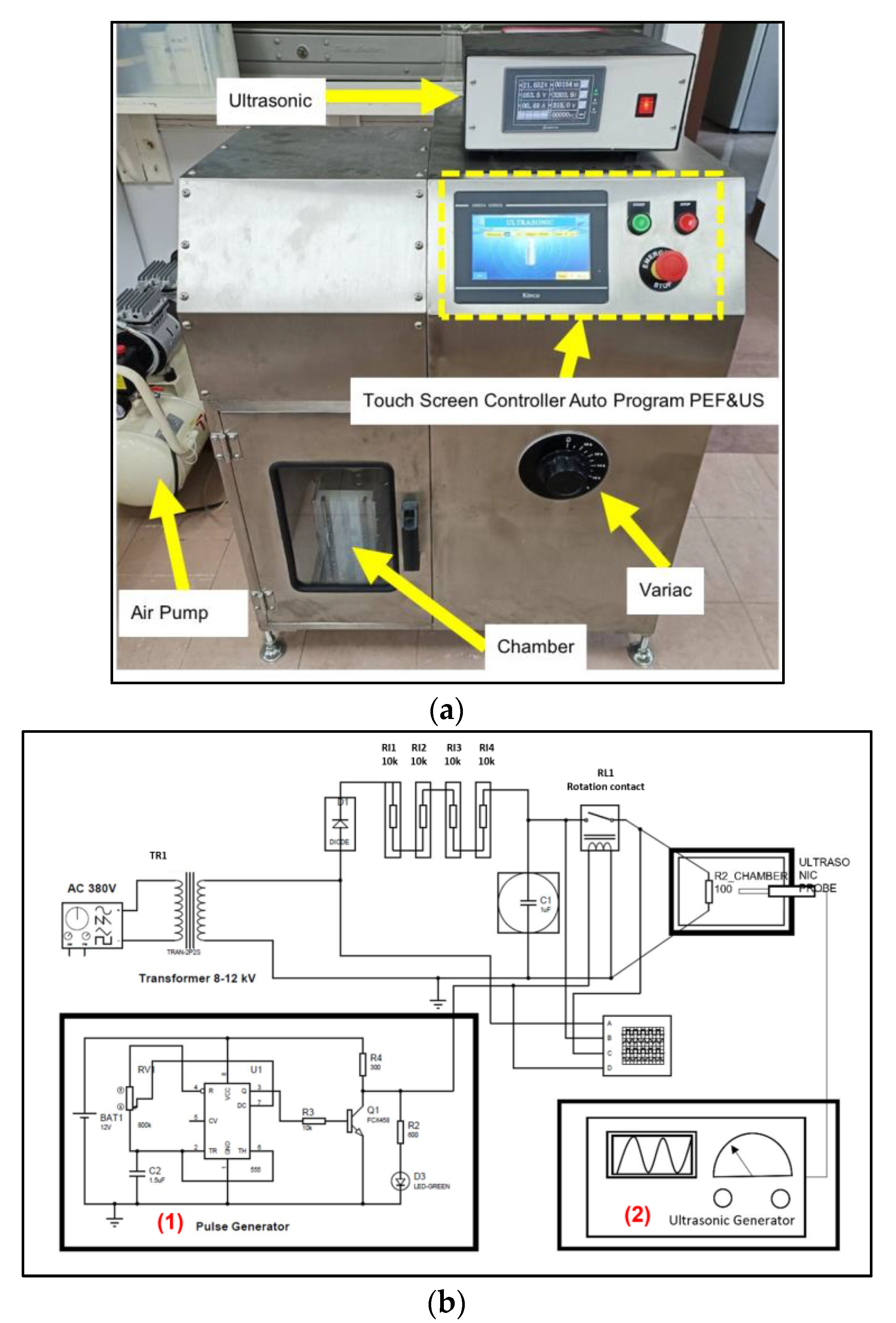
2.2. Pulsed Electric Field and Ultrasonic (PEF-US) Apparatus

The PEF-US prototype was designed and built at the Research Center of Methodology in Producing Active Ingredients from Cannabis and Herbs by Bioreactor (RPAH), Payap University, Chiang Mai, Thailand (Figure 1). The configuration of the PEF-US equipment included several key components, such as a high-voltage generator capable of reaching a maximum voltage of 20 kV, a pulse generator, an ultrasonic horn, a chamber, and an air pump (Figure 1a). A schematic diagram of a PEF-US system is shown in Figure 1b. The systems typically include a pulse generator (1) and an ultrasonic generator (2) as their primary components. The electrical system of the PEF-US comprised a 380 V AC power source, 8–12 kV transformers, and four resistances. This apparatus is versatile, offering four operational modes: PEF, US, US + PEF, and PEF + US.
The voltage, frequency, pulse duration, and ultrasonic conditions were all controlled via a digital touchscreen. A polypropylene enclosure and two stainless steel electrodes made up the treatment chambers. Inside the treatment chamber, cat- and anion electrodes were arranged in parallel, separated by a 100 mm interval. The chamber’s maximum capacity was 5 L (recommended extraction capacity is 1–4 L to obtain the best extraction results). The US was operated with the maximum power of 300 W at a frequency of 20 kHz. The tip of the US horn was placed 50% of the way into the extraction solvent.

2.3. Kratom Extraction Procedures

2.3.1. PEF Extraction

The PEF extraction was performed after the sample mixture was loaded into the treatment chamber. An intensity of 1.3 kV/cm of PEF was applied to the sample mixture at a 1 Hz frequency and with 100 pulses [18]. The experiment was performed at room temperature (30 °C). The temperature of the sample was measured after the extraction and was 30 ± 1 °C on average. The sample was collected after 20 min of extraction time and stored at −20 °C. It was utilized within a 24 h timeframe.

2.3.2. US Extraction

After transferring the sample mixture into the treatment chamber, the US mode was operated following the modified protocol [5] by fixing the operation at 20 min. The initial temperature was 30 °C, while the final temperature was 33 ± 2 °C on average.

2.3.3. PEF + US Extraction

After the sample mixture was loaded into the treatment chamber, PEF was operated by following the conditions described in Section 2.3.1, as the first extraction step; then, the auto-control operation was switched to US using the same conditions as described in Section 2.3.2. The final temperature of the PEF + US extraction was 33 ± 2 °C.

2.3.4. US + PEF Extraction

After the sample mixture was loaded into the treatment chamber, US was applied first and was followed by the PEF mode. The operating conditions followed the procedures described in Section 2.3.1 and Section 2.3.2, respectively. The final temperature of the US + PEF extraction was 31 ± 2 °C.

2.3.5. Maceration Extraction

The mixture of the sample and extraction solvent was left at room temperature (30 ± 2 °C) for 24 h. This sample served as a control sample.

2.4. Liquid Chromatography Analysis of Kratom Extracts

The mitragynine content was determined using the UHPLC model QSight LX50 and the LC-MS/MS model QSight 110 (PerkinElmer, Waltham, MA, USA). Before injection, the samples were filtered via a 0.22 µm nylon filter. The gradient program for mobile phase A (water containing 0.1% formic acid) and mobile phase B (methanol) was adjusted at 0–1 min, 25% B; 1–3 min, 30% B; and 3–4 min, 25% B. A 10 µL needle and a 20 µL loop were connected to the solvent delivery module. The MS operation was set at a nebulizer pressure of 350 psi, an electrospray voltage of 5000 V, and a source temperature of 340 °C. The drying gas was set at 120 °C, while the HSID was set at 320 °C. In positive mode, the mass scan mode was performed from 100 to 1000 m/z. A Quasar SPP C18 column (100 × 2.1 mm, 2.6 µm, PerkinElmer, Buckinghamshire, UK) was used to separate mitragynine at 40 °C. The aforementioned studies were used to identify the MS/MS data [6,7,8,19].
A mitragynine standard with a purity of 97% (cat No. FM26024) was provided by Biosynth International, Inc. (Lane Gardner, Gardner, MA, USA). Standard solutions at concentrations of 1.0, 2.5, 5.0, 7.5, and 10.0 ppm were freshly prepared before the analyses (n = 3) by weighing a specific amount of the mitragynine and dissolving it in methanol. The mitragynine content of the kratom leaves was quantified and evaluated by calculating the linearity of the standard calibration curve.

2.5. Extraction Efficiency

The extraction efficiency was calculated in comparison to the maceration technique using the following equation:
E f f i c i e n c y % = 100   × M t     M 0 M 0
where:
  • M0 = mitragynine content of control or PEF/US alone (mg/L);
  • Mt = mitragynine content of sample using PEF-US apparatus mode (mg/L).

2.6. Energy Consumption Determination

The amount of electricity consumed under US extraction was measured using an electricity meter (OKELE, Wenzhou, China) and calculated according to Equation (2).
E = P   ×   t
where E is the energy measured in J or kWh, P is the power (W), and t is the time (h).
The energy consumption of the PEF technique was calculated using Equations (3) and (4) [20].
E J = N × V ( t ) 2 × t p R
R = d σ A
where EJ is the Joules of energy (J), N is the applied pulse number, V(t) is the voltage across the treatment cell (kV/cm), tp is the operation duration (s), R is the resistance (Ω), d is the separation between the two electrodes (cm), σ is the conductivity of the liquid suspension (mS/cm), and A is the cross-sectional area of the exposed electrode surface (cm2).

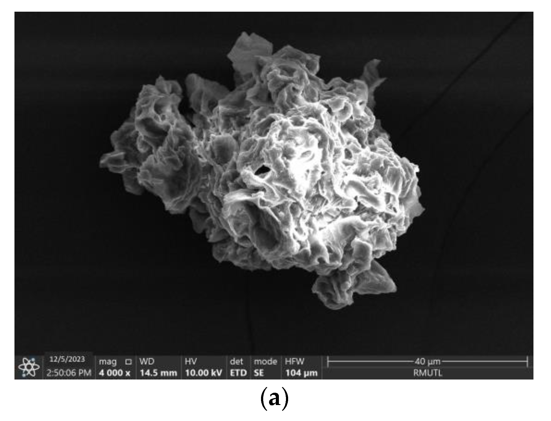
2.7. Scanning Electron Microscopy

A change in the morphology of the kratom powder was observed using scanning electron microscopy (SEM; PentaFETTM precision, X-act, Oxford Instruments, Abingdon, UK). The kratom powder after maceration and the PEF, US, PEF + US, or US + PEF extraction was placed on the stubs of the SEM using double-sided tape and examined at a magnification of 4000×, with an accelerating voltage of 10 kV.

2.8. Fourier-Transform Infrared Spectroscopy

Fourier-transform infrared spectroscopy (FTIR) was performed to detect the functional groups in the extract using an infrared spectrophotometer (Jasco FTIR Analyzer, FT/IR-4700, Jasco, Japan). The spectrum was analyzed in the range of 500 to 3500 cm1, with a resolution of 4 cm1.

2.9. Statistical Analysis

All the experiments were performed at least three times. The mean and standard deviation (S.D.) of the experimental values were provided. SPSS version 18 (SPSS Inc., Chicago, IL, USA) was used to assess the differences across the extraction modalities using a one-way ANOVA test. The threshold for a meaningful difference was fixed at p < 0.05.

3. Results and Discussion

3.1. Validation of UHPLC Measurement

The linearity of the mitragynine standard calibration curve (y = 116,823x + 13,862) with the corresponding correlation coefficient (R2) of 0.9984 is shown in Figure 2a. The calibration curve had an R2 value with a good linearity. The chromatographic separation of standard mitragynine is presented in Figure 2b at a retention time of 1.25 min. The retention time of mitragynine in the kratom leaf extracts was 1.22 min (Figure 2c).

3.2. Mitragynine Content

The mitragynine content for the different extraction modes of the developed PEF-US apparatus was assessed and expressed in mg/L (Table 1). The mitragynine content ranged from 73.13 ± 0.40 to 106.63 ± 0.85 mg/L and decreased in the following order: PEF + US > US + PEF > PEF > US > maceration. The PEF + US mode exhibited a higher mitragynine content (106.63 ± 0.85 mg/L) than the other modes. The four different modes significantly increased the mitragynine content by 45.81 ± 0.59%, 33.00 ± 1.85%, 23.06 ± 1.87%, and 13.77 ± 0.47%, respectively, compared to the control (maceration). From these results, it can be inferred that the mitragynine content of the advanced methods (PEF, US, US + PEF, and PEF + US) was superior to that of the conventional method (maceration). However, the combination of the PEF and US techniques increased the mitragynine content by 8.08–18.48% in comparison to PEF extraction alone and by 16.91–28.16% in comparison to US extraction alone. These findings support the results of Parniakov et al. [21,22], who found that mixing PEF and US to pretreat microalgae suspensions in a binary combination of water and an organic solvent increased the pigment yields and extraction efficiency for Nannochloropsis spp. Furthermore, the combination of US and PEF might boost the virgin olive oil extraction yield from 16.3% to 18.1% [15]. The pretreatment of rosemary and thyme by-products with PEF before extracting with US was found to improve their antioxidant activities and phenolic profiles such as luteolin-7-O-glucoside, luteolin-7-O-glucuronide, and rosmarinic acid [17].
The electroporation of cell membranes (electrically induced pore creation of membranes) can explain the efficacy of PEF in aiding with extraction procedures [23], as shown in Figure 3a. When exposed to an electric field, cell membranes behave like capacitors with a low dielectric constant. As a result of the rising buildup of charges across the membrane, the membrane grows thinner due to electrostatic attraction between opposing charges. When the outer layer of the membrane breaks down, trans-membrane holes develop, and the crucial disintegration of the voltage is reached by increasing the external field strength [23]. PEF-assisted extraction, according to Kumari et al. [23], involves the application of short pulses of moderate electrical power (approximately 0.5–20 kV/cm). PEF intensities of this magnitude are regarded as an effective pretreatment strategy for increasing extraction yields [24].
During the rarefaction phase, ultrasound waves with a frequency greater than 20 kHz created negative pressure, creating cavitation bubbles from the solvent’s gas nuclei [23]. These bubbles expanded over several repetitions until they were unstable and eventually violently collapsed/became exposed, a process known as acoustic cavitation (Figure 3b). This also produces intense micro-streaming currents that can break the cell wall, leading to increased diffusion and a quicker mass transfer rate and resulting in enhanced biological chemical release [23]. Furthermore, the cavitation bubbles also broke the covalent bonds and allowed the phenolic compounds to release from the cell walls of the plant materials.
Due to the application of PEF with a 1.3 kV/cm intensity before the US extraction, the PEF procedure acted as a pretreatment method. Therefore, the application of US increased the extraction efficiency by increasing pore formation and enhancing solvent diffusion (Figure 3c). The use of ultrasound followed by the PEF technique (Figure 3d) resulted in a mitragynine content of 97.27 ± 1.33 mg/L, which represented a decreased efficiency by 12% in comparison with the PEF + US method; this phenomenon might be caused by irreversible electroporation, which causes the mechanical breakdown of the cell membrane and renders cells unviable [21,23].
In this study, dried leaf powders were used as the samples for the extraction processes. Therefore, the soaking time and solvent absorbability of the samples should have had some effect. In the dried samples, the cells contained a small amount of water inside the cells; when the PEF was applied, the electric current could still achieve the electroporation of the membrane due to the polarity of the membrane, and the absorbability of the small-size sample powders allowed electricity to be conducted though the samples. The US extraction technique relied on making cavitation bubbles from the solvent’s gas nuclei [23]. In this study, dried sample powders were used, and the dried cells had a very low moisture content. Therefore, it took a few minutes for the solvent to be absorbed by the sample powders and for the US to make cavitation bubbles from the solvent’s gas nuclei. Therefore, the PEF extraction method resulted in more mitragynine in the extracts than the US extraction method. The results also agreed that the PEF + US extraction method gave better extraction results than US + PEF.

3.3. Energy Consumption

The total energy required to extract mitragynine from kratom leaves was 4.94 ± 0.31 kJ/kg for PEF alone, 1.03 ± 0.01 kJ/kg for US alone, 3.72 ± 0.13 kJ/kg for PEF + US, and 3.64 ± 0.02 kJ/kg for US + PEF (Table 1). The results found that the combination techniques consumed less energy than PEF alone by approximately 24.70% for PEF + US and 26.31% for US + PEF. Therefore, the utilization of PEF and US in combination presents an opportunity to enhance the mitragynine extraction while simultaneously reducing the energy consumption. Consequently, this approach holds promise for a potential application as an eco-friendly technology in industrial extraction processes. Moreover, the energy consumption under this prototype was almost equivalent to the extraction of aromatic plants [25] and rosemary [26] by microwave extraction at 4.2 kJ/kg.

3.4. Change in Surface Structure

An SEM analysis was used to confirm the deformation of the M. speciosa surface after the application of different extraction techniques of the prototype (Figure 4). As shown in Figure 4a, conventional M. speciosa extraction showed closed cells and a tight surface form. After being subjected to different extraction modes, physical modifications to the M. speciosa cell wall were noticed. The use of PEF formed a porous layer on the surface of M. speciosa (Figure 4b), caused by the electroporation of PEF, which strengthened the electric field (cations and anions) on the plant cell wall [27]. The surface of the US-treated kratom leaves had a hollow structure (Figure 4c) due to the formation of cavitation bubbles during US extraction. Thus, a porous and smooth surface was found when PEF was used before the US extraction (Figure 4d). The SEM micrographs of the sample after US + PEF (Figure 4e) were not considerably different from those of the US samples (Figure 4c), but only minor damage was observed on the external surface. This indicates that the US and PEF treatments affected the structure of the cell due to the high, localized pressures induced by the cavitation from US and the electroporation from PEF. Therefore, the mitragynine content was related to the physical modification of the M. speciosa surface. According to Xing et al. [28], the considerable level of cell wall breakdown increases the release of intracellular compounds from the raw materials and improves the extraction efficacy.

3.5. Fourier-Transform Infrared Spectroscopy Analysis

Figure 5 illustrates the FTIR functional group of the M. speciosa powder extracted with various conditions of the prototype in the range of 500 to 3500 cm1. The peaks at 2912.9 and 2848.3 cm−1 correspond to the C-H stretching vibration, which was present due to the polyphenolic compound and the TPC concentration. At 1617.0 cm−1, the absorption bands correspond to the C=C in the aromatic groups. The absorbance peak at around 1625–1430 cm−1 corresponds to C-C stretching bonds. The distinctive band at 1047.2 cm−1 was associated with the C-O deformation of phenolic compounds [29]. The weak intensities of the C-H bonds at 2912.9 and 2848.3 cm−1, the C=C bond at 1617.0 cm−1, the C-C at 1625–1430 cm−1, and the C-O bond at 1047.2 cm−1 of the M. speciosa powder treated using the PEF + US and US + PEF methods were attributed to the higher number of active molecules being disintegrated into the extraction solvents. As a result, the FTIR confirmed that the mitragynine in the kratom was released into the extraction solvents, which was consistent with the mitragynine level shown in Table 1.

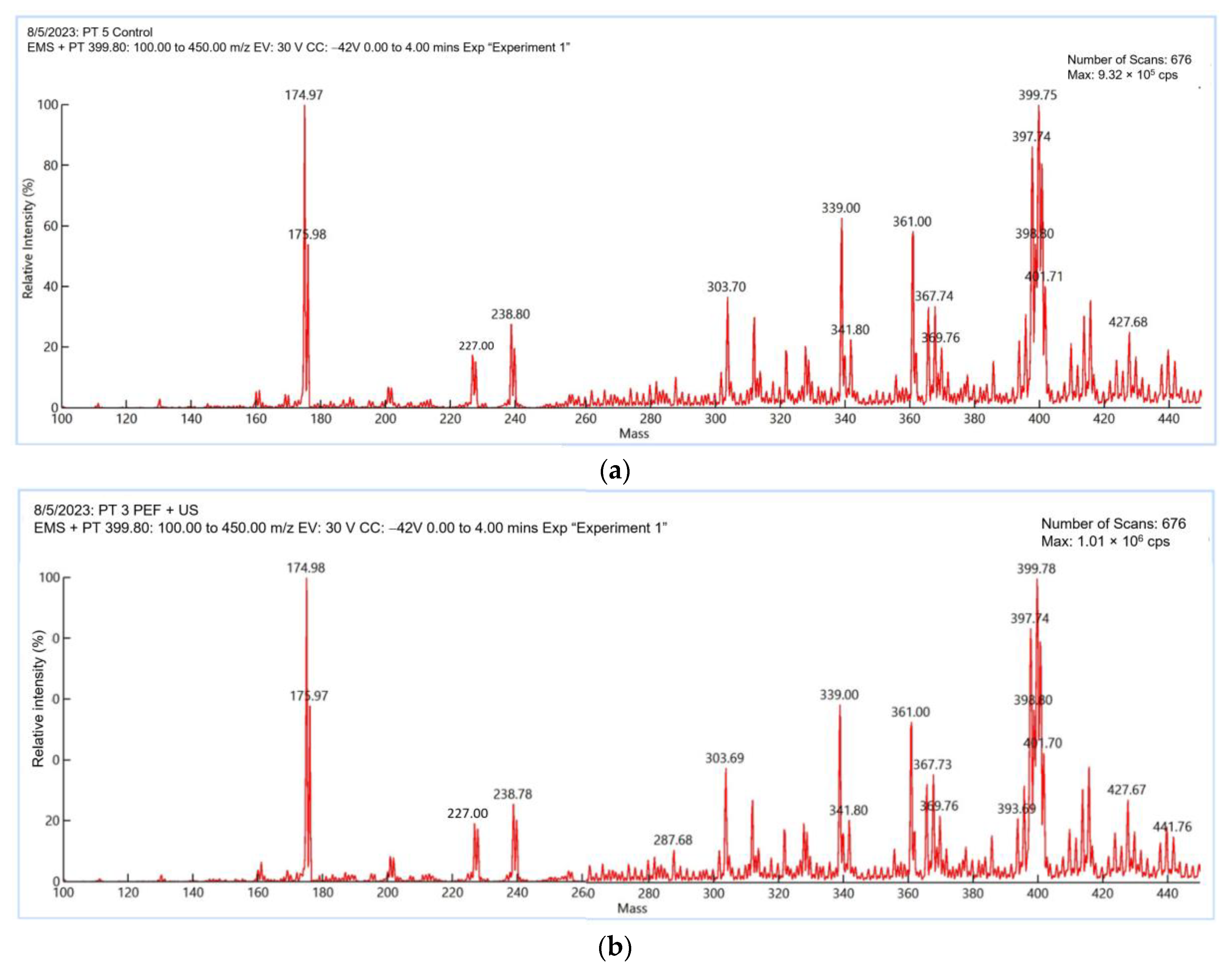
3.6. LC-MS/MS Profiles

The mass spectra of M. speciosa leaves extracted using maceration and PEF + US are shown in Figure 6. A similar pattern was observed for the control (Figure 6a) and PEF + US extraction (Figure 6b). Table 2 summarizes the identification of the mitragynine, confirmed using published studies [6,7,8,19]. The precursor ion and product ion of mitragynine were m/z 399.75→174.97 for maceration and 399.78→174.98 for the PEF + US extracts. The fragmentation pattern was a helpful tool to resolve unknown alkaloids in the M. speciosa extracts.

4. Conclusions

The purpose of this research was to establish whether a combination of PEF and US equipment could be used to obtain plant extracts with an enhanced content of valuable compounds. During this investigation, four different mechanisms, including PEF, US, US + PEF, and PEF + US, for mitragynine extraction from dried M. speciosa leaves were employed. Regarding the evaluation of extractability, the PEF + US device proved to be very efficient at extracting mitragynine from the dried leaves of kratom, significantly increasing the efficiency of the mitragynine extraction compared to the control (maceration). Together with a higher mitragynine content, less energy was required for the extraction when PEF and US were used in a contact way. The physical modifications of the kratom leaves obtained by the different extraction techniques were evaluated using SEM and an FTIR analysis. The results indicated that a combination of PEF and US devices may be considered a green alternative approach and can aid in the streamlining of agricultural product implementation. However, developing low-cost, environmentally friendly extraction methods that yield a viable extract rich in bioactive chemicals remains a difficult task. Therefore, further work needs to be conducted to focus on the impact of the PEF and US parameters on the cell wall material of kratom leaves to better improve the alkaloid compounds, especially the mitragynine content.

5. Patents

The prototype of the pulsed electric field and ultrasonic (PEF-US) apparatus is the subject of the Petty Patent No. 2303000855, with the title extraction processing of mitragynine from kratom leaves using the combination of pulse electric field and ultrasonic technique (in Thai).

Author Contributions

Conceptualization, S.S. and A.K.-a.; methodology, S.S. and A.K.-a.; validation, A.K.-a. and T.P.; investigation, P.J., R.J., N.P. and R.H.; data curation, A.K.-a., R.J., N.P. and R.H.; writing—original draft preparation, R.J., A.K.-a. and N.P.; writing—review and editing, A.K.-a., P.J., N.P., S.S. and T.P.; visualization, A.K.-a., S.S. and T.P.; supervision, S.S. and T.P.; project administration, S.S. and T.P.; funding acquisition, T.P. All authors have read and agreed to the published version of the manuscript.

Funding

This research received no external funding.

Informed Consent Statement

Not applicable.

Data Availability Statement

Not applicable.

Acknowledgments

This study was partially supported by APA industries Co., Ltd. and Payap University.

Conflicts of Interest

The authors declare no conflict of interest.

References

  1. Trakulsrichai, S.; Sathirakul, K.; Auparakkitanon, S.; Krongvorakul, J.; Sueajai, J.; Noumjad, N.; Sukasem, C.; Wananukul, W. Pharmacokinetics of mitragynine in man. Drug Des. Devel. Ther. 2015, 9, 2421–2429. [Google Scholar] [CrossRef] [PubMed]
  2. Eastlack, S.C.; Cornett, E.M.; Kaye, A.D. Kratom—Pharmacology, clinical implications, and outlook: A comprehensive review. Pain. Ther. 2020, 9, 55–69. [Google Scholar] [CrossRef] [PubMed]
  3. Fluyau, D.; Revadigar, N. Biochemical Benefits, Diagnosis, and Clinical Risks Evaluation of Kratom. Front. Psychiatry 2017, 8, 1–8. [Google Scholar] [CrossRef] [PubMed]
  4. Orio, L.; Alexandru, L.; Cravotto, G.; Mantegna, S.; Barge, A. UAE, MAE, SFE-CO2 and classical methods for the extraction of Mitragyna speciosa leaves. Ultrason. Sonochem. 2012, 19, 591–595. [Google Scholar] [CrossRef] [PubMed]
  5. Zakaria, F.; Tan, J.-K.; Mohd Faudzi, S.M.; Abdul Rahman, M.B.; Ashari, S.E. Ultrasound-assisted extraction conditions optimisation using response surface methodology from Mitragyna speciosa (Korth.) Havil leaves. Ultrason. Sonochem. 2021, 81, 105851. [Google Scholar] [CrossRef]
  6. Karunakaran, T.; Goh, Y.S.; Santhanam, R.; Murugaiyah, V.; Abu Bakar, M.H.; Ramanathan, S. RP-HPLC-DAD analysis of mitragynine content in Mitragyna speciosa Korth. (Ketum) leaf extracts prepared using ultrasound assisted extraction technique and their cytotoxicity. Separations 2022, 9, 345. [Google Scholar] [CrossRef]
  7. Limcharoen, T.; Pouyfung, P.; Ngamdokmai, N.; Prasopthum, A.; Ahmad, A.R.; Wisdawati, W.; Prugsakij, W.; Warinhomhoun, S. Inhibition of alpha-glucosidase and pancreatic lipase properties of Mitragyna speciosa (Korth.) Havil. (Kratom) leaves. Nutrients 2022, 14, 3909. [Google Scholar] [CrossRef]
  8. Goh, Y.S.; Karunakaran, T.; Murugaiyah, V.; Santhanam, R.; Abu Bakar, M.H.; Ramanathan, S. Accelerated solvent extractions (ASE) of Mitragyna speciosa Korth. (Kratom) leaves: Evaluation of its cytotoxicity and antinociceptive activity. Molecules 2021, 26, 3704. [Google Scholar] [CrossRef]
  9. Soquetta, M.B.; Terra, L.d.M.; Bastos, C.P. Green technologies for the extraction of bioactive compounds in fruits and vegetables. CyTA J. Food 2018, 16, 400–412. [Google Scholar] [CrossRef]
  10. Bocker, R.; Silva, E.K. Pulsed electric field assisted extraction of natural food pigments and colorings from plant matrices. Food Chem. X 2022, 15, 100398. [Google Scholar] [CrossRef]
  11. Toepfl, S.; Mathys, A.; Heinz, V.; Knorr, D. Potential of high hydrostatic pressure and pulsed electric fields for energy efficient and environmentally friendly food processing. Food Rev. Int. 2006, 22, 405–423. [Google Scholar] [CrossRef]
  12. Toepfl, S.; Heinz, V.; Knorr, D. High intensity pulsed electric fields applied for food preservation. Chem. Eng. Process. Process Intensif. 2007, 46, 537–546. [Google Scholar] [CrossRef]
  13. Ranjha, M.M.A.N.; Kanwal, R.; Shafique, B.; Arshad, R.N.; Irfan, S.; Kieliszek, M.; Kowalczewski, P.Ł.; Irfan, M.; Khalid, M.Z.; Roobab, U.; et al. A Critical Review on pulsed electric field: A novel technology for the extraction of phytoconstituents. Molecules 2021, 26, 4893. [Google Scholar] [CrossRef] [PubMed]
  14. Lal, A.M.N.; Prince, M.V.; Kothakota, A.; Pandiselvam, R.; Thirumdas, R.; Mahanti, N.K.; Sreeja, R. Pulsed electric field combined with microwave-assisted extraction of pectin polysaccharide from jackfruit waste. Innov. Food Sci. Emerg. Technol. 2021, 74, 102844. [Google Scholar] [CrossRef]
  15. Grillo, G.; Boffa, L.; Calcio Gaudino, E.; Binello, A.; Rego, D.; Pereira, M.; Martínez, M.; Cravotto, G. Combined ultrasound and pulsed electric fields in continuous-flow industrial olive-oil production. Foods 2022, 11, 3419. [Google Scholar] [CrossRef] [PubMed]
  16. Nowosad, K.; Sujka, M.; Pankiewicz, U.; Kowalski, R. The application of PEF technology in food processing and human nutrition. J. Food Sci. Technol. 2021, 58, 397–411. [Google Scholar] [CrossRef]
  17. Tzima, K.; Brunton, N.P.; Lyng, J.G.; Frontuto, D.; Rai, D.K. The effect of pulsed electric field as a pre-treatment step in ultrasound assisted extraction of phenolic compounds from fresh rosemary and thyme by-products. Innov. Food Sci. Emerg. Technol. 2021, 69, 102644. [Google Scholar] [CrossRef]
  18. Shiekh, K.A.; Olatunde, O.O.; Zhang, B.; Huda, N.; Benjakul, S. Pulsed electric field assisted process for extraction of bioactive compounds from custard apple (Annona squamosa) leaves. Food Chem. 2021, 359, 129976. [Google Scholar] [CrossRef] [PubMed]
  19. Avula, B.; Sagi, S.; Wang, Y.-H.; Wang, M.; Ali, Z.; Smillie, T.J.; Zweigenbaum, J.; Khan, I.A. Identification and characterization of indole and oxindole alkaloids from leaves of Mitragyna speciosa Korth using liquid chromatography–accurate QToF mass spectrometry. J. AOAC Int. 2015, 98, 13–21. [Google Scholar] [CrossRef]
  20. Qin, S.; Timoshkin, I.V.; Maclean, M.; Wilson, M.P.; MacGregor, S.J.; Given, M.J.; Anderson, J.G.; Wang, T. Pulsed electric field treatment of microalgae: Inactivation tendencies and energy consumption. IEEE Trans. Plasma Sci. 2014, 42, 3191–3196. [Google Scholar] [CrossRef]
  21. Parniakov, O.; Barba, F.J.; Grimi, N.; Marchal, L.; Jubeau, S.; Lebovka, N.; Vorobiev, E. Pulsed electric field assisted extraction of nutritionally valuable compounds from microalgae Nannochloropsis spp. using the binary mixture of organic solvents and water. Innov. Food Sci. Emer. Technol. 2015, 27, 79–85. [Google Scholar] [CrossRef]
  22. Parniakov, O.; Apicella, E.; Koubaa, M.; Barba, F.J.; Grimi, N.; Lebovka, N.; Pataro, G.; Ferrari, G.; Vorobiev, E. Ultrasound-assisted green solvent extraction of high-added value compounds from microalgae Nannochloropsis spp. Bioresour. Technol. 2015, 198, 262–267. [Google Scholar] [CrossRef]
  23. Kumari, B.; Tiwari, B.K.; Hossain, M.B.; Brunton, N.P.; Rai, D.K. Recent advances on application of ultrasound and pulsed electric field technologies in the extraction of bioactives from agro-industrial by-products. Food Bioproc. Tech. 2018, 11, 223–241. [Google Scholar] [CrossRef]
  24. Balasa, A.; Janositz, A.; Knorr, D. Electric field stress on plant systems. Encycl. Biotechnol. Agric. Food 2011, 2, 208–211. [Google Scholar] [CrossRef]
  25. Lucchesi, M.E.; Chemat, F.; Smadja, J. Solvent-free microwave extraction of essential oil from aromatic herbs: Comparison with conventional hydro-distillation. J. Chromatogr. A 2004, 1043, 323–327. [Google Scholar] [CrossRef] [PubMed]
  26. Bousbia, N.; Vian, M.A.; Ferhat, M.A.; Petitcolas, E.; Meklati, B.Y.; Chemat, F. Comparison of two isolation methods for essential oil from rosemary leaves: Hydrodistillation and microwave hydrodiffusion and gravity. Food Chem. 2009, 114, 355–362. [Google Scholar] [CrossRef]
  27. Supasin, S.; Kantala, C.; Intra, P.; Rattanadecho, P. Postharvest preservation of Thai mango var. Chok-Anan by the combination of pulsed electric field and chemical pickling. Horticulturae 2022, 8, 584. [Google Scholar] [CrossRef]
  28. Xing, C.; Cui, W.-Q.; Zhang, Y.; Zou, X.-S.; Hao, J.-Y.; Zheng, S.-D.; Wang, T.-T.; Wang, X.-Z.; Wu, T.; Liu, Y.-Y.; et al. Ultrasound-assisted deep eutectic solvents extraction of glabridin and isoliquiritigenin from Glycyrrhiza glabra: Optimization, extraction mechanism and in vitro bioactivities. Ultrason. Sonochem. 2022, 83, 105946. [Google Scholar] [CrossRef] [PubMed]
  29. Manik, U.P.; Nande, A.; Raut, S.; Dhoble, S.J. Green synthesis of silver nanoparticles using plant leaf extraction of Artocarpus heterophylus and Azadirachta indica. Results Mater. 2020, 6, 100086. [Google Scholar] [CrossRef]
Figure 1. The prototype (a) and diagram (b) of the pulsed electric field and ultrasonic (PEF-US) apparatus. 1 is the pulsed generator and 2 is ultrasonic generator.
Agriengineering 05 00115 g001
Figure 2. Standard calibration curve of PEF mitragynine using UHPLC measurement (a); example base peak chromatogram of standard mitragynine (b); and mitragynine in kratom leaf extracts (c). A black dot represents the observed value of the standard mitragynine peak; a green line represents a linearity curve; and a gray line represents an observed peak of mitragynine by UHPLC.
Agriengineering 05 00115 g002
Figure 3. Illustration of PEF extraction (a), US extraction (b), the dual extraction of PEF + US (c), and the dual extraction of US + PEF (d).
Agriengineering 05 00115 g003aAgriengineering 05 00115 g003b
Figure 4. SEM images of M. speciosa powder after extraction with various conditions: (a) conventional extraction, (b) PEF extraction, (c) US extraction, (d) PEF + US extraction, and (e) US + PEF extraction (×4000). Yellow arrows indicate disintegrated cell areas.
Agriengineering 05 00115 g004aAgriengineering 05 00115 g004b
Figure 5. The FTIR spectrum of M. speciosa leaves after extraction with various conditions is represented by a red line for conventional extraction, a yellow line for PEF extraction, a black dotted line for US extraction, a blue line for US + PEF extraction, and a green line for PEF + US extraction.
Agriengineering 05 00115 g005
Figure 6. The full-scan mass spectra of M. speciosa leaves extracted by maceration (a) and using a PEF + US device (b).
Agriengineering 05 00115 g006
Table 1. A comparison of the effect of different extraction modes of the developed PEF-US device on the mitragynine content, extraction efficiency, and energy consumption of kratom leaves.
Apparatus ModeMitragynine Content 1 (mg/L)Efficiency (%)Energy Consumption (kJ/kg)
PEF90.00 ± 1.37 c 23.06 ± 1.874.94 ± 0.31
US83.20 ± 0.17 d13.77 ± 0.471.03 ± 0.01
PEF + US106.63 ± 0.85 a45.81 ± 0.593.72 ± 0.13
US + PEF97.27 ± 1.33 b33.00 ± 1.853.64 ± 0.02
Control (maceration)73.13 ± 0.40 e0
1 Duncan’s multiple range tests (n = 3) showed that a–e within the same column are statistically different at p < 0.05.
Table 2. UHPLC-MS/MS analysis results: mitragynine MS/MS data in macerated and PEF + US extracts.
Extraction MethodRT (min)Calculated m/z [M+H]+Precursor Ion Experimental m/z [M+H]+% Error (mDa)Chemical
Formula
Major Ions
(Key Fragment Ions)
Maceration1.22399.2278399.75+0.5222C23H30N2O4238.80 (27%)
227.00 (12%)
174.97 (100%)
110.09 (0%)
PEF + US1.22399.2278399.78+0.5522C23H30N2O4238.78 (26%)
227.00 (14%)
174.98 (100%)
110.09 (0%)
Disclaimer/Publisher’s Note: The statements, opinions and data contained in all publications are solely those of the individual author(s) and contributor(s) and not of MDPI and/or the editor(s). MDPI and/or the editor(s) disclaim responsibility for any injury to people or property resulting from any ideas, methods, instructions or products referred to in the content.

