Review

GABAA-ρ Receptors in the CNS: Their Functional, Pharmacological, and Structural Properties in Neurons and Astroglia

by
Abraham Rosas-Arellano
1,
Argel Estrada-Mondragón
2,
Ataúlfo Martínez-Torres
3 and
Daniel Reyes-Haro
3,*
1
Unidad de Imagenología, Instituto de Fisiología Celular, Universidad Nacional Autónoma de México, Ciudad de Mexico 04510, Mexico
2
Department of Biomedical and Clinical Sciences, Linköping University, 58183 Linköping, Sweden
3
Departamento de Neurobiología Celular y Molecular, Instituto de Neurobiología, Campus Juriquilla, Universidad Nacional Autónoma de México, Querétaro 76230, Mexico
*
Author to whom correspondence should be addressed.
Neuroglia 2023, 4(4), 239-252; https://doi.org/10.3390/neuroglia4040017
Submission received: 1 September 2023 / Revised: 25 September 2023 / Accepted: 29 September 2023 / Published: 8 October 2023

Abstract

:
Gamma-aminobutyric acid (GABA) is known as the main inhibitory transmitter in the central nervous system (CNS), where it hyperpolarizes mature neurons through activation of GABAA receptors, pentameric complexes assembled by combination of subunits (α1–6, β1–3, γ1–3, δ, ε, θ, π and ρ1–3). GABAA-ρ subunits were originally described in the retina where they generate non-desensitizing Cl- currents that are insensitive to bicuculline and baclofen. However, now is known that they are widely expressed throughout the brain including glial cells. For example, whole-cell patch-clamp recordings demonstrated the functional expression of GABAA-ρ receptors in primary cultures of cerebellar astrocytes, as well as in cerebellar ependymal cells and striatal astrocytes. In these cells GABA-currents were partially blocked by TPMPA and insensitive to barbiturates. These receptors are proposed to be involved in extrasynaptic communication and dysfunction of the signaling is accompanied by reduced expression of GABAA-ρ receptors in Huntington’s disease and autism spectrum disorders (ASD). Thus, the aim of this review is to present an overview about GABAA-ρ receptors including their structure and function, as well as their importance in the excitatory/inhibitory (E/I) balance in neurodevelopment and in disease.

1. Introduction

Gamma-aminobutyric acid (GABA) is a non-protein amino acid present in invertebrates and vertebrates, where is considered the main neural transmitter exerting inhibitory activity on neurons and their intricate networks [1,2]. However, this neurotransmitter depolarizes astrocytes and neural precursors through the activation of ionotropic GABAA receptors, pentameric proteins that are modulated by barbiturates and benzodiazepines [3,4]. Interneuron–astrocyte communication regulates excitatory/inhibitory (E/I) balance and gliotransmission is accepted to play an important role. Astroglia setting includes the expression of GABAA receptors to detect interneuron signaling and calcium transients derived from this communication promote the release of gliotransmitters like GABA, known as an important extrasynaptic source for tonic inhibition. In this scenario, the expression of GABAA-ρ subunits becomes relevant under physiological and pathological conditions. For example, during earlier postnatal neurodevelopment [5,6,7,8,9,10] and as a potential therapeutic target for post-stroke motor recovery [11].

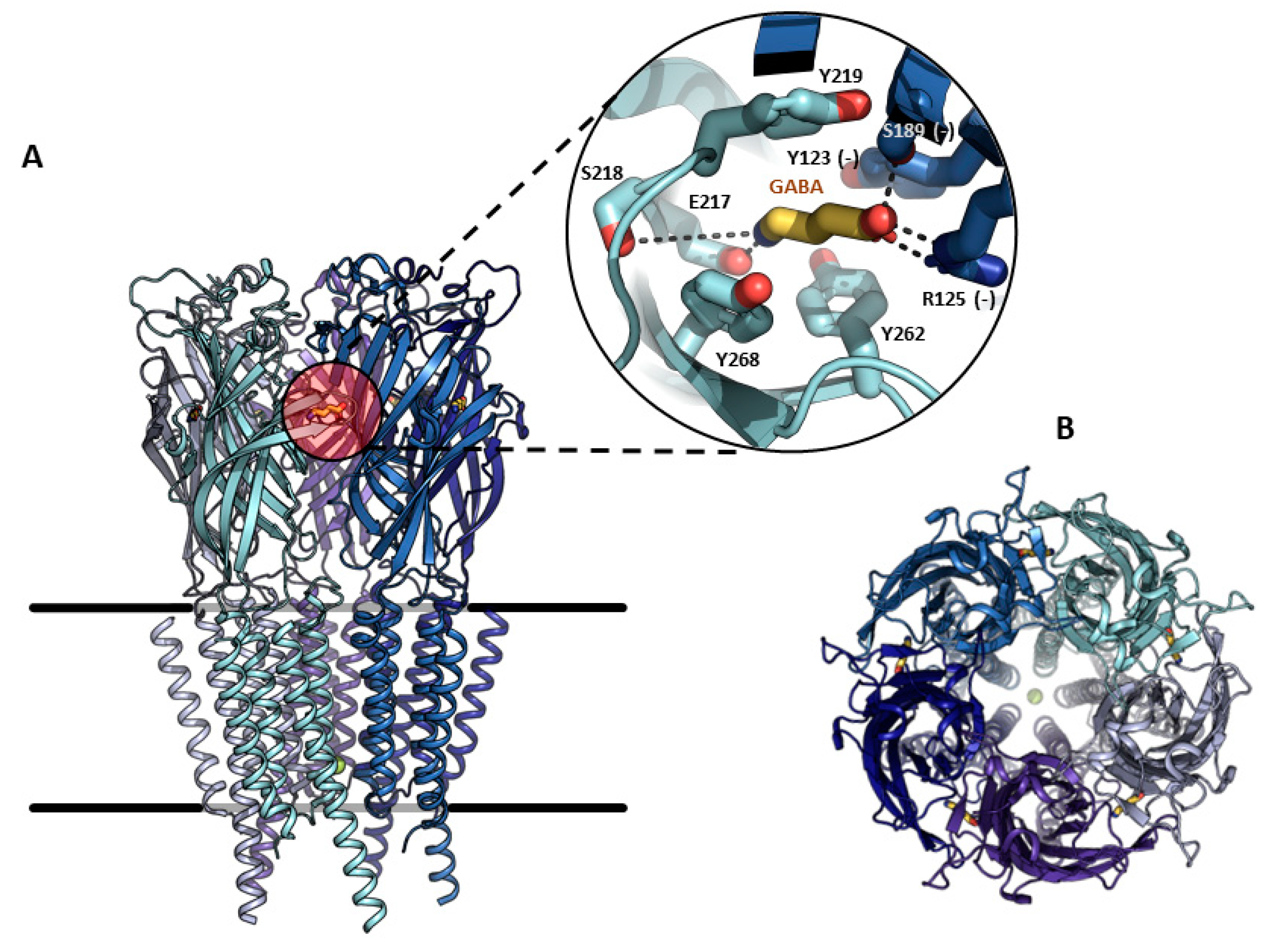
2. Structural Properties, Characterization, and Functions of GABAA-ρ Receptors

The GABAA receptor is an ion channel member of the ligand-gated ion channel (pLGIC) superfamily that opens transiently, inducing the inhibition of action potentials through an inward membrane chloride conductance [4,12]. It was identified through bicuculline sensitivity in autonomic brain terminals and in mammalian brain slices [13]. This receptor is widely distributed in the central nervous system (CNS) and combinations of nineteen subunits (α1–6, β1–3, γ1–3, δ, ε, θ, π and ρ1–3) occur to coordinate differential neural inhibitions, such as phasic or tonic receptors, depending on the stoichiometry of each channel [8,9,10]. In the adult brain, the most common arrangement is: α1 (2), β2/3 (1), and γ2 (2) [14,15,16]. Topologically, each subunit contains an amino-terminal domain, where the neurotransmitter recognition site is located, a short extracellular carboxy-terminal domain, two cytoplasmic loops, and four transmembrane regions (Figure 1).
Each subunit is predicted to have a molecular weight between 48 and 64 kDa, based on the amino acid sequence, and form a central pore or ion channel. Pentameric complexes weigh approximately 275 kDa [4]. Classical GABAA receptors are known to desensitize upon continuous exposure to the ligand; however, GABAA-ρ subunits form functional homomeric receptors that do not desensitize [18]. GABAA-mediated signaling is a key element for the E/I balance of the brain and its dysfunction is linked to motor, cognitive, and psychiatric disorders [16]. GABAA-ρ was originally described as a retinal component (Table 1). There are three genes of GABAA-ρ1-3 subunits; the first two were originally cloned from human retinal cDNA libraries [18,19], and the third from a rat retinal cDNA library [20]. The human GABAA-ρ1 subunit shows alternative spliced variants and two deletions of 51 and 450 nucleotides, respectively (GABAA-ρ1-51 and GABAA-ρ1-450), which brought speculations as to the regulation of the expression of full-length GABAA-ρ1 subunits, possibly co-assembling with some of the alternative spliced subunits and leading to variants with tissue-specific properties [21]. The first electrophysiological characterization was performed with the archetypical heterologous expression system for the study of ion channels, developed by Ricardo Miledi and collaborators, by injecting exogenous mRNA in Xenopus oocytes and using a two-electrode voltage clamp (TEVC) [22]. This pioneer group injected mRNA preparations from either the brain or retina in Xenopus oocytes and found different GABA responses, which were kinetically and pharmacologically different [4]. For example, a slight decay and the magnitude of the current remained almost at maximum as long as the agonist was present in the perfusion bath. On the other hand, GABA responses decayed quickly near the basal level [4]. The sensitivity to the agonist was also very different, as the retinal GABAA receptor presented an EC50 of 1.48 μM, in contrast to the brain cortex, (EC50 = 84.6 μM). The cloned cDNA coding for the GABAA-ρ1 subunit expressed in Xenopus oocytes displayed a GABA EC50 of 2 μM and was insensitive to bicuculline [4,18]; these results were like those obtained from oocytes injected with retinal mRNA. Several reports have shown heteromerization in different GABAA-ρ subunits [23,24], as well as in other GABAA subunits [8,25,26,27], or even in subunits belonging to different members of the pLGIC superfamily [28]. The homomeric GABAA-ρ1 subunit is insensitive to bicuculline and baclofen, commonly used to discriminate between heteromeric GABAA channels and metabotropic GABAB receptors, respectively [29,30,31]. The pharmacological distinctiveness that allowed the discriminating of GABAA-ρ subunits from different subunits of heteromeric GABAA receptors was the synthesis of conformationally restricted GABA analogues [32]. The cis-isomer, cis-4-amino-crotonic acid (CACA) and trans-4-amino-crotonic acid (TACA) are GABAA-ρ1 partial and potent agonists, respectively. Nonetheless, TACA is not selective and is also able to activate typical heteromeric GABAA receptors; this gives an advantage to using CACA to clearly identify GABAA-ρ subunits [33,34]. Homomeric GABAA-ρ1 receptors are insensitive to many of the heteromeric GABAA allosteric modulators, such as benzodiazepines and barbiturates. However, some drugs have powerful allosteric properties, like the anticonvulsant loreclezole [35] or drugs designed for specific purposes, such as (1,2,5,6-tetrahydropyridine-4-yl) methylphosphinic acid (TPMPA), -cis-/trans-3-aminocyclopentane) butylphosphinic acid ([±]-cis and [±]-trans-3-ACPBuPA, Piperidin-4-yl) methylphosphinic acid (P4MPA), Piperidin-4-yl seleninic acid (SEPI), and 3-(guanido)-1-oxo-1-hydroxy-phospholane (3-GOHP) [36]; all of them have been used as a source to investigate the involvement of GABAA-ρ receptors in sleep–waking behavior and antinociception in the peripheral nervous system or in learning, and also to investigate memory enhancers and other cognitive activities [36,37]. The importance of the GABAA-ρ receptor becomes increasingly clear through its wide expression in the CNS (Table 1). Additionally, it is highly related to ammonia-induced apoptosis, retinitis pigmentosa, myopia, and may be important for some specific in vivo effects of ethanol and Huntington’s disease (HD) [16,38]. At the last stage of the preparation of this manuscript the cryogenic electron microscopy structures of GABAA-ρ1 subunits was reported, in apo state as well as in the presence of TPMPA, picrotoxin (PTX), and GABA, generating different conformations [17]. This milestone is an exciting point in the state-of-the-art of the current research that will help us to understand the intricated mechanisms that pharmacologically and functionally define this family of proteins, as a natural step following the pioneering experimental achievements described here.

3. Trafficking and Scaffold Proteins of GABAA-ρ Receptors

It has been hypothesized that the assembly of a complete pentameric GABAA-ρ receptor occurs in the rough endoplasmic reticulum and is followed by trafficking via the Golgi network to the cell surface. Accordingly, GABAA-ρ2 tagged with green fluorescent protein was observed in clusters distributed at the soma and along the axons of retinal bipolar neurons, while in cerebellar astrocytes it was restricted only to the soma [60].
The N-terminus of the GABAA-ρ contains signals that are critical for the correct assembly of subunits into functional pentameric GABAA receptors, as revealed via heterologous expression studies in Xenopus leavis [30]. A key element supporting this assumption are the first 46 residues of the N-terminal domain, prior to the extracellular α-helix, known as the N-terminal extension. In the GABAA-ρ1 receptor the N-terminal extension is much larger than other members of the pLGIC superfamily, and its function has been tested and confirmed through the functional impact of consecutive serial deletions [80]. Nevertheless, our understanding of the whole process of trafficking remains rudimentary, but it is becoming increasingly clear that trafficking is facilitated by the proteins described below.
The cytoskeletal protein microtubule-associated protein 1B (MAP1B) is a critical factor for the development and function of the nervous system and is viewed as the most significant GABAA-ρ receptor subunit anchoring protein. A 12 amino acid sequence (RI(D/N)THAIDKYSR) at the C-terminal end of the second intracellular domain of GABAA-ρ1 interacts physically with MAP1B [81,82]. Clustering of GABAA-ρ subunits occurs at synapses on bipolar axon terminals of the retina [39,40]. GABAA-ρ subunits can also bind to a glycine transporter splice variant, GLYT-1E/F, in a distinct region of the MAP1B binding site. This transporter protein is also involved in the anchoring and signaling of excitatory and metabotropic receptors that interact with different proteins, thus playing pivotal roles in linking membrane proteins to the actin cytoskeleton [83].
The GABAA receptor-associated protein (GABARAP) interacts with the intracellular domain of GABAA receptors [84]. It binds to microtubules and to the N-ethylmaleimide-sensitive factor (NSF), a protein that is linked to events of intracellular vesicular fusion [85]. GABARAP is abundant in the Golgi apparatus and in intracellular vesicles, but it is not present at GABAergic synapses, suggesting its participation in the intracellular transport of both homomeric and heteromeric GABAA receptors [86].
Some evidence suggests that the crosstalk between GABAA-ρ1 and GABAA-ρ2 receptors and purinergic P2X2 receptors co-localize in the same clusters in the plasma membrane [87]. On the other hand, GABAA-ρ1 directly interacts with protein kinase C-ζ (PKCζ) via adapter proteins of the ZIP protein family, as well as with protein phosphatase 1 (PP1). This interaction is important in promoting (or not) the cellular internalization of GABAA-ρ subunits [88]. ZIP3 is a C-terminal splice variant of the ZIP protein family, and it is ubiquitously expressed in non-neuronal and neuronal tissues, including the retina. The GABAA-ρ intracellular binding regions of ZIP3 contain ZZ-zinc finger domains that interact with 10 amino acids conserved in GABAA-ρ1–3 subunits but not in heteromeric GABAA receptors. Consistently, only GABAA-ρ1–3 subunits can bind to ZIP3, which dimerizes with another ZIP subunit (ZIP1–3), interacting with PKC-ζ. This complex is considered determinant for the formation of the scaffold of GABAA-ρ subunits in the cytoskeleton [89].

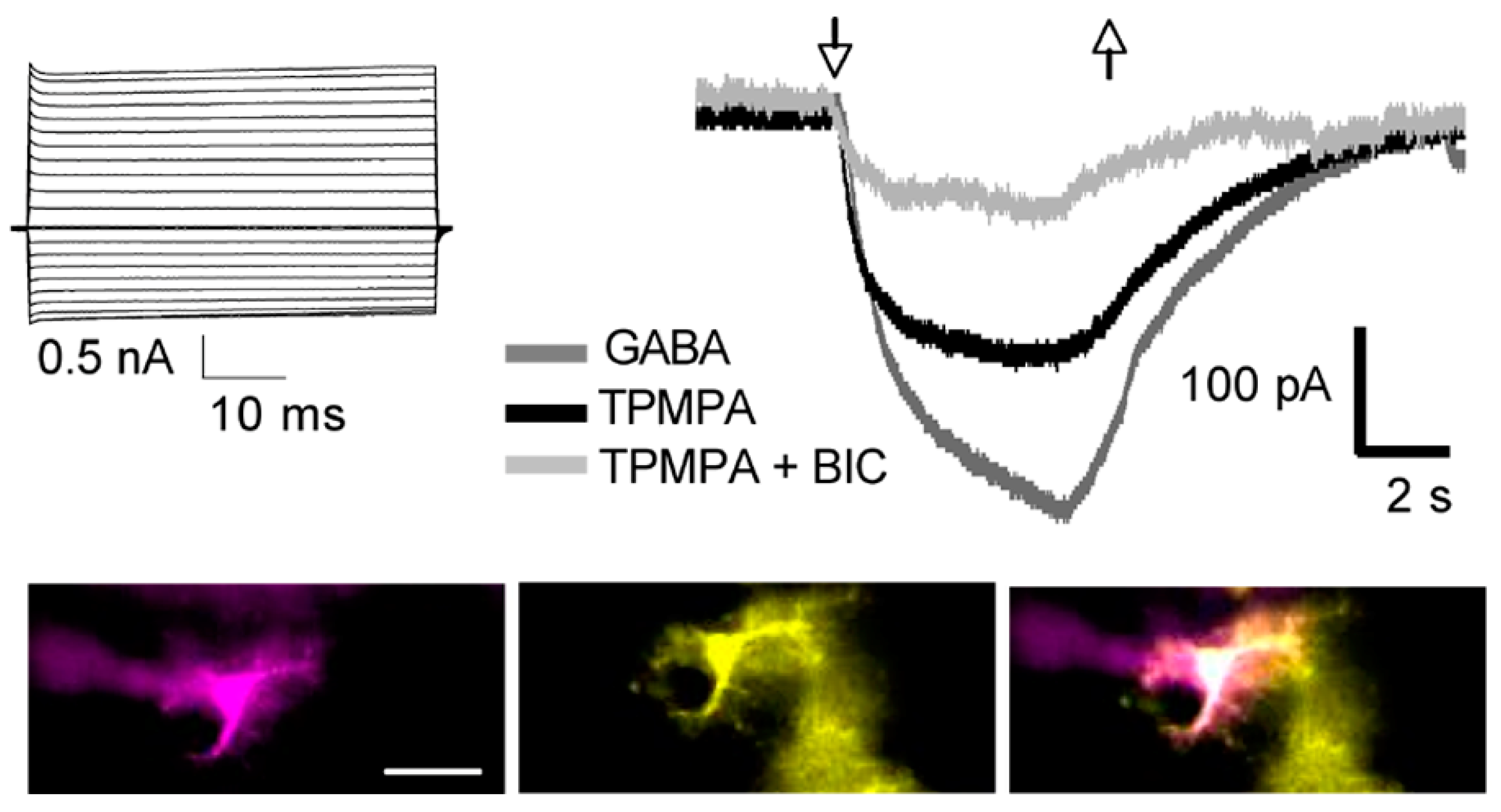
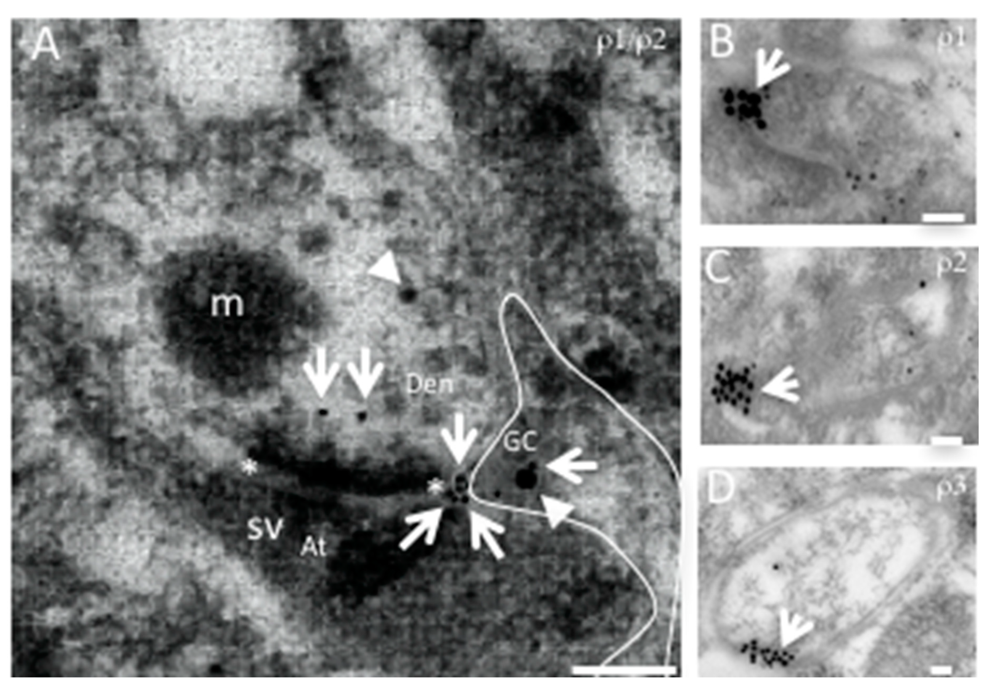
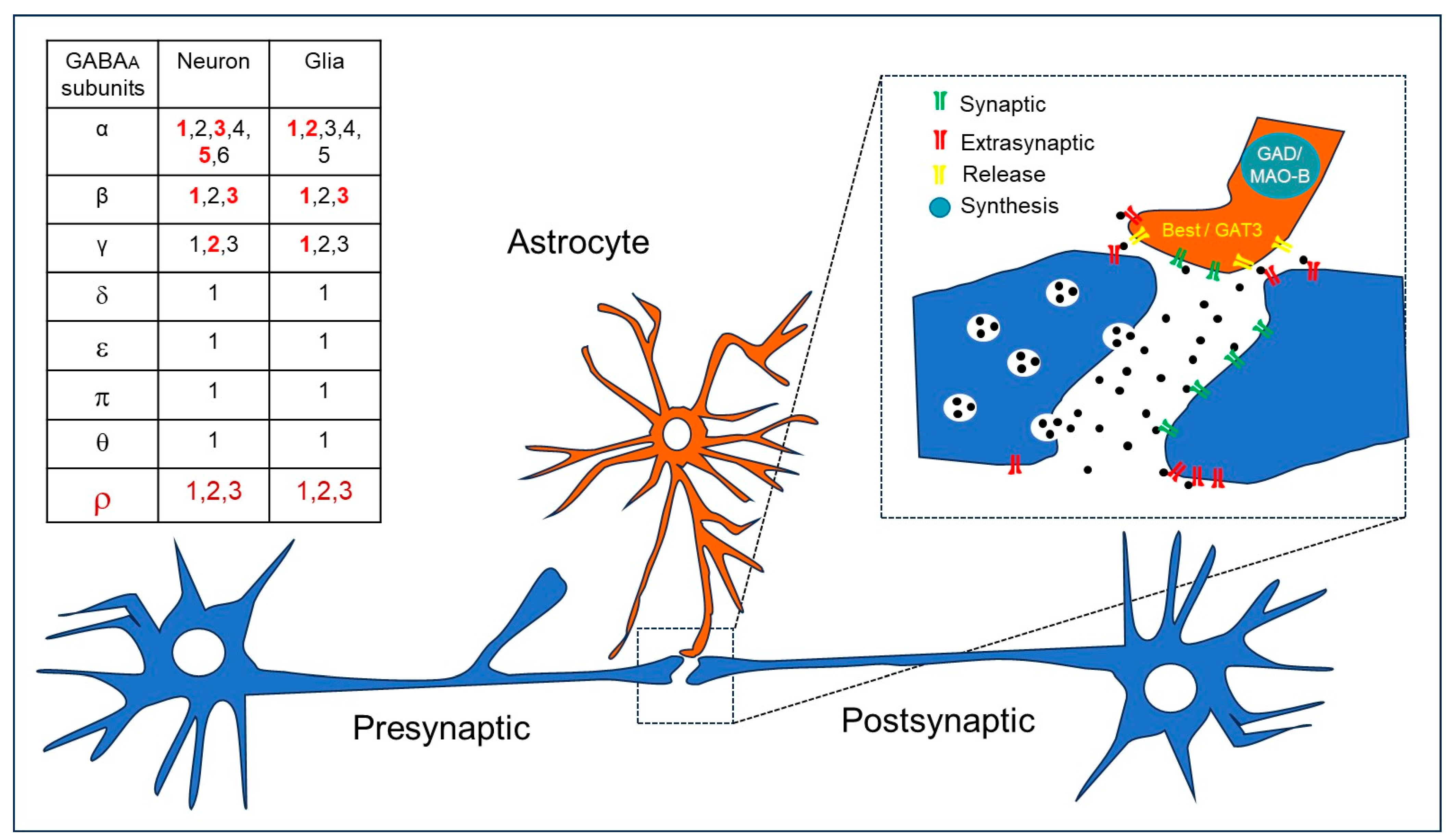
4. GABAA-ρ Receptors in Astroglia

Neurons are considered the basic unit for information processing associated with perception and cognition. However, this neurocentric definition has been challenged because they only represent a fraction of the brain cells. Glial cells, on the other hand, are the dominant population and respond to neuronal activity through neurotransmitter receptor activation [90]. Although the precise decoding of neuron–glia communication in brain function is unknown, glial cells contribute to synapse formation, cell development, myelination, brain microcirculation, neuroprotection, and the modulation of neural activity [91,92]. In this context, the GABAA receptors mediating synaptic (phasic) or extrasynaptic (tonic) transmission are molecularly and functionally distinct for both neurons and glial cells [93]. Astroglial cells include astrocytes, ependymal cells, Bergmann and Múller glia; all of them are identified by the expression of the cytoskeletal glial fibrillary acidic protein (GFAP). Astroglia express functional GABAA receptors, but their role in the CNS physiology is intriguing, since glial cells do not produce action potentials. Nevertheless, GABA depolarizes astrocytes through GABAA receptors but has unitary conductances and gating properties similar to those recorded in neurons [94]. Furthermore, specific differences in GABAA subunit expression were observed between primary cultures of rat cerebellar astrocytes and granular neurons [95]. Initial studies with RT-PCR reported that the total amount of GABAA receptor subunit mRNA in astrocytes was two orders of magnitude lower than in neuronal cells. Moreover, of all the GABAA subunits expressed by granular neurons, only α6 and γ2 subunits were not expressed by astrocytes. Another difference was that GABAA-α1, α3, and α5 subunits were abundantly expressed in granular neurons, while GABAA-α1 and α2 were predominant in cerebellar astrocytes. Likewise, GABAA-β1 and β3 were abundantly expressed in both cell types. Finally, GABAA-γ2 and GABAA-γ1 were prevalent in cerebellar granular neurons and astrocytes, respectively [95]. Studies in astrocytes isolated from the thalamus showed that all of them express functional GABAA receptors that responded to the selective agonist muscimol [96]. The same study showed a GABAA subunit expression profile through single cell RT-PCR studies. The prevalent subunits were GABAA-α2, GABAA-α5, GABAA-β1, and GABA A-γ1 [96]. Overall, these studies reported the heterogeneous expression of GABAA subunits associated with a brain region. Accordingly, astroglia showed differences in their responses to pharmacological modulators of GABAA receptors such as benzodiazepines and barbiturates [9,93,97]. For example, DMCM, an inverse benzodiazepine agonist that enhanced GABAA-mediated currents in astrocytes and inhibited them in neurons, suggests a different subunit composition [93]. Electrophysiological recordings on cerebellar astrocytes in situ showed a heterogeneous array of GABAA subunits because modulation by benzodiazepines was absent in Bergmann glia [97]. Moreover, modulation by barbiturates, such as pentobarbital, was present in Bergmann glia but absent in ependymal glial cells of the cerebellum [6,97]. These differences are related to a specific GABAA subunit array that includes GABAA-∆ or GABAA-ρ subunits in Bergmann glia and ependymal cells, respectively [6,97]. Additionally, the functional expression of GABAA receptors was reported in situ via in striatal astrocytes; pentobarbital modulation was absent and GABAA-ρ subunits were found in more than half of them [9] (Figure 2).
Although a role for the GABAA-ρ subunit expressed in glial cells is not yet evident, the three genes have been localized by means of immunogold electron microscopy in astrocytes from the neostriatum and cerebellum [5,9] (Figure 3).
The role of the GABAA-ρ subunit in cerebellar or neostriatal astroglia during early postnatal development or in the control of precise movements in adults remains unexplored.

5. GABAA Receptors and Extrasynaptic Communication in Brain Diseases

Huntington disease (HD). Patients with HD show motor, cognitive, and mental illnesses. The synthesis and release of GABA is compromised by the loss of striatal neurons, resulting in an increase in thalamo-cortical activity that produces “dance-like” movements. The expression of GABAA receptors displays a variability expression in HD [98] and the use of agonists is a common therapeutic strategy. Moreover, experimental evidence in HD transgenic R6/2 mouse brain showed that GABAA-ρ3 immunoreactivity decreases significantly in the striatum, hippocampus, and cortex [50]. Accordingly, RT-PCR studies reported that the mRNA expression of GABAA-ρ3 subunits occurs through different stages of the mouse postnatal development and double immunofluorescence studies showed its expression in calretinin+ interneurons and astrocytes, indicating a relevant role of this subunit in the physiology and pathology of the striatum [9].
Parkinson’s disease (PD). The main symptoms of PD are tremor and motor problems, a hallmark of the disease is the increased expression of GABAA receptors in the susbstantia nigra and caudate nucleus in postmortem brains of diagnosed patients. GABAergic transmission through ionotropic receptors has two modalities known as phasic and tonic. The first is involved in the regulation of neuronal activity while the second is mediated by extrasynaptic receptors that are activated mainly by astrocytes and influence the gain in neuronal excitability. Thus, astrocytes exhibit the augmented synthesis and release of GABA in PD, resulting in an increased tonic inhibition because of the augmented expression of GABA transporters (GATs) and high-affinity GABAA receptors [99].
Alzheimer’s disease (AD). This mental illness is associated with elderly people and memory loss is evident because of the accumulation of β-amyloid plaques in the brain. The number of GABAergic terminals contacting cortical neurons is reduced, while GABAA receptors are increased in AD. Furthermore, GABA synthesis (GAD67 or MAOB) by astrocytes is increased and released through GAT3 or bestrophin 1 channels, causing an increased tonic inhibition in the dentate gyrus of a mouse model of AD. Thus, astrocytic GABA maybe relevant in the pathology of AD [99].
Autism spectrum disorder (ASD). This developmental disorder is characterized by stereotyped and repetitive behaviors, social interaction deficits, and communication impairments. Dysfunction of the GABAergic signaling is associated with ASD, particularly, the GABAA receptors are involved in neuronal proliferation and differentiation [10]. The depolarization induced by GABA during early neurodevelopment leads to a decrease in DNA synthesis and the proliferation of GFAP+ progenitor cells in the subventricular zone (SVZ). This mechanism was also observed in the adult hippocampus where progenitor cells showed tonic currents induced by GABAA receptors containing the α4 subunit. The signaling requires an autocrine/paracrine release of GABA. Lastly, the agonist muscimol induces the proliferation of granule cells in the developing cerebellum of the rat. Moreover, postmortem studies in patients diagnosed with ASD reported an increased expression of GAD67 in cerebellar interneurons, together with Purkinje cell loss and diminished expression of the GABAA subunits α1 and β3, resulting in a significant E/I imbalance ([100]). The preclinical model of autism induced by prenatal exposure to valproic acid (VPA) reproduced Purkinje cell loss and the increased expression of the astrocytic marker GFAP [10]. The expression of GABAA-ρ3 increases linearly throughout neurodevelopment of the cerebellum and homomeric receptors show high affinity for the neurotransmitter (EC50 ∼ 3 μM). Immunofluorescence studies reported that, besides Purkinje cells, GABAA-ρ3 is expressed in ependymal cells and whole-cell patch-clamp recordings confirmed the functional expression of GABAA-ρ receptors, because GABA-evoked currents were partially blocked with TPMPA and completely abolished when bicuculline was added, pentobarbital potentiation was absent. Interestingly, GABAρ3 expression is disrupted in the VPA model, suggesting that this subunit is relevant in ASD [6,10].

6. Discussion

Interneuron–astrocyte communication through GABAergic signaling induces calcium transients that can actively regulate E/I balance through gliotransmission [101]. Indeed, extracellular chloride concentration is important for inhibitory transmission and is partly modulated by astrocytic depolarization, resulting from chloride efflux after GABAA receptor activation. The Cl-mediated depolarization favors the influx of Ca2+ from the extracellular space through voltage-gated calcium channels, promoting additional calcium release from intracellular stores and consequently, the release of different gliotransmitters [102] (Figure 4).
The astrocytic release of GABA was identified as one of the main extrasynaptic sources for tonic inhibition [103] and it controls the migration of neuronal precursors through GABAA receptor activation at the subventricular zone, while a similar mechanism operates for cortical development through GABAA-ρ receptors [61,104]. Moreover, interneurons and astrocytes express GABAA-ρ subunits during early postnatal development of the striatum [5,9], while GABAρ3 is expressed before the onset of hearing and later replaced by GABAA-ρ1 and GABAA-ρ2 subunits in the medial nucleus of the trapezoid body [6]. The GABAA-ρ3 subunit in cerebellar ependymal cells is reduced in a preclinical model of autism induced by prenatal exposure to valproic acid, suggesting a key role of GABAA-ρ receptors in neurodevelopment disorders [10]. On the other hand, therapeutic strategies targeting astroglial GABAA-ρ receptors were recently explored in post-stroke motor recovery [11]. Thus, this review highlights GABAA-ρ expressing astroglia as another population that can potentially modulate synaptic function. Accordingly, astrocytes with a specific molecular signature were recently shown to contribute to LTP strengthening and hippocampal memories, opposing hyperexcitation during seizures and possible sub-thalamic nucleus overactivation in Parkinson’s disease after astrocyte-targeted genetic VGLUT deletion [105]. Future studies should explore the precise inhibitory synapse–astrocyte organization at the molecular (scRNAseq) and functional level (3D Ca2+ imaging).

7. Conclusions

GABAA-ρ receptors are functionally expressed by neurons and astroglia. They are involved in extrasynaptic-mediated tonic currents and are relevant in earlier postnatal neurodevelopment. However, further studies are required to understand their role in the physiology and pathology of the central nervous system.

Author Contributions

A.R.-A., A.E.-M., A.M.-T. and D.R.-H. wrote the original draft and edited the final version of the manuscript. All authors contributed to the article and approved the submitted version. All authors have read and agreed to the published version of the manuscript.

Funding

This work was supported by CONAHCYT 319209 and UNAM-DGAPA-PAPIIT IN209121 to DR-H.

Acknowledgments

The authors acknowledge Jessica Norris for her proofreading of the English version of the manuscript. D.R.-H. is grateful to R. Arellano and E. Garay, for academic and technical support.

Conflicts of Interest

The authors declare that the research was conducted in the absence of any commercial or financial relationships that could be construed as a potential conflict of interest.

References

  1. Roberts, E.; Frankel, S. gamma-Aminobutyric acid in brain: Its formation from glutamic acid. J. Biol. Chem. 1950, 187, 55–63. [Google Scholar] [CrossRef] [PubMed]
  2. Krnjevic, K.; Phillis, J.W. Iontophoretic studies of neurones in the mammalian cerebral cortex. J. Physiol. 1963, 165, 274–304. [Google Scholar] [CrossRef] [PubMed]
  3. Parker, I.; Gundersen, C.B.; Miledi, R. Actions of pentobarbital on rat brain receptors expressed in Xenopus oocytes. J. Neurosci. 1986, 6, 2290–2297. [Google Scholar] [CrossRef] [PubMed]
  4. Polenzani, L.; Woodward, R.M.; Miledi, R. Expression of mammalian gamma-aminobutyric acid receptors with distinct pharmacology in Xenopus oocytes. Proc. Natl. Acad. Sci. USA 1991, 88, 4318–4322. [Google Scholar] [CrossRef]
  5. Rosas-Arellano, A.; Machuca-Parra, A.I.; Reyes-Haro, D.; Miledi, R.; Martínez-Torres, A. Expression of GABAρ receptors in the neostriatum: Localization in aspiny, medium spiny neurons and GFAP-positive cells. J. Neurochem. 2012, 122, 900–910. [Google Scholar] [CrossRef]
  6. Reyes-Haro, D.; González-González, M.A.; Pétriz, A.; Rosas-Arellano, A.; Kettenmann, H.; Miledi, R.; Martínez-Torres, A. γ-Aminobutyric acid-ρ expression in ependymal glial cells of the mouse cerebellum. J. Neurosci. Res. 2013, 4, 527–534. [Google Scholar] [CrossRef]
  7. Reyes-Haro, D.; Rosas-Arellano, A.; González-González, M.A.; Mora-Loyola, E.; Miledi, R.; Martínez-Torres, A. GABAρ expression in the medial nucleus of the trapezoid body. Neurosci. Lett. 2013, 532, 23–28. [Google Scholar] [CrossRef]
  8. Pétriz, A.; Reyes-Haro, D.; González-González, M.A.; Miledi, R.; Martínez-Torres, A. GABAρ subunits confer a bicuculline-insensitive component to GFAP+ cells of cerebellum. Proc. Natl. Acad. Sci. USA 2014, 111, 17522–17527. [Google Scholar] [CrossRef]
  9. Reyes-Haro, D.; Hernández-Santos, J.A.; Miledi, R.; Martínez-Torres, A. GABAρ selective antagonist TPMPA partially inhibits GABA-mediated currents recorded from neurones and astrocytes in mouse striatum. Neuropharmacology 2017, 113, 407–415. [Google Scholar] [CrossRef]
  10. Varman, D.R.; Soria-Ortíz, M.B.; Martínez-Torres, A.; Reyes-Haro, D. GABAρ3 expression in lobule X of the cerebellum is reduced in the valproate model of autism. Neurosci. Lett. 2018, 687, 158–163. [Google Scholar] [CrossRef]
  11. van Nieuwenhuijzen, P.S.; Parker, K.; Liao, V.; Houlton, J.; Kim, H.L.; Johnston, G.A.R.; Hanrahan, J.R.; Chebib, M.; Clarkson, A.N. Targeting GABAC Receptors Improves Post-Stroke Motor Recovery. Brain Sci. 2021, 11, 315. [Google Scholar] [CrossRef]
  12. Barnard, E.A. Receptor classes and the transmitter-gated ion channels. Trends Biochem. Sci. 1992, 17, 368–374. [Google Scholar] [CrossRef]
  13. Hill, D.R.; Bowery, N.G. 3H-baclofen and 3H-GABA bind to bicuculline-insensitive GABA B sites in rat brain. Nature 1981, 290, 149–152. [Google Scholar] [CrossRef]
  14. Olsen, R.W.; Sieghart, W. International Union of Pharmacology. LXX. Subtypes of gamma-aminobutyric acid(A) receptors: Classification on the basis of subunit composition, pharmacology, and function. Update. Pharmacol. Rev. 2008, 60, 243–260. [Google Scholar] [CrossRef] [PubMed]
  15. Collingridge, G.L.; Olsen, R.W.; Peters, J.; Spedding, M. A nomenclature for ligand-gated ion channels. Neuropharmacology 2009, 56, 2–5. [Google Scholar] [CrossRef]
  16. Rosas-Arellano, A.; Estrada-Mondragón, A.; Mantellero, C.A.; Tejeda-Guzmán, C.; Castro, M.A. The adjustment of γ-aminobutyric acidA tonic subunits in Huntington’s disease: From transcription to translation to synaptic levels into the neostriatum. Neural Regen. Res. 2018, 13, 584–590. [Google Scholar] [CrossRef] [PubMed]
  17. Cowgill, J.; Fan, C.; Haloi, N.; Tobiasson, V.; Zhuang, Y.; Howard, R.J.; Lindahl, E. Structure and dynamics of dif-ferential ligand binding in the human ρ-type GABAA receptor. Neuron 2023, S0896-6273(23)00587-1. [Google Scholar] [CrossRef]
  18. Cutting, G.R.; Lu, L.; O’Hara, B.F.; Kasch, L.M.; Montrose-Rafizadeh, C.; Donovan, D.M.; Shimada, S.; Antonarakis, S.E.; Guggino, W.B.; Uhl, G.R.; et al. Cloning of the gamma-aminobutyric acid (GABA) rho 1 cDNA: A GABA receptor subunit highly expressed in the retina. Proc. Natl. Acad. Sci. USA 1991, 88, 2673–2677. [Google Scholar] [CrossRef]
  19. Wang, T.L.; Guggino, W.B.; Cutting, G.R. A novel gamma-aminobutyric acid receptor subunit (rho 2) cloned from human retina forms bicuculline-insensitive homooligomeric receptors in Xenopus oocytes. J. Neurosci. 1994, 14, 6524–6531. [Google Scholar] [CrossRef]
  20. Ogurusu, T.; Shingai, R. Cloning of a putative gamma-aminobutyric acid (GABA) receptor subunit rho 3 cDNA. Biochim. Biophys. Acta 1996, 1305, 15–18. [Google Scholar] [CrossRef]
  21. Martínez-Torres, A.; Vazquez, A.E.; Panicker, M.M.; Miledi, R. Cloning and functional expression of alternative spliced variants of the rho1 gamma-aminobutyrate receptor. Proc. Natl. Acad. Sci. USA 1998, 95, 4019–4022. [Google Scholar] [CrossRef] [PubMed]
  22. Miledi, R.; Parker, I.; Sumikawa, K. Properties of acetylcholine receptors translated by cat muscle mRNA in Xenopus oocytes. EMBO J. 1982, 1, 1307–1312. [Google Scholar] [CrossRef] [PubMed]
  23. Hackam, A.S.; Wang, T.L.; Guggino, W.B.; Cutting, G.R. The N-terminal domain of human GABA receptor rho1 subunits contains signals for homooligomeric and heterooligomeric interaction. J. Biol. Chem. 1997, 272, 13750–13757. [Google Scholar] [CrossRef] [PubMed]
  24. Pan, Y.; Ripps, H.; Qian, H. Random assembly of GABA rho1 and rho2 subunits in the formation of heteromeric GABA(C) receptors. Cell Mol. Neurobiol. 2006, 26, 289–305. [Google Scholar] [CrossRef]
  25. Qian, H.; Ripps, H. Response kinetics and pharmacological properties of heteromeric receptors formed by coassembly of GABA rho- and gamma 2-subunits. Proc. Biol. Sci. 1999, 266, 2419–2425. [Google Scholar] [CrossRef]
  26. Milligan, C.J.; Buckley, N.J.; Garret, M.; Deuchars, J.; Deuchars, S.A. Evidence for inhibition mediated by coassembly of GABAA and GABAC receptor subunits in native central neurons. J. Neurosci. 2004, 24, 7241–7250. [Google Scholar] [CrossRef]
  27. Pan, Y.; Qian, H. Interactions between rho and gamma2 subunits of the GABA receptor. J. Neurochem. 2005, 94, 482–490. [Google Scholar] [CrossRef]
  28. Pan, Z.H.; Zhang, D.; Zhang, X.; Lipton, S.A. Evidence for coassembly of mutant GABAC rho1 with GABAA gamma2S, glycine alpha1 and glycine alpha2 receptor subunits in vitro. Eur. J. Neurosci. 2000, 12, 3137–3145. [Google Scholar] [CrossRef]
  29. Woodward, R.M.; Polenzani, L.; Miledi, R. Characterization of bicuculline/baclofen-insensitive (rho-like) gamma-aminobutyric acid receptors expressed in Xenopus oocytes. II. Pharmacology of gamma-aminobutyric acidA and gamma-aminobutyric acidB receptor agonists and antagonists. Mol. Pharmacol. 1993, 43, 609–625. [Google Scholar]
  30. Enz, R.; Cutting, G.R. Molecular composition of GABAC receptors. Vision Res. 1998, 38, 1431–1441. [Google Scholar] [CrossRef]
  31. Zhang, J.; Xue, F.; Chang, Y. Structural determinants for antagonist pharmacology that distinguish the rho1 GABAC receptor from GABAA receptors. Mol. Pharmacol. 2008, 74, 941–951. [Google Scholar] [CrossRef] [PubMed]
  32. Johnston, G.A.; Curtis, D.R.; Beart, P.M.; Game, C.J.; McCulloch, R.M.; Twitchin, B. Cis- and trans-4-aminocrotonic acid as GABA analogues of restricted conformation. J. Neurochem. 1975, 24, 157–160. [Google Scholar] [CrossRef] [PubMed]
  33. Kusama, T.; Spivak, C.E.; Whiting, P.; Dawson, V.L.; Schaeffer, J.C.; Uhl, G.R. Pharmacology of GABA rho 1 and GABA alpha/beta receptors expressed in Xenopus oocytes and COS cells. Br. J. Pharmacol. 1993, 109, 200–206. [Google Scholar] [CrossRef] [PubMed]
  34. Kerr, D.I.; Ong, J. GABAB receptors. Pharmacol. Ther. 1995, 67, 187–246. [Google Scholar] [CrossRef]
  35. Thomet, U.; Baur, R.; Dodd, R.H.; Sigel, E. Loreclezole as a simple functional marker for homomeric rho type GABA(C) receptors. Eur. J. Pharmacol. 2000, 408, 1–2. [Google Scholar] [CrossRef]
  36. Ng, C.K.; Kim, H.L.; Gavande, N.; Yamamoto, I.; Kumar, R.J.; Mewett, K.N.; Johnston, G.A.; Hanrahan, J.R.; Chebib, M. Medicinal chemistry of ρ GABAC receptors. Future Med. Chem. 2011, 3, 197–209. [Google Scholar] [CrossRef]
  37. Arnaud, C.; Gauthier, P.; Gottesmann, C. Study of a GABAC receptor antagonist on sleep-waking behavior in rats. Psychopharmacology 2001, 154, 415–419. [Google Scholar] [CrossRef]
  38. Naffaa, M.M.; Hung, S.; Chebib, M.; Johnston, G.A.R.; Hanrahan, J.R. GABAρ receptors: Distinctive functions and molecular pharmacology. Br. J. Pharmacol. 2017, 174, 1881–1894. [Google Scholar] [CrossRef]
  39. Enz, R.; Brandstätter, J.H.; W‚àö¬ßssle, H.; Bormann, J. Immunocytochemical localization of the GABAc receptor rho subunits in the mammalian retina. J. Neurosci. 1996, 16, 4479–4490. [Google Scholar] [CrossRef]
  40. Koulen, P.; Brandstätter, J.H.; Enz, R.; Bormann, J.; Wässle, H. Synaptic clustering of GABA(C) receptor rho-subunits in the rat retina. Eur. J. Neurosci. 1998, 10, 115–127. [Google Scholar] [CrossRef]
  41. Dong, C.J.; Picaud, S.A.; Werblin, F.S. GABA transporters and GABAC-like receptors on catfish cone- but not rod-driven horizontal cells. J. Neurosci. 1994, 14, 2648–2658. [Google Scholar] [CrossRef] [PubMed]
  42. Du, J.L.; Yang, X.L. Subcellular localization and complements of GABA(A) and GABA(C) receptors on bullfrog retinal bipolar cells. J. Neurophysiol. 2000, 84, 666–676. [Google Scholar] [CrossRef] [PubMed]
  43. Qian, H.; Dowling, J.E. GABAA and GABAC receptors on hybrid bass retinal bipolar cells. J. Neurophysiol. 1995, 74, 1920–1928. [Google Scholar] [CrossRef] [PubMed]
  44. Zhang, J.; Slaughter, M.M. Preferential suppression of the ON pathway by GABAC receptors in the amphibian retina. J. Neurophysiol. 1995, 74, 1583–1592. [Google Scholar] [CrossRef] [PubMed]
  45. Fletcher, E.L.; Koulen, P.; Wässle, H. GABAA and GABAC receptors on mammalian rod bipolar cells. J. Comp. Neurol. 1998, 396, 351–365. [Google Scholar] [CrossRef]
  46. Delaney, A.J.; Sah, P. GABA receptors inhibited by benzodiazepines mediate fast inhibitory transmission in the central amygdala. J. Neurosci. 1999, 19, 9698–9704. [Google Scholar] [CrossRef]
  47. Fujimura, J.; Nagano, M.; Suzuki, H. Differential expression of GABA(A) receptor subunits in the distinct nuclei of the rat amygdala. Brain Res. Mol. Brain Res. 2005, 138, 17–23. [Google Scholar] [CrossRef]
  48. Flores-Gracia, C.; Nuche-Bricaire, A.; Crespo-Ramírez, M.; Miledi, R.; Fuxe, K.; Pérez de la Mora, M. GABA(A) ρ receptor mechanisms in the rat amygdala and its role in the modulation of fear and anxiety. Psychopharmacology 2010, 212, 475–484. [Google Scholar] [CrossRef]
  49. Rosas-Arellano, A.; Parodi, J.; Machuca-Parra, A.I.; Sánchez-Gutiérrez, A.; Inestrosa, N.C.; Miledi, R.; Martínez-Torres, A. The GABA(A)ρ receptors in hippocampal spontaneous activity and their distribution in hippocampus, amygdala and visual cortex. Neurosci. Lett. 2011, 500, 20–25. [Google Scholar] [CrossRef]
  50. Kumar, U.; Heer, M.; Somvanshi, R.K. Regional and subcellular distribution of GABAC ρ3 receptor in brain of R6/2 mouse model of Huntington’s disease. Neurosci. Lett. 2017, 640, 81–87. [Google Scholar] [CrossRef]
  51. Wegelius, K.; Pasternack, M.; Hiltunen, J.O.; Rivera, C.; Kaila, K.; Saarma, M.; Reeben, M. Distribution of GABA receptor rho subunit transcripts in the rat brain. Eur. J. Neurosci. 1998, 10, 350–357. [Google Scholar] [CrossRef] [PubMed]
  52. López-Chávez, A.; Miledi, R.; Martínez-Torres, A. Cloning and functional expression of the bovine GABA(C) rho2 subunit. Molecular evidence of a widespread distribution in the CNS. Neurosci. Res. 2005, 53, 421–427. [Google Scholar] [CrossRef] [PubMed]
  53. Frazao, R.; Nogueira, M.I.; Wässle, H. Colocalization of synaptic GABA(C)-receptors with GABA (A)-receptors and glycine-receptors in the rodent central nervous system. Cell Tissue Res. 2007, 330, 1–15. [Google Scholar] [CrossRef] [PubMed]
  54. Rosas-Arellano, A.; Ochoa-de la Paz, L.D.; Miledi, R.; Martínez-Torres, A. Brain distribution and molecular cloning of the bovine GABA rho1 receptor. Neurosci. Res. 2007, 57, 347–353. [Google Scholar] [CrossRef]
  55. Chesnoy-Marchais, D. Persistent GABAA/C responses to gabazine, taurine and beta-alanine in rat hypoglossal motoneurons. Neuroscience 2016, 330, 191–204. [Google Scholar] [CrossRef]
  56. Albrecht, B.E.; Breitenbach, U.; Stühmer, T.; Harvey, R.J.; Darlison, M.G. In situ hybridization and reverse transcription polymerase chain reaction studies on the expression of the GABA(C) receptor rho1- and rho2-subunit genes in avian and rat brain. Eur. J. Neurosci. 1997, 9, 2414–2422. [Google Scholar] [CrossRef]
  57. Boue-Grabot, E.; Roudbaraki, M.; Bascles, L.; Tramu, G.; Bloch, B.; Garret, M. Expression of GABA receptor rho subunits in rat brain. J. Neurochem. 1998, 70, 899–907. [Google Scholar] [CrossRef]
  58. Harvey, V.L.; Duguid, I.C.; Krasel, C.; Stephens, G.J. Evidence that GABA rho subunits contribute to functional ionotropic GABA receptors in mouse cerebellar Purkinje cells. J. Physiol. 2006, 577, 127–139. [Google Scholar] [CrossRef]
  59. Mejía, C.; García-Alcocer, G.; Berumen, L.C.; Rosas-Arellano, A.; Miledi, R.; Martínez-Torres, A. Expression of GABArho subunits during rat cerebellum development. Neurosci. Lett. 2008, 432, 1–6. [Google Scholar] [CrossRef]
  60. Martínez-Delgado, G.; Reyes-Haro, D.; Espino-Saldaña, A.E.; Rosas-Arellano, A.; Pétriz, A.; Juárez-Mercado, P.; Miledi, R.; Martínez-Torres, A. Dynamics of GABAρ2 receptors in retinal bipolar neurons and cerebellar astrocytes. Neuroreport 2011, 22, 4–9. [Google Scholar] [CrossRef]
  61. Denter, D.G.; Heck, N.; Riedemann, T.; White, R.; Kilb, W.; Luhmann, H.J. GABAC receptors are functionally expressed in the intermediate zone and regulate radial migration in the embryonic mouse neocortex. Neuroscience 2010, 167, 124–134. [Google Scholar] [CrossRef] [PubMed]
  62. Limon, A.; Reyes-Ruiz, J.M.; Miledi, R. Loss of functional GABA(A) receptors in the Alzheimer diseased brain. Proc. Natl. Acad. Sci. USA 2012, 109, 10071–10076. [Google Scholar] [CrossRef] [PubMed]
  63. Fatemi, S.H.; Reutiman, T.J.; Folsom, T.D.; Rustan, O.G.; Rooney, R.J.; Thuras, P.D. Downregulation of GABAA receptor protein subunits α6, β2, δ, ε, γ2, τ, and ρ2 in superior frontal cortex of subjects with autism. J. Autism Dev. Disord. 2014, 44, 1833–1845. [Google Scholar] [CrossRef] [PubMed]
  64. Mohammadi, E.; Shamsizadeh, A.; Salari, E.; Fatemi, I.; Allahtavakoli, M.; Roohbakhsh, A. Effect of TPMPA (GABAC receptor antagonist) on neuronal response properties in rat barrel cortex. Somatosens. Mot. Res. 2017, 34, 108–115. [Google Scholar] [CrossRef] [PubMed]
  65. Cheng, Z.Y.; Chebib, M.; Schmid, K.L. Identification of GABA receptors in chick cornea. Mol. Vis. 2012, 18, 1107–1114. [Google Scholar] [PubMed]
  66. Didelon, F.; Sciancalepore, M.; Savic’, N.; Mladinic’, M.; Bradbury, A.; Cherubini, E. gamma-Aminobutyric acidA rho receptor subunits in the developing rat hippocampus. J. Neurosci. Res. 2002, 67, 739–744. [Google Scholar] [CrossRef]
  67. Rozzo, A.; Armellin, M.; Franzot, J.; Chiaruttini, C.; Nistri, A.; Tongiorgi, E. Expression and dendritic mRNA localization of GABAC receptor rho1 and rho2 subunits in developing rat brain and spinal cord. Eur. J. Neurosci. 2002, 15, 1747–1758. [Google Scholar] [CrossRef]
  68. Hartmann, K.; Stief, F.; Draguhn, A.; Frahm, C. Ionotropic GABA receptors with mixed pharmacological properties of GABAA and GABAC receptors. Eur. J. Pharmacol. 2004, 497, 139–146. [Google Scholar] [CrossRef]
  69. Liu, B.; Hattori, N.; Jiang, B.; Nakayama, Y.; Zhang, N.Y.; Wu, B.; Kitagawa, K.; Taketo, M.; Matsuda, H.; Inagaki, C. Single cell RT-PCR demonstrates differential expression of GABAC receptor rho subunits in rat hippocampal pyramidal and granule cells. Brain Res. Mol. Brain Res. 2004, 123, 1–6. [Google Scholar] [CrossRef]
  70. Alakuijala, A.; Palgi, M.; Wegelius, K.; Schmidt, M.; Enz, R.; Paulin, L.; Saarma, M.; Pasternack, M. GABA receptor rho subunit expression in the developing rat brain. Brain Res. Dev. Brain Res. 2005, 154, 15–23. [Google Scholar] [CrossRef]
  71. Bhandage, A.K.; Jin, Z.; Bazov, I.; Kononenko, O.; Bakalkin, G.; Korpi, E.R.; Birnir, B. GABA-A and NMDA receptor subunit mRNA expression is altered in the caudate but not the putamen of the postmortem brains of alcoholics. Front. Cell. Neurosci. 2014, 8, 415. [Google Scholar] [CrossRef]
  72. Chen, Y.; Zhou, D.; Zhou, K.; Ren, Y.; Dai, W.; Xu, M.; Lu, L.; Lu, Z. Study on olfactory function in GABAC receptor/channel rho1 subunit knockout mice. Neurosci. Lett. 2007, 427, 10–15. [Google Scholar] [CrossRef] [PubMed]
  73. Sivilotti, L.; Nistri, A. Pharmacology of a novel effect of gamma-aminobutyric acid on the frog optic tectum in vitro. Eur. J. Pharmacol. 1989, 164, 205–212. [Google Scholar] [CrossRef] [PubMed]
  74. Cheng, Z.Y.; Wang, X.P.; Schmid, K.L.; Liu, L. Identification of GABA receptors in chick retinal pigment epithelium. Neurosci. Lett. 2013, 539, 43–47. [Google Scholar] [CrossRef]
  75. Ekema, G.M.; Zheng, W.; Lu, L. Interaction of GABA receptor/channel rho(1) and gamma(2) subunit. Investig. Ophthalmol. Vis. Sci. 2002, 43, 2326–2333. [Google Scholar] [PubMed]
  76. Zheng, W.; Xie, W.; Zhang, J.; Strong, J.A.; Wang, L.; Yu, L.; Xu, M.; Lu, L. Function of gamma-aminobutyric acid receptor/channel rho 1 subunits in spinal cord. J. Biol. Chem. 2003, 278, 48321–48329. [Google Scholar] [CrossRef]
  77. Grabert, J.; Jost, B.; Patz, S.; Wahle, P. GABA(C) receptors are expressed in GABAergic and non-GABAergic neurons of the rat superior colliculus and visual cortex. Exp. Brain Res. 2009, 199, 245–252. [Google Scholar] [CrossRef]
  78. Pasternack, M.; Boller, M.; Pau, B.; Schmidt, M. GABA(A) and GABA(C) receptors have contrasting effects on excitability in superior colliculus. J. Neurophysiol. 1999, 82, 2020–2023. [Google Scholar] [CrossRef]
  79. Schlicker, K.; McCall, M.A.; Schmidt, M. GABAC receptor-mediated inhibition is altered but not eliminated in the superior colliculus of GABAC rho1 knockout mice. J. Neurophysiol. 2009, 101, 2974–2983. [Google Scholar] [CrossRef]
  80. Wong, L.W.; Tae, H.S.; Cromer, B.A. Role of the ρ GABA(C) receptor N-terminus in assembly, trafficking and function. ACS Chem. Neurosci. 2014, 5, 1266–1277. [Google Scholar] [CrossRef]
  81. Hanley, J.G.; Koulen, P.; Bedford, F.; Gordon-Weeks, P.R.; Moss, S.J. The protein MAP-1B links GABA(C) receptors to the cytoskeleton at retinal synapses. Nature 1999, 397, 66–69. [Google Scholar] [CrossRef]
  82. Billups, D.; Hanley, J.G.; Orme, M.; Attwell, D.; Moss, S.J. GABAC receptor sensitivity is modulated by interaction with MAP1B. J. Neurosci. 2000, 20, 8643–8650. [Google Scholar] [CrossRef] [PubMed]
  83. Hanley, J.G.; Jones, E.M.; Moss, S.J. GABA receptor rho1 subunit interacts with a novel splice variant of the glycine transporter, GLYT-1. J. Biol. Chem. 2000, 275, 840–846. [Google Scholar] [CrossRef] [PubMed]
  84. Chen, L.; Wang, H.; Vicini, S.; Olsen, R.W. The gamma-aminobutyric acid type A (GABAA) receptor-associated protein (GABARAP) promotes GABAA receptor clustering and modulates the channel kinetics. Proc. Natl. Acad Sci. USA 2000, 97, 11557–11562. [Google Scholar] [CrossRef] [PubMed]
  85. Kittler, J.T.; Rostaing, P.; Schiavo, G.; Fritschy, J.M.; Olsen, R.; Triller, A.; Moss, S.J. The subcellular distribution of GABARAP and its ability to interact with NSF suggest a role for this protein in the intracellular transport of GABA(A) receptors. Mol. Cell Neurosci. 2001, 18, 13–25. [Google Scholar] [CrossRef] [PubMed]
  86. Wang, H.; Bedford, F.K.; Brandon, N.J.; Moss, S.J.; Olsen, R.W. GABA(A)-receptor-associated protein links GABA(A) receptors and the cytoskeleton. Nature 1999, 397, 69–72. [Google Scholar] [CrossRef]
  87. Boué-Grabot, E.; Emerit, M.B.; Toulmé; Séguéla, P.; Garret, M. Cross-talk and co-trafficking between rho1/GABA receptors and ATP-gated channels. J. Biol. Chem. 2004, 279, 6967–6975. [Google Scholar] [CrossRef]
  88. Linck, L.; Binder, J.; Haynl, C.; Enz, R. Endocytosis of GABA(C) receptors depends on subunit composition and is regulated by protein kinase C-ζ and protein phosphatase 1. J. Neurochem. 2015, 134, 233–246. [Google Scholar] [CrossRef]
  89. Croci, C.; Brändstatter, J.H.; Enz, R. ZIP3, a new splice variant of the PKC-zeta-interacting protein family, binds to GABAC receptors, PKC-zeta, and Kv beta 2. J. Biol. Chem. 2003, 278, 6128–6135. [Google Scholar] [CrossRef]
  90. Reyes-Haro, D.; Bulavina, L.; Pivneva, T. Glia, el pegamento de las ideas [Glia, the glue of ideas]. Ciencia 2014, 65, 12–18. [Google Scholar]
  91. Barres, B.A. The mystery and magic of glia: A perspective on their roles in health and disease. Neuron 2008, 60, 430–440. [Google Scholar] [CrossRef]
  92. Vélez-Fort, M.; Audinat, E.; Angulo, M.C. Central role of GABA in neuron-glia interactions. Neuroscientist 2012, 18, 237–250. [Google Scholar] [CrossRef] [PubMed]
  93. Bormann, J.; Kettenmann, H. Patch-clamp study of gamma-aminobutyric acid receptor Cl- channels in cultured astrocytes. Proc. Natl. Acad Sci. USA 1988, 85, 9336–9340. [Google Scholar] [CrossRef]
  94. Kettenmann, H.; Backus, K.H.; Schachner, M. Aspartate, glutamate and gamma-aminobutyric acid depolarize cultured astrocytes. Neurosci. Lett. 1984, 23, 25–29. [Google Scholar] [CrossRef] [PubMed]
  95. Bovolin, P.; Santi, M.R.; Puia, G.; Costa, E.; Grayson, D. Expression patterns of gamma-aminobutyric acid type A receptor subunit mRNAs in primary cultures of granule neurons and astrocytes from neonatal rat cerebella. Proc. Natl. Acad Sci. USA 1992, 89, 9344–9348. [Google Scholar] [CrossRef]
  96. Höft, S.; Griemsmann, S.; Seifert, G.; Steinhäuser, C. Heterogeneity in expression of functional ionotropic glutamate and GABA receptors in astrocytes across brain regions: Insights from the thalamus. Philos. Trans. R. Soc. Lond. B Biol. Sci. 2014, 369, 20130602. [Google Scholar] [CrossRef] [PubMed]
  97. Müller, T.; Fritschy, J.M.; Grosche, J.; Pratt, G.D.; Möhler, H.; Kettenmann, H. Developmental regulation of voltage-gated K+ channel and GABAA receptor expression in Bergmann glial cells. J. Neurosci. 1994, 14 Pt 1, 2503–2514. [Google Scholar] [CrossRef]
  98. Rosas-Arellano, A.; Tejeda-Guzmán, C.; Lorca-Ponce, E.; Palma-Tirado, L.; Mantellero, C.A.; Rojas, P.; Missirlis, F.; Castro, M.A. Huntington’s disease leads to decrease of GABA-A tonic subunits in the D2 neostriatal pathway and their relocalization into the synaptic cleft. Neurobiol. Dis. 2018, 110, 142–153. [Google Scholar] [CrossRef] [PubMed]
  99. Kim, Y.S.; Yoon, B.E. Altered GABAergic Signaling in Brain Disease at Various Stages of Life. Exp. Neurobiol. 2017, 26, 122–131. [Google Scholar] [CrossRef]
  100. Ghit, A.; Assal, D.; Al-Shami, A.S.; Hussein, D.E.E. GABAA receptors: Structure, function, pharmacology, and related disorders. J. Genet. Eng. Biotechnol. 2021, 19, 123. [Google Scholar] [CrossRef]
  101. Mederos, S.; Perea, G. GABAergic-astrocyte signaling: A refinement of inhibitory brain networks. Glia 2019, 67, 1842–1851. [Google Scholar] [CrossRef] [PubMed]
  102. Liu, J.; Feng, X.; Wang, Y.; Xia, X.; Zheng, J.C. Astrocytes: GABAceptive and GABAergic Cells in the Brain. Front. Cell. Neurosci. 2022, 16, 892497. [Google Scholar] [CrossRef]
  103. Yoon, B.E.; Lee, C.J. GABA as a rising gliotransmitter. Front. Neural. Circuits 2014, 17, 141. [Google Scholar] [CrossRef]
  104. Bolteus, A.J.; Bordey, A. GABA release and uptake regulate neuronal precursor migration in the postnatal subventricular zone. J. Neurosci. 2004, 24, 7623–7631. [Google Scholar] [CrossRef]
  105. de Ceglia, R.; Ledonne, A.; Litvin, D.G.; Lind, B.L.; Carriero, G.; Latagliata, E.C.; Bindocci, E.; Di Castro, M.A.; Savtchouk, I.; Vitali, I.; et al. Specialized astrocytes mediate glutamatergic gliotransmission in the CNS. Nature, 2023; online ahead of print. [Google Scholar] [CrossRef]
Figure 1. Molecular structure and binding-site of the human GABAA-ρ1 receptor. (A). View of the pentameric receptor embedded in the plasma membrane, expanding in detail the orthosteric GABA binding site formed by the interface of two subunits in the amino-terminal extracellular domain. Residues interacting with GABA (yellow) from the main subunit (cyan), or complementary (–) subunit (deep blue), are indicated. (B). View of the pentamer from the outside of the membrane, showing the central ion-conducting pore. Structure representation was prepared using PyMOL v 2.5.2 (PDB file: 8OP9, from [17]).
Neuroglia 04 00017 g001
Figure 2. Functional expression of the GABAA-ρ subunit in astrocytes. Whole-cell patch-clamp recording of an astrocyte in a coronal slice of the dorsal striatum of a GFAP-EGFP mouse. A classic passive current profile obtained from a sulforhodamine B (magenta)-injected cell that was also GFAP-EGFP+ (yellow). The overlay of both signals confirms cell identity. Scale bar represents 20 μm. Membrane currents were evoked in 50 ms voltage steps ranging from −160 to +40 mV, from a holding potential of −70 mV. Astrocytes responded to GABA (10 μM) and the current was partially reduced by the selective GABAA-ρ antagonist TPMPA (100 μM); a further inhibition was observed when the selective GABA-A antagonist bicuculline (BIC, 100 μM) was added. The arrows indicate the start (up) and end (down) of the agonist/antagonist application.
Neuroglia 04 00017 g002
Figure 3. Representative electron micrographs of GABAA-ρ1-3 immunogold localization in mouse neostriatum. (A). Axon–dendrite synaptic connection co-expressing GABAA-ρ1 (arrow) and -ρ2, (arrowhead) and a positive glial cell process (GC) for both subunits (outlined). GABAA-ρ1 is located at peri- and extrasynaptic spaces. The synaptic active zone is marked with asterisks; presynaptic region is marked with (At: axon terminal), and postsynaptic with (Den: dendrite). GCs with perisynaptic processes showing GABAA-ρ1 (B), GABAA-ρ2 (C) and GABAA-ρ3 (D) immunogold localization. Scale bar 100 nm. (BD). Expression of GABAA-ρ subunits in glial processes. Abbreviations: m: mitochondria, sv: synaptic vesicles. Scale bar 200 nm.
Neuroglia 04 00017 g003
Figure 4. GABAergic Astrocytes modulate synaptic communication. The table summarizes GABAA subunits expressed by neurons and astrocytes; notice the common expression of GABAρ subunits. Thus, astrocytes express functional GABAA receptors, although with different subunit arrangements, as suggested by pharmacological evidence. Synaptic release of GABA by interneurons activates postsynaptic and extrasynaptic receptors. Activation of astrocytic GABAA receptors results in chloride efflux and depolarization. This opens voltage-gated calcium channels (VGCC) leading to an influx of extracellular Ca2+. Activation of GABAB receptors induces release of Ca2+ from the intracellular IP3-sensitive stores, resulting in calcium transients that promote gliotransmission. Astrocytes fine-tune synaptic communication through the release of gliotransmitters. Among them, GABA synthesis is mediated by glutamate decarboxylase (GAD) or monoamino oxidase B (MAO-B) and the release of this gliotransmitter is through bestrophin channels (Best) or the GABA transporter 3 (GAT-3). The release of astrocytic GABA can activate the presynaptic receptors of excitatory neurons and/or tonic currents through extrasynaptic receptors, regulating the excitability of interneurons.
Neuroglia 04 00017 g004
Table 1. Distribution of GABAA-ρ receptors in the CNS.
CNS Region or Cell TypeGABAA-ρ1GABAA-ρ2GABAA-ρ3References
Retina [20,39,40,41,42,43,44,45]
    Outer nuclear layerXXX
      Retinal rod bipolar cells
    Outer plexiform layerXX
      Dendritic tree of bipolar cells
    Inner nuclear layerXX
      Bodies of horizontal cellsXX
      Bodies of bipolar cells
    Inner plexiform layerXX
      Axons of bipolar cells
AmygdalaXX [46,47,48,49,50,51]
    Basolateralxxx
    CentralXX
    Cortical basomedialxxx
    Intercalated paracapsular islandsXX
    LateralXX
    Postmortem nuclei
BrainstemX [7,26,52,53,54,55,56]
    Hypoglosal motoneuronsX
    Dorsal vagal nucleus X
    Lateral parabrachial nucleusXXX
    Medial nucleus of trapezoid bodyX
   Motoneurons
   Rachidian bulbXX
      OlivarXX
      CuneiformXX
      Cuneiform accessoryXX
      Reticular nucleiX
   Solitary tract nucleusXX
   Varolio’s pons [6,53,57,58,59,60]
Cerebellum X
   AstrocytesXX
   Basket cellsX X
   Ependymal cells X
     (paraventricular zone)XX
   Purkinje cell layer X
Cerebral cortex X [49,50,51,61,62,63,64]
   Entorhinal X
   Frontal X
    Layer II (pyramidal-parvalbumin neurons
       Lateral X
       Medial XX
       MediolateralXXX
    Layer VI (pyramidal-parvalbumin neurons
       Mediolateral X
   ParietalXXX
   Somatosensory X
       Barrel (principal and adjacent whisker)
   Temporal
   Visualxx
Cornea Xx
Corpus callosumXX
HippocampusXXX[49]
   CA1XXX[65]
   CA2 [52]
   CA3XX [50,51,52,66,67,68,69,70]
   Dendate gyrusXXX
     GranularXXX
     Polymorph X
     MolecularXX
   Stratum oriensXX
   SubiculumXX
   Stratum radiatumXXX
Isthmo-opticXX
NeostriatumXX
   Polygonal and fusiform cellsXX
   Calbindin interneurons [56]
   Calretinin interneuronsXX [5,9,51,52,54,71]
   D2 projection neuronsXX
   AstrocytesXX
Olfactory bulb X
  Mitral cellsXX
Optic tectum X
Optic nerve and tractX [72]
Pituitary glandXX
Retinal pigment epitheliumXX [73]
Spinal cordXXX[52]
  Dorsal root ganglionXX [52]
  Laminae I X[74]
  Laminae IIX [51,52,53,67,75,76]
  MotoneuronsX X
  InterneuronsX
  Ventral hornXX
Superior colliculusXX [51,53,57,77,78,79]
  Dorsal lateral geniculate nucleusX
  Superficial gray layer
  Calbindin neuronsXX
ThalamusXXX
  Dorsal X
X
x Only determined by functionality [51,56]
Disclaimer/Publisher’s Note: The statements, opinions and data contained in all publications are solely those of the individual author(s) and contributor(s) and not of MDPI and/or the editor(s). MDPI and/or the editor(s) disclaim responsibility for any injury to people or property resulting from any ideas, methods, instructions or products referred to in the content.

