Article

In Vitro Antibacterial Activity of Different Bioceramic Root Canal Sealers

1
Department of Clinical-Surgical, Diagnostic and Pediatric Sciences-Section of Dentistry, University of Pavia, 27100 Pavia, Italy
2
Department of Molecular Medicine, Unit of Biochemistry, University of Pavia, 27100 Pavia, Italy
*
Authors to whom correspondence should be addressed.
Ceramics 2022, 5(4), 901-907; https://doi.org/10.3390/ceramics5040065
Submission received: 14 September 2022 / Revised: 21 October 2022 / Accepted: 24 October 2022 / Published: 1 November 2022
(This article belongs to the Special Issue Ceramic Materials in Oral Applications)

Abstract

:
Bioceramic root canal sealers have been introduced in clinical dental use, but less is known about the antibacterial activity against Streptococcus mutans, Streptococcus salivarius, and Streptococcus sanguis. The purpose of the study is to compare new bioceramic sealers with a traditional zinc-oxide eugenol material considered as a control. The different bioceramic root canal sealants tested were FillRoot ST, BioRoot™RCS, Well-Root™ PT, and CeraSeal. In vitro antibacterial activity against Streptococci was assessed using the agar disc diffusion test at two different intervals, 24 h and 48 h. A non-parametric statistical analysis was performed to compare the inhibition zones for each of the different materials. Bioceramic root canal sealers showed mild antibacterial activity, while zinc-oxide eugenol-based material showed a stronger inhibition of Streptococci diffusion. No differences were detected for the measurements of inhibition zones between 24 h and 48 h except for FillRoot ST and BioRoot™RCS.

1. Introduction

In endodontic treatments, effective hermetic closure of the root canal system achievable through the application of root canal sealants turns out to be crucial for successful therapy, thus creating an interface between the root dentin and gutta-percha [1]. They can inhibit the proliferation of microorganisms and pathogens, thus preventing contamination of periapical tissues: in this way, they prevent the growth of periapical lesions or aid the healing of periapical tissues [2]. Many authors have defined the precise properties of an ideal endodontic sealant: biocompatibility, bacteriostatic or antibacterial properties, dimensional stability, long processing time, adhesion to dentin walls, radiopacity, absence of discoloration and salivary solubility, potential solubility in solvents [3,4]. Insolubility or low solubility of a sealer is one of the most desirable physical properties since it most influences the success of endodontic treatment [5,6]. Sealant dissolution can lead to gaps or voids along the dentin/sealant/gutta-percha interface, thus providing a proliferative reservoir and consequently contamination of periapical tissues [7,8,9]. In addition, antibacterial properties allow the healing process to take place and ensure the prevention of infection [10,11,12]. Bacterial load reduction in the endodontic system is essential for treatment outcomes because pathogens and their products are the main factors involved in dentinal, pulpal, and periapical diseases [13,14,15,16,17,18]. However, it is known that sterilization of the root canal system is not possible [19]. Many bacterial species and other microorganisms are involved in the primary or persistent infection of the endodontic space [20]. Root canal filling materials should have antimicrobial activities to reduce the number of residual microorganisms, remove periapical contamination and prevent recurrence [21]. Therefore, recently introduced root canal sealers are tested for the antibacterial analysis against a control using the agar diffusion test. The main bacteria involved in the in vitro analysis are Streptococci or Enterococci strains [22,23].
Today many kinds of endodontic sealers exist for daily practice: zinc-oxide eugenol, resin-based, calcium hydroxide-containing, MTA, and bioceramic-based root canal sealers [24]. The quality and efficacy of ZnOE-based sealants are widely shown in the literature [4]. Calcium hydroxide sealants should have antimicrobial effects and dentinogenic properties, stimulating apical barrier formation [4]. Epoxy resins exhibit antimicrobial and adhesive properties to dentin walls, good sealing ability, and insolubility [4]. Root canal sealants based on mineral trioxide aggregate (MTA) have been introduced due to their extremely elevated biocompatibility [25,26]. However, due to the handling characteristics of MTA, its use as a sealant is precluded without the addition of chemicals (gels or water-soluble polymers) to increase its fluidity [26,27,28]. The biocompatibility of MTA endodontic sealants is reported in several studies in the literature, and they also stimulate the mineralization and nucleation of hydroxyapatite [29]. Recently, bioceramic sealants containing calcium silicate and/or calcium phosphate have produced considerable attention due to their physical and biological properties, such as alkaline pH, insolubility, and dimensional stability [30,31]. Calcium phosphate in bioceramic materials improves setting properties and sealant adhesion to root dentin [32].
The purpose of this study was to evaluate and compare the antimicrobial activity of different bioceramic root canal sealants by agar disc diffusion test: FillRoot ST, BioRoot™RCS, Well-Root™ PT, CeraSeal. Streptococcus mutans, Streptococcus salivarius, and Streptococcus sanguis microbial strains were selected.

2. Materials and Methods

Bioceramic root canal sealants FillRoot ST, BioRoot™RCS, Well-Root™ PT, and CeraSeal were chosen for this in vitro research (Table 1). Pulp Canal Sealer™ EWT, a traditional eugenol zinc-oxide sealer, was selected as a control. Table 1 shows the chemical composition of materials tested and prepared by closely observing the manufacturer’s instructions.

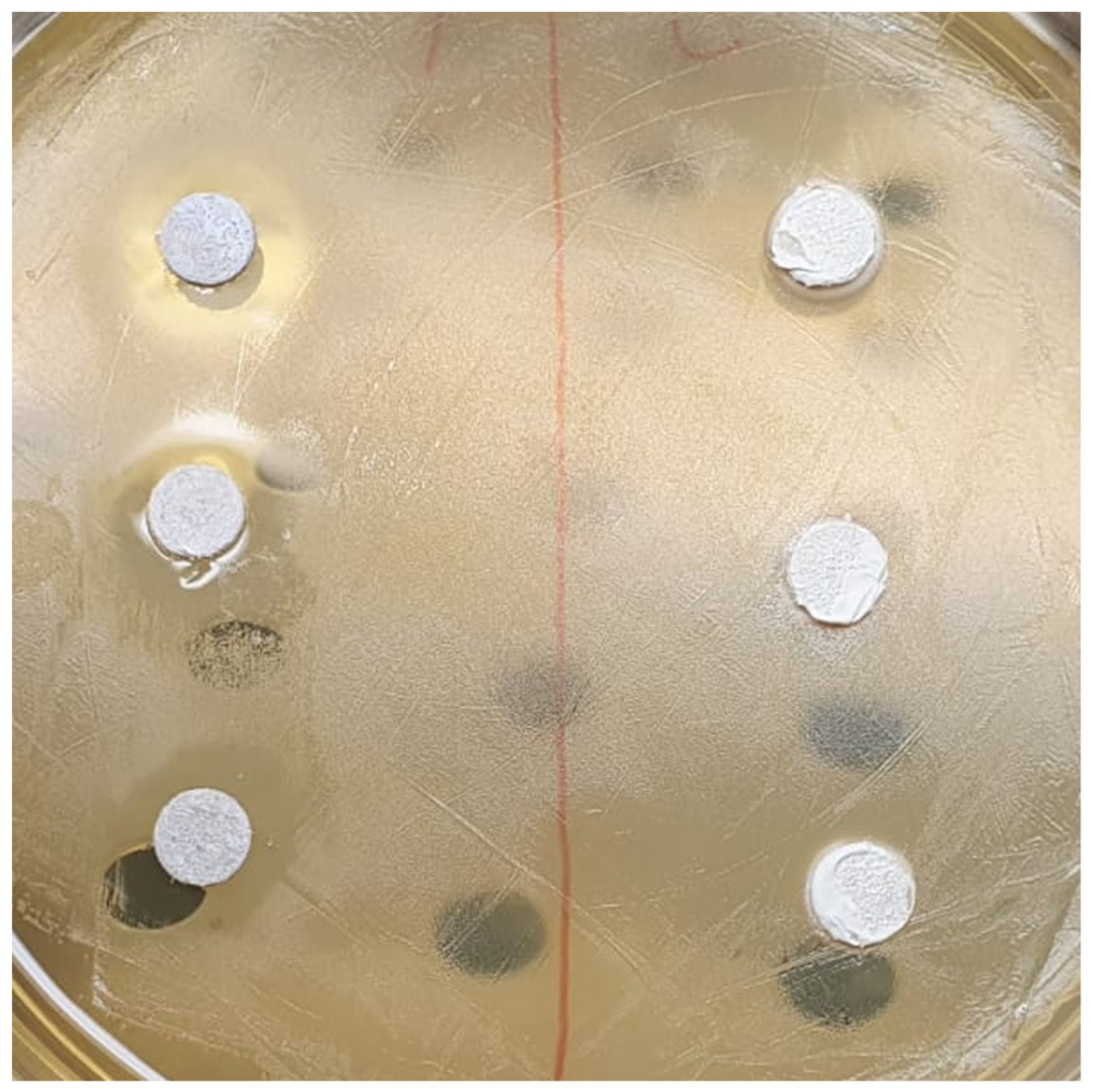
2.1. Bacterial Strains and Growth Conditions

The streptococcal strains used in this study were from the Culture Collection of University of Goteborg (CCUG): S. mutans (CCUG 35176), S. salivarius (CCUG 11878), and S. sanguis (CCUG 17826). The cultures were grown and maintained in a Brain Heart Infusion (BHI, Difco, Detroit, MI, USA). S. mutans culture medium was supplemented with 10% (v/v) heat-inactivated horse blood serum (Oxoid, Rodano, Milano, Italy) to improve its growth. The culture of all bacterial strains was statically incubated for 16 h at 37 °C under aerobic conditions. This overnight culture, used as source for the experiments, was reduced at a final density of 1 × 10¹⁰ cells/mL as determined by comparing the OD600 of the sample with a standard curve relating OD600 to cell number.

2.2. Agar Disc Diffusion Test

Sterile paper discs (diameter: 6 mm, thickness: 1 mm) (Watman International, Maidstone, UK) were impregnated with 10 μL of each root canal sealer. All materials were prepared according to manufacturers’ recommendations as shown in Figure 1. Then, BHI-agar plates were incubated with 1 × 10⁷ cells/mL of an overnight culture of each streptococcal strain at 37 °C for 20 min. The excess of bacterial suspension was removed from the plates and incubated with the paper disks impregnated with the root canal sealer at 37 °C for 24 h. The diameter of the halo formed around the paper disc (inhibition zone) was measured by the same operator in two perpendicular locations with a millimeter ruler (sliding callipers) with accuracy of 0.5 mm, after 24 h and 48 h. The size of the inhibition zone was calculated as follows:
size of inhibition zone = (diameter of halo − diameter of specimen) × ½.
All the assays were conducted in triplicate and the results were recorded in terms of the average diameter of inhibition zone.

2.3. Statistical Analysis

Data of the diameters of the growth inhibition zones, expressed in cm, were collected separately for each culture, and analyzed using R (The R Foundation for Statistical Computing). Data were assessed to be normal by means of the Kolmogorov–Smirnov test, revealing that data were not normally distributed. A non-parametric statistical method was used to investigate intra-group and inter-group comparisons. The Wilcoxon test was used to assess the differences that occurred after 24 h and after 48 h for each material tested. Kruskal–Wallis analysis of variance was used to assess the differences among the materials tested. Significance was predetermined for p < 0.05.

3. Results

The medians (minimum-maximum) of the growth inhibition results (mm) of different root canal sealants are shown in Table 2.
All Streptococci strains tested showed a significant inhibition zone (p < 0.05). The antimicrobial activity resulted in quite similar among the three streptococcal species, while the statistical analysis showed significant differences among the materials tested.
When testing the inhibition zones in cultures of S. mutans, the analysis did not evidence statistically significant differences between 24 h and 48 h for Groups C, D, and E, while A and B showed a significant increase in the inhibition zones after 48 h. Similar results were obtained for cultures of S. sanguis and S. salivarius. Statistical intergroup analysis performed with Kruskal–Wallis ANOVA showed significantly wider inhibition zones for Group E (p < 0.05).

4. Discussion

Survival of bacteria in endodontic space after root canal treatment may lead to persistent infection, healing difficulties, and treatment failure: chemical action of irrigating solution is essential in promoting the cleaning and the disinfection of the complex root canal space, even if its complete sterility is not feasible [3,4]. Endodontic sealants are used in root canal therapy to ensure the adhesion of gutta-percha to the root dentin, to seal any gaps, and finally also to inhibit the proliferation of any microorganisms remaining in the endodontic system after chemo-mechanical preparation, thus preventing recolonization of root canals [20,21]. The sealant should be biocompatible, without dimensional change, and have a long-lasting antibacterial effect [33]. The antibacterial effects of endodontic sealers have been investigated several times using the agar diffusion test (ADT) and direct contact test (DCT) [34]. ADT represents one of the most common and simple methods to study the antimicrobial activity of root canal sealants. The main limitations associated with its use are the lack of standardization of oculus density, adequate culture medium, agar viscosity, storage conditions of the plates, and dependence on the solubility and diffusion characteristics of the test material and culture medium [35]. Thus, only water-soluble materials can be tested using the ADT method [35,36]. Consequently, ADT is not the only recommended test to assess the antibacterial activity of endodontic sealants. DCT, instead, has several advantages such as reproducibility, quantitative dosing, and, in addition, reproducing direct contact between endodontic sealants within the root canal system. However, both methods have their own specific characteristics, and it is difficult to compare their results, even if these variables were carefully controlled, consistent, and reproducible results can be obtained.
All the tested materials showed antibacterial effects against the different pathogens: the antimicrobial activity resulted quite similarly among the three streptococcal species, while the statistical analysis showed significant differences among the materials tested. In fact, the traditional eugenol zinc-oxide sealer (Pulp Canal Sealer EWT), selected as control, showed significantly wider inhibition zones, probably due to its composition and biophysical characteristics. The bioceramic root canal sealers showed similar results between them, even if they appeared less efficient than the Pulp Canal Sealer EWT. The Streptococci tested are gram-positive facultative anaerobes and are able to grow in the presence or absence of oxygen. We selected the Streptococci strains because they include the most frequent bacteria and microorganism found in persistent endodontic infections and in failed root canal treatment cases [20,21]. They are resistant against intracanal medicament, such as calcium hydroxide, and they penetrate into secondary accessory canals and isthmuses [37]. These results confirmed the antibacterial activity of the bioceramic root canal sealers, as reported in previous studies [38]: their alkaline pH may contribute not only to their osteogenic potential and biocompatibility but also to their antibacterial ability against Streptococci strains. This ability of different root canal bioceramic sealers should be tested even against Enterococcus faecalis, which is a commonly isolated species involved in persistent endodontic infection. Based on the results of the present study, bioceramic root canal sealers show an adequate antibacterial ability to inhibit the diffusion of Streptococci strains.

5. Conclusions

The use of root canal filling materials that have antimicrobial activity is considered advantageous in the effort to reduce the number of remaining microorganisms, prevent recurrent root canal infection, and aid in the healing of periapical tissues. The results of this in vitro study could be verified with a clinical study, which could confirm the differences in the antibacterial activity of different products. Within the conditions of this in vitro study, the antibacterial activity performed with bioceramic root canal sealers is encouraging and effective for endodontic aims. Beside these results, bioceramic root canal sealers represent a favorable option for further research in regards to its potential application.

Author Contributions

Conceptualization, A.D., C.P. and R.B.; methodology, M.C.; software, G.P.; validation, M.C., A.D. and R.B.; formal analysis, G.R. and M.P.; investigation, G.R., M.P. and G.P.; resources, C.P.; data curation, G.P.; writing—original draft preparation, G.R., M.P., M.C. and R.B.; writing—review and editing, A.D. and C.P.; visualization, G.P.; supervision, C.P.; project administration, A.D., M.C., C.P. and R.B. All authors have read and agreed to the published version of the manuscript.

Funding

This research received no external funding.

Institutional Review Board Statement

Not applicable.

Informed Consent Statement

Not applicable.

Data Availability Statement

Data supporting reported results are available on request from the corresponding authors.

Conflicts of Interest

The authors declare no conflict of interest.

References

  1. Viapiana, R.; Flumignan, D.L.; Guerreiro-Tanomaru, J.M.; Camilleri, J.; Tanomaru-Filho, M. Physicochemical and mechanical properties of zirconium oxide and niobium oxide modified Portland cement-based experimental endodontic sealers. Int. Endod. J. 2014, 47, 437–448. [Google Scholar] [CrossRef] [PubMed]
  2. Schafer, E.; Zandbiglari, T. Solubility of root-canal sealers in water and artificial saliva. Int. Endod. J. 2003, 36, 660–669. [Google Scholar] [CrossRef] [PubMed]
  3. Grossman, L. Endodontic Practice, 11th ed.; Lea & Febiger: Philadelphia, PA, USA, 1988. [Google Scholar]
  4. Torabinejad, M.; Walton, R.E. Endodontics: Principles and Practice; Saunders Elsevier: St. Louis, MO, USA, 2009. [Google Scholar]
  5. Orstavik, D.; Nordhal, I.; Tibbals, J.E. Dimensional change following setting of root canal sealer materials. Dent. Mater. 2001, 17, 146–151. [Google Scholar] [CrossRef]
  6. Silva, E.J.; Rosa, T.P.; Herrera, D.R.; Jacinto, R.C.; Gomes, B.P.; Zaia, A.A. Evaluation of cytotoxicity and physicochemical properties of calcium silicate-based endodontic sealer MTA Fillapex. J. Endod. 2013, 39, 274–277. [Google Scholar] [CrossRef] [PubMed]
  7. American National Standards/American Dental Association. Endodontic Sealing Material; ANSI/ADA Speci Cation no. 57; American National Standards/American Dental Association: Chicago, IL, USA, 2000. [Google Scholar]
  8. ISO 6876; Dental Root Canal Sealing Materials. International Organization for Standardization: Geneva, Switzerland, 2001.
  9. Poggio, C.; Lombardini, M.; Conti, A.; Rindi, S. Solubility of root-end-filling materials: A comparative study. J. Endod. 2007, 33, 1094–1097. [Google Scholar] [CrossRef] [PubMed]
  10. McHugh, C.P.; Zhang, P.; Michalek, S.; Eleazer, P.D. pH required to kill Enterococcus faecalis in vitro. J. Endod. 2004, 30, 218–219. [Google Scholar] [CrossRef]
  11. Stuart, C.H.; Schwartz, S.A.; Beeson, T.J.; Owatz, C.B. Enterococcus faecalis: Its role in root canal treatment failure and current concepts in retreatment. J. Endod. 2006, 32, 93–98. [Google Scholar] [CrossRef]
  12. Okabe, T.; Sakamoto, M.; Takeuchi, H.; Matsushima, K. Effects of pH on mineralization ability of human dental pulp cells. J. Endod. 2012, 32, 198–201. [Google Scholar] [CrossRef]
  13. Sjogren, U.; Figdor, D.; Persson, S.; Sundqvist, G. Influence of infection at the time of root filling on the outcome of endodontic treatment of teeth with apical periodontitis. Int. Endod. J. 1997, 30, 297–306. [Google Scholar] [CrossRef]
  14. Kakehashi, S.; Stanley, S.R.; Fitzgerald, R.J. The effect of surgical exposures of dental pulps in germ free and conventional laboratory rats. J. Oral Surg. 1965, 20, 340–349. [Google Scholar] [CrossRef]
  15. Brannstrom, M.; Nordenvali, K.J. Bacterial penetration, pulpul reaction and the inner surface of concise enamel bond composite filling in etched and unetched cavities. J. Dent. Res. 1978, 57, 3–10. [Google Scholar] [CrossRef] [PubMed]
  16. Fabricus, I.; Dahlen, G.; Holm, S.E.; Moller, A.J.R. Influence of combinations of oral bacteria on peri-apical tissues of monkeys. Scand. J. Dent. Res. 1982, 90, 200–206. [Google Scholar]
  17. Barnett, F.; Stevens, R.; Tronstad, L. Demonstration of Bacteroides intermedius in peri-apical tissue using indirect immmunofluoroscence microscopy. Endod. Dent. Traumatol. 1990, 6, 153–156. [Google Scholar] [CrossRef]
  18. Siqueira, J.F.; Favieri, A.; Gahyva, S.M.; Moraes, S.R.; Lima, K.C.; Lopes, H.P. Antimicrobial activity and flow rate of newer and established root canal sealers. J. Endod. 2000, 26, 274–277. [Google Scholar] [CrossRef] [PubMed]
  19. Singh, G.; Gupta, I.; Elshamy, F.M.; Boreak, N.; Homeida, H.E. In vitro comparison of antibacterial properties of bioceramic- based sealer, resin-based sealer and zinc oxide eugenol-based sealer and two mineral trioxide aggregates. Eur. J. Dent. 2016, 10, 366–369. [Google Scholar] [CrossRef] [PubMed] [Green Version]
  20. Matigatti, S.; Jain, D.; Ratnakar, P.; Moturi, S. Antimicrobial effect of conventional root canal medicament vs. propolis against Enterococcus faecalis, staph aureus and candida albicans. J. Contemp. Dent. Pract. 2012, 13, 305–309. [Google Scholar] [CrossRef]
  21. Zhang, H.; Shen, Y.; Ruse, N.D.; Haapasalo, M. Antibacterial activity of endodontic sealers by modi ed direct contact test against Enterococcus faecalis. J. Endod. 2009, 35, 1051–1055. [Google Scholar] [CrossRef] [PubMed]
  22. López-García, S.; Myong-Hyun, B.; Lozano, A.; García-Bernal, D.; Forner, L.; Llena, C.; Guerrero-Gironés, J.; Murcia, L.; Rodríguez-Lozano, F.J. Cytocompatibility, bioactivity potential, and ion release of three premixed calcium silicate-based sealers. Clin. Oral Investig. 2020, 24, 1749–1759. [Google Scholar] [CrossRef]
  23. Tonini, R.; Giovarruscio, M.; Gorni, F.; Ionescu, A.; Brambilla, E.; Mikhailovna, I.M.; Luzi, A.; Maciel Pires, P.; Sauro, S. In Vitro Evaluation of Antibacterial Properties and Smear Layer Removal/Sealer Penetration of a Novel Silver-Citrate Root Canal Irrigant. Materials 2020, 13, 194. [Google Scholar] [CrossRef] [Green Version]
  24. Zhou, H.; Shen, Y.; Zheng, W.; Li, L.; Zheng, Y.; Haapasalo, M. Physical Properties of 5 root canal sealers. J. Endod. 2013, 39, 1281–1286. [Google Scholar] [CrossRef]
  25. Zhou, H.M.; Du, T.F.; Shen, Y.; Wang, Z.J.; Zheng, Y.F.; Haapasalo, M. In vitro cytotoxicity of calcium silicate-containing endodontic sealers. J. Endod. 2015, 41, 56–61. [Google Scholar] [CrossRef] [PubMed]
  26. Xuereb, M.; Vella, P.; Damidot, D.; Sammut, C.V.; Camilleri, J. In situ assessment of the setting of tricalcium silicate-based sealers using a dentin pressure model. J. Endod. 2015, 41, 111–124. [Google Scholar] [CrossRef] [PubMed]
  27. Kogan, P.; He, J.; Glickman, G.N.; Watanabe, I. The effects of various additives on setting properties of MTA. J. Endod. 2006, 32, 569–572. [Google Scholar] [CrossRef] [PubMed]
  28. Gomes-Filho, J.E.; Rodrigues, G.; Watanabe, S.; Estrada Bernabe, P.F.; Lodi, C.S.; Gomes, A.C.; Faria, D.M.; dos Sanots, D.A.; Moraes, J.C.S. Evaluation of the tissuereaction to fast endodontic cement (CER) and Angelus MTA. J. Endod. 2009, 35, 1377–1380. [Google Scholar] [CrossRef] [PubMed]
  29. Salles, L.P.; Gomes-Cornelio, A.L.; Guimaraes, F.C.; Herrera, B.S.; Bao, S.N.; Rossa-Junior, C.; Guerreiro-Tanaomaru, J.M.; Tanomaru-Filho, M. Mineral trioxide aggregate-based endodontic sealer stimulates hydroxyapatite nucleation inhuman osteoblast-like cell culture. J. Endod. 2012, 38, 971–976. [Google Scholar] [CrossRef]
  30. Candeiro, G.T.; Correia, F.C.; Duarte, M.A.; Ribeiro-Siqueira, D.C.; Gavini, G. Evaluation of radiopacity, pH, release of calcium ions, and flow of a bioceramic root canal sealer. J. Endod. 2012, 38, 842–845. [Google Scholar] [CrossRef] [Green Version]
  31. Loushine, B.A.; Bryan, T.E.; Looney, S.W.; Gillen, B.M.; Loushine, R.J.; Weller, R.N.; Pashley, D.H.; Tay, F.R. Setting properties and cytotoxicity evaluation of a premixed bioceramic root canal sealer. J. Endod. 2011, 37, 673–677. [Google Scholar] [CrossRef]
  32. Atmeh, A.R.; Chong, E.Z.; Richard, G.; Festy, F.; Watson, T.F. Dentin-cement interfacial interaction: Calcium silicates and polyalkenoates. J. Dent. Res. 2012, 91, 454–459. [Google Scholar] [CrossRef]
  33. Baumgartner, G.; Zehnder, M.; Paqué, F. Enterococcus faecalis type strain leakage through root canals filled with gutta-percha/AH Plus or Resilon/Epiphany. J. Endod. 2007, 33, 45–47. [Google Scholar] [CrossRef]
  34. Morgental, R.D.; Vier-Pelisser, F.V.; Oliveira, S.D.; Antunes, F.C.; Cogo, D.M.; Kopper, P.M.P. Antibacterial activity of two MTA-based root canal sealers. Int. Endod. J. 2011, 44, 1128–1133. [Google Scholar] [CrossRef]
  35. Cobankara, F.K.; Altinöz, H.C.; Ergani, O.; Kav, K.; Belli, S. In vitro antibacterial activities of root-canal sealers by using two different methods. J. Endod. 2004, 30, 57–60. [Google Scholar] [CrossRef] [PubMed]
  36. Rôças, I.N.; Jung, I.Y.; Lee, C.Y.; Siqueira, J.F., Jr. Polymerase chain reaction identification of microorganisms in previously root-filled teeth in a South Korean population. J. Endod. 2004, 30, 504–508. [Google Scholar] [CrossRef] [PubMed]
  37. Hancock, H.H.; Sigurdsson, A.; Trope, M.; Moiseiwitsch, J. Bacteria isolated after unsuccessful endodontic treatment in a North Am population. Oral Surg. Oral Med. Oral Pathol. Oral Radiol. Endod. 2001, 91, 579–586. [Google Scholar] [CrossRef] [PubMed]
  38. Lee, J.K.; Kwak, S.W.; Ha, J.H.; Lee, W.; Kim, H.C. Physicochemical Properties of Epoxy Resin-Based and Bioceramic-Based Root Canal Sealers. Bioinorg. Chem. Appl. 2017, 2017, 2582849. [Google Scholar] [CrossRef]
Figure 1. All materials were prepared according to manufacturers’ recommendations and collected on glass plates.
Ceramics 05 00065 g001
Table 1. Tested materials and composition.
GroupMaterialCompositionManufacturer
AFillRoot STaluminosilicates, zirconium dioxide, fillers, thickening agentsDental World srl. Molfetta, BA, Italy
BBioRoot RCSPowder: zirconium dioxide, tricalcium silicate and povidone.Liquid: calcium chloride and polycarboxylateSeptodont, Saint-Maur-des-Fosses,
France
CWell-Root PTaluminosilicates, zirconium dioxide, fillers, thickening agentsVericom Co., Chuncheon, Korea
DCeraSealCalcium silicates, zirconium dioxide, thickening agentsMeta Biomed Co., Cheongju, Korea
EPulp Canal Sealer EWTPowder: zinc oxide, silver powder, thymol iodide, dimeric acid resin.
Liquid: 4-allyl-2-methoxyphenol, balsam resin and water
Kerr, Romulus, MI, USA
Table 2. Median (minimum-maximum) of growth inhibition results (mm) of the different root canal sealants.
S. mutansS. sanguisS. salivariusControl
GroupMaterial24 h48 h24 h48 h24 h48 h24 h 48 h
AFillRoot ST0.005 (0.003–0.005)0.008 (0.005–0.01)0.007 (0.006–0.007)0.01 (0.007–0.012)0.003 (0.002–0.003)0.006 (0.003–0.008)<0.000<0.000
BBioRoot™RCS0.012 (0.01–0.013)0.019 (0.014–0.022)0.014 (0.012–0.015)0.021 (0.016–0.024)0.010 (0.008–0.011)0.017 (0.012–0.020)<0.000<0.000
CWell-Root™ PT0.007 (0.006–0.008)0.007 (0.005–0.008)0.009 (0.008–0.01)0.009 (0.007–0.01)0.005 (0.004–0.006)0.005 (0.003–0.006)<0.000<0.000
DCeraSeal0.006 (0.005–0.007)0.005 (0.004–0.007)0.008 (0.007–0.009)0.007 (0.006–0.009)0.004 (0.003–0.005)0.003 (0.002–0.005)<0.000<0.000
EPulp Canal Sealer™ EWT0.31 (0.24–0.45)0.37 (0.26–0.49)0.32 (0.27–0.46)0.36 (0–0.42)0.28 (0.23–0.42)0.31 (0–0.28)<0.000<0.000
Publisher’s Note: MDPI stays neutral with regard to jurisdictional claims in published maps and institutional affiliations.

