Review

Three-Dimensional Printing and Digital Flow in Human Medicine: A Review and State-of-the-Art

by
Rodrigo Salazar-Gamarra
1,*,
Hans Contreras-Pulache
2,
Gloria Cruz-Gonzales
3,
Salvatore Binasco
4,
William Cruz-Gonzales
5 and
Jeel Moya-Salazar
1,6,*
1
Digital Transformation Center, Universidad Norbert Wiener, Lima 51001, Peru
2
South America Center for Education and Research in Public Health, Universidad Norbert Wiener, Lima 51001, Peru
3
Faculties of Medical Technologies, Universidad Nacional Federico Villareal, Lima 51001, Peru
4
Postgraduation Program in Engineering, Universidade Paulista (UNIP), São Paulo 01001-000, Brazil
5
Faculty of Financial and Accounting Sciences, Universidad Nacional Federico Villarreal, Lima 51001, Peru
6
School of Biomedicine, Faculties of Engineering, Universidad Tecnológica del Perú, Lima 51001, Peru
*
Authors to whom correspondence should be addressed.
Appl. Syst. Innov. 2022, 5(6), 126; https://doi.org/10.3390/asi5060126
Submission received: 26 September 2022 / Revised: 24 November 2022 / Accepted: 29 November 2022 / Published: 15 December 2022

Abstract

:
The use of exponential technologies is changing how people live and interact; this has been called the “Fourth Industrial Revolution”. Within these technologies, 3D printing is playing a leading role, especially in health. In this context, this literature review aims to present the state of the art of 3D printing, its digital workflow and applications in medicine, and the advantages of its use in public health. Consequently, it describes the benefits for the patient and the medical team from a diagnostic stage, a brief history of its development, what is the digital flow when working with a 3D printer, what experiences of its use in medicine, and finally, how this technology used in medicine and public health can be part of the Digital Transformation in Peru.

1. Introduction

Klaus Schwab in 2016 published a book called “The Fourth Industrial Revolution”; this compiled information from the World Economic Forum in which it was mentioned that 3D printing is an exponential technology that will see its deployment in the 2020s, as one of the drivers of the fourth industrial revolution (Figure 1), and as the limitations of size, costs, and speed are overcome, 3D printing will become even more widespread. It will include electronic components such as printed circuits and up to human cells and organs [1].
In 2018 in Peru, the Digital Government Law was published [2], and in 2019 using a resolution, the Government and Digital Transformation Laboratory was created; this state body has several lines of action and one of them is to promote the use of emerging technologies [3]. Consequently, it is time for medical and health sciences personnel to find scientific and technical information on these technologies, specifically 3D printing, in an integrated manner so that they can adapt and apply the knowledge that will generate progress in Peruvian health systems [4].
This narrative review aims to present the state of the art of 3D printing, digital workflow, and its applications in medicine, as well as to mention the advantages of its use in public health in the context of digital transformation in Peru. First, the benefits of 3D printing for the medical team and the patient from the diagnostic stage are covered. A brief review of the history of 3D printing follows this. Next, an attempt is made to answer the question: what is the mechanism of operation of a 3D printer? This question will be answered by explaining its digital flow. Next, a selection of the applications found in the scientific evidence of 3D technology in medicine is presented. Finally, we reflect on the use of 3D printing in the context of the digital transformation of the Peruvian State and its use in medicine and public health.

2. Search Strategy

The bibliographic search of scientific articles for the present review was performed in three primary databases Scopus, MedLine/PubMed, and Scielo. In addition, the search for documents on Digital Transformation legislation in Peru was performed on the official web portal of the Peruvian State. The investigation was conducted from 15 December 2019 to 9 September 2020. It was performed with the following MeSH/DeCS terms: “Three-dimensional printing”, “quality of life”, “computer-aided design”, “medicine”, “evidence-based medicine”, “public health”, and “health policy”, and their translations in Spanish and Portuguese. These terms were restricted to their appearance in the scientific articles’ titles, abstracts, or keywords.

3. A Brief History of 3D Technology

In 1970, Herbert Voelcker was the first to propose a mathematical theory and algorithms for modeling a solid object in three dimensions [5]. In 1984, Charles Hull invented the first rapid prototyping technology called Stereolithography (SLA) [6] to manufacture solid objects by “printing” thin layers of photo-curable material one on top of the other; this technology uses an ultraviolet light laser as an energy source and is highly dependent on the materials used.
In 1987, Carl Deckard invented the second rapid prototyping technology called Selective Laser Sintering (SLS) [7], which consisted of building models layer by layer based on powdered materials, using a laser beam to fuse and solidify the powder particles. In 1988, Scott Crump invented the third rapid prototyping technology, Fused Deposition Modeling (FDM) [7], in which, by heating and extruding material in the form of filaments, layer by layer, a 3D object is built.
In 1991, rapid prototyping machines began to be commercialized by three companies: Stratasys (Minnesota, USA), Cubital (Ra’anana, Israel), and Helisys (California, USA). In particular, Helisys developed the fourth rapid prototyping technology, the Laminated Object Manufacturing (LOM) system; this system involves heating and bonding a laser-cut sheet material and the sheet cuts (layers) are superimposed and connected on top of the other until the desired object is formed. Later, in 1993, the Massachusetts Institute of Technology (MIT) invented 3D printing, which injects successive layers of material to create a 3D object such as a standard desktop inkjet printer. Based on 3D printing technology, new machines and products have been developed. Since 1996, the term “3D printing” has been extended to refer to a set of technologies that were born under the name of Rapid Prototyping (RP) and are also referred to as Additive Manufacturing (AM) or Solid Freeform Fabrication (SFF) [8,9,10]. By 2020, 3D printed electronic circuits, tissues, and human organs were already available [11,12].

4. Three-Dimensional Technology for Multi-Professional Collaboration and Patient Quality of Life

Three-dimensional technologies have been progressively applied in healthcare areas over the course of the last three decades. This has brought integrated, objective advantages for all generations of medical teams to work under genuine, effective, and empathic collaboration focused on patient diagnosis and treatment [13,14,15,16,17]. Today the experiences gathered by the Medical Boards have enabled a disruptive approach in the integration and total understanding of the team and, more importantly, that the public sector patient can be informed in a more didactic way [18] because it allows effective communication with the patient. This is due to the possibility of showing in virtual or physical a medical-grade 3D impression in this diagnostic stage to the patient [14,19]. For example, suppose a patient with cancer, who has suffered an accident or congenital malformation, is on the verge of sacrificing their bodily structures to survive. In that case, decisions should be as objective as possible, and the patient should be well-informed. Virtual 3D surgical–reconstructive and prosthetic planning to achieve the rehabilitation of the individual in society are found in a few hospitals in the world. There is evidence that it transmits to the patient a global awareness of the collective effort carried out in his treatment, including what is demanded from the patient so that he has adequate emotional preparation and instruction about the therapy and the hope of his recovery and improvement [20,21].

5. Digital Flow in 3D Printing

Three-dimensional technologies group a set of methods or workflows for digital manufacturing. In the medical area, three stages, often sequential, must be understood:
  • Three-dimensional image acquisition;
  • Three-dimensional Modeling;
  • Three-dimensional printing.
If the objective is to print, for example, a surgical guide, we must first model it from the tomography images for a specific case. If only the tomography is to be visualized to make a clinical decision, we are left at the first stage of image acquisition. In other words, multiple flows depend on the need [22]. On the other hand, specific hardware and software are at all stages. The hardware can be a computer, a tomography, a cellphone, a 3D printer, etc. The software is the computer program used in any of the three stages, highlighting a function of this, perhaps the most important, which is the one that allows modeling the desired file before being printed. A variable as crucial as the above is the choice of the 3D printing technology and the appropriate material, which define functionality together. In other words, the function is determined by the shape and material [23].

5.1. Image Acquisition

Usually, for healthcare applications, we start with medical images, especially when discussing diagnosis and treatment. The flow chosen will depend on the clinical needs. When they are internal structures such as those obtained by MRI, CT, and 3D Ultrasound, the information will be stored in the DICOM (Digital Imaging and Communication on Medicine) format. In a complementary way, there are surface scanners such as intraoral scanners of structured light to scan teeth, mucous membranes, and others, as well as for extraoral scanning that can be Photogrammetry (Monoscopic and Stereophotogrammetry), Laser, Structured light in its variants of white, blue, and infrared light, which allow us to create a digital copy of surfaces of faces. Depending on the technology, the latter is saved directly in the STL format, OBJ combined with JPEG texture maps, or others. Of these files, the oldest and most commonly used is STL, which loads only the geometry of the 3D file. This article does not focus on making an in-depth comparison between 3D file types. Still, each can load different information, such as geometry, appearance, scene, and motion [24,25,26]. Of these, only geometry can be 3D printed, hence the still widespread use of STL files and software that allows the transformation of DICOM.
In many cases, the visualization software of recent equipment allows rendering for viewing only (diagnostic/therapeutic evaluation). On the other hand, transforming to STL is not visualizing but directly changing the format to another type of communication file to continue with the next steps. The act of converting the DICOM format to STL using specialized software is called “Segmentation”. This term should not be confused with surgical segmentation of structures. It is important to remember that a tomography or MRI scan in each slice also captures unwanted structures, the air surrounding the face, and even artifacts. In this sense, segmentation is understood as cleaning each of the tomography slices, based on the desired Housenfield units to obtain an accurate and professional model.
The manipulation of software (such as Invesalius®, Slicer®, or others) in inexperienced hands can produce severe errors in the identification of structures, thicknesses, and distances and, consequently, severe mistakes in diagnosis and treatment. Because of that, it is essential to have a formally trained and experienced technical professional to use medical manipulation.

5.2. Three-Dimensional Modeling

Three-dimensional modeling is a concept originating from computer graphics sciences. It refers to developing a “mathematical representation” of any surface of an object in three dimensions through specialized software [22,24,25]. The product is called a “3D model”. Visually it is similar to creating a clay sculpture, only with digital tools developed by mathematical programming. One of the main advantages of digital 3D modeling is that it is easy to save versions and manipulate the geometries (mesh) completely. When we work on biomedical image manipulation, the 3D modeling stage is after image acquisition because we edit the geometry obtained from the previous step. If the case is to create a medical device, such as a spatula, or a retractor, we do not necessarily require prior image acquisition, and the process starts with 3D modeling. However, for example, if the objective is to create a fracture immobilization splint, the 3D modeling is from a previous surface image of the arm that requires immobilization, and the digital sculpting of the splint is completed using specialized software. Delving a little deeper into the principles, if the objective is to print, the nature of the design must obey not only the shape but also the manufacturing mechanism and final material. Due to physical, chemical, and biological properties, the overall safety assessment must be considered in terms of fidelity, reproduction, precision, compatibility, and others. Several specialized computer graphics programs exist, and their nature is in commercial licensing. They range from free and open source software (“Free and Open Source Software—FOSS”, which allows modification of algorithms in the hands of programmers), free (“Freeware”), free with special paid functions (“Freemium”), and exclusive to paid commercial license (“Commercial license”). For the health area, there are examples such as Blender® (FOSS), Meshmixer® (Freeware), Zbrush®, and Mimics® (Commercial license), among others. They can be exported in STL files and loaded to a 3D printer for the next step of additive manufacturing [22,24,26,27].

5.3. Three-Dimensional Printing

Few classifications still contribute to this tool’s applications and benefits. The learning curve for medical teams has been hampered by access to information and the technologies and materials themselves [28]. The principles to consider are defining the type of 3D technology and the material to be used. There are hundreds of materials, including polymers, ceramics, metals, resins, waxes, gypsum, etc. The technology with which the raw material is transformed, whether by deposition, polymerization, or synthesis, among others, will define the universe of desired properties for the surface, mechanical strength, melting point, dimensional stability, hardness, texture, biocompatibility, degradation, among many other variables [11].
Among all the necessary considerations to evaluate the material used, these last two depend significantly on the application and its planning since patient safety must be guaranteed. Degradation is a gradual breakdown of material naturally due to its exposure to both the environment and biological processes within the body. It is required that this of a very low ratio and non-toxic so that it is biocompatible, in addition to not having an immune response from the organism, among others [29,30]. On the one hand, in the case of bone implants, the most biocompatible material is titanium because it is stable and inert [31,32,33], and its high mechanical properties make it ideal for the procedure. There are also cases where the implant aims to help regenerate a body structure, and then the material biodegrades to be absorbed with minor side effects. On the other hand, in the case of facial prostheses, medical-grade silicone is the most accepted. While no other material guarantees the safety of patients, it will be defined as the gold standard. Different materials are being studied, such as flexible resins [34], but still, more research is needed. Although biodegradation is not the main issue here, aging is one of the difficulties, compounded by color stability, microbial colonization, silicone dehydration, edge cracking, and others. Two years is the global average time, with eight months–3 years for change, according to the nature of the anatomical area, the use, and the environmental conditions [35].
As the cost of materials and technology has decreased, its progressive development has been seen more and more in different applications. As a result, the size of the industry around 3D printing is growing rapidly, which is expected to have an impact in socio-economic terms, especially in the framework of the fourth industrial revolution [1]. Table 1 shows a proposal by the authors for the classification and definitions of 3D printing applications in healthcare.

6. Experiences with the Use of 3D Technology in Medicine

Currently, research in 3D printing has increased due to the impact on people’s quality of life, the accuracy of 3D printed images and objects, the emergence of new printing materials, and the reduction in manufacturing costs [36,37,38]. It is used in medicine to improve patient communication, medical education, diagnosis, planning, and non-surgical or individualized surgical treatments, decreasing operative time and possible post-operative complications [39,40,41]. This signifies (at least in part) a new industrial revolution [1,42].
In head and neck surgery, its main applications are placing implants using surgical guides, reconstructing lost facial structures or bone defects, and the simulation of surgical interventions [43]. In neurosurgery, in the case of aneurysms, it allows predetermining the size of the microcatheter to be used, as well as practicing clipping procedures to understand the vascular pathology preoperatively; planning electrode placement, based on anatomical models of the brain and skull; creating individualized and functional neurosurgical devices, such as the creation of compensators, to protect the organs surrounding the target tumor tissue during radiotherapy; printing 3D templates for the placement of pedicle screws with initial findings of no lesion incidence; and finally, it is currently used for trans-nasal sphenoidal endoscopy to remove pituitary tumors [44].
Cardiology’s leading application for congenital heart diseases is surgical planning using anatomical models [45]. In cardiovascular diseases, useful in case of severe aortic stenosis, where it is possible to replicate not only the anatomy but also the functional properties of aortic valve stenosis, for communication between the patient and the patient’s relatives, with the medical team, besides facilitating teaching and the level of knowledge of residents [46], including Benjamin et al., [47] in a systematic review indicated that education through 3D anatomical models is better than teaching through dissection of cadavers.
In hepatopathies, anatomical models are of great utility; in addition, 3D images allow intraoperative guidance of procedures, such as hepatectomies of small tumors in infants; it will enable simulating of the placement of a stent during the transjugular intrahepatic portosystemic shunt (TIPS) procedure or the placement of a drain in percutaneous cholecystostomy [40]. In nephrology, it is used to replicate the complex anatomical structure of the kidney and its pathologies, which allows the visualization and planning of the surgical treatment, for example, in the intervention of renal tumors or transplants [48]. In the case of colorectal surgery, it is used to facilitate laparoscopic surgery; for example, in the case of 3D laparoscopic lymphadenectomy of right colon cancer, it has been shown that there were no intra or postoperative complications [39]. In anesthesiology, airways have been replicated to plan and practice tracheal intubation techniques [49]. In pediatrics, the surgical treatment of cleft palate is an application of this technology, but also in non-surgical treatments, such as prosthetic rehabilitations in which the printing material plays a fundamental role [41]. In oncology, 3D printing, in addition to surgical procedures, is being applied in radiotherapy, bolus printing, and molds for brachytherapy [50,51].
Bioprinting tissues and organs is another way of using this technology; thus, Celprogen Inc (California, USA) achieved the first 3D printed human heart and pancreas. Bioprinting of tissues and organs occurs because the three-dimensional structure of the print allows the vascularization of the tissue or organ by seeding with endothelial cells, using an extracellular matrix of proteins, and the isolation and differentiation of stem cells [11,52]. Researchers are already working on 4D printing, a process based on 3D printing but with intelligent materials, which modify themselves in response to external stimuli such as heat, light, pH, electrical or magnetic forces, and humidity over time [1,53,54].

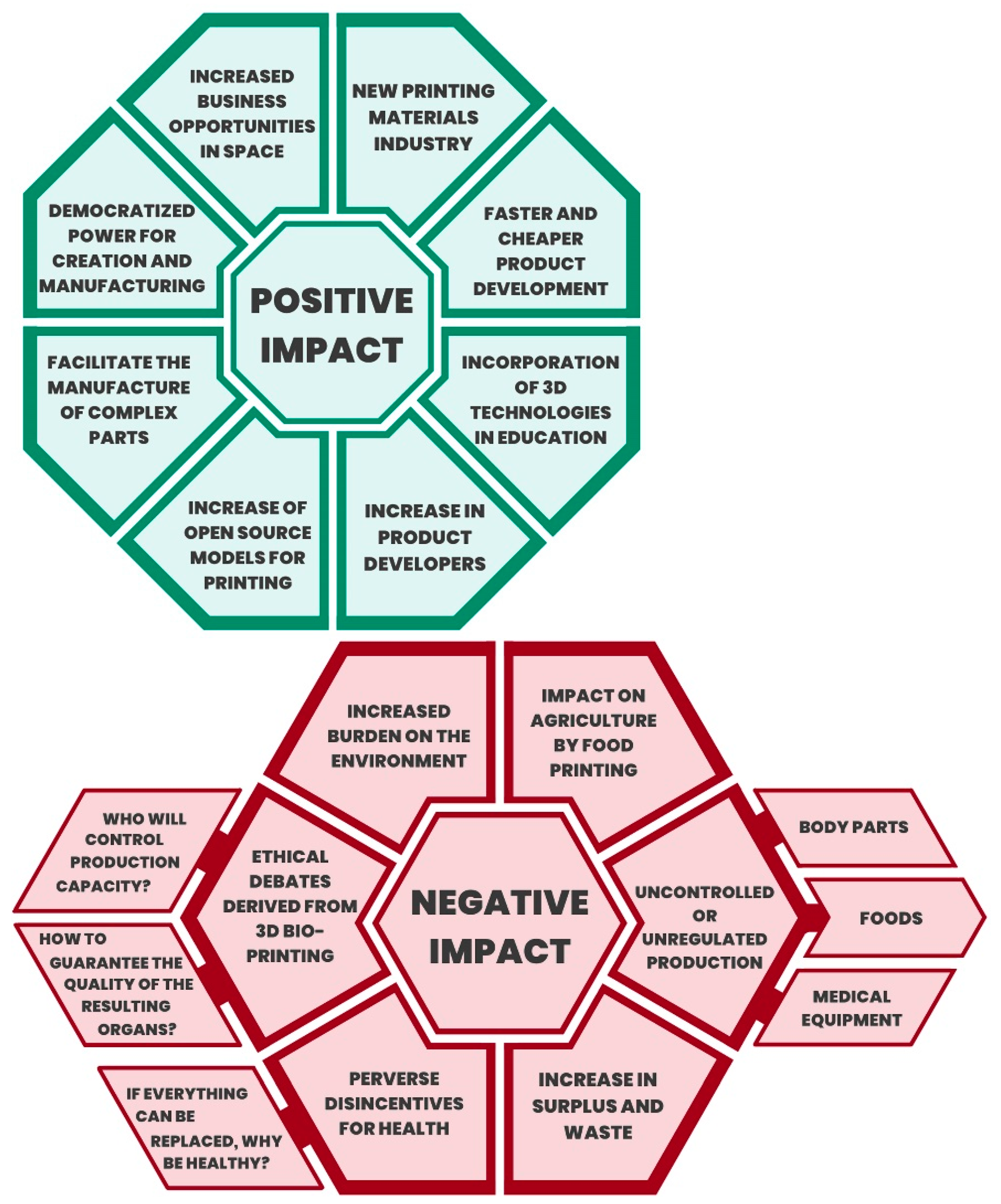
7. Three-Dimensional Technology and Digital Transformation and the Impact on Medicine and Public Health

In medicine, research and development of innovative methods now make it possible to provide an approach that is either more efficient or only possible using 3D technologies. The learning curves in healthcare for new professionals are shortened, potential public expenditures are cost-effective, and state innovation policies can improve the population’s health conditions [40,48,53].
On the other hand, health technologies have played an instrumental role in Peru; however, exponential technologies (including 3D printing) are only some of the instruments for solving problems. With a view to the future, the use of exponential technologies in conjunction with the design of a healthcare system will be discussed. A system that prides itself not only on using digital technologies but that is digital from the design of the digital transformation in health [54,55,56,57,58].
In public health, this digital transformation consists of having systems for patient follow-up, citizen control and reporting of drug prices, updating epidemiological information in real-time, designing health campaigns in social networks, re-founding the logic of social programs, mass health education in digital media, community monitoring in real-time and geo-referenced, the automated definition of risks and vulnerabilities, solving health problems without mediating geographical barriers, improving public service (queues, appointments), ensuring that a doctor has access to the necessary information about his patient, reformulating the role of the patient (considering that the essential merit of the digital world is the democratization of access to information) [57,58,59].
Moreover, the history of the Peruvian health system has shown that the “reforms” in this system have not occurred from the “top-down” but silently, from the “bottom-up” [60]. This would lead us to think that changes can also come from agents external to the health system and not necessarily by resolution or decree of the Ministry of Health (without this meaning that the Ministry of Health does not play any transcendental role, quite the contrary) [61]. In this sense, exponential technologies in healthcare should not be merely instrumental but the significant axis of an entire institutional reengineering. Perhaps implementing the National Digital Transformation System, announced in January 2020, will be the ideal framework to promote this categorical leap forward [4]. A concrete positive impact of these public implementations is the example of The National Pediatric Public Hospital “Instituto Nacional de Salud del Niño de San Borja”, which is the national instance from the Ministry of Health of Perú, more recently published an Official Procedure Guide for 3D Printed Anatomical Models, (Resolución Directorial 118-2022-DG-INSNSB) [54] which contains the instruction and permission to the Diagnostic and Treatment Support Unit and the Diagnostic Imaging Service of the Hospital. This is the only concrete document in the Peruvian Public Health System. It was written by a professional multidisciplinary team who identified the patient and system needs and self-acquired experience in 3D technologies. They used the “bottom-up” strategy, designing and elevating the proposal to the decision makers of the Hospital, and now it is shared with the whole Ministry of Health of Perú to be helpful. This may be one of the most efficient examples in which the technical professionals who are in the daily basic needs and understand details of processes gather the arguments and write the structure of policies jointly with decision makers to create legal pathways to democratize accessibility.
In this context, 3D printing positively impacts medicine and has enormous potential for use in public health; for example, it played an essential role during the pandemic caused by Severe Acute Respiratory Syndrome Coronavirus 2 (SARS-CoV-2) [62]. There is evidence that 3D printing was used during the pandemic to manufacture personal protective equipment (PPE) for healthcare personnel [63,64,65,66], and also medical devices such as mechanical ventilators [67], as well as to manufacture objects that support sterilization processes [68]. Therefore, 3D printing is playing a relevant role and is being used by several countries in the world, with the advantage of allowing the development of virtual collaborative work and cost-effective products [62].

8. Conclusions

The scientific literature shows that 3D printing in medicine is currently playing an increasingly important role due to its digital flow (images, modeling, and 3D printing), the possibility of effective communication between the medical team and the patient, the degree of individualization it achieves, and the fact that it is a cost-effective technology. The “bottom-up” strategy may efficiently promote guidelines and policies in complex public health systems.

Author Contributions

Conceptualization, R.S.-G. and H.C.-P.; methodology, R.S.-G. and J.M.-S.; validation, R.S.-G., G.C.-G. and H.C.-P.; formal analysis, J.M.-S.; investigation, R.S.-G. and H.C.-P.; resources, W.C.-G.; data curation, J.M.-S. and S.B.; writing—original draft preparation, R.S.-G., H.C.-P., S.B. and J.M.-S.; writing—review and editing, H.C.-P., G.C.-G., J.M.-S. and W.C.-G.; visualization, S.B.; supervision, R.S.-G.; project administration, H.C.-P. All authors have read and agreed to the published version of the manuscript.

Funding

This study was funded by the UNW (Registration 011-2022-UNW).

Data Availability Statement

Not applicable.

Conflicts of Interest

The authors declare no conflict of interest.

References

  1. Schwab, K. La Cuarta Revolución Industrial; DEBATE: Madrid, Spain, 2016. [Google Scholar]
  2. Presidencia de consejo de Ministros. Decreto Legislativo N° 1412 Normativa sobre Transformación Digital, Normatividad del Centro Nacional de Seguridad Digital; PCM: Lima, Peru, 2018. Available online: https://www.gob.pe/institucion/pcm/normas-legales/289706-1412 (accessed on 28 November 2022).
  3. Presidencia del Consejo de Ministros. Laboratorio de Gobierno y Transformación Digital. Plataforma Digital única del Estado Peruano; PCM: Lima, Peru, 2019. Available online: https://www.gob.pe/8256 (accessed on 28 November 2022).
  4. Diario El Peruano. Decreto de Urgencia que crea el Sistema Nacional de Transformación Digital—Decreto de Urgencia—n° 006-2020—Poder Ejecutivo—Decretos de Urgencia [Internet]; El Peruano. 2020. Available online: https://busquedas.elperuano.pe/normaslegales/decreto-de-urgencia-que-crea-el-sistema-nacional-de-transfor-decreto-de-urgencia-n-006-2020-1844001-1/ (accessed on 28 November 2022).
  5. Voelcker, H.B.; Requicha, A.A.G. Geometric Modeling of Mechanical Parts and Processes. Computer 1977, 10, 48–57. [Google Scholar] [CrossRef]
  6. Kietzmann, J.; Pitt, L.; Berthon, P. Disruptions, decisions, and destinations: Enter the age of 3-D printing and additive manufacturing. Bus. Horiz. 2015, 58, 209–215. [Google Scholar] [CrossRef]
  7. George, M.; Aroom, K.R.; Hawes, H.G.; Gill, B.S.; Love, J. 3D Printed Surgical Instruments: The Design and Fabrication Process. World J. Surg. 2017, 41, 314–319. [Google Scholar] [CrossRef] [PubMed]
  8. Chang, K.-H.; Chang, K.-H. Chapter 5—Rapid Prototyping; University of Salford: Amsterdam, The Netherlands, 2013. [Google Scholar] [CrossRef]
  9. Jasveer, S.; Jianbin, X. Comparison of Different Types of 3D Printing Technologies. Int. J. Sci. Res. Publ. 2018, 8, 1–9. [Google Scholar] [CrossRef]
  10. Peng, Q.; Tang, Z.; Liu, O.; Peng, Z. Rapid prototyping-assisted maxillofacial reconstruction. Ann. Med. 2015, 47, 186–208. [Google Scholar] [CrossRef]
  11. Trenfield, S.J.; Awad, A.; Madla, C.M.; Hatton, G.B.; Firth, J.; Goyanes, A.; Gaisford, S.; Basit, A.W. Shaping the future: Recent advances of 3D printing in drug delivery and healthcare. Expert Opin. Drug Deliv. 2019, 16, 1081–1094. [Google Scholar] [CrossRef]
  12. Espera, A.H.; Dizon, J.R.C.; Chen, Q.; Advincula, R.C. 3D-printing and advanced manufacturing for electronics. Prog. Addit. Manuf. 2019, 4, 245–267. [Google Scholar] [CrossRef]
  13. Verhoeven, T.J.; Coppen, C.; Barkhuysen, R.; Bronkhorst, E.M.; Merkx, M.A.W.; Bergé, S.J.; Maal, T. Three dimensional evaluation of facial asymmetry after mandibular reconstruction: Validation of a new method using stereophotogrammetry. Int. J. Oral Maxillofac. Surg. 2013, 42, 19–25. [Google Scholar] [CrossRef]
  14. Salazar-Gamarra, R.; Seelaus, R.; Da Silva, J.V.L.; Da Silva, A.M.; Dib, L.L. Monoscopic photogrammetry to obtain 3D models by a mobile device: A method for making facial prostheses. J. Otolaryngol. Head Neck Surg. 2016, 45, 33. [Google Scholar] [CrossRef]
  15. Lauren, M.; McIntyre, F. 4D clinical imaging for dynamic CAD. Int. J. Dent. 2013, 2013, 690265. [Google Scholar] [CrossRef]
  16. Bader, C.; Kolb, D.; Weaver, J.C.; Sharma, S.; Hosny, A.; Costa, J.; Oxman, N. Making data matter: Voxel printing for the digital fabrication of data across scales and domains. Sci. Adv. 2018, 4, eaas8652. [Google Scholar] [CrossRef] [PubMed] [Green Version]
  17. Tack, P.; Victor, J.; Gemmel, P.; Annemans, L. 3D-printing techniques in a medical setting: A systematic literature review. Biomed Eng Online 2016, 15, 1–21. [Google Scholar] [CrossRef] [PubMed] [Green Version]
  18. Rogers, S.N.; El-Sheikha, J.; Lowe, D. The development of a Patients Concerns Inventory (PCI) to help reveal patients concerns in the head and neck clinic. Oral Oncol. 2009, 45, 555–561. [Google Scholar] [CrossRef] [PubMed]
  19. Salazar-Gamarra, R.; Moraes, C.A.D.C.; Seelaus, R.M.; Silva JVLDa Dib, L.L.; Ulloa, J.J. Introdução à metodologia “Mais Identidade”: Próteses faciais 3D com a utilização de tecnologias acessíveis para pacientes sobreviventes de câncer no rosto. Comun. Cient Técn Odontol. 2019, 2, 251–272. [Google Scholar] [CrossRef]
  20. Salazar-Gamarra, R.; Oliveira, J.A.P.; Dib, L.L. A estética em reabilitação bucomaxilofacial. Rev. APCD Estética 2019, 5, 20–27. [Google Scholar]
  21. Denise, H.C.; Ting, S.; Lin, C.Y.; Lin, C.L.K.; Lin, Y.L.C. Relationships of exercise timing with sleep, fatigue and rest—Activity rhythms of lung cancer patients in Taiwan: An exploratory study. Cancer Care 2020, 29, e13233. [Google Scholar] [CrossRef]
  22. Bibb, R.; Eggbeer, D.; Paterson, A. Medical Modelling: The Application of Advanced Design and Rapid Prototyping Techniques in Medicine; Woodhead Pub.: New York, NY, USA, 2020. [Google Scholar]
  23. Jakus, A.E. An Introduction to 3D Printing—Past, Present, and Future Promise. In 3D Printing in Orthopaedic Surgery; Elsevier: Amsterdam, The Netherlands, 2019; pp. 1–15. [Google Scholar] [CrossRef]
  24. Lobat, T. Applications of Biomedical Engineering in Dentistry; Springer Nature: Cham, Switzerland, 2020. [Google Scholar] [CrossRef]
  25. Tincopa, J.P.; Salazar-Gamarra, R.; Lopez-Hinostroza, M.; Moya-Salazar, B.; Contreras-Pulache, H.; Moya-Salazar, J. Comparison of Models for 3D Printing of Solitary Fibrous Tumor Obtained Using Open-Source Segmentation Software. Appl. Syst. Innov. 2022, 5, 116. [Google Scholar] [CrossRef]
  26. Driscoll, R.M. Clinical Applications of Digital Dental Technology; Wiley Library: Hoboken, NJ, USA, 2015. [Google Scholar] [CrossRef]
  27. Tamimi, F.; Hirayama, H. Digital Restorative Dentistry a Guide to Materials, Equipment and Clinical Procedures; Springer Nature: Cham, Switzerland, 2020. [Google Scholar] [CrossRef]
  28. Mishra, S. Application of 3D printing in medicine. Indian Heart J. 2016, 68, 108–109. [Google Scholar] [CrossRef] [Green Version]
  29. Lane, J.; Mait, J.; Unnanuntana, A.; Hirsch, B.; Shaffer, A.; Shonuga, O. Materials in Fracture Fixation. Compr. Biomater. 2011, 6, 219–235. [Google Scholar] [CrossRef]
  30. Murphy, S.V.; Atala, A. 3D bioprinting of tissues and organs. Nat. Biotechnol. 2014, 32, 773–785. [Google Scholar] [CrossRef]
  31. Karageorgiou, V.; Kaplan, D. Porosity of 3D biomaterial scaffolds and osteogenesis. Biomaterials 2005, 26, 5474–5491. [Google Scholar] [CrossRef] [PubMed]
  32. Sidambe, A. Biocompatibility of Advanced Manufactured Titanium Implants—A Review. Materials 2014, 7, 8168–8188. [Google Scholar] [CrossRef] [PubMed] [Green Version]
  33. Khorsandi, D.; Fahimipour, A.; Abasian, P.; Saber, S.S.; Seyedi, M.; Ghanavati, S.; Ahmad, A.; De Stephanis, A.A.; Taghavinezhaddilami, F.; Leonova, A.; et al. 3D and 4D printing in dentistry and maxillofacial surgery: Printing techniques, materials, and applications. Acta Biomater. 2021, 122, 26–49. [Google Scholar] [CrossRef] [PubMed]
  34. Salazar-Gamarra, R.; Cárdenas-Bocanegra, A.; Masch, U.; Da Costa Moraes, C.A.; Seelaus, R.; Lopes Da Silva, J.V.; Lauria Dib, L. Color translation from monoscopic photogrammetry +ID Methodology into a Polyjet final 3D printed facial prosthesis. F1000Research 2022, 11, 582. [Google Scholar] [CrossRef]
  35. Malateaux, G.; Salazar-Gamarra, R.; de Souza Silva, J.; Gallego Arias Pecorari, V.; Suffredini, I.B.; Dib, L.L. Ultraviolet C as a method of disinfecting medical silicone used in facial prostheses: An in vitro study. J. Prosthet. Dent. 2021, 126, 452.e1–452.e6. [Google Scholar] [CrossRef]
  36. Sun, L.; Hua, G.; Cheng, T.C.E.; Wang, Y. How to price 3D-printed products? Pricing strategy for 3D printing platforms. Int. J. Prod. Econ. 2020, 226, 107600. [Google Scholar] [CrossRef]
  37. Arbabian, M.E.; Wagner, M.R. The Impact of 3D Printing on Manufacturer-Retailer Supply Chains. Eur. J. Oper. Res. 2020, 285, 538–552. [Google Scholar] [CrossRef]
  38. Ngo, T.D.; Kashani, A.; Imbalzano, G.; Nguyen, K.T.Q.; Hui, D. Additive manufacturing (3D printing): A review of materials, methods, applications and challenges. Compos. Part B Eng. 2018, 143, 172–196. [Google Scholar] [CrossRef]
  39. Emile, S.H.; Wexner, S.D. Systematic review of the applications of three-dimensional printing in colorectal surgery. Color. Dis. 2019, 21, 261–269. [Google Scholar] [CrossRef]
  40. Perica, E.R.; Sun, Z. A Systematic Review of Three-Dimensional Printing in Liver Disease. J. Digit. Imaging 2018, 31, 692–701. [Google Scholar] [CrossRef]
  41. Francoisse, C.A.; Sescleifer, A.M.; King, W.T.; Lin, A.Y. Three-dimensional printing in medicine: A systematic review of pediatric applications. Pediatr. Res. 2020, 89, 415–425. [Google Scholar] [CrossRef] [PubMed]
  42. Prince, J.D. 3D Printing: An Industrial Revolution. J. Electron. Resour. Med. Libr. 2014, 11, 39–45. [Google Scholar] [CrossRef]
  43. Hong, C.J.; Giannopoulos, A.A.; Hong, B.Y.; Witterick, I.J.; Irish, J.C.; Lee, J.; Vescan, A.; Mitsouras, D.; Dang, W.; Campisi, P.; et al. Clinical applications of three-dimensional printing in otolaryngology–head and neck surgery: A systematic review. Laryngoscope 2019, 129, 2045–2052. [Google Scholar] [CrossRef] [PubMed]
  44. Randazzo, M.; Pisapia, J.; Singh, N.; Thawani, J. 3D printing in neurosurgery: A systematic review. Surg. Neurol. Int. 2016, 7, S801–S809. [Google Scholar] [CrossRef] [Green Version]
  45. Lau, I.; Sun, Z. Three-dimensional printing in congenital heart disease: A systematic review. J. Med. Radiat. Sci. 2018, 65, 226–236. [Google Scholar] [CrossRef]
  46. Sun, Z.; Lee, S.Y. A systematic review of 3-D printing in cardiovascular and cerebrovascular diseases. Anatol. J. Cardiol. 2017, 17, 423–435. [Google Scholar] [CrossRef]
  47. Langridge, B.; Cantab, B.A.; Momin, S.; Coumbe, B.; Hons, B.A.; Woin, E.; Griffin, M.; Butler, P. Systematic Review of the Use of 3-Dimensional Printing in Surgical Teaching and Assessment 2017. J. Surg. Educ. 2018, 75, 209–221. [Google Scholar] [CrossRef]
  48. Sun, Z.; Liu, D. A systematic review of clinical value of three-dimensional printing in renal disease. Quant. Imaging Med. Surg. 2018, 8, 311–325. [Google Scholar] [CrossRef] [Green Version]
  49. Chao, I.; Young, J.; Chuen, J.; Weinberg, L.; Rachbuch, C. The application of three-dimensional printing technology in anaesthesia: A systematic review 2017. Anaesthesia 2017, 72, 641–650. [Google Scholar] [CrossRef] [PubMed] [Green Version]
  50. Tino, R.; Yeo, A.; Leary, M.; Brandt, M.; Kron, T. A systematic review on 3D-Printed imaging and dosimetry phantoms in radiation therapy. Technol. Cancer Res. Treat. 2019, 18, 1–14. [Google Scholar] [CrossRef] [Green Version]
  51. Rooney, M.K.; Rosenberg, D.M.; Braunstein, S.; Cunha, A.; Damato, A.L.; Ehler, E.; Pawlicki, T.; Robar, J.; Tatebe, K.; Golden, D.W. Three-dimensional printing in radiation oncology: A systematic review of the literature. J. Appl. Clin. Med. Phys. 2020, 21, 15–26. [Google Scholar] [CrossRef] [PubMed]
  52. Crafts, T.D.; Ellsperman, S.E.; Wannemuehler, T.J.; Bellicchi, T.D.; Shipchandler, T.Z.; Mantravadi, A.V. Three-Dimensional Printing and Its Applications in Otorhinolaryngology–Head and Neck Surgery. Otolaryngol. Head Neck Surg. 2017, 156, 999–1010. [Google Scholar] [CrossRef] [Green Version]
  53. Bertana, V.; Catania, F.; Cocuzza, M.; Ferrero, S.; Scaltrito, L.; Pirri, C.F. Medical and biomedical applications of 3D and 4D printed polymer nanocomposites. In 3D and 4D Printing of Polymer Nanocomposite Materials; Elsevier: Amsterdam, The Netherlands, 2020; pp. 325–366. [Google Scholar] [CrossRef]
  54. Ministerio de Salud. Resolución Directoral 000117-2022-DG-INSNSB del 08 de Junio de 2022. Por lo cual se Aprueba la “Guía de Procedimiento: Impresión de Modelos Anatómicos en 3D”; MINSA: Lima, Peru, 2022.
  55. Ma, S.Q.; Zhang, Y.P.; Wang, M.; Liang, Y.H.; Ren, L.; Ren, L.Q. Recent progress in 4D printing of stimuli-responsive polymeric materials. Sci. China Technol. Sci. 2019, 63, 532–544. [Google Scholar] [CrossRef]
  56. Menegaz, A.M.; de Ávila Quevedo, L.; Muniz, L.C.; Finlayson, T.L.; Ayala, G.X.; Cascaes, A.M. Changes in young children’s oral health-related behaviours and caregiver knowledge: A cluster randomized controlled trial in Brazil. Community Dent. Oral Epidemiol. 2020, 48, 81–87. [Google Scholar] [CrossRef] [PubMed]
  57. Marques, I.C.P.; Ferreira, J.J.M. Digital transformation in the area of health: Systematic review of 45 years of evolution. Health Technol. 2020, 10, 575–586. [Google Scholar] [CrossRef]
  58. Delloite, T.D.C. Digital transformation Shaping the future of European healthcare. Digit Transform 2020, 3, 1–61. [Google Scholar]
  59. Mesko, B. Digital Health Technologies and Well-Being in the Future. IT Prof. 2020, 22, 20–23. [Google Scholar] [CrossRef]
  60. Moya-Salazar, J.; Gomez-Saenz, L.; Cañari, B.; Contreras-Pulache, H. Scientific research and innovation response to COVID-19 in Peru. F1000research 2021, 10, 399. [Google Scholar] [CrossRef]
  61. Arroyo, J. Salud: La Reforma Silenciosa; Universidad Peruana Cayetano Heredia: Lima, Peru, 2015. [Google Scholar] [CrossRef]
  62. Salmi, M.; Akmal, J.S.; Pei, E.; Wolff, J.; Jaribion, A.; Khajavi, S.H. 3D printing in COVID-19: Productivity estimation of the most promising open source solutions in emergency situations. Appl. Sci. 2020, 10, 4004. [Google Scholar] [CrossRef]
  63. Pedraja, J.; Maestre, J.M.; Rabanal, J.M.; Morales, C.; Aparicio, J.; del Moral, I. Role of 3D printing for the protection of surgical and critical care professionals in the COVID-19 pandemic. Rev. Esp. Anestesiol. Reanim. 2020, 67, 417–424. [Google Scholar] [CrossRef]
  64. Tarfaoui, M.; Nachtane, M.; Goda, I.; Qureshi, Y.; Benyahia, H. 3D printing to support the shortage in personal protective equipment caused by COVID-19 pandemic. Materials 2020, 13, 3339. [Google Scholar] [CrossRef] [PubMed]
  65. Tino, R.; Moore, R.; Antoline, S.; Ravi, P.; Wake, N.; Ionita, C.N.; Morris, J.M.; Decker, S.J.; Sheikh, A.; Rybicki, F.J.; et al. COVID-19 and the role of 3D printing in medicine. 3D Print Med. 2020, 6, 11. [Google Scholar] [CrossRef] [PubMed] [Green Version]
  66. Ford, J.; Goldstein, T.; Trahan, S.; Neuwirth, A.; Tatoris, K.; Decker, S. A 3D-printed nasopharyngeal swab for COVID-19 diagnostic testing. 3D Print Med. 2020, 6, 21. [Google Scholar] [CrossRef] [PubMed]
  67. Gino, B.; Wang, Z.; D’Entremont, P.; Renouf, T.S.; Dubrowski, A. Automated Inflating Resuscitator (AIR): Design and Development of a 3D-Printed Ventilator Prototype and Corresponding Simulation Scenario Based on the Management of a Critical COVID-19 Patient. Cureus 2020, 12, e9134. [Google Scholar] [CrossRef] [PubMed]
  68. Skrzypczak, N.G.; Tanikella, N.G.; Pearce, J.M. Open source high-temperature RepRap for 3-D printing heat-sterilizable PPE and other applications. HardwareX 2020, 8, e00130. [Google Scholar] [CrossRef]
Figure 1. Expected impact of 3D printing on the industry. Positive impact in green and negative in red.
Asi 05 00126 g001
Table 1. Classification and definition of 3D printing applications.
Classification and Applications of 3D Printing
BIOMODEL PRINTINGPrinting of an anatomical model whose added value is in visualizing and interacting with an anatomical fragment. No connotation of surgical planning; they are intended to be a materialized reproduction of the anatomy. Among its advantages is to improve the dimension of understanding the anatomy. It is widely used in multiple specialties that carry surgical connotations for diagnostic and planning purposes, as well as creating a value of confidence in the patient’s environment.
PROSTHESIS PRINTINGIntermediate prototypesThese materialize the desired shape, finally transforming into another biomaterial yet to be made available to be printed directly. An example of this application is buccomaxillofacial rehabilitation for missing parts of the face, worked on by a specialist dental surgeon.
Direct prosthesisInternalThey aimed at printing biomaterials that can substitute another autologous substance to be implanted. Their regulation is as tightly restricted as pharmacology since they interact with the physiological environment. The most commonly used materials are Titanium, PEEK, and PMMA. Depending on each country, these materials require demonstration and approval by governmental agencies, such as FDA, ANVISA, DIGEMID, or others. An example of these prostheses is Neurosurgical Prostheses, worked by the transdisciplinary team of neurosurgery.
ExternalThey aimed at coupling bioinert materials on the surface of the body. Commonly used materials are PET, PLA, ABS, and Resins with biocompatibility certificates. An example of these prostheses is those of the extremities. They are worked by the transdisciplinary team composed of orthopedic physicians, engineers specialized in biomedical equipment, and other related technicians.
SURGICAL GUIDESNon-strictThey are surgical guides that only serve as an intraoperative reference but do not have a strip or hole where a cutting or drilling instrument can strictly perform active action on the patient. An example of this would be that a jaw is printed with the shape of the cut that is needed and that has the necessary measurements, but there is no guarantee that these measurements can be transported to the act of cutting.
StrictNon-functionalThey have strips and holes that can be placed on the corresponding anatomy and effectively fixed, guaranteeing cuts and perforations as virtually planned. The contact between the active part of the cutting or drilling instrument has to be metal to metal to ensure tightness.
FunctionalWhen strict surgical guides are used, at the same time, a functional surgical–prosthetic result is planned. For example, airway or antagonist teeth ensure masticatory, swallowing, speech, and other stomatognathic functions in head and neck reconstructions.
Publisher’s Note: MDPI stays neutral with regard to jurisdictional claims in published maps and institutional affiliations.

