
Tracing Raw Material Sources of Prehistoric Stone Artefacts by Non-Invasive Techniques: The Case of the Early Bronze Age (3rd Mill. BCE) Site of Vathy, Astypalaia, Greece


Abstract
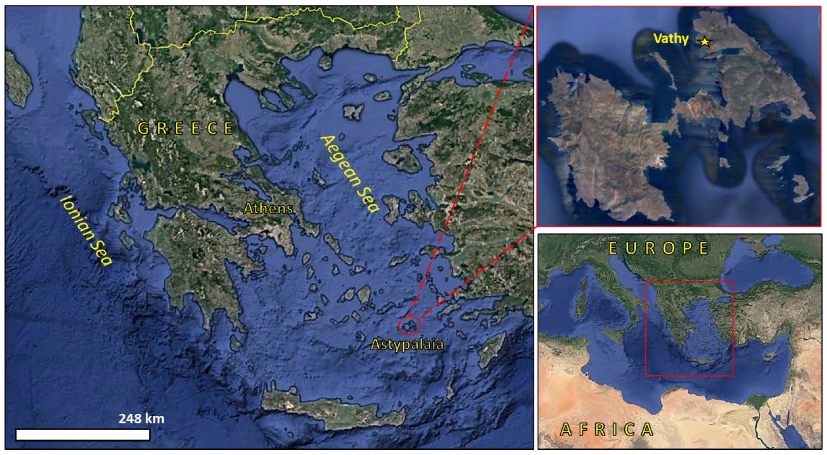
:1. Introduction
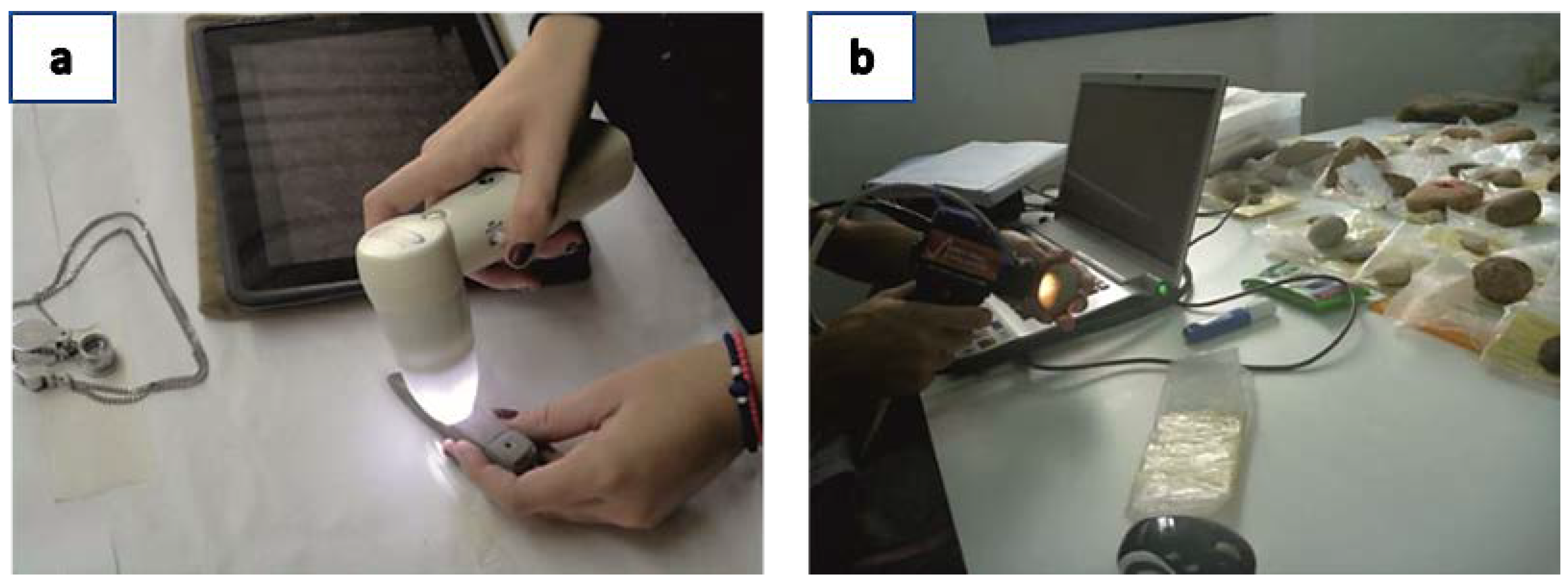
2. Materials and Methods
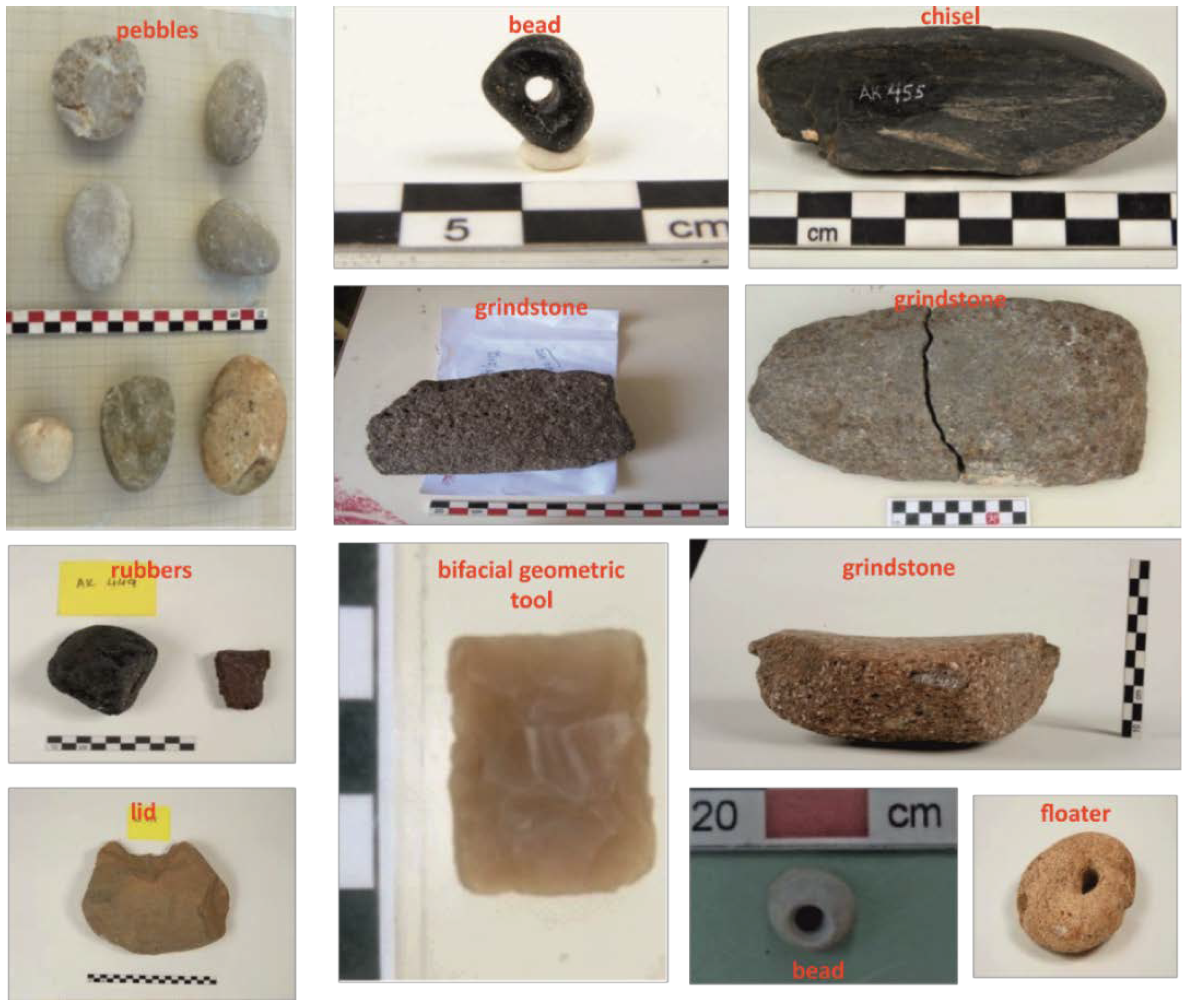
2.1. Archaeological Artefacts
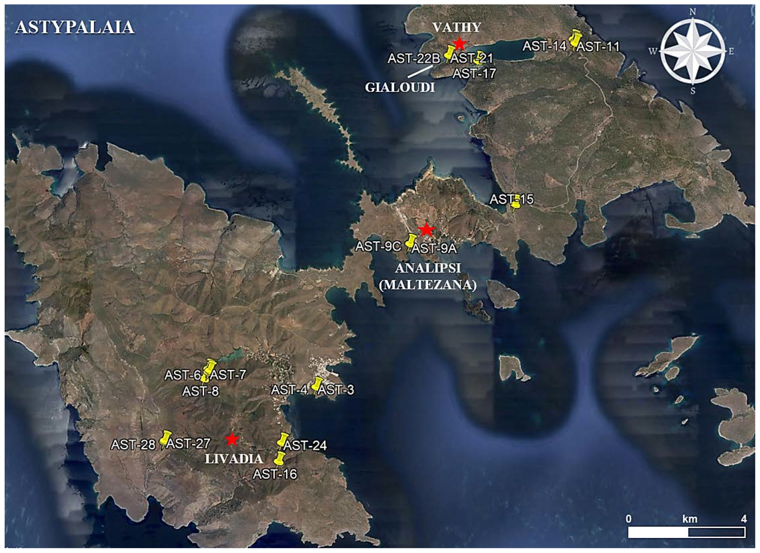
2.2. Raw Materials
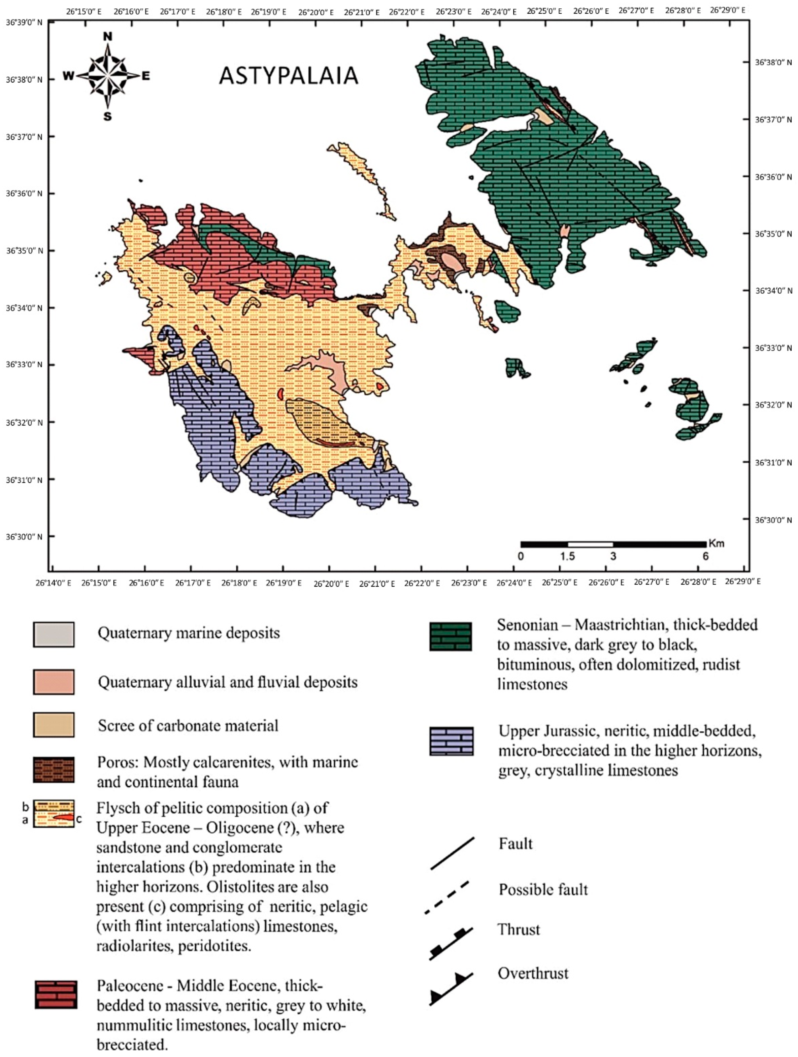
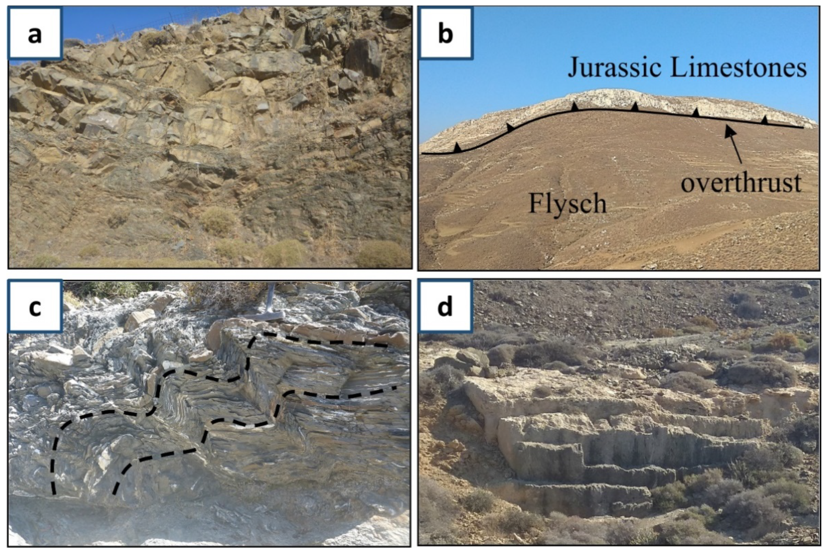
3. Geologic Setting of Astypalaia and Field Observations
4. Results and Discussion
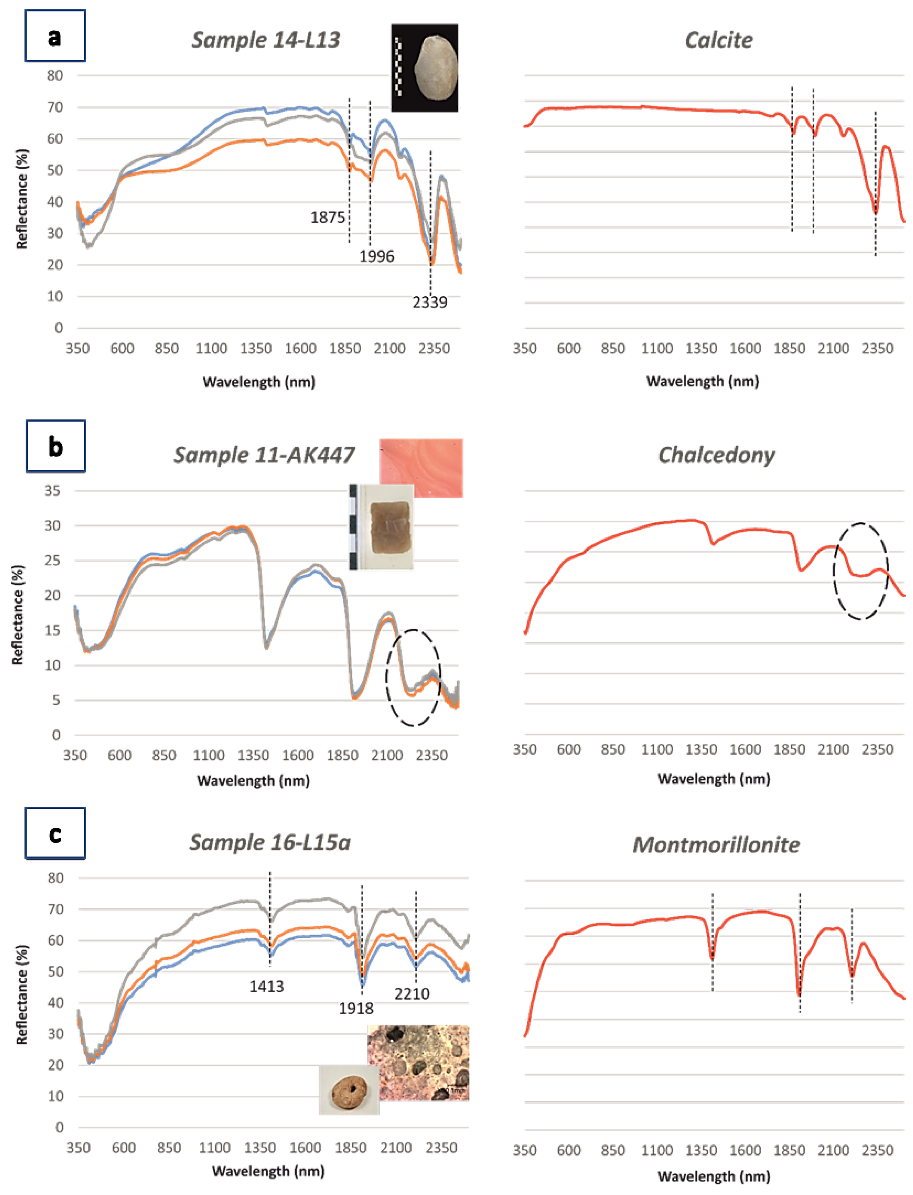
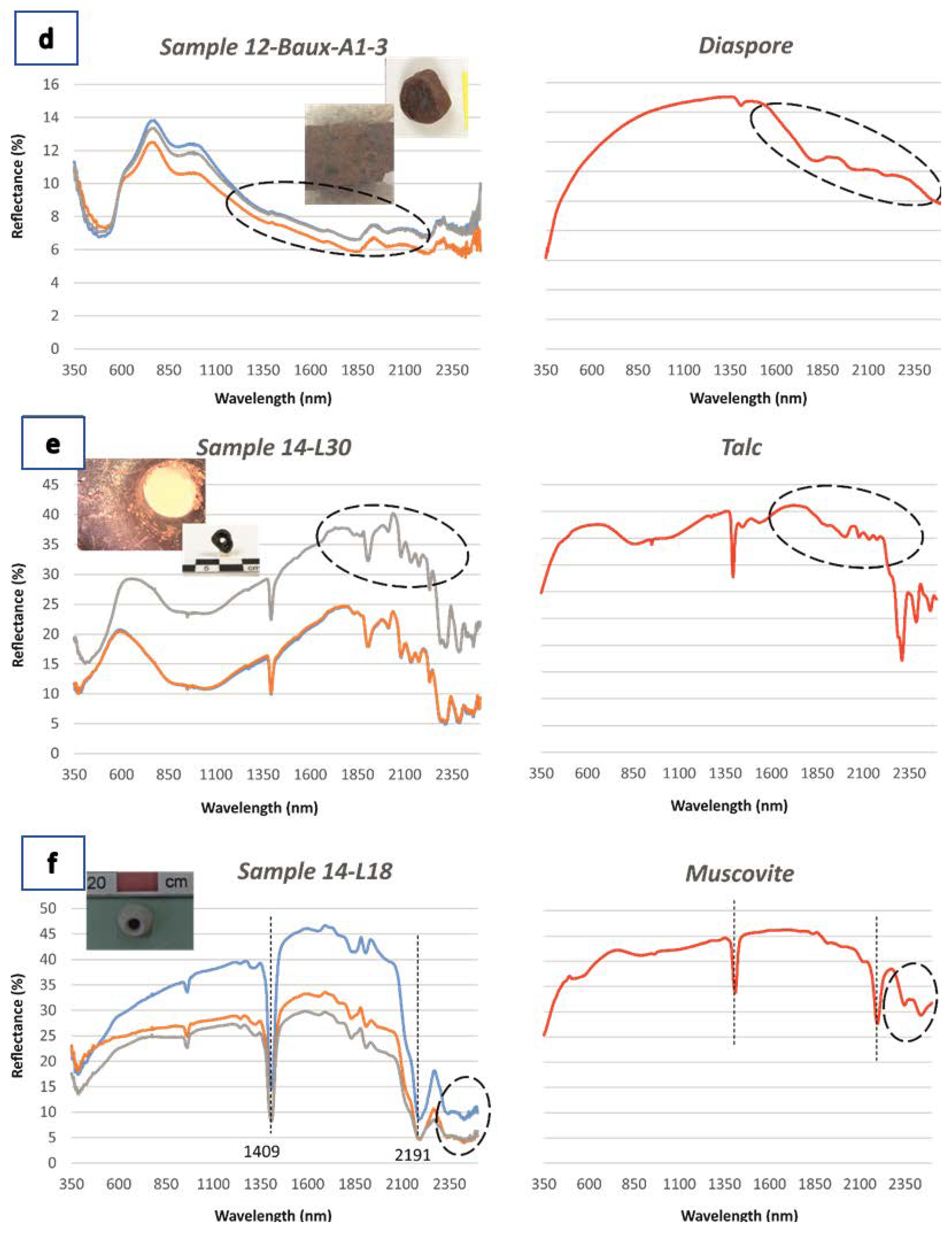
4.1. Microscopic Observation and Spectroscopic Characteristics of the Prehistoric Artefacts


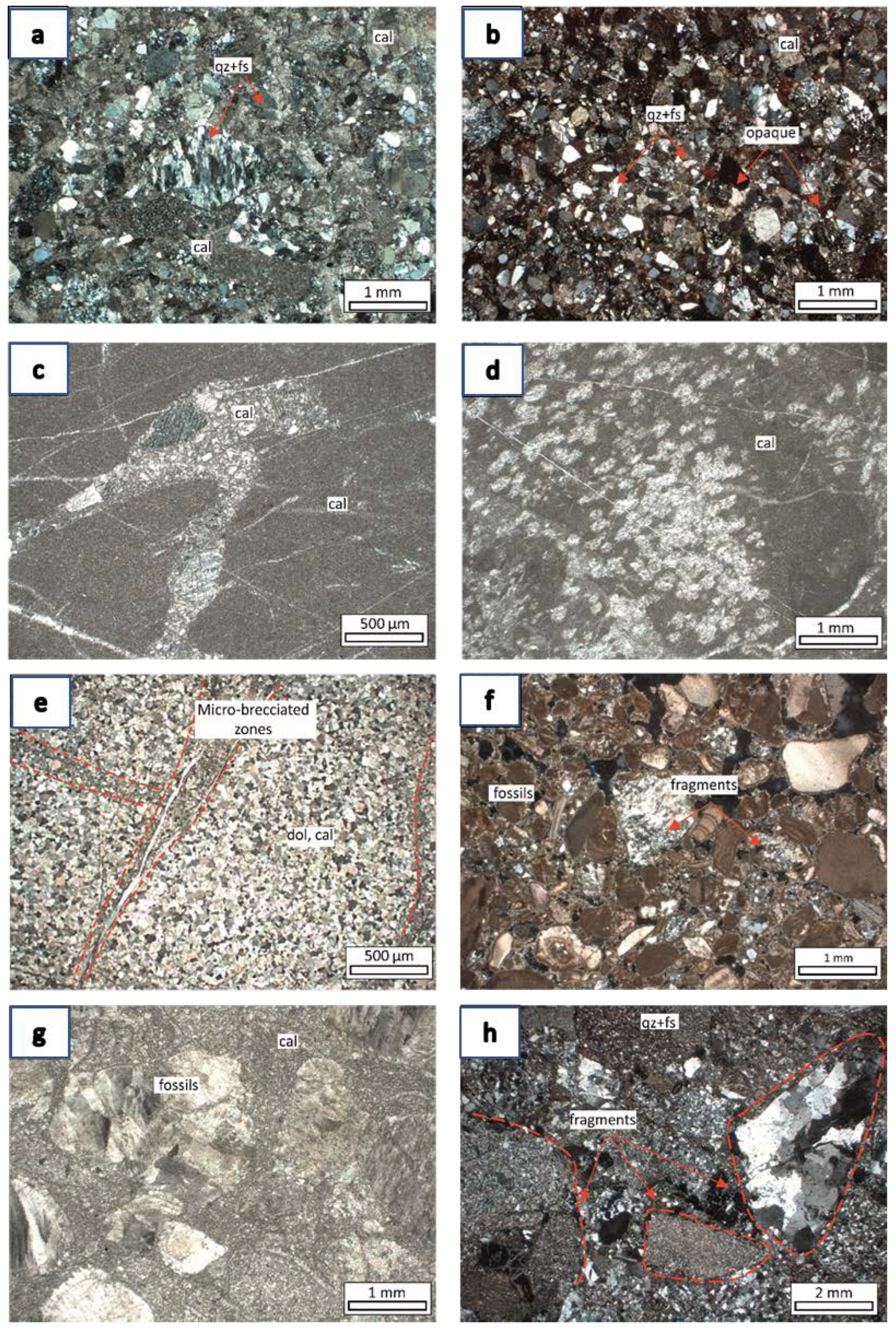
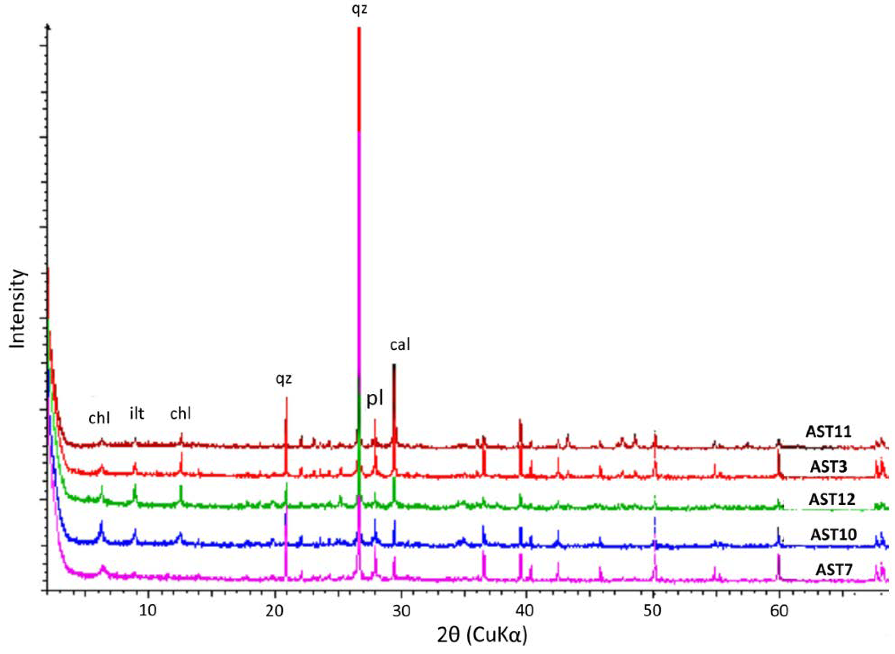
4.2. Petrographic and Mineralogical Analysis of Astypalaian Lithotypes
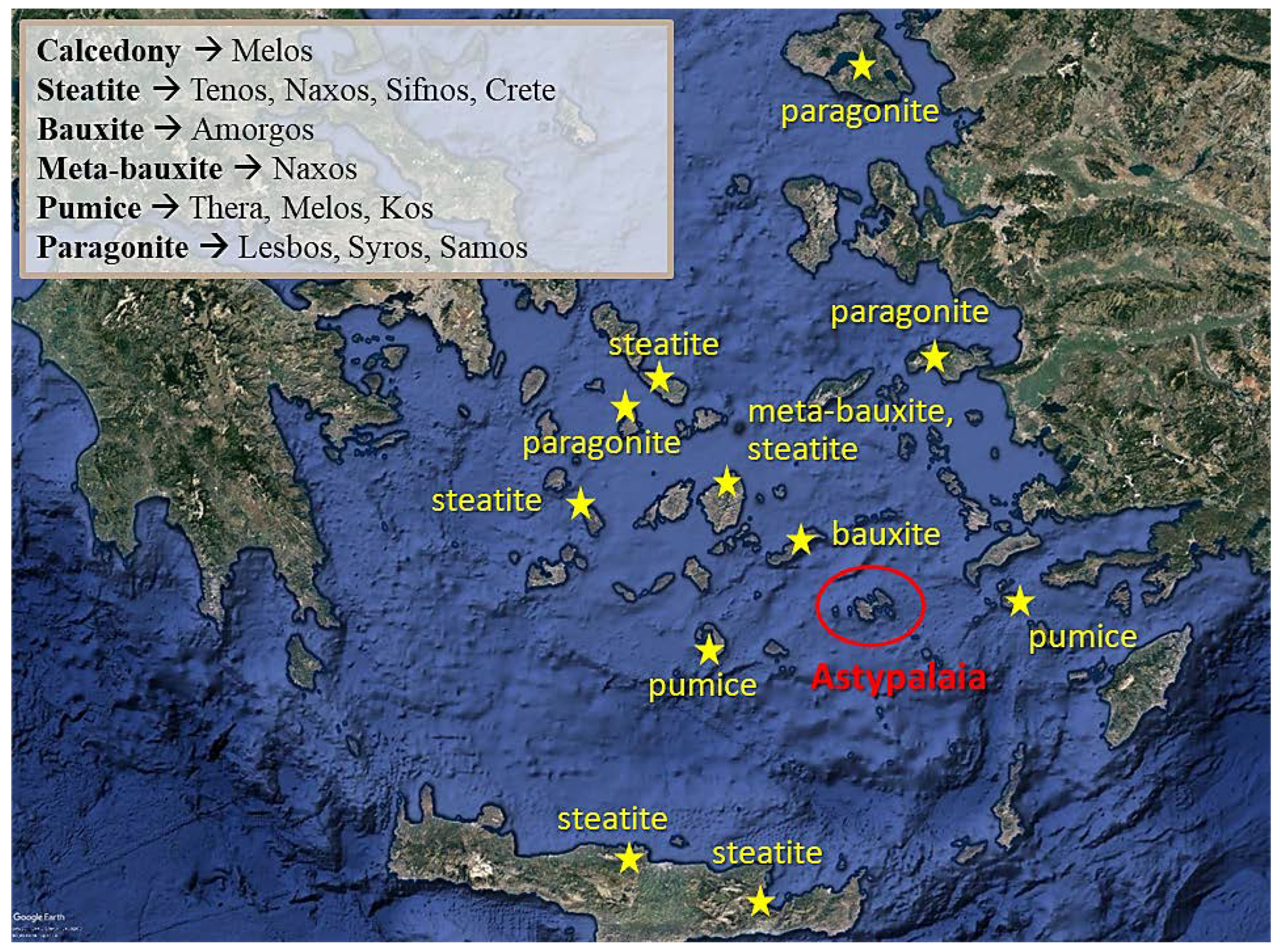
4.3. Preliminary Results on the Provenance of the Raw Materials
5. Conclusions
Author Contributions
Funding
Data Availability Statement
Conflicts of Interest
References
- Biró, K.T. Non-destructive research in archaeology. J. Radioanal. Nucl. Chem. 2005, 265, 235–240. [Google Scholar] [CrossRef]
- Anderson, C.; Törnberg, A.; Törnberg, P. An Evolutionary Developmental Approach to Cultural Evolution. Curr. Anthropol. 2014, 55, 154–174. [Google Scholar] [CrossRef] [Green Version]
- Kintigh, K.; Altschul, J.; Beaudry, M.; Drennan, R.; Kinzig, A.; Kohler, T.; Limp, W.F.; Maschner, H.; Michener, W.; Pauketat, T.; et al. Grand Challenges for Archaeology. Am. Antiq. 2014, 79, 5–24. [Google Scholar] [CrossRef] [Green Version]
- Liritzis, I. Twelve thousand years of non-linear cultural evolution: The physics of chaos in Archaeology. Synesis J. Sci. Technol. Ethics Policy 2013, 4, G19–G31. [Google Scholar]
- Liritzis, I.; Laskaris, N.; Vafiadou, A.; Karapanagiotis, I.; Volonakis, P.; Papageorgopoulou, C.; Bratitsi, M. Archaeometry: An overview. Sci. Cult. 2020, 6, 49–98. [Google Scholar] [CrossRef]
- Philokyprou, M. The initial appearance of ashlar stone in Cyprus. Issues of provenance and use. Mediterr. Archaeol. Archaeom. 2011, 2011, 37–53. [Google Scholar]
- Columbu, S.; Piras, G.; Sitzia, F.; Pagnotta, S.; Raneri, S.; Legnaioli, S.; Palleschi, V.; Lezzerini, M.; Giamello, M. Petrographic and mineralogical characterization of volcanic rocks and surface-depositions on Romanesque Monuments. Mediterr. Archaeol. Archaeom. 2018, 18, 37–64. [Google Scholar] [CrossRef]
- Cau Ontiveros, M.A.; Martinez Farreras, V.; Pecci, A.; Mas Florit, C.; Fantuzzi, L. Archaeometric analysis for provenance and content of roman amphorae from the site of Sa Mesquida (Mallorca, Spain). Mediterr. Archaeol. Archaeom. 2018, 18, 87–105. [Google Scholar] [CrossRef]
- Glascock, M.D. A systematic approach to geochemical sourcing of obsidian artifacts. Sci. Cult. 2020, 6, 35–47. [Google Scholar] [CrossRef]
- Lara, C.; Iliopoulos, I. Ceramic technology, ethnic identification and multiethnic contacts: The archaeological example of the Cuyes river valley (Southeastern Ecuadorian highlands). J. Archaeol. Sci. Rep. 2020, 33, 102557. [Google Scholar] [CrossRef]
- Xanthopoulou, V.; Iliopoulos, I.; Liritzis, I. Characterizations techniques of clays for the archaeometric study of ancient ceramics: A review. Sci. Cult. 2020, 6, 73–86. [Google Scholar] [CrossRef]
- Skarpelis, N. Silcrete and chert as source rocks of early prehistoric artifacts: The case of central Evia (Greece). Mediterr. Archaeol. Archaeom. 2021, 21, 1–19. [Google Scholar] [CrossRef]
- Hunt, G.R. Spectral Signatures of Particulate Minerals in the Visible and near Infrared. Geophysics 1977, 42, 501–513. [Google Scholar] [CrossRef] [Green Version]
- Bruni, S. Etruscan Fine Ware Pottery: Near-Infrared (NIR) Spectroscopy as a Tool for the Investigation of Clay Firing Temperature and Atmosphere. Minerals 2022, 12, 412. [Google Scholar] [CrossRef]
- Inagaki, T.; Yonenobu, H.; Tsuchikawa, S. Near-Infrared Spectroscopic Investigation of the Hydrothermal Degradation Mechanism ofWood as an Analogue of Archaeological Objects. Part I: Softwood. Appl. Spectrosc. 2008, 62, 1209–1215. [Google Scholar] [CrossRef]
- Lichtblau, D.; Strlič, M.; Trafela, T.; Kolar, J.; Ander, M. Determination of mechanical properties of historical paper based on NIR spectroscopy and chemometrics—A new instrument. Appl. Phys. 2008, 29, 19872. [Google Scholar] [CrossRef] [Green Version]
- Delaney, J.K.; Thoury, M.; Zeibel, J.G.; Ricciardi, P.; Morales, K.M.; Dooley, K.A. Visible and infrared imaging spectroscopy of paintings and improved reflectography. Herit. Sci. 2016, 4, 6. [Google Scholar] [CrossRef] [Green Version]
- Tanyaş, H.; Dirican, Μ.; Lütfi Süzen, Μ.; Türkmenoğlu, A.G.; Kolat, Ç.; Atakuman, Ç. Identification of possible source areas of stone raw materials combining remote sensing and petrography. Int. J. Remote Sens. 2017, 38, 3923–3942. [Google Scholar] [CrossRef] [Green Version]
- Vlachopoulos, A. Archaeological fieldwork at Vathy, Astypalaia, Dodona. History—Archaeology. MΓ-MΔ 2017, 2014, 371–411. (In Greek) [Google Scholar]
- Vlachopoulos, A. Vathy, Astypalaia. Research and material studies in a diachronic palimpsest of Aegean Archaeology. Dodekan. Chron. Kθ Rhodes 2021, 2021, 83–131. (In Greek) [Google Scholar]
- Tsigkas, G.; Sfikas, G.; Pasialis, A.; Vlachopoulos, A. Nikou, C. Markerless detection of ancient rock carvings in the wild: Rock art in Vathy, Astypalaia. Pattern Recognit. Lett. 2020, 135, 337–345. [Google Scholar] [CrossRef]
- Adam, Ε. Ground stone tools and utensils from the Vathy peninsula. The surface finds. In Vathy, Astypalaia. Ten Years of Research in a Diachronic Palimpsest of the Aegean (2011–2020); Vlachopoulos, A., Ed.; Hellenic Ministry of Culture: Athens, Greece; University of Ioannina: Ioannina, Greece; Melissa Publishing House: Athens, Greece, 2022; (In Greek; abstracts in English). [Google Scholar]
- Adam, E. Ground stone tools and utensils, finds. In Vathy, Astypalaia. Ten Years of Research in a Diachronic Palimpsest of the Aegean (2011–2022); Vlachopoulos, A., Ed.; Hellenic Ministry of Culture: Athens, Greece; University of Ioannina: Ioannina, Greece; Melissa Publishing House: Athens, Greece, 2022; (In Greek; abstracts in English). [Google Scholar]
- Metaxas, O. The chipped stone assemblage from the field survey of Vathy, Astypalaia. In Vathy, Astypalaia. Ten Years of Research in a Diachronic Palimpsest of the Aegean (2011–2022); Vlachopoulos, A., Ed.; Hellenic Ministry of Culture: Athens, Greece; University of Ioannina: Ioannina, Greece; Melissa Publishing House: Athens, Greece, 2022; (In Greek; abstracts in English). [Google Scholar]
- Vlachopoulos, A.; Angelopoulou, A. Early Cycladic Figurines from Vathy, Astypalaia. In Early Cycladic Sculpture in Context from Beyond the Cyclades: Mainland Greece, the North and East Aegean; Marthari, M., Renfrew, C., Boyd, M., Eds.; Oxbow Books: Oxford, PA, USA, 2019; pp. 202–226. [Google Scholar]
- Iliopoulos, I.; Kokkaliari, M. Accessing provenance issues of prehistoric stone artefacts from Vathy (Astypalaia, Greece) through the minero-petrographic characterization of local lithologies. In Vathy, Astypalaia. Ten Years of Research in a Diachronic Palimpsest of the Aegean, (2011–2022); Vlachopoulos, A., Ed.; Hellenic Ministry of Culture: Athens, Greece; University of Ioannina: Ioannina, Greece; Melissa Publishing House: Athens, Greece, 2022; (In Greek; abstracts in English). [Google Scholar]
- Clark, R.N.; King, T.V.; Klejwa, M.; Swayze, G.A.; Vergo, N. High spectral resolution reflectance spectroscopy of minerals. J. Geophys. Res. 1990, 95, 12653–12680. [Google Scholar] [CrossRef] [Green Version]
- Christodoulou, G. About the geology of Astypalaia Island. Sci. Yearb. Issued Fac. Nat. Math. Sci. 1966, 10, 169–180. [Google Scholar]
- Marnelis, P.; Bonneau, M. Stratigraphie et structure de l’Île d’Astypalea (Dodécanèse, Grèce). In VI. Colloquium on the Geology of the Aegean Region; Kallergis, G., Ed.; 6th Aegeiscollathens; Institute of Geological and Mining Research: Yaoundé, Cameroon, 1977; Volume 1, pp. 323–332. [Google Scholar]
- Ring, U. Structure and deformation history of Astypalea island, Aegean Sea. Bull. Geol. Soc. Greece 2001, 34, 329–335. [Google Scholar] [CrossRef] [Green Version]
- Drouga, A. The Effect of Volcanic Ash Sedimentation and Dispersion at the Present and Modern Marine Sediments of Kos, Nissyros and Astypalea Islands (SE Volcanic Arc), Aegean Sea, Greece. Ph.D. Dissertation, Aristotle University of Thessaloniki, Thessaloniki, Greece, 2002. [Google Scholar]
- Chatzaras, V. Geotraverse across the Hellenides between Western Crete and the Cycladic Islands. Ph.D. Dissertation, University of Patras, Patras, Greece, 2010; 206p. [Google Scholar]
- Papoulia, M.; Karymbalis, E.; Gaki-Papanastassiou, K.; Maroukian, H. Assessment of the susceptibility of the coast of Astypalea Island (SE Aegean Sea) to sea-level rise. Bull. Geol. Soc. Greece 2013, 47, 305–314. [Google Scholar]
- IGME. Geological Map of Greece 1986, 1:50000; Astipalaia Sheet; IGME: Athens, Greece, 1986. [Google Scholar]
- Clark, R.N. Spectroscopy of rocks and minerals, and principles of spectroscopy. In Remote Sensing for Earth Sciences—Manual of Remote Sensing, 3rd ed.; Renz, A.N., Ed.; John Wiley and Sons: New York, NY, USA, 1999; pp. 3–58. [Google Scholar]
- Gaffey, S.J. Spectral reflectance of carbonate minerals in the visible and near infra-red (0.35–2.55 microns): Calcite, aragonite, and dolomite. Am. Mineral. 1986, 71, 151–162. [Google Scholar]
- Crowley, J.K. Visible and near-infrared spectra of carbonate rocks-reflectance variations related to petrographic texture and impurities. J. Geophys. Res. 1986, 91, 5001–5012. [Google Scholar] [CrossRef]
- Aines, R.D.; Rossman, G.R. Water in minerals? A peak in the infrared. J. Geophys. Res. 1984, 89, 4059–4407. [Google Scholar] [CrossRef]
- Clark, R.N.; Swayze, G.A.; Wise, R.; Livo, E.; Hoefen, T.; Kokaly, R.; Sutley, S.J. USGS Digital Spectral Library splib06a; Digital Data Series; U.S. Geological Survey: Denver, CO, USA, 2007; p. 231.
- Rice, M.S.; Cloutis, E.A.; Bell, J.F., III; Bish, D.L.; Horgan, B.H.; Mertzman, S.A.; Craig, M.A.; Renaut, R.W.; Gautason, B. Mountain, B. Reflectance spectra diversity of silica-rich materials: Sensitivity to environment and implications for detections on Mars. Icarus 2013, 223, 499–533. [Google Scholar] [CrossRef]
- Doublier, M.P.; Roache, A.; Potel, S. Application of SWIR Spectroscopy in very low-grade metamorphic environments: A comparison with XRD methods. Geol. Surv. West. Aust. 2010, 61, 2992. [Google Scholar]
- Fleet, M.E.; Deer, W.A.; Howie, R.A.; Zussman, J. Rock-Forming Minerals: Micas, 2nd ed.; Geological Society of London: London, UK, 2003. [Google Scholar]
- Doublier, M.P.; Roache, T.; Potel, S. Short-wavelength infrared spectroscopy: A new petrological tool in low-grade to very low-grade pelites. Geology 2010, 38, 1031–1034. [Google Scholar] [CrossRef]
- Whitney, L.; Evans, W. Abbreviations for names of rock forming minerals. Am. Mineral. 2010, 3, 227. [Google Scholar] [CrossRef]
- Jones, R.E.; Kilikoglou, V.; Olive, V.; Bassiakos, Y.; Ellam, R.; Bray, I.S.J.; Sanderson, D.C.W. A new protocol for the chemical characterisation of steatite—Two case studies in Europe: The Shetland Islands and Crete. J. Archaeol. Sci. 2007, 34, 626–641. [Google Scholar] [CrossRef]
- Richards, H.; Mondillo, N.; Boni, M.; Thorne, R.; Tavlan, M. Bauxite and nickel-cobalt lateritic deposits of the Tethyan belt. Bauxite. Soc. Econ. Geol. 2016, 19, 349–387. [Google Scholar]
- Papavasiliou, K.; Voudouris, P.; Kanellopoulos, C.; Alfieris, D.; Xydous, S. Mineralogy and Geochemistry of the Triades-Galana Pb-Zn-Ag-Au intermediate-high sulfidation epithermal deposit, Milos island, Greece. Bull. Geol. Soc. Greece 2016, 50, 1969–1979. [Google Scholar] [CrossRef] [Green Version]
- Hatzipanagiotou, K.; Tsikouras, B.; Migiros, G.; Gartzos, E.; Serelis, K. Origin of rodingites in ultramafic rocks from Lesvos island (NE Aegean, Greece). Ofioliti 2003, 28, 13–23. [Google Scholar]
- Seck, H.A.; Koetz, J.; Okrusch, M.; Seidel, E.; Stosch, H.G. Geochemistry of a meta-ophiolite suite; an associated metagabbros, eclogites and glaucophanites on the island of Syros, Greece. Eur. J. Mineral. 1996, 8, 607–623. [Google Scholar] [CrossRef]
- Ockenga, E.; Yalçin, Ü.; Medenbach, O.; Schreyer, W. Zincohögbomite, a new mineral from eastern Aegean metabauxites. Eur. J. Mineral. 1998, 10, 1361–1366. [Google Scholar] [CrossRef]
- Allen, S.R.; Cas, R.A.F. Transport of pyroclastic flows across the sea during the explosive, rhyolitic eruption of the Kos Plateau Tuff, Greece. Bull. Volcanol. 2001, 62, 41–456. [Google Scholar] [CrossRef]
- Dufek, J.; Bergantz, G.W. Dynamics and deposits generated by the Kos Plateau Tuff eruption: Controls of basal particle loss on pyroclastic flow transport. Geochem. Geophys. Geosyst. 2007, 8, Q12007. [Google Scholar] [CrossRef]
- Dufek, J.; Manga, M.; Staedter, M. Littoral blasts: Pumice-water heat transfer and the conditions for steam explosions when pyroclastic flows enter the ocean. J. Geophys. Res. Solid Earths 2007, 112, B11201. [Google Scholar] [CrossRef]


| Type | No of |
|---|---|
| discs | 2 |
| pull weights | 1 |
| pumice | 2 |
| adze | 1 |
| grinding slabs | 2 |
| pounders/polishers | 5 |
| utilized pebbles | 14 |
| stone beads | 2 |
| pebbles | 54 |
| bauxite | 2 |
| serpentine | 1 |
| flint nodule | 1 |
| copper slags | 3 |
| unidentified | 27 |
| geometric tool | 1 |
| Total | 118 |
| Wavelength (nm) | Mechanism | Mineral Group |
|---|---|---|
| ~1400 | OH- and water | clay minerals, hydroxyls, zeolites |
| ~1900 | water | smectites |
| ~2200 | Al-OH | clay minerals, amphiboles, mica |
| ~2250 | Fe-OH | epidote, biotite, tourmaline, chlorite, phlogopite |
| ~2300–2400 | Mg-OH | amphiboles, epidote, chlorite |
| ~2340 | CO32− | carbonates |
| Sample | XRD | Lithotype |
|---|---|---|
| AST-3 | qz, pl, cal, ms/ilt, chl | Calcitic sandstone |
| AST-4B | cal | Biosparite |
| AST-7 | qz, pl, cal, ms/ilt | Calcitic sandstone |
| AST-9A1 | qz, pl, afs, cal, arg | Biomicrite |
| AST-9A2 | qz, pl, afs, cal, arg | Biomicrite |
| AST-9B | qz, pl, afs, cal, arg | Biomicrite |
| AST-10 | qz, pl, cal, ms/ilt, chl | Calcitic sandstone |
| AST-11 | qz, pl, cal, ms/ilt, chl | Calcitic sandstone |
| AST-13 | cal | Limestone |
| AST-16B | cal, qz | Calcitic olistholite |
| AST-21 | cal | Calcarenite |
| AST-22A | cal, arg | Limestone |
| AST-22B | cal | Limestone |
| AST-25 | cal, dol | Limestone |
| AST-26 | dol, cal | Crystalline limestone/Marble? |
| AST-27 | cal | Calcarenite |
| AST-28 | qz, pl, cal, ms/ilt, chl, clay minerals | Calcitic sandstone |
| Local | Non-Local |
|---|---|
| Calcitic sandstone | Chalcedony |
| Shale | Bauxite/Meta-bauxite |
| Marl | Steatite |
| Limestone/marble | Paragonite |
| Pumice and volcanic rocks (rhyodacites to andesites, tuffs) |
Publisher’s Note: MDPI stays neutral with regard to jurisdictional claims in published maps and institutional affiliations. |
© 2022 by the authors. Licensee MDPI, Basel, Switzerland. This article is an open access article distributed under the terms and conditions of the Creative Commons Attribution (CC BY) license (https://creativecommons.org/licenses/by/4.0/).
Share and Cite
Kokkaliari, M.; Adam, E.; Vlachopoulos, A.; Iliopoulos, I. Tracing Raw Material Sources of Prehistoric Stone Artefacts by Non-Invasive Techniques: The Case of the Early Bronze Age (3rd Mill. BCE) Site of Vathy, Astypalaia, Greece. Quaternary 2022, 5, 42. https://doi.org/10.3390/quat5040042
Kokkaliari M, Adam E, Vlachopoulos A, Iliopoulos I. Tracing Raw Material Sources of Prehistoric Stone Artefacts by Non-Invasive Techniques: The Case of the Early Bronze Age (3rd Mill. BCE) Site of Vathy, Astypalaia, Greece. Quaternary. 2022; 5(4):42. https://doi.org/10.3390/quat5040042
Chicago/Turabian StyleKokkaliari, Maria, Eugenia Adam, Andreas Vlachopoulos, and Ioannis Iliopoulos. 2022. "Tracing Raw Material Sources of Prehistoric Stone Artefacts by Non-Invasive Techniques: The Case of the Early Bronze Age (3rd Mill. BCE) Site of Vathy, Astypalaia, Greece" Quaternary 5, no. 4: 42. https://doi.org/10.3390/quat5040042