Proceeding Paper

The Utilization of Urolithin A—A Natural Polyphenol Metabolite of Ellagitannins as a Modulator of the Gut Microbiota for Its Potential Use in Obesity Therapy †

by
Abdulrasheed O. Abdulrahman
1,
Mohammed Yahya Alzubaidi
1,
Muhammad Shahid Nadeem
1,
Jalaluddin Awlia Khan
1,
Irfan A. Rather
2 and
Mohammad Imran Khan
1,3,*
1
Department of Biochemistry, Faculty of Science, King Abdulaziz University, Jeddah 21589, Saudi Arabia
2
Department of Biology, Faculty of Science, King Abdulaziz University, Jeddah 21589, Saudi Arabia
3
Cancer Metabolism and Epigenetic Unit, Faculty of Science, King Abdulaziz University, Jeddah 21589, Saudi Arabia
*
Author to whom correspondence should be addressed.
Presented at the 1st International Electronic Conference on Biomolecules: Natural and Bio-Inspired Therapeutics for Human Diseases, 1–13 December 2020; Available online: https://iecbm2020.sciforum.net/.
Proceedings 2021, 79(1), 12; https://doi.org/10.3390/IECBM2020-08806
Published: 1 December 2020

Abstract

:
Obesity, a global health concern, is associated with the dysbiosis of intestinal microbial composition. This study aims to investigate the potential of urolithin A (Uro-A) in reducing body weight gain through modulation of the gut microbiota. Rats were fed a high-fat diet and administered with Uro-A. Changes in the composition of the gut microbial community were analyzed using 16S rDNA gene sequencing. Our results showed that Uro-A significantly decreased body weight gain and modulated the compositions of gut microbes that are related to body weight, inflammation, impaired glucose, and dysfunctional lipid metabolism, which are all associated with obesity.

1. Introduction

Obesity is a critical nutritional health disorder caused by the imbalance between energy intake and its expenditure where caloric intake exceeds energy expenditure [1]. The prevalence of obesity is on the rise globally, constituting a critical health care challenge [2].
The human gut houses trillions of bacterial species that play key roles in nutrient utilization and maintenance of energy balance [3]. Recent findings have revealed the role of gut microbiota in reducing obesity and co-morbidities [4]. However, prolonged high-fat diet (HFD) feeding can alter the composition of the gut microbiota, thereby increasing the risk of the development of obesity and other related diseases [5]. Thus, the gut microbiota may play a significant role in controlling obesity through regulating body weight gain, chronic inflammation, lipid metabolism, and hepatic fat storage [6,7].
Urolithin A (Figure 1a) is one of the naturally occurring secondary gut-derived metabolites obtained from ellagitannin and ellagic acid-rich food sources like pomegranates, grapes, nuts, and berries [8]. It is also the most studied metabolite among the urolithins (Urolithins B, C, D, and iso-urolithin A). Recent studies have pointed towards a potential anti-obesity activity for this metabolite [9,10]. Thus, this study aims to investigate the potential of urolithin A (Uro-A) in reducing body weight gain through modulation of the gut microbiota.

2. Materials and Methods

2.1. Preparation of Urolithin Stock Solution

The urolithin A (purchased from BLD Pharma, Shanghai, China) stock solution was prepared by dissolving it in dimethyl sulfoxide (DMSO) at a concentration of 20 mg/mL and an aliquot of the stock solution was made by diluting it in phosphate-buffered saline (PBS).

2.2. Animals and Treatment

Wistar rats numbering 18 were obtained from the animal house of King Fahd Medical and Research Centre (KFMRC), King Abdulaziz University, Jeddah, Saudi Arabia. Animals were acclimatized for 7 days at 22–24 °C and on a 12/12-h light on/off cycle, after which they were randomly segregated into 3 groups (n = 6) and were fed either a normal diet (ND) or an HFD (containing 40% normal diet, with 40% lard, 10% sucrose, and 10% cholesterol). Animals in the ND group (group 1) were fed a normal diet, and the HFD group (groups two and three) were fed a high-fat diet for 10 weeks. The animals in the HFD group were considered obese when they achieved a 10% higher body weight as compared to the control group [11,12]. Following this, a 2.5 mg/kg [13] intraperitoneal (IP) injection of Uro-A was given to group three (HFD + Uro-A) four times a week for four weeks.
At the end of this treatment period, animals were placed on an overnight fast and were euthanized under diethyl ether anesthesia; blood was withdrawn from the abdominal aorta. The intestinal contents were then collected under aseptic conditions and placed at −80 °C until analysis.

2.3. Serum Biochemical Analysis

Whole blood was centrifugated at 3000 rpm for 10 min and the upper serum samples were collected and stored at −80 °C until use. The levels of low-density lipoprotein cholesterol (LDL), triglycerides (TG), high-density lipoprotein cholesterol (HDL), and total cholesterol were quantified with a commercial kit (Crescent diagnostics, Jeddah, Saudi Arabia).

2.4. Sequencing of the 16S rDNA and Data Processing

The total genomic DNA from the cecal content of the intestine was extracted using QIAamp Fast DNA Stool Mini Kit (Qiagen, Hilden, Germany) according to the manufacturer’s instructions and then subjected to 16S rDNA gene sequencing targeting the V3–V4 region with barcoded 341F and 805R universal primers according to the procedure of Dowd et al. [14]. Following the minimal cycle of PCR amplification, the purified products were amplified by pyrosequencing on an Illumina MiSeq platform from Macrogen, Seoul, Korea. The overlapped Illumina MiSeq data, 2 × 300 bp, were paired-end, merged, and the sequences were grouped into operational taxonomy units (OTU) at 97% similarity using QIIME 1.9.

2.5. Statistical Analysis

Data are presented as mean ± SE. Plot fitting and statistical analysis with either one-way ANOVA followed by Dunnett’s multiple comparisons test or Kruskal–Wallis test followed by Dunn’s multiple comparisons test were performed using GraphPad Prism V6.0 software (GraphPad Software, San Diego, CA, USA). * p < 0.05 was taken as significant.

3. Results and Discussion

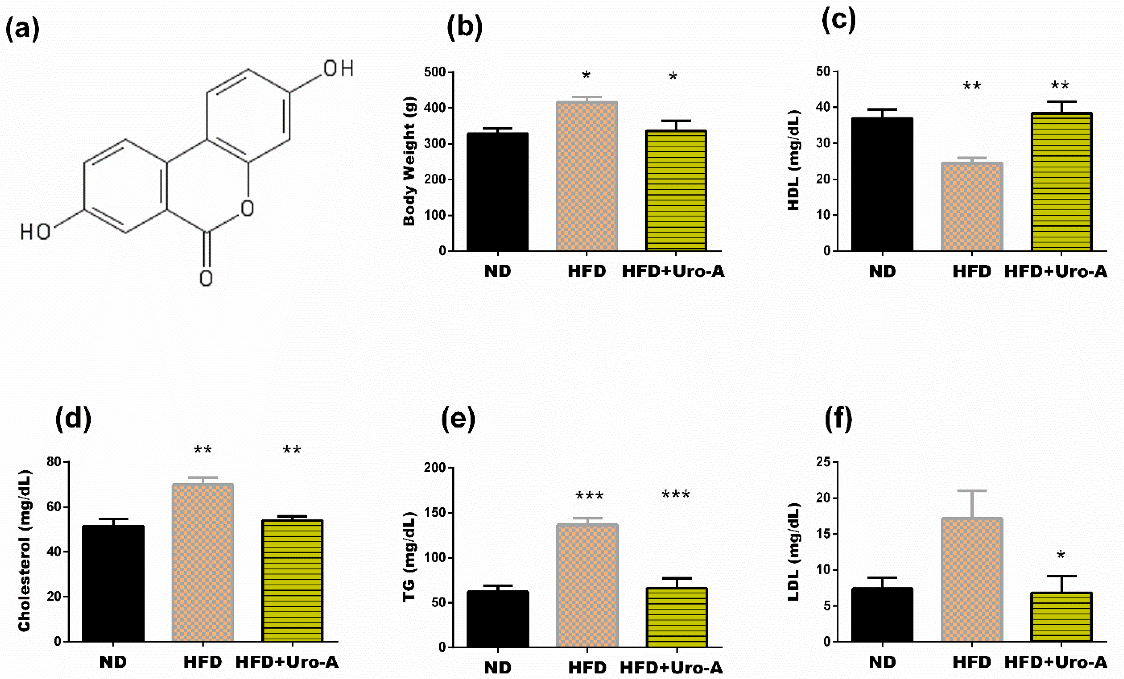
3.1. Urolithin A Administration Improved Altered Metabolism in HFD Rats

As shown in (Figure 1b), the final body weight of animals fed an HFD was significantly (p < 0.05) higher than the weight of animals fed a normal diet. However, animals treated with Uro-A showed a significant (p < 0.05) decrease in the final body weight when compared with that of untreated animals fed an HFD. In addition, compared with a normal diet, HFD resulted in a significant reduction in HDL levels (p < 0.01) and a significant increase in serum cholesterol (p < 0.01) and triglycerides (p < 0.001) levels (Figure 1c–e). Compared with untreated animals fed an HFD, Uro-A-treated rats showed a significant increase in HDL level (p < 0.01) and a significant decrease in serum cholesterol (p < 0.01), triglycerides (p < 0.001), and LDL (p < 0.05) levels. This result suggests a potential anti-obesity role for urolithin A.

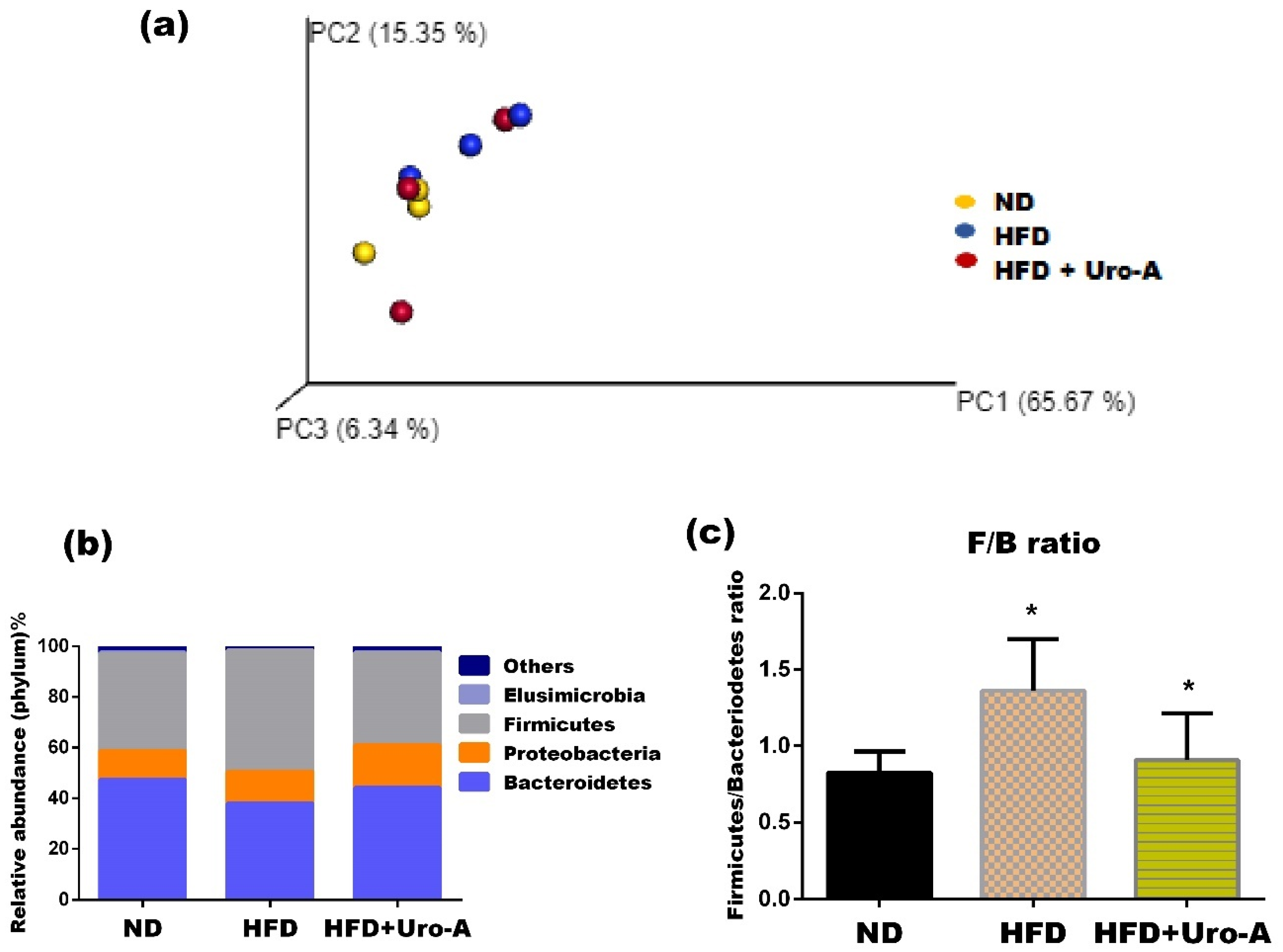
3.2. Urolithin A Administration Altered Microbial Diversity

Analysis of the metagenomics amplicon from the Miseq platform revealed that a total of 969,801 high-quality reads were obtained with an average of 21,435, 25,530 and 31,166 reads for ND, HFD, and HFD + Uro-A groups, respectively.
Compared with the normal diet, HFD feeding resulted in the compositional reshape of the gut microbiota community. For example, the beta diversity from the principal coordinate analysis (unweighted UniFrac) showed that the gut microbiota population in the animals fed an HFD clustered differentially from animals fed a normal diet (Figure 2a). A similar observation was made in Uro-A-treated animals fed with an HFD (Figure 2a). The alpha diversity index with Chao1 showed that animals fed with a normal diet have the maximum species diversity followed by Uro-A-treated animals fed a high-fat diet. This species diversity changes with the type of diet or treatment administered (Table 1). The Shannon microbial diversity richness was lower in the HFD groups than in the normal diet-fed rats, and animals fed an HFD treated with Uro-A had a higher species richness compared with that in untreated animals fed an HFD. There was no visible difference in the Simpson diversity amongst the groups (Table 1).

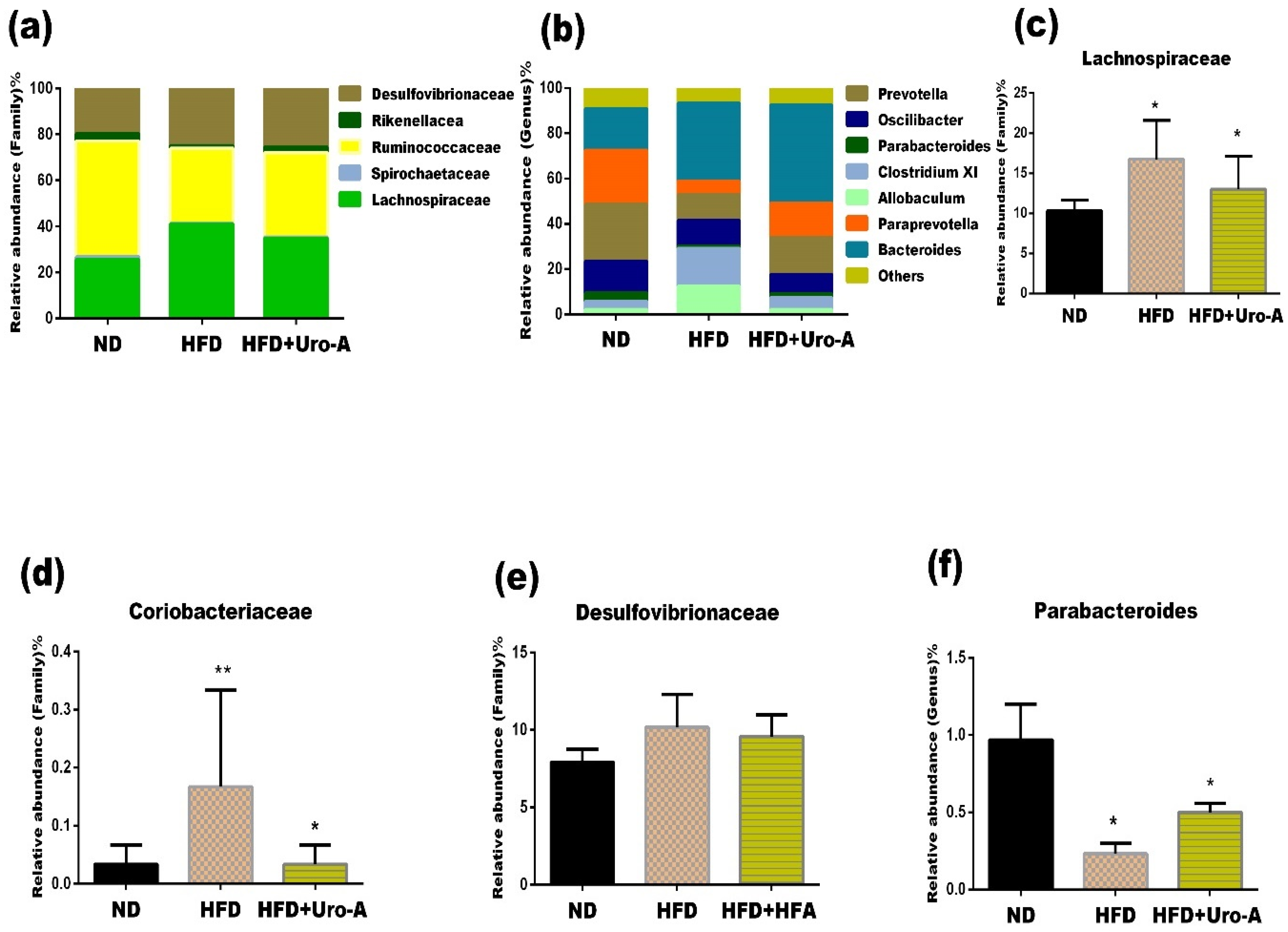
3.3. Urolithin A Administration Altered the Composition of Specific Microbes

Previous reports have shown that the gut microbiota plays an important role in obesity development [6,7]. Therefore, the characterization of the gut microbiota in obese individuals and normal-weight subjects would help to increase our understanding of the pathophysiology of obesity-induced gut alteration and provide insights into the development of therapeutic options for obesity management. Urolithin A treatment modified the composition of key gut microbes at the phylum, family, and genus levels. For example, at the phylum level, HFD increased the phylum Firmicutes as compared with the normal diet. Uro-A treatment restored the level of Firmicutes to normal levels (Figure 2b). However, it has been previously shown that an increased ratio of Firmicutes to Bacteroidetes in HFD-fed mice contributes to the development of obesity [15]. Our result revealed an increased abundance of Firmicutes to the abundance of Bacteroidetes ratio (F/B ratio) in untreated HFD-fed rats compared with that in animals on a normal diet (Figure 2c). This change in the gut microbial composition has been reported to be synonymous with obesity-driven dysbiosis found in humans and animals [16]. Uro-A treatment significantly decreased the F/B ratio compared to untreated animals fed HFD (Figure 2c).
At the family level, compared with untreated animals fed a normal diet, the levels of Lachnospiraceae and Coriobacteriaceae were higher in animals fed an HFD. On the other hand, treatment with Uro-A decreased the levels of Lachnospiraceae and Coriobacteriaceae compared to untreated animals fed an HFD (Figure 3c,d). Previous studies report that the level of Lachnospiraceae is elevated in an HFD and is associated with an increase in body weight in germ-free ob/ob mice [17]. This family of microbes continuously impair glucose metabolism and is thought to be associated with type 2 diabetes and obesity [17,18]. Consistent with these reports, we noted an increase in the abundance of Lachnospiraceae in the cecum of rats fed an HFD (Figure 3c), which was reduced by Uro-A treatment (Figure 3c).
Similarly, Coriobacteriaceae are anaerobic, Gram-positive bacteria belonging to the dominant bacterial community found in the digestive system of humans and rodents [19]. Campbell et al. [20] observed an increased abundance of the Coriobacteriaceae in HFD mice which resulted in a significant increase in their body weight [20]. An increased abundance of this family has been reported in obese children and adolescents [21]. The Coriobacteriaceae family takes part in the metabolism of bile acid, which is linked to gut barrier impairment and metabolic dysfunctions [22]. A decrease in the abundance of this family might, thus, be important in partly regulating lipid metabolism and ameliorate metabolic dysfunction associated with the consumption of HFD. Our results showed that Uro-A administration decreased the relative abundance of Coriobacteriaceae. This effect agrees with our result on serum lipids in which Uro-A treatment significantly decreased the serum lipid profiles (Figure 1).
Furthermore, Desulfovibrionaceae are Gram-negative bacteria shown to be related to inflammation that could lead to disruption in the gut microbial community [23]. Failure in the regulation of Desulfovibrionaceae in obese humans triggers cecum inflammation and can lead to systemic inflammation, resulting in metabolic dysfunction [24,25]. Our study shows the disproportion in the levels of Desulfovibrionaceae in the microbiome in HFD-fed rats compared with the normal diet-fed rats (Figure 3e). However, rats treated with Uro-A showed decreased Desulfovibrionaceae levels when compared with untreated HFD-fed rats.
At the genus level, an HFD resulted in a decrease in Parabacteroides, Oscillibacter, and Prevotella when compared to untreated animals fed a normal diet (Figure 3b). Urolithin A treatment increased the abundance of Parabacteroides and Prevotella. Parabacteroides are SCFA-producing microbes whose relatively low abundance was noted in patients with obesity and nonalcoholic fatty liver [26,27]. Oral administration of Parabacteroides distasonis to both ob/ob mice and HFD mice resulted in improved glucose homeostasis, decreased weight gain, and restored lipid metabolism [28]. In this study, we noticed that treatment with Uro-A resulted in an increase in the relative abundance of Parabacteroides following an HFD (Figure 3f), which suggests that the decrease in body weight of the rats treated with Uro-A might be partly due to the increase in the population of Parabacteroides, as these microbes have been shown to reduce body weight [28,29].

4. Conclusions

In this study, we showed that the administration of Uro-A modulated the gut microbial community of HFD-fed rats, resulting in decreased bodyweight, decreased serum levels of cholesterol, triglycerides, and LDL while increasing the level of HDL. More importantly, we also showed that Uro-A modulated the relative abundance of specific microbes such as Lachnospiraceae, Coriobacteriaceae, Parabacteroides, and Desulfovibrionaceae. These modulated microbes are related to body weight gain, inflammation, impaired glucose, and dysfunctional lipid metabolism, which are all closely associated with obesity.
Taken together, our results suggest that Uro-A possesses anti-obesity properties, which may be related to the modulation of gut microbiota composition. Urolithin A, therefore, might be useful in the development of new therapies for obesity management.

Institutional Review Board Statement

The study was conducted according to the guidelines of the Declaration of Helsinki, and approved by approved the departmental ethics committee, Department of Biochemistry, King Abdulaziz University, Jeddah, Saudi Arabia (no.: 1801825-1013096, 24 January 2019).

Informed Consent Statement

Not applicable.

Data Availability Statement

All relevant data have been included in this manuscript.

Acknowledgments

Special thanks to the Deanship of Graduate Studies, King Abdulaziz University, Jeddah, Saudi Arabia for the grant of Ph.D. scholarship to A.O.A.

Conflicts of Interest

The authors declare no conflict of interest.

References

  1. Hong, S.J.; Lee, J.-H.; Kim, E.J.; Yang, H.J.; Park, J.-S.; Hong, S.-K. Anti-obesity and anti-diabetic effect of neoagarooligosaccharides on high-fat diet-induced obesity in mice. Mar. Drugs 2017, 15, 90. [Google Scholar] [CrossRef] [PubMed]
  2. Abarca-Gómez, L.; Abdeen, Z.A.; Hamid, Z.A.; Abu-Rmeileh, N.M.; Acosta-Cazares, B.; Acuin, C.; Adams, R.J.; Aekplakorn, W.; Afsana, K.; Aguilar-Salinas, C.A. Worldwide trends in body-mass index, underweight, overweight, and obesity from 1975 to 2016: A pooled analysis of 2416 population-based measurement studies in 128· 9 million children, adolescents, and adults. Lancet 2017, 390, 2627–2642. [Google Scholar] [CrossRef]
  3. Bäckhed, F.; Ding, H.; Wang, T.; Hooper, L.V.; Koh, G.Y.; Nagy, A.; Semenkovich, C.F.; Gordon, J.I. The gut microbiota as an environmental factor that regulates fat storage. Proc. Natl. Acad. Sci. USA 2004, 101, 15718–15723. [Google Scholar] [CrossRef] [PubMed]
  4. Shen, J.; Obin, M.S.; Zhao, L. The gut microbiota, obesity and insulin resistance. Mol. Asp. Med. 2013, 34, 39–58. [Google Scholar] [CrossRef]
  5. Musso, G.; Gambino, R.; Cassader, M. Interactions between gut microbiota and host metabolism predisposing to obesity and diabetes. Annu. Rev. Med. 2011, 62, 361–380. [Google Scholar] [CrossRef]
  6. Canfora, E.E.; Meex, R.C.; Venema, K.; Blaak, E.E. Gut microbial metabolites in obesity, NAFLD and T2DM. Nat. Rev. Endocrinol. 2019, 15, 261–273. [Google Scholar] [CrossRef]
  7. Eid, H.M.; Wright, M.L.; Anil Kumar, N.; Qawasmeh, A.; Hassan, S.T.; Mocan, A.; Nabavi, S.M.; Rastrelli, L.; Atanasov, A.G.; Haddad, P.S. Significance of microbiota in obesity and metabolic diseases and the modulatory potential by medicinal plant and food ingredients. Front. Pharmacol. 2017, 8, 387. [Google Scholar] [CrossRef]
  8. González-Sarrías, A.; Giménez-Bastida, J.A.; García-Conesa, M.T.; Gómez-Sánchez, M.B.; García-Talavera, N.V.; Gil-Izquierdo, A.; Sánchez-Álvarez, C.; Fontana-Compiano, L.O.; Morga-Egea, J.P.; Pastor-Quirante, F.A. Occurrence of urolithins, gut microbiota ellagic acid metabolites and proliferation markers expression response in the human prostate gland upon consumption of walnuts and pomegranate juice. Mol. Nutr. Food Res. 2010, 54, 311–322. [Google Scholar] [CrossRef]
  9. Espín, J.C.; Larrosa, M.; García-Conesa, M.T.; Tomás-Barberán, F. Biological significance of urolithins, the gut microbial ellagic acid-derived metabolites: The evidence so far. Evid.-Based Complement. Altern. Med. 2013, 2013. [Google Scholar] [CrossRef]
  10. Kang, I.; Kim, Y.; Tomás-Barberán, F.A.; Espín, J.C.; Chung, S. Urolithin A, C, and D, but not iso-urolithin A and urolithin B, attenuate triglyceride accumulation in human cultures of adipocytes and hepatocytes. Mol. Nutr. Food Res. 2016, 60, 1129–1138. [Google Scholar] [CrossRef]
  11. Klop, B.; Elte, J.W.F.; Cabezas, M.C. Dyslipidemia in obesity: Mechanisms and potential targets. Nutrients 2013, 5, 1218–1240. [Google Scholar] [CrossRef] [PubMed]
  12. Srinivasan, K.; Patole, P.; Kaul, C.; Ramarao, P. Reversal of glucose intolerance by pioglitazone in high fat diet-fed rats. Methods Find. Exp. Clin. Pharmacol. 2004, 26, 327–333. [Google Scholar] [CrossRef] [PubMed]
  13. Savi, M.; Bocchi, L.; Mena, P.; Dall’Asta, M.; Crozier, A.; Brighenti, F.; Stilli, D.; Del Rio, D. In vivo administration of urolithin A and B prevents the occurrence of cardiac dysfunction in streptozotocin-induced diabetic rats. Cardiovasc. Diabetol. 2017, 16, 1–13. [Google Scholar] [CrossRef]
  14. Del Rio, D.; Rodriguez-Mateos, A.; Spencer, J.P.; Tognolini, M.; Borges, G.; Crozier, A. Dietary (poly) phenolics in human health: Structures, bioavailability, and evidence of protective effects against chronic diseases. Antioxid. Redox Signal. 2013, 18, 1818–1892. [Google Scholar] [CrossRef] [PubMed]
  15. Everard, A.; Lazarevic, V.; Derrien, M.; Girard, M.; Muccioli, G.G.; Neyrinck, A.M.; Possemiers, S.; Van Holle, A.; François, P.; de Vos, W.M. Responses of gut microbiota and glucose and lipid metabolism to prebiotics in genetic obese and diet-induced leptin-resistant mice. Diabetes 2011, 60, 2775–2786. [Google Scholar] [CrossRef] [PubMed]
  16. Cotillard, A.; Kennedy, S.P.; Kong, L.C.; Prifti, E.; Pons, N.; Le Chatelier, E.; Almeida, M.; Quinquis, B.; Levenez, F.; Galleron, N. ANR MicroObes consortium. Dietary intervention impact on gut microbial gene richness. Nature 2013, 500, 585–588. [Google Scholar] [CrossRef]
  17. Kameyama, K.; Itoh, K. Intestinal colonization by a Lachnospiraceae bacterium contributes to the development of diabetes in obese mice. Microbes Environ. 2014, ME14054. [Google Scholar] [CrossRef]
  18. Meehan, C.J.; Beiko, R.G. A phylogenomic view of ecological specialization in the Lachnospiraceae, a family of digestive tract-associated bacteria. Genome Biol. Evol. 2014, 6, 703–713. [Google Scholar] [CrossRef]
  19. Rosenberg, E.; DeLong, E.F.; Lory, S.; Stackebrandt, E.; Thompson, F. The Prokaryotes: Actinobacteria; Springer: Berlin/Heidelberg, Germany, 2014. [Google Scholar]
  20. Campbell, C.L.; Yu, R.; Li, F.; Zhou, Q.; Chen, D.; Qi, C.; Yin, Y.; Sun, J. Modulation of fat metabolism and gut microbiota by resveratrol on high-fat diet-induced obese mice. Diabetes Metab. Syndr. Obes. Targets Ther. 2019, 12, 97. [Google Scholar] [CrossRef]
  21. Nirmalkar, K.; Murugesan, S.; Pizano-Zárate, M.L.; Villalobos-Flores, L.E.; García-González, C.; Morales-Hernández, R.M.; Nuñez-Hernández, J.A.; Hernández-Quiroz, F.; Romero-Figueroa, M.D.S.; Hernández-Guerrero, C. Gut microbiota and endothelial dysfunction markers in obese Mexican children and adolescents. Nutrients 2018, 10, 2009. [Google Scholar] [CrossRef]
  22. Stenman, L.K.; Holma, R.; Eggert, A.; Korpela, R. A novel mechanism for gut barrier dysfunction by dietary fat: Epithelial disruption by hydrophobic bile acids. Am. J. Physiol.-Gastrointest. Liver Physiol. 2013, 304, G227–G234. [Google Scholar] [CrossRef] [PubMed]
  23. Rather, I.A.; Bajpai, V.K.; Ching, L.L.; Majumder, R.; Nam, G.-J.; Indugu, N.; Singh, P.; Kumar, S.; Hajrah, N.H.; Sabir, J.S. Effect of a bioactive product SEL001 from Lactobacillus sakei probio65 on gut microbiota and its anti-colitis effects in a TNBS-induced colitis mouse model. Saudi J. Biol. Sci. 2020, 27, 261–270. [Google Scholar] [CrossRef] [PubMed]
  24. Boutagy, N.E.; McMillan, R.P.; Frisard, M.I.; Hulver, M.W. Metabolic endotoxemia with obesity: Is it real and is it relevant? Biochimie 2016, 124, 11–20. [Google Scholar] [CrossRef] [PubMed]
  25. Carvalho, F.A.; Koren, O.; Goodrich, J.K.; Johansson, M.E.; Nalbantoglu, I.; Aitken, J.D.; Su, Y.; Chassaing, B.; Walters, W.A.; González, A. Transient inability to manage proteobacteria promotes chronic gut inflammation in TLR5-deficient mice. Cell Host Microbe 2012, 12, 139–152. [Google Scholar] [CrossRef] [PubMed]
  26. Del Chierico, F.; Nobili, V.; Vernocchi, P.; Russo, A.; De Stefanis, C.; Gnani, D.; Furlanello, C.; Zandonà, A.; Paci, P.; Capuani, G. Gut microbiota profiling of pediatric nonalcoholic fatty liver disease and obese patients unveiled by an integrated meta-omics-based approach. Hepatology 2017, 65, 451–464. [Google Scholar] [CrossRef]
  27. Zhang, J.; Yang, G.; Wen, Y.; Liu, S.; Li, C.; Yang, R.; Li, W. Intestinal microbiota are involved in the immunomodulatory activities of longan polysaccharide. Mol. Nutr. Food Res. 2017, 61, 1700466. [Google Scholar] [CrossRef]
  28. Wang, K.; Liao, M.; Zhou, N.; Bao, L.; Ma, K.; Zheng, Z.; Wang, Y.; Liu, C.; Wang, W.; Wang, J. Parabacteroides distasonis alleviates obesity and metabolic dysfunctions via production of succinate and secondary bile acids. Cell Rep. 2019, 26, 222–235. e5. [Google Scholar] [CrossRef]
  29. Wu, T.-R.; Lin, C.-S.; Chang, C.-J.; Lin, T.-L.; Martel, J.; Ko, Y.-F.; Ojcius, D.M.; Lu, C.-C.; Young, J.D.; Lai, H.-C. Gut commensal Parabacteroides goldsteinii plays a predominant role in the anti-obesity effects of polysaccharides isolated from Hirsutella sinensis. Gut 2019, 68, 248–262. [Google Scholar] [CrossRef]
Figure 1. Uro-A reduced body weight and restored serum lipids level. Chemical structure of Uro-A (a), final body weight (b), HDL (c), serum cholesterol (d), triglycerides (e), LDL (f). Data are mean ± SE (n = 6). * p < 0.05; ** p < 0.01; *** p < 0.001 against HFD.
Proceedings 79 00012 g001
Figure 2. Diversity and gut microbial composition. Unweighted UniFrac Principal coordinates analysis (PCoA) (a), the relative abundance of bacterial taxa at the phylum level (b), Firmicutes to Bacteroidetes ratio (c). * p < 0.05.
Proceedings 79 00012 g002
Figure 3. Effects of Uro-A on gut microbial composition. Relative abundance at the family level (a), relative abundance at the genus level (b), Relative abundance of specific bacterial at the family, and genus level (cf). * p < 0.05; ** p < 0.01.
Proceedings 79 00012 g003
Table 1. Bacterial diversity from cecal sample.
GroupChao 1ShannonSimpson
ND640.00 ± 3.257.19 ± 0.100.98 ± 0.00
HFD439.40 ± 13.606.08 ± 0.270.96 ± 0.01
HFD + Uro-A459.30 ± 13.706.45 ± 0.160.97 ± 0.00
Publisher’s Note: MDPI stays neutral with regard to jurisdictional claims in published maps and institutional affiliations.

