Article

Characteristics of Pathogenic Escherichia coli Associated with Diarrhea in Children under Five Years in Northwestern Ethiopia

by
Berihun Mossie Mulu
1,
Mequanint Addisu Belete
2,3,*,
Tiliksew Bialfew Demlie
1,
Habtamu Tassew
1 and
Tesfaye Sisay Tessema
3
1
Department of Veterinary Science, College of Agriculture and Environmental Sciences, Bahir Dar University, Bahir Dar 79, Ethiopia
2
Department of Veterinary Laboratory Technology, College of Agriculture and Natural Resource, Debre Markos University, Debre Markos 269, Ethiopia
3
Institute of Biotechnology, Addis Ababa University, Addis Ababa 1176, Ethiopia
*
Author to whom correspondence should be addressed.
Trop. Med. Infect. Dis. 2024, 9(3), 65; https://doi.org/10.3390/tropicalmed9030065
Submission received: 5 February 2024 / Revised: 9 March 2024 / Accepted: 19 March 2024 / Published: 21 March 2024

Abstract

:
Diarrheagenic Escherichia coli (DEC) are the leading cause of infectious diarrhea and pose a significant global, regional, and national burden of disease. This study aimed to investigate the prevalence of six DEC pathotypes in children with diarrhea and determine their antibiotic resistance patterns. Samples from 107 diarrheagenic children were collected and processed for Escherichia coli (E. coli). Single-plex PCR was used to detect target virulence genes as well as characterize and categorize DEC pathotypes. Antibiotic resistance patterns were determined by the Kirby–Bauer disk diffusion method. E. coli was detected in 79 diarrheal stool samples, accounting for 73.8% of the samples collected. Additionally, 49.4% (39 out of 79) of the isolates harbored various typical virulence factors. Results revealed six pathotypes of virulence: enterotoxigenic E. coli (ETEC) (53.8%), enteropathogenic E. coli (EPEC) (12.8%), enteroaggregative E. coli (EAEC) (10.3%), Heteropathotypes (7.8%), Shiga toxin-producing E. coli (STEC), and enterohemorrhagic E. coli (EHEC) (7.7% each). The isolates exhibited high antibiotic resistance against trimethoprim/sulfamethoxazole (82.1%), amoxicillin (79.5%), ampicillin (74.4%), gentamicin (69.2%), and streptomycin (64.1%). An overall occurrence of 84.6% of multiple-drug resistance was observed in the isolates, with resistance ranging from three to four antibiotic classes. Our findings revealed a high level of pathogenic E. coli that were highly resistant to multiple categories of antibiotics among children in the Awi zone. These findings highlight the potential role of pathogenic E. coli in childhood diarrhea in tropical low-resource settings and underscore the need for continued research on the characteristics of pathogenic and antibiotic-resistant strains.

1. Introduction

Escherichia coli was previously recognized as a harmless commensal and normal intestinal microorganism that provided some benefit to its hosts. However, the ability of E. coli to acquire virulent genes allows them to be highly diverse and adaptable pathogens [1]. The genes encoding E. coli virulence factors are located in plasmids, in large genome regions called pathogenicity islands (10 to 200 kb), or in integrated bacteriophages [2]. It can cause a wide range of diseases from the gastrointestinal tract to extraintestinal sites such as the urinary tract, bloodstream, and central nervous system [3].
The acquisition of various virulence genes has resulted in the formation of specific pathotypes involved in diarrheal diseases called diarrheagenic E. coli (DEC) [4]. DEC isolates are classified based on the phenotypic traits and the presence of individual and combined virulence factors: enterotoxigenic E. coli (ETEC), enteroinvasive E. coli (EIEC), enteroaggregative E. coli (EAEC), enteropathogenic E. coli (EPEC), enterohemorrhagic E. coli (EHEC), Shiga toxin-producing E. coli (STEC), diffusely adherent E. coli (DAEC) [5,6].
EIEC is characterized by the ability to invade and destroy enterocytes. The invasion of EIEC is mediated by a gene on a large invasion plasmid (plnv) and bacterial chromosome [7]. ETEC produces heat-labile (LT) and heat-stable (ST) toxins encoded by the eltAB and estAB genes [8,9]. In the pathogenesis of EAHC, aggregative adherence fimbriae (AAFs) play a role in adherence and epithelial damage. The pAA plasmid contains the master regulator aggR, which regulates the expression of genes and classifies EAEC into typical and atypical strains [10]. EPEC pathotypes are characterized by the ability to cause attaching and effacing (A/E) lesions and are identified by the existence of particular virulence factors including intimin (encoded by the eae gene) and bundle-forming pili (Bfp). It is subdivided into typical and atypical strains based on the presence of the E. coli adherence factor (EAF) plasmid. EPEC strains that possess the virulence plasmid EAF are considered typical [11]. The major virulence factors in STEC infection are potent Shiga toxins encoded by the stx1 and stx2 genes. The subgroup EHEC is determined by the acquisition of an additional intimin (eae) gene and an entero-hemolysin (hlyA) gene [12]. Watery diarrhea and dysentery with blood and mucus are the typical characteristics of DEC infections in young children [13].
There are more than 1.7 billion cases of childhood diarrhea disease globally, resulting in around half a million (525,000) deaths among children under five each year [14]. Indeed, diarrheal diseases are among the leading causes of child morbidity and mortality in low–middle-income countries (LMICs), accounting for over 90% of deaths in children under five years of age. The 2009–2018 demographic and health survey (DHS) of 34 Sub-Saharan African countries revealed a 15.3% overall prevalence of diarrhea among children under five [15,16]. DEC pathotypes, particularly EAEC, EPEC, and ETEC, are among the primary agents of moderate–severe diarrhea, responsible for about 30–40% of cases of diarrhea episodes and 70,000 diarrhea deaths of children younger than five years [17]. In Ethiopia, deaths due to diarrhea have shown a decline over time following the implementation of various strategies like Community-Led Total Sanitation and Hygiene (CLTSH) and Sustainable Development Goals (SDGs). However, it remains the greatest threat to the health of children under five years with a pool prevalence of 22% [18,19,20]. For example, 25,139 deaths due to diarrhea were recorded in 2019 among children under five [21].
Gram-negative bacteria can acquire and transmit antimicrobial resistance (AMR), thereby becoming resistant to multiple classes of antibiotics, making it difficult to effectively treat common infections. Multidrug-resistant Enterobacteriaceae, including E. coli, has been recognized by WHO as a critical priority pathogen that poses a serious threat to human health [22]. In low-resource settings, the emergence and spread of AMR have been associated with a link between humans, food-producing animals, and the environment. However, there is a scarcity of microbiological, epidemiological, and social science research at the community and population level to fill a large gap in the knowledge of AMR [23,24].
Several previous studies have shown the prevalence and distribution of DEC as a cause of diarrhea in children under five years old in different areas of Ethiopia [25,26,27,28,29]. However, research on the molecular-based detection and pathotyping of E. coli isolates in the Awi zone, Northwest Ethiopia, is limited. Awi is a zone in the country that has one of the highest rates of diarrhea in children under the age of five [30,31,32]. Thus, there is a need to investigate whether the DEC is responsible for the high prevalence of childhood diarrhea in this area. Therefore, the overall aim of this study was to characterize pathogenic E. coli associated with intestinal infections in children in Northwest Ethiopia.

2. Materials and Methods

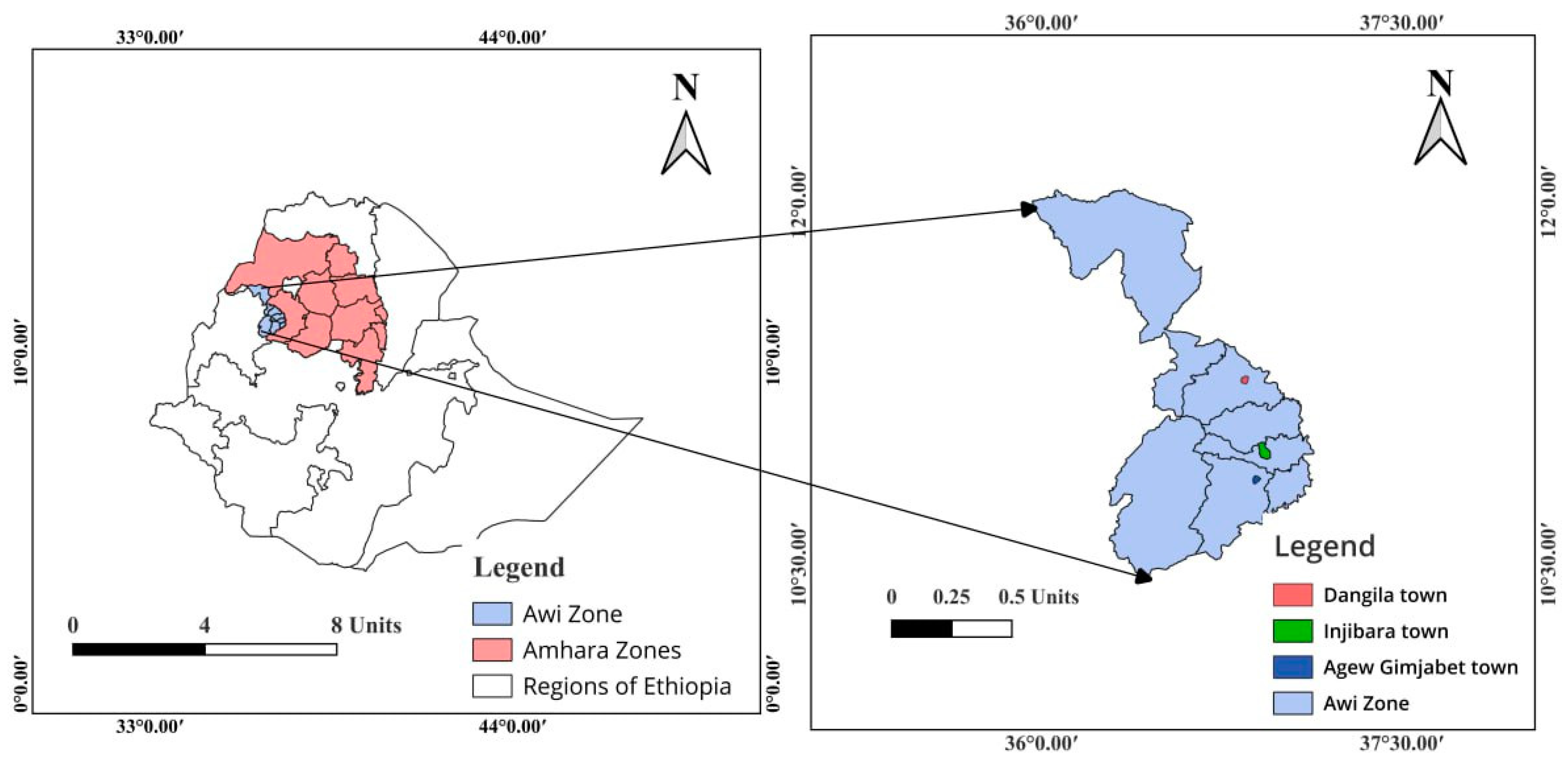
2.1. Description of the Study Area

This study was conducted in selected hospitals (Injibara General Hospital, Dangila, and Agew Gimjabet Primary Hospitals) in the administrative zone of Awi of the Amhara Regional State in Northwest Ethiopia. The global positions of Injibara, Dangila, and Agew Gimjabet on the Google Earth tool (2020) are indicated by their latitude and longitude of 10°57′ N, 36°56′ E; 11°16′ N, 36°50′ E; and 10°51′ N, 36°52′60′′ E, with an approximate elevation of 2560, 2137, and 2329 m above sea level, respectively (Figure 1).

2.2. Study Design and Subjects, and Sampling

A cross-sectional study was conducted from November 2021 to May 2022. Convenient and purposive sampling methods were used to select study sites and collect samples. Children under five years of age who visited those three hospitals with acute diarrhea were included in this study. Acute diarrhea is defined as the passage of loose or watery stools usually occurring at least three times in 24 h or three or more days and lasts no longer than 14 days [33]. However, all children who received antibiotic therapy within the previous two weeks and older than five years were excluded from this study. The questionnaires were designed to evaluate information about anamnesis of children. Approximately 25 g of diarrheal stool samples was were collected by laboratory technicians from the respective hospitals using sterile stool cups containing buffered peptone water.

2.3. Bacterial Isolation and Identification

Each diarrheal sample was directly cultured on Eosin Methylene Blue Agar (EMB) (Oxoid, Basingstoke, Hampshire, UK). Colonies showing green metallic sheen characteristics on EMB agar were considered presumptive E. coli isolates. Next, pure colonies stored in nutrient broth were further characterized for biochemical activity using biochemical tests: indole, methyl red, Vogues, Proskauer, and citrate utilization (IMViC). Bacterial isolates exhibiting an IMViC (+ + − −) pattern were confirmed as E. coli isolates [34].

2.4. DNA Extraction and Polymerase Chain Reaction

DNA from E. coli was extracted using the boiling method from overnight Tryptone Soya Broth (TSB) cultures. The Nanodrop 2000 spectrophotometer (Thermo Scientific, Wilmington, DE, USA) was used to determine the concentration and purity of DNA. The extracted DNA was stored at −20 °C and used for the molecular detection of the virulence genes. Each PCR reaction was performed in a total of 25 µL mixtures containing 0.5 µL of each primer (0.2 µM, Bioneer, Daejeon, South Korea), 2.5 µL of the PCR buffer (10X, Solis Bio Dyne, Tartu, Estonia), 1 µL of dNTP (100 mM, HiMedia, Mumbai, India), 2 µL of MgCl2 (25 mM, Solis Bio Dyne), 0.5 µL of the Taq polymerase enzyme (5 U/µL, Solis Bio Dyne), 15 µL of nuclease-free water, and 3 µL of template DNA.
PCR amplifications were performed on the A300 fast gradient thermal cycler (LongGene Scientific Instruments, Hangzhou, China). The reaction mixtures were amplified with an initial denaturation at 95 °C for 3 min., followed by 35 cycles of a denaturation step at 95 °C/40 s, annealing (Table 1)/40 s, and extension at 72 °C for 1 min, and the final extension step was at 72 °C/10 min. The PCR products were resolved using 1.5% agarose gel electrophoresis (w/v), stained with ethidium bromide in 1× TAE buffer (40 mM Tris, 20 mM acetic acid, 1 mM EDTA, pH 8.3). The size of the PCR amplicon was verified by comparison with a 100 bp DNA ladder (HiMedia, Mumbai, India). The gel result was visualized and photographed under a Gel Doc EZ Gel Documentation System (Bio-Rad, Hercules, CA, USA). The primer gene sequences, target genes, respective pathotypes of E. coli, and amplicon size in the base pairs are shown in Table 1.

2.5. Susceptibility Testing

Antimicrobial susceptibility testing (AST) was carried out using ten different antibiotics from six classes comprising fluoroquinolones [ciprofloxacin (5 µg), nalidixic acid (30 µg)]; penicillins [amoxicillin (10 µg), ampicillin (10 µg)]; aminoglycosides [gentamycin (10 µg), streptomycin (10 µg)]; folate antagonists [trimethoprim/sulfamethoxazole (25 µg), trimethoprim (5 µg)]; cephalosporins [cefotaxime (30 µg)]; and tetracyclines [tetracycline (30 µg)] (Oxoid, Hampshire, United Kingdom) on a 100 mm plate. The disk diffusion method was used for the AST of all isolates following Clinical Laboratory Standards Institute (CLSI) guidelines [44]. Mueller Hinton agar plates (HiMedia, Mumbai, India) were inoculated with a suspension of a pure culture equilibrated to 0.5 McFarland standards. The results were recorded after 24 h of incubation at 37 °C, and the diameter of the inhibition zone was interpreted as susceptible, intermediate, and resistant following the breakpoints. Isolates with nonsusceptibility to at least one agent in three or more antimicrobial categories were considered to have multidrug resistance (MDR) [45].

2.6. Data Analysis

All data were entered and analyzed using Microsoft Office Excel® 2019 (Microsoft Corporation, Redmond, WA, USA) and Graph Pad Prism (v9.5.1) (GraphPad Software, Boston, MA, USA). Descriptive statistics were computed by determining absolute frequencies along with their respective percentages. The occurrence of diarrheagenic E. coli, the detection of the virulence gene, and the extent of antibiotic resistance were compared among study subjects in selected hospitals.

3. Results

3.1. Escherichia coli Isolation

In total, 107 stool samples were collected from children with diarrhea in three selected public or government hospitals. The samples were collected from three geographically distinct areas, as follows: Injibara town (n = 43), Dangila town (n = 38), and Agew Gimjabet town (n = 26) (Table 2).
The conventional analysis revealed that E. coli was recovered in 79 (73.8%) diarrheal stool samples. E. coli isolates were more prevalent in males (51.4%) than in females (48.1%). Bacterial infection was high in the age group 25–60 months (63.3%), followed by the medium age group 7–24 months (27.8%) (Table 2). The rates for E. coli infection were 35.4%, 34.2%, and 7.6% in the groups of children fed mothers’ milk and supplemented with complementary foods, fed solid food, and exclusively breastfed, respectively. Basic information on diarrheal children and the distribution of E. coli and DEC concerning various variables is displayed in Table 2.

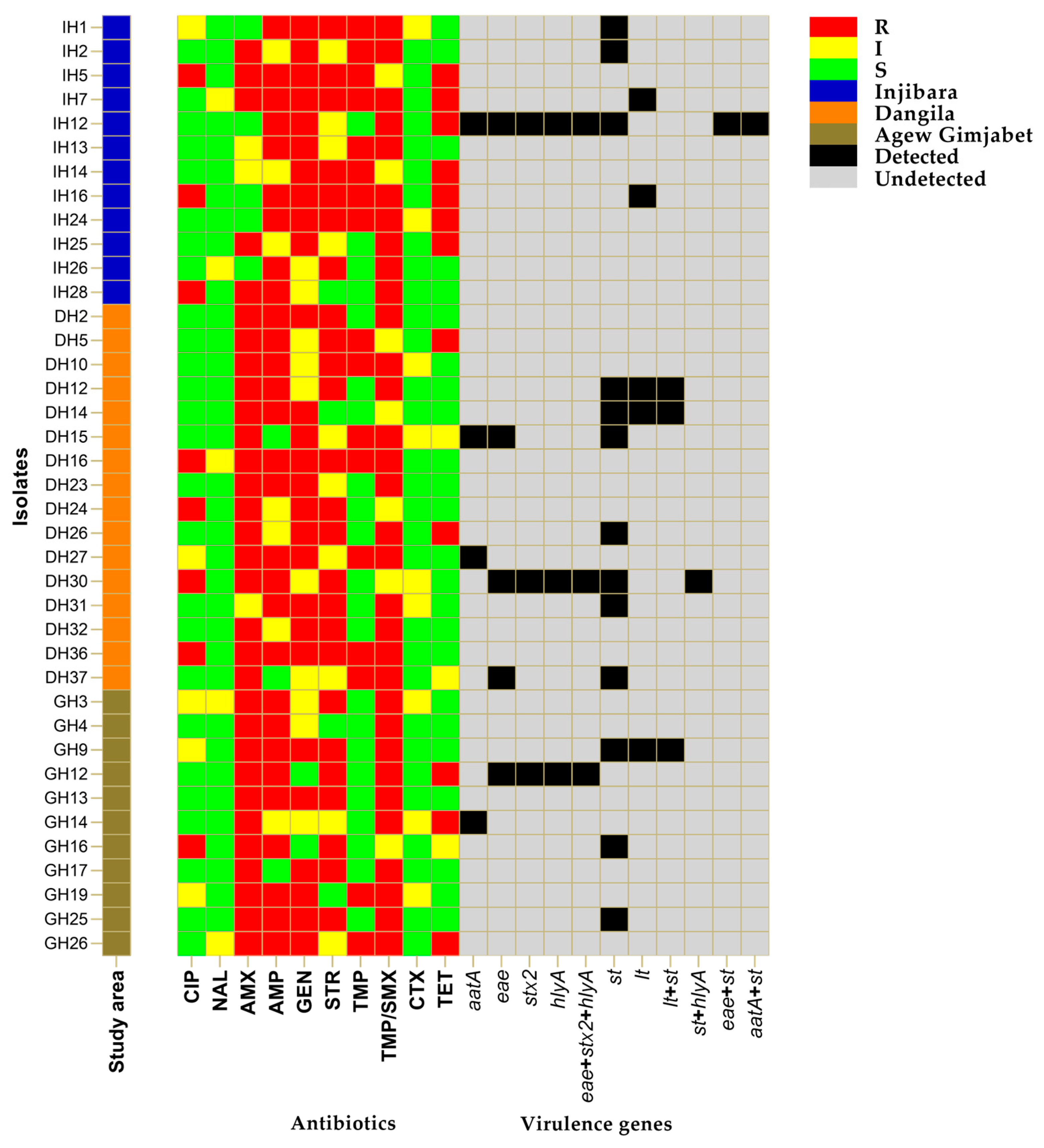
3.2. Occurrence of Virulence Genes and DEC Pathotypes

Of 79 isolates, 39 (49.4%) carried one or more putative genes associated with virulence and were confirmed as DEC. The highest detection rate was recorded in diarrhea stool samples from Dangila Primary Hospital (41%), followed by Injibara General Hospital (31%), and Agew Gimjabet Primary Hospital (28%). The detection of DEC is more frequent in male (58.9%) children who used tap water sources (61.5%) and those with a history of contact with animals (56.4%) (Table 2). The presence of the st gene was observed in 33.3% of DEC isolates, followed by the lt gene, which was detected in 12.8% of cases. Notably, 7.7% of isolates harbored both lt and st genes. Additionally, the eae and aatA genes were identified in 12.8% and 10.3% of the DEC isolates, respectively. The stx2 gene was detected in 7.7% of isolates. Heteropathotypes, characterized by the presence of a combination of various virulence genes from two pathotypes (st/hlyA, eae/st/, and aatA/st), were detected at an overall rate of 7.8%. The st gene was the most prevalent while the genes bfpA, ial, and daaE were not found in any of the isolates (Figure 2 and Figure S1).
Overall, six different DEC pathotypes were identified based on the combination of virulence genotypes: ETEC, aEPEC, EAEC, Heteropathotypes, STEC, and EHEC. Pathotype detection rates of 51.3%, 25.6%, and 23.1% were recorded at the Dangila Primary, Injibara General, and Agew Gimjabet Primary Hospitals, respectively. All pathotypes were detected in the hospitals sampled except for STEC and Heteropathotypes, which were absent at the Agew Gimjabet Primary Hospitals (Figure 2). The most common pathotype was ETEC, presented in 53.8% of cases, followed by EPEC in 12.8%. To a lesser extent, the STEC and EHEC strains were detected with an equal percentage of 7.7%. Heteropathotypes (ETEC-EHEC, aEPEC-ETEC, EAEC-ETEC) were detected in 7.8% of DEC isolates, with an equal percentage (2.6%) of gene combinations (Figure 2).

3.3. Antimicrobial Resistance in DEC Isolates

Figure 3 shows the distribution of the phenotypic antibiotic resistance profiles of DEC isolates. High levels of antibiotic resistance were found against trimethoprim/sulfamethoxazole (82.1%), amoxicillin (79.5%), ampicillin (74.4%), gentamicin (69.2%), and streptomycin (64.1%). On the other hand, DEC isolates from diarrheal children revealed a high level of sensitivity to nalidixic acid (87.2%), cefotaxime (76.9%), ciprofloxacin (66.6%), tetracycline (61.5%), and trimethoprim (56.4%) (Figure 3). Furthermore, 84.6% of the isolates exhibited MDR and showed ten antimicrobial resistance patterns. The predominant MDR pattern in DEC isolates was resistance to three antibiotic categories (84.9%), followed by resistance to four classes of antibiotics (15.1%). The less frequently observed phenotype was resistance to seven antibiotic agents (3%) (Table 3).

4. Discussion

The identification and characterization of DEC pathotypes are critical to understanding the underlying mechanisms of E. coli-related diarrheal diseases. However, the incidence and impact of DEC infections in low-resource settings, including Ethiopia, remain poorly understood primarily due to the absence of coordinated epidemiological surveillance systems and advanced laboratory services [46]. The present study identified DEC pathotypes in diarrheal children. We determined each virulence factor and antibiotic resistance profile using a combination of molecular techniques and a phenotypic analysis. Our findings revealed that 39 (49.4%) DEC possess various virulence genes, including st, lt, aatA, eae, stx2, and hlyA that enable them to cause diarrheagenic illness. The proportion of DEC detected in this study was higher than the 20.6% to 36.4% reported from Iran [47], Kenya [48], and Egypt [49]. A similar finding with a higher frequency of pathogenic E. coli was documented in Nigeria [46], Gabon [50], Iraq [51], Guinea Bissau [52], and Sudan [53].
In this study, an elevated occurrence of DEC infection was observed in children who were fed solid food in comparison to those exclusively fed mothers’ milk. The difference in the rate of infection could be attributed to the protective capacity of maternal immunity transmitted through breastfeeding [54,55]. In particular, there is a prevailing notion that breastfeeding plays a crucial role in the protection of children against E. coli infection due to the presence of antibodies specifically targeting virulence factors [56]. Observations in previous studies demonstrate that components in the milk provide protective factors that prevent STEC attachment to the intestine and block the path of pathogenesis [57]. The impact of breastfeeding on the morbidity and mortality associated with diarrhea in infants and children has been well documented in recent studies, particularly in developing countries. These studies have consistently demonstrated that partial or no breastfeeding is correlated with an increased incidence of diarrhea in young children [58]. On the contrary, the introduction of solid and complementary foods has been implicated as a source of contamination potentially to the development of intestinal infections in infants and children under five years old. Supportive findings from low-resource settings indicate that children fed with complementary foods often exhibit high levels of contamination with pathogenic microbes [59,60,61].
The intestinal tract of animals serves as a prominent natural reservoir for human DEC [62]. The frequent occurrence of DEC among children who have a history of contact with animals in the present study suggests direct or indirect contact with an animal reservoir or with their feces as the possible route of transmission for intestinal pathogenic E. coli. A comprehensive meta-analysis of demographic health survey data from 30 Sub-Saharan African countries examined the association between child health outcomes and household ownership of livestock, revealing animals as a significant risk factor for both acute enteric infections and overall mortality in children [63]. There is formative evidence from Ethiopia that underscores the pivotal role of animal feces and animal husbandry practices as an important source of the bacterial contamination and transmission pathway to infants [64]. The study of Belete et al. [65] revealed a frequent detection of DEC isolates among children who have a history of contact with diarrheic calves, highlighting the significance of these interactions.
The DEC pathotypes, specifically ETEC, aEPEC, EAEC, STEC, EHEC, and Heteropathotypes, were responsible for the diarrhea of children in the study area. ETEC was the most common pathotype detected (53.8%) among DEC. These strains are the most common cause of acute childhood diarrhea in LMICs, where hygiene standards are often deplorable [66,67]. The current result showed a higher detection rate than reports from India, with percentages of 5.7% and 6% [68,69]. In our study, 33.3% of ETEC isolates produced heat-stable toxins (STs), 12.8% produced heat-labile toxins (LTs), and 7.7% produced both enterotoxins. These findings are consistent with those of a study conducted in Kenya [70] and Indonesia [71], where ST toxins were the most common, followed by LT, and both enterotoxins were the least common. In contrast, studies from Iran [72] and Bolivia [73] found that LT toxins were detected at a higher rate than ST toxins.
aEPEC was the second most common pathotype, accounting for 12.8% of all cases. All EPEC strains detected in this study were eae-positive and thus classified as atypical EPEC. Our findings are in line with those of Borujerdi et al. [47], who reported that the EPEC strain was the second most common pathotype and only possessed eae genes. The current finding, however, contradicts reports from developed and developing countries demonstrating an increase in the detection rate of aEPEC strains over tEPEC strains [49,74]. The failure to identify the bfp gene is most often because bfp is the structural gene that encodes the bundle-forming pilus (bfp), and these fimbriae are produced only under specific cultural conditions [75].
EAEC is a significant emerging pathogen associated with persistent diarrhea in children in developing countries. It causes watery diarrhea without blood and intermittent abdominal cramps with no fever [76]. In this study, the proportion of EAEC was found to be 10.3%, which contradicts findings in Nigeria [77], China [78], and Qatar [79]. However, these findings are close to the proportions (3.8%) and (6.5%) reported by Mabika et al. [50] in Gabon and Shatub et al. [51] in Iraq. The disparity in EAEC prevalence reported in these studies may be due to differences in sample types and geographical areas.
The present study detected a low (7.7%) proportion of STEC that harbored stx2 genes. This finding agrees with a recent study in China [79], which reported a percentage of 3.7%. The results of the current study were slightly higher than the reports of Huang et al. [80], with a detection rate of 0.4%. Furthermore, Bonkoungou et al. [81] found 1% of STEC strains in human diarrheic stool samples in Ouagadougou, Burkina Faso. We detected 7.7% of EHEC strains, slightly higher than a study by Shatub et al. [51] that reported a detection rate of 4.3%. In contrast, Al-Dulaimi et al. [82] and Abbasi et al. [17] revealed a comparable prevalence of EHEC, with 7.8% and 9.3%, respectively.
Escherichia coli exhibits remarkable genomic plasticity, facilitating the emergence of new strains capable of harboring virulence genes with the characteristics of two or more DEC pathotypes. This enables E. coli to adapt to different environments and host immune responses, increasing its pathogenic potential [83]. Heteropathotypes with a combination of two virulence genes accounting for 7.8% were identified in this study. The result was higher than a study conducted in India [84], which found a detection rate of 3.8%. In other studies, hybrid pathotypes were identified among children suffering from diarrhea with a corresponding rate of 6% in Pakistan [76], 3.9% in Nigeria [85], and 14% in Ethiopia [65].
The current study has revealed distinctive distribution patterns of antimicrobial susceptibility among hospitals, suggesting the presence of varying local therapeutic practices by healthcare providers, influenced by several factors. Prescription practices play a crucial role in determining the types and frequencies of antibiotic agents used, thereby impacting the development of antibiotic resistance. The preference for antibiotic usage patterns is primarily influenced by educational qualifications, experience, sources of updated knowledge, and the practice setting of the clinician. Another significant factor that influences prescription patterns is the presence of hospital protocols and local guidelines for antibiotic use. In low-resource settings, access to appropriate diagnostic tests for pathogen identification and drug availability are identified as the major factors influencing prescription decisions [86,87].
Escherichia coli has been shown to play a significant role in the emergence of antimicrobial resistance due to its capacity to accumulate resistance genes. On the other hand, E. coli can also serve as an indicator organism to estimate the burden and trend of antimicrobial resistance in humans and animals [88,89]. Antibiotics including cotrimoxazole, amoxicillin, and cephalosporins are among the commonly used antibiotics for the treatment of bacterial diarrhea in Ethiopia [90,91]. In the present study, DEC of children with diarrhea showed higher resistance to trimethoprim/sulfamethoxazole (82.1%), amoxicillin (79.5%), ampicillin (74.4%), and gentamicin (69.2%). This finding agrees with the resistance reported recently in Kenya [48]. In Burkina Faso, Bonko et al. [92] also reported that isolates from children under five exhibited a high resistance rate to trimethoprim-sulphamethoxazole (100%), ampicillin (100%), ciprofloxacin (71.4%). Another study in the Maasai community of Kenya revealed high sensitivity to ampicillin, chloramphenicol, gentamycin, tetracycline, and trimethoprim/sulfamethoxazole [93].
Multidrug-resistant bacteria pose a significant threat to global public health. This highlighted that high resistance not only reduces treatment options but also profoundly impairs the efficacy of managing bacterial diseases. In addition, these MDR isolates can transfer antimicrobial resistance characteristics to susceptible (non-resistance) strains through genetic change. In this study, 84.6% of E. coli isolates were MDR with three or more antibiotic classes, which agree with the findings of Estrada-Garcia et al. [94]. A probable reason for such widespread resistance could be the extensive use of antibiotics in the treatment of diarrheal disease in children, often without performing antibiotic sensitivity tests or frequent prescription practices from professionals, or the acquisition of certain common antibiotic resistance genes through horizontal gene transfer from donor bacteria, phages, or free DNA [95]. This calls for further understanding of the molecular mechanisms of the development of antibiotic resistance in E. coli and other related bacteria.
In this study, we examined the phenotypic, genotypic, and antibiotic resistance profiles of DEC isolated from children under five suffering from diarrhea. The study further reported a high prevalence of multiple antibiotic-resistant diarrheagenic Escherichia coli (MRDEC) strains. These strains pose a significant threat to public health in terms of disease management difficulties and incidence of infection. The findings could provide essential information for the prevention and treatment of diarrhea in low-income settings. The limitation of this study is that results from a cross-sectional study and purposive sampling may not accurately reflect the overall prevalence and identify the primary-causing co-pathogens in children under five in the area. Further studies using case–control studies and probability sampling methods are needed to provide a more comprehensive understanding of the prevalence and causative agents of diarrheal diseases. Additionally, our research is only restricted to samples from children with diarrhea, and suggests various source samples, such as food products, environmental samples, and animal samples, to clarify the sources of contamination in the future. Further research using molecular typing methods should also explore the genetic similarities among DEC isolates and transmission dynamics in household and hospital settings.

5. Conclusions

The result of this study indicates that pathogenic E. coli play a role in child diarrhea in Northwest Ethiopia. ETEC and EPEC pathotypes were detected more frequently and can be a major source of diarrhea in low-income settings. This study revealed high rates of resistance to the class of folate antagonists, penicillins, and aminoglycosides, alongside alarming rates of MRDEC strains. Routine surveillance systems, the development of new antibiotics, and the exploration of alternative treatment strategies that emphasize antisense oligonucleotide and bacteriophage therapy technologies could offer a relevant solution to combating antibiotic-resistant Escherichia coli strains. In conclusion, continued epidemiological and genetic studies are required to better understand emerging strains of pathogenic E. coli and the risk factors and genetic mechanisms associated with antibiotic resistance.

Supplementary Materials

The following supporting information can be downloaded at: https://www.mdpi.com/article/10.3390/tropicalmed9030065/s1, Figure S1: Representative gel pictures of amplified products of E. coli virulence genes separated by 1.5% agarose gel electrophoresis and stained with ethidium bromide.

Author Contributions

B.M.M., M.A.B., T.B.D., H.T. and T.S.T. participated in the conceptualization and methodology of the study. B.M.M. carried out data collection. B.M.M. and M.A.B. performed the investigation and data analysis. T.B.D., H.T. and T.S.T. supervised and validated the study. B.M.M. and T.S.T. contributed to data curation. B.M.M., T.B.D. and H.T. drafted the original manuscript. All authors have read and agreed to the published version of the manuscript.

Funding

This research received no external funding.

Institutional Review Board Statement

The study protocol (ERB. Ref/L-477) was reviewed and approved by the Ethical Review Boards of Bahir Dar University, the College of Agriculture and Environmental Science, and the School of Animal Science and Veterinary Medicine.

Informed Consent Statement

The purpose and procedures of the study were carefully explained, and written consent was obtained from the parents of the children.

Data Availability Statement

All data generated or analyzed during this study are included in this manuscript.

Acknowledgments

The authors would like to express gratitude to all the management and staff of the respective hospitals for their necessary administrative guidance and cooperation throughout sample collection. The technical assistance of the division of the biotechnology laboratory of Bahir Dar University and Addis Ababa University is acknowledged.

Conflicts of Interest

The authors declare that they have no conflicts of interest with respect to this work.

References

  1. Croxen, M.A.; Finlay, B.B. Molecular mechanisms of Escherichia coli pathogenicity. Nat. Rev. Microbiol. 2010, 8, 26–38. [Google Scholar] [CrossRef]
  2. Lambertini, E.; Karns, J.S.; Van Kessel, J.A.S.; Cao, H.; Schukken, Y.H.; Wolfgang, D.R.; Smith, J.M.; Pradhan, A.K. Dynamics of Escherichia coli virulence factors in dairy herds and farm environments in a longitudi-nal study in the United States. Appl. Environ. Microbiol. 2015, 81, 4477–4488. [Google Scholar] [CrossRef]
  3. Kaper, J.B.; Nataro, J.P.; Mobley, H.L.T. Pathogenic Escherichia coli. Nat. Rev. Microbiol. 2004, 2, 123–140. [Google Scholar] [CrossRef]
  4. Li, D.; Shen, M.; Xu, Y.; Liu, C.; Wang, W.; Wu, J.; Luo, X.; Jia, X.; Ma, Y. Virulence gene profiles and molecular genetic characteristics of diarrheagenic Escherichia coli from a hospital in western China. Gut Pathog. 2018, 10, 35. [Google Scholar] [CrossRef]
  5. Jesser, K.J.; Levy, K. Updates on defining and detecting diarrheagenic Escherichia coli pathotypes. Curr. Opin. Infect. Dis. 2020, 33, 372–380. [Google Scholar] [CrossRef]
  6. Bendary, M.M.; El-Hamid, M.I.A.; Alhomrani, M.; Alamri, A.S.; Elshimy, R.; Mosbah, R.A.; Bahnass, M.M.; Omar, N.N.; Al-Sanea, M.M.; Elmanakhly, A.R.; et al. What Is behind the Correlation Analysis of Diarrheagenic E. coli Pathotypes? Biology 2022, 11, 1004. [Google Scholar] [CrossRef]
  7. Pasqua, M.; Michelacci, V.; Di Martino, M.L.; Tozzoli, R.; Grossi, M.; Colonna, B.; Morabito, S.; Prosseda, G. The intriguing evolutionary journey of enteroinvasive E. coli (EIEC) toward pathogenicity. Front. Microbiol. 2017, 8, 2390. [Google Scholar] [CrossRef]
  8. Rodrigues, J.F.; Lourenço, R.F.; Maeda, D.L.N.F.; Cintra, M.d.J.; Nakao, N.; Mathias-Santos, C.; Luiz, W.B.; Ferreira, L.C.d.S. Strain-specific transcriptional and posttranscriptional regulation of heat-labile toxin expression by enterotoxigenic Escherichia coli. Braz. J. Microbiol. 2020, 51, 455–465. [Google Scholar] [CrossRef] [PubMed]
  9. Wang, H.; Zhong, Z.; Luo, Y.; Cox, E.; Devriendt, B. Heat-stable enterotoxins of enterotoxigenic Escherichia coli and their impact on host immunity. Toxins 2019, 11, 24. [Google Scholar] [CrossRef] [PubMed]
  10. Joffré, E.; Rojas, V.I. Molecular epidemiology of enteroaggregative Escherichia coli (Eaec) isolates of hospitalized children from bolivia reveal high heterogeneity and multidrug-resistance. Int. J. Mol. Sci. 2020, 21, 9543. [Google Scholar] [CrossRef] [PubMed]
  11. Jafari, A.; Aslani, M.; Bouzari, S. Escherichia coli: A brief review of diarrheagenic pathotypes and their role in diarrheal diseases in Iran. Iran J. Microbiol. 2012, 4, 102–117. [Google Scholar]
  12. Kuhnert, P.; Boerlin, P.; Frey, J. Target genes for virulence assessment of Escherichia coli isolates from water, food and the environment. FEMS Microbiol. Rev. 2000, 24, 107–117. [Google Scholar] [CrossRef] [PubMed]
  13. Gomes, T.A.; Elias, W.P.; Scaletsky, I.C.; Guth, B.E.C.; Rodrigues, J.F.; Piazza, R.M.; Ferreira, L.; Martinez, M.B. Diarrheagenic Escherichia coli. Braz. J. Microbiol. 2016, 47, 3–30. [Google Scholar] [CrossRef] [PubMed]
  14. World Health Organization. Diarrhoeal Disease. 2017. Available online: https://www.who.int/news-room/fact-sheets/detail/diarrhoeal-disease (accessed on 1 March 2024).
  15. Cohen, A.L.; Platts-Mills, J.A.; Nakamura, T.; Operario, D.J.; Antoni, S.; Mwenda, J.M.; Weldegebriel, G.; Rey-Benito, G.; de Oliveira, L.H.; Ortiz, C.; et al. Aetiology and incidence of diarrhoea requiring hospitalisation in children under 5 years of age in 28 low-income and middle-income countries: Findings from the Global Pediatric Diarrhea Surveillance network. BMJ Glob. Health 2022, 7, e009548. [Google Scholar] [CrossRef] [PubMed]
  16. Demissie, G.D.; Yeshaw, Y.; Aleminew, W.; Akalu, Y. Diarrhea and associated factors among under five children in sub-Saharan Africa: Evidence from demographic and health surveys of 34 sub-Saharan countries. PLoS ONE 2021, 16, e0257522. [Google Scholar] [CrossRef] [PubMed]
  17. Abbasi, E.; Mondanizadeh, M.; van Belkum, A.; Ghaznavi-Rad, E. Multi-drug-resistant diarrheagenic Escherichia coli pathotypes in pediatric patients with gastroen-teritis from central Iran. Infect. Drug Resist. 2020, 13, 1387–1396. [Google Scholar] [CrossRef]
  18. Mernie, G.; Kloos, H.; Adane, M. Prevalence of and factors associated with acute diarrhea among children under five in rural areas in Ethiopia with and without implementation of community-led total sanitation and hygiene. BMC Pediatr. 2022, 22, 148. [Google Scholar] [CrossRef] [PubMed]
  19. Mesfin, Y.; Argaw, M. Burden of Diarrheal Disease among Under Five Children in Ethiopia, 2000–2016: Findings from the Global Health Estimates 2016. Health Sci. J. 2021, 15, 801. Available online: http://www.hsj.gr/ (accessed on 1 March 2024).
  20. Alebel, A.; Tesema, C.; Temesgen, B.; Gebrie, A.; Petrucka, P.; Kibret, G.D. Prevalence and determinants of diarrhea among under-five children in Ethiopia: A systematic review and meta-analysis. PLoS ONE 2018, 13, e0199684. [Google Scholar] [CrossRef]
  21. Global Burden of Disease Collaborative Network. Global Burden of Disease Study 2019 (GBD 2019) Results. Seattle, United States: Institute for Health Metrics and Evaluation (IHME). 2021. Available online: https://ourworldindata.org/grapher/causes-of-death-in-children-under-5?country=~ETH (accessed on 28 February 2024).
  22. Breijyeh, Z.; Jubeh, B.; Karaman, R. Resistance of gram-negative bacteria to current antibacterial agents and approaches to resolve it. Molecules 2020, 25, 1340. [Google Scholar] [CrossRef]
  23. Rousham, E.K.; Unicomb, L.; Islam, M.A. Human, animal and environmental contributors to antibiotic resistance in low-resource settings: Integrating behavioural, epidemiological and One Health approaches. Proc. R. Soc. B Biol. Sci. 2018, 285, 20180332. [Google Scholar] [CrossRef]
  24. Gemeda, B.A.; Assefa, A.; Jaleta, M.B.; Amenu, K.; Wieland, B. Antimicrobial resistance in Ethiopia: A systematic review and meta-analysis of prevalence in foods, food handlers, animals, and the environment. One Health 2021, 13, 100286. [Google Scholar] [CrossRef]
  25. Wolde, A.; Deneke, Y.; Sisay, T.; Mathewos, M.; Fesseha, H. Isolation of Escherichia coli and Its Associated Risk Factor from Diarrheic Children in Wolaita Sodo Town, Southern Ethiopia. Res. Rep. Trop. Med. 2021, 12, 227–234. [Google Scholar] [CrossRef] [PubMed]
  26. Adugna, A.; Kibret, M.; Abera, B.; Nibret, E.; Adal, M. Antibiogram of E. coli serotypes isolated from children aged under five with acute diarrhea in Bahir Dar town. Afr. Health Sci. 2015, 15, 656–664. [Google Scholar] [CrossRef] [PubMed]
  27. Zenebe, T.; Mitiku, M.; Alem, Y. Prevalence of Escherichia coli in Under-Five Children with Diarrhea in Ethiopia: A Systematic Review and Meta-Analysis. Int. J. Microbiol. 2020, 2020, 8844294. [Google Scholar] [CrossRef] [PubMed]
  28. Getaneh, D.K.; Hordofa, L.O.; Ayana, D.A.; Tessema, T.S.; Regassa, L.D. Prevalence of Escherichia coli O157:H7 and associated factors in under-five children in Eastern Ethiopia. PLoS ONE 2021, 16, e0246024. [Google Scholar] [CrossRef] [PubMed]
  29. Zelelie, T.Z.; Eguale, T.; Yitayew, B.; Abeje, D.; Alemu, A.; Seman, A.; Jass, J.; Mihret, A.; Abebe, T. Molecular epidemiology and antimicrobial susceptibility of diarrheagenic Escherichia coli isolated from children under age five with and without diarrhea in Central Ethiopia. PLoS ONE 2023, 18, e0288517. [Google Scholar] [CrossRef] [PubMed]
  30. Gessesse, D.N.; Tarekegn, A.A. Prevalence and associated factors of diarrhea among under-five children in the Jawi district, Awi Zone Ethiopia, 2019. Community based comparative cross-sectional study. Front. Pediatr. 2022, 10, 890304. [Google Scholar] [CrossRef] [PubMed]
  31. Azage, M.; Kumie, A.; Worku, A.; Bagtzoglou, A.C. Childhood diarrhea in high and low hotspot districts of Amhara Region, northwest Ethiopia: A multilevel modeling. J. Health. Popul. Nutr. 2016, 35, 13. [Google Scholar] [CrossRef] [PubMed]
  32. Hailu, B.; Ji-Guo, W.; Hailu, T. Water, Sanitation, and Hygiene Risk Factors on the Prevalence of Diarrhea among Under-Five Children in the Rural Community of Dangila District, Northwest Ethiopia. J. Trop. Med. 2021, 2021, 2688500. [Google Scholar] [CrossRef]
  33. Kosek, M.; Bern, C.; Guerrant, R.L. The global burden of diarrhoeal disease, as estimated from studies published between 1992 and 2000. Bull. World Health Organ. 2003, 81, 197–204. [Google Scholar]
  34. Quinn, P.J.; Markey, B.K.; Leonard, F.C.; Hartigan, P.; Fanning, S.; Fitzpatrick, E.S. Veterinary Microbiology and Microbial Disease, 2nd ed.; John Wiley & Sons: Hoboken, NJ, USA, 2011. [Google Scholar]
  35. Pal, A.; Ghosh, S.; Ramamurthy, T.; Yamasaki, S.; Tsukamoto, T.; Bhattacharya, S.K.; Nair, G.B.; Takeda, Y. Shiga-toxin producing Escherichia coli from healthy cattle in a semi-urban community in Calcutta, India. Indian J. Med. Res. 1999, 110, 83–85. [Google Scholar]
  36. Khan, A.; Yamasaki, S.; Sato, T.; Ramamurthy, T.; Pal, A.; Datta, S.; Chowdhury, N.R.; Das, S.C.; Sikdar, A.; Tsukamoto, T.; et al. Prevalence and Genetic Profiling of Virulence Determinants of Non-O157 Shiga Toxin-Producing Escherichia coli Isolated from Cattle, Beef, and Humans, Calcutta, India. Emerg. Infect. Dis. 2002, 8, 54–62. [Google Scholar] [CrossRef]
  37. Nada, R.A.; Shaheen, H.I.; Touni, I.; Fahmy, D.; Armstrong, A.W.; Weiner, M.; Klena, J.D. Design and validation of a multiplex polymerase chain reaction for the identification of enterotoxigenic Escherichia coli and associated colonization factor antigens. Diagn. Microbiol. Infect. Dis. 2010, 67, 134–142. [Google Scholar] [CrossRef]
  38. da Cruz, C.B.N.; de Souza, M.C.S.; Serra, P.T.; Santos, I.; Balieiro, A.; Pieri, F.A.; Nogueira, P.A.; Orlandi, P.P. Virulence factors associated with pediatric shigellosis in Brazilian Amazon. BioMed Res. Int. 2014, 2014, 539697. [Google Scholar] [CrossRef]
  39. Tornieporth, N.G.; John, J.; Salgado, K.; de Jesus, P.; Latham, E.; Melo, M.C.; Gunzburg, S.T.; Riley, L.W. Differentiation of Pathogenic Escherichia coli Strains in Brazilian Children by PCR. J. Clin. Microbiol. 1995, 33, 1371–1374. [Google Scholar] [CrossRef] [PubMed]
  40. Havt, A.; Lima, I.F.; Medeiros, P.H.; Clementino, M.A.; Santos, A.K.; Amaral, M.S.; Veras, H.N.; Prata, M.M.; Lima, N.L.; Di Moura, A.; et al. Prevalence and virulence gene profiling of enteroaggregative Escherichia coli in malnourished and nourished Brazilian children. Diagn. Microbiol. Infect. Dis. 2017, 89, 98–105. [Google Scholar] [CrossRef] [PubMed]
  41. Vidal, M.; Kruger, E.; Durán, C.; Lagos, R.; Levine, M.; Prado, V.; Toro, C.; Vidal, R. Single multiplex PCR assay to identify simultaneously the six categories of diarrheagenic Escherichia coli associated with enteric infections. J. Clin. Microbiol. 2005, 43, 5362–5365. [Google Scholar] [CrossRef] [PubMed]
  42. Gunzburg, S.T.; Tornieporth, N.G.; Riley, L.W. Riley, Identification of Enteropathogenic Escherichia coli by PCR-Based Detection of the Bun-dle-Forming Pilus Gene. J. Clin. Microbiol. 1995, 33, 1375–1377. [Google Scholar] [CrossRef]
  43. Selim, S.A.; Ahmed, S.F.; Aziz, M.H.A.; Zakaria, A.M.; Klena, J.D.; Pangallo, D. Prevalence and characterization of shiga-toxin O157:H7 and non-O157:H7 enterohemorrhagic Escherichia coli isolated from different sources. Biotechnol. Biotechnol. Equip. 2013, 27, 3834–3842. [Google Scholar] [CrossRef]
  44. Clinical and Laboratory Standards Institute (CLSI). Performance Standards for Antimicrobial Susceptibility Testing, 30th ed.; Supplement M100; Clinical and Laboratory Standards Institute: Wayne, PA, USA, 2020. [Google Scholar]
  45. Magiorakos, A.-P.; Srinivasan, A.; Carey, R.B.; Carmeli, Y.; Falagas, M.E.; Giske, C.G.; Harbarth, S.; Hindler, J.F.; Kahlmeter, G.; Olsson-Liljequist, B.; et al. Multidrug-resistant, extensively drug-resistant and pandrug-resistant bacteria: An international expert proposal for interim standard definitions for acquired resistance. Clin. Microbiol. Infect. 2012, 18, 268–281. [Google Scholar] [CrossRef]
  46. Saka, H.K.; Dabo, N.T.; Muhammad, B.; García-Soto, S.; Ugarte-Ruiz, M.; Alvarez, J. Diarrheagenic Escherichia coli Pathotypes from Children Younger Than 5 Years in Kano State, Nigeria. Front. Public Health 2019, 7, 348. [Google Scholar] [CrossRef]
  47. Broujerdi, S.M.; Ardakani, M.R.; Rezatofighi, S.E. Characterization of diarrheagenic Escherichia coli strains associated with diarrhea in children, Khouzestan, Iran. J. Infect. Dev. Ctries. 2018, 12, 649–656. [Google Scholar] [CrossRef]
  48. Webale, M.K.; Guyah, B.; Wanjala, C.; Nyanga, P.L.; Webale, S.K.; Abonyo, C.; Kitungulu, N.; Kiboi, N.; Bowen, N. Phenotypic and Genotypic Antibiotic Resistant diarrheagenic Escherichia coli pathotypes isolated from Children with Diarrhea in Nairobi City, Kenya. Ethiop. J. Health Sci. 2020, 30, 881–890. [Google Scholar] [CrossRef] [PubMed]
  49. Khairy, R.M.M.; Fathy, Z.A.; Mahrous, D.M.; Mohamed, E.S.; Abdelrahim, S.S. Prevalence, phylogeny, and antimicrobial resistance of Escherichia coli pathotypes isolated from children less than 5 years old with community acquired- diarrhea in Upper Egypt. BMC Infect. Dis. 2020, 20, 908. [Google Scholar] [CrossRef] [PubMed]
  50. Mabika, R.M.; Liabagui, S.L.O.; Moundounga, H.K.; Mounioko, F.; Souza, A.; Yala, J.F. Molecular Prevalence and Epidemiological Characteristics of Diarrheagenic E. coli in Children under 5 Years Old in the City of Koula-Moutou, East-Central Gabon. Open J. Med. Microbiol. 2021, 11, 157–175. [Google Scholar] [CrossRef]
  51. Shatub, W.T.; Jafar, N.A.-H.; Melconian, A.K. Detection of diarrheagenic E. coli among children under 5 year’s age in Tikrit city of Iraq by using single multiplex PCR technique. Plant Arch. 2021, 21, 1230–1237. [Google Scholar] [CrossRef]
  52. Mero, S.; Timonen, S.; Lääveri, T.; Løfberg, S.; Kirveskari, J.; Ursing, J.; Rombo, L.; Kofoed, P.-E.; Kantele, A. Prevalence of diarrhoeal pathogens among children under five years of age with and without diarrhoea in guinea-bissau. PLoS Neglected Trop. Dis. 2021, 15, e0009709. [Google Scholar] [CrossRef] [PubMed]
  53. Saeed, A.; Abd, H.; Sandstrom, G. Microbial aetiology of acute diarrhoea in children under five years of age in Khartoum, Sudan. J. Med. Microbiol. 2015, 64, 432–437. [Google Scholar] [CrossRef] [PubMed]
  54. Sereme, Y.; Toumi, E.; Saifi, E.; Faury, H.; Skurnik, D. Maternal immune factors involved in the prevention or facilitation of neonatal bacterial infections. Cell. Immunol. 2024, 395–396, 104796. [Google Scholar] [CrossRef]
  55. Langel, S.N.; Blasi, M.; Permar, S.R. Maternal immune protection against infectious diseases. Cell Host Microbe 2022, 30, 660–674. [Google Scholar] [CrossRef]
  56. Torres, A.G. Maternal immunity, a way to confer protection against enteropathogenic Escherichia coli. J. Pediatr. 2017, 93, 548–550. [Google Scholar] [CrossRef] [PubMed]
  57. Herrera-Insua, I.; Gomez, H.F.; Diaz-Gonzalez, V.A.; Chaturvedi, P.; Newburg, D.S.; Cleary, T.G. Human milk lipids bind Shiga toxin. Adv. Exp. Med. Biol. 2001, 501, 333–339. [Google Scholar] [PubMed]
  58. Turin, C.G.; Ochoa, T.J. The Role of Maternal Breast Milk in Preventing Infantile Diarrhea in the Developing World. Curr. Trop. Med. Rep. 2014, 1, 97–105. [Google Scholar] [CrossRef] [PubMed]
  59. Palmieri, J.R.; Meacham, S.L.; Warehime, J.; Stokes, S.A.; Ogle, J.; Leto, D.; Bax, M.; Dauer, A.M.; Lozovski, J.M. Relationships between the weaning period and the introduction of complementary foods in the transmission of gastrointestinal parasitic infections in children in Honduras. Res. Rep. Trop. Med. 2018, 9, 113–122. [Google Scholar] [CrossRef] [PubMed]
  60. Ogbo, F.A.; Nguyen, H.; Naz, S.; Agho, K.E.; Page, A. The association between infant and young child feeding practices and diarrhoea in Tanzanian children. Trop. Med. Health 2018, 46, 2. [Google Scholar] [CrossRef]
  61. Müller-Hauser, A.A.; Sobhan, S.; Huda, T.M.N.; Waid, J.L.; Wendt, A.S.; Islam, M.A.; Rahman, M.; Gabrysch, S. Key Food Hygiene Behaviors to Reduce Microbial Contamination of Complementary Foods in Rural Bangladesh. Am. J. Trop. Med. Hyg. 2022, 107, 709–719. [Google Scholar] [CrossRef] [PubMed]
  62. Lange, M.E.; Uwiera, R.R.E.; Inglis, G.D. Enteric Escherichia coli O157:H7 in Cattle, and the Use of Mice as a Model to Elucidate Key Aspects of the Host-Pathogen-Microbiota Interaction: A Review. Front. Vet. Sci. 2022, 9, 937866. [Google Scholar] [CrossRef]
  63. Kaur, M.; Graham, J.P.; Eisenberg, J.N.S. Eisenberg, Livestock ownership among rural households and child morbidity and mortality: An analysis of demographic health survey data from 30 Sub-Saharan African Countries (2005–2015). Am. J. Trop. Med. Hyg. 2017, 96, 741–748. [Google Scholar] [CrossRef]
  64. Budge, S.; Hutchings, P.; Parker, A.; Tyrrel, S.; Tulu, T.; Gizaw, M.; Garbutt, C. Do domestic animals contribute to bacterial contamination of infant transmission pathways? Formative evidence from Ethiopia. J. Water Health 2019, 17, 655–669. [Google Scholar] [CrossRef]
  65. Belete, M.A.; Demlie, T.B.; Chekole, W.S.; Tessema, T.S. Molecular identification of diarrheagenic Escherichia coli pathotypes and their antibiotic resistance patterns among diarrheic children and in contact calves in Bahir Dar city, Northwest Ethiopia. PLoS ONE 2022, 17, e0275229. [Google Scholar] [CrossRef]
  66. Vilchez, S.; Reyes, D.; Paniagua, M.; Bucardo, F.; Möllby, R.; Weintraub, A. Prevalence of diarrhoeagenic Escherichia coli in children from León, Nicaragua. J. Med. Microbiol. 2009, 58, 630–637. [Google Scholar] [CrossRef]
  67. David, E.E.; Yameen, M.A.; Igwenyi, I.O.; Okafor, A.C.; Obeten, U.N.; Obasi, D.O.; Ezeilo, U.R.; David, C.N. The frequency of virulent genes and antimicrobial resistance patterns of diarrheagenic Escherichia coli isolated from stools of children presenting with diarrhea in a tertiary hospital in Abakaliki, Nigeria. Int. J. One Health 2020, 6, 147–152. [Google Scholar] [CrossRef]
  68. Thakur, N.; Jain, S.; Changotra, H.; Shrivastava, R.; Kumar, Y.; Grover, N.; Vashistt, J. Molecular characterization of diarrheagenic Escherichia coli pathotypes: Association of virulent genes, serogroups, and antibiotic resistance among moderate-to-severe diarrhea patients. J. Clin. Lab. Anal. 2018, 32, e22388. [Google Scholar] [CrossRef]
  69. Verma, S.; Venkatesh, V.; Kumar, R.; Kashyap, S.; Kumar, M.; Maurya, A.K.; Dhole, T.N.; Singh, M. Etiological agents of diarrhea in hospitalized pediatric patients with special emphasis on diarrheagenic Escherichia coli in North India. J. Lab. Physicians 2019, 11, 068–074. [Google Scholar] [CrossRef]
  70. Kipkirui, E.; Koech, M.; Ombogo, A.; Kirera, R.; Ndonye, J.; Kipkemoi, N.; Kirui, M.; Philip, C.; Roth, A.; Flynn, A.; et al. Molecular characterization of enterotoxigenic Escherichia coli toxins and colonization factors in children under five years with acute diarrhea attending Kisii Teaching and Referral Hospital, Kenya. Trop. Dis. Travel Med. Vaccines 2021, 7, 31. [Google Scholar] [CrossRef] [PubMed]
  71. Subekti, D.; Lesmana, M.; Tjaniadi, P.; Machpud, N.; Sriwati; Sukarma; Daniel, J.; Alexander, W.; Campbell, J.; Corwin, A.; et al. Prevalence of enterotoxigenic Escherichia coli (ETEC) in hospitalized acute diarrhea patients in Denpasar, Bali, Indonesia. Diagn. Microbiol. Infect. Dis. 2003, 47, 399–405. [Google Scholar] [CrossRef] [PubMed]
  72. Haghi, F.; Zeighami, H.; Hajiahmadi, F.; Khoshvaght, H.; Bayat, M. Frequency and antimicrobial resistance of diarrhoeagenic Escherichia coli from young children in Iran. J. Med. Microbiol. 2014, 63, 427–432. [Google Scholar] [CrossRef]
  73. Rodas, C.; Mamani, R.; Blanco, J.; Blanco, J.E.; Wiklund, G.; Svennerholm, A.-M.; Sjöling, Å.; Iniguez, V. Enterotoxins, colonization factors, serotypes and antimicrobial resistance of enterotoxigenic Escherichia coli (ETEC) strains isolated from hospitalized children with diarrhea in Bolivia. Braz. J. Infect. Dis. 2011, 15, 132–137. [Google Scholar] [CrossRef]
  74. Ochoa, T.J.; Contreras, C.A. Enteropathogenic Escherichia coli infection in children. Curr. Opin. Infect. Dis. 2011, 24, 478–483. [Google Scholar] [CrossRef]
  75. Nguyen, T.V.; Le Van, P.; Le Huy, C.; Gia, K.N.; Weintraub, A. Detection and characterization of diarrheagenic Escherichia coli from young children in Hanoi, Vi-etnam. J. Clin. Microbiol. 2005, 43, 755–760. [Google Scholar] [CrossRef] [PubMed]
  76. Zil-E-Huma, Z.-E.; Tareen, A.M.; Ullah, K.; Asmat, T.M.; Samad, A.; Iqbal, A.; Mustafa, M.Z.; Ahmad, I.; Rahman, S.U. Incidence of diarrheagenic Escherichia coli pathotypes in children suffering from diarrhea in Ter-tiary Care Hospitals, Quetta, Pakistan. Pak. J. Zool. 2019, 51, 2015–2021. [Google Scholar] [CrossRef]
  77. Onanuga, A.; Igbeneghu, O.; Lamikanra, A. A study of the prevalence of diarrhoeagenic Escherichia coli in children from Gwagwalada, Federal Capital Territory, Nigeria. Pan Afr. Med. J. 2014, 17, 146. [Google Scholar] [CrossRef] [PubMed]
  78. Zhou, Y.; Zhu, X.; Hou, H.; Lu, Y.; Yu, J.; Mao, L.; Mao, L.; Sun, Z. Characteristics of diarrheagenic Escherichia coli among children under 5 years of age with acute diarrhea: A hospital based study. BMC Infect. Dis. 2018, 18, 63. [Google Scholar] [CrossRef] [PubMed]
  79. Eltai, N.O.; Al Thani, A.A.; Al Hadidi, S.H.; Al Ansari, K.; Yassine, H.M. Antibiotic resistance and virulence patterns of pathogenic Escherichia coli strains associated with acute gastroenteritis among children in Qatar. BMC Microbiol. 2020, 20, 54. [Google Scholar] [CrossRef] [PubMed]
  80. Huang, F.; Deng, Y.; Qu, M.; Liu, G.R.; Liu, Y.; Zhang, X.; Li, J.; Yan, H.Q.; Gao, Z.Y.; Liu, B.W.; et al. Etiological surveillance and analysis of infectious diarrhea in Beijing in year 2010. Chin. J. Prev. Med. 2011, 45, 820–824. [Google Scholar] [CrossRef]
  81. Bonkoungou, I.J.O.; Somda, N.S.; Traoré, O.; Zoma, S.; Garba, Z.; Drabo, K.M.; Barro, N. Detection of diarrheagenic Escherichia coli in human diarrheic stool and drinking water samples in Ouagadougou, Burkina Faso. J. Infect. Dis. 2021, 15, 53–58. [Google Scholar] [CrossRef]
  82. Al-Dulaimi, T.H.; Aziz, H.W.; Al-Marzoqi, A.H.; Al-Aziz, S.A.; Mohsin, S.A.A. Molecular Characterization and Antibiotic Susceptibility of Diarrheagenic Escherichia coli from Children. Med. J. Babylon 2015, 12, 541–550. [Google Scholar]
  83. Braz, V.S.; Melchior, K.; Moreira, C.G. Escherichia coli as a Multifaceted Pathogenic and Versatile Bacterium. Front. Cell. Infect. Microbiol. 2020, 10, 548492. [Google Scholar] [CrossRef]
  84. Rajendran, P.; Ajjampur, S.S.R.; Chidambaram, D.; Chandrabose, G.; Thangaraj, B.; Sarkar, R.; Samuel, P.; Rajan, D.P.; Kang, G. Pathotypes of diarrheagenic Escherichia coli in children attending a tertiary care hospital in South India. Diagn. Microbiol. Infect. Dis. 2010, 68, 117–122. [Google Scholar] [CrossRef]
  85. Ogunbiyi, T.S.; Fayemi, O.E.; Akanni, G.B.; Ayolabi, C.I.; Hald, T. Molecular Characterization of Hetero-Pathogenic and Diarrheagenic Escherichia coli Pathotypes in Diarrheic Children under Five Years and Exposure Environment in Ogun State, South-West Nigeria. Pathogens 2023, 12, 1358. [Google Scholar] [CrossRef]
  86. Wiboonchutikula, C.; Bin Kim, H.; Honda, H.; Loo, A.Y.X.; Cheng, V.C.-C.; Camins, B.; Jantarathaneewat, K.; Apisarnthanarak, P.; Rutjanawech, S.; Apisarnthanarak, A. Antibiotic prescribing behavior among physicians in Asia: A multinational survey. Antimicrob. Steward. Health Epidemiol. 2023, 3, e112. [Google Scholar] [CrossRef] [PubMed]
  87. Chem, E.D.; Anong, D.N.; Akoachere, J.-F.K.T. Prescribing patterns and associated factors of antibiotic prescription in primary health care facilities of Kumbo East and Kumbo West Health Districts, North West Cameroon. PLoS ONE 2018, 13, e0193353. [Google Scholar] [CrossRef]
  88. Poirel, L.; Madec, J.-Y.; Lupo, A.; Schink, A.-K.; Kieffer, N.; Nordmann, P.; Schwarz, S. Antimicrobial Resistance in Escherichia coli. Microbiol. Spectr. 2018, 6, 1–27. [Google Scholar] [CrossRef] [PubMed]
  89. Leekitcharoenphon, P.; Johansson, M.H.K.; Munk, P.; Malorny, B.; Skarżyńska, M.; Wadepohl, K.; Moyano, G.; Hesp, A.; Veldman, K.T.; Bossers, A.; et al. Genomic evolution of antimicrobial resistance in Escherichia coli. Sci. Rep. 2021, 11, 15108. [Google Scholar] [CrossRef] [PubMed]
  90. Addis, G.T.; Dagnew, S.B.; Anagaw, A.; Ayele, T.M.; Tadesse, T.Y. Evaluation of antibiotic utilization pattern in the treatment of acute diarrheal diseases at Debre Tabor comprehensive specialized hospital, Debre Tabor, Ethiopia: A retrospective cross-sectional study. Heliyon 2023, 9, e18049. [Google Scholar] [CrossRef] [PubMed]
  91. Tekleab, A.M.; Asfaw, Y.M.; Weldetsadik, A.Y.; Amaru, G.M. Antibiotic prescribing practice in the management of cough or diarrhea among children attending hospitals in Addis Ababa: A cross-sectional study. Pediatr. Health Med. Ther. 2017, 8, 93–98. [Google Scholar] [CrossRef] [PubMed]
  92. Bonko, M.D.A.; Tahita, M.C.; Kiemde, F.; Lompo, P.; Yougbaré, S.; Some, A.M.; Tinto, H.; Mens, P.F.; Menting, S.; Schallig, H.D.F.H. Antibiotic susceptibility profile of bacterial isolates from febrile children under 5 years of age in Nanoro, Burkina Faso. Trop. Med. Int. Health 2021, 26, 1220–1230. [Google Scholar] [CrossRef] [PubMed]
  93. Sang, W.K.; Kariuki, S.M.; Schnabel, D.; Boga, H.I.; Waiyaki, P.G.; Wamae, C.N. Antibiotic susceptibility of Enteric pathogens from the Maasai community, Narok and Kajiado Districts, Kenya. Afr. J. Health Sci. 2011, 19, 70–75. [Google Scholar]
  94. Estrada-García, T.; Cerna, J.F.; Paheco-Gil, L.; Velázquez, R.F.; Ochoa, T.J.; Torres, J.; DuPont, H.L. Drug-resistant Diarrheogenic Escherichia coli, Mexico. Emerg. Infect. Dis. 2005, 11, 1306–1308. [Google Scholar] [CrossRef]
  95. Gupta, S.; Shek, A.; Shrivastava, S.; Verma, A.K. Isolation, Identification, Molecular Characterization and Antibiogram of E. coli Isolates from Neonatal Calves. Int. J. Curr. Microbiol. Appl. Sci. 2019, 8, 1996–2007. [Google Scholar] [CrossRef]
Figure 1. Map of the study area. The map was created by using Esri’s ArcGIS® 10.8 desktop GIS software.
Tropicalmed 09 00065 g001
Figure 2. Frequency of virulent genes and diarrheagenic E. coli pathotypes detected in children with diarrhea and hospitals. IGH: Injibara General Hospital, DPH: Dangila Primary Hospital, AGPH: Agew Gimjabet Primary Hospital, eae: attaching–effacing gene, stx2: Shiga-like toxin II gene, hlyA: E. coli hemolysin gene, aatA: aggregative adherence gene, lt: heat-liable toxin gene, st: heat-stable toxin gene.
Tropicalmed 09 00065 g002
Figure 3. Heat map showing the distribution of antimicrobial susceptibility and virulence profiles in 39 pathogenic E. coli isolates exhibited from samples of diarrheic children. CIP: ciprofloxacin, NAL: nalidixic acid, AMX: amoxicillin, AMP: ampicillin, GEN: gentamicin, STR: streptomycin, TMP: trimethoprim, TMP/SMX: trimethoprim/sulfamethoxazole, CTX: cefotaxime, TET: tetracycline, eae: attaching–effacing gene, stx2: Shiga-like toxin II gene, hlyA: E. coli hemolysin gene, aatA: aggregative adherence gene, lt: heat-liable toxin gene, st: heat-stable toxin gene.
Tropicalmed 09 00065 g003
Table 1. Oligonucleotide primers used in single-plex PCR for amplification of diarrheagenic virulence genes.
PrimerNucleotide Sequence (5′ to 3′)Target GenePathotypesAnnealing Temperature Amplicon Size (bp)Reference
EVT1
EVT2
F: CAACACTGGATGATCTCAGC
R: CCCCCTCAACTGCTAATA
stx2STEC/EHEC55 °C350[35]
EAE-1
EAE-2
F: AAACAGGTGAAACTGTTGCC
R: CTCTGCAGATTAACCTCTGC
eaeEPEC/EHEC55 °C490[36]
ST1
ST2
F: TTT ATT TCT GTA TTG TCT T
R: GCAGGATTACAACACAATTC
stETEC55 °C294[37]
IAL F
IAL R
F: CTGGATGGTATGGTGAGG
R: GGAGGCCAACAACATTATTTCC
ialEIEC55 °C320[38]
LT1
LT2
F: GGCGACAGATTATACCGTGC
R: CCGAATTCTGTTATATATGTC
ltETEC48 °C696[39]
EAEC F
EAEC R
F: CTGGCGAAAGACTGTATCAT
R: CAATGTATAGAAATCCGCTGTT
aatAEAEC48 °C630[40]
daaE1
daaE2
F: GAACGTTGGTTAATGTGGGGT
R: TATTCACCGGTCGGTTATCAG
daaEDAEC47 °C542[41]
BFPF
BFPR
F: AATGGTGCTTGCGCTTGCTGC
R: GCCGCTTTATCCAACCTGGTA
bfpAEPEC57 °C324[42]
EHEC F
EHEC R
F: ACGATGTGGTTTATTCTGGA
R: CTTCACGTCACCATACATAT
hlyAEHEC45 °C167[43]
Table 2. Background information of 107 diarrheal children and the occurrence of DEC with different factors in three public hospitals in the Awi zone, Ethiopia.
VariablesNo. of Tested (N = 107)Culture-Positive (N = 79)PCR-Positive (DEC) (N = 39)Total
IGH
n = 43
DPH
n = 38
AGPH
n = 26
IGH
n = 32
DPH
n = 29
AGPH
n = 18
IGH
n = 12
DPH
n = 16
AGPH
n = 11
Totalt
n = 107
Totalc
n = 79
Totalp
n = 39
Sex
 Female20 (38.5)18 (34.6)14 (26.9)15 (39.5)17 (44.7)6 (15.8)5 (31.3)8 (50)3 (18.8)52 (48.6)38 (48.1)16 (41)
 Male23 (41.8)20 (36.4)12 (21.8)17 (41.5)12 (29.3)12 (29.3)7 (30.4)8 (34.8)8 (34.8)55 (51.4)41 (51.9)23 (58.9)
Age class
 0–6 months6 (37.5)4 (25)6 (37.5)3 (42.8)2 (28.6)2 (28.6)2 (66.6)01 (33.3)16 (14.9)7 (8.9)3 (7.7)
 7–24 months20 (58.8)12 (72.8)2 (5.8)15 (68.2)7 (31.8)03 (33.3)6 (66.6)034 (31.8)22 (27.8)9 (23.1)
 25–60 months17 (29.8)22 (38.6)18 (31.6)14 (28)20 (40)16 (32)7 (25.9)10 (37)10 (37)57 (53.3)50 (63.3)27 (69.2)
Feeding
 Mother’s milk4 (36.4)4 (36.4)3 (27.3)2 (33.3)3 (50)1 (16.6)2 (66.6)1 (33.3)011 (10.3)6 (7.6)3 (7.7)
 Breast + complementary23 (63.8)8 (22.2)5 (13.8)18 (64.3)6 (21.4)4 (14.3)7 (43.8)4 (25)3 (18.8)36 (33.6)28 (35.4)16 (41)
 Solid food16 (26.6)26 (43.3)18 (30)12 (44.4)20 (74.1)13 (48.1)3 (13.6)11 (50)8 (36.4)60 (56.1)27 (34.2)22 (56.4)
Source of water
 Tap water31 (42.5)22 (30.1)20 (27.4)22 (42.3)16 (30.8)14 (26.9)8 (33.3)7 (29.2)9 (37.5)73 (68.2)52 (65.8)24 (61.5)
 Well10 (34.5)14 (48.3)5 (17.2)9 (37.5)11 (45.8)4 (16.6)2 (16.6)8 (66.6)2 (16.6)29 (27.1)24 (30.4)12 (30.8)
 Boiled2 (40)2 (40)1 (20)1 (33.3)2 (66.6)02 (66.6)1 (33.3)05 (4.7)3 (3.8)3 (7.7)
Contact with animals
 Yes26 (45.6)16 (28.1)15 (26.3)19 (46.3)12 (29.3)10 (21.4)8 (36.4)8 (36.4)6 (27.3)57 (53.3)41 (51.9)22 (56.4)
 No17 (34)22 (44)11 (22)13 (34.2)17 (44.7)8 (21.1)4 (23.5)8 (47)5 (29.4)50 (46.8)38 (48.1)17 (43.6)
IGH: Injibara General Hospital, DPH: Dangila Primary Hospital, AGPH: Agew Gimjabet Primary Hospital, DEC: diarrheagenic Escherichia coli, Totalt: total tested, Totalc: total of culture-positive, Totalp: total of PCR-positive.
Table 3. Phenotypic multiple-antibiotic resistance in diarrheagenic Escherichia coli.
Pattern No.Antibiotic-Resistant PatternsNo. of Antibiotics (Classes)MDR Isolates
n (%)
1 *GEN, AMX2 (2)4 *
2 *AMX, TMP/SMX2 (2)2 *
3AMP, TMP/SMX, STR3 (3)4 (12.1)
4TET, AMX, TMP/SMX3 (3)3 (9.1)
5TMP, TET, GEN, TMP/SMX4 (3)6 (18.2)
6GEN, AMX, TMP/SMX, STR4 (3)3 (9.1)
7AMP, AMX, TMP/SMX, STR4 (3)4 (12.1)
8TET, AMP, GEN, AMX, STR5 (3)3 (9.1)
9TMP, AMP, AMX, GEN, STR5 (3)2 (6.1)
10TMP, TET, AMP, GEN, AMX, STR6 (4)4 (12.1)
11TMP, AMP, GEN, AMX, TMP/SMX, STR6 (3)3 (9.1)
12TMP, TET, AMP, GEN, AMX, TMP/SMX, STR7 (4)1 (3)
MDR: multidrug-resistant, AMX: amoxicillin, AMP: ampicillin, GEN: gentamicin, STR: streptomycin, TMP: trimethoprim, TMP/SMX: trimethoprim/sulfamethoxazole, TET: tetracycline, * non-multidrug-resistant.
Disclaimer/Publisher’s Note: The statements, opinions and data contained in all publications are solely those of the individual author(s) and contributor(s) and not of MDPI and/or the editor(s). MDPI and/or the editor(s) disclaim responsibility for any injury to people or property resulting from any ideas, methods, instructions or products referred to in the content.

