Article

Detection of Crimean–Congo Haemorrhagic Fever Virus from Livestock Ticks in Northern, Central and Southern Senegal in 2021

1
Pôle de Zoologie Médicale, Institut Pasteur de Dakar, Dakar 220, Senegal
2
Pôle de Virologie, Institut Pasteur de Dakar, Dakar 220, Senegal
3
St. Petersburg Pasteur Institute, Federal Service for Consumer Rights Protection and Human Well-Being Surveillance, St. Petersburg 190013, Russia
*
Author to whom correspondence should be addressed.
Trop. Med. Infect. Dis. 2023, 8(6), 317; https://doi.org/10.3390/tropicalmed8060317
Submission received: 27 April 2023 / Revised: 5 June 2023 / Accepted: 7 June 2023 / Published: 12 June 2023
(This article belongs to the Section Vector-Borne Diseases)

Abstract

:
Crimean–Congo haemorrhagic fever virus (CCHFV) occurs sporadically in Senegal, with a few human cases each year. This active circulation of CCHFV motivated this study which investigated different localities of Senegal to determine the diversity of tick species, tick infestation rates in livestock and livestock infections with CCHFV. The samples were collected in July 2021 from cattle, sheep and goats in different locations in Senegal. Tick samples were identified and pooled by species and sex for CCHFV detection via RT-PCR. A total of 6135 ticks belonging to 11 species and 4 genera were collected. The genus Hyalomma was the most abundant (54%), followed by Amblyomma (36.54%), Rhipicephalus (8.67%) and Boophilus (0.75%). The prevalence of tick infestation was 92%, 55% and 13% in cattle, sheep and goats, respectively. Crimean–Congo haemorrhagic fever virus (CCHFV) was detected in 54/1956 of the tested pools. The infection rate was higher in ticks collected from sheep (0.42/1000 infected ticks) than those from cattle (0.13/1000), while all ticks collected from goats were negative. This study confirmed the active circulation of CCHFV in ticks in Senegal and highlights their role in the maintenance of CCHFV. It is imperative to take effective measures to control tick infestation in livestock to prevent future CCHFV infections in humans.

1. Introduction

Ticks are the first and second most important vectors of animal and human diseases, respectively [1]. In the context of global and environmental changes, many tropical and subtropical countries are facing major public health crises related to the emergence or re-emergence of tick-borne diseases. These phenomena are explained by the expansion into new geographical areas suitable for the development of tick vectors and/or reservoir hosts [2]. Factors contributing to the emergence or re-emergence of zoonoses include adaptation to the host’s immune system, rapid spread through livestock and migratory bird movements, and widespread invasion of natural habitats by humans through their economic activities (agriculture, mining, fruit or honey collection, road construction, and extensive livestock farming) [3]. In response to this situation, several studies have been conducted to determine the diversity of ticks and the viruses they transmit, with a focus on the Crimean–Congo haemorrhagic fever virus (CCHFV) [4].
Crimean–Congo haemorrhagic fever (CCHF) is a disease caused by a tick-borne virus of the order Bunyavirales, family Nairoviridae and genus Orthonairovirus [5]. It has the widest geographic distribution of all tick-borne viral diseases, being found in more than 30 countries in Africa, Asia, Eastern Europe and the Middle East [6]. Crimean–Congo haemorrhagic fever virus circulates mainly in rural areas. As with all vector-borne diseases, the presence and persistence of zoonotic outbreaks of CCHF depend on the biological and ecological relationships between three types of organisms: the virus, the tick and the vertebrate hosts [7]. Although CCHFV has been isolated from several species of hard ticks (Ixodidae), the primary vector group appears to be ticks of the genus Hyalomma [8].
Tick bites remain the primary mode of transmission of CCHFV to humans and animals. However, human infections can also occur via direct contact with infected blood, fluids or other tissues of animals or patients during the acute phase of the disease. Animals are asymptomatic reservoirs of the virus. However, infection in humans can cause a non-specific febrile illness characterised by fever, myalgia, diarrhoea, nausea and vomiting after a short incubation period (1–5 days). In some cases, symptoms may progress to severe haemorrhagic disease [9].
In Senegal, evidence of CCHFV circulation has been reported since 1960 [10]. Between 2003 and 2020, nine confirmed cases and one death were recorded in different regions (Matam, Thiès, Saint-Louis, Kaolack, Fatick, and Tambacounda) [11]. More recently, eight confirmed cases and two deaths (between 2021 and 2022) were reported (unpublished data). Epidemiological and environmental data indicate that the northern regions of Senegal are at the highest risk of CCHF outbreaks [12]. This may be due to their ecological parameters and proximity to Mauritania where the CCHF epidemic is recurrent [13,14]. Indeed, the most recent strains/isolates were detected in Northern Senegal in the Matam region. Additionally, genetic characterisation showed that some of these strains belong to the same group as a strain isolated in Mauritania in 1984.
The present study was initiated and conducted in this national context of active CCHFV circulation in several localities in Senegal. The main objective was to investigate emerging and re-emerging tick-borne viruses circulating in Senegal. More specifically, the aim was to create an inventory of the diversity of ticks infesting livestock (cattle, goats and sheep) but also to estimate the infection rate of CCHFV in ticks in several localities in the north, centre and south of Senegal where CCHF cases have been reported.

2. Materials and Methods

2.1. Collection Sites

The study was conducted in different geographical regions (Table 1) from the north to the south of the country (Figure 1). The climate is subtropical, with a long dry season (approximately mid-October to mid-June in the north and early November to mid-May in the south) and a rainy season (approximately, late June to early October in the north, and late May to late October in the south). The north of the country (Nabadji, Haere Lao, Tatki, Tessekere, Kassack and Saint-Louis) is considered a semi-desert area with an annual rainfall of about 300 mm [15]. In these areas, the main economic activity of the populations is nomadic pastoralism with livestock including cattle, sheep and goats. The long dry seasons cause people and livestock to move from the north to the south of the country, where water and pasture are abundant after the rainy season. The other sites are located in the centre of the country (Bandia), with wooded savannah, and in the south (Kolda), a forested area characterised by an annual rainfall of up to 1000 mm. These collection sites were chosen because of the high prevalence of ticks on livestock and the isolation of CCHFV from ticks collected in these areas in the past.

2.2. Tick Sampling

Sampling was carried out from 1 to 14 July 2021. We visited 24 herds with a total of 720 animals, including 240 cattle, 240 sheep and 240 goats kept under semi-extensive conditions. The animals were kept in cattle sheds and sheepfolds at night, and during the day they were turned out onto pastures around the villages and drink from the watering point. An average of 30 animals were randomly selected from each farm during each visit. Tick collections were carried out on domestic animals. All ticks, irrespective of their stage of development (adults, nymphs, or larvae) were collected via extirpation using hard forceps from the entire body of the animal (ear, head, neck, back, fanon, abdomen, anogenital region, tail and feet). The tick was pulled firmly by the hypostome to avoid damage that could make species identification difficult. Once collected, the ticks were placed in 2 mL microfuge tubes labelled with the date and location of collection, ID number, sex of animal and site of tick attachment. All tubes were then stored in liquid nitrogen at −180 °C until treatment.

2.3. Identification and Processing of Tick Samples

In the laboratory, the ticks were washed with sterile water and then identified on a chill table under a binocular stereo-zoom microscope using appropriate morphological characteristics [16,17]. After identification, the ticks were grouped into monospecific pools of 1 to 28 individuals according to sex, date, collection site and feeding status. Each pool was triturated in 0.5 or 2 mL of L15 medium (10% foetal bovine serum, 100 U/mL penicillin, 100 μg/mL streptomycin, and 1 μL/mL amphotericin B) under BSL-3 laboratory conditions using a sterile bead homogeniser (Tissue Lyser II—QIAGEN, Hilden, Germany), depending on size. The tick homogenates were centrifuged at 2500 rpm for 5 min at 4 °C and the supernatants were stored at −80 °C until use.

2.4. Detection of CCHFV by RT-PCR

RNA was extracted from the tick supernatant using QIAamp RNA Viral kit (Qiagen GmbH, Heiden, Germany) according to the manufacturer’s recommendations. The RNA was eluted in 60 μL of AVE buffer and stored at −80 °C until use. Detection of CCHFV was performed using the AmpliSens CCHFV-FRT PCR kit (Amplisens, Bratislava 47, Slovak Republic) according to the manufacturer’s recommendations. A total of 10 μL of RNA was added to a 15 μL reaction mixture consisting of 12.5 μL of the buffer, 4 μL of nuclease-free water, 1 μL of each primer, 0.5 μL of the probe and 1 μL of the enzyme. The qRT- PCR was performed on CFX96 (Biorad, Biorad Laboratories, Marnes-La-Coquette, France). The cycling conditions were 50 °C for 30 min and 95 °C for 15 min, followed by 5 cycles of 95 °C for 10 s, 54 °C for 30 s and 72 °C for 15 s and finally 45 cycles of 95 °C for 10 s, 50 °C for 30 s (where signal acquisition was performed) and 72 °C for 15 s. The signal of the CCHFV cDNA amplification product was detected in the channel for the JOE fluorophore.

2.5. Data Analysis

The number of ticks collected from animals was recorded by collection date, sampling location and animal species in a 2016 Excel spreadsheet. The relative abundance for a sampling location was calculated by dividing the number of ticks collected at that site by the total number of ticks sampled. For each tick species, the relative abundance was calculated as the ratio of the total number of ticks collected from a vertebrate host of that species to the total number of species collected from all hosts inspected at a given site. The tick infestation prevalence (IP) was the ratio of the total number of infested animals to the total number of animals examined [18]. The X2 test was used to compare IP and relative abundance values. The biodiversity of the species collected at the different sites was estimated using the following indices.
One index is species richness, which is the total number of species in a sample. The other is the Shannon–Weaver index [19], which quantifies the number of species collected and their relative abundance, and measures the heterogeneity of an environment or a study period. It is calculated using the following formula:
  H = i = 1 S   p i   ln p i
where H′ is the Shannon–Weaver diversity index and pi is the proportion of species i compared to the total number of species (S) in the study area (or species richness of the area), which is calculated as follows:
p (i) = ni/N
where ni is the number of individuals of species i and N is the total population (n individuals of all species). The Pooled InfRate Version 4.0 software was used to calculate and compare viral infection rates (IRs), or more precisely to perform viral RNA detection from tick pools [20]. The infection rates were calculated taking into account the number of pools tested, the number of ticks in each pool and the number of positive pools. Infection rates were reported per 1000 ticks for ease of interpretation. A pair of IRs was considered not to be significantly different (p ≥ 0.05) if the confidence interval of the difference included zero. The correlations between tick attachment sites and each of the three hosts were assessed using principal component analysis (PCA). The R software [21] (version 4.1.3) was used for statistical analyses and graphical presentations of the data.

3. Results

3.1. Tick Infestation Prevalence in Vertebrate Hosts

A total of 90 animals (30 cattle, 30 sheep and 30 goats) were examined at each site (Table 2). The total IP was 53 ± 0.03% (383/720). The tick infestation prevalence was significantly higher in cattle (IP = 92 ± 0.03%; 220/240) than in sheep (IP = 55 ± 0.1%; 131/240) and goats (IP = 13 ± 0.05%; 32/240) (p ≤ 0.05). A comparison between the latter two hosts showed that sheep were significantly more infested than goats were (X2 = 89.217, df = 1, p ≤ 0.05).
In the localities of Tatki (IP = 70%), Haere Lao (IP = 61%), Saint-Louis (IP = 60%), Tessekere (IP = 52%) and Bandia (IP = 51%), more than half of the inspected animals were infested with ticks. In the other localities, the IPs were below 50% (48% in both Kassack and Kolda and 36% in Nabadji). In all the localities visited, the cattle were more affected than the sheep and goats (p ≤ 0.05), except in Tatki and Kassack where the IPs were comparable between cattle and sheep (p ≤ 0.05). Goats were the least infested hosts in these different localities, except in Nabadji and Saint-Louis where they were as infested as sheep were.

3.2. Specific Richness and Diversity

Table 3 shows the specific richness and diversity of the tick population collected from the different localities. The specific richness was higher in Kolda (10 species), followed by Kassack and Saint-Louis (8 species each), Bandia and Tessekere (7 species each), Haere Lao (6 species), Nabadji (5 species), and Tatki (4 species). The Shannon index indicates that the site of Saint-Louis had a higher species diversity (H = 1.68).

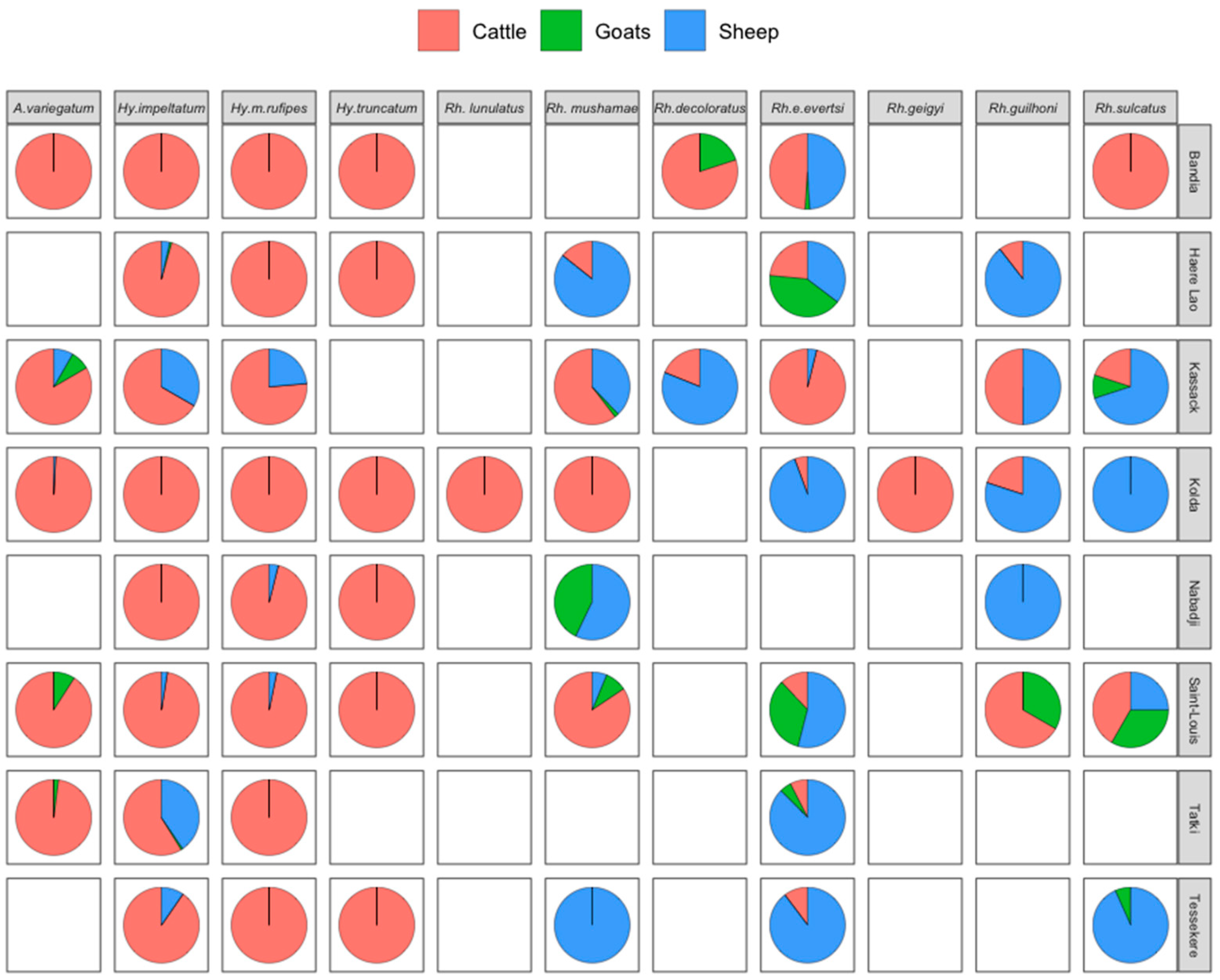
3.3. Relative Abundance of Tick Species by Host and Locality

A total of 5216, 848 and 71 ticks were collected from cattle, sheep and goats, respectively. This population included 4317 males (70.37%), 1792 females (29.2%) and 26 nymphs (0.6%) belonging to four genera (Hyalomma, Amblyomma, Rhipicephalus and Boophilus) and eleven species collected from the 720 animals that were examined (Figure 2).
The genus Hyalomma was the most abundant (53.95% of the sample) and was present in all surveyed areas. Within this genus, three species were recorded, Hy. impeltatum, Hy. marginatum rufipes and Hy. Truncatum, representing 57.91%, 29.33% and 12.74%, respectively. Hy. impeltatum and Hy. marginatum rufipes were present in all study areas, while Hy. truncatum was absent in Kassack and Tatki. Hy. truncatum was collected only from cattle, while Hy. impeltatum and Hy. marginatum rufipes were present on all vertebrate hosts inspected, although mainly on cattle (Figure 2). The highest number of Hy. impeltatum was recorded in Tatki, while Hy. marginatum rufipes was mainly collected in Bandia.
The genus Amblyomma was represented by a single species (A. variegatum) and was the second most abundant genus (36.79% of the sample). This species was present at five collection sites and was collected from all three hosts, but mainly from cattle. The highest abundance of this species was recorded in Kolda (81.64%) and Bandia (64.05%).
Five species of the genus Rhipicephalus were collected, representing 8.51% of the tick sample. This genus was present at all collection sites. The different species recorded were Rh. evertsi evertsi (46.93% of this genus), Rh. muhsamae (29.31%), Rh. guilhoni (10.54%), Rh. sulcatus (8.43%) and Rh. lunulatus (4.79%). The latter was found only in Kolda and only in cattle.
The genus Boophilus was the least abundant, representing only 0.75% of the total sample. Two species were recorded in this genus during the study: B. decoloratus (91.3% of this genus) and B. geigyi (8.7%). The latter was present only in Kolda and was collected from cattle, while B. decoloratus was present in two areas (Kassack and Bandia) and was collected from all hosts, but mainly from cattle.

3.4. Tick Attachment Sites on Hosts

Figure 3 shows the tick species collected according to their hosts and their preferred attachment sites. The anogenital region was the most infested attachment site regardless of the host. The smaller the angle formed by two attachment sites, the higher the probability of finding the same species on these sites. In fact, the species found in the anogenital region were also found on the abdomen and feet in cattle, whereas in sheep the species found in the anogenital region were found on the abdomen, back and tail. This correlation was not observed in goats due to the small number of tick species collected from these hosts. The preferred secondary sites of tick attachment were the abdomen, tail and feet in cattle, ears, abdomen and neck in goats and ears and abdomen in sheep.

3.5. Detection of CCHFV in Ticks

Of the 1956 pools tested, CCHFV was detected in 33 pools of the Hyalomma genus, 14 pools of the Amblyomma genus, 6 pools of the Rhipicephalus genus and 1 pool of the Boophilus genus.
CCHFV was detected in ticks collected from cattle and sheep. All pools of ticks collected from goats were negative for CCHFV. Hyalomma impeltatum accounted for 44.4% of the positive pools, followed by A. variegatum (25.9%) and Rh. evertsi evertsi and Hy. marginatum rufipes (11.1% for each species). Hyalomma truncatum and B. decoloratus represented less than 6% of the positive pools.
The overall IR of CCHFV in ticks was 0.15/1000 infected ticks (Table 4). The IR was higher in ticks collected from sheep (0.42/1000 infected ticks) than from cattle (0.13/1000 infected ticks) (p ≤ 0.05). The virus was more frequently detected in Hyalomma tick species, but the IR was higher in B. decoloratus (IR = 6.46/1000 ticks), followed by Rh. evertsi evertsi (3.47/1000 ticks), Hy. truncatum (0.26/1000 ticks), Hy. marginatum rufipes (0.25/1000 ticks), Hy. impeltatum (0.2372/1000 ticks) and A. variegatum (0.17/1000 ticks). The infection rates in the north (Kassack, Haere Lao, Saint-Louis, Tessekere and Tatki), centre (Bandia) and south (Kolda) were 0.58/1000 ticks, 0.41/1000 ticks and 0.142/1000, respectively. These rates were significantly higher in the north than in the centre and south (p ≤ 0.05). In the northern zone, the locality of Kassack had a higher infection rate (1.032/1000) compared to that in Tessekere (0.635/1000), Haere Lao (0.0483/1000), Saint-Louis (0.355/1000) and Tatki (0,223/1000) (p ≤ 0.05). CCHFV was not detected in any of the tick samples collected in the locality of Nabadji.
Our results showed that species of different genera (Hy. m. rufipes vs. Rh. evertsi evertsi or A. variegatum vs. Hy. truncatum) or male and female ticks of the same species collected from the same host can be infected with CCHFV.

4. Discussion

The main objective of this study was to investigate emerging and re-emerging tick-borne viruses circulating in Senegal. The Crimean–Congo haemorrhagic fever virus was detected in different tick species infesting cattle and sheep in seven of the eight localities visited. The prevalence of tick infestation in livestock was 53.19%. Cattle were the most tick-infested hosts (92%), which could be explained by their less hairy skin, which is easier for ticks to attach themselves to, but also by their long-distance movements compared to those of small ruminants. They are therefore more likely to be exposed to different ecological zones suitable for the development of ticks and thereby become infested. The high infestation rate of ovine hosts compared to that of caprine hosts has been described in previous studies of tick infestation in livestock in Senegal and Pakistan [22,23]. The relatively low level of parasitism in goats could be explained by an aversion towards goats, as the quality of their fur is not favourable for tick attachment [24].
Although the Kolda site was richer in species, Saint-Louis was more diverse, as shown by the Shannon index. This phenomenon could be explained by the presence of species (more common in the north) in the Kolda area due to transhumance. In fact, the period of tick collection coincided with the “Tabaski” festival, when the animals are transported to the south of the country to be sold. A new report of a tick species in an area could be linked to transhumance and new ecological conditions favourable to the establishment of this species. This is the case for the Hy. impeltatum that was present in the Kolda area where it was absent during a study carried out by Gueye et al. (1989) [25]. Hyalomma impeltatum is thought to be native to the northern zone of West Africa, which is its natural habitat [26].
The diversity of 11 species in 4 genera was reported in our collections. Previous studies on livestock in the same areas showed the same high diversity with four additional species (Hy. dromadarii, Hy. Impressum, Rh. cuspitatus and Rh. senegalensis) [22,27,28]. These missing species in our study could have been related to the shorter sampling period and smaller number of sites visited compared to the longer sampling period (15 months) and the greater number of sites visited during the previous study, which allowed for a greater chance of collecting more species. Climate-related ecological changes (high temperature associated with low humidity and sparse rainfall) may also contribute to species decline [29].
In agreement with previous observations [22,27,28], our results confirm the absence of B. decoloratus in the south and B. geigyi and Rh. lunulatus in the north and centre of the country. The absence of B. decoloratus in the south of the country shows that its range coincides with the Sahelian xerophilic steppe and the northern Sudanian savannah in West Africa [28]. This species is replaced by B. geigyi in the South Sudanian and Guinean savannah of West Africa.
Cattle were infested by all tick species, mainly by species of the genus Hyalomma and Amblyomma. This trophic preference for this host may be due to its availability, the adaptation of these tick species through their morphological characteristics (the length of the hypostome) and the phenomenon of aggregation [29].
Sheep were infested by all tick species except Hy. truncatum and B. geigyi. Hyalomma impeltatum was the most abundant species collected from sheep, followed by Rh. evertsi evertsi. Thus, sheep seem to be the secondary host of Hy. impeltatum after cattle. However, sheep may be the primary host of Rh. evertsi evertsi given the higher infestation rate compared to that in cattle and goats.
As with sheep, all tick species collected from cattle were found in goats, with the exception of Hy. m. rufipes, Hy. truncatum and B. geigyi. Goats were mainly infested with Rh. evertsi evertsi. The low infestation rate by other tick species is probably due to the restricted movements of goats in the domestic environment [27]. Indeed, the presence of this species on small ruminants could indicate a domestic cycle for Rh. everstsi evertsi. These results are in agreement with the data obtained by Gueye et al. (1987) [28] in the Niayes area. In fact, their results showed a high infestation rate of Rh. evertsi evertsi in calves in permanent housing, whereas in the same study, only one Rh. evertsi evertsi tick was collected from native cattle in the field.
The ano-genital region was the most infested attachment site regardless of host species. Secondary preferred tick sites were the abdomen, tail and feet in cattle, the abdomen, neck and ears in goats, and the abdomen and ears in sheep. Most of the ticks collected were found on sites with little fur and thinner skin. The high tick infestation in these areas may be due to ticks’ preference for warm, moist, hidden, well-vascularised and thin sites [30,31].
Of the 11 tick species identified in this study, 6 (Hy. impeltatum, Hy. marginatum rufipes, Hy. truncatum, B. decoloratus, A. variegatum and Rh. evertsi evertsi) were naturally infected with CCHFV. These species have been found to be already naturally infected with the virus in Senegal. According to Camicas and colleagues [32], four species of these ticks can be considered major vectors of CCHFV in Senegal, namely Hy. m rufipes, Hy. truncatum, Rh. evertsi evertsi and A. variegatum. In addition, the role of vectors in the maintenance and transmission cycles of CCHFV in Senegal has been studied experimentally [33,34]. This study showed infection rates of 100% in Hy. marginatum rufipes and Hy. truncatum (one of the vectors of CCHFV in humans), 60% in A. variegatum and 100% for Rh. evertsi evertsi females after an incubation period of 15 days post-inoculation [34]. Given its high anthropophilic rate, A. variegatum may play an important role in human infections, often resulting in mild symptoms in areas where the tick is present [2]. Although the Rh. evertsi evertsi species is an inefficient vector from an epidemiological point of view, its role during epizootic periods should not be underestimated [34].
The CCHFV infection rates were higher in B. decoloratus than those in other species. However, its low density reflects its low involvement in the ecology of CCHFV [32].
In this study, CCHFV was most frequently detected in tick species of the genus Hyalomma. Hyalomma impeltatum had a relatively low infection rate compared to other tick species of the same genus. This species, although found to be naturally associated with the virus in other studies in Senegal, was removed from the list of major CCHFV vectors by Camicas and colleagues [32] because of its low epidemiological importance. However, an experimental study suggested its possible role as a vector after the detection of high levels (63%) of the CCHF viral antigen in adult ticks emerging from virus-exposed larvae [35]. Therefore, this species should be closely monitored to determine its potential epidemiological role.
The risk of CCHFV infection was higher in ticks collected from northern localities than in Central and Southern Senegal. This confirms that the northern localities of Senegal have the highest exposure to CCHFV. These results are in agreement with those of previous studies carried out in Senegal [12,13,36]. This high infection rate could be explained by the high relative abundance of the main CCHFV vectors (Hy. m. rufipes and Hy. truncatum) in the north, but also by the geographical proximity of this area to Mauritania, where the virus often circulates in a quasi-endemic form [37].
Our results showed that infection rates were higher in sheep than in cattle, which were more heavily tick-infested. These results are similar to the data obtained in Pakistan by the authors of [38,39]. Domestic ruminants, especially sheep, may play an important role in virus amplification as they become viremic in about one week [6], allowing the virus to infect more ticks during this period.
Our study showed that different species of ticks, as well as the same species of ticks of different sexes collected from a single animal host, were infected with CCHFV. These infections may be due to different modes of transmission of CCHFV, including co-feeding, transovarial and trans-stadial transmission [32,40,41]. Once infected, ticks remain positive for CCHFV throughout their lives [42]. Therefore, parallel blood sampling of livestock for the detection of CCHFV would be necessary to better understand the modes of virus transmission.
This study confirms the active circulation of CCHFV in ticks collected from livestock in different locations in Senegal, highlighting their role in the maintenance of CCHFV. Effective measures, including the development of a tick vaccine to control tick infestation in livestock, are urgently needed to prevent future CCHFV infections in humans.

Author Contributions

Conceptualisation, M.D., V.G.D. and A.B.; formal analysis, A.B., M.D., B.D. and I.D. (Ibrahim Dia); investigation, A.B., A.G., M.M. and E.H.N.; methodology, A.B., A.G. and E.H.N.; project administration, M.D. and V.G.D.; resources, M.N., I.D. (Idrissa Dieng), O.F. and M.M.; software, A.B., B.D. and I.D. (Ibrahim Dia); supervision, M.D. and A.G.; validation, M.D., A.B. and A.G.; visualisation, A.B., A.G. and M.D.; writing—original draft, A.B., B.D. and M.D.; Writing—review and editing, A.B., M.N., A.G., B.D., E.H.N., I.D. (Ibrahim Dia), A.S.D., M.M., V.G.D., O.F., M.D. and I.D. (Idrissa Dieng); funding acquisition, M.D. and V.G.D. All authors have read and agreed to the published version of the manuscript.

Funding

This research was supported the by the Russian Science Foundation, grant no. 20-64-46014.

Institutional Review Board Statement

Not applicable.

Informed Consent Statement

Not applicable.

Data Availability Statement

All data included in this work are provided in tables and figures.

Acknowledgments

The authors would like to thank the veterinarians of the different health districts visited and the farmers for their technical assistance and collaboration.

Conflicts of Interest

The authors declare no conflict of interest.

References

  1. Duvallet, G.; Fontenille, D.; Robert, V. Entomologie Médicale et Vétérinaire; IRD Éditions: Marseille, France, 2018; Available online: http://books.openedition.org/irdeditions/21923 (accessed on 20 December 2022).
  2. Sylla, M.; Molez, J.-F.; Cornet, J.-P.; Camicas, J.-L.; Pourrut, X. Variabilité climatique et répartition de la fièvre hémorragique de Crimée-Congo et de la cowdriose, maladies à tiques au Sénégal. Acarologia 2008, 48, 155–161. [Google Scholar]
  3. Sonenshine, D.E. Range Expansion of Tick Disease Vectors in North America: Implications for Spread of Tick-Borne Disease. Int. J. Environ. Res. Public Health 2018, 15, 478. [Google Scholar] [CrossRef] [PubMed] [Green Version]
  4. Fakoorziba, M.R.; Golmohammadi, P.; Moradzadeh, R.; Moemenbellah-Fard, M.D.; Azizi, K.; Davari, B.; Alipour, H.; Ahmadnia, S.; Chinikar, S. Reverse Transcription PCR-Based Detection of Crimean-Congo Hemorrhagic Fever Virus Isolated from Ticks of Domestic Ruminants in Kurdistan Province of Iran. Vector-Borne Zoonotic Dis. 2012, 12, 794–799. [Google Scholar] [CrossRef]
  5. Whitehouse, C.A. Crimean-Congo hemorrhagic fever. Antivir. Res. 2004, 64, 145–160. [Google Scholar] [CrossRef] [PubMed]
  6. Ergönül, O. Crimean-Congo haemorrhagic fever. Lancet Infect Dis. 2006, 6, 203–214. [Google Scholar] [CrossRef] [PubMed]
  7. Papa, A.; Velo, E.; Papadimitriou, E.; Cahani, G.; Kota, M.; Bino, S. Ecology of the Crimean-Congo hemorrhagic fever endemic area in Albania. Vector Borne Zoonotic Dis. 2009, 9, 713–716. [Google Scholar] [CrossRef]
  8. Nabeth, P.; Cheikh, D.O.; Lo, B.; Faye, O.; Vall, I.O.M.; Niang, M.; Wague, B.; Diop, D.; Diallo, M.; Diallo, B. Crimean-Congohemorrhagic fever, Mauritania. Emerg. Infect. Dis. 2004, 10, 2143–2149. [Google Scholar] [CrossRef] [PubMed]
  9. Bente, D.A.; Forrester, N.L.; Watts, D.M.; McAuley, A.J.; Whitehouse, C.A.; Bray, M. Crimean-Congo hemorrhagic fever: History, epidemiology, pathogenesis, clinical syndrome and genetic diversity. Antivir. Res. 2013, 100, 159–189. [Google Scholar] [CrossRef] [Green Version]
  10. Chunikhin, S.; Chumakov, M.; Butenko, A.; Smirnova, S.E.; Taufflieb, R.; Camicas, J.L.; Robin, Y.; Cornet, M.; Shabon, Z. Results from investigating human and domestic and wild animal blood sera in the Senegal Republic (Western Africa) for antibodies to crimean hemorrhagic fever virus. Nauchn Sess Polio Virus Entsefalitov Mosc. 1969, 2, 158–160. [Google Scholar]
  11. Temur, A.I.; Kuhn, J.H.; Pecor, D.B.; Apanaskevich, D.A.; Keshtkar-Jahromi, M. Epidemiology of Crimean-Congo Hemorrhagic Fever (CCHF) in Africa—Underestimated for Decades. Am. J. Trop. Med. Hyg. 2021, 104, 1978–1990. [Google Scholar] [CrossRef]
  12. Wilson, M.L.; Gonzalez, J.-P.; LeGuenno, B.; Cornet, J.-P.; Guillaud, M.; Calvo, M.-A.; Digoutte, J.-P.; Camicas, J.-L. Epidemiology of Crimean-Congo hemorrhagic fever in Senegal: Temporal and spatial patterns. In Hemorrhagic Fever with Renal Syndrome Tick- and Mosquito-Borne Viruses; Calisher, C.H., Ed.; Springer: Vienna, Austria, 1990; pp. 323–340. Available online: http://link.springer.com/10.1007/978-3-7091-9091-3_35 (accessed on 10 November 2022).
  13. Dieng, I.; Barry, M.A.; Diagne, M.M.; Diop, B.; Ndiaye, M.; Faye, M.; Ndione, M.H.D.; Dieng, M.M.; Bousso, A.; Fall, G.; et al. Detection of Crimean Congo haemorrhagic fever virus in North-eastern Senegal, Bokidiawé 2019. Emerg. Microbes Infect. 2020, 9, 2485–2487. [Google Scholar] [CrossRef] [PubMed]
  14. Mhamadi, M.; Badji, A.; Dieng, I.; Gaye, A.; Ndiaye, E.H.; Ndiaye, M.; Mhamadi, M.; Toure, C.T.; Barry, A.; Ndiaye, O.; et al. Multiple genotypes of Crimean-Congo hemorrhagic fever virus detected in ticks during a one health survey in Agnam, Northeastern Senegal. Emerg. Microbes Infect. 2022, 11, 2711–2714. [Google Scholar] [CrossRef]
  15. ANSD/RGPHAE-Sénégal. 2019. Agence National de la Statistique et de la Démographie. Available online: http://www.ansd.sn/index.php (accessed on 10 November 2022).
  16. Hoogstraal, H. African Ixodoidea. Ticks of le Soudan (avec Référence Spéciale à la Province d’Équatoria et avec Examens Préliminaires pf les Genres Boophilus, Margaropus, et Hyalomma); Report No, 005 050.29.07; Departement of Medical Zoology: Caire, Egypt, 1956; p. 1101. Available online: https://www.biodiversitylibrary.org/part/97784 (accessed on 18 October 2022).
  17. Walker, A.R.; Bouattour, A.; Camicas, J.-L.; Estrada-Pena, A.; Horak, I.G.; Latif, A.A.; Pegram, R.G.; Preston, P.M. Ticks of Domestic Animals in Africa: A Guide to Identification of Species; Bioscience Reports: Edinburgh, Scotland, 2003. [Google Scholar]
  18. Bush, A.O.; Lafferty, K.D.; Lotz, J.M.; Shostak, A. Parasitology Meets Ecology on Its Own Terms: Margolis et al. Revisited. J. Parasitol. 1997, 83, 575–583. [Google Scholar] [CrossRef]
  19. Nolan, K.; Callahan, J. Beachcomber Biology: The Shannon-Weiner Species Diversity Index. Proc. Workshop ABLE 2006, 27, 334–338. [Google Scholar]
  20. Biggerstaff, B. PooledInRate, Version 4.0: An Excel® Add-In to Compute Infection Rates from Pooled Data. Centers for Disease Control and Prevention Fort Collins: Colorado. 2016. Available online: https://github.com/CDCgov/PooledInfRate (accessed on 1 April 2023).
  21. R Core Team. R: A Language and Environment for Statistical Computing; R Foundation for Statistical Computing: Vienna, Austria, 2016; Volume 145, Available online: https://www.r-project.org/ (accessed on 1 April 2023).
  22. Gueye, A.; Mbengue, M.; Diouf, A. Tiques et hémoparasitoses du bétail au Sénégal. VI. La zone soudano-sahélienne. Rev. D’élevage Médecine Vétérinaire Pays Trop. 1994, 47, 39–46. [Google Scholar] [CrossRef]
  23. Ramzan, M.; Naeem-Ullah, U.; Abbas, H.; Adnan, M.; Rasheed, Z.; Khan, S. Diversity of hard ticks in goats and sheep in Multan, Punjab, Pakistan. Int. J. Agric. Biol. Res. 2019, 35, 7–9. [Google Scholar]
  24. Khan, S.S.; Ahmed, H.; Afzal, M.S.; Khan, M.R.; Birtles, R.J.; Oliver, J.D. Epidemiology, Distribution and Identification of Ticks on Livestock in Pakistan. Int. J. Environ. Res. Public Health 2022, 19, 3024. [Google Scholar] [CrossRef]
  25. Gueye, A.; Mbengue, M.; Diouf, A. Tiques et hémoparasitoses du bétail au Sénégal. IV. La zone Sud-soudanienne. Rev. D’élevage Médecine Vétérinaire Pays Trop. 1989, 42, 517–528. [Google Scholar]
  26. Morel, P.-C. Contribution à la connaissance de la distribution des tiques (acariens, Ixodidae et Amblyomnisdae), en Afrique ethiopienne continentale. Ph.D Thesis, Université de Paris-Sud, Paris, France, 1969. Available online: https://agritrop.cirad.fr/355829/ (accessed on 10 November 2022).
  27. Gueye, A.; Mbengue, M.; Diouf, A.; Sonko, M.L. Tiques et hémoparasitoses du bétail au Sénégal. V. La zone Nord-guinéenne. Rev. D’élevage Médecine Vétérinaire Pays Trop. 1993, 46, 551–561. [Google Scholar] [CrossRef]
  28. Gueye, A.; Camicas, J.-L.; Diouf, A.; Mbengue, M. Tiques et hémoparasitoses du bétail au Sénégal. II. La zone sahélienne. Rev. D’élevage Médecine Vétérinaire Pays Trop. 1987, 40, 119–125. [Google Scholar]
  29. Boulanger, N.; McCoy, K.D. Tiques et Maladies à Tiques: Biologie, Ecologie Evolutive, Épidémiologie; IRD Éditions: Marseille, France, 2016; Volume 1, 669p. [Google Scholar]
  30. Stachurski, F. Invasion of West African cattle by the tick Amblyomma variegatum. Med. Vet. Èntomol. 2000, 14, 391–399. [Google Scholar] [CrossRef] [PubMed]
  31. Muchenje, V.; Dzama, K.; Chimonyo, M.; Raats, J.G.; Strydom, P. Tick susceptibility and its effects on growth performance and carcass characteristics of Nguni, Bonsmara and Angus steers raised on natural pasture. Animal 2008, 2, 298–304. [Google Scholar] [CrossRef] [PubMed] [Green Version]
  32. Camicas, J.L.; Cornet, J.-P.; Wilson, M.L.; Adam, F.; Zeller, H.G. La fièvre hémorragique de Crimée-Congo au Sénégal: Dernières données sur l’écologie du virus CCHF- fdi:41933-Horizon. Bull. Société Pathol. Exot. 1994, 87, 11–16. [Google Scholar]
  33. Faye, O.; Cornet, J.P.; Camicas, J.L.; Fontenille, D.; Gonzalez, J.P. Experimental transmission of Crimean-Congo hemorrhagic fever virus: Role of 3 vector species in the maintenance and transmission cycles in Senegal. Parasite 1999, 6, 27–32. [Google Scholar] [CrossRef] [PubMed] [Green Version]
  34. Faye, O.; Fontenille, D.; Thonnon, J.; Gonzalez, J.P.; Cornet, J.P.; Camicas, J.L. Experimental transmission of Crimean-Congo hemorrhagic fever virus by Rhipicephalus evertsi evertsi (Acarina: Ixodidae). Bull. Soc. Pathol. Exot. 1999, 92, 143–147. [Google Scholar] [PubMed]
  35. Dohm, D.J.; Logan, T.M.; Linthicum, K.J.; Rossi, C.A.; Turell, M.J. Transmission of Crimean-Congo Hemorrhagic Fever Virus by Hyalomma impeltatum (Acari: Ixodidae) after Experimental Infection. J. Med. Entomol. 1996, 33, 848–851. [Google Scholar] [CrossRef]
  36. Mhamadi, M.; Badji, A.; Dieng, I.; Gaye, A.; Ndiaye, E.H.; Ndiaye, M.; Mhamadi, M.; Touré, C.T.; Mbaye, M.R.; Barry, M.A.; et al. Crimean–Congo Hemorrhagic Fever Virus Survey in Humans, Ticks, and Livestock in Agnam (Northeastern Senegal) from February 2021 to March 2022. Trop. Med. Infect. Dis. 2022, 7, 324. [Google Scholar] [CrossRef]
  37. Boushab, B.M.; Kelly, M.; Kébé, H.; Bollahi, M.A.; Basco, L.K. Crimean-Congo Hemorrhagic Fever, Mauritania. Emerg. Infect. Dis. 2020, 26, 817–818. [Google Scholar] [CrossRef]
  38. Kasi, K.K.; von Arnim, F.; Schulz, A.; Rehman, A.; Chudhary, A.; Oneeb, M.; Sas, M.A.; Jamil, T.; Maksimov, P.; Sauter-Louis, C.; et al. Crimean-Congo haemorrhagic fever virus in ticks collected from livestock in Balochistan, Pakistan. Transbound. Emerg. Dis. 2020, 67, 1543–1552. [Google Scholar] [CrossRef] [Green Version]
  39. Shahid, M.F.; Yaqub, T.; Ali, M.; Ul-Rahman, A.; Bente, A.D. Prevalence and phylogenetic analysis of Crimean-Congo hemorrhagic fever virus in ticks collected from Punjab province of Pakistan. Acta Trop. 2021, 218, 105892. [Google Scholar] [CrossRef]
  40. Gonzalez, J.P.; Camicas, J.L.; Cornet, J.P.; Faye, O.; Wilson, M.L. Sexual and transovarian transmission of Crimean-Congo haemorrhagic fever virus in Hyalomma truncatum ticks. Res. Virol. 1992, 143, 23–28. [Google Scholar] [CrossRef] [PubMed]
  41. Logan, T.M.; Watts, D.M.; Linthicum, K.J.; Moulton, J.R.; Bailey, C.L. Experimental Transmission of Crimean-Congo Hemorrhagic Fever Virus by Hyalomma Truncatum Koch. Am. J. Trop. Med. Hyg. 1989, 40, 207–212. [Google Scholar] [CrossRef] [PubMed]
  42. Gargili, A.; Estrada-Peña, A.; Spengler, J.R.; Lukashev, A.; Nuttall, P.A.; Bente, D.A. The role of ticks in the maintenance and transmission of Crimean-Congo hemorrhagic fever virus: A review of published field and laboratory studies. Antivir. Res. 2017, 144, 93–119. [Google Scholar] [CrossRef] [PubMed]
Figure 1. Map showing tick sampling sites in Senegal, July 2021.
Tropicalmed 08 00317 g001
Figure 2. Relative abundance of tick species collected according to host and collection site, July 2021, Senegal.
Tropicalmed 08 00317 g002
Figure 3. Principal component analysis (PCA) showing tick attachment sites according to species and hosts, July 2021, Senegal.
Tropicalmed 08 00317 g003
Table 1. Geographic coordinates of the different collection sites, Senegal, July 2021.
LocalitiesLatitude NLongitude W
Nabadji15°43′44.78″13°24′09.76″
Haere Lao16°23′59.71″14°19′26.91″
Tatki16°06′49.36″15°16′19.28″
Tessekere15°48′55.50″15°08′09.88″
Kassack16°25′29.52″15°59′03.81″
Saint-Louis15°51′58.27″16°29′15.26″
Bandia14°35′19.13″17°01′06.43″
Kolda12°53′02.54″14°56′19.88″
Table 2. Tick infestation prevalence in livestock in different localities of Senegal, July 2021.
LocalitiesHostTotal
CattleGoatsSheep
PIIP (%)PIIP (%)PIIP (%)PIIP (%)
Nabadji3023773041330517903235
Haere Lao303010030413302170905561
Tatki302893305173030100906370
Tessekere3028933000301963904752
Kassack30227330413301757904348
Saint-louis302997301343301240905460
Bandia30301003027301447904651
Kolda30301003000301343904348
Total2402209224032132401315572038353
P: prospected; I: infected; IP: tick infestation prevalence.
Table 3. Species richness and Shannon diversity index of tick populations in the different localities of Senegal, July 2021.
LocalityFemaleMaleNymphAbundance TotalRelative Abundance (RA%)Shannon (H′)Specific Richness (S)
Bandia41112193163326.61.0507
Haere Lao14933604857.911.2406
Kassack7415092333.801.5708
Kolda373100313138922.60.7410
Nabadji10818302914.740.6805
Saint-Louis15238405368.741.6808
Tatki4608901135122.00.4904
Tessekere6515202173.541.3707
Table 4. Crimean–Congo haemorrhagic fever virus infection rate by tick species, location and host, July 2021, Senegal.
LocalitiesSpeciesHostIR95% CITOTAL (95% CI)
BandiaA. variegatumCattle0.09(0.05–0.17)0.14 (0.09–0.22)
Hy.m rufipesCattle0.18(0.03–0.58)
Hy. truncatumCattle0.25(0.01–1.21)
Rh. evertsi evertsiCattle16.3(3.04–52.91)
Rh. evertsi evertsisheep7.46(1.36–24.41)
KoldaA. variegatumCattle0.03(0.01–0.08)0.04 (0.01–0.09)
Hy. truncatumCattle0.21(0.01–0.99)
Haere LaoHy. impeltatumCattle0.36(0.09–0.98)0.48 (0.19–1.00)
Hy. impeltatumSheep107(8.20–422.82)
Hy.m rufipesCattle0.49(0.09–1.62)
KassackB. decoloratusCattle35.2(2.19–158.44)1.03 (0.19–3.33)
Hy. impeltatumSheep1000(206.55–1000)
Saint-LouisHy. impeltatumCattle0.50(0.09–1.65)0.35 (0.02–1.94)
Hy.m rufipesSheep1000(206.55–1000)
Hy. truncatumCattle0.39(0.02–1.94)
TatkiHy. impeltatumCattle0.22(0.12–0.37)0.22 (0.14–0.34)
Hy. impeltatumSheep1.16(0.05–0.39)
Rh. evertsi evertsiSheep6.17(1.13–20.17)
TessekereHy. impeltatumCattle0.42(0.02–2.03)0.63 (0.11–2.08)
Hy.m rufipesCattle1.49(0.09–7.28)
Total speciesB. decoloratus6.46(0.38–30.72)0.15 (0.11–0.19)
A. variegatum0.06(0.03–0.10)
Hy. impeltatum0.24(0.16–0.35)
Hy. m rufipes0.25(0.10–0.53)
Hy. truncatum0.260.07–0.71)
Rh. evertsi evertsi3.47(1.44–7.15)
Total hostCattle0.13(0.09–0.17)
Sheep0.42(0.22–0.72)
IR: infection rate; 95% CI: 95% confidence interval.
Disclaimer/Publisher’s Note: The statements, opinions and data contained in all publications are solely those of the individual author(s) and contributor(s) and not of MDPI and/or the editor(s). MDPI and/or the editor(s) disclaim responsibility for any injury to people or property resulting from any ideas, methods, instructions or products referred to in the content.

