Article

Assessment of an Immuno-Diagnostic Method for Hookworm-Related Cutaneous Larva Migrans Using Crude Extracts of Ancylostoma caninum

by
Sitthithana Adam
1,
Paron Dekumyoy
1,
Duangporn Nacapunchai
2,
Thawatchai Ketboonlue
1,
Prakaykaew Charunwatthana
3,
Jittima Dhitavat
3,
Khuanchai Koompapong
4,
Putza Chonsawat
5 and
Dorn Watthanakulpanich
1,*
1
Department of Helminthology, Faculty of Tropical Medicine, Mahidol University, 420/6 Ratchawithi Road, Ratchathewi, Bangkok 10400, Thailand
2
College of Allied Health Sciences, Suan Sunandha Rajabhat University, Bangkok 10300, Thailand
3
Department of Clinical Tropical Medicine, Faculty of Tropical Medicine, Mahidol University, 420/6 Ratchawithi Road, Ratchathewi, Bangkok 10400, Thailand
4
Department of Protozoology, Faculty of Tropical Medicine, Mahidol University, 420/6 Ratchawithi Road, Ratchathewi, Bangkok 10400, Thailand
5
Hospital for Tropical Diseases, Diagnostic Laboratory Unit, Faculty of Tropical Medicine, Mahidol University, 420/6 Ratchawithi Road, Ratchathewi, Bangkok 10400, Thailand
*
Author to whom correspondence should be addressed.
Trop. Med. Infect. Dis. 2023, 8(4), 209; https://doi.org/10.3390/tropicalmed8040209
Submission received: 3 March 2023 / Revised: 25 March 2023 / Accepted: 27 March 2023 / Published: 30 March 2023
(This article belongs to the Section Neglected and Emerging Tropical Diseases)

Abstract

:
People can become infected with cutaneous larva migrans (CLM) through skin penetration by the infective zoonotic larvae of hookworms. Few studies have investigated CLM’s immunodiagnosis, and the existing studies were limited to crude somatic or excretory/secretory antigens (Ags) from adult worms. Here, we aimed to develop an indirect enzyme-linked immunosorbent assay (ELISA) to differentiate and diagnose hwCLM by detecting immunoglobulin (Ig)E, IgG, and IgG subclasses 1–4 (IgG1–4) against the somatic Ag of adult Ancylostoma caninum checkerboard titrations of adult A. caninum worm extract. Pooled serum controls were immunocharacterized using an indirect ELISA. The IgG1–4 and IgE results were unsatisfactory; however, the use of total IgG achieved results comparable to those of immunoblotting. Thus, we continued to analyze the IgG-ELISA using serum samples from patients with hwCLM and heterologous infections as well as from healthy controls. The sensitivity and excellent specificity of the total IgG-ELISA were 93.75% and 98.37%, respectively, and its positive and negative predictive values were 75% and 99.67%, respectively. Antibodies from five cases of angiostrongyliasis, gnathostomiasis, and dirofilariasis cross-reacted with the somatic Ag of adult A. caninum. This new assay can adequately serodiagnose hwCLM when combined with clinical features and/or histological examination.

1. Introduction

Cutaneous larva migrans (CLM), also known as creeping eruption, sandworm eruption, plumber’s itch, and serpiginous dermatitis, is caused by the intradermal penetration and migration of several larvae of helminths, mainly canine hookworms (zoonotic hookworms) and also including Ancylostoma caninum, A. braziliense, and A. ceylanicum, which are well-known causative agents of hookworm-related CLM (hwCLM), and other helminths such as Gnathostoma spp., Loa loa, Uncinaria stenocephala, Pelodera strongyloides, Strongyloides stenocephala, Dirofilaria repens, Fasciola (ectopic feature), and Schistosoma may mimic similar migratory skin lesions [1,2,3,4,5,6]. HwCLM infections are mainly distributed across tropical and subtropical countries [2,4]. The species of zoonotic hookworms that infect humans vary throughout different regions [3,7]. Globally, 1.3 billion people are infected by hookworms, and about 878 million school-age children are at risk, according to the WHO. In addition, hookworm infection can lead to approximately 65,000 deaths annually, thereby resulting in 845 thousand DALYs (disability-adjusted life years) per year. The hw infection here focuses on N. americanus and A. duodenale; however, A. caninum zoonotic hookworms also develop into adults in humans [8]. Definitive hosts, dogs and cats, are the main CLM transmitters to humans, and several hookworm infection studies reported their prevalence in different regions, such as 77 of 80 dogs infected with A. caninum (96.3%) and A. braziliense (49.4%) in Uruguay [9], 19% of 63 stray dogs infected with A. braziliense and 27% infected with A. caninum in South Africa [10], and 66.3% of 178 dogs infected with A. caninum in China [11]. In humans, a report of infected travelers stated that 98 patients with hwCLM visited Southeast Asia (31 patients, 31.6%), the Caribbean or Central America (27, 27.6%), South America (13, 13.4%), East Africa (10, 10.2%), the Indian Subcontinent (10, 10.2%), West Africa (5, 5.1%), South Africa (1, 1%) and North Africa (1, 1%) [12]. The larvae of zoonotic hookworms infect humans by penetrating the skin via contaminated soil, sand on the ground, or consumption of larvae on grass/vegetables. HwCLM demonstrates its clinical symptoms on the skin of humans in the epidermis and infrequently in the upper dermis [7,13,14,15,16,17,18]. Symptoms develop within a few days after larval penetration but often requires only symptomatic treatment, even for severe cases. Moreover, other organs and tissues are also reported to be affected by rare infections, such as those in the lungs, including migratory pulmonary infiltrates and Loeffler’s syndrome, in which the larva in the sputum was found to possibly be A. braziliense or A. caninum [19,20]; the clinical symptoms involved are coughing with green sputum and erythematous eruption on the palate after presenting complicated symptoms and after having serpiginous eruption on the feet [21]. Mouth infections present in the tongue, lips, cheeks, floor of mouth, palate, and oral mucosa oropharynx [22,23,24,25,26]. Reported small intestinal infections include those by A. braziliense larvae [27], visceral larval migrans manifesting as hepatomegalia caused by A. caninum in a child [28], and those probably caused by A. caninum larvae in skeletal muscle fiber [29] and in corneas [30,31]. In addition, the sporadic infections by adult A. caninum in human intestine were reported in the Philippines, South America, and Israel [32,33,34,35,36].
Methods for diagnosing hwCLM are usually based on the clinical presentation of pruritus and erythematous scaly lesions as well as a history of recent travel to tropical or subtropical regions with exposure to a beach or jungle [12,37]. However, misdiagnosis and inappropriate treatment (58%) were found in patients vacationing in tropics or subtropics [38] in which zoonotic hookworms could possibly infect other tissues and organs of the infected patients, not the skin, as mentioned previously. Few publications have reported on the developing serodiagnostic tests for human zoonotic hookworm infections [12,13,38,39,40,41]. An IgG-ELISA based on the A. caninum excretory-secretory antigen was attempted in eosinophilic enteritis cases, and its efficacy was proven [40,42]. Human hookworm (A. duodenale)-infected patients were diagnosed by ELISA using an animal hookworm (A. caninum) larvae antigen, which gave higher results in IgG- and IgG4-ELISAs than when using a N. americanus adult antigen [43]. Clinical presentation and patient history alone may not be enough to predict these infections because inexperienced physicians misdiagnose these parasites as those that cause similar symptoms, including larvae and animal hookworm itself in ectopic foci. Therefore, we aimed to develop an indirect enzyme-linked immunosorbent assay (ELISA) to differentiate and diagnose hwCLM from other CLM etiologies by detecting IgE, IgG, and IgG subclasses 1–4 (IgG1–4) against the somatic Ag of adult A. caninum.

2. Materials and Methods

2.1. Antigens

Twenty female and twenty male adult A. caninum worms were morphologically identified and stored at −80 °C for seventeen years at the Immunodiagnostic Unit for Helminthic Infections, Department of Helminthology, Faculty of Tropical Medicine, Mahidol University (Bangkok, Thailand). Prior to analysis, the worms were washed with normal saline solution and distilled water and homogenized using a mortar and pestle with distilled water and alumina in an ice tray. The homogenate was sonicated with an ultrasonicator (Ultrasonic Processor XL; Qsonica, LLC, Newtown, CT, USA) at 1-min intervals for 10 min and then centrifuged (Eppendorf AG, Hamburg, Germany) at 18,000× g at 4 °C. Afterward, the supernatant was collected to determine its protein content using the Pierce™ Bicinchoninic Acid (BCA) Protein Assay Kit (Thermo Fisher Scientific, Waltham, MA, USA). Then, the supernatant was stored at −80 °C until used. The quality of the crude somatic extract was analyzed by using gel electrophoresis and staining.

2.2. Serum Samples

The ethical committee of the Faculty of Tropical Medicine, Mahidol University, approved the use of human sera in this study (Ethical Approval No. MUTM 2014-071-01). Serum samples (n = 326) were stored as antibody stocks at −80 °C for between 3 and 34 years at the Immunodiagnostic Unit, Department of Helminthology, Mahidol University. The serum samples were divided into the homologous hwCLM sera (n = 16), heterologous sera with various helminth and protozoan infections (n = 280), and healthy sera (n = 30) groups. Table 1 and Figure 1, Figure 2 and Figure 3 provide additional information on some hwCLM samples.

2.3. ELISA

A checkerboard titration method with a twofold condition was used to optimize the indirect ELISA protocol. Immunocharacterizations of the ELISA checkerboard titration curves of the optical density (OD) values showed that total IgG had the best separation between positive and negative pooled controls, but the IgG1–4 and IgE curves were unsatisfactory (Figure 4). Therefore, only total IgG was used for the ELISA, carried out in accordance with the procedure of Yoonuan et al. [46]. The optimal ELISA conditions were 0.25 µg/mL A. caninum Ag in 0.05 M carbonate–bicarbonate buffer at pH 9.6 with a serum dilution of 1:1600 and an anti-human IgG conjugate dilution of 1:2000. Briefly, the wells of a 96-well plate were coated with the Ag, washed, and blocked with 1% skim milk- and 0.02% NaN3 in phosphate-buffered saline (PBS). Each diluted serum sample reacted with Ag at 37 °C for 1.5 h. After washing, the immunoreaction was combined with diluted anti-human IgG Ab conjugated with horseradish peroxidase (SouthernBiotech, Birmingham, AL, USA). After incubating the plate at 37 °C for 1.5 h, the reaction was visualized via incubation for 30 min with ABTS substrate solution (2,2-azino-di-[3-ethyl-benzthiazoline sulfonate]; Sigma-Aldrich Canada Co., Oakville, ON, Canada). The reaction was stopped by adding 1% sodium dodecyl sulfate (SDS), and absorbance was measured using a microplate reader at a wavelength of 405 nm.

2.4. Observation of Crude Extract Profiles via Staining and Antibodies

Because the worms were stored at −80 °C for several years, ice crystals may have damaged the internal structures of the frozen worms and proteins during processing, which involved various temperatures from −12 °C to −75 °C [47,48]. Somatic extract patterns were demonstrated by transferring 15 and 30 µg of the extract into SDS-polyacrylamide gel via electrophoresis (ATTO Corporation, Tokyo, Japan) with 5% stacking and 13% separating gels, which was followed by simple staining. The two extract concentrations were treated with sample buffer (1.5×; 0.25 M Tris-HCl, pH 6.8, 1.75% SDS, 3.75% mercaptoethanol, 7.5% glycerol, and 0.008% bromophenol blue) at 100 °C for 3 min in a heat block (Accublock™; Labnet International Inc., Edison, NJ, USA); then, the proteins were separated via SDS-PAGE at a constant current of 20 mA, performed in accordance with the protocol of Dekumyoy et al. [49] with slight modifications. Finally, the gel was stained with Coomassie brilliant blue and destained.
As the IgG1–4 and IgE checkerboard titration results were unsatisfactory (Figure 4), we performed immunoblotting [49] to observe the reactions between the Ag and IgG1–4 and IgE. A blot of the somatic extracts was analyzed based on the Ab reactions. The electrophoresed extracts (30 µg) were transferred to a nitrocellulose membrane using a semi-dry transfer cell (ATTO Corporation). The nonbinding sites on the membrane were blocked with 3% skim milk- 0.02% NaN3 in PBS for 1 h on a rocking platform. Each strip was incubated in a 1:50 dilution of pooled positive serum from the hwCLM samples in 0.02% NaN3-PBS-Tween20 (PBS-T) overnight on a rocking platform. The strips were then incubated with individual anti-human Abs against total IgG, IgG1–4, and IgE (SouthernBiotech) and conjugated with horseradish peroxidase at a dilution of 1:1000 in PBS-T for 2 h. Afterward, the membrane strips were treated with the substrate solution (2,6-dichlorophenol indophenols)-H2O2. The reaction was stopped by washing them with distilled water, and the Ag–Ab complex formations were observed.

2.5. Data Analysis

The cut-off values of the indirect ELISA were determined via analysis of a receiver operating characteristic curve (95% confidence interval) using PASW Statistics for Windows, version 18.0. (SPSS Inc., Chicago, IL, USA). Sensitivity, specificity, and predictive values were calculated using the 2 × 2 table method [50]. Scatter plots of the OD values obtained from the indirect ELISA were created using GraphPad Prism, version 6.00 for Windows (GraphPad Software, Inc., La Jolla, CA, USA).

3. Results

3.1. Analysis of Crude Extract Profiles

The concentration of the extract containing both female and male worms was 1 mg/mL. For SDS-PAGE protein analysis, 15 and 30 µg of the extract demonstrated good quality with defined protein bands after Coomassie brilliant blue staining. Most of the protein profiles had many distinct bands with molecular weights of >31 kDa and relatively few bands with molecular weights of <31 kDa. Strong bands were detected at 64, 58.5, 49, 42, 40, 35.7, and 33 kDa (Figure 5A).

3.2. Correlations between Immunoglobulins and Antigens

After the checkerboard titrations (Figure 4), the OD value curves of the total IgG from the pooled positive controls (hwCLM) differed from those of the pooled negative controls in the same dilutions. Twofold antigen concentrations of 0.25−4 μg/mL, a 1:2000 dilution of the secondary Ab, and a 1:200 dilution of hwCLM serum showed high OD values ranging from 2.0 to 2.5. The 1:1600 dilution showed OD values ranging from 1.0 to 1.5 on a Y scale. The negative control diluted at 1:200 showed high OD values ranging from 1.0 to 1.5. The negative control diluted at 1:1600 showed OD values ranging from 0 to 0.5 on a Y scale (Figure 4A). For IgG1–4, low OD values of each dilution of the pooled positive (hwCLM) and negative controls were similar and ranged from 0.05 to 0.15. The OD values of all diluted sera from both the pooled positive (hwCLM) and negative controls did not separate from each other at those antigen concentrations (Figure 4B−E). For IgG1 and IgG2 at the OD values of the 1:200 dilutions, the pooled positive control (hwCLM) values were less separated from the negative control values following twofold antigen dilution. For IgE, its OD value was separated in the same manner as the IgG1–4 values between the pooled positive and negative controls and ranged from 0.05 to 0.10. After separating the OD values, IgG was further analyzed at an Ag concentration of 0.25 μg/mL, a serum dilution of 1:1600, and a secondary Ab dilution of 1:2000. The curves of the Ag–Ab complexes from checkerboard titration could not be appropriately differentiated from the pooled positive and negative serum controls when analyzed using IgG1–4 and IgE. The reaction curves were in groups for all serum dilutions and antigen concentrations. Additionally, the Ag–Ab complexes could not be appropriately differentiated via immunoblot analysis. The reactions produced many strong distinct reactive bands for total IgG but not for IgG1–4 or IgE (Figure 5B). This thereby demonstrates the poor IgE and IgG1–4 reactions of the positive pooled serum controls at 1:200 to 1:1600 dilutions in the checkerboard titrations via immunoblot.

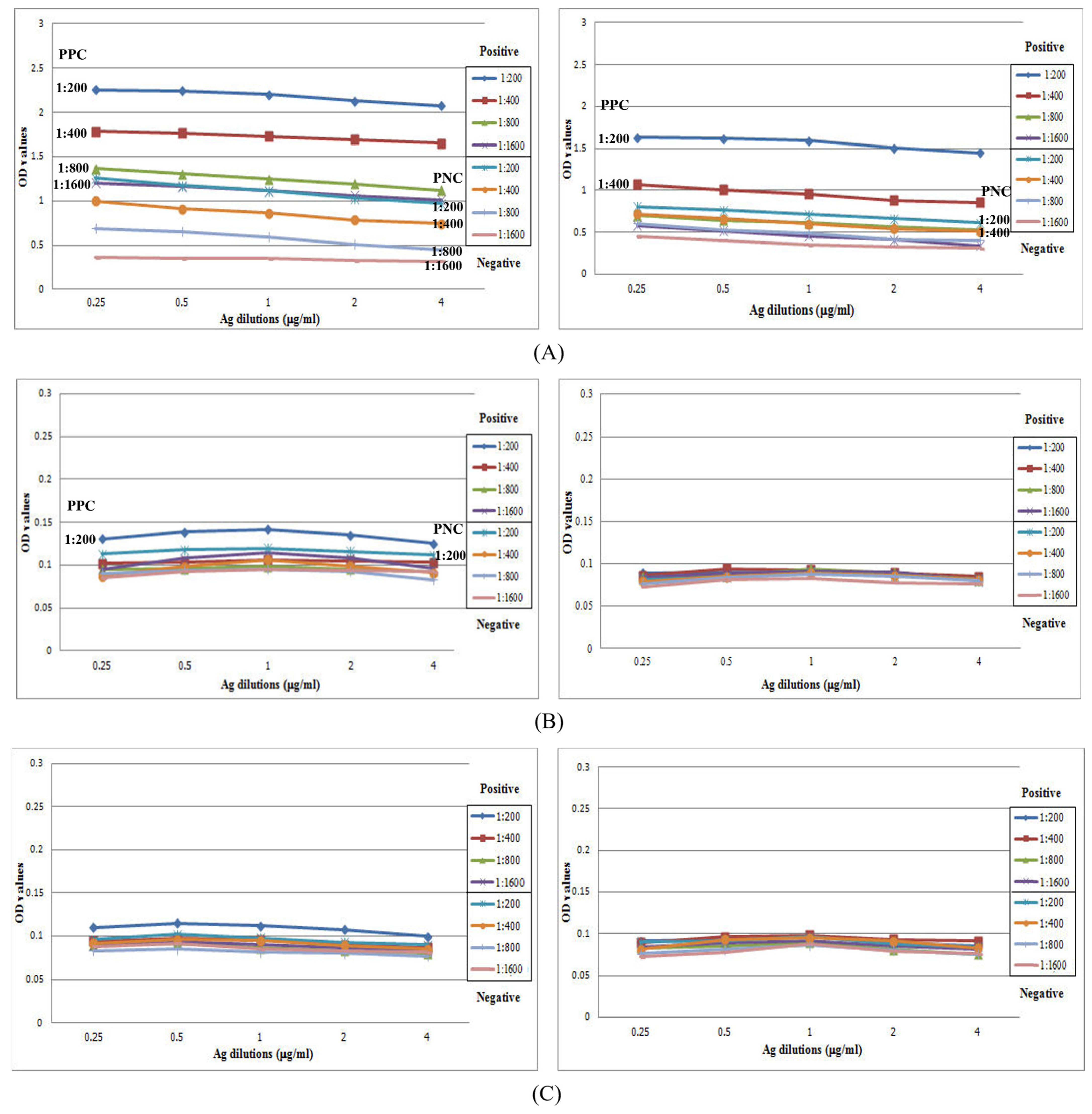
3.3. Full Scale IgG-ELISA

All serum samples were subjected to IgG-ELISA. Of the 16 hwCLM samples, 1 was a false negative, and the OD values of the other 15 samples ranged from 1.111 to 0.452 (cut-off value: 0.451). Most of the hwCLM OD values ranged from 0.452 to 0.556. The false negative case had an OD value of 0.286. There were two high OD values in the hwCLM cases, 1.111 and 1.078, which separated them from the group. Therefore, the sensitivity and specificity of the IgG-ELISA were 93.75% and 98.39%, respectively, and its positive and negative predictive values were 75% and 99.67%, respectively. Only three nematodiases yielded false positives toward the crude adult A. caninum Ag, i.e., angiostrongyliasis (2/15), gnathostomiasis (2/15), and dirofilariasis (1/4). Nonetheless, we achieved satisfactory results because we found no cross-reactions in the serum samples for 24 diseases: nematodiases (ascariasis, capillariasis, Bancroftian filariasis, Brugian filariasis, hookworm infection, strongyloidiasis, trichostrongyliasis, toxocariasis, trichinellosis, and trichuriasis), cestodiases (taeniasis saginata, neurocysticercosis, cystic echinococcosis, sparganosis, and hymenolepiasis nana), trematodiases (paragonimiasis, opisthorchiasis, fascioliasis, minute intestinal fluke infection, and schistosomiasis), and protozoan infections (amoebiasis, giardiasis, Blastocystis infection, and malaria) (Figure 6). It was found that strongyloidiasis (11 cases), fascioliasis (3 cases), and schistsomiasis (4 cases) can cause CLM symptoms, but no false positive occurred. Negative control sera demonstrated good discrimination from the hwCLM cases; all negative control sera yielded true negative results, and only two samples were less distinct from others in this group.

4. Discussion

When focusing on IgG, several previous studies assessed total IgG but yielded poor results. In contrast, the selected crude A. caninum somatic Ags after our checkerboard titration reacted less with Abs against cestodiasis, trematodiasis, and protozoan infections, including ten nematode-associated diseases, and all OD values were below the cut-off value. This may be because of the small amount of the Ags used (0.025 μg/100 μL of coating buffer) and their low antigenicity to the Abs in those tested serum samples. Finally, our proposed ELISA conditions for evaluating IgG yielded 93.75% sensitivity and 98.39% specificity. All healthy control sera had low OD-ELISA values in response to the Ags and yielded similar OD values.
One hwCLM sample in our study had few IgG Abs to A. caninum somatic Ag; thus, it gave a false negative result. This patient had a clinical history of subcutaneous swelling and other erythematous skin changes and larval migrans, and his sero-tests were negative for gnathostomiasis and strongyloidiasis. The gnathostomiasis test was done via detection of a 24-kDa diagnostic band of the G. spinigerum Ag on an IgG immunoblot, and the strongyloidiasis test was performed via IgG-ELISA using a molecular weight cut-off Ag of Strongyloides stercoralis infective larvae; the test was performed at the Department of Helminthology, Faculty of Tropical Medicine. In 2003, a previous study of a patient showed clinical features of erythematous linear and serpiginous plaques. The patient tested positive for crude A. caninum Ags and negative for G. doloresi Ags using the IgG-ELISA. Researchers attempted to screen this patient’s antibodies for other infections using a multiple-dot ELISA with 12 Ags: Dirofilaria immitis, Toxocara canis, Ascaris suum, Anisakis simplex, G. doloresi, Strongyloides ratti, Paragonimus westermani, P. miyazakii, Fasciola hepatica, Clonorchis sinensis, Spirometra erinacei, and Cysticercus cellulosae. The tests were negative, and the patient was diagnosed with A. caninum infection [7]. Additionally, the sensitivity and specificity of the proposed ELISA for diagnosing a single case of hwCLM in that study were suboptimal.
In our study, A. caninum somatic Ag was screened and did not cross-react with Abs from 24 parasitic diseases or with the negative controls. Only five sera samples from three nematodiasis patients cross-reacted with A. caninum somatic Ag. These false positives may not be encountered for serodifferentiation if the targeted habitats of those helminths are considered, i.e., Angiostrongylus worms mostly infect the brain. Dirofilaria immitis exists as nodules in the lungs or tissues because humans are the dead-end host for this worm, and Gnathostoma worms travel throughout the human body, organs, and especially the cutaneous tissues. All hwCLM cases in the present study were negative for gnathostomiasis (Table 1).
The larval and immature stages of many helminths, including Gnathostoma, Strongyloides, sparganum, Paragonimus, and Fasciola, can cause CLM cases similar to those of hookworms [2,5,51,52,53,54,55], but Abs against these helminths in our study did not cross-react with the A. caninum somatic Ag. Additionally, a few Abs in two of fifteen cases of gnathostomiasis showed false positives. However, animal hookworms mostly migrate in the dermis of human skin, but Gnathostoma worms can move in the skin and into deeper tissues and organs.
After several years of frozen storage, the worms were not degraded and demonstrated sharp and defined protein bands on Coomassie brilliant blue-stained gel. Ice crystal damage did not appear to affect the frozen worms. However, the adult worm Ag in our study did not react well with the pooled positive and negative control sera on the ELISA checkerboard titration for individual IgG subclasses and IgE, which is possibly because the serum samples from the controls contained low levels of individual immunoglobulins. The duration of storage of serum samples at −80 °C did not affect antibody reactivity because two high OD values, 1.111 and 1.078, were from two young boys whose sera were kept for 34 and 3 years, respectively. The false negative case was an adult patient, and his serum sample was kept for 11 years. To support low IgE in hwCLM, IgE is produced after all other isotypes in the blood [56], with the lowest concentration among serum Igs of a sequential Ig response [57]. Some reports of hookworm-related CLM presented laboratory findings with normal serum IgE levels [14,39], and the Ags from canine hookworm larvae were mostly confined to the dermis of humans (an abnormal host), thereby revealing a limited IgE response to infection [12]. Hence, the low or absent IgE levels in the positive controls for hwCLM in our study may have led to their being indistinguishable from the negative controls in the checkerboard titration. The increasing IgE levels may have depended on the magnitude of infection such as in schistosomiasis, filarial tropical eosinophilia [58], and N. americanus self-infection, which requires three to four infections to measure increases in IgE [59]. Helminth species other than A. caninum can cause increased IgE levels because their locations are not limited to the dermis [12].
Finding a different result when comparing antigens from A. caninum worms, Loukas and colleagues [13] used a different Ag from A. caninum worms, excretory-secretory (ES) Ag, against four helminthiases, which did not react with Abs from human hookworm infections but reacted with Abs to strongyloidiasis, toxocariasis, and schistosomiasis. In our study, crude A. caninum somatic Ag did not cross-react with Abs against human-associated hookworm diseases, including strongyloidiasis, toxocariasis, and schistosomiasis, which differs from the study using ES-Ag. In Thailand, N. americanus is mainly reported via molecular confirmation, and minor species include A. duodenale and A. ceylanicum in humans at the studied sites [60,61]. Human-associated hookworms (15 sera used) in our study were confirmed only via detection of eggs and larvae; however, Abs from those hookworm infections may be induced by the aforementioned three species. Antibodies of those fifteen cases did not cross-react with the A. caninum Ag and thereby yielded true negative results (Figure 6). When comparing the crude somatic and crude ES Ags of A. caninum, Loukas and colleagues used an IgG-ELISA to study Abs from human eosinophilic enteritis with A. caninum infections and found a higher detection rate using ES-Ag than using a crude somatic Ag, although cross-reactions still occurred with parasitological soils [40]. Improvement of the diagnostic method using immunoblot with Ac68 of the ES product from A. caninum Ag detected eight ancylostomiasis caninum cases, five of which were positive for IgG and IgG1–4, one was positive for IgG and negative for IgG1–4, one was negative for IgG and positive for IgG1–4, and one was negative for all IgGs. All IgGs showed 75% sensitivity in immunoblot results, and IgG4 showed the best specificity (100%), which was determined from only three helminthiases (strongyloidiasis, trichinellosis, and schistosomiasis), but all human-associated hookworm infections (A. duodenale, N. americanus and unknown species) reacted with the Ac68 Ag. These authors found that Ac68 Ag was hookworm-specific [41].
Occasionally, when a serodiagnosis from a total IgG-ELISA cannot be interpreted because of high cross-reactivity, IgG1–4 can be analyzed to obtain a better diagnosis. However, previous studies recommended using an individual IgG subclass or co-interpretation of IgG subclasses for serodiagnosis, such as for gnathostomiasis (i.e., co-interpretation of IgG1-screening tests and IgG2-confirmed diagnosis via ELISA using the CSAg of Gnathostoma larvae) [62]. Laummaunwai and colleagues [63] reported that the highest sensitivity for immunoblotting for diagnosing gnathostomiasis using crude Gnathostoma larvae Ag was obtained with total IgG rather than IgG1–4, although specificity with IgG4 was slightly better than with total IgG (93.9% vs. 91.6%, respectively). HwCLM may be difficult to diagnose because of misdiagnoses and prescribed treatments from physicians and laboratory practitioners [64,65]. A previous study reported a misdiagnosis and improper treatment rate of 58% [38], including the observation of reported information over a long duration of increasing Igs because patients often visit a physician at the early onset of clinical symptoms. Thus, our results suggest that IgG1–4 and IgE from hwCLM sera cannot be used as mediators between diagnoses using ELISA and immunoblotting. However, the crude somatic Ag used in the present study had good sensitivity and excellent specificity once the optimal conditions for the IgG-ELISA were determined. Use of a crude Ag can demonstrate cross-reactivity with Abs of heterologous diseases; thus, proper ELISA conditions can improve the results.

5. Conclusions

Serodifferentiation with crude A. caninum somatic Ag for the proposed IgG-ELISA can potentially be used to diagnose hwCLM from A. caninum hookworms in humans. However, IgE and IgG1–4 could not be differentiated via the ELISA for the positive and negative controls following checkerboard titration; also in addition, the antigen–antibody banding pattern via immunoblot used pooled positive controls. Further studies are warranted to evaluate the proposed ELISA using more hwCLM cases and incorporating a purification technique during Ag preparation.

Author Contributions

Conceptualization, P.D. and D.W.; methodology, P.D. and D.W.; formal analysis, P.D. and D.W.; investigation, S.A. and T.K.; resources, D.N., P.C. (Prakaykaew Charunwatthana), J.D., K.K. and P.C. (Putza Chonsawat); data curation, P.D. and D.W.; writing—original draft preparation, S.A.; writing—review and editing, P.D. and D.W.; supervision, P.D. and D.W.; project administration, P.D. and D.W.; funding acquisition, P.D. and D.W. All authors have read and agreed to the published version of the manuscript.

Funding

This research was partially supported by Head of the Immunodiagnostic Unit for Helminthic Infections of the Department (P.D.) and the Master program scholarship of the Faculty of Tropical Medicine.

Institutional Review Board Statement

The study was conducted in accordance with the Declaration of Helsinki and approved by the Ethics Committee of the Faculty of Tropical Medicine, Mahidol University (Ethical Approval No. MUTM 2014-071-01) for studies involving human sera.

Informed Consent Statement

Not applicable.

Data Availability Statement

Not applicable.

Acknowledgments

The authors thank the staff of the Department of Helminthology, Faculty of Tropical Medicine, Mahidol University, Thailand for providing laboratory facilities and assistance in laboratory techniques, especially Akkarin Poodeepiyasawad for his preparation of photos and photographic work, Rapeepun Prasertbun from the Department of General Medicine, Juntendo University, Faculty of Medicine, Tokyo, Japan for her assistance in data citation, and Kaung Myat Naing, College of Advanced Manufacturing Innovation, King Mongkut’s Institute of Technology Ladkrabang (KMITL), Thailand for his assistance in formatting and data citation. We also thank Traci Raley, MS, ELS, from Edanz (https://www.edanz.com/ac accessed on 15 March 2022) for editing a draft of this manuscript.

Conflicts of Interest

The authors declare no conflict of interest.

References

  1. Antolová, D.; Miterpáková, M.; Paraličová, Z. Case of human Dirofilaria repens infection manifested by cutaneous larva migrans syndrome. Parasitol. Res. 2015, 114, 2969–2973. [Google Scholar] [CrossRef] [PubMed]
  2. Brenner, M.A.; Patel, M.B. Cutaneous larva migrans: The creeping eruption. Cutis 2003, 72, 111–115. [Google Scholar] [PubMed]
  3. Caumes, E. It’s time to distinguish the sign ‘creeping eruption’ from the syndrome ‘cutaneous larva migrans’. Dermatology 2006, 213, 179–181. [Google Scholar] [CrossRef]
  4. Hochedez, P.; Caumes, E. Hookworm-Related Cutaneous Larva Migrans. J. Trop. Med. 2007, 14, 326–333. [Google Scholar] [CrossRef]
  5. Xuan, L.T.; Hung, N.T.; Waikagul, J. Cutaneous fascioliasis: A case report in Vietnam. Am. J. Trop. Med. Hyg. 2005, 72, 508–509. [Google Scholar] [PubMed] [Green Version]
  6. Katz, M.; Despommier, D.D.; Gwadz, R. Parasitic Diseases; Springer: New York, NY, USA, 1988. [Google Scholar]
  7. Feldmeier, H.; Schuster, A. Mini review: Hookworm-related cutaneous larva migrans. Eur. J. Clin. Microbiol. Infect. Dis. 2012, 31, 915–918. [Google Scholar] [CrossRef]
  8. Anisuzzaman; Tsuji, N. Schistosomiasis and hookworm infection in humans: Disease burden, pathobiology and anthelmintic vaccines. Parasitol. Int. 2020, 75, 102051. [Google Scholar] [CrossRef]
  9. Malgor, R.; Oku, Y.; Gallardo, R.; Yarzábal, I. High prevalence of Ancylostoma spp. infection in dogs, associated with endemic focus of human cutaneous larva migrans, in Tacuarembo, Uruguay. Parasite 1996, 3, 131–134. [Google Scholar] [CrossRef] [PubMed] [Green Version]
  10. Minnaar, W.N.; Krecek, R.C.; Fourie, L.J. Helminths in dogs from a peri-urban resource-limited community in Free State Province, South Africa. Vet. Parasitol. 2002, 107, 343–349. [Google Scholar] [CrossRef]
  11. Wang, C.R.; Qiu, J.H.; Zhao, J.P.; Xu, L.M.; Yu, W.C.; Zhu, X.Q. Prevalence of helminthes in adult dogs in Heilongjiang Province, the People’s Republic of China. Parasitol. Res. 2006, 99, 627–630. [Google Scholar] [CrossRef] [PubMed]
  12. Jelinek, T.; Maiwald, H.; Nothdurft, H.D.; Löscher, T. Cutaneous larva migrans in travelers: Synopsis of histories, symptoms, and treatment of 98 patients. Clin. Infect. Dis. 1994, 19, 1062–1066. [Google Scholar] [CrossRef] [Green Version]
  13. Loukas, A.; Opdebeeck, J.; Croese, J.; Prociv, P. Immunologic incrimination of Ancylostoma caninum as a human enteric pathogen. Am. J. Trop. Med. Hyg. 1994, 50, 69–77. [Google Scholar] [CrossRef] [PubMed]
  14. Morsy, H.; Mogensen, M.; Thomsen, J.; Thrane, L.; Andersen, P.E.; Jemec, G.B. Imaging of cutaneous larva migrans by optical coherence tomography. Travel. Med. Infect. Dis. 2007, 5, 243–246. [Google Scholar] [CrossRef]
  15. O’Connell, E.; Suarez, J.; Leguen, F.; Zhang, G.; Etienne, M.; Torrecilla, A.; Jimenez, A.; Farahi, F.; Alzugaray, M.; Rodriguez, D. Outbreak of cutaneous larva migrans at a children’s camp-Miami, Florida, 2006. MMWR Morb. Mortal. Wkly. Rep. 2007, 56, 1285–1287. [Google Scholar]
  16. Tremblay, A.; MacLean, J.D.; Gyorkos, T.; Macpherson, D.W. Outbreak of cutaneous larva migrans in a group of travellers. Trop. Med. Int. Health 2000, 5, 330–334. [Google Scholar] [CrossRef] [PubMed] [Green Version]
  17. Yap, F.B. Creeping eruption. Int. J. Infect. Dis. 2010, 14, e545. [Google Scholar] [CrossRef] [Green Version]
  18. Hotez, P.J.; Narasimhan, S.; Haggerty, J.; Milstone, L.; Bhopale, V.; Schad, G.A.; Richards, F.F. Hyaluronidase from infective Ancylostoma hookworm larvae and its possible function as a virulence factor in tissue invasion and in cutaneous larva migrans. Infect. Immun. 1992, 60, 1018–1023. [Google Scholar] [PubMed]
  19. Muhleisen, J.P. Demonstration of pulmonary migration of the causative organism of creeping eruption. Ann. Intern. Med. 1953, 38, 595–600. [Google Scholar] [CrossRef] [PubMed]
  20. Tan, S.K.; Liu, T.T. Cutaneous larva migrans complicated by Löffler syndrome. Arch. Dermatol. 2010, 146, 210–212. [Google Scholar] [CrossRef] [PubMed]
  21. Soo, J.K.; Vega-Lopez, F.; Stevens, H.P.; Chiodini, P.L. Cutaneous larvae migrans and beyond-a rare association. Travel. Med. Infect. Dis. 2003, 1, 41–43. [Google Scholar] [CrossRef]
  22. Burrill, D.Y.; Kotcher, E.; Childers, J.K. Nematode infestation of the buccal submucosa. Oral Surg. Oral Med. Oral Pathol. 1957, 10, 612–613. [Google Scholar] [CrossRef]
  23. Jaeger, R.G.; Araújo, V.C.D.; Marcucci, G.; Araújo, N.S.D. Larva migrans in the oral mucosa. J. Oral Med. 1987, 42, 246–247. [Google Scholar]
  24. André, J.; Bernard, M.; Ledoux, M.; Achten, G. Larva migrans of the Oral Mucosa. Dermatology 1988, 176, 296–298. [Google Scholar] [CrossRef]
  25. Horman, R. Larva migrans. In Textbook of Dermatology, 4th ed.; Blackwell: Oxford, UK, 1986; pp. 995–997. [Google Scholar]
  26. Lopes, M.A.; Zaia, A.A.; de Almeida, O.P.; Scully, C. Larva migrans that affect the mouth. Oral Surg. Oral Med. Oral Pathol. 1994, 77, 362–367. [Google Scholar] [CrossRef] [PubMed]
  27. Bonne, C. Invasion of the Submucosa of the Human Small Intestine by Ancylostoma Braziliense. Am. J. Trop. Med. 1937, s1–s17, 587–594. [Google Scholar] [CrossRef]
  28. Gandullia, E.; Lignana, E.; Rabagliati, A.; Penna, R. Visceral larva migrans caused by Ancylostoma caninum. Minerva Pediatr. 1981, 33, 917–923. [Google Scholar] [PubMed]
  29. Little, M.; Halsey, N.A.; Cline, B.L.; Katz, S.P. Ancylostoma larva in a muscle fiber of man following cutaneous larva migrans. Am. J. Trop. Med. Hyg. 1983, 32, 1285–1288. [Google Scholar]
  30. Nadbath, R.P.; Lawlor, P.P. Nematode (Ancylostoma) in the cornea; a case report. Am. J. Ophthalmol. 1965, 59, 486–490. [Google Scholar] [CrossRef]
  31. Beaver, P.C. The nature of visceral larva migrans. J. Parasitol. 1969, 55, 3–12. [Google Scholar] [CrossRef] [PubMed]
  32. Manalang, C. Studies on Ankylostomiasis in the Philippines. In Proceedings of the Far Eastern Association of Tropical Medicine Transactions Sixth Biennial Congress, Tokyo, Japan, 1925; Kyorinsha Medical Publishing Co.: Tokyo, Japan, 1926; Volume 1, pp. 351–368. [Google Scholar]
  33. Deane, M. Helminths eliminated from a group of Amazon residents after treatment with hexilresorcinol. Rev. Saude Publica 1950, 3, 443–464. [Google Scholar]
  34. Komma, M.; Queiroz, D.S.; Silva, A.; Jardim, C. Occurrence and prevalence of hookworms in 14 children in Goiânia. Rev. Goiana Med. 1969, 15, 169–174. [Google Scholar]
  35. Carrias, V.; de Vargas, P. Infection of the human intestine by Ancylostoma caninum Ercolani 1859. Rev. Soc. Bras. Med. Trop. 1985, 18, 57. [Google Scholar]
  36. Witenberg, G. Some unusual observations on helminthiasis in Israel. Harefuah 1951, 41, 178–180. [Google Scholar]
  37. Caumes, E.; Ly, F.; Bricaire, F. Cutaneous larva migrans with folliculitis: Report of seven cases and review of the literature. Br. J. Dermatol. 2002, 146, 314–316. [Google Scholar] [CrossRef] [PubMed]
  38. Davies, H.D.; Sakuls, P.; Keystone, J.S. Creeping eruption. A review of clinical presentation and management of 60 cases presenting to a tropical disease unit. Arch. Dermatol. 1993, 129, 588–591. [Google Scholar] [CrossRef]
  39. Kwon, I.H.; Kim, H.S.; Lee, J.H.; Choi, M.H.; Chai, J.Y.; Nakamura-Uchiyama, F.; Nawa, Y.; Cho, K.H. A serologically diagnosed human case of cutaneous larva migrans caused by Ancylostoma caninum. Korean J. Parasitol. 2003, 41, 233–237. [Google Scholar] [CrossRef] [PubMed] [Green Version]
  40. Loukas, A.; Croese, J.; Opdebeeck, J.; Prociv, P. Detection of antibodies to secretions of Ancylostoma caninum in human eosinophilic enteritis. Trans. R. Soc. Trop. Med. Hyg. 1992, 86, 650–653. [Google Scholar] [CrossRef]
  41. Loukas, A.; Opdebeeck, J.; Croese, J.; Prociv, P. Immunoglobulin G subclass antibodies against excretory/secretory antigens of Ancylostoma caninum in human enteric infections. Am. J. Trop. Med. Hyg. 1996, 54, 672–676. [Google Scholar] [CrossRef] [PubMed]
  42. Croese, J.; Loukas, A.; Opdebeeck, J.; Prociv, P. Occult enteric infection by Ancylostoma caninum: A previously unrecognized zoonosis. Gastroenterology 1994, 106, 3–12. [Google Scholar] [CrossRef]
  43. Haichou, X.; Yan, W.; Shuhua, X.; Sen, L.; Yong, W.; Guangjin, S.; Weituo, W.; Bin, Z.; Drake, L.; Zheng, F.; et al. Epidemiology of human ancylostomiasis among rural villagers in Nanlin County (Zhongzhou Village), Anhui Province, China: II. Seroepidemiological studies of the age relationships of serum antibody levels and infection status. Southeast Asian J. Trop. Med. Public Health 2000, 31, 736–741. [Google Scholar]
  44. Pothong, K.; Komalamisra, C.; Kalambaheti, T.; Watthanakulpanich, D.; Yoshino, T.P.; Dekumyoy, P. ELISA based on a recombinant Paragonimus heterotremus protein for serodiagnosis of human paragonimiasis in Thailand. Parasit Vectors 2018, 11, 322. [Google Scholar] [CrossRef] [PubMed]
  45. Ieamsuwan, I.; Watthanakulpanich, D.; Chaisri, U.; Adisakwattana, P.; Dekumyoy, P. Evaluation of immunodiagnostic tests for human gnathostomiasis using different antigen preparations of Gnathostoma spinigerum larvae against IgE, IgM, IgG, IgG1-4 and IgG1 patterns of post-treated patients. Trop. Med. Int. Health 2021, 26, 1634–1644. [Google Scholar] [CrossRef]
  46. Yoonuan, T.; Nuamtanong, S.; Dekumyoy, P.; Phuphisut, O.; Adisakwattana, P. Molecular and immunological characterization of cathepsin L-like cysteine protease of Paragonimus pseudoheterotremus. Parasitol. Res. 2016, 115, 4457–4470. [Google Scholar]
  47. Deardorff, T.L.; Throm, R. Commercial blast-freezing of third-stage Anisakis simplex larvae encapsulated in salmon and rockfish. J. Parasitol. 1988, 74, 600–603. [Google Scholar] [PubMed]
  48. Shikama, K. Effect of Freezing and Thawing on the Stability of Double Helix of DNA. Nature 1965, 207, 529–530. [Google Scholar] [CrossRef]
  49. Dekumyoy, P.; Insun, D.; Waikagul, J.; Tanantaphruti, M.; Rongsriyam, Y.; Coochote, W. IgG- and IgG4-detected antigens of Dirofilaria immitis adult worms for bancroftian filariasis by enzyme-linked immunoelectrotransfer blot. Southeast Asian J. Trop. Med. Public Health 2000, 31 (Suppl. S1), 58–64. [Google Scholar]
  50. Florkowski, C.M. Sensitivity, specificity, receiver-operating characteristic (ROC) curves and likelihood ratios: Communicating the performance of diagnostic tests. Clin. Biochem. Rev. 2008, 29 (Suppl. S1), S83–S87. [Google Scholar] [PubMed]
  51. Alvarez, P.; Melgarejo, C.; Beltran, G.; Santos, R.; Ferrer, K.; Elescano, I.; Victorero, E.; Meza, B.; Ortiz, N.; León, A.; et al. Four Case Reports of Cutaneous Sparganosis From Peruvian Amazon. Am. J. Dermatopathol. 2022, 44, 510–514. [Google Scholar] [CrossRef]
  52. Dainichi, T.; Nakahara, T.; Moroi, Y.; Urabe, K.; Koga, T.; Tanaka, M.; Nawa, Y.; Furue, M. A case of cutaneous paragonimiasis with pleural effusion. Int. J. Dermatol. 2003, 42, 699–702. [Google Scholar] [CrossRef]
  53. Singh, T.S.; Devi Kh, R.; Singh, S.R.; Sugiyama, H. A case of cutaneous paragonimiasis presented with minimal pleuritis. Trop. Parasitol. 2012, 2, 142–144. [Google Scholar] [CrossRef]
  54. Catchpole, B.N.; Snow, D. Human ectopic fascioliasis. Lancet 1952, 2, 711–712. [Google Scholar] [CrossRef]
  55. Griffin, M.P.; Tompkins, K.J.; Ryan, M.T. Cutaneous sparganosis. Am. J. Dermatopathol. 1996, 18, 70–72. [Google Scholar] [CrossRef] [PubMed]
  56. Xiong, H.; Dolpady, J.; Wabl, M.; Curotto de Lafaille, M.A.; Lafaille, J.J. Sequential class switching is required for the generation of high affinity IgE antibodies. J. Exp. Med. 2012, 209, 353–364. [Google Scholar] [CrossRef] [PubMed] [Green Version]
  57. Martins, T.B.; Bandhauer, M.E.; Bunker, A.M.; Roberts, W.L.; Hill, H.R. New childhood and adult reference intervals for total IgE. J. Allergy Clin. Immunol. 2014, 133, 589–591. [Google Scholar] [CrossRef]
  58. Neva, F.A.; Kaplan, A.P.; Pacheco, G.; Gray, L.; Danaraj, T.J. Tropical eosinophilia. A human model of parasitic immunopathology, with observations on serum IgE levels before and after treatment. J. Allergy Clin. Immunol. 1975, 55, 422–429. [Google Scholar] [CrossRef] [PubMed]
  59. Ogilvie, B.M.; Bartlett, A.; Godfrey, R.C.; Turton, J.A.; Worms, M.J.; Yeates, R.A. Antibody responses in self-infections with Necator americanus. Trans. R. Soc. Trop. Med. Hyg. 1978, 72, 66–71. [Google Scholar] [CrossRef]
  60. Phosuk, I.; Intapan, P.M.; Thanchomnang, T.; Sanpool, O.; Janwan, P.; Laummaunwai, P.; Aamnart, W.; Morakote, N.; Maleewong, W. Molecular detection of Ancylostoma duodenale, Ancylostoma ceylanicum, and Necator americanus in humans in northeastern and southern Thailand. Korean J. Parasitol. 2013, 51, 747–749. [Google Scholar] [CrossRef] [PubMed]
  61. Traub, R.J.; Inpankaew, T.; Sutthikornchai, C.; Sukthana, Y.; Thompson, R.C. PCR-based coprodiagnostic tools reveal dogs as reservoirs of zoonotic ancylostomiasis caused by Ancylostoma ceylanicum in temple communities in Bangkok. Vet. Parasitol. 2008, 155, 67–73. [Google Scholar] [CrossRef] [PubMed]
  62. Nuchprayoon, S.; Sanprasert, V.; Suntravat, M.; Kraivichian, K.; Saksirisampant, W.; Nuchprayoon, I. Study of specific IgG subclass antibodies for diagnosis of Gnathostoma spinigerum. Parasitol. Res. 2003, 91, 137–143. [Google Scholar] [CrossRef]
  63. Laummaunwai, P.; Sawanyawisuth, K.; Intapan, P.M.; Chotmongkol, V.; Wongkham, C.; Maleewong, W. Evaluation of human IgG class and subclass antibodies to a 24 kDa antigenic component of Gnathostoma spinigerum for the serodiagnosis of gnathostomiasis. Parasitol. Res. 2007, 101, 703–708. [Google Scholar] [CrossRef]
  64. Gill, N.; Somayaji, R.; Vaughan, S. Exploring Tropical Infections: A Focus on Cutaneous Larva Migrans. Adv. Skin. Wound Care 2020, 33, 356–359. [Google Scholar] [CrossRef] [PubMed]
  65. Tan, S.; Firmansyah, Y. New Approachment of Creeping Eruption Management. J. Dermatol. Res. Ther. 2020, 6, 1–5. [Google Scholar] [CrossRef]
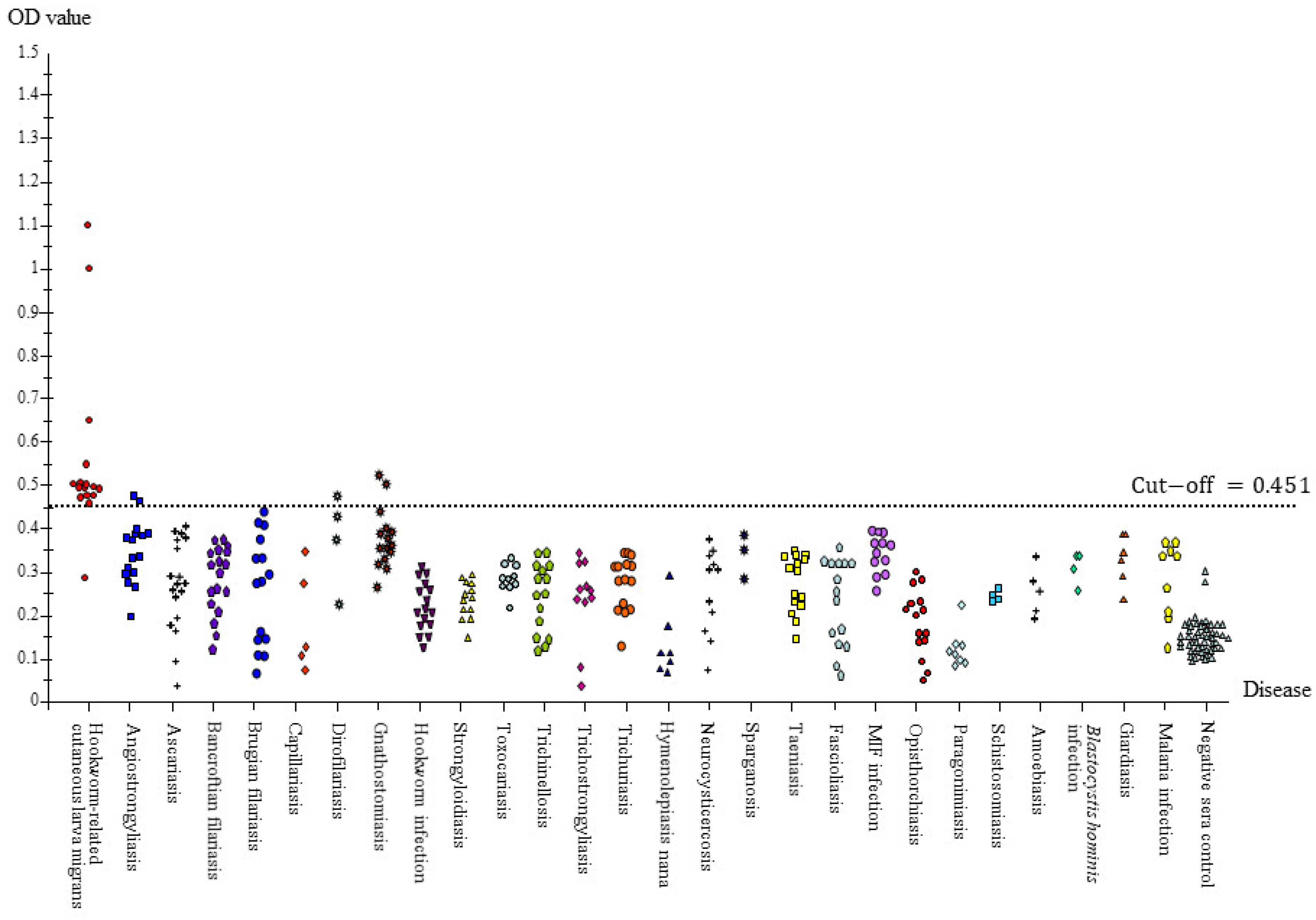
Figure 1. Hookworm-related cutaneous larva migrans of a female patient presenting a zigzag track on the abdomen.
Tropicalmed 08 00209 g001
Figure 2. Section of biopsied larva from the abdomen showing the cuticle, body surface, intestine-containing cavity, several intestinal cells, and muscle cell lateral cords.
Tropicalmed 08 00209 g002
Figure 3. Hookworm-related cutaneous larva migrans on the thumb of the right hand of a young male patient showing a thread-like line of serpigenous inflammation.
Tropicalmed 08 00209 g003
Figure 4. Checkerboard titration curves for determining the optimal concentration of A. caninum crude antigens (0.25–4 μg/mL) against various dilutions of pooled positive controls (PPC) and pooled negative controls (PNC) using 1:2000 (L, Left) and 1:4000 (R, Right) dilutions of peroxidase-conjugated anti-human IgGs ((A): IgG, (B): IgG1, (C): IgG2, (D): IgG3, (E): IgG4, and (F): IgE). Lines represent OD values of dilutions of either pooled positive serum or pooled negative serum at ratios of 1:200, 1:400, 1:800, and 1:1600.
Tropicalmed 08 00209 g004aTropicalmed 08 00209 g004b
Figure 5. (A) Protein profile comparison of A. caninum crude extracts at 15 (A) and 30 (B) µg/well shown by Coomassie brilliant blue staining. M: low molecular weight markers (kDa). Demonstration of strong bands at 58.5 and 49 kDa and faint bands from 90 to 27 kDa. (B) Anti-human immunoglobulins IgG, IgG1–4, and IgE reacted with pooled human antibodies of hwCLM against crude A. caninum somatic antigens via immunoblot. M: low molecular weight markers; A: IgG; B–E: IgG1–4; F: IgE. Reaction of total IgG (lane A) was strong in a range of standard markers from 97.4 to 21.5 kDa and below 14.4 kDa. IgG1–4 (lanes B–E) and IgE (lane F) showed weak and undefined bands, with more bands in IgG2 (lane C) and IgG3 (lane D) in a range of markers > 45 kDa and weak bands below 14.4 kDa in lanes C, D, and E (IgE). Several weak and smeared bands were found in all reactions of IgE and IgG1–4.
Tropicalmed 08 00209 g005
Figure 6. Scatter patterns of IgG-ELISA absorbance values of serum samples from hookworm-related CLM, 27 different diseases, and negative control against A. caninum crude antigen. Ag–Ab reactions were accomplished under ELISA conditions, 0.25 μg/mL of Ag concentration, 1:1600 diluted serum samples, and an anti-human IgG dilution of 1:2000 at a cut-off value of 0.451.
Tropicalmed 08 00209 g006
Table 1. Descriptions of serum samples of 27 diseases and from healthy controls using different diagnostic methods, including clinical pictures of hwCLM cases.
DiseaseNumber of Serum SamplesSpecific or Proven Diagnosis
Homologous Sera
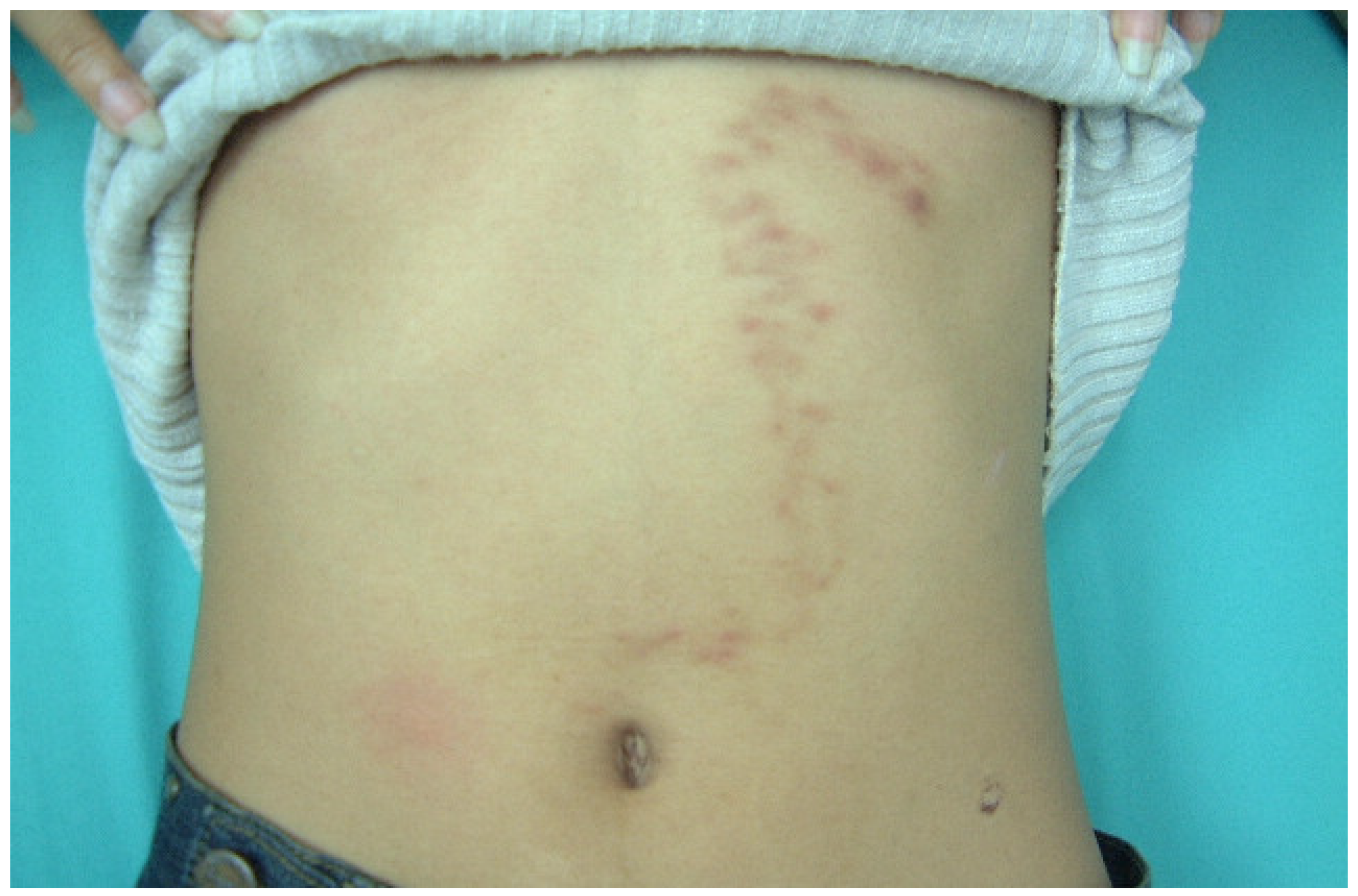
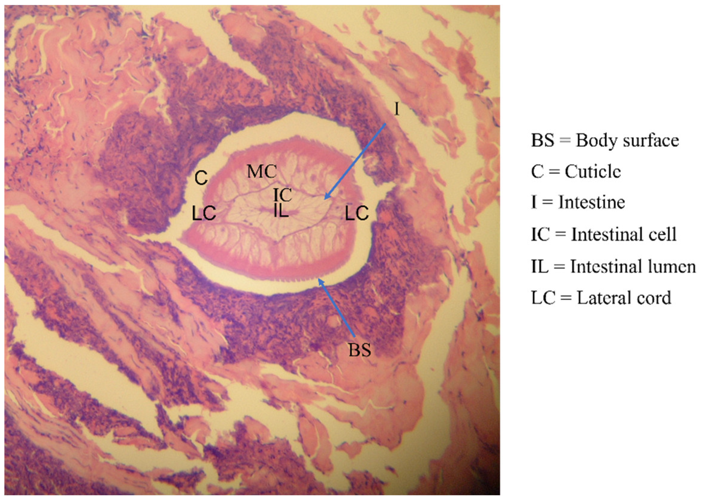
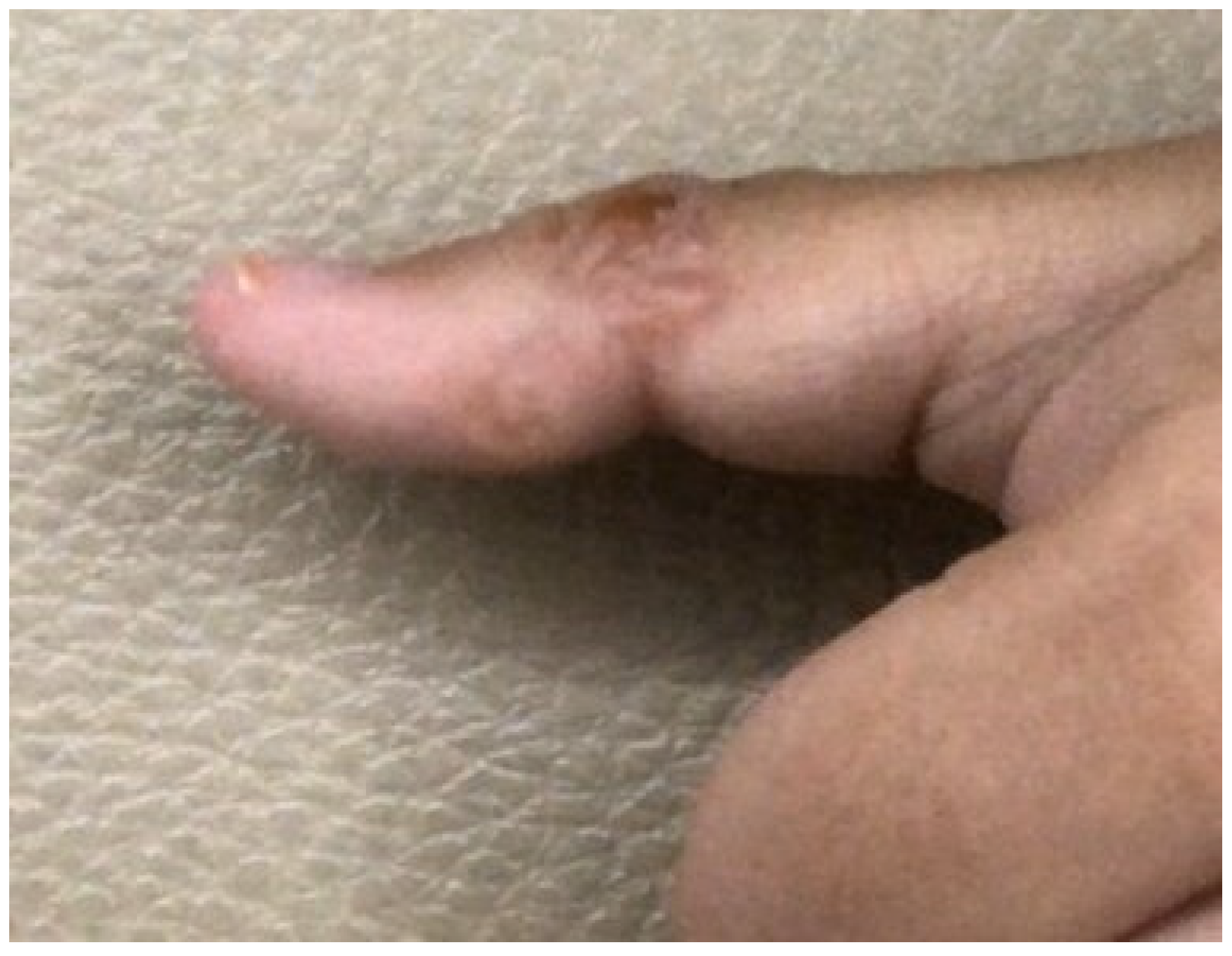
Human-related cutaneous larva migrans (hwCLM)
16Clinical manifestations related to cutaneous larva migrans or creeping eruption, such as pruritus, swelling pain, and the appearance of an erythematous track on the skin of the trunk, buttock, hand, leg, etc.
History of traveling or/and exposure to soil or beach land where dogs were around
Tested negative for gnathostomiasis with an IgG-immunoblotting test and for strongyloidiasis with an IgG-ELISA, including a demonstration of two hwCLM cases and a section of larva structure from the patient via tissue biopsy (Figure 1, Figure 2 and Figure 3)
Heterologous sera Nematodiasis
Angiostrongyliasis15Ocular angiostrongyliasis, larval stage found
Larva in CSF sample found
Consumption of snail intermediate hosts, stiff neck, and positive immunoblot
Ascariasis18Eggs in feces found by using the Kato thick smear technique
Bancroftian filariasis17Microfilariae on blood films
Brugian filariasis15Microfilariae on blood films and positive ELISA results
Capillariasis5Eggs and adults in feces found by using the simple smear technique and simple sedimentation
Dirofilaria infection4Sectioned worms in lung tissues and worms from the eyes and/or positive immunoblot results
Gnathostomiasis15Detection of worms and positive immunoblot results
Hookworm infection15Eggs in feces found by using the Kato thick smear technique or detection of L3 by using the polyethylene tube culture method
Strongyloidiasis11Larvae found in polyethylene tube culture
Toxocariasis10Clinical symptoms and positive immunoblot results
Trichinellosis15Biopsy and/or history of eating meat of wild pigs, clinical symptom manifestation, and positive immunoblot results
Trichostrongyliasis 11Culture technique and larvae confirmation
Trichuriasis15Eggs found by using the Kato thick smear technique
Cestodiasis
Cystic echinocossosis6Scolices found or positive immunoblot results; 1 indigenous case and 5 imported cases
Hymenolepiasis nana8Eggs in feces found by using the simple smear technique
Neurocysticercosis11Biopsy or clinical manifestation accompanying a computerized axial tomographic (CAT) scan of the brain and positive immunoblot results
Sparganosis3Spargana found
Taeniasis 15Taenia saginata segments or Taenia eggs found in feces
Trematodiasis
Fascioliasis3Eggs found or positive immunoblot results; 1 indigenous case and 2 cases supported by US Naval Medical Research Unit No. 3, Egypt
Opisthorchiasis15Worms detected post-treatment
Paragonimiasis heterotremus15Eggs in feces and sputum found by using the simple smear technique
Schistosomiasis 4Schistosoma mansoni eggs found; cases supported by US Naval Medical Research Unit No.3, Egypt
Small intestinal fluke infection 10Worms detected post-treatment
Protozoa infections
Amoebiasis5Cysts in feces found by using the simple smear technique
Blastocystis hominis infection4Cysts in feces found by using the simple smear technique
Giardiasis6Giardia intestinalis cysts in feces found by using the simple smear technique
Malaria 9Plasmodium falciparum or P. vivax found in blood smears
Negative control sera30Negative results from stool examinations using the simple smear and formalin-ether concentration techniques for other parasitic infections.
Negative screening results from IgG-ELISA using ten antigens, namely crude antigens of Gnathostoma spinigerum larvae, Strongyloides stercolaris larvae, Toxocara canis male and female adult worms, Angiostrongylus cantonensis adult worms, Dirofilaria immitis female adult worms, Trichinella spiralis muscle larvae, Taenia solium metacestode, Paragonimus heterotremus worms, Fasciola gigantica worms, and fluid of hydatid cyst [44,45]
Disclaimer/Publisher’s Note: The statements, opinions and data contained in all publications are solely those of the individual author(s) and contributor(s) and not of MDPI and/or the editor(s). MDPI and/or the editor(s) disclaim responsibility for any injury to people or property resulting from any ideas, methods, instructions or products referred to in the content.

