Communication

Serological Detection of Toxoplasma gondii among Free-Grazing Ducks from Central and Western Thailand—A One Health Perspective on Integrated Farming

1
Department of Parasitology, Faculty of Veterinary Medicine, Kasetsart University, Bangkok 10900, Thailand
2
Department of Veterinary Medicine, Faculty of Animal Science and Veterinary Medicine, College of Agriculture and Forestry, Hue University, Hue 49000, Vietnam
3
Department of Farm Resources and Production Medicine, Faculty of Veterinary Medicine, Kasetsart University, Nakhon Pathom 73000, Thailand
4
Department of Veterinary Public Health, Faculty of Veterinary Medicine, Kasetsart University, Bangkok 10900, Thailand
5
Faculty of Veterinary Medicine, Mahanakorn University of Technology, Bangkok 10530, Thailand
6
Laboratory of Molecular Parasitology, Institute of Parasitology, University of Zurich, 8000 Zurich, Switzerland
*
Author to whom correspondence should be addressed.
Trop. Med. Infect. Dis. 2023, 8(2), 103; https://doi.org/10.3390/tropicalmed8020103
Submission received: 26 December 2022 / Revised: 31 January 2023 / Accepted: 1 February 2023 / Published: 6 February 2023
(This article belongs to the Special Issue Feature Papers in One Health)

Abstract

:
Toxoplasmosis is one of the most common zoonotic parasitic diseases infecting nearly all warm-blooded animals, including poultry (geese, turkeys, chickens, and ducks). It is caused by Toxoplasma gondii (T. gondii), which is an obligate intracellular protozoan belonging to the Apicomplexa group. In Thailand, duck meat production for domestic consumption and international trade is mainly bred and produced in the central and western parts of the country. Free-grazing ducks in integrated duck–rice production have significant benefits in rice cultivation, accounting for the popularity of this farming system in Thailand. However, ducks are considered particularly susceptible to consuming T. gondii oocysts from water contaminated with cat feces due to the fact of their feeding habits of free-grazing and dabbling. Hence, the prevalence of this zoonotic parasite in a large-scale integrated farming context is particularly challenging with respect to the contamination of the food chain of humans and farm animals. In the present study, we examined the overall prevalence of T. gondii infection in slaughtered free-grazing ducks originating from Central and Western Thailand, setting the stage for an in-depth One Health approach to assess and manage the risks of integrated farming practices. A representative sample size of 161 ducks was calculated using a two-stage sampling method. Specifically, serum samples were collected from 217 slaughtered free-grazing ducks originating in six provinces in Central and Western Thailand. Serum antibodies against T. gondii were detected using an indirect fluorescent antibody test (IFAT). The positive control serum samples were prepared from ducks experimentally immunized with T. gondii. Sixty-eight (31.3%) of the two hundred and seventeen ducks were seropositive with T. gondii. Two groups of fattening ducks and spent layers showed similar seropositivity rates at 29% and 32.3%, with the majority of positive samples being found in the low titer. In addition, a wide distribution of positive serum samples was observed in all six provinces in the present study. To the best of our knowledge, this is the first report on a serological prevalence snapshot in commercially produced duck populations that have high interaction with farmed environments in Thailand, revealing a high infection pressure in areas of integrated duck–rice farming. Importantly, contaminated duck meat for commercial use, as well as offal and carcasses from slaughterhouses, completes the transmission of T. gondii from the environment into the food chain of humans and domestic animals. Hence, from a One Health perspective, it is important to clarify whether this transmission chain extends further to the wild, i.e., predator–prey cycles that are independent of duck farming or are self-contained.

1. Introduction

Toxoplasma gondii (T. gondii) is an obligate intracellular protozoan causing toxoplasmosis which is of significant medical and veterinary importance. Felids are the only definitive hosts producing environmentally resistant, highly infectious oocysts, which are shed with feces and survive for up to two years in the environment. Conversely, intermediate hosts of T. gondii, consisting of all warm-blooded animals, are of great significance in harboring infectious tissue cysts [1].
A systematic review previously revealed an average global T. gondii seropositivity rate of 25.7% in humans over the last three decades [2]. Water and food contaminated with T. gondii oocysts or undercooked meat of chronically infected animals containing tissue cysts can become sources of infection for humans [1]. Healthy individuals infected with Toxoplasma are typically asymptomatic during the self-limiting acute phase and become chronically infected for life, whereas some such clinical signs as cervical or occipital lymphadenopathy, microphthalmia (ocular toxoplasmosis), encephalitis, and hydrocephaly or microcephaly (congenital toxoplasmosis) can be seen in immune-compromised patients [3,4].
In Thailand, T. gondii infections have been reported in various animal species but lack a study in ducks [5,6,7,8,9,10]. Approximately 2.27 M metric tons of Thai duck meat products, including cooked, refrigerated, frozen, and otherwise treated duck meat, were exported to Japan, Hongkong, Europe, South Africa, Bangladesh, and neighboring countries, such as Myanmar, Laos, and Cambodia, in 2021 [11,12]. Central and Western Thailand are the major areas for producing duck meat and eggs for domestic consumption and international exportation [13,14].
Thailand is the world’s second-largest rice exporter, with production mostly in the central plains aided by local irrigation systems and yielding two or even three crops each year. Rice production is traditionally linked to “ped lai thoong”, the co-cultivation of Khaki Campbell ducks released in large flocks to feed in freshly harvested paddies. This development of rice cultivation has been closely associated with the free-grazing duck farming system in which large flocks of ducks are rotated among paddy fields after the harvest to scavenge leftover rice grains, husks, insects, small fishes, and snails for ducks [14]. This is beneficial for rice production in several ways including lowering the requirement for combating pests. Fattening ducks, reared for meat production only, are usually slaughtered after two months in the paddies. In addition, ducks are raised for egg production. They are kept in paddy fields for 5–6 months prior to a nesting period of one to two years at farms and slaughtered as spent layer ducks for meat [14,15]. Hence, because of their feeding habits free-grazing ducks are highly exposed to T. gondii oocysts while grazing in large areas in these irrigated environments. The prevalence of seropositivity in these ducks can thus be considered a reliable indicator of the infection pressure by T. gondii in the farmed environment. As a consequence, the consumption of undercooked duck meat constitutes a risk for T. gondii infection in humans and animals [1,16,17]. From a One Health perspective, environmental and animal health are the relevant focus areas of this system at first glance. However, the fact that the waterborne zoonotic pathogen T. gondii enters the human food chain via duck meat, contaminated with tissue cysts containing infectious bradyzoites, adds human health as the third pillar of the One Health concept. The consequences for human health as a result of this particular form of integrated farming remains to be determined. Ultimately, we want to bring to bear a systems approach to assess and evaluate this problem and use this approach to design strategies for intervention. As a first assessment of the importance of this medical and economical threat, we conducted a prevalence snapshot in commercially reared free-grazing duck populations at the time of slaughter to determine the presence of antibodies against T. gondii in ducks from areas of Central and Western Thailand.

2. Materials and Methods

2.1. Study Design and Sample Size Calculation

Khaki Campbell ducks raised in the central and western parts of Thailand were included in this study. The fattening ducks and spent layers, which are typically raised in the traditional free-grazing system, were collected. To detect antibodies to T. gondii in the free-grazing ducks, a sample size calculation was performed using a two-stage sampling method: n h / i = [ 1 ( 1 CL ) 1 / e   ] × ( N h / i e 1 2 ) , where CL is the level of confidence, at 95%; e is the number of detectable individuals with the event in the population: e = N × p × Se, with p being the expected prevalence of 15% at the individual level [18] and 32% at farm level [19], and Se being the sensitivity of IFAT, at 80% [20]; Nh/i is the number of flocks/individuals per flock within 2 months (estimated sampling period in a slaughterhouse), at 90 flocks/1000 ducks; and nh/i is the number of flocks/individuals per flock. A minimal sample size calculation was seven flocks and 23 ducks/flock. Before stunning, blood samples from individual birds were collected in a local slaughterhouse located in Nakhon Pathom province.

2.2. Blood Sample Collection

Between April and May 2021, the blood was sampled in the duck superficial plantar metatarsal vein using serum tubes (BD, Belliver Industrial Estate, Plymouth, UK) by sterile 3–5 milliliter syringes with 20–21# needles. The sample tubes were left standing for at least 30 min at ambient temperature for coagulation before transporting them to the laboratory. The blood was then centrifuged at 1100× g for 5 min, and serum fractions were removed and stored at −20 °C for serological examination.

2.3. Positive and Negative Control Sera

T. gondii-positive and -negative duck sera were generated. Briefly, 500 microliters of 104 tachyzoites of in vitro cultivated T. gondii PLK strain were intravenously inoculated into five Khaki Campbell ducks aged one month for the positive control. Negative control serum samples were collected from three ducks injected with 500 microliters of sterile PBS (phosphate-buffered saline, pH 7.4). All ducks were examined as clinically healthy and seronegative with T. gondii before inoculation. The serum samples were collected on the 1st, 2nd, 3rd, 4th, 5th, 6th, 7th, and 8th week postinoculation (wpi.). The positive and negative control sera were confirmed by an inhouse indirect fluorescent antibody test (IFAT) (see Section 2.4 below).

2.4. Serological Examination

An inhouse qualitative indirect fluorescent antibody test (IFAT) was used for the detection of T. gondii antibodies in the duck serum samples. Preparation of IFAT slides: T. gondii tachyzoites of the RH strain were cultivated in vitro in African green monkey kidney (Vero) cells [21,22]. The monolayer of the infected cells was harvested, and T. gondii tachyzoites were released and enriched using several passages through syringe needles and a 5 μm filter. The tachyzoites were then washed three times with cold PBS and diluted to 106 tachyzoites/mL. Twenty microliters of a T. gondii tachyzoite suspension were pipetted into each well of Teflon-coated slides (Cel-Line Associates, Newfield, NJ, USA). The slides were then air-dried at room temperature overnight and fixed with ice-cold acetone for 10 min and air-drying before storing at −80 °C until used.
Detection of anti-T. gondii antibodies in duck serum: The serum samples were titrated at a two-fold dilution, starting at 1:25, in diluting buffer (NaH2PO4, 0.11 g; bovine serum albumin, 5 g; Na2HPO4, 0.595 g; NaCl, 4.275 g; and ddH2O, 500 mL) before and incubated with acetone-fixed tachyzoite-coated slides. The secondary antibodies (fluorescein-labeled antibody to duck IgG (H + L) generated in goats (KPL, Gaithersburg, MD 20878, USA)) were used at a dilution of 1:150. The positive and negative control sera were included in each test. The slides were incubated with diluted serum or secondary antibody solutions at 37 °C for 30 min and washed three times with FA Rinse buffer (Na2CO3, 1.14 g; NaHCO3, 3.36 g; NaCl, 0.85 g; and ddH2O, 1000 mL) before embedding under coverslips. The slides were examined microscopically under fluorescence light. Only a bright, linear peripheral fluorescence of the tachyzoites was considered positive. A titer of 1:100 was used as the positive cut-off value for T. gondii [19].

2.5. Statistical Analyses

The statistically significant difference between the frequencies of the seropositive samples in the two duck types was considered at p < 0.05 using the χ2 test. The normal distribution of antibody titers was determined using the Shapiro test, and the Wilcoxon’s test was then employed to determine the statistically significant difference in the antibody titer distribution between the two duck types. These statistical analyses were conducted using the R software package version 3.6.3.

3. Results

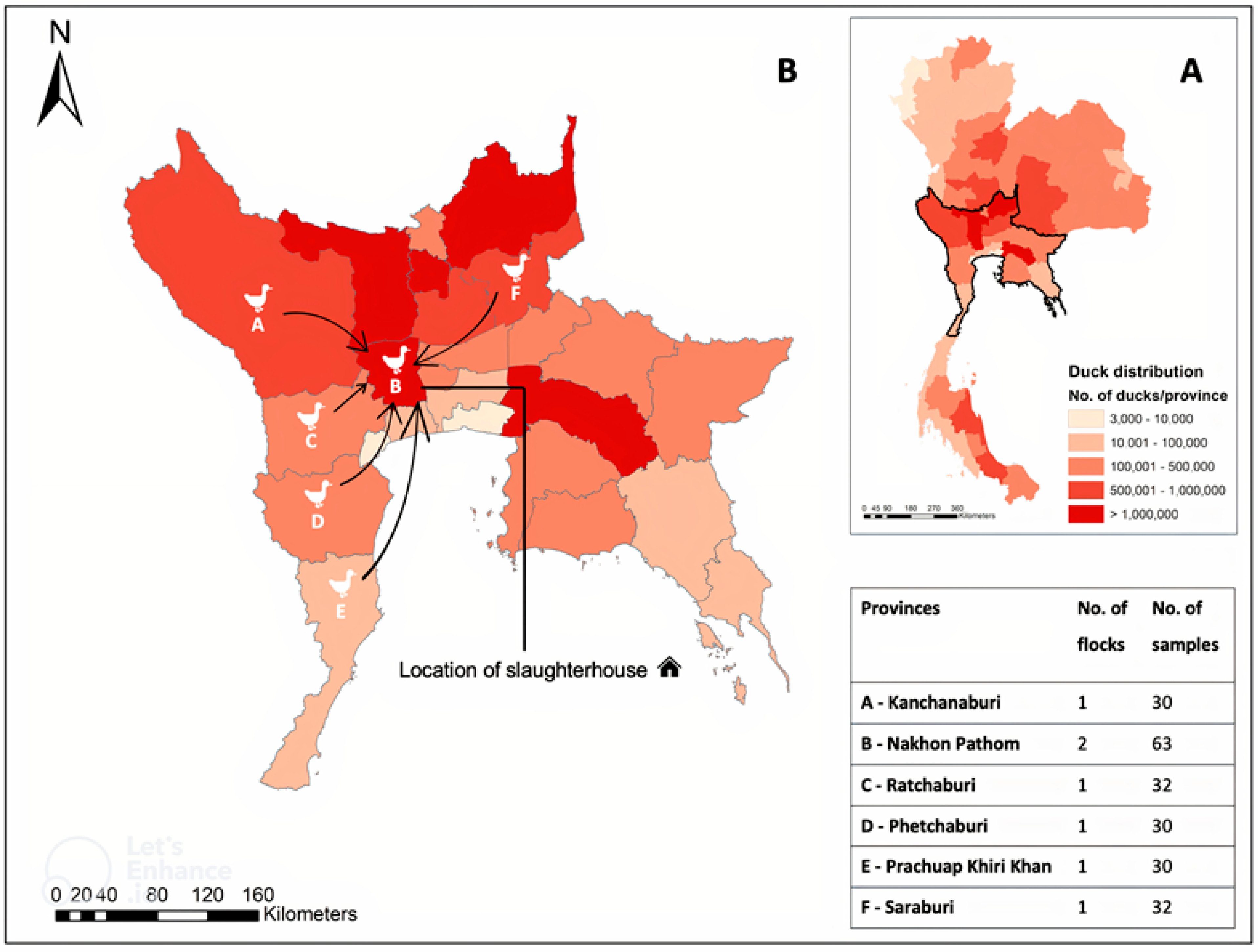
3.1. Samples, Geographical Areas, and Data Characteristics

The antemortem serum samples were conveniently collected from 217 Khaki Campbell ducks (at least 30 ducks/flock) at the slaughterhouse. Five flocks of spent layers and two flocks of fattening ducks originated from six different provinces, including two flocks in Nakhon Pathom and one flock in each other provinces (Kanchanaburi, Phetchaburi, Prachuap Khiri Khan, Ratchaburi, and Saraburi) (Figure 1).

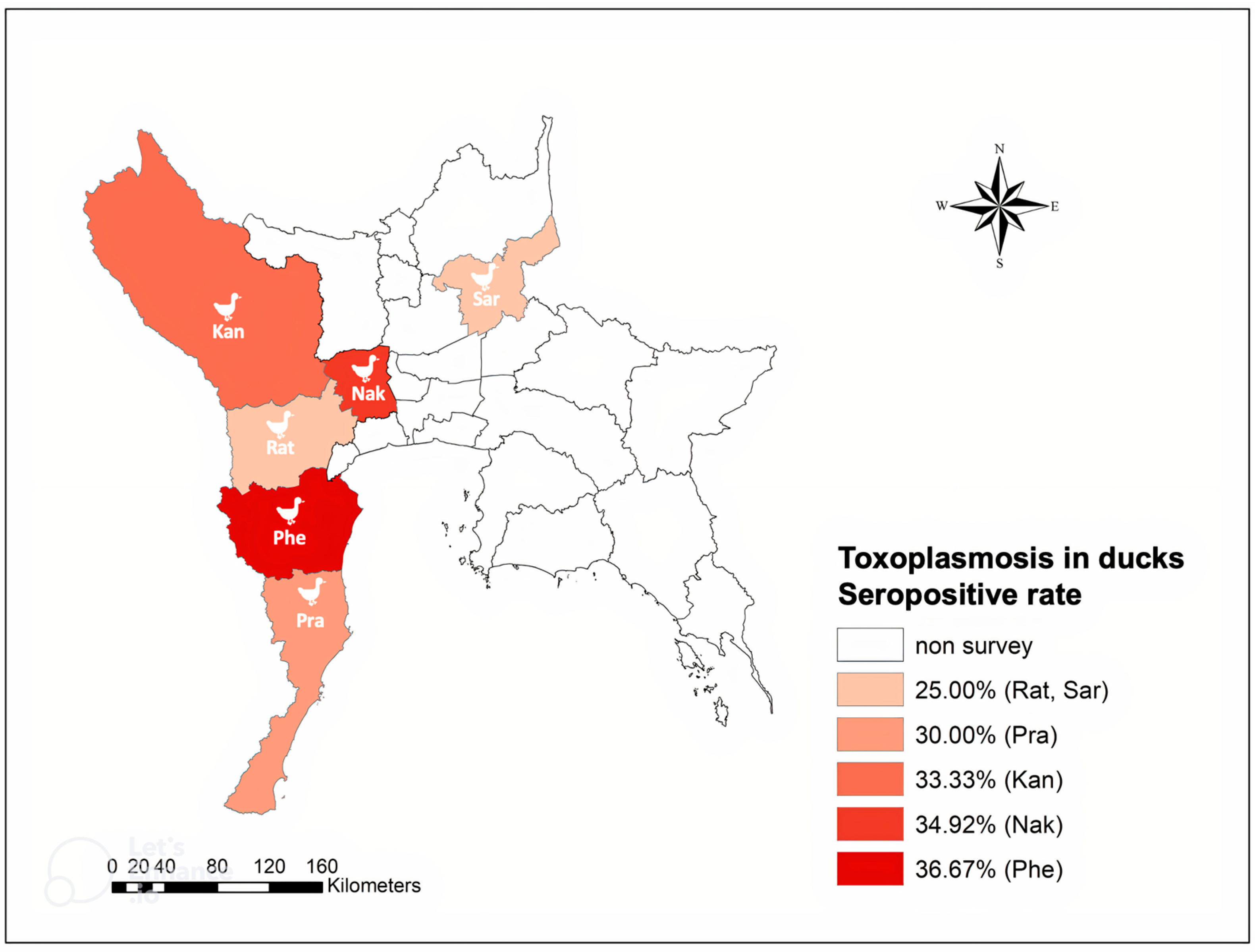
3.2. Seropositive Rate and Geographical Distribution of T. gondii Infection in Free-Grazing Ducks

Positive T. gondii antibody titers were detected in 68/217 (31.3%) ducks, with a titer of 1:100 being used as the cut-off value. The seropositive rates of T. gondii in the fattening and the spent layers were 29% and 32.3%, respectively. No significant variation was found between the two duck groups (𝜒2 = 0.1, df = 1, p = 0.8). The majority of the positive samples showed a low IFAT titer of 1:100 (75%), followed by a titer of 1:200 (23.5%). The Wilcoxon’s test showed a significantly higher titer distribution in the fattening ducks compared to the spent layers (W = 326.5, p = 0.02). Eighty-two % of the positive sera were observed at the titer of 1:100 in the spent layers, making it the most frequent titer in this type of duck. The titer of 1:200 accounted for the remaining 18% of the positive sera. Meanwhile, over 55% of the positive sera of the fattening ducks were recorded at a 1:100 titer. Only one sample had a titer of 1:400, and 38.9% of positive samples showed a 1:200 titer in this group (Table 1).
The seropositive ducks were widely distributed among all investigated provinces: Kanchanaburi, 10/30 (33.3%); Nakhon Pathom, 22/63 (34.9%); Phetchaburi, 11/30 (36.7%); Prachuap Khiri Khan, 9/30 (30%); and Ratchaburi and Saraburi, 8/32 (25%) (Figure 2). The differences in the seropositive rates of the flocks from the investigated provinces were not significant (p = 0.8).

4. Discussion

The present investigation revealed a high seropositivity rate for T. gondii (31.3%) in free-grazing ducks from the central and western provinces of Thailand compared to that in ducks from different sites throughout the world; varying seropositivity rates of T. gondii infection in ducks were also reported at 5.7% in Germany [19], 14.6% in Malaysia [18], 6.6–13.2% in China [23], and 10.6–55% in Egypt [24,25,26]. These differences might be due to the presence of technical or sampling differences (serological tests, cut-off values, sources for animals, sample sizes, age, feeding range, etc.) but also the magnitude of infection pressure from T. gondii oocysts or tissue cysts in animal carcasses (e.g., rodents) in the study areas (Table 2). The cut-off value of 1:100, which is considerably higher than those of previous studies, was chosen to improve the specificity of IFAT in the present study, thus avoiding false positives during the examination [19].
In Thailand, the tradition of rotating free-grazing ducks is closely linked to rice crop cycles and is widely distributed and commercialized throughout the central, western, and eastern regions [14]. Free-grazing duck flocks forage in the paddy fields until the flowering stage and then again after harvest time. They scavenge for insects, snails, husks, weeds, and leftover grains, thus reducing the need for chemical fertilizers and pesticides. Since no biosecurity aspects are considered in the free-grazing systems, grazing and dabbling ducks are constantly exposed to peroral transmission of waterborne pathogens from the environment [15]. In the case of T. gondii, this risk for individuals in a flock is cumulative with several dimensions, e.g., total surface area used for feeding (including transports of flocks to remote paddies), cumulative time of exposure, degree of oocyst contamination in feeding areas (linked to the density of the cat population), and management of offal from local slaughtering (use as cat food). This could be a reason why the free-grazing ducks in this study were relatively highly seropositive with T. gondii than caged or farm ducks. Indeed, they can ingest T. gondii tissue cysts in carcasses of intermediate hosts or oocysts released by cats in the fields, which in turn might indicate environmental contamination [19]. Moreover, the flocks are transported to other villages, districts, or even provinces over substantial distances after the exhaustion of the food supply in the paddy fields, perhaps also contributing to a wider distribution of oocysts [15]. In the present study, a similar significant prevalence of T. gondii infection in the free-grazing ducks sampled in all investigated provinces of Central and Western Thailand suggested the strong possibility of exposure to the parasite of the livestock and humans in these regions. Hence, to address these risks, a One Health concept is required, which takes all aspects of this integrated farming system into consideration [31]. This includes also abiotic environmental factors, such as rainfall and temperature. These climatic factors govern the circulation T. gondii oocysts in the natural environment [32,33]. A tropical savanna climate is recorded in the central and western parts of Thailand, with the annual rainfall and temperature being approximately 1390.8 mm and 29 °C, respectively. Especially, the highest mean temperature of approximately 36 °C, monthly precipitation of 188 mm, and relatively high humidity of 73% were recorded during the sample collection between April and May [34]. This humid and hot condition suits T. gondii oocysts to sporulate and prolong [32], thus increasing the risk of infection with sporulated oocysts in animals when feeding in the pastures.
Even though there was no statistically significant difference between the frequencies of positive sera in the two duck types, significantly higher antibody titers against T. gondii infection were found in the fattening ducks compared to the spent layer ducks in this study (p = 0.02). Maksimov et al. (2011) experimentally inoculated ducks with tachyzoites and oocysts of T. gondii, which revealed that their antibody response levels were partly dependent on the stages of the parasite with which the ducks were infected [19]. Indeed, the group inoculated with T. gondii oocysts developed stronger and longer-lasting antibody responses than those injected with tachyzoites. Additionally, antibodies to T. gondii were firstly observed in the first week postinoculation (wpi), and they reached the highest levels between the third and fourth wpi in both groups of duck. The antibody responses could then last for over eleven wpi and decrease more or less rapidly, eventually persisting for life at the residual level [19,35], indicating that antibody levels also depend on when the ducks are diagnosed during the infectious period. In this study, the fattening ducks (males) were normally released in the paddy fields for approximately a couple of months until slaughtered. Meanwhile, the spent layer ducks were also rotated in the fields for around a half year. Then, they were slaughtered after a nesting period of one to two years at the farms [15]. In other words, the fattening was sampled much earlier (2 months) than the spent layers (over a year of nesting period) after their exposure to pathogens. Therefore, a general tendency for higher antibody titers could be seen in the fattening ducks.
The domestic ducks were demonstrated to be relatively susceptible to T. gondii infection [19,36]. Although the disease does not frequently develop symptoms in poultry, especially in the cases of avirulent T. gondii strains, tissue cysts may be still distributed in some parts of the duck body [36]. From the aspect of food safety, skeletal muscles and hearts are considered the most intensively infected tissues [1]. Furthermore, T. gondii cysts from tissues on day 28 postinfection were successfully isolated [36]. In the present study, the antibodies against T. gondii infection were detected in both the fattening and spent layer ducks at 3 months of age and over, indicating that cysts are likely to present in their tissues. Therefore, determining the presence of tissue cysts in free-grazing ducks and oocysts in the environment is necessary to clarify the possibility of zoonosis in Thailand. Indeed, integrative research requiring a combination of spatial, geographic, and other mathematical models, field and laboratory methods, and veterinary and medical practices is obviously needed to assess the risk factors. This is also crucial in devising preventive measures for toxoplasmosis transmitted from livestock and the natural environment to humans, particularly in sheltering and outdoor cat management policies.

5. Conclusions

In summary, the present study indicated a high seropositivity rate of toxoplasmosis, and it is widely distributed in free-grazing ducks from Central and Western Thailand. This study revealed a considerable risk of infection for livestock and humans via the environment contaminated with T. gondii in the study areas. Therefore, the potential risk of human infection should be avoided by the restriction of consuming undercooked duck meat. Further studies on the genetic characterization are needed to clarify which T. gondii strains are present in ducks reared in farmed environments in Thailand. Furthermore, a prospective longitudinal study in a free-grazing duck flock might indicate the impact of seasonal changes on T. gondii infection.

Author Contributions

Conceptualization, T.T.N., K.K., W.P. and T.I.; methodology, T.T.N. and T.I.; software, T.T.N.; validation, T.T.N., K.K., W.P. and T.I.; formal analysis, T.T.N.; investigation, T.T.N.; resources, K.K. and T.I.; data curation, T.T.N., R.J.-o., P.P. and T.I.; writing—original draft preparation, T.T.N.; writing—review and editing, K.K., A.B.H. and T.I.; visualization, T.T.N.; supervision K.K., W.P., A.B.H. and T.I.; project administration, T.I.; funding acquisition, T.I. All authors have read and agreed to the published version of the manuscript.

Funding

This work was financially supported by a grant from the Faculty of Veterinary Medicine, Kasetsart University, Bangkok, Thailand (grant number: TWP.64_03), and the DAAD In-Country/In-Region Scholarship Programme 2019 (grant number: 57454966) offered by DAAD–SEARCA scholarship (German Academic Exchange Service–Southeast Asian Regional Center for Graduate Study and Research in Agriculture). The APC was financially funded by the faculty of Veterinary Medicine, Kasetsart University, Bangkok, Thailand.

Institutional Review Board Statement

The study was conducted in accordance with the Ethical Review Board of the Office of the National Research Council of Thailand (NRCT), and approved by Kasetsart University’s Institution Animal Care and Use Committee (approval number: ACKU63-VET035).

Informed Consent Statement

Not applicable.

Data Availability Statement

Not applicable.

Acknowledgments

The authors would like to thank Thaweesak Songserm (Faculty of Veterinary Medicine, Kasetsart University, Thailand) for his dedication to consulting on the research question, contacting the slaughterhouse owner, and collecting the samples. We also thank the staff and graduate students in the Department of Parasitology, Faculty of Veterinary Medicine, Kasetsart University, for their support during our project.

Conflicts of Interest

The authors declare no conflict of interest.

References

  1. Webster, J.P. Review of “Toxoplasmosis of Animals and Humans (Second Edition)” by J.P. Dubey. Parasite Vectors 2010, 3–112. [Google Scholar]
  2. Molan, A.; Nosaka, K.; Wang, W.; Hunter, M. Global status of Toxoplasma gondii infection: Systematic review and prevalence snapshots. Trop. Biomed. 2019, 36, 898–925. [Google Scholar] [CrossRef]
  3. Pleyer, U.; Gross, U.; Schluter, D.; Wilking, H.; Seeber, F. Toxoplasmosis in Germany. Dtsch Arztebl Int. 2019, 116, 435–444. [Google Scholar]
  4. Wanachiwanawin, D.; Sutthent, R.; Chokephaibulkit, K.; Mahakittikun, V.; Ongrotchanakun, J.; Monkong, N. Toxoplasma gondii antibodies in HIV and non-HIV infected Thai pregnant women. Asian Pac. J. Allergy Immunol. 2001, 19, 291–293. [Google Scholar] [PubMed]
  5. Chaichan, P. Epidemiology of Toxoplasma gondii in Thailand. Ph.D. Thesis, University of Limoges, Limoges, France, 2017. [Google Scholar]
  6. Inpankaew, T.; Pinyopanuwut, N.; Chimnoi, W.; Kengradomkit, C.; Sununta, C.; Zhang, G.; Nishikawa, Y.; Igarashi, I.; Xuan, X.; Jittapalapong, S. Serodiagnosis of Toxoplasma gondii infection in dairy cows in Thailand. Transbound. Emerg. Dis. 2010, 57, 42–45. [Google Scholar] [CrossRef] [PubMed]
  7. Jittapalapong, S.; Inpankaew, T.; Pinyopanuwat, N.; Chimnoi, W.; Kengradomkij, C.; Wongnarkpet, S.; Maruayama, S.; Lekkla, A.; Sukthana, Y. Epidemiology of Toxoplasma gondii infection of stray cats in Bangkok, Thailand. Southeast Asian J. Trop. Med. Public Health 2010, 41, 13–18. [Google Scholar]
  8. Jittapalapong, S.; Sittisan, P.; Sakpuaram, T.; Kabeya, H.; Maruyama, S.; Inpankaew, T. Coinfection of Leptospira spp and Toxoplasma gondii among stray dogs in Bangkok, Thailand. Southeast Asian J. Trop. Med. Public Health 2009, 40, 247–252. [Google Scholar]
  9. Kengradomkij, C.; Kamyingkird, K.; Pinyopanuwat, N.; Chimnoi, W.; Jittapalapong, S.; Inpankaew, T. Seroprevalence of Toxoplasma gondii from stray cats residing in temples, Bangkok, Thailand. J. Trop. Med. Parasitol. 2018, 41, 8–14. [Google Scholar]
  10. Tuntasuvan, D.; Mohkaew, K.; Dubey, J.P. Seroprevalence of Toxoplasma gondii in elephants (Elephus maximus indicus) in Thailand. J. Parasitol. 2001, 87, 229–230. [Google Scholar] [CrossRef]
  11. Tridge. Top Export Destinations of Duck Meat from Thailand 2022. Available online: https://www.tridge.com/intelligences/duck-meat/TH (accessed on 15 June 2022).
  12. Volza Grow Global. Duck Meat Exports from Thailand 2021. Available online: https://www.volza.com/p/duck-meat/export/export-from-thailand/ (accessed on 15 June 2022).
  13. Department of Livestock Development. Data from: Number of livestock inventory in Thailand. 2017. Available online: https://ict.dld.go.th/webnew/index.php/th/service-ict/report/289-report-thailand-livestock/reportservey2560 (accessed on 15 June 2022).
  14. Gilbert, M.; Chaitaweesub, P.; Parakamawongsa, T.; Premashthira, S.; Tiensin, T.; Kalpravidh, W.; Wagner, H.; Slingenbergh, J. Free-grazing ducks and highly pathogenic avian influenza, Thailand. Emerg. Infect. Dis. 2006, 12, 227–234. [Google Scholar] [CrossRef]
  15. Songserm, T.; Jam-on, R.; Sae-Heng, N.; Meemak, N.; Hulse-Post, D.; Sturm-Ramirez, K.; Robert, G.W. Domestic Ducks and H5N1 Influenza Epidemic, Thailand. Emerg. Infect. Dis. 2006, 12, 575–581. [Google Scholar] [CrossRef]
  16. El-Massry, A.; Mahdy, O.A.; El-Ghaysh, A.; Dubey, J.P. Prevalence of Toxoplasma gondii antibodies in sera of turkeys, chickens, and ducks from Egypt. J. Parasitol. 2000, 86, 627–628. [Google Scholar] [CrossRef] [PubMed]
  17. Dubey, J.P. Toxoplasma gondii infections in chickens (Gallus domesticus): Prevalence, clinical disease, diagnosis and public health significance. Zoonoses Public Health 2010, 57, 60–73. [Google Scholar] [CrossRef]
  18. Puvanesuaran, V.; Noordin, R.; Balakrishnan, V. Isolation and genotyping of Toxoplasma gondii from free-range ducks in Malaysia. Avian Dis. 2013, 57, 128–132. [Google Scholar] [CrossRef] [PubMed]
  19. Maksimov, P.; Buschtons, S.; Herrmann, D.C.; Conraths, F.J.; Gorlich, K.; Tenter, A.M.; Dubey, J.P.; Nagel-Kohl, U.; Thoms, B.; Botcher, L.; et al. Serological survey and risk factors for Toxoplasma gondii in domestic ducks and geese in Lower Saxony, Germany. Vet. Parasitol. 2011, 182, 140–149. [Google Scholar] [CrossRef] [PubMed]
  20. Casartelli-Alves, L.; Boechat, V.C.; Macedo-Couto, R.; Ferreira, L.C.; Nicolau, J.L.; Neves, L.B.; Millar, P.R.; Vicente, R.T.; Oliveira, R.V.C.; Muniz, A.G.; et al. Sensitivity and specificity of serological tests, histopathology and immunohistochemistry for detection of Toxoplasma gondii infection in domestic chickens. Vet. Parasitol. 2014, 204, 346–351. [Google Scholar] [CrossRef] [PubMed]
  21. Nguyen, T.T.; Kengradomkij, C.; Inpankaew, T. Detection of antibodies to Toxoplasma gondii among owned dogs in Cambodia. Food Waterborne Parasitol. 2020, 22, e00103. [Google Scholar] [CrossRef]
  22. Sabin, A.B. Toxoplasmic encephalitis in children. JAMA 1941, 116, 801–807. [Google Scholar] [CrossRef]
  23. Zhao, G.; Song, Z.; Wang, S.; Hassan, I.A.; Wang, W.; Cheng, F.; Yang, X. A seroepidemiological survey of Toxoplasma gondii infection in free-range and caged ducks in Southwest China. Isr. J. Vet. Med. 2015, 70, 41–45. [Google Scholar]
  24. Harfoush, M.; Tahoon Ael, N. Seroprevalence of Toxoplasma gondii antibodies in domestic ducks, free-range chickens, turkeys and rabbits in Kafr El-Sheikh Governorate Egypt. J. Egypt Soc. Parasitol. 2010, 40, 295–302. [Google Scholar]
  25. Aboulaila, M.; ElBahy, N.; Hilali, M.; Yokoyama, N.; Igarashi, I. Serodiagnosis of Toxoplasma gondii in ducks from Behera Governorate, Egypt. J. Protozool. Res. 2011, 21, 45–49. [Google Scholar]
  26. Ibrahim, H.M.; Osman, G.Y.; Mohamed, A.H.; Al-Selwi, A.G.M.; Nishikawa, Y.; Abdel-Ghaffar, F. Toxoplasma gondii: Prevalence of natural infection in pigeons and ducks from middle and upper Egypt using serological, histopathological, and immunohistochemical diagnostic methods. Vet. Parasitol. Reg. Stud. Rep. 2018, 13, 45–49. [Google Scholar] [CrossRef] [PubMed]
  27. Alkhaled, M.J.A.; Yakoob, A.Y.; Al-Hamadani, A.H.U. An investigation of Toxoplasmosis in Free Range chickens, Industrial chickens and Duck in mid Euphrates area of Iraq. AL-Qadisiya J. VetMedSci 2012, 1, 17. [Google Scholar] [CrossRef]
  28. Amouei, A.; Sharif, M.; Hosseini, S.A.; Sarvia, S.; Mizani, A.; Salehi, S.; Gholami, S.; Jafar-Ramaji, T.; Daryani, A. Prevalence of Toxoplasma gondii infection in domestic and migrating birds from Mazandaran province, Northern Iran. Avian Biol. Res. 2018, 11, 12–15. [Google Scholar] [CrossRef]
  29. Bártová, E.; Sedlák, K.; Literák, I. Serologic survey for toxoplasmosis in domestic birds from the Czech Republic. Avian Pathol. 2009, 38, 317–320. [Google Scholar] [CrossRef]
  30. Sroka, J.; Wojcik-Fatla, A.; Szymanska, J.; Dutkiewicz, J.; Zajac, V.; Zwolinski, J. The occurrence of Toxoplasma gondii infection in people and animals from rural environment of Lublin region-estimate of potential role of water as a source of infection. Ann. Agric. Environ. Med. 2010, 17, 125–132. [Google Scholar]
  31. One Health High-Level Expert Panel (OHHLEP); Adisasmito, W.B.; Almuhairi, S.; Behravesh, C.B.; Bilivogui, P.; Bukachi, S.A.; Casas, N.; Becerra, N.C.; Charron, D.F.; Chaudhary, A.; et al. One Health: A new definition for a sustainable and healthy future. PLOS Pathog. 2022, 18, e1010537. [Google Scholar]
  32. Yan, C.; Liang, L.J.; Zheng, K.Y.; Zhu, X.Q. Impact of environmental factors on the emergence, transmission and distribution of Toxoplasma gondii. Parasit Vectors 2016, 9, 137. [Google Scholar] [CrossRef]
  33. Simon, A.; Poulin, M.B.; Rousseau, A.N.; Ogden, N.H. Fate and transport of Toxoplasma gondii oocysts in seasonally snow-covered watersheds: A conceptual framework from a melting snowpack to the Canadian arctic coasts. Int. J. Environ. Res. Public Health 2013, 10, 994–1005. [Google Scholar] [CrossRef]
  34. Thai Meteorological Department Automatic Weather System. Climate Data. Available online: http://www.aws-observation.tmd.go.th/web/climate/climate_past.asp (accessed on 20 October 2022).
  35. Robert-Gangneux, F.; Dardé, M.L. Epidemiology of and diagnostic strategies for toxoplasmosis. Clin. Microbiol. Rev. 2012, 25, 264–296. [Google Scholar] [CrossRef] [PubMed] [Green Version]
  36. Bártová, E.; Dvoráková, H.; Bárta, J.; Sedlák, K.; Literák, I. Susceptibility of the domestic duck (Anas platyrhynchos) to experimental infection with Toxoplasma gondii oocysts. Avian Pathol. 2004, 33, 153–157. [Google Scholar] [CrossRef] [PubMed]
Figure 1. Study area: (A) distribution of free-grazing ducks in Thailand (13); (B) six investigated provinces with the number of flocks and ducks collected.
Tropicalmed 08 00103 g001
Figure 2. Geographical distribution of T. gondii infection in free-grazing ducks in the central and western areas of Thailand.
Tropicalmed 08 00103 g002
Table 1. T. gondii seropositive free-gazing ducks in this study.
Type of DucksNo. of TestedNo. of Positive (%)Statistical AnalysisTiter (%)
1:1001:2001:400
Fattening6218 (29)𝜒2 = 0.1, df = 1,
p = 0.8
10 (55.6)7 (38.9)1 (5.6)
Spent layer 15550 (32.3)41 (82)9 (18)0 (0)
Total21768 (31.3)-7523.51.5
Table 2. Worldwide seropositive rates of T. gondii in ducks.
CountrySourceSeropositive Rate TestCut-OffReference
ThailandS 1/FR 231.3IFAT1:100This study
MalaysiaF 3/FR14.6MAT 61:6[18]
ChinaFR13.2MAT1:25[23]
C 46.6
IraqM 556LAT 71:2[27]
IranM/FR46MAT1:20[28]
GermanyF/FR5.7ELISA8 (SAG1) 90.14[19]
Czech RepublicF14IFAT1:40[29]
PolandF21.2MAT1:40[30]
EgyptF55IHA 101:80[24]
S13.6MAT1:25[25]
M10.6ELISA (SAG2t) 110.014[26]
1 Slaughterhouse; 2 free range; 3 farm; 4 caged; 5 market; 6 modified agglutination test; 7 latex agglutination test; 8 enzyme-linked immunosorbent assay; 9 protein used in test; 10 indirect hemagglutination; 11 protein used in test.
Disclaimer/Publisher’s Note: The statements, opinions and data contained in all publications are solely those of the individual author(s) and contributor(s) and not of MDPI and/or the editor(s). MDPI and/or the editor(s) disclaim responsibility for any injury to people or property resulting from any ideas, methods, instructions or products referred to in the content.

