
Primary Cutaneous Cryptococcosis Caused by Cryptococcus gatti in an Elderly Patient

 , , and
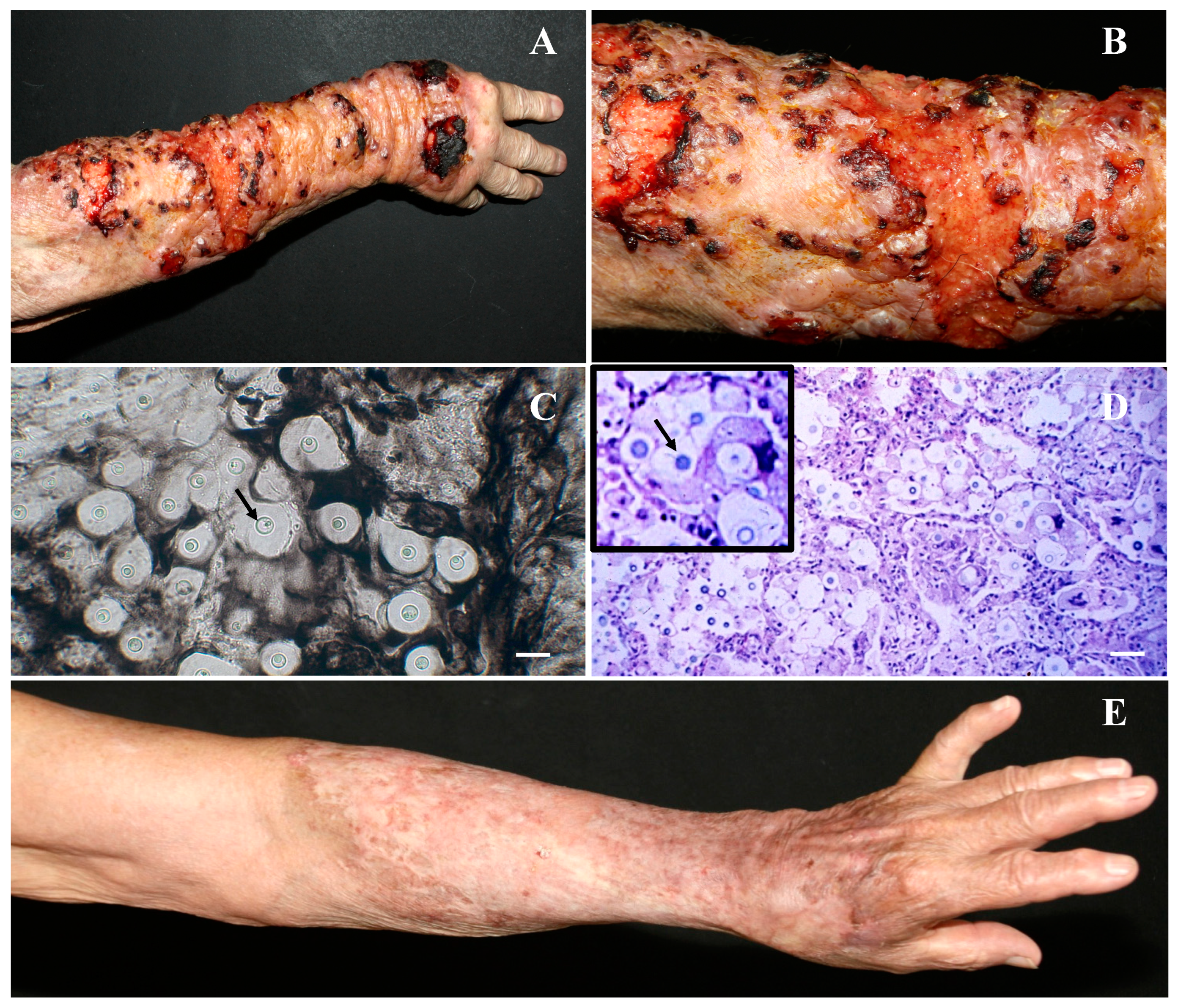
Abstract
:1. Introduction
2. Case Report
3. Discussion
4. Conclusions
Author Contributions
Funding
Institutional Review Board Statement
Informed Consent Statement
Data Availability Statement
Acknowledgments
Conflicts of Interest
References
- Firacative, C.; Trilles, L.; Meyer, W. Recent Advances in Cryptococcus and Cryptococcosis. Microorganisms 2021, 10, 13. [Google Scholar] [CrossRef] [PubMed]
- Hsu, E.; Webster, S.M.; Nanes, M. Disseminated Cryptococcosis in An Immunocompetent Host Presenting As Osteomyelitis and Leading to Adrenal Insufficiency. Am. J. Med. Sci. 2022, 363, 75–79. [Google Scholar] [CrossRef] [PubMed]
- Christianson, J.C.; Engber, W.; Andes, D. Primary cutaneous cryptococcosis in immunocompetent and immunocompromised hosts. Med. Mycol. 2003, 41, 177–188. [Google Scholar] [CrossRef] [PubMed]
- Pappas, P.G.; Perfect, J.R.; Cloud, G.A.; Larsen, R.A.; Pankey, G.A.; Lancaster, D.J.; Henderson, H.; Kauffman, C.A.; Haas, D.W.; Saccente, M.; et al. Cryptococcosis in Human Immunodeficiency Virus—Negative Patients in the Era of Effective Azole Therapy. Clin. Infect. Dis. 2001, 33, 690–699. [Google Scholar] [CrossRef] [Green Version]
- Vandersmissen, G.; Meuleman, L.; Tits, G.; Verhaeghe, A.; Peetermans, W.E. Cutaneous Cryptococcosis In Corticosteroid-Treated Patients Without Aids. Acta Clin. Belg. 1996, 51, 111–117. [Google Scholar] [CrossRef]
- Rathore, S.S.; Sathiyamoorthy, J.; Lalitha, C.; Ramakrishnan, J. A holistic review on Cryptococcus neoformans. Microb. Pathog. 2022, 166, 105521. [Google Scholar] [CrossRef]
- Muselius, B.; Durand, S.-L.; Geddes-McAlister, J. Proteomics of Cryptococcus neoformans: From the Lab to the Clinic. Int. J. Mol. Sci. 2021, 22, 12390. [Google Scholar] [CrossRef]
- Florek, M.; Korzeniowska-Kowal, A.; Wzorek, A.; Włodarczyk, K.; Marynowska, M.; Pogorzelska, A.; Brodala, M.; Ploch, S.; Buczek, D.; Balon, K.; et al. Prevalence, Genetic Structure, and Antifungal Susceptibility of the Cryptococcus neoformans/C. gattii Species Complex Strains Collected from the Arboreal Niche in Poland. Pathogens 2021, 11, 8. [Google Scholar] [CrossRef]
- Xiujiao, X.; Ai’e, X. Two cases of cutaneous cryptococcosis. Mycoses 2005, 48, 238–241. [Google Scholar] [CrossRef]
- Maciey, S.; Santa Maria, C.; Oshima, S.; Newberry, J. Cryptococcus gattii Meningitis in a Previously Healthy Young Woman: A Case Report. Clin. Pr. Cases Emerg. Med. 2021, 5, 345–349. [Google Scholar] [CrossRef]
- MacDougall, L.; Fyfe, M.; Romney, M.; Starr, M.; Galanis, E. Risk Factors for Cryptococcus gattii Infection, British Columbia, Canada. Emerg. Infect. Dis. 2011, 17, 193–199. [Google Scholar] [CrossRef] [PubMed]
- da Lacaz, C.S.; Heins-Vaccari, E.M.; Hernández-Arriagada, G.L.; Martins, E.L.; Prearo, C.A.L.; Corim, S.M.; Martins, M.d.A. Primary cutaneous cryptococcosis due to Cryptococcus neoformans var. gattii serotype B, in an immunocompetent patient. Rev. Inst. Med. Trop. Sao. Paulo. 2002, 44, 225–228. [Google Scholar] [CrossRef] [PubMed] [Green Version]
- Hurst, S.; Lysen, C.; Cooksey, G.; Vugia, D.J.; Litvintseva, A.P.; Lockhart, S.R. Molecular typing of clinical and environmental isolates of Cryptococcus gattii species complex from southern California, United States. Mycoses 2019, 62, 1029–1034. [Google Scholar] [CrossRef] [PubMed]
- Lockhart, S.R.; Roe, C.C.; Engelthaler, D.M. Timing the Origin of Cryptococcus gattii sensu stricto, Southeastern United States. Emerg. Infect. Dis. 2018, 24, 2095–2097. [Google Scholar] [CrossRef] [PubMed] [Green Version]
- Campuzano, A.; Wormley, F. Innate Immunity against Cryptococcus, from Recognition to Elimination. J. Fungi 2018, 4, 33. [Google Scholar] [CrossRef] [Green Version]
- Hill, J.O. CD4+ T cells cause multinucleated giant cells to form around Cryptococcus neoformans and confine the yeast within the primary site of infection in the respiratory tract. J. Exp. Med. 1992, 175, 1685–1695. [Google Scholar] [CrossRef]
- Tan, S.; Nasr, G.; Harding, C. Lung Masses as a Presenting Sign of Disseminated Cryptococcus. Cureus 2021, 13, e15185. [Google Scholar] [CrossRef]
- Jarvis, J.N.; Harrison, T.S. HIV-associated cryptococcal meningitis. AIDS 2007, 21, 2119–2129. [Google Scholar] [CrossRef]
- Graybill, J.R.; Sobel, J.; Saag, M.; van Der Horst, C.; Powderly, W.; Cloud, G.; Riser, L.; Hamill, R.; Dismukes, W. Diagnosis and management of increased intracranial pressure in patients with AIDS and cryptococcal meningitis. The NIAID Mycoses Study Group and AIDS Cooperative Treatment Groups. Clin. Infect. Dis. 2000, 30, 47–54. [Google Scholar] [CrossRef] [Green Version]
- Amphornphruet, A.; Silpa-Archa, S.; Preble, J.M.; Foster, C.S. Endogenous Cryptococcal Endophthalmitis in Immunocompetent Host: Case Report and Review of Multimodal Imaging Findings and Treatment. Ocul. Immunol. Inflamm. 2018, 26, 518–522. [Google Scholar] [CrossRef]
- Beyt, B.E.; Waltman, S.R. Cryptococcal endophthalmitis after corneal transplantation. N. Engl. J. Med. 1978, 298, 825–826. [Google Scholar] [CrossRef] [PubMed]
- Chen, S.C.-A.; Meyer, W.; Sorrell, T.C. Cryptococcus gattii infections. Clin. Microbiol. Rev. 2014, 27, 980–1024. [Google Scholar] [CrossRef] [PubMed] [Green Version]
- Zavala, S.; Baddley, J.W. Cryptococcosis. Semin. Respir. Crit. Care Med. 2020, 41, 69–79. [Google Scholar] [CrossRef] [PubMed]
- Setianingrum, F.; Rautemaa-Richardson, R.; Denning, D.W. Pulmonary cryptococcosis: A review of pathobiology and clinical aspects. Med. Mycol. 2019, 57, 133–150. [Google Scholar] [CrossRef] [PubMed]
- Chang, W.-C.; Tzao, C.; Hsu, H.-H.; Lee, S.-C.; Huang, K.-L.; Tung, H.-J.; Chen, C.-Y. Pulmonary cryptococcosis: Comparison of clinical and radiographic characteristics in immunocompetent and immunocompromised patients. Chest 2006, 129, 333–340. [Google Scholar] [CrossRef]
- Noguchi, H.; Matsumoto, T.; Kimura, U.; Hiruma, M.; Kusuhara, M.; Ihn, H. Cutaneous Cryptococcosis. Med. Mycol. J. 2019, 60, 101–107. [Google Scholar] [CrossRef] [Green Version]
- Zhou, H.-X.; Lu, L.; Chu, T.; Wang, T.; Cao, D.; Li, F.; Ning, G.; Feng, S. Skeletal cryptococcosis from 1977 to 2013. Front. Microbiol. 2015, 5, 740. [Google Scholar] [CrossRef] [Green Version]
- Desalermos, A.; Kourkoumpetis, T.K.; Mylonakis, E. Update on the epidemiology and management of cryptococcal meningitis. Expert Opin. Pharmacother. 2012, 13, 783–789. [Google Scholar] [CrossRef] [Green Version]
- Park, B.J.; Wannemuehler, K.A.; Marston, B.J.; Govender, N.; Pappas, P.G.; Chiller, T.M. Estimation of the current global burden of cryptococcal meningitis among persons living with HIV/AIDS. AIDS 2009, 23, 525–530. [Google Scholar] [CrossRef]
- Gushiken, A.C.; Saharia, K.K.; Baddley, J.W. Cryptococcosis. Infect. Dis. Clin. N. Am. 2021, 35, 493–514. [Google Scholar] [CrossRef]
- Nascimento, E.; Barião, P.H.G.; Kress, M.R.V.Z.; Vilar, F.C.; Santana, R.D.C.; Gaspar, G.G.; Martinez, R. Cryptococcosis by Cryptococcus neoformans/Cryptococcus gattii Species Complexes in non-HIV-Infected Patients in Southeastern Brazil. Rev. Soc. Bras. Med. Trop. 2021, 54, e01692021. [Google Scholar] [CrossRef] [PubMed]
- Trilles, L.; dos Lazéra, M.S.; Wanke, B.; Oliveira, R.V.; Barbosa, G.G.; Nishikawa, M.M.; Morales, B.P.; Meyer, W. Regional pattern of the molecular types of Cryptococcus neoformans and Cryptococcus gattii in Brazil. Mem. Inst. Oswaldo Cruz 2008, 103, 455–462. [Google Scholar] [CrossRef] [PubMed]
- Firacative, C.; Lizarazo, J.; Illnait-Zaragozí, M.T.; Castañeda, E.; Latin American Cryptococcal Study Group. The status of cryptococcosis in Latin America. Mem. Inst. Oswaldo Cruz 2018, 113, e170554. [Google Scholar] [CrossRef] [PubMed] [Green Version]
- Firacative, C.; Meyer, W.; Castañeda, E. Cryptococcus neoformans and Cryptococcus gattii Species Complexes in Latin America: A Map of Molecular Types, Genotypic Diversity, and Antifungal Susceptibility as Reported by the Latin American Cryptococcal Study Group. J. Fungi 2021, 7, 282. [Google Scholar] [CrossRef]
- Perfect, J.R.; Dismukes, W.E.; Dromer, F.; Goldman, D.L.; Graybill, J.R.; Hamill, R.J.; Harrison, T.S.; Larsen, R.A.; Lortholary, O.; Nguyen, M.H.; et al. Clinical practice guidelines for the management of cryptococcal disease: 2010 update by the infectious diseases society of america. Clin. Infect. Dis. 2010, 50, 291–322. [Google Scholar] [CrossRef] [Green Version]
- de Spadari, C.C.; Wirth, F.; Lopes, L.B.; Ishida, K. New Approaches for Cryptococcosis Treatment. Microorganisms 2020, 8, 613. [Google Scholar] [CrossRef]
- Klein, K.R.; Hall, L.; Deml, S.M.; Rysavy, J.M.; Wohlfiel, S.L.; Wengenack, N.L. Identification of Cryptococcus gattii by Use of l-Canavanine Glycine Bromothymol Blue Medium and DNA Sequencing. J. Clin. Microbiol. 2009, 47, 3669–3672. [Google Scholar] [CrossRef] [Green Version]
- Lewis, E.D.; Wu, D.; Meydani, S.N. Age-associated alterations in immune function and inflammation. Prog Neuro-Psychopharmacol. Biol Psychiatry 2022, 118, 110576. [Google Scholar] [CrossRef]
- Romiopoulos, I.; Pana, Z.D.; Pyrpasopoulou, A.; Linardou, I.; Avdelidi, E.; Sidiropoulou, M.; Chatzidrosou, E.; Ioannides, D.; Karagiannis, A.; Roilides, E. Fulminant cryptococcal meningoencephalitis after successful treatment of primary cutaneous cryptococcosis. GERMS 2020, 10, 388–391. [Google Scholar] [CrossRef]
- Bardhi, R.; Lawrence, K.; Bedford, L.M.; Deirawan, H.; Moossavi, M. Disseminated cryptococcosis presenting with cutaneous involvement in an immunocompromised patient. Dermatol. Online J. 2020, 26, 15. [Google Scholar] [CrossRef]
- Lu, Y.-Y.; Wu, C.-S.; Hong, C.-H. Primary cutaneous cryptococcosis in an immunocompetent man: A case report. Dermatol. Sin. 2013, 31, 90–93. [Google Scholar] [CrossRef] [Green Version]
- Nowak, M.A.; Putynkowska, A.; Barańska-Rybak, W.; Czarnacka, K.; Dębska-Ślizień, M.A. Cutaneous cryptococcosis: An underlying immunosuppression? Clinical manifestations, pathogenesis, diagnostic examinations and treatment. Adv. Dermatol. Allergol./Postępy Dermatol. i Alergol. 2020, 37, 154–158. [Google Scholar] [CrossRef] [PubMed]
- do Amaral, D.M.; de Rocha, R.C.C.; Carneiro, L.E.P.; Vasconcelos, D.M.; de Abreu, M.A.M.M. Disseminated cryptococcosis manifested as a single tumor in an immunocompetent patient, similar to the cutaneous primary forms. An. Bras. Dermatol. 2016, 91, 29–31. [Google Scholar] [CrossRef] [PubMed] [Green Version]
- Carvalho, A.K.; Carvalho, K.; Passero, L.F.D.; Sousa, M.G.T.; da Matta, V.L.R.; Gomes, C.M.C.; Corbett, C.E.P.; Kallas, G.E.; Silveira, F.T.; Laurenti, M.D. Differential Recruitment of Dendritic Cells Subsets to Lymph Nodes Correlates with a Protective or Permissive T-Cell Response during Leishmania (Viannia) Braziliensis or Leishmania (Leishmania) Amazonensis Infection. Mediat. Inflamm. 2016, 2016, 7068287. [Google Scholar] [CrossRef] [Green Version]
- Ribeiro, S.P.; Milush, J.M.; Cunha-Neto, E.; Kallas, E.G.; Kalil, J.; Passero, L.F.D.; Hunt, P.W.; Deeks, S.; Nixon, D.F.; Sengupta, D. P16INK4a expression and immunologic aging in chronic HIV infection. PLoS ONE. 2016, 11, e0166759. [Google Scholar] [CrossRef] [Green Version]
- Patel, P.; Ramanathan, J.; Kayser, M.; Baran, J. Primary cutaneous cryptococcosis of the nose in an immunocompetent woman. J. Am. Acad. Dermatol. 2000, 43, 344–345. [Google Scholar] [CrossRef]
- Inoue, N.; Endo, Y.; Kabashima, K. Extensive leg ulcer caused by primary cutaneous cryptococcosis in an immunocompromised patient. Eur. J. Dermatol. 2021, 31, 657–658. [Google Scholar] [CrossRef]

Publisher’s Note: MDPI stays neutral with regard to jurisdictional claims in published maps and institutional affiliations. |
© 2022 by the authors. Licensee MDPI, Basel, Switzerland. This article is an open access article distributed under the terms and conditions of the Creative Commons Attribution (CC BY) license (https://creativecommons.org/licenses/by/4.0/).
Share and Cite
Belda, W., Jr.; Casolato, A.T.S.; Luppi, J.B.; Passero, L.F.D.; Criado, P.R. Primary Cutaneous Cryptococcosis Caused by Cryptococcus gatti in an Elderly Patient. Trop. Med. Infect. Dis. 2022, 7, 206. https://doi.org/10.3390/tropicalmed7090206
Belda W Jr., Casolato ATS, Luppi JB, Passero LFD, Criado PR. Primary Cutaneous Cryptococcosis Caused by Cryptococcus gatti in an Elderly Patient. Tropical Medicine and Infectious Disease. 2022; 7(9):206. https://doi.org/10.3390/tropicalmed7090206
Chicago/Turabian StyleBelda, Walter, Jr., Ana T. S. Casolato, Juliana B. Luppi, Luiz Felipe D. Passero, and Paulo R. Criado. 2022. "Primary Cutaneous Cryptococcosis Caused by Cryptococcus gatti in an Elderly Patient" Tropical Medicine and Infectious Disease 7, no. 9: 206. https://doi.org/10.3390/tropicalmed7090206