
Progress and Perspectives of Spectroscopic Studies on Carbon K-Edge Using Novel Soft X-ray Pulsed Sources
 , , , , , and
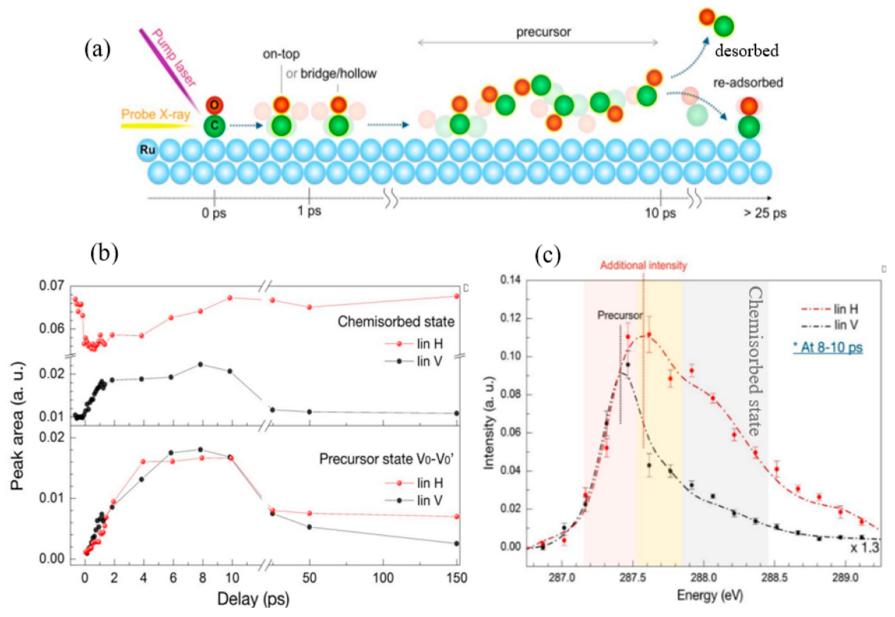
Abstract
:1. Introduction
2. Carbon K-Edge Studies Using FELs
2.1. Probing Catalysis in Real Time: Observation of Transient Precursor States
2.2. Intense FEL-Induced Nonlinear Effects
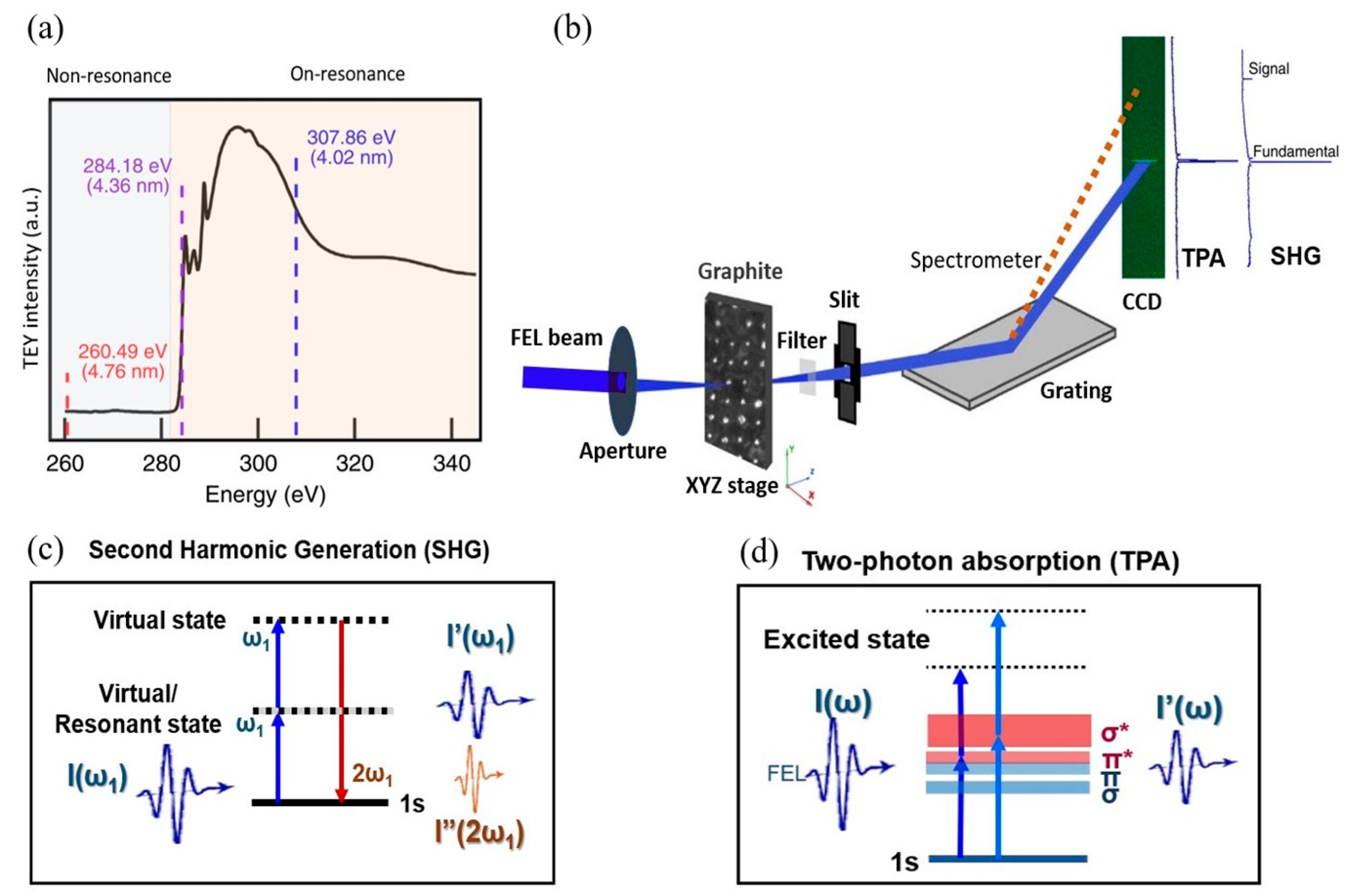
2.2.1. Second Harmonic Generation by Graphite
2.2.2. Two-Photon Absorption by Graphite
2.2.3. Saturable Absorption by Graphite
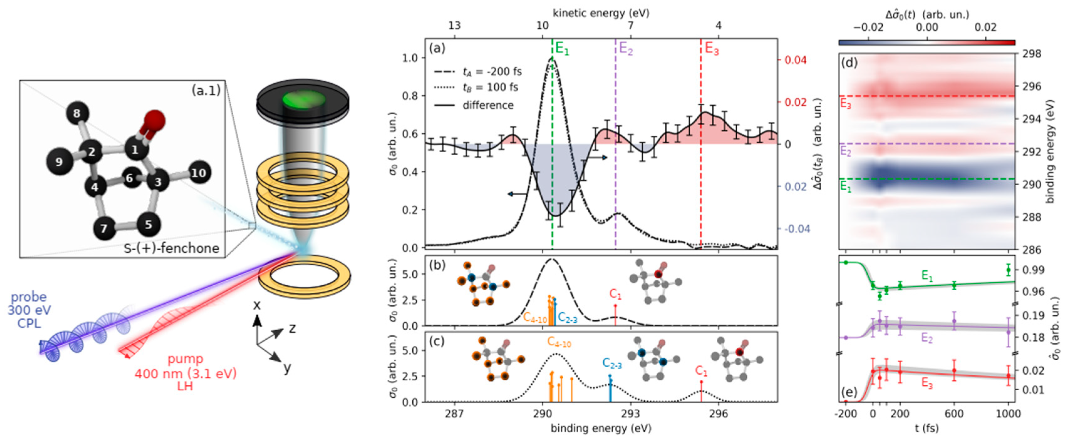
2.3. The Electronic Structure of Photo-Excited Chiral Molecules
3. Carbon K-Edge Studies with HHG Sources
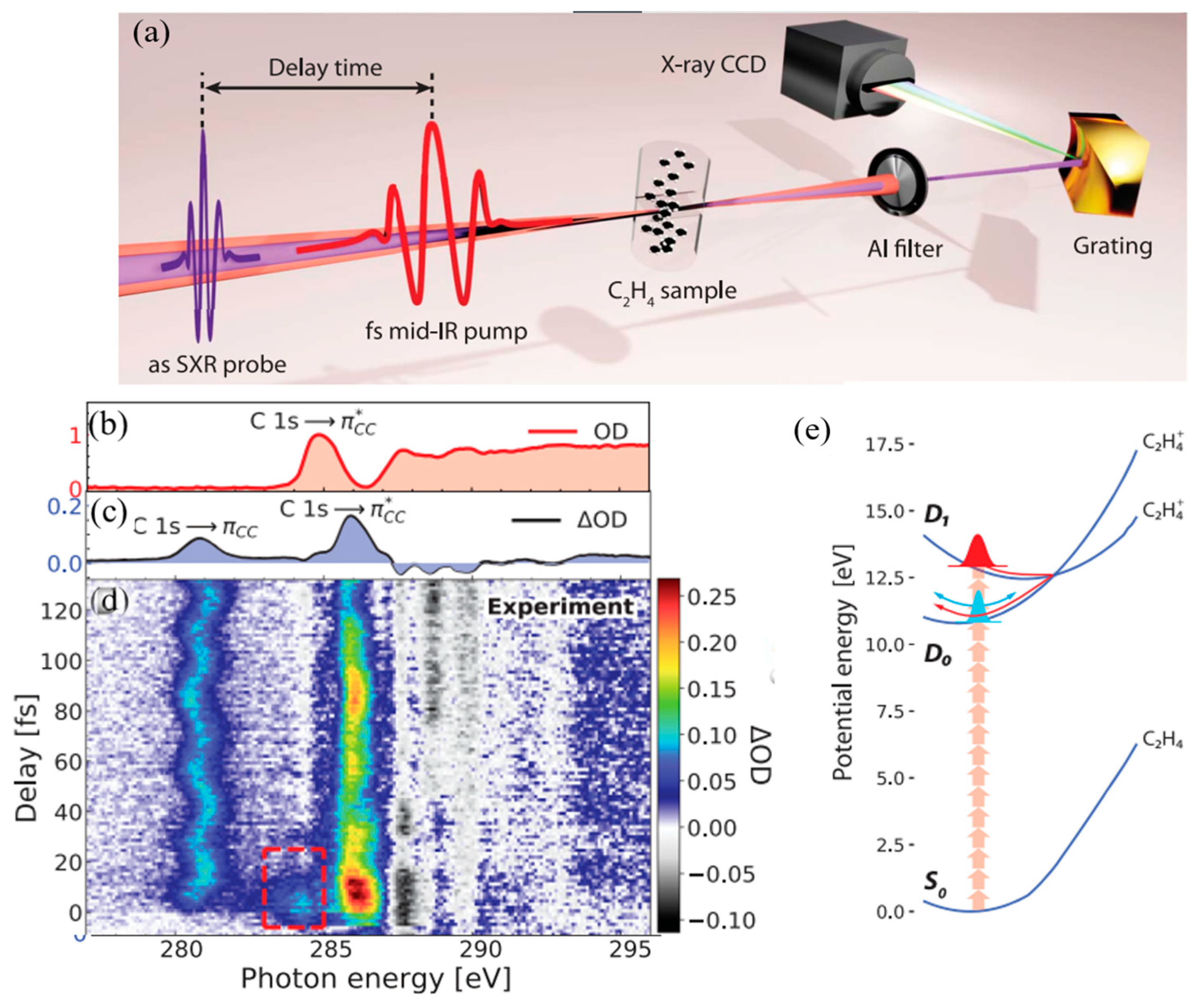
3.1. Ultrafast Electronic and Structural Dynamics of Organic Molecules at Conical Intersections
3.2. Time-Resolved Photodissociation Reactions of Molecular Cations
4. Conclusions and Outlook
Funding
Data Availability Statement
Conflicts of Interest
References
- Smith, N. Science with Soft X Rays. Phys. Today. 2001, 54, 29–34. [Google Scholar] [CrossRef]
- Wong, L.J.; Kaminer, I. Prospects in x-ray science emerging from quantum optics and nanomaterials. Appl. Phys. Lett. 2021, 119, 130502. [Google Scholar] [CrossRef]
- Nascimento, D.R.; Zhang, Y.; Bergmann, U.; Govind, N. Near-Edge X-ray Absorption Fine Structure Spectroscopy of Heteroatomic Core-Hole States as a Probe for Nearly Indistinguishable Chemical Environments. J. Phys. Chem. Lett. 2020, 11, 556–561. [Google Scholar] [CrossRef] [PubMed]
- Rabe, P.; Kamps, J.J.A.G.; Sutherlin, K.D.; Linyard, J.D.S.; Aller, P.; Pham, C.C.; Makita, H.; Clifton, I.; McDonough, M.A.; Leissing, T.M.; et al. X-ray free-electron laser studies reveal correlated motion during isopenicillin N synthase catalysis. Sci. Adv. 2021, 7, eabh0250. [Google Scholar] [CrossRef] [PubMed]
- Rezvani, S.; D’Elia, A.; Macis, S.; Nannarone, S.; Lupi, S.; Schütt, F.; Rasch, F.; Adelung, R.; Lu, B.; Zhang, Z.; et al. Structural anisotropy in three dimensional macroporous graphene: A polarized XANES investigation. Diam. Relat. Mater. 2021, 111, 108171. [Google Scholar] [CrossRef]
- Di Cicco, A.; Rezvani, S.J.; Nannarone, S. Revisiting the Probing Depths of Soft X-ray Absorption Techniques by Constant Initial State Photoemission Experiments. Springer Proc. Phys. 2021, 220, 85–97. [Google Scholar] [CrossRef]
- Di Cicco, A.; Polzoni, G.; Gunnella, R.; Trapananti, A.; Minicucci, M.; Rezvani, S.J.; Catone, D.; Di Mario, L.; Cresi, J.S.P.; Turchini, S.; et al. Broadband optical ultrafast reflectivity of Si, Ge and GaAs. Sci. Rep. 2020, 10, 17363. [Google Scholar] [CrossRef]
- Dell’Angela, M.; Anniyev, T.; Beye, M.; Coffee, R.; Föhlisch, A.; Gladh, J.; Katayama, T.; Kaya, S.; Krupin, O.; LaRue, J.; et al. Real-Time Observation of Surface Bond Breaking with an X-ray Laser. Science 2013, 339, 1302–1306. [Google Scholar] [CrossRef]
- Matsuda, I.; Kubota, Y. Recent Progress in Spectroscopies Using Soft X-ray Free-electron Lasers. Chem. Lett. 2021, 50, 1336–1344. [Google Scholar] [CrossRef]
- Rossbach, J.; Schneider, J.R.; Wurth, W. 10 years of pioneering X-ray science at the Free-Electron Laser FLASH at DESY. Phys. Rep. 2019, 808, 1–74. [Google Scholar] [CrossRef]
- Geloni, G.; Huang, Z.; Pellegrini, C. X-Ray Free Electron Lasers: Applications in Materials, Chemistry and Biology; Bergmann, U., Yachandra, J.Y., Eds.; Royal Society of Chemistry: London, UK, 2017; pp. 1–44. [Google Scholar] [CrossRef]
- Gorobtsov, O.Y.; Mercurio, G.; Capotondi, F.; Skopintsev, P.; Lazarev, S.; Zaluzhnyy, I.A.; Danailov, M.B.; Dell’Angela, M.; Manfredda, M.; Pedersoli, E.; et al. Seeded X-ray free-electron laser generating radiation with laser statistical properties. Nat. Commun. 2018, 9, 8–13. [Google Scholar] [CrossRef] [PubMed] [Green Version]
- Costantini, R.; Morgante, A.; Angela, M.D. Excitation density in time-resolved water window soft X-ray spectroscopies: Experimental constraints in the detection of excited states. J. Electron Spectrosc. Relat. Phenom. 2022, 254, 147141. [Google Scholar] [CrossRef]
- Chapman, H.; Barty, A.; Bogan, M.J.; Boutet, S.; Frank, M.; Hau-Riege, S.P.; Marchesini, S.; Woods, B.W.; Bajt, S.; Benner, W.H.; et al. Femtosecond diffractive imaging with a soft-X-ray free-electron laser. Nat. Phys. 2006, 2, 839–843. [Google Scholar] [CrossRef] [Green Version]
- Li, S.; Driver, T.; Alexander, O.; Cooper, B.; Garratt, D.; Marinelli, A.; Cryan, J.P.; Marangos, J.P. Time-resolved pump-probe spectroscopy with spectral domain ghost imaging. Faraday Discuss. 2021, 228, 488–501. [Google Scholar] [CrossRef] [PubMed]
- Ebrahimpour, Z.; Pliekhova, O.; Cabrera, H.; Abdelhamid, M.; Korte, D.; Gadedjisso-Tossou, K.S.; Niemela, J.; Stangar, U.L.; Franko, M. Photodegradation mechanisms of reactive blue 19 dye under UV and simulated solar light irradiation. Spectrochim. Acta Part A Mol. Biomol. Spectrosc. 2021, 252, 119481. [Google Scholar] [CrossRef]
- Ebrahimpour, Z.; Mansour, N. Plasmonic Near-Field Effect on Visible and Near-Infrared Emissions from Self-Assembled Gold Nanoparticle Films. Plasmonics 2018, 13, 1335–1342. [Google Scholar] [CrossRef]
- Maoz, B.M.; Chaikin, Y.; Tesler, A.B.; Bar Elli, O.; Fan, Z.; Govorov, A.O.; Markovich, G. Amplification of chiroptical activity of chiral biomolecules by surface plasmons. Nano Lett. 2013, 13, 1203–1209. [Google Scholar] [CrossRef]
- Shibuya, T.; Takahashi, T.; Sakaue, K.; Dinh, T.-H.; Hara, H.; Higashiguchi, T.; Ishino, M.; Koshiba, Y.; Nishikino, M.; Ogawa, H.; et al. Deep-hole drilling of amorphous silica glass by extreme ultraviolet femtosecond pulses. Appl. Phys. Lett. 2018, 113, 171902. [Google Scholar] [CrossRef]
- Ahmadi, F.; Ebrahimpour, Z.; Asgari, A. Titania nanoparticles embedded Er3+-Sm3+ co-doped sulfophosphate glass: Judd-Ofelt parameters and spectroscopic properties enhancement. J. Alloys Compd. 2020, 843, 155982. [Google Scholar] [CrossRef]
- Bergmann, U.; Kern, J.; Schoenlein, R.W.; Wernet, P.; Yachandra, V.K.; Yano, J. Using X-ray free-electron lasers for spectroscopy of molecular catalysts and metalloenzymes. Nat. Rev. Phys. 2021, 3, 264–282. [Google Scholar] [CrossRef]
- Föhlisch, A.; Nyberg, M.; Hasselström, J.; Karis, O.; Pettersson, L.G.M.; Nilsson, A. How carbon monoxide adsorbs in different sites. Phys. Rev. Lett. 2000, 85, 3309–3312. [Google Scholar] [CrossRef] [PubMed]
- Bowker, M. The Role of Precursor States in Adsorption, Surface Reactions and Catalysis. Top. Catal. 2016, 59, 663–670. [Google Scholar] [CrossRef] [Green Version]
- Nilsson, A.; LaRue, J.; Öberg, H.; Ogasawara, H.; Dell’Angela, M.; Beye, M.; Öström, H.; Gladh, J.; Nørskov, J.; Wurth, W.; et al. Catalysis in real time using X-ray lasers. Chem. Phys. Lett. 2017, 675, 145–173. [Google Scholar] [CrossRef]
- Wang, H.-Y.; Schreck, S.; Weston, M.; Liu, C.; Ogasawara, H.; LaRue, J.; Perakis, F.; Dell’Angela, M.; Capotondi, F.; Giannessi, L.; et al. Time-resolved observation of transient precursor state of CO on Ru(0001) using carbon K-edge spectroscopy. Phys. Chem. Chem. Phys. 2020, 22, 2677–2684. [Google Scholar] [CrossRef] [PubMed]
- LaRue, J.L.; Katayama, T.; Lindenberg, A.; Fisher, A.S.; Öström, H.; Nilsson, A.; Ogasawara, H. THz-Pulse-Induced Selective Catalytic CO Oxidation on Ru. Phys. Rev. Lett. 2015, 115, 036103. [Google Scholar] [CrossRef] [Green Version]
- Boyd, R.W. The nonlinear optical susceptibility-Chapter 1. In Nonlinear Optics; O’Reilly: Sebastopol, CA, USA, 1961; pp. 1–67. [Google Scholar]
- Lam, R.K.; Raj, S.L.; Pascal, T.A.; Pemmaraju, C.; Foglia, L.; Simoncig, A.; Fabris, N.; Miotti, P.; Hull, C.J.; Rizzuto, A.M.; et al. Two-photon absorption of soft X-ray free electron laser radiation by graphite near the carbon K-absorption edge. Chem. Phys. Lett. 2018, 703, 112–116. [Google Scholar] [CrossRef] [Green Version]
- Lam, R.K.; Raj, S.L.; Pascal, T.A.; Pemmaraju, C.D.; Foglia, L.; Simoncig, A.; Fabris, N.; Miotti, P.; Hull, C.J.; Rizzuto, A.M.; et al. Soft X-Ray Second Harmonic Generation as an Interfacial Probe. Phys. Rev. Lett. 2018, 120, 023901. [Google Scholar] [CrossRef] [Green Version]
- Hoffmann, L.; Jamnuch, S.; Schwartz, C.P.; Helk, T.; Raj, S.L.; Mizuno, H.; Mincigrucci, R.; Foglia, L.; Principi, E.; Saykally, R.J.; et al. Saturable absorption of free-electron laser radiation by graphite near the carbon K-edge. J. Phys. Chem. Lett. 2021, 13, 39. [Google Scholar] [CrossRef]
- Yamamoto, S.; Omi, T.; Akai, H.; Kubota, Y.; Takahashi, Y.; Suzuki, Y.; Hirata, Y.; Yamamoto, K.; Yukawa, R.; Horiba, K.; et al. Element Selectivity in Second-Harmonic Generation of GaFeO3 by a Soft-X-Ray Free-Electron Laser. Phys. Rev. Lett. 2018, 120, 223902. [Google Scholar] [CrossRef]
- Miao, J.; Ercius, P.; Billinge, S.J.L. Atomic electron tomography: 3D structures without crystals. Science 2016, 353, aaf2157. [Google Scholar] [CrossRef] [Green Version]
- Blanchet, V.; Descamps, D.; Petit, S.; Mairesse, Y.; Pons, B.; Fabre, B. Ultrafast relaxation investigated by photoelectron circular dichroism: An isomeric comparison of camphor and fenchone. Phys. Chem. Chem. Phys. 2021, 23, 25612–25628. [Google Scholar] [CrossRef] [PubMed]
- Ilchen, M.; Schmidt, P.; Novikovskiy, N.M.; Hartmann, G.; Rupprecht, P.; Coffee, R.N.; Ehresmann, A.; Galler, A.; Hartmann, N.; Helml, W.; et al. Site-specific interrogation of an ionic chiral fragment during photolysis using an X-ray free-electron laser. Commun. Chem. 2021, 4, 199. [Google Scholar] [CrossRef]
- Mayer, D.; Lever, F.; Picconi, D.; Metje, J.; Alisauskas, S.; Calegari, F.; Düsterer, S.; Ehlert, C.; Feifel, R.; Niebuhr, M.; et al. Following excited-state chemical shifts in molecular ultrafast x-ray photoelectron spectroscopy. Nat. Commun. 2022, 13, 198. [Google Scholar] [CrossRef] [PubMed]
- Faccialà, D.; Devetta, M.; Beauvarlet, S.; Besley, N.; Calegari, F.; Callegari, C.; Catone, D.; Cinquanta, E.; Ciriolo, A.G.; Colaizzi, L.; et al. Time-resolved chiral X-Ray photoelectron spectroscopy with transiently enhanced atomic site-selectivity: A Free Electron Laser investigation of electronically excited fenchone enantiomers. arXiv 2022, arXiv:2202.13704. [Google Scholar] [CrossRef]
- Cerullo, G.; Garavelli, M. A novel spectroscopic window on conical intersections in biomolecules. Proc. Natl. Acad. Sci. USA 2020, 117, 26553–26555. [Google Scholar] [CrossRef] [PubMed]
- Keefer, D.; Schnappinger, T.; de Vivie-Riedle, R.; Mukamel, S. Visualizing conical intersection passages via vibronic coherence maps generated by stimulated ultrafast X-ray Raman signals. Proc. Natl. Acad. Sci. USA 2020, 117, 24069–24075. [Google Scholar] [CrossRef]
- Zinchenko, K.S.; Ardana-Lamas, F.; Seidu, I.; Neville, S.P.; van der Veen, J.; Lanfaloni, V.U.; Schuurman, M.S.; Wörner, H.J. Sub-7-femtosecond conical-intersection dynamics probed at the carbon K-edge. Science 2021, 371, 489–494. [Google Scholar] [CrossRef]
- Bhattacherjee, A.; Leone, S.R. Ultrafast X-ray Transient Absorption Spectroscopy of Gas-Phase Photochemical Reactions: A New Universal Probe of Photoinduced Molecular Dynamics. Acc. Chem. Res. 2018, 51, 3203–3211. [Google Scholar] [CrossRef] [Green Version]
- Ross, A.D.; Hait, D.; Scutelnic, V.; Haugen, E.A.; Ridente, E.; Balkew, M.B.; Neumark, D.M.; Head-Gordon, M.; Leone, S.R. Jahn-Teller distortion and dissociation of CCl4+ by transient X-ray spectroscopy simultaneously at the carbon K- and chlorine L-edge. Chem. Sci. 2022, 13, 9310–9320. [Google Scholar] [CrossRef]
- Attar, A.R.; Bhattacherjee, A.; Pemmaraju, C.D.; Schnorr, K.; Closser, K.D.; Prendergast, D.; Leone, S.R. Femtosecond x-ray spectroscopy of an electrocyclic ring-opening reaction. Science 2017, 356, 54–59. [Google Scholar] [CrossRef] [Green Version]
- Wolf, T.J.A.; Sanchez, D.M.; Yang, J.; Parrish, R.M.; Nunes, J.P.F.; Centurion, M.; Coffee, R.; Cryan, J.P.; Gühr, M.; Hegazy, K.; et al. The photochemical ring-opening of 1,3-cyclohexadiene imaged by ultrafast electron diffraction. Nat. Chem. 2019, 11, 504–509. [Google Scholar] [CrossRef] [PubMed] [Green Version]
- Rossos, A.; Kochman, M.; Miller, R.J.D. Ultrafast ring-opening and solvent-dependent product relaxation of photochromic spironaphthopyran. Phys. Chem. Chem. Phys. 2019, 21, 18119–18127. [Google Scholar] [CrossRef] [Green Version]
- Pertot, Y.; Schmidt, C.; Matthews, M.; Chauvet, A.; Huppert, M.; Svoboda, V.; von Conta, A.; Tehlar, A.; Baykusheva, D.; Wolf, J.-P.; et al. Time-resolved X-ray absorption spectroscopy with a water-window highharmonic source. Science 2017, 355, 264–267. [Google Scholar] [CrossRef] [Green Version]
- Köppel, H.; Yarkony, D.R.; Barentzen, H. (Eds.) The Jahn-Teller Effect: Fundamentals and Implications for Physics and Chemistry; Springer Science & Business Media: Berlin/Heidelberg, Germany, 2009; Volume 97. [Google Scholar]
- Mukherjee, S.; Streit, J.K.; Gann, E.; Saurabh, K.; Sunday, D.F.; Krishnamurthy, A.; Ganapathysubramanian, B.; Richter, L.J.; Vaia, R.A.; DeLongchamp, D.M. Polarized X-ray scattering measures molecular orientation in polymer-grafted nanoparticles. Nat. Commun. 2021, 12, 4896. [Google Scholar] [CrossRef] [PubMed]
- Stephens, A.D.; Qaisrani, M.N.; Ruggiero, M.T.; Mirón, G.D.; Morzan, U.N.; Lebrero, M.C.G.; Jones, S.T.E.; Poli, E.; Bond, A.D.; Woodhams, P.J.; et al. Short hydrogen bonds enhance nonaromatic protein-related fluorescence. Proc. Natl. Acad. Sci. USA 2021, 118, e2020389118. [Google Scholar] [CrossRef] [PubMed]
- Mirian, N.S.; Di Fraia, M.; Spampinati, S.; Sottocorona, F.; Allaria, E.; Badano, L.; Danailov, M.B.; Demidovich, A.; De Ninno, G.; Di Mitri, S.; et al. Generation and measurement of intense few-femtosecond superradiant extreme-ultraviolet free-electron laser pulses. Nat. Photonics. 2021, 15, 523–529. [Google Scholar] [CrossRef]
- Giannessi, L.; Allaria, E.; Badano, L.; Bencivenga, F.; Callegari, C.; Capotondi, F.; Castronovo, D.; Cinquegrana, P.; Coreno, M.; Danailov, M.B.; et al. FERMI 2.0 UPGRADE STRATEGY. J. Accel. Conf. Website 2022, 1041–1043. [Google Scholar] [CrossRef]
- Nam, Y.; Keefer, D.; Nenov, A.; Conti, I.; Aleotti, F.; Segatta, F.; Lee, J.Y.; Garavelli, M.; Mukamel, S. Conical Intersection Passages of Molecules Probed by X-ray Diffraction and Stimulated Raman Spectroscopy. J. Phys. Chem. Lett. 2021, 12, 12300–12309. [Google Scholar] [CrossRef]
- Fu, Y.; Nishimura, K.; Shao, R.; Suda, A.; Midorikawa, K.; Lan, P.; Takahashi, E.J. High efficiency ultrafast water-window harmonic generation for single-shot soft X-ray spectroscopy. Commun. Phys. 2020, 3, 92. [Google Scholar] [CrossRef]
- Jadoun, D.; Kowalewski, M. Time-Resolved Photoelectron Spectroscopy of Conical Intersections with Attosecond Pulse Trains. J. Phys. Chem. Lett. 2021, 12, 8103–8108. [Google Scholar] [CrossRef]
- Collins, B.A.; Gann, E. Resonant soft X-ray scattering in polymer science. J. Polym. Sci. 2022, 60, 1199–1243. [Google Scholar] [CrossRef]
- Koliyadu, J.C.P.; Letrun, R.; Kirkwood, H.J.; Liu, J.; Jiang, M.; Emons, M.; Bean, R.; Bellucci, V.; Bielecki, J.; Birnsteinova, S.; et al. Pump–probe capabilities at the SPB / SFX instrument of the European XFEL. J. Synchrotron Radiat. 2022, 29, 1273–1283. [Google Scholar] [CrossRef] [PubMed]
- Morzan, U.N.; Videla, P.E.; Soley, M.B.; Nibbering, E.T.J.; Batista, V.S. Vibronic Dynamics of Photodissociating ICN from Simulations of Ultrafast X-Ray Absorption Spectroscopy. Angew. Chem. Int. Ed. 2020, 59, 20044–20048. [Google Scholar] [CrossRef] [PubMed]
- Mincigrucci, R.; Kowalewski, M.; Rouxel, J.R.; Bencivenga, F.; Mukamel, S.; Masciovecchio, C. Impulsive UV-pump/X-ray probe study of vibrational dynamics in glycine. Sci. Rep. 2018, 8, 2–10. [Google Scholar] [CrossRef] [Green Version]
- Ferrario, M.; Alesini, D.; Anania, M.; Artioli, M.; Bacci, A.; Bartocci, S.; Bedogni, R.; Bellaveglia, M.; Biagioni, A.; Bisesto, F.; et al. EuPRAXIA@SPARC_LAB Design study towards a compact FEL facility at LNF. Nucl. Instrum. Methods Phys. Res. Sect. A Accel. Spectrometers Detect. Assoc. Equip. 2018, 909, 134–138. [Google Scholar] [CrossRef]
- Pompili, R.; Chiadroni, E.; Cianchi, A.; Ferrario, M.; Gallo, A.; Shpakov, V.; Villa, F. From SPARC_LAB to EuPRAXIA@SPARC_LAB. Instruments. 2019, 3, 45. [Google Scholar] [CrossRef] [Green Version]
- Petrillo, V.; Bacci, A.; Chiadroni, E.; Dattoli, G.; Ferrario, M.; Giribono, A.; Marocchino, A.; Petralia, A.; Conti, M.R.; Rossi, A.; et al. Free Electron Laser in the water window with plasma driven electron beams. Nucl. Instrum. Methods Phys. Res. Sect. A Accel. Spectrometers Detect. Assoc. Equip. 2018, 909, 303–308. [Google Scholar] [CrossRef]
- Villa, F.; Coreno, M.; Ebrahimpour, Z.; Giannessi, L.; Marcelli, A.; Opromolla, M.; Petrillo, V.; Stellato, F. ARIA—A VUV Beamline for EuPRAXIA@SPARC_LAB. Condens. Matter 2022, 7, 11. [Google Scholar] [CrossRef]
- Villa, F.; Balerna, A.; Chiadroni, E.; Cianchi, A.; Coreno, M.; Dabagov, S.A.; Cicco, D.; Gunnella, R.; Marcelli, A.; Masciovecchio, C.; et al. Photon beam line of the water window FEL for the EuPRAXIA@SPARC-LAB project. J. Phys. Conf. Ser. 2020, 1596, 012039. [Google Scholar] [CrossRef]
- Balerna, A.; Bartocci, S.; Batignani, G.; Cianchi, A.; Chiadroni, E.; Coreno, M.; Cricenti, A.; Dabagov, S.; Di Cicco, A.; Faiferri, M.; et al. The potential of eupraxia@sparc_lab for radiation based techniques. Condens. Matter 2019, 4, 30. [Google Scholar] [CrossRef] [Green Version]
- Guyader, L.L.; Eschenlohr, A.; Beye, M.; Schlotter, W.; Döring, F.; Carinan, C.; Hickin, D.; Agarwal, N.; Boeglin, C.; Bovensiepen, U.; et al. Photon shot-noise limited transient absorption soft X-ray spectroscopy at the European XFEL. arXiv 2022. [Google Scholar] [CrossRef]
- Gerasimova, N.; La Civita, D.; Samoylova, L.; Vannoni, M.; Villanueva, R.; Hickin, D.; Carley, R.; Gort, R.; Van Kuiken, B.E.; Miedema, P.; et al. The soft X-ray monochromator at the SASE3 beamline of the European XFEL: From design to operation. J. Synchrotron Radiat. 2022, 29, 1299–1308. [Google Scholar] [CrossRef] [PubMed]
- Mazuritskiy, M.I.; Lerer, A.M. Focusing of Long-Wavelength X-Rays by Means of Spherical and Planar Microchannel Plates. JETP Lett. 2020, 112, 138–144. [Google Scholar] [CrossRef]
- Mazuritskiy, M.I.; Lerer, A.M.; Marcelli, A.; Dabagov, S.B.; Coreno, M.; D’Elia, A.; Rezvani, S.J. Wave propagation and focusing of soft X-rays by spherical bent microchannel plates. J. Synchrotron Radiat. 2021, 28, 383–391. [Google Scholar] [CrossRef] [PubMed]
- Mazuritskiy, M.I.; Lerer, A.M.; Dabagov, S.B.; Marcelli, A. Coherent X-ray Fluorescent Excitation inside MCP Microchannels: Axial Channeling and Wave Propagation. J. Surf. Investig. 2021, 15, 513–519. [Google Scholar] [CrossRef]
- Mazuritskiy, M.I.; Lerer, A.M.; Marcelli, A.; Dabagov, S.B. Synchrotron radiation transmission by two coupled flat microchannel plates: New opportunities to control the focal spot characteristics. J. Synchrotron Radiat. 2022, 29, 355–362. [Google Scholar] [CrossRef]
- Schulz, J.; Bielecki, J.; Doak, R.B.; Dörner, K.; Graceffa, R.; Shoeman, R.L.; Sikorski, M.; Thute, P.; Westphal, D.; Mancuso, A.P. A versatile liquid-jet setup for the European XFEL. J. Synchrotron Radiat. 2019, 26, 339–345. [Google Scholar] [CrossRef] [Green Version]
- Kirian, R.A.; Awel, S.; Eckerskorn, N.; Fleckenstein, H.; Wiedorn, M.; Adriano, L.; Bajt, S.; Barthelmess, M.; Bean, R.J.; Beyerlein, K.R.; et al. Simple convergent-nozzle aerosol injector for single-particle diffractive imaging with X-ray free-electron lasers. Struct. Dyn. 2015, 2, 041717. [Google Scholar] [CrossRef]
- Klein, Y.; Strizhevsky, E.; Capotondi, F.; de Angelis, D.; Giannessi, L.; Pancaldi, M.; Pedersoli, E.; Penco, G.; Prince, K.C.; Sefi, O.; et al. High-resolution absorption measurements with free-electron lasers using ghost spectroscopy. arXiv 2022, arXiv:2203.00688. [Google Scholar] [CrossRef]
- Driver, T.; Li, S.; Champenois, E.G.; Duris, J.; Ratner, D.; Lane, T.J.; Rosenberger, P.; Al-Haddad, A.; Averbukh, V.; Barnard, T.; et al. Attosecond transient absorption spooktroscopy: A ghost imaging approach to ultrafast absorption spectroscopy. Phys. Chem. Chem. Phys. 2020, 22, 2704–2712. [Google Scholar] [CrossRef]


Publisher’s Note: MDPI stays neutral with regard to jurisdictional claims in published maps and institutional affiliations. |
© 2022 by the authors. Licensee MDPI, Basel, Switzerland. This article is an open access article distributed under the terms and conditions of the Creative Commons Attribution (CC BY) license (https://creativecommons.org/licenses/by/4.0/).
Share and Cite
Ebrahimpour, Z.; Coreno, M.; Giannessi, L.; Ferrario, M.; Marcelli, A.; Nguyen, F.; Rezvani, S.J.; Stellato, F.; Villa, F. Progress and Perspectives of Spectroscopic Studies on Carbon K-Edge Using Novel Soft X-ray Pulsed Sources. Condens. Matter 2022, 7, 72. https://doi.org/10.3390/condmat7040072
Ebrahimpour Z, Coreno M, Giannessi L, Ferrario M, Marcelli A, Nguyen F, Rezvani SJ, Stellato F, Villa F. Progress and Perspectives of Spectroscopic Studies on Carbon K-Edge Using Novel Soft X-ray Pulsed Sources. Condensed Matter. 2022; 7(4):72. https://doi.org/10.3390/condmat7040072
Chicago/Turabian StyleEbrahimpour, Zeinab, Marcello Coreno, Luca Giannessi, Massimo Ferrario, Augusto Marcelli, Federico Nguyen, Seyed Javad Rezvani, Francesco Stellato, and Fabio Villa. 2022. "Progress and Perspectives of Spectroscopic Studies on Carbon K-Edge Using Novel Soft X-ray Pulsed Sources" Condensed Matter 7, no. 4: 72. https://doi.org/10.3390/condmat7040072