Article

Comparison of Immune Response of Litopenaeus vannamei Shrimp Naturally Infected with Vibrio Species, and after Being Fed with Florfenicol

by
Medhat S. Shakweer
1,
Gehad E. Elshopakey
2,
Abdelwahab A. Abdelwarith
3,
Elsayed M. Younis
3,
Simon John Davies
4 and
Samia Elbahnaswy
1,*
1
Department of Internal Medicine, Infectious and Fish Diseases, Faculty of Veterinary Medicine, Mansoura University, Mansoura 35516, Egypt
2
Department of Clinical Pathology, Faculty of Veterinary Medicine, Mansoura University, Mansoura 35516, Egypt
3
Department of Zoology, College of Science, King Saud University, P.O. Box 2455, Riyadh 11451, Saudi Arabia
4
College of Science and Engineering, University of Galway, H91 TK33 Galway, Ireland
*
Author to whom correspondence should be addressed.
Fishes 2023, 8(3), 148; https://doi.org/10.3390/fishes8030148
Submission received: 4 January 2023 / Revised: 12 February 2023 / Accepted: 27 February 2023 / Published: 2 March 2023
(This article belongs to the Section Nutrition and Feeding)

Abstract

:
The outbreaks caused by Vibrio spp. are a notable threat to the potential growth of the economy of penaeid culture, which is still controlled by the administration of antibiotics. At first, the infected group was subjected to phenotypic bacteriological examination with subsequent molecular identification via 16S rRNA gene sequencing, which confirmed four strains of Vibrio spp., V. atlanticus, V. natriegens, V. alginolyticus, and V. harveyi, from moribund-infected shrimp during mortality events in an Egyptian hatchery. To better understand the defense mechanism of the most effective antibiotic against Vibrio strains, the immune responses were compared and evaluated in infected Litopenaeus vannamei broodstock after being fed 5 mg kg−1 of florfenicol antibiotic, which was first determined through in vitro antibiogram tests. Therefore, our study aimed to determine the immune response of L. vannamei during Vibrio spp. infection in Egyptian hatcheries and after antibiotic medication. The parameters assessed were the total and differential hemocyte count (THC), granular cells (GC), semi-granular cells (SGC), and hyaline cells (HC). As well as the metabolic and immune enzymes: alanine aminotransferases (ALT), aspartate aminotransferases (AST), alkaline phosphatase (ALP), acid phosphatase (ACP), and lysozyme activity; an antioxidant index, such as superoxide dismutase (SOD) and glutathione (GSH); a phagocytic assay; changes in reactive oxygen species (ROS); and bactericidal activity in the hemolymph of the control, infected, and treated groups. Further evaluation of the mRNA expression levels of the prophenoloxidase (LvproPO), toll-like receptor 1 (LvToll1), and haemocyanin (LvHc) genes were performed in the hepatopancreas of the same groups. A significant drop in the THC, GC, SGC, and HC counts, as well as lysozyme and bactericidal activities, phagocytic assay, ROS, SOD, and GSH index, were represented in infected shrimp compared to control shrimp; however, a marked increase in the activity of ALT, AST, ALP, and ACP was observed. These activities were significantly restored in the treated shrimp compared to the infected shrimp. Nevertheless, no significant changes were noted in the transcriptional levels of the LvproPO and LvToll1 genes in the treated shrimp when compared to the infected shrimp; however, a significant suppression of the LvHc gene was noted. Our study aimed to determine the immune response of L. vannamei during Vibrio spp. infection in Egyptian hatcheries and after antibiotic medication. We concluded that florfenicol in medicated feed could be effective in controlling vibriosis and ameliorating the immune response of shrimp.

1. Introduction

Shrimp farming is recognized as a major economic source in the aquaculture sector for its high protein content for human nutrition [1]. Pacific white-leg shrimp, Litopenaeus vannamei, has become one of the main economically cultivated species in Latin America and Southeast Asia [2]. Egypt produces marine shrimp in semi-intensive culture systems with an annual production of more than 7000 tons; however, the majority of shrimp hatcheries still depend on wild broodstock to produce the postlarvae [3,4]. Infectious microbes comprise one of the leading constraining factors in L. vannamei mariculture industries, causing global economic losses of more than 1 billion USD, especially from Vibrio spp. [5,6]. The most serious bacterial pathogens are the gram-negative Vibrio spp. in the family Vibrionaceae, which include more than 100 species, such as V. anguillarum, V. alginolyticus, V. parahaemolyticus, V. harveyi, V. penaeicida, and V. campbelli [7,8]. Vibriosis is the most important and challenging disease in penaeid shrimp hatcheries, resulting in low survival rates of commercial postlarvae and broodstock shrimp culture worldwide [9]. Moreover, the widespread dissemination of Vibrio spp. in shrimp culture ecosystems is related to their rapid multiplication rates and ability to acclimate to environmental alterations [10]. Most of the virulence factors of Vibrio spp. involve hemolysins, cytotoxins, and iron-acquisition systems [11]. In particular, the bioluminescent marine bacterium V. harveyi, which causes penaeid bacterial septicemia, has been reported to induce 100% mortality in the early larval stages of shrimp, with enormous production losses in the shrimp industry [2,12]. A variety of virulence factors, such as toxin-A production, adhesion factors, extracellular polysaccharides, biofilm formation, lytic enzymes, siderophores, type III secretion systems, and bacteriophages, induce V. harveyi infections [13]. In addition, V. alginolyticus is a recurrent pathogen in shrimp hatcheries worldwide [14]. In the past three decades, an array of phenotypic and molecular methods has been applied for Vibrio spp. identification. However, traditional phenotypic characterization methods of Vibrio spp. are time-consuming and have restricted discriminatory power because Vibrio spp. have highly similar phenotypes, particularly V. alginolyticus, which is biochemically very similar to V. parahaemolyticus [15]. Advanced genomic techniques, such as DNA-sequence-based identification and 16S rRNA, have increased the diagnostic accuracy of Vibrio spp. [8]. The control of bacterial pathogens triggered by Vibrio primarily depends on antibiotic usage in hatcheries and shrimp farms [16,17]. However, studies have confirmed that overuse of antibiotics in shrimp farming has resulted in the evolution of several types of resistant Vibrio spp. [18], and they could also destroy microbially mature systems combined with their ineffectiveness against luminescent V. harveyi [19]. Florfenicol (FLO), one of the most widely used antibacterial drugs, penetrates the cells via facilitated transport by blocking the union site of the 50S ribosomal subunit. This antibiotic is effective against chloramphenicol-resistant bacteria but lacks the functional set, which is specific to human toxicity [20,21]. Consequently, it has been licensed by the Food and Drug Administration (FDA) in the USA [22] and in many other countries for the control of mortality in farmed catfish and salmonids infected with columnaris disease [23], nile tilapia in Brazil [24], and cod Gadus morhua against V. anguillarum [25]. Relevant studies have evaluated the potential impact of florfenicol in controlling shrimp mortality induced by Vibrio spp. [26,27,28].
Shrimp have an innate immune response mediated by hemocytes, which includes cellular and humoral reactions [29,30]. Cell-mediated immunity includes phagocytosis by hyaline cells (HCs) and encapsulation and nodule formation by granular cells (GCs) and semi-granular cells (SGCs) [31,32]. The primary humoral response involves the secretion of pattern-recognition proteins and a variety of humoral effectors, like the prophenoloxidase (proPO) system, as well as antimicrobial peptides (AMPs), and clotting proteins to degrade the invading pathogens. Regarding antimicrobial defense, the production of both reactive oxygen intermediates (ROIs) and toxic intermediates quinones through the proPO system is a potent mechanism in killing the pathogens in crustaceans [33,34]. Evaluation of shrimp immunity during a bacterial infection can provide important clues to understanding vulnerability to disease in shrimp and subsequently identify immunomarkers, which could be valuable in the diagnosis and management of outbreaks of disease in penaeids. Therefore, the goal of this work was to determine, through in vitro analyses, the most effective antibiotic against pathogenic Vibrio spp. isolated from L. vannamei from a hatchery suffering high winter mortalities in the Dibah Triangle Zone (DTZ), Damietta-Port-Said Province, Egypt. We performed a molecular characterization of the isolated Vibrio spp. using Vibrio 16S rRNA as a target marker, followed by sequencing and phylogenetic analyses. In addition, the antibiotic effectiveness of florfenicol was investigated by evaluating the immune response of shrimp and the immune-related gene expression in response to natural infection by Vibrio spp. and after florfenicol administration.

2. Materials and Methods

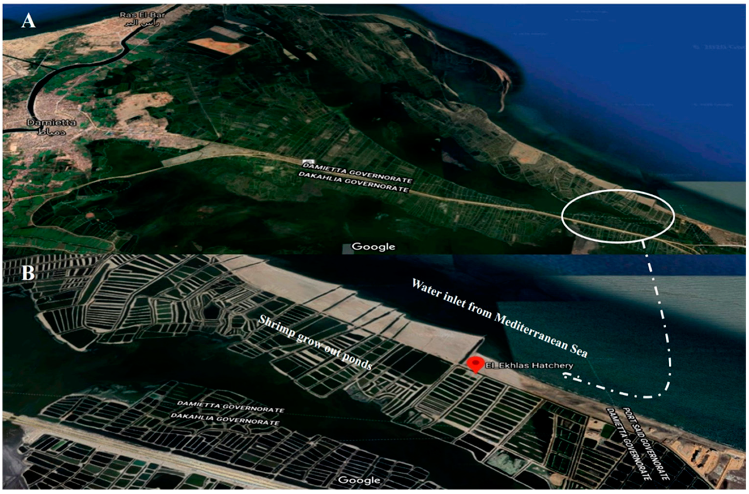
2.1. Collection of Shrimp Samples from Study Area

Diseased broodstock of L. vannamei (body weight 45.68 g ± 0.18 g) were randomly collected from three large raceways (100 shrimp per raceway, 3 m × 8 m × 1 m) set in a shrimp hatchery in DTZ, Damietta-Port-Said Province, Egypt (Figure 1), during a mortality event in the winter season from January to April, 2021. These raceways (El-Ekhlas shrimp hatchery, DTZ, Damietta-Port-Said Province, Egypt) were classified as the ‘infected’ group where moribund shrimp were observed to have high mortality rates (55%) and displayed lethargic swimming behavior and congestion of almost all body appendages and telson with brown and hemorrhagic discoloration of the hepatopancreas (HP). The feeding regime was 5% of the total body weight of a basal commercial feed containing 40% protein (Skretting, Nutreco Company, Amersfoort, Netherlands). Water quality parameters, such as ammonia (mg L−1) via the LaMotte®Aquaponics Test Kit (code no. 3637) (LaMotte Company, Chestertown, MD, USA), temperature (°C) using a water thermometer (Yellow Springs Comp., Yellow Springs, OH, USA), dissolved oxygen (mg L−1) using an oximeter model YK-22DO, and salinity (‰) using a salinometer alongside the pH value, were monitored daily. Shrimp samples (10 random moribund shrimp/raceway) were transported from the hatchery to the research facilities of the Laboratory of Fish Diseases and Management, Veterinary Medicine, Mansoura University, with a maximum time interval of 2 h. Polyethylene bags filled with marine water (one third) and oxygen (two thirds) prior to sealing were used for the transportation of shrimp. Consequently, sampling of the diseased shrimp was performed within two days of the disease outbreak. At first, hemolymph of 100 µL (n = 6) was collected from the ventral sinus of L. vannamei samples via a syringe prefilled with 500 µL of an anticoagulant buffer (trisodium citrate 30 mM, sodium chloride 0.34 M, EDTA 10 mM, pH 7.5, osmolality 780 mOsm kg L−1) for the evaluation of total hemocyte count, phagocytosis, and respiratory burst activity. Another hemolymph sample (100 µL) was individually collected from each shrimp in different raceways, stored at 4 °C for 12 h, and then centrifuged at 4000 rpm for 10 min. The supernatant was collected and kept at −80 °C until further analyses of enzyme activities, antioxidant index, and immunological parameters. Simultaneously, shrimp hepatopancreas (HP) from the three groups was aseptically tested for bacterial isolation and then carefully dissected (vertical cut) into two longitudinal sections, and one section from each sample was immediately fixed in RNAlater® (Sigma-Aldrich, Inc., St. Louis, MO 68178, USA) and stored at −80 °C for gene expression analysis.

2.2. Isolation and Identification of Vibrio spp. Using 16S Ribosomal RNA Sequence Analysis

The surface of the diseased shrimp was disinfected with 75% alcohol spray (EasyCare, El-Giza, Egypt), and the sampled hepatopancreas was collected under aseptic conditions and streaked directly on thiosulfate-citrate-bile-salts-sucrose agar (TCBS, DifcoTM, Becton and Dickinson, New York, NY, USA), which was incubated for 18–24 h at 28 ± 2 °C, and then purified colonies were restreaked on TCBS to obtain pure bacterial colonies.
Bacterial isolates were initially identified by biochemical characterization as per the criteria in Bergey’s Manual of Determinative Bacteriology [35]. Further presumptive characterization of the retrieved isolates was achieved using a commercial miniaturized API®20NE system (BioMérieux, Inc., Durham, NC, USA) according to the manufacturer’s instructions. The isolates were identified according to previously described diagnostic schemes [36,37]. The purified strains were stored in brain heart infusion broth 15% (CM1135, Oxoid, Basingstoke, UK) (vol/vol) glycerol (Sigma-Aldrich, Inc., St. Louis, MO, USA) at 20 °C.
Molecular identification of retrieved Vibrio spp. was performed using qualitative PCR. Identified bacterial colonies were cultured on tryptic soya agar +2% NaCl and then were picked up for DNA extraction using a QIAamp DNA Mini kits (Qiagen, Redwood, CA, USA) with some modifications according to the manufacturer’s protocol. Briefly, 200 µL of the sample suspension was incubated with 10 µL of proteinase K and 200 µL of lysis buffer at 56 °C for 10 min. After incubation, 200 µL of 100% ethanol was added to the lysate, which was then washed and centrifuged. The nucleic acid was then eluted with 100 µL of elution buffer. Two primer sets for Vibrio 16S rRNA (5′-CGGTGAAATGCGTAGAGAT-3′) and (5′-TTACTAGCGATTCCGAGTTC-3′) were used to perform PCR, which amplified a 663-bp fragment [38]. PCR amplification was performed in a 25-µL reaction mixture containing 12.5 µL of Emerald Amp Max PCR Master Mix (Takara, Tokyo, Japan), 1 µL of each primer (20 pmol; Metabion, Planegg, Germany), 4.5 µL of distilled water, and 6 µL of DNA template. V. parahaemolyticus (ATCC® 17802) and Escherichia coli (ATCC®25922) were used as positive and negative controls, respectively. The reaction proceeded in an Applied Biosystems 2720 thermal cycler (Applied Biosystems, Foster City, CA, USA). The thermal profile of PCR consisted of an initial denaturation step at 94 °C for 5 min, followed by 35 cycles of 94 °C for 30 s, 56 °C for 45 s, and 72 °C for 45 s, and a final extension step at 72 °C for 10 min. Next, 15 µL of PCR products were analyzed by agarose (1.5%) gel electrophoresis (AppliChem GmbH, Darmstadt, Germany) using gradients of 5 V cm−1, and a 100-bp DNA ladder (Fermentas, Thermo Fisher Scientific, Waltham, MA, USA) was used to determine the fragment sizes. The bands were photographed using a gel documentation system (Alpha Innotech, Biometra, San Leonardo, CA, USA).
The QIAquick PCR product extraction kits (Qiagen, Valencia, Spain) were used to purify PCR products. Subsequently, Bigdye Terminator V3.1 cycle sequencing kits (Perkin Elmer Applied Biosystems, Foster City, CA, USA) were employed for the sequencing reaction. Relevant bacterial DNA sequences of Vibrio spp. were obtained using the Applied Biosystems 3130 × Genetic Analyzer (HITACHI, Minato-ku, Tokyo, Japan). These sequences were analyzed for homology using the BLASTn program (http://www.ncbi.nlm.nih.gov/Blast (accessed on 3 January 2023)). Multiple alignment through Muscle tool and maximum likelihood phylogenetic analysis were conducted using the Mega software version 7.0 [39].

2.3. Susceptibility of Pathogenic Vibrio Strains to Antibiotics

The susceptibility of the two identified Vibrio strains (V. harvei and V. alginoticus) to antibiotics was screened by the in vitro agar diffusion method [40,41,42] using seven different antimicrobial disks (diameter 6 mm, Bioanalyse®, Ankara, Turkey) [chloramphenicol (C, 30 μg), ciprofloxacin (CIP, 5 μg), amoxicillin (AML, 25 μg), norfloxacin (NOR, 10 μg), doxycycline (DO, 30 μg), erythromycin (E, 15 μg), and florfenicol (F, 30 μg)], through an antibiogram. Purified colonies from each isolate were inoculated onto Mueller-Hinton agar (MHA) plates (Oxoid, Hampshire, UK) supplemented with 1.5% (w/v) sodium chloride using sterile cotton buds. Plates were incubated agar down between 24 and 48 h at 30 °C. The diameters of the inhibition halos surrounding the disks were measured in millimeters and interpreted as sensitive, intermediate, or resistant, according to the instructions of the Clinical and Laboratory Standards Institute [43] and the standardization of monitoring of antimicrobial usage and subsequent antimicrobial resistance in shrimp farming [17].

2.4. Experimental Control of Vibrio Infection by Antibiotic Application

After the antibiotic sensitivity test, the efficacy of in vivo antibiotic application against vibriosis in Pacific white shrimp was evaluated. A total of 120 alive, healthy, and infected broodstock shrimp (20 shrimp/group), which were divided into two groups (control and treated) in ss, were used in the experimental trial; each group was kept in a 20 L glass aquaria and supplied with hatchery water with mechanical aeration. For this purpose, florfenicol (10% Pan Flor, Marcyrl Animal Health, Cairo, Egypt) was mixed with a commercial feed (Skretting, Nutreco Company, Amersfoort, Netherlands) for diseased broodstock shrimp at a dose of 5 mg kg−1 body weight. The antimicrobial was added to the commercial feed and vigorously homogenized, followed by the addition of 10 μL of soybean oil per gram of feed to avoid a lack of antimicrobial hydro-solubilization during feeding. The control group of healthy shrimp were fed a basal commercial feed. The diseased broodstock were fed the medicated diet at a rate of 5% of their body weight per day for 10 consecutive days. The cumulative mortality was recorded during the antibiotic application. Thereafter, the hemolymph and hepatopancreas (n = 6) from each group were sampled on day 11 as previously described for the immunological assessment and gene expression analysis, respectively.

2.5. Total Hemocyte Count

One hundred microliters of hemolymph-anticoagulant mixtures were incubated with 100 µL fixative solution (10% formalin in 0.45 M NaCl) for 20 min at room temperature. The hemocytes were counted using a light microscope and hemocytometer (Boeco, Hamburg, Germany) [44]. Fixed hemocyte suspensions were used for the preparation of smears. The smears were completely air-dried before staining with Giemsa solution. The numbers and relative proportions of different hemocytes were calculated by counting at least 200 cells on each slide. The absolute differential hemocyte count was determined using the following equation: [(number of different hemocyte cell types/200) × THC] [45].

2.6. Enzyme Activity Assays and Antioxidant Index

2.6.1. Hepatopancreatic Enzymes

The activities of alanine and aspartate aminotransferases (ALT & AST) were tested in the supernatant of hemolymph using commercial kits (BioMed, Bayern, Germany) with a spectrophotometer (5010, Photometer, ROBERT RIELE GmbH & Co KG, Berlin, Germany). The optical density was evaluated at 546 nm.

2.6.2. Alkaline Phosphatase and Acid Phosphatase Enzymes and Antioxidants

Alkaline (ALP) and acid phosphatase (ACP) activities were evaluated using p-nitrophenyl phosphate (PNPP) as the standard substrate [46]. ALP and ACP were –estimated using p-nitro phenyl phosphate 16 mM as a standard substrate. Briefly, the substrate-hemolymph mixtures were incubated at 37 °C for 30 min after adding glycine NaOH and sodium acetate buffers for ALP and ACP, respectively. The released p-nitrophenol in the resulting supernatants was measured at 410 nm (5010, Photometer, ROBERT RIELE GmbH & Co KG, Berlin, Germany), and the amount was calculated from the standard curve.
The lysozyme activity was assessed following the previously described protocol [47] based on its ability to lyse Micrococcus lysodeikticus (Sigma Chemical Co., Saint Louis, MO, USA). Briefly, after centrifugation of the hemolymph-anticoagulant mixture, the precipitate was mixed with 0.02% Micrococcus lysodeikticus diluted in shrimp saline. The reaction was applied at room temperature (25 °C) and the absorbance was measured at 1 min intervals for 5 min at 540 nm using a microplate reader (Optica, Mikura Ltd., Horsham, UK). The lysozyme concentration was calculated using the calibration curve performed using lyophilized chicken egg-white lysozyme (Sigma Chemical Co., Saint Louis, MO, USA).
Superoxide dismutase (SOD) activity was estimated in the supernatant of hemolymph using the Ransod kit (Randox, Crumlin, UK) and evaluation of SOD’s ability to inhibit superoxide radical-dependent reactions [48]. The reaction mixture consisted of xanthine and 2-(4-iodophenyl)-3-(4-nitrophenol)-5-phenyltetrazolium chloride (INT); the xanthine oxidase enzyme was prepared. The superoxide radicals resulting from the xanthine were directly reacted with INT to produce a red formazan dye. The optical density was evaluated at 505 nm (5010, Photometer, ROBERT RIELE GmbH & Co KG, Berlin, Germany). One unit of SOD was the amount desired to suppress the rate of xanthine reduction by 50%.
Glutathione (GSH) level was evaluated depending on its capacity to reduce 5,5′dithiobis or 5,5′-dithiobis (2-nitrobenzoic acid) (Sigma Chemical Co., Saint Louis, MO, USA) with GSH to produce a yellow compound [49]. This reduced chromogen is directly proportional to GSH concentration and its absorbance can be measured at 405 nm (5010, Photometer, ROBERT RIELE GmbH & Co KG, Berlin, Germany).

2.7. Non-Specific Immune Response Assays

2.7.1. Phagocytosis Percent Assay

The phagocytic activity was conducted following the protocol described in a previous study [50,51], with some modifications. Briefly, 500 μL of cell suspension (5 × 10⁵ cells mL−1) were gently mixed with the same volume of latex beads (~107 beads mL1; 1.094 μL; Polysciences Inc., Warrington, PA, USA) and incubated at room temperature for 30 min. Strictly, 200 μL of the hemocyte-bead mixture was mixed with 0.1 mL of 10% formalin for 20 min at room temperature. Twenty microliters of fixed-hemocyte were smeared on glass slides, air-dried, and fixed with methanol for 5 min before staining with Giemsa stain. Numbers of phagocytizing cells were counted from any 200 cells observed using a light microscope. The phagocytic activity was defined as follows: phagocytosis (%) = (number of cells ingesting beads/200) × 100.

2.7.2. Respiratory Burst Assay

The superoxide anion of hemocytes was estimated by measuring the formazan formed from the reduction of nitroblue tetrazolium (NBT) [52]. Briefly, hemolymph-anticoagulant mixtures (100 mL) collected from control, infected, and treated groups were pipetted into the wells of a microtiter plate and then incubated for 2 h at room temperature. After incubation, the supernatant was removed; the hemocytes were washed three times with Hank’s buffered salt solution, and then one hundred microliters of NBT-PMA (NBT; 0.3% in PBS, 100 mL: phorbol 12-myristate 13-acetate PMA, Sigma-Aldrich; 1 mg mL−1 PBS) were added to different wells and incubated for 30 min at 25°C. Later, the NBT solution was removed and absolute methanol was added. Then, the hemocytes were washed three times with 70% methanol and air-dried. Formazan deposits were dissolved by adding dimethyl sulfoxide (DMSO, Sigma) and 2 M potassium hydroxide (KOH, Sigma Chemical Co., Saint Louis, MO, USA). The respiratory burst activity was expressed as optical density (OD) that was measured at 630 nm using a microplate reader (Optica, Mikura Ltd., Horsham, UK).

2.7.3. Bactericidal Activity

The bactericidal activity of hemolymph supernatant (serum) was determined as described previously with some modifications [50]. A volume of the serum samples (100 µL) from each group were pipetted into different sets of wells with the same volume of V. harveyi bacterial suspension (1 × 10⁸ CFU mL−1). Another set of wells containing shrimp PBS (sodium chloride 136 mM, potassium chloride 2.68 mM, disodium phosphate 8 mM, potassium dihydrogenphosphate 1.76 mM, in distilled water, pH 7.5) plus bacteria was prepared as a blank control. The microtiter plate was then incubated for 3 h at 25 °C before adding diphenyltetrazolium bromide solution (3 mg mL−1 PBS) for a continuous 30 min incubation at room temperature. Later, the supernatant was completely removed, and the formazan precipitate was suspended in dimethyl sulfoxide (DMSO, Sigma Chemical Co., Saint Louis, MO, USA). The bactericidal activity was expressed as absorbance units (O.D.) after measuring the absorbance at 560 nm using a microtiter plate reader (Optica, Mikura Ltd., Horsham, UK).

2.8. Total RNA Extraction, cDNA Synthesis, and Real-Time Quantitative PCR (qRT-PCR) Assay

Total RNA from the tested hepatopancreas samples collected in RNAlater® (Sigma-Aldrich, Saint Louis, MO, USA) from control, infected, and treated shrimp was extracted using Easy-SpinTM [DNA Free] Total RNA Extraction Kits (iNtRON Biotechnology, Inc., Sagimakgol-ro, Republic of Korea) according to the manufacturer’s instructions. RNA integrity was confirmed by agarose gel electrophoresis, and the concentrations and purities of the samples were examined using a NanoDrop spectrophotometer (Thermo Fisher Scientific, Waltham, MA, USA). First-strand cDNA was synthesized in a reaction volume (20 μL) containing 1 μg of total RNA using a TOPscriptTM cDNA Synthesis Kit (Enzynomics Co. Ltd., Sagimakgol-ro, Republic of Korea). Subsequently, L. vannamei hepatopancreas cDNAs (1 μL) were quantified using TOPreal™ 2× PreMIX SYBR Green qPCR master mix with low ROX (Enzynomics Co. Ltd., Daejeon, Republic of Korea) according to the manufacturer’s recommendations using a Rotor-Gene Q MDx 6 plex real-time PCR system (Qiagen, Germantown, MD, USA). Specific primers, proPO (LvproPO), hemocyanin (LvHc), and toll-like receptor 1 (LvToll1) were employed to amplify the selected genes together with β-actin as the housekeeping gene (Table 1). The PCR program consisted of 40 cycles at 95 °C (10 s), 60 °C (15 s), and 72 °C (30 s), followed by melt curve generation at 72–95 °C for seconds. The relative gene expression levels were evaluated in triplicates included on template controls using the 2−ΔΔCT formula [53].

2.9. Data Analysis

Data related to immune parameters as well as gene expression were analyzed using GraphPad Prism v. 8.0 (GraphPad Software, Inc., San Diego, CA, USA) and statistically presented as mean ± standard error (SEM). The comparison of the data related to enzyme activities, the antioxidant index, and immunological parameters was performed using a one-way analysis of variance (ANOVA) followed by a Tukey’s test among the three groups. Variations of the related gene expression values were estimated and applied. Fold changes were displayed using a log-base-two scale to highlight the changes in transcriptional levels, where p < 0.05 (*) and p < 0.01 (**) were considered to indicate significant differences, whereas ns was considered non-significant.

3. Results

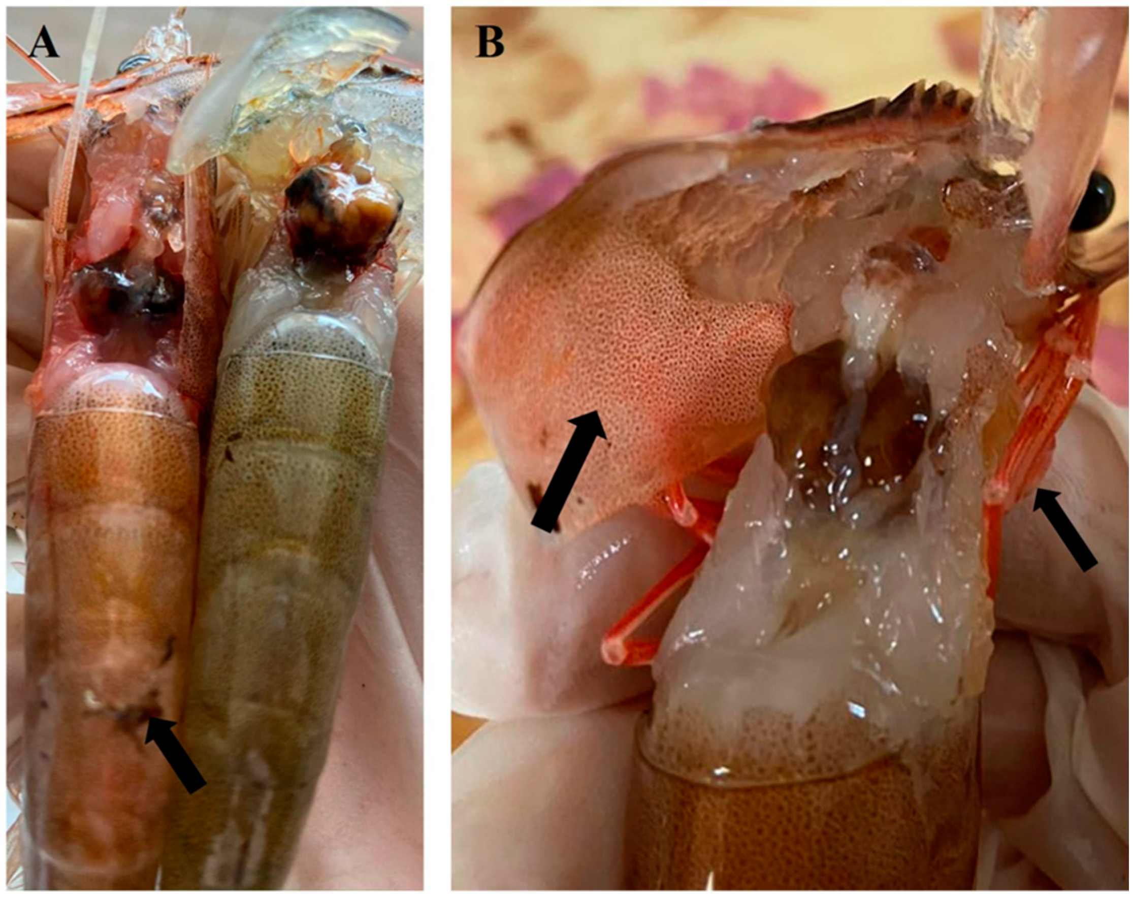
3.1. Clinical Signs of Diseased Shrimp

Compared to healthy shrimp, the hepatopancreas and body of diseased L. vannamei were significantly reddish looking and lethargic, and the hepatopancreas exhibited a petechial hemorrhage in the abdominal muscle and red coloration of the body and pleopods (Figure 2A). Moreover, reddish spots appeared on the anterior carapace, including pleopods (Figure 2B).

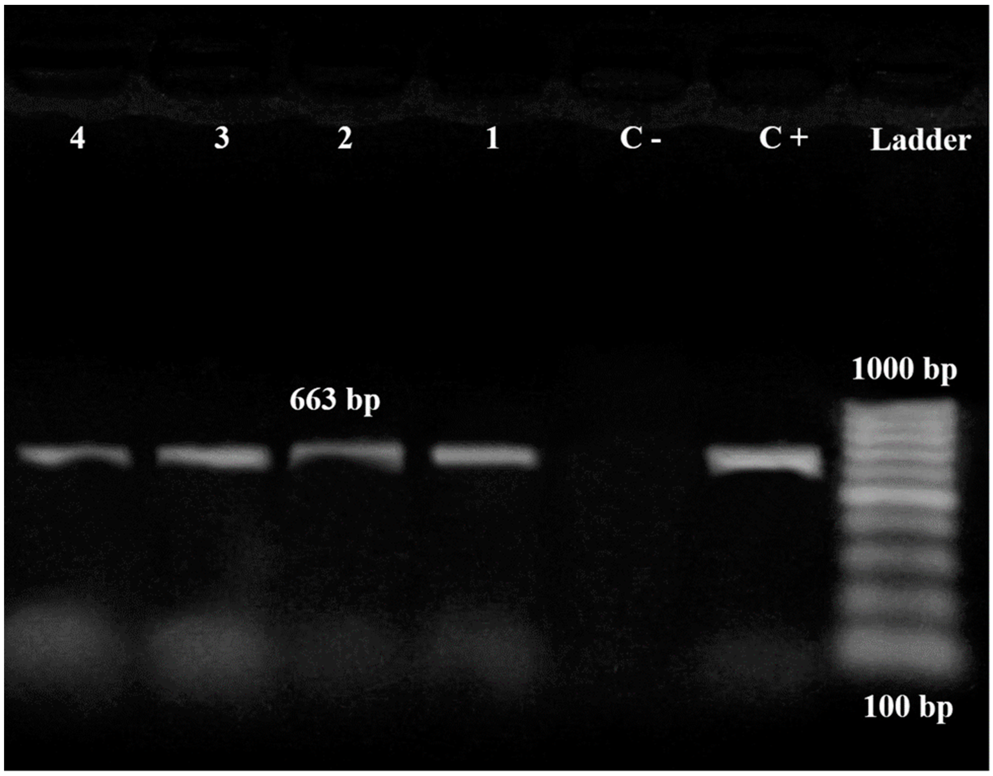
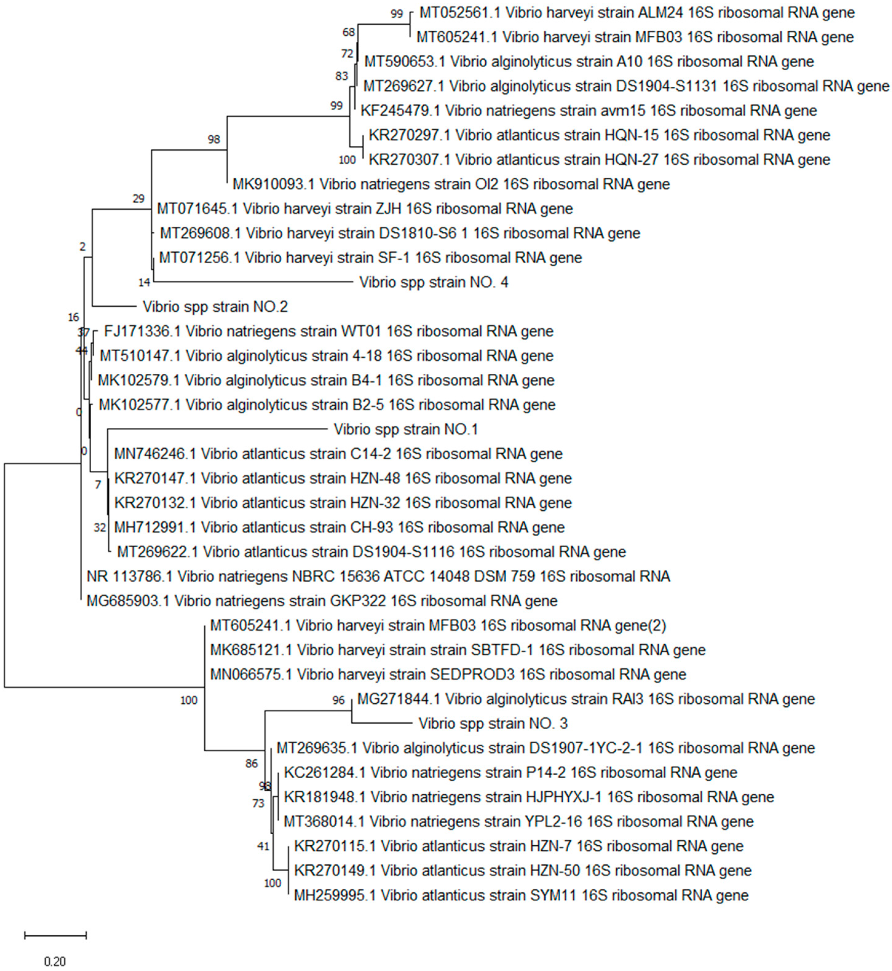
3.2. Characterization of Bacterial Vibrio Strains

A total of 45 bacterial isolates of Vibrio spp. Were recovered from diseased L. vannamei based on bacterial biochemical and morphological characterization. The API 20E identification system sorted the isolates into V. atlanticus (5 isolates), V. natriegens (7 isolates), V. alginolyticus (15 isolates), and V. harveyi (18 isolates).
A PCR assay of bacterial isolates using Vibrio 16S rRNA primers showed amplified bands of a 663-bp size that was characteristic of Vibrio spp. Isolated from infected L. vannamei (Figure 3). The amplicon sizes of the band obtained from the positive control sample corresponded to the predicted sizes, whereas no amplicon was observed in the negative control (Figure 3).
In this study, the BLAST analysis of the nucleotide sequence of the four 16S rRNA genes from Vibrio spp. Shared 97.31–98.5% identity with the 16S rRNA partial sequences and the complete genomes of V. atlanticus, V. natriegens, V. alginolyticus, and V. harveyi, respectively, obtained from the GenBank database. Based on the phylogenetic tree constructed from 16S rRNA gene partial sequences, Vibrio strain-1 was grouped with V. atlanticus isolates; however, Vibrio strain-2 was grouped with a different Vibrio spp., including V. natriegens (Figure 4). Meanwhile, Vibrio strain-3 and strain-4 were grouped with V. alginolyticus and also V. harveyi (Figure 4), respectively. No bacterial load was detected on the samples isolated from the treated and control shrimp.

3.3. Susceptibility of Pathogenic Vibrio Strains to Antibiotics

Bacterial Vibrio strains were highly sensitive to chloramphenicol, doxycycline, and florfenicol but resistant to amoxicillin. They exhibited moderate sensitivity to norfloxacin. The other Vibrio strain was moderately sensitive to erythromycin and ciprofloxacin; however, the other strain showed dissimilar results (Table 2).
Cumulative mortality rates were initially estimated to be around 55% on day 1, which continued at the same percent for the successive five days. Subsequently, the FLO application decreased the cumulative mortality to 20% for the medicated shrimp. There were no mortalities in the non-infected broodstock shrimp (control group).

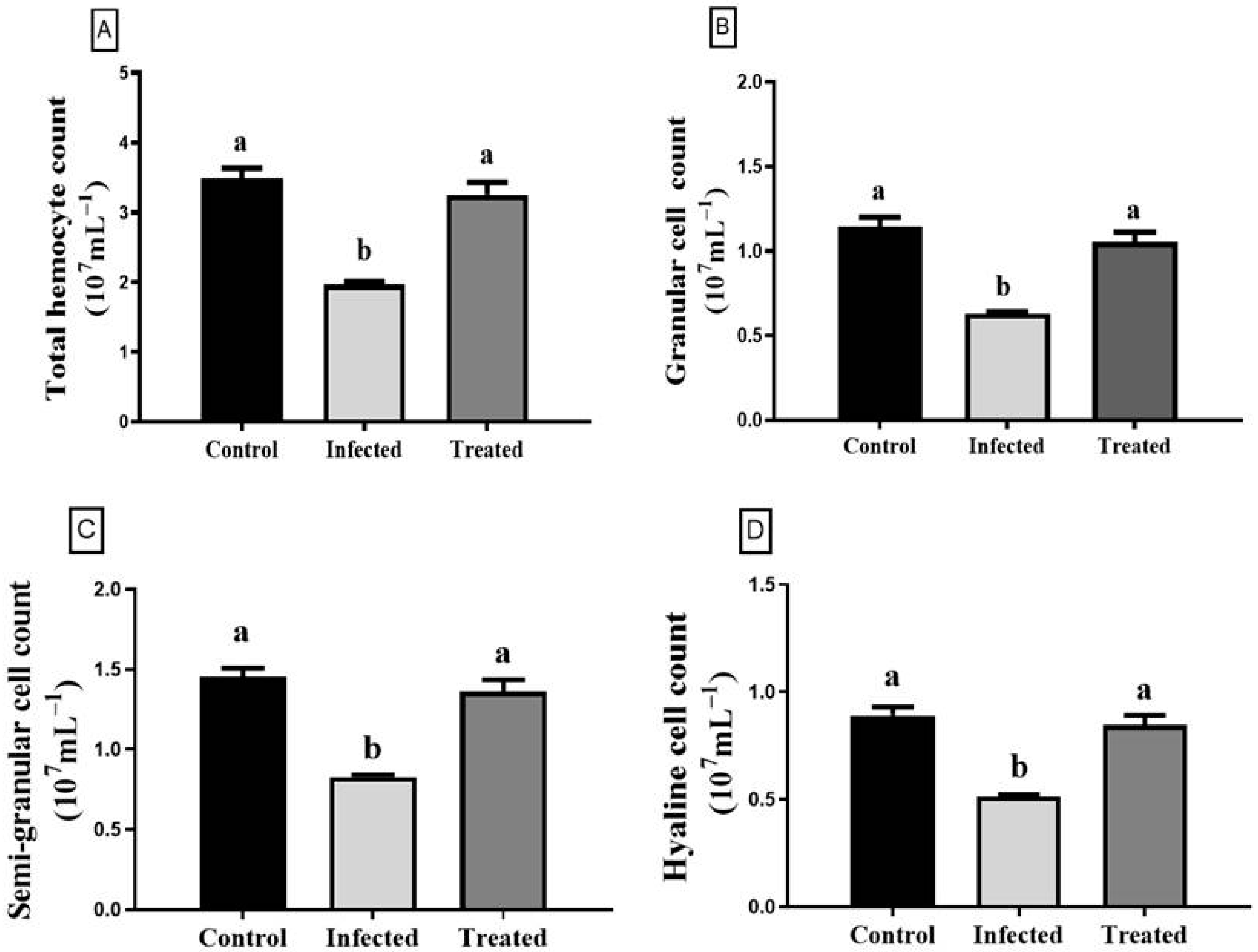
3.4. Total Hemocyte Count

Total hemocyte count (THC), granular cell (GC), semi-granular cell (SGC), and hyaline cell (HC) counts of all experimental groups are shown in Figure 5. The Vibrio-infected group exhibited a significant drop in the THC (t = 10.58; df = 8; p = 0.0001), GC (t = 9.12; df = 8; p = 0.0001), SGC (t = 10.55; df = 8; p = 0.0001), and HC (t = 9.73; df = 8; p = 0.0001) count compared to the control group (p < 0.001). However, the treatment of infected shrimp with florfenicol significantly restored the total hemocyte cell counts (t = 5.86; df = 8; p = 0.0004), GC (t = 7.32; df = 8; p < 0.0001), SGC (t = 7.11; df = 8; p = 0.0001), and HC (t = 7.19; df = 8; p < 0.0001) counts to their normal levels, relative to the infected group (Figure 5).

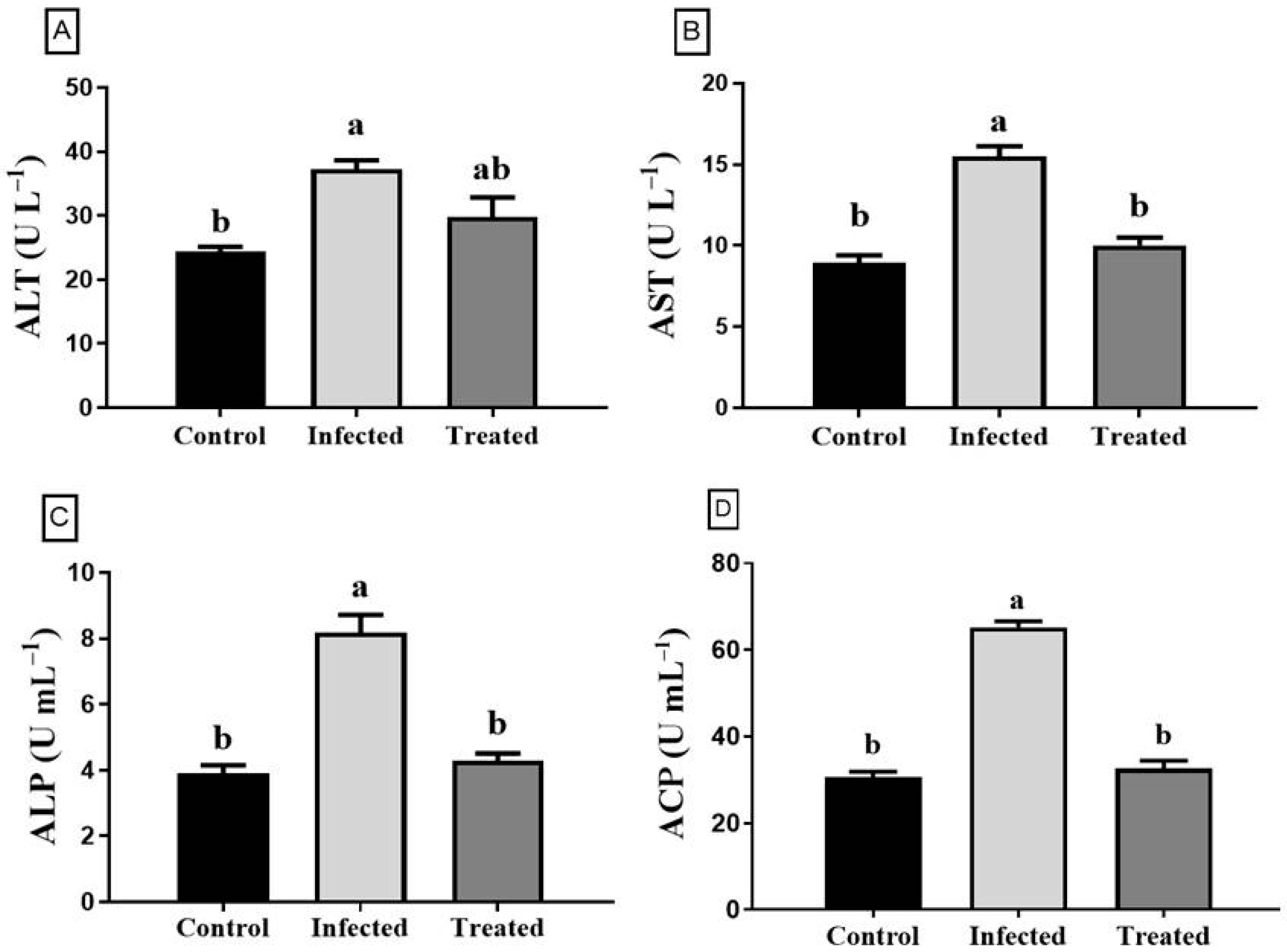
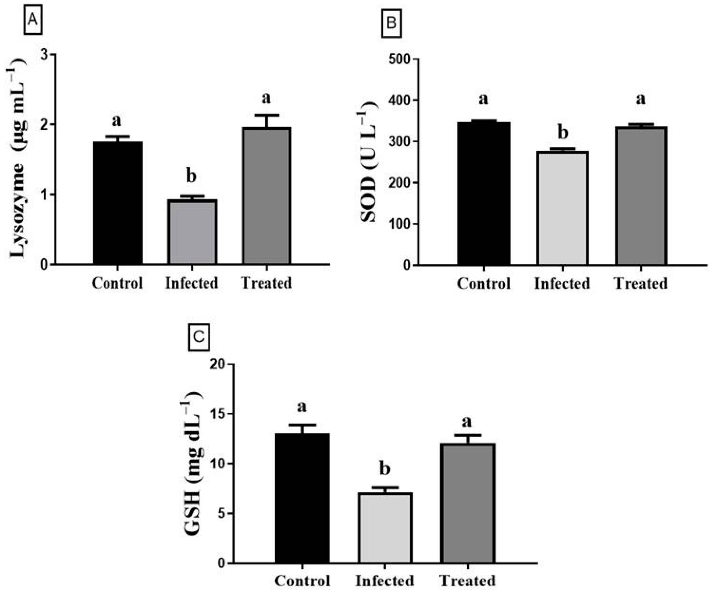
3.5. Enzyme Activity Assays and Antioxidant Index

The enzyme activities and antioxidants estimated in the supernatant of hemolymph are enumerated in Figure 6 and Figure 7. The ALT (t = 8.31; df = 8; p = 0.0001), AST (t = 8.52; df = 8; p = 0.0001), ALP (t = 7.19; df = 8; p < 0.0001), and ACP (t = 18.68; df = 8; p < 0.0001) activities were significantly elevated in L. vannamei infected with Vibrio spp. compared to the control (Figure 6). Meanwhile, the previous enzyme activities (ALT (t = 2.25; df = 8; p = 0.0542), AST (t = 6.81; df = 8; p = 0.0001), ALP (t = 6.67; df = 8; p = 0.0002), and ACP (t = 14.58; df = 8; p < 0.0001) were significantly reestablished in the antibiotic-treated group compared to the infected group (Figure 6).

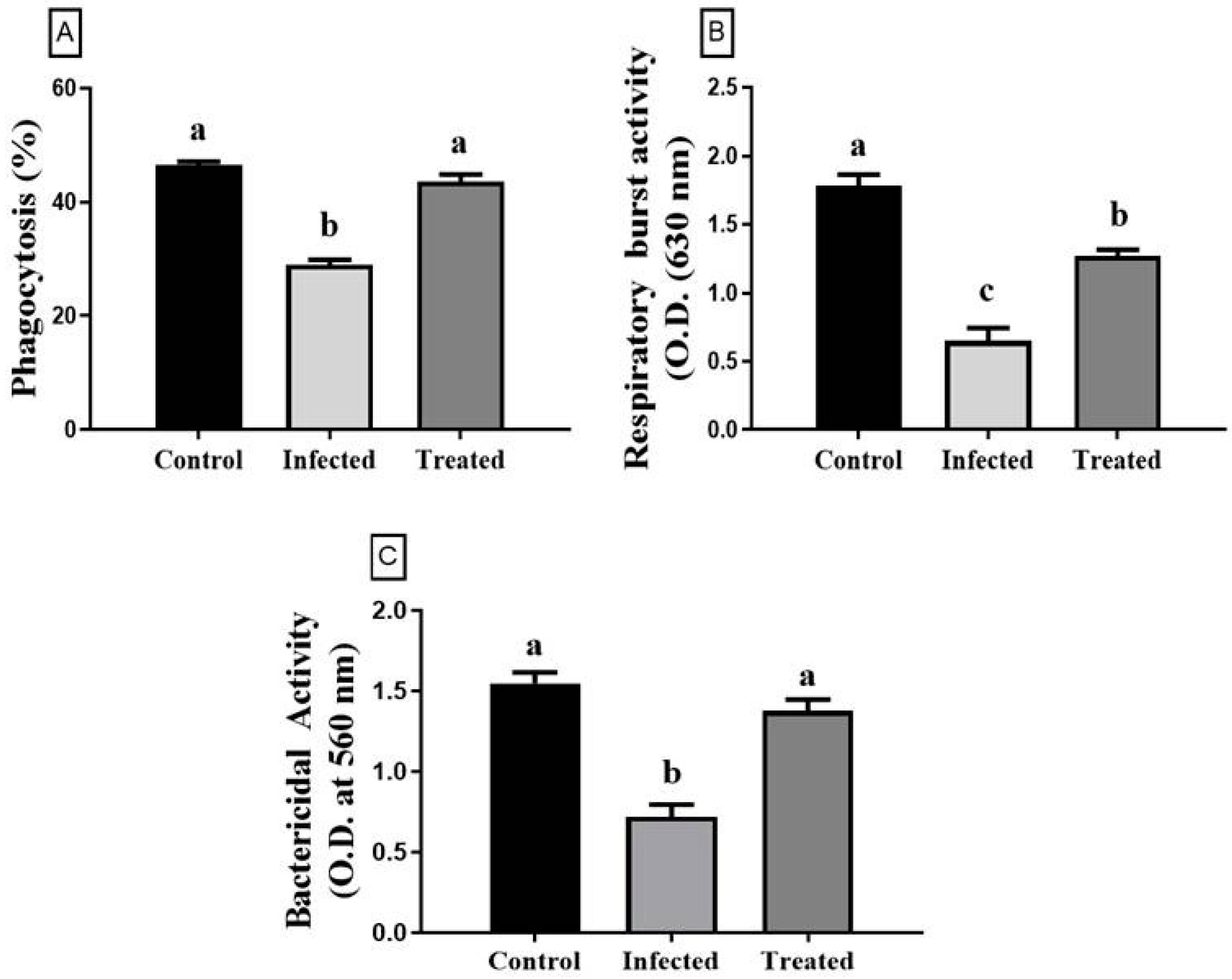
3.6. Immune Parameters

The effects of the florfenicol treatment on the phagocytic, respiratory burst, and bactericidal activities of shrimp against Vibrio spp. infection are displayed in Figure 8. Phagocytosis % (t = 9.60; df = 8; p < 0.0001), respiratory burst (t = 6.18; df = 8; p = 0.0003), and bactericidal (t = 6.67; df = 8; p = 0.0002) activities of shrimp treated with florfenicol were significantly increased compared to those of Vibrio-infected shrimp which represented significant suppression in the different immune responses (Figure 8).

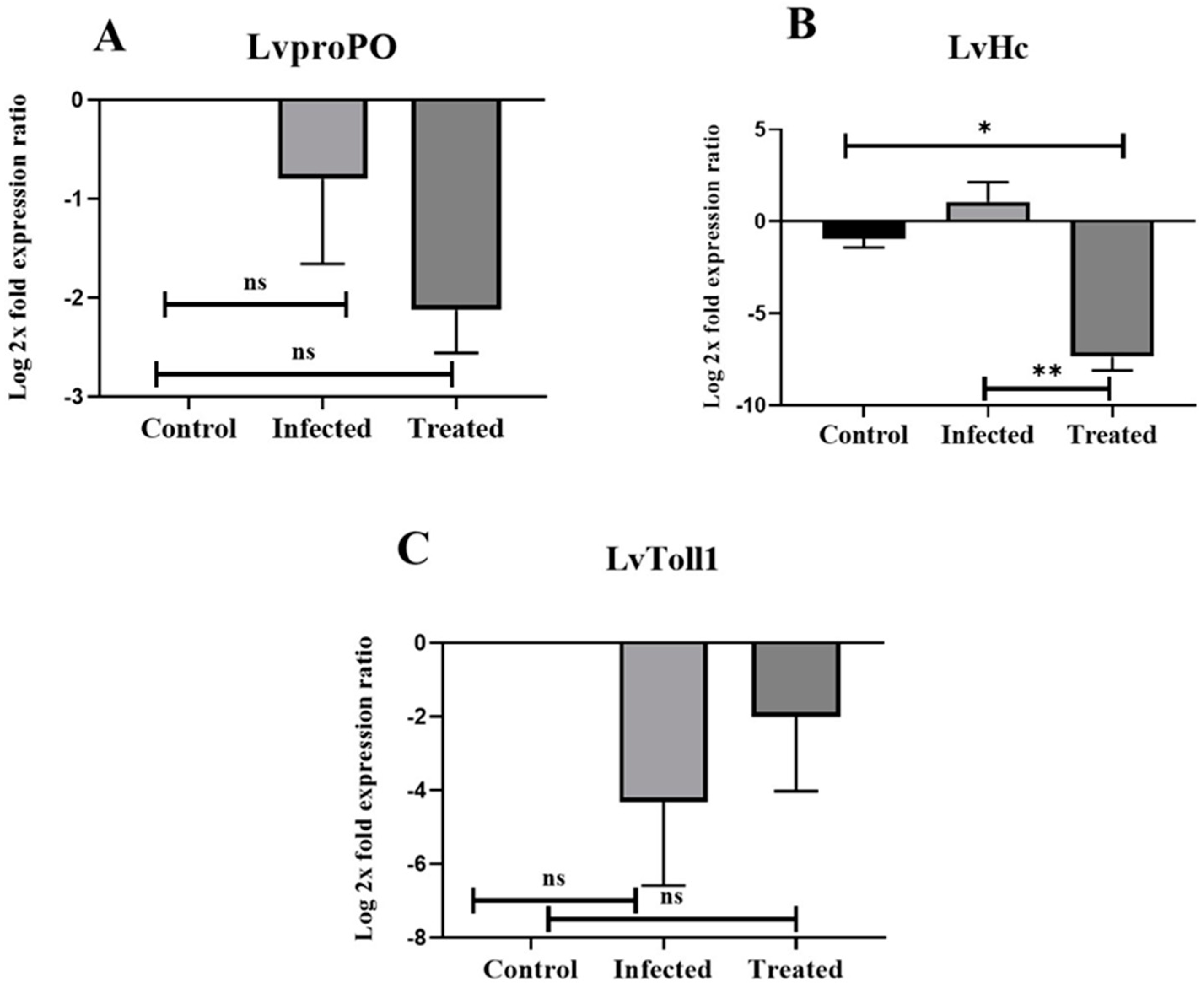
3.7. Expression Profiles of LvHc, LvToll1, and LvproPO

The transcript levels of the LvproPO and LvToll1 genes in the hepatopancreas were not statistically different between groups (Figure 9A,C) although lower expression levels were observed in the infected and treated groups compared with the control group. Similarly, the mRNA expression of LvHc in the hepatopancreas was markedly downregulated in the treated group (p = 0.0103, 0.0038) when compared with the control and infected groups (Figure 9B).

4. Discussion

Vibrio species remain a life-threatening pathogen in shrimp hatcheries that have a crucial demand for shrimp culture. Owing to vibriosis, several countries, such as India, Thailand, and Mexico, have reported severe economic losses in L. vannamei aquaculture [58,59]. The stress associated with diminished resistance to Vibrio spp. infection has primarily promoted high mortalities in shrimp culture, especially diseases caused by V. harveyi and V. alginolyticus [12,60].
In the present study, Vibrio strains were identified by 16S rRNA gene sequencing from naturally infected L. vannamei broodstocks in a shrimp hatchery in the Egyptian coastal province of DTZ. Among these, V. harveyi has been previously reported to promote bright-red syndrome in Pacific white shrimp [58,59]. In addition, V. natriegens has been reported to cause high mortalities in shrimp culture [61]. Moreover, diseased shrimp displayed cuticular lesions with a red coloration of the body, hepatopancreas, and pleopods; these symptoms are similar to those of shrimp bright-red syndrome, especially the disease caused by V. harveyi and V. alginolyticus [9,56].
Regarding the phylogenetic analysis, most of the isolated strains retrieved from the diseased L. vannamei shared almost identical sequences in the 16S rRNA gene (98.8% identity) and belonged to the Harveyi clade, consisting of V. harveyi, V. alginolyticus, and V. natriegens, which shared high levels of phenotypic and genotypic homology and are known to be pathogenic for shrimp [13,62]. This finding was consistent with the results observed in the Ecuadorian L. vannamei hatcheries suffering mortalities [26]. However, new strains are identified in our studies, such as V. atlanticus and V. natriegens in Egypt, suggesting the diversity of the circulating bacteria and indicating the crucial importance of studying the effectiveness of the shrimp immune response.
Antibiotics are the most effective therapeutic agents against several pathogenic bacteria in shrimp farming [63]. We evaluated several antibiotics in our study to demonstrate the patterns of antibiotic resistance; however, most of these antibiotics are not authorized for use in aquaculture. In our screening, both strains were resistant to amoxicillin similar to reports of several resistant Vibrio spp. isolated from diseased shrimp to first-generation penicillins or/and β-lactam antibiotics [59,64]. Moderate sensitivity was shown for enrofloxacin, which is one of the currently used antibiotics in several countries against vibriosis [58,59]. Interestingly, Vibrio strains have been documented to be sensitive to most of the tested antibiotics. Florfenicol is used in our study to decrease the mortality rates of shrimp owing to its potent activity against bacterial pathogens. In general, FLO was also approved by the FDA for use in shrimp aquaculture because of its high potency for the control of necrotizing hepatopancreatitis and vibriosis infections in farm-raised Pacific white shrimp [65]. Many studies have used FLO for controlling the mortality caused by Vibrio spp. isolated in Ecuador, the USA, Japan, Thailand, and Mexico [66,67]. Additionally, absorption of FLO from the intestine has been shown to be rapid with extensive distribution and prompt elimination in Pacific white shrimp, Litopenaeus vannamei [68]. A recent study showed that low concentrations of florfenicol (minimal inhibitory concentration, MIC of 8 μg mL−1) were highly effective in controlling bacterial growth for many pathogenic Vibrio strains [26]. Moreover, FLO had a greater favorable effect on the survivability of adult shrimp (Litopenaeus vannamei) with necrotizing hepatopancreatitis (NHP) disease by diminishing the bacterial load, allowing the shrimp to mount an immune response to the pathogens [27]. However, it has been reported that oral administration of FLO at 100 and 200 mg kg had a suppressive effect on the antioxidant activity of juvenile Litopenaeus vannamei [69]. Regarding our results from this study, it is reasonable to assume that a dose of 5 mg kg given orally for FLO should be applicable for the control of vibriosis in Pacific white shrimp.
Hemocytes play a critical role in the immune response of crustaceans, including phagocytosis, mediation of cytotoxicity, encapsulation, and nodule formation [70]. The hemocytes are categorized into three types based on the presence of granules or relative size: large granular (granular, GC), small granular (semi-granular, SGC), and granular (hyaline, HC) hemocytes [32]. The decline in THC counts in infected L. vannamei was revealed in our study. Generally, exposure to infectious pathogens or environmental stress reduces THC counts in shrimp, which in turn boosts the risk of secondary infection [71]. On the other hand, the THC of shrimp declined in softshell clams, Mya arenaria infected with V. splendidus [72], and L. vannamei infected with V. harveyi [73]. This decline in THCs was associated with the bacterial inflammatory response as hemocytes leave the circulation and migrate to the site of infection [74]. As well, hemocytes aggregate into hemocytic nodules with cell adhesion molecules to eliminate bacteria from the circulation [34]. The current study showed that florfenicol medication could reestablish THC, GC, SGC, and HC in the white shrimp infected with Vibrio spp. A previous report enumerated that the supplementation of chitosan–gentamicin conjugates increased THCs in L. vannamei infected with V. parahaemolyticus through enhancement of hemocyte proliferation and their phagocytic activity [75]. Enrofloxacin promoted disease resistance against V. parahaemolyticus and restored THCs in shrimp [76].
The main biomarkers for hepatopancreas function, AST and ALT activities, can reflect the degree of hepatopancreas injury in shrimp [77]. The elevation of AST and ALT activities suggested possible damage to the hepatopancreatic tissue as in necrotizing hepatopancreatitis disease caused by Vibrio spp., which was significantly decreased following oxytetracycline administration in L. vannamei [27]. In our study, shrimp treated with florfenicol had significantly lower hepatopancreatic enzyme levels (ALT and AST) compared to the infected and control groups [78]. During infection with V. parahaemolyticus, the epithelial cells of the hepatopancreatic tubules rupture following infection. When treated with florfenicol, the hepatopancreas showed intact epithelial cells with the improved structural integrity of the tubules, explaining the decrease in the hepatopancreas enzyme activity in the hemolymph [78].
Various enzymes in the hemolymph, such as ALP, ACP, SOD, and lysozyme, are considered indicators for the evaluation of disease resistance and immune status in shrimp [79]. ALP is one of the regulatory enzymes connected to the metabolism process and phagolysis; whereas, ACP is an identical lysosomal enzyme that has a key role in the elimination and hydrolyzing of microbes [80,81]. Both ALP and ACP are involved in the regulation of the phosphorylation and dephosphorylation processes [82]. The ACP activity of Chlamys farreri was significantly elevated following the challenge with V. anguillarum [83]. Other comparable studies demonstrated that ACP activities of L. vannamei are more sensitive to V. parahaemolyticus and their increase is attributed to the disturbance of cell metabolism and immune function as well as the stress caused by Vibrio infection [75,76,84], which is consistent with our results. Moreover, the administration of florfenicol significantly lowered ALP and ACP activities, which may be due to its ability to strengthen resistance against infection as the activity of these immune enzymes was significantly elevated in the cell-free hemolymph of shrimp following Vibrio infection in L. vannamei [78].
Lysozyme is a functional antibacterial protein and a pivotal component of the invertebrates’ innate immune system as it can hydrolyze the mucopolysaccharides present in the bacterial cell wall to kill pathogens [85]. SOD is one of the remarkable antioxidant enzymes that protect the host against oxidative stress through the degradation of excess O2− to produce molecular oxygen and hydrogen peroxide [86]. GSH is a valuable antioxidant in cells that can reduce hydrogen peroxide to water together with glutathione peroxidase to maintain the integrity of the red blood cell membrane and prevent damage by oxidants [87]. Our results exhibited lowered activities of lysozyme, SOD, and GSH following Vibrio infection, which were then restored following antibiotic administration. Similarly, previous reports presented a significant decrease in SOD activity in L. vannamei injected with V. parahaemolyticus and V. alginolyticus, respectively [84,88]. In contrast to our results, previous studies demonstrated increased lysozyme activity after V. parahaemolyticus and V. alginolyticus infection in L. vannamei and P. trituberculatus [76,89]. The lower GSH level and SOD activity may be correlated to the oxidative stress mediated by singlet oxygen that causes SOD inactivation during infection [90]. Whereas, lower lysozyme activity may be correlated with the inactivation of the immune response in shrimp challenged with Vibrio spp. [27]. Similar to our results, the SOD activity and GSH level of L. vannamei infected with V. parahaemolyticus were restored upon the oral administration of chitosan–gentamicin conjugate due to a great reduction in ROS production and lipid peroxidation after antibiotic treatment [75]. Dietary administration of a low dose of Astragalus and florfenicol increased lysozyme activity levels in shrimp challenged with V. parahaemolyticus [78].
Phagocytosis is the initial internal defense mechanism against any foreign objects, and one of the fundamental roles of hemolymph in the invertebrate defense process [91]. The phagocytic cells produce superoxide anion and its reactive derivatives that have powerful bactericidal activity, during the mechanism of respiratory burst [50]. Our results showed that infection with Vibrio reduced the phagocytosis and bactericidal activity of L. vannamei, as also demonstrated by previous studies [92,93]. The reduced phagocytosis of V. splendidus-challenged hemocytes could result from the loss of pseudopodia due to the toxic effect of extracellular products produced by bacteria [72]. V. tapetis also decreased the phagocytosis activity of hemocytes in the Japanese carpet clam R. philippinarum [94]. The lower respiratory burst activity may be due to either V. splendidus lacking a receptor that stimulates respiratory burst activity or it actively curbs the hemocyte’s response [72]. In the current study, there was a significant decline in the phagocytic, respiratory burst, and bactericidal activities, which returned to normal in florfenicol-treated shrimp.
We detected slight differences in the transcript levels of the immune response genes in the hepatopancreas of L. vannamei. Activation of the proPO system during infection inhibits the damage caused by the proteases synthesized by pathogens [95]. Our study revealed that expression of the proPO gene displayed a decreasing pattern in the infected and treated groups. This result indicates that once pathogens are removed through the proPO system during infection or treatment, protease inhibitors capture the prophenoloxidase-activating enzyme (proPOAE), and thus counteract the proPO-activation to the phenoloxidase isoform [96]. This result is consistent with other studies that reported the modulation of this gene in L. vannamei challenged with V. harveyi and the white spot syndrome virus [56,97]. ProPOs are primarily expressed in L. vannamei hemocytes but are present at very low levels in the hepatopancreas, as assessed by RT-PCR and Northern blotting analysis [54,97]. Likewise, the significant decrease in the expression of LvHc in our study can justify the lower expression levels of LvproPO, i.e., when hemocyanin proteolysis is generated, it is converted into a phenoloxidase-like enzyme, possessing a phenoloxidase activity [98]. The downregulation of immune-related genes, such as penaeidin and proPO, has been reported after oxytetracycline and oxolinic acid treatment in shrimp [99]. A similar trend regarding the expression of LvToll1 was observed in our study, with no significant changes being detected in the treated group compared with the control group. However, shrimp tolls are involved in the regulation of AMPs as they are primarily synthesized in the hemocytes. Our results are consistent with another study that demonstrated that LvToll1 could have other potential roles in the penaeid immune response, and the expression level of LvToll1 was also low in the hepatopancreas of L. vannamei [57]. The results obtained in the FLO application sought greater effectiveness for the control of bacterial infections in shrimp hatcheries. However, we need to highlight the importance of withdrawal times for the antibiotic, with respect to eliminating the residual presence of these compounds from the edible tissues, and from the cultivation system to diminish the development of antibiotic resistance in the bacteria.

5. Conclusions

In summary, four Vibrio strains were isolated in this study from the hepatopancreas of L. vannamei shrimp in an Egyptian hatchery-raised shrimp. FLO supplementation scavenged most of the hemato-immune parameters as well as the pathogen load in L. vannamei shrimp infected with Vibrio spp. Herein, our data might evaluate the immunological screening of hatchery-diseased shrimp in indicating the effect of antibiotic medication in the management and control of vibriosis through regaining the immune response of L. vannamei shrimp after the antibiotic clears the infection rather than a direct response. Further investigation and exploration should be undertaken on the quality and usage of antibiotics in shrimp aquaculture.

Author Contributions

S.E. and G.E.E.: Methodology, Formal analysis, Investigation, Validation, and Writing—original draft, Writing—review and editing. S.E.: Conceptualization, Supervision, and Final revision. M.S.S.: Investigation, Visualization, and Methodology. A.A.A., E.M.Y.: Funding acquisition, Resources, Validation, Writing—review & editing. S.J.D.: Conceptualization, data support & analysis, Scientific critical input & Citation linkage, Literature comparative dialogue, Revisions. Funding acquisition and Resources. All authors have read and agreed to the published version of the manuscript.

Funding

This research was funded by the researchers supporting project number (RSPD2023R700), King Saud University, Riyadh, Saudi Arabia.

Institutional Review Board Statement

This study abides by the Medical Research Ethics Committee of Mansoura University and follows the general guidelines of the Canadian Council on Animal Care with the code number (R/65).

Informed Consent Statement

Not applicable.

Data Availability Statement

The data that support the findings of this study are available on reasonable request from the corresponding author, S. Elbahnaswy. The data are not publicly available due to their containing information that could compromise the privacy of research participants.

Acknowledgments

This research was funded by the researchers supporting project number (RSPD2023R700), King Saud University, Riyadh, Saudi Arabia.

Conflicts of Interest

The authors do not report any financial or personal connections with other persons or organizations that might negatively affect the contents of this publication and/or claim authorship rights to this publication.

References

  1. Okamura, Y.; Inada, M.; Elshopakey, G.E.; Itami, T. Characterization of xanthine dehydrogenase and aldehyde oxidase of Marsupenaeus japonicus and their response to microbial pathogen. Mol. Biol. Rep. 2018, 45, 419–432. [Google Scholar] [CrossRef] [PubMed]
  2. Thirugnanasambandam, R.; Inbakandan, D.; Kumar, C.; Subashni, B.; Vasantharaja, R.; Abraham, L.S.; Ayyadurai, N.; Murthy, P.S.; Kirubagaran, R.; Khan, S.A. Genomic insights of Vibrio harveyi RT-6 strain, from infected “Whiteleg shrimp”(Litopenaeus vannamei) using Illumina platform. Mol. Phylogenetics Evol. 2019, 130, 35–44. [Google Scholar] [CrossRef] [PubMed]
  3. Adeleke, B.; Robertson-Andersson, D.; Moodley, G.; Taylor, S. Aquaculture in Africa: A Comparative Review of Egypt, Nigeria, and Uganda Vis-À-Vis South Africa. Rev. Fish. Sci. Aquac. 2020, 29, 1–31. [Google Scholar] [CrossRef]
  4. Megahed, M.E. A comparison of the severity of white spot disease in cultured shrimp (Fenneropenaeus indicus) at a farm level in Egypt. I-Molecular, histopathological and field observations. Egypt. J. Aquat. Biol. Fish. 2019, 23, 613–637. [Google Scholar] [CrossRef] [Green Version]
  5. Feng, B.; Liu, H.; Wang, M.; Sun, X.; Pan, Y.; Zhao, Y. Diversity analysis of acute hepatopancreatic necrosis disease-positive Vibrio parahaemolyticus strains. Aquac. Fish. 2017, 2, 278–285. [Google Scholar] [CrossRef]
  6. Culot, A.; Grosset, N.; Bruey, Q.; Auzou, M.; Giard, J.-C.; Favard, B.; Wakatsuki, A.; Baron, S.; Frouel, S.; Techer, C.; et al. Isolation of Harveyi clade Vibrio spp. collected in aquaculture farms: How can the identification issue be addressed? J. Microbiol. Methods 2021, 180, 106106. [Google Scholar] [CrossRef] [PubMed]
  7. Dubert, J.; Nelson, D.R.; Spinard, E.J.; Kessner, L.; Gomez-Chiarri, M.; da Costa, F.; Prado, S.; Barja, J.L. Following the infection process of vibriosis in Manila clam (Ruditapes philippinarum) larvae through GFP-tagged pathogenic Vibrio species. J. Invertebr. Pathol. 2016, 133, 27–33. [Google Scholar] [CrossRef] [Green Version]
  8. Chatterjee, S.; Haldar, S. Vibrio related diseases in aquaculture and development of rapid and accurate identification methods. J. Mar. Sci. Res. Dev. S 2012, 1, 1–7. [Google Scholar]
  9. Mastan, S.; Begum, S.A. Vibriosis in farm reared white shrimp, Litopenaeus vannamei in Andhra Pradesh-natural occurrence and artificial challenge. Int. J. Appl. Sci. Biotechnol. 2016, 4, 217–222. [Google Scholar] [CrossRef] [Green Version]
  10. Baker-Austin, C.; Oliver, J.D.; Alam, M.; Ali, A.; Waldor, M.K.; Qadri, F.; Martinez-Urtaza, J. Vibrio spp. infections. Nat. Rev. Dis. Prim. 2018, 4, 1–19. [Google Scholar] [CrossRef]
  11. Kumaran, T.; Citarasu, T. Isolation and characterization of Vibrio species from shrimp and Artemia culture and evaluation of the potential virulence factor. Intel. Prop. Rights 2016, 4, 2. [Google Scholar]
  12. Zhang, X.-H.; He, X.; Austin, B. Vibrio harveyi: A serious pathogen of fish and invertebrates in mariculture. Mar. Life Sci. Technol. 2020, 2, 1–15. [Google Scholar] [CrossRef] [PubMed] [Green Version]
  13. Darshanee Ruwandeepika, H.A.; Sanjeewa Prasad Jayaweera, T.; Paban Bhowmick, P.; Karunasagar, I.; Bossier, P.; Defoirdt, T. Pathogenesis, virulence factors and virulence regulation of vibrios belonging to the Harveyi clade. Rev. Aquac. 2012, 4, 59–74. [Google Scholar] [CrossRef]
  14. Hasan, M.A.R.; Siddique, M.A.; Hasan, M.; Hossain, M.A.; Rahman, M.S. 16S rRNA gene sequence based identification of Vibrio spp. in shrimp and tilapia hatcheries of Bangladesh. Dhaka Univ. J. Biol. Sci. 2017, 26, 45–58. [Google Scholar] [CrossRef] [Green Version]
  15. Janda, J.M.; Abbott, S.L. 16S rRNA gene sequencing for bacterial identification in the diagnostic laboratory: Pluses, perils, and pitfalls. J. Clin. Microbiol. 2007, 45, 2761–2764. [Google Scholar] [CrossRef] [PubMed] [Green Version]
  16. Karim, M.; Uddin, M.; Rahman, M. Microbiological study of costal shrimp aquaculture production system of Bangladesh. J. Biol. Life Sci. 2018, 9, 17–30. [Google Scholar]
  17. Thornber, K.; Verner-Jeffreys, D.; Hinchliffe, S.; Rahman, M.M.; Bass, D.; Tyler, C.R. Evaluating antimicrobial resistance in the global shrimp industry. Rev. Aquac. 2020, 12, 966–986. [Google Scholar] [CrossRef] [Green Version]
  18. Cabello, F.C. Heavy use of prophylactic antibiotics in aquaculture: A growing problem for human and animal health and for the environment. Environ. Microbiol. 2006, 8, 1137–1144. [Google Scholar] [CrossRef]
  19. De Schryver, P.; Defoirdt, T.; Sorgeloos, P. Early mortality syndrome outbreaks: A microbial management issue in shrimp farming? PLoS Pathog. 2014, 10, e1003919. [Google Scholar] [CrossRef]
  20. Yanong, R.P.; Curtis, E.W.; Simmons, R.; Bhattaram, V.A.; Gopalakrishnan, M.; Ketabi, N.; Nagaraja, N.V.; Derendorf, H. Pharmacokinetic studies of florfenicol in koi carp and threespot gourami Trichogaster trichopterus after oral and intramuscular treatment. J. Aquat. Anim. Health 2005, 17, 129–137. [Google Scholar] [CrossRef]
  21. Brausch, J.M.; Connors, K.A.; Brooks, B.W.; Rand, G.M. Human pharmaceuticals in the aquatic environment: A review of recent toxicological studies and considerations for toxicity testing. Rev. Environ. Contam. Toxicol. 2012, 218, 1–99. [Google Scholar] [PubMed]
  22. Bowker, J.D.; Ostland, V.E.; Carty, D.; Bowman, M.P. Effectiveness of Aquaflor (50% florfenicol) to control mortality associated with Streptococcus iniae in freshwater-reared subadult sunshine bass. J. Aquat. Anim. Health 2010, 22, 254–265. [Google Scholar] [CrossRef]
  23. Carty, D.; Bowker, J.; Bowman, M.; Erdahl, D. Calculate Amount of Aquaflor (Florfenicol, 501‰) to Add to Fish Feed; AADAP Drug Research Information Bulletin of the US Fish and Wildlife Services; U.S. Fish & Wildlife Service: Washington, DC, USA, 2007.
  24. de Oliveira, T.F.; Queiroz, G.A.; Teixeira, J.P.; Figueiredo, H.C.P.; Leal, C.A.G. Recurrent Streptoccoccus agalactiae infection in Nile tilapia (Oreochromis niloticus) treated with florfenicol. Aquaculture 2018, 493, 51–60. [Google Scholar] [CrossRef]
  25. Samuelsen, O.B.; Bergh, Ø.; Ervik, A. Pharmacokinetics of florfenicol in cod Gadus morhua and in vitro antibacterial activity against Vibrio anguillarum. Dis. Aquat. Org. 2003, 56, 127–133. [Google Scholar] [CrossRef] [PubMed]
  26. Sotomayor, M.A.; Reyes, J.K.; Restrepo, L.; Domínguez-Borbor, C.; Maldonado, M.; Bayot, B. Efficacy assessment of commercially available natural products and antibiotics, commonly used for mitigation of pathogenic Vibrio outbreaks in Ecuadorian Penaeus (Litopenaeus) vannamei hatcheries. PloS ONE 2019, 14, e0210478. [Google Scholar] [CrossRef] [PubMed]
  27. Martínez-Córdova, L.R.; Gollas-Galván, T.; Garibay-Valdez, E.; Valenzuela-Gutiérrez, R.; Porchas, M.M.; Porchas-Cornejo, M.A.; Sánchez-Paz, A.; Mendoza-Cano, F. Physiological and immune response of Litopenaeus vannamei undergoing the acute phase of the necrotizing hepatopancreatitis disease and after being treated with oxytetracycline and FF. Lat. Am. J. Aquat. Res. 2016, 44, 535–545. [Google Scholar] [CrossRef]
  28. Soto-Rodríguez, S.; Armenta, M.; Gomez-Gil, B. Effects of enrofloxacin and florfenicol on survival and bacterial population in an experimental infection with luminescent Vibrio campbellii in shrimp larvae of Litopenaeus vannamei. Aquaculture 2006, 255, 48–54. [Google Scholar] [CrossRef]
  29. Hauton, C. The scope of the crustacean immune system for disease control. J. Invertebr. Pathol. 2012, 110, 251–260. [Google Scholar] [CrossRef] [Green Version]
  30. Okamura, Y.; Mekata, T.; Elshopakey, G.E.; Itami, T. Molecular characterization and gene expression analysis of hypoxia-inducible factor and its inhibitory factors in kuruma shrimp Marsupenaeus japonicus. Fish Shellfish. Immunol. 2018, 79, 168–174. [Google Scholar] [CrossRef]
  31. Elbahnaswy, S.; Koiwai, K.; Zaki, V.H.; Shaheen, A.A.; Kondo, H.; Hirono, I. A novel viral responsive protein (MjVRP) from Marsupenaeus japonicus haemocytes is involved in white spot syndrome virus infection. Fish Shellfish. Immunol. 2017, 70, 638–647. [Google Scholar] [CrossRef]
  32. Lin, X.; Söderhäll, I. Crustacean hematopoiesis and the astakine cytokines. Blood J. Am. Soc. Hematol. 2011, 117, 6417–6424. [Google Scholar] [CrossRef] [PubMed] [Green Version]
  33. Tassanakajon, A.; Rimphanitchayakit, V.; Visetnan, S.; Amparyup, P.; Somboonwiwat, K.; Charoensapsri, W.; Tang, S. Shrimp humoral responses against pathogens: Antimicrobial peptides and melanization. Dev. Comp. Immunol. 2018, 80, 81–93. [Google Scholar] [CrossRef] [PubMed]
  34. Jiravanichpaisal, P.; Lee, B.L.; Söderhäll, K. Cell-mediated immunity in arthropods: Hematopoiesis, coagulation, melanization and opsonization. Immunobiology 2006, 211, 213–236. [Google Scholar] [CrossRef]
  35. Bergey, D.H. Bergey’s Manual of Determinative Bacteriology; Lippincott Williams & Wilkins: Philadelphia, PA, USA, 1994. [Google Scholar]
  36. Buller, N.B. Bacteria from Fish and Other Aquatic Animals: A Practical Identification Manual; CABI Publishing: Wallingford, UK, 2004. [Google Scholar]
  37. Costinar, L.; Herman, V.; Pascu, C.; Marcu, A.; Marcu, A.; Faur, B. Isolation and characterization of Vibrio alginolyticus and Pasteurella spp. from Siberian Sturgeon (Acipenser Baerii). Lucr. Stiinłifice Med. Vet. 2010, 43, 125–127. [Google Scholar]
  38. Tarr, C.L.; Patel, J.S.; Puhr, N.D.; Sowers, E.G.; Bopp, C.A.; Strockbine, N.A. Identification of Vibrio isolates by a multiplex PCR assay and rpoB sequence determination. J. Clin. Microbiol. 2007, 45, 134–140. [Google Scholar] [CrossRef] [Green Version]
  39. Kumar, S.; Stecher, G.; Tamura, K. MEGA7: Molecular Evolutionary Genetics Analysis Version 7.0 for Bigger Datasets. Mol. Biol. Evol. 2016, 33, 1870–1874. [Google Scholar] [CrossRef] [Green Version]
  40. Zidour, M.; Chevalier, M.; Belguesmia, Y.; Cudennec, B.; Grard, T.; Drider, D.; Souissi, S.; Flahaut, C. Isolation and characterization of bacteria colonizing Acartia tonsa copepod eggs and displaying antagonist effects against Vibrio anguillarum, Vibrio alginolyticus and other pathogenic strains. Front. Microbiol. 2017, 8, 1919. [Google Scholar] [CrossRef]
  41. Balouiri, M.; Sadiki, M.; Ibnsouda, S.K. Methods for in vitro evaluating antimicrobial activity: A review. J. Pharm. Anal. 2016, 6, 71–79. [Google Scholar] [CrossRef] [Green Version]
  42. El-Son, M.A.; Elbahnaswy, S.; Ibrahim, I. Molecular and histopathological characterization of Photobacterium damselae in naturally and experimentally infected Nile tilapia (Oreochromis niloticus). J. Fish Dis. 2020, 43, 1505–1517. [Google Scholar] [CrossRef]
  43. CLSI. Performance Standards for Antimicrobial Susceptibility Testing; Twenty-Fifth Informational Supplement M100-S25; Clinical and Laboratory Standards Institute (CLSI): Wayne, PA, USA, 2015. [Google Scholar]
  44. Liu, C.-H.; Yeh, S.-T.; Cheng, S.-Y.; Chen, J.-C. The immune response of the white shrimp Litopenaeus vannamei and its susceptibility to Vibrio infection in relation with the moult cycle. Fish Shellfish. Immunol. 2004, 16, 151–161. [Google Scholar] [CrossRef]
  45. Elshopakey, G.E.; Risha, E.F.; Abdalla, O.A.; Okamura, Y.; Hanh, V.D.; Ibuki, M.; Sudhakaran, R.; Itami, T. Enhancement of immune response and resistance against Vibrio parahaemolyticus in kuruma shrimp (Marsupenaeus japonicus) by dietary supplementation of β-1, 4-mannobiose. Fish Shellfish. Immunol. 2018, 74, 26–34. [Google Scholar] [CrossRef] [PubMed]
  46. Vaseeharan, B.; Ramasamy, P.; Wesley, S.G.; Chen, J.C. Influence of acute salinity changes on biochemical, hematological and immune characteristics of Fenneropenaeus indicus during white spot syndrome virus challenge. Microbiol. Immunol. 2013, 57, 463–469. [Google Scholar] [CrossRef] [PubMed]
  47. Lin, Y.-C.; Tayag, C.M.; Huang, C.-L.; Tsui, W.-C.; Chen, J.-C. White shrimp Litopenaeus vannamei that had received the hot-water extract of Spirulina platensis showed earlier recovery in immunity and up-regulation of gene expressions after pH stress. Fish Shellfish. Immunol. 2010, 29, 1092–1098. [Google Scholar] [CrossRef] [PubMed]
  48. Biagini, G.; Sala, D.; Zini, I. Diethyldithiocarbamate, a superoxide dismutase inhibitor, counteracts the maturation of ischemic-like lesions caused by endothelin-1 intrastriatal injection. Neurosci. Lett. 1995, 190, 212–216. [Google Scholar] [CrossRef]
  49. Jollow, D.; Mitchell, J.; Zampaglione, N.a.; Gillette, J. Bromobenzene-induced liver necrosis. Protective role of glutathione and evidence for 3, 4-bromobenzene oxide as the hepatotoxic metabolite. Pharmacology 1974, 11, 151–169. [Google Scholar] [CrossRef]
  50. Elshopakey, G.E.; Risha, E.F.; Abdalla, O.A.; Okamura, Y.; Harada, S.; Kishida, S.; Matsuura, Y.; Sudhakaran, R.; Itami, T. Efficacy of dietary fermented vegetable product on immune response, up-regulation of immune-related genes and protection of kuruma shrimp (Marsupenaeus japonicus) against Vibrio parahaemolyticus. Aquaculture 2018, 497, 431–439. [Google Scholar] [CrossRef]
  51. Elbahnaswy, S.; Elshopakey, G.E.; Ibrahim, I.; Habotta, O.A. Potential role of dietary chitosan nanoparticles against immunosuppression, inflammation, oxidative stress, and histopathological alterations induced by pendimethalin toxicity in Nile tilapia. Fish Shellfish. Immunol. 2021, 118, 270–282. [Google Scholar] [CrossRef]
  52. Andrino, K.G.S.; Apines-Amar, M.J.S.; Janeo, R.L.; Corre, V.L., Jr. Effects of dietary mannan oligosaccharide (MOS) and β-glucan on growth, immune response and survival against white spot syndrome virus (WSSV) infection of juvenile tiger shrimp Penaeus monodon. Aquac. Aquar. Conserv. Legis. 2014, 7, 321–332. [Google Scholar]
  53. Livak, K.J.; Schmittgen, T.D. Analysis of relative gene expression data using real-time quantitative PCR and the 2(-Delta Delta C(T)) Method. Methods 2001, 25, 402–408. [Google Scholar] [CrossRef]
  54. Wang, Y.-C.; Chang, P.-S.; Chen, H.-Y. Tissue distribution of prophenoloxidase transcript in the Pacific white shrimp Litopenaeus vannamei. Fish Shellfish. Immunol. 2006, 20, 414–418. [Google Scholar] [CrossRef]
  55. Soto-Alcalá, J.; Álvarez-Ruiz, P.; Audelo-Naranjo, J.M.; Esparza-Leal, H.M.; Luis-Villaseñor, J.; Estrada-Godínez, A.; Luna-González, A.; Gámez-Jiménez, C.; Diarte-Plata, G. Transcriptional response of immune-related genes in Litopenaeus vannamei post-larvae cultured in recirculating aquaculture systems with and without biofloc. Aquacult. Int. 2019, 27, 209–225. [Google Scholar] [CrossRef]
  56. Aguilera-Rivera, D.; Escalante-Herrera, K.; Gaxiola, G.; Prieto-Davó, A.; Rodríguez-Fuentes, G.; Guerra-Castro, E.; Hernández-López, J.; Chávez-Sánchez, M.C.; Rodríguez-Canul, R. Immune response of the Pacific white shrimp, Litopenaeus vannamei, previously reared in biofloc and after an infection assay with Vibrio harveyi. J. World Aquac. Soc. 2019, 50, 119–136. [Google Scholar] [CrossRef] [Green Version]
  57. Wang, P.-H.; Liang, J.-P.; Gu, Z.-H.; Wan, D.-H.; Weng, S.-P.; Yu, X.-Q.; He, J.-G. Molecular cloning, characterization and expression analysis of two novel Tolls (LvToll2 and LvToll3) and three putative Spätzle-like Toll ligands (LvSpz1–3) from Litopenaeus vannamei. Dev. Comp. Immunol. 2012, 36, 359–371. [Google Scholar] [CrossRef] [PubMed]
  58. Soto-Rodriguez, S.A.; Gomez-Gil, B.; Lozano, R. ‘Bright-red’syndrome in Pacific white shrimp Litopenaeus vannamei is caused by Vibrio harveyi. Dis. Aquat. Org. 2010, 92, 11–19. [Google Scholar] [CrossRef] [Green Version]
  59. Soto-Rodriguez, S.A.; Gomez-Gil, B.; Lozano, R.; del Rio-Rodríguez, R.; Diéguez, A.L.; Romalde, J.L. Virulence of Vibrio harveyi responsible for the “Bright-red” Syndrome in the Pacific white shrimp Litopenaeus vannamei. J. Invertebr. Pathol. 2012, 109, 307–317. [Google Scholar] [CrossRef]
  60. Xiao, J.; Liu, L.; Ke, Y.; Li, X.; Liu, Y.; Pan, Y.; Yan, S.; Wang, Y. Shrimp AHPND-causing plasmids encoding the PirAB toxins as mediated by pirAB-Tn903 are prevalent in various Vibrio species. Sci. Rep. 2017, 7, 42177. [Google Scholar] [CrossRef] [Green Version]
  61. Fu, S.; Song, B.; Liu, Y.; Liu, Z. Screening of probiotic strains and their protection against vibriosis in juvenile shrimp (Litopenaeus vannamei). China Environ. Sci. 2009, 29, 867–872. [Google Scholar]
  62. Cano-Gomez, A.; Bourne, D.G.; Hall, M.R.; Owens, L.; Høj, L. Molecular identification, typing and tracking of Vibrio harveyi in aquaculture systems: Current methods and future prospects. Aquaculture 2009, 287, 1–10. [Google Scholar] [CrossRef]
  63. Tendencia, E.A.; de la Peña, L.D. Antibiotic resistance of bacteria from shrimp ponds. Aquaculture 2001, 195, 193–204. [Google Scholar] [CrossRef]
  64. Chiou, J.; Li, R.; Chen, S. CARB-17 family of β-lactamases mediates intrinsic resistance to penicillins in Vibrio parahaemolyticus. Antimicrob. Agents Chemother. 2015, 59, 3593–3595. [Google Scholar] [CrossRef] [PubMed] [Green Version]
  65. Morales-Covarrubias, M.S.; Tlahuel-Vargas, L.; Martínez-Rodríguez, I.E.; Lozano-Olvera, R.; Palacios-Arriaga, J.M. Necrotising hepatobacterium (NHPB) infection in Penaeus vannamei with florfenicol and oxytetracycline: A comparative experimental study. Rev. Científica 2012, 22, 72–80. [Google Scholar]
  66. Tipmongkolsilp, N.; Limpanon, Y.; Patamalai, B.; Lusanandana, P.; Wongtavatchai, J. Oral medication with florfenicol for black tiger shrimps Penaeus monodon. Thai J. Vet. Med. 2006, 36, 39–47. [Google Scholar]
  67. Roque, A.; Molina-Aja, A.; Bolán-Mejıa, C.; Gomez-Gil, B. In vitro susceptibility to 15 antibiotics of Vibrios isolated from penaeid shrimps in Northwestern Mexico. Int. J. Antimicrob. Agents 2001, 17, 383–387. [Google Scholar] [CrossRef]
  68. Fang, W.; Li, G.; Zhou, S.; Li, X.; Hu, L.; Zhou, J. Pharmacokinetics and tissue distribution of thiamphenicol and florfenicol in Pacific white shrimp Litopenaeus vannamei in freshwater following oral administration. J. Aquat. Anim. Health 2013, 25, 83–89. [Google Scholar] [CrossRef]
  69. Ren, X.; Pan, L.; Wang, L. Effect of florfenicol on selected parameters of immune and antioxidant systems, and damage indexes of juvenile Litopenaeus vannamei following oral administration. Aquaculture 2014, 432, 106–113. [Google Scholar] [CrossRef]
  70. Ramalingam, K.; Shyamala, D.; Kumaran, N.S.; Karthik, R.; Vanitha, M. Manifestations Studies on Enzyme Profile of Vibrio parahaemolyticus MTCC451 Inoculated Black Tiger Prawn Penaeus monodon. J. Fish. Aquat. Sci. 2015, 10, 477. [Google Scholar] [CrossRef]
  71. Chang, Z.Q.; Ge, Q.Q.; Sun, M.; Wang, Q.; Lv, H.Y.; Li, J. Immune responses by dietary supplement with Astragalus polysaccharides in the Pacific white shrimp, Litopenaeus vannamei. Aquac. Nutr. 2018, 24, 702–711. [Google Scholar] [CrossRef]
  72. Araya, M.T.; Siah, A.; Mateo, D.R.; Markham, F.; McKenna, P.; Johnson, G.R.; Berthe, F.C. Morphological and molecular effects of Vibrio splendidus on hemocytes of softshell clams, Mya arenaria. J. Shellfish. Res. 2009, 28, 751–758. [Google Scholar] [CrossRef]
  73. Vieira, F.; Buglione, C.; Mourino, J.; Jatobá, A.; Martins, M.; Schleder, D.; Andreatta, E.; Barraco, M.; Vinatea, L. Effect of probiotic supplemented diet on marine shrimp survival after challenge with Vibrio harveyi. Arq. Bras. De Med. Veterinária E Zootec. 2010, 62, 631–638. [Google Scholar] [CrossRef] [Green Version]
  74. Van de Braak, C.; Botterblom, M.; Taverne, N.v.; Van Muiswinkel, W.; Rombout, J.; Van der Knaap, W. The roles of haemocytes and the lymphoid organ in the clearance of injected Vibrio bacteria in Penaeus monodon shrimp. Fish Shellfish. Immunol. 2002, 13, 293–309. [Google Scholar] [CrossRef]
  75. Liang, F.; Li, C.; Hou, T.; Wen, C.; Kong, S.; Ma, D.; Sun, C.; Li, S. Effects of chitosan–gentamicin conjugate supplement on non-specific immunity, aquaculture water, intestinal histology and microbiota of pacific white shrimp (Litopenaeus vannamei). Mar. Drugs 2020, 18, 419. [Google Scholar] [CrossRef]
  76. Zhai, Q.; Li, J. Effectiveness of traditional Chinese herbal medicine, San-Huang-San, in combination with enrofloxacin to treat AHPND-causing strain of Vibrio parahaemolyticus infection in Litopenaeus vannamei. Fish Shellfish. Immunol. 2019, 87, 360–370. [Google Scholar] [CrossRef] [PubMed]
  77. Chen, S.; Zhuang, Z.; Yin, P.; Chen, X.; Zhang, Y.; Tian, L.; Niu, J.; Liu, Y. Changes in growth performance, haematological parameters, hepatopancreas histopathology and antioxidant status of pacific white shrimp (Litopenaeus vannamei) fed oxidized fish oil: Regulation by dietary myo-inositol. Fish Shellfish. Immunol. 2019, 88, 53–64. [Google Scholar] [CrossRef]
  78. Zhai, Q.; Li, J.; Feng, Y.; Ge, Q. Evaluation of combination effects of Astragalus polysaccharides and florfenicol against acute hepatopancreatic necrosis disease-causing strain of Vibrio parahaemolyticus in Litopenaeus vannamei. Fish Shellfish. Immunol. 2019, 86, 374–383. [Google Scholar] [CrossRef] [PubMed]
  79. Liu, X.-L.; Xi, Q.-Y.; Yang, L.; Li, H.-Y.; Jiang, Q.-Y.; Shu, G.; Wang, S.-B.; Gao, P.; Zhu, X.-T.; Zhang, Y.-L. The effect of dietary Panax ginseng polysaccharide extract on the immune responses in white shrimp, Litopenaeus vannamei. Fish Shellfish. Immunol. 2011, 30, 495–500. [Google Scholar] [CrossRef] [PubMed]
  80. Yin, X.-L.; Li, Z.-J.; Yang, K.; Lin, H.-Z.; Guo, Z.-X. Effect of guava leaves on growth and the non-specific immune response of Penaeus monodon. Fish Shellfish. Immunol. 2014, 40, 190–196. [Google Scholar] [CrossRef]
  81. Sarlin, P.a.; Philip, R. Efficacy of marine yeasts and baker’s yeast as immunostimulants in Fenneropenaeus indicus: A comparative study. Aquaculture 2011, 321, 173–178. [Google Scholar] [CrossRef]
  82. Deng, B.; Wang, Z.; Tao, W.; Li, W.; Wang, C.; Wang, M.; Ye, S.; Du, Y.; Wu, X.; Wu, D. Effects of polysaccharides from mycelia of Cordyceps sinensis on growth performance, immunity and antioxidant indicators of the white shrimp Litopenaeus vannamei. Aquac. Nutr. 2015, 21, 173–179. [Google Scholar] [CrossRef]
  83. Wang, X.; Wang, L.; Zhang, H.; Ji, Q.; Song, L.; Qiu, L.; Zhou, Z.; Wang, M.; Wang, L. Immune response and energy metabolism of Chlamys farreri under Vibrio anguillarum challenge and high temperature exposure. Fish Shellfish. Immunol. 2012, 33, 1016–1026. [Google Scholar] [CrossRef]
  84. Pang, H.; Wang, G.; Zhou, S.; Wang, J.; Zhao, J.; Hoare, R.; Monaghan, S.J.; Wang, Z.; Sun, C. Survival and immune response of white shrimp Litopenaeus vannamei following single and concurrent infections with WSSV and Vibrio parahaemolyticus. Fish Shellfish. Immunol. 2019, 92, 712–718. [Google Scholar] [CrossRef]
  85. Zhao, Y.; Yuan, L.; Wan, J.; Sun, H.; Wang, Y.; Zhang, Q. Effects of a potential autochthonous probiotic Bacillus subtilis 2-1 on the growth and intestinal microbiota of juvenile sea cucumber, Apostichopus japonicus Selenka. J. Ocean. Univ. China 2018, 17, 363–370. [Google Scholar] [CrossRef]
  86. Kohen, R.; Nyska, A. Invited review: Oxidation of biological systems: Oxidative stress phenomena, antioxidants, redox reactions, and methods for their quantification. Toxicol. Pathol. 2002, 30, 620–650. [Google Scholar] [CrossRef] [PubMed] [Green Version]
  87. Couto, N.; Malys, N.; Gaskell, S.J.; Barber, J. Partition and turnover of glutathione reductase from Saccharomyces cerevisiae: A proteomic approach. J. Proteome Res. 2013, 12, 2885–2894. [Google Scholar] [CrossRef] [PubMed]
  88. Li, C.-C.; Yeh, S.-T.; Chen, J.-C. The immune response of white shrimp Litopenaeus vannamei following Vibrio alginolyticus injection. Fish Shellfish. Immunol. 2008, 25, 853–860. [Google Scholar] [CrossRef] [PubMed]
  89. Chen, P.; Wang, Q.; Li, J.; Li, J.T.; Liu, Q.; Liu, P. Effects on lysozyme and phosphatase activities of Portunus trituberculatus infected by Vibrio alginolyticus. Prog. Fish. Sci. 2009, 30, 78–82. [Google Scholar]
  90. Munoz, M.; Cedeño, R.; Rodríguez, J.; van der Knaap, W.P.; Mialhe, E.; Bachère, E. Measurement of reactive oxygen intermediate production in haemocytes of the penaeid shrimp, Penaeus vannamei. Aquaculture 2000, 191, 89–107. [Google Scholar] [CrossRef] [Green Version]
  91. Saptiani, G.; Sidik, A.S.; Ardhani, F.; Hardi, E.H. Response of hemocytes profile in the black tiger shrimp (Penaeus monodon) against Vibrio harveyi induced by Xylocarpus granatum leaves extract. Vet. World 2019, 13, 751. [Google Scholar] [CrossRef] [Green Version]
  92. Cheng, A.-C.; Lin, H.-L.; Shiu, Y.-L.; Tyan, Y.-C.; Liu, C.-H. Isolation and characterization of antimicrobial peptides derived from Bacillus subtilis E20-fermented soybean meal and its use for preventing Vibrio infection in shrimp aquaculture. Fish Shellfish. Immunol. 2017, 67, 270–279. [Google Scholar] [CrossRef]
  93. Tseng, D.-Y.; Ho, P.-L.; Huang, S.-Y.; Cheng, S.-C.; Shiu, Y.-L.; Chiu, C.-S.; Liu, C.-H. Enhancement of immunity and disease resistance in the white shrimp, Litopenaeus vannamei, by the probiotic, Bacillus subtilis E20. Fish Shellfish. Immunol. 2009, 26, 339–344. [Google Scholar] [CrossRef]
  94. Allam, B.; Paillard, C.; Auffret, M.; Ford, S.E. Effects of the pathogenic Vibrio tapetis on defence factors of susceptible and non-susceptible bivalve species: II. Cellular and biochemical changes following in vivo challenge. Fish Shellfish. Immunol. 2006, 20, 384–397. [Google Scholar] [CrossRef]
  95. Baucheun, P.; Somboonwiwat, K.; Tassanakajon, A. Inhibitory activity of alpha-2-macroglobulin from Pacific white shrimp Litopenaeus vannamei against Vibrio harveyi proteases. In Proceedings of the The 26th annual meeting of the Thai Society for Biotechnology and International Conference, Chiang Rai, Thailand, 26–29 November 2014; pp. 559–566. [Google Scholar]
  96. Tassanakajon, A.; Somboonwiwat, K.; Supungul, P.; Tang, S. Discovery of immune molecules and their crucial functions in shrimp immunity. Fish Shellfish. Immunol. 2013, 34, 954–967. [Google Scholar] [CrossRef] [PubMed]
  97. Ai, H.-S.; Liao, J.-X.; Huang, X.-D.; Yin, Z.-X.; Weng, S.-P.; Zhao, Z.-Y.; Li, S.-D.; Yu, X.-Q.; He, J.-G. A novel prophenoloxidase 2 exists in shrimp hemocytes. Dev. Comp. Immunol. 2009, 33, 59–68. [Google Scholar] [CrossRef] [PubMed]
  98. Coates, C.J.; Talbot, J. Hemocyanin-derived phenoloxidase reaction products display anti-infective properties. Dev. Comp. Immunol. 2018, 86, 47–51. [Google Scholar] [CrossRef] [PubMed] [Green Version]
  99. Aoki, T.; Wang, H.-C.; Unajak, S.; Santos, M.D.; Kondo, H.; Hirono, I. Microarray analyses of shrimp immune responses. Mar. Biotechnol. 2011, 13, 629–638. [Google Scholar] [CrossRef]
Figure 1. (A): Satellite map from Google Earth (https://earth.google.com/web/search/El-Ekhlas+shrimp+hatchery+in+damietta/ (accessed on 3 January 2023)) shows the Dibah Triangle Zone (DTZ), Damietta-Port-Said Province, Egypt. (B): El-Ekhlas shrimp hatchery (sampling site) surrounded by shrimp growout ponds.
Fishes 08 00148 g001
Figure 2. (A): Hatchery field cases of infected and normal broodstock shrimp, Litopenaeus vannamei. (B): infected shrimp show reddish discoloration of abdominal muscles, hepatopancreas, and pleopods, with a melanized area on the reddish abdominal cuticle (arrow). Red-colored spots are shown on the carapace with pereopods (arrow).
Fishes 08 00148 g002
Figure 3. PCR amplification of the 16S rRNA gene of Vibrio spp. Isolated from Litopenaeus vannamei. The PCR products displayed corresponded to the predicted molecular mass of 663-bp (16S rRNA gene). Lane (C+): positive control sample. Lane (C−): negative control, and to its right is the 100-bp DNA ladder. Lanes (1–4): represent the bacterial DNA samples.
Fishes 08 00148 g003
Figure 4. Phylogenetic tree of the four Vibrio strains isolated from diseased shrimp, Litopenaeus vannamei was constructed using maximum likelihood based on the 16S rRNA sequences of Vibrio spp. The numbers above the branches are the values calculated through a bootstrap analysis (1000 replicates).
Fishes 08 00148 g004
Figure 5. Total hemocyte count (THC; (A)), granular cell (GC; (B)), semi-granular cell (SGC; (C)), and hyaline cell (HC; (D)) of infected Litopenaeus vannamei with Vibrio spp. and those treated with florfenicol antibiotic. Values are expressed as the mean ± SME (n = 6 per group). Means with different superscripts are significantly different (p < 0.05).
Fishes 08 00148 g005
Figure 6. Alanine aminotransferase (ALT; (A)), aspartate aminotransferase (AST; (B)), alkaline phosphatase (ALP; (C)), and acid phosphatase (ACP; (D)) activities of infected Litopenaeus vannamei with Vibrio spp. and those treated with florfenicol antibiotic. Values are expressed as the mean ± SME (n = 6 per group). Means with different superscripts are significantly different (p < 0.05). Lysozyme activity (t = 10.17; df = 8; p < 0.0001), SOD (t = 11.91; df = 8; p < 0.0001), and GSH (t = 6.14; df = 8; p = 0.0003) indexes were significantly lower in Vibrio-infected shrimp (p < 0.001) than in the control group (Figure 7). The treatment with florfenicol elevated the lowered levels of lysozyme (t = 6.20; df = 8; p = 0.0003), SOD (t = 8.76; df = 8; p < 0.0001), and GSH (t = 5.58; df = 8; p = 0.0005) caused by Vibrio infections as demonstrated in the treated group when compared to the infected group (Figure 7).
Fishes 08 00148 g006
Figure 7. Lysozyme activity (A), superoxide dismutase (SOD; (B)), and glutathione (GSH; (C)) of infected Litopenaeus vannamei with Vibrio spp. and those treated with florfenicol antibiotic. Values are expressed as the mean ± SME (n = 6 per group). Means with different superscripts are significantly different (p < 0.05).
Fishes 08 00148 g007
Figure 8. (A): Phagocytosis (%). (B): Respiratory bursts, and (C): Bactericidal activities of infected Litopenaeus vannamei with Vibrio spp. and those treated with florfenicol antibiotic. Values are expressed as the mean ± SME (n = 6 per group). Means with different superscripts are significantly different (p < 0.05).
Fishes 08 00148 g008
Figure 9. Relative qPCR expression analysis of the LvproPO (A), LvHc (B), and LvToll1 (C) genes in the hepatopancreas of Litopenaeus vannamei in the control, infected, and treated groups. Data are normalized to β-actin. The data were analyzed by a one-way ANOVA and presented as a fold change between groups. Values are expressed as the mean ± SE (n = 3 per group). Asterisks refer to significant differences between groups at * p < 0.05, ** p < 0.01, and ns: non-significant.
Fishes 08 00148 g009
Table 1. Sequences of primer pairs used in the quantitative real-time PCR reactions.
GeneqPCR Primers (5′–3′)Reference
β-Actin-FCCACGAGACCACCTACAAC[54]
β-Actin-RAGCGAGGGCAGTGATTTC
Lv PPO-FCTGGGCCCGGGAACTCAAG[55]
Lv PPO-RGGTGAGCATGAAGAAGAGCTGGA
Lv Hc-FGTCTTAGTGGTTCTTGGGCTTGTC[56]
Lv Hc-RGGTCTCCGTCCTGAATGTCTCC
LvToll1-FTCGACCATCCCTTTTACACC [57]
LvToll1-RTTGCCTGGAAGGTCTGATTC
Table 2. Susceptibility of two pathogenic Vibrio strains to antibiotics through the results of the antibiogram tests.
AntibioticConcentration
(µg)
Vibrio harveyiVibrio alginolyticus
amoxicillin (AML)25RR
erythromycin I15sR
chloramphenicI(C)30SS
doxycycline (DO) 30SS
florfenicol (FFC)30SS
ciprofloxacin (CIP) 5sS
norfloxacin (NOR) 10ss
The sensitivity criterion used was based on the standardization of surveillance of antimicrobial usage and antimicrobial resistance in shrimp aquaculture. S: Highly sensitive; R: Resistant; s: moderately sensitive.
Disclaimer/Publisher’s Note: The statements, opinions and data contained in all publications are solely those of the individual author(s) and contributor(s) and not of MDPI and/or the editor(s). MDPI and/or the editor(s) disclaim responsibility for any injury to people or property resulting from any ideas, methods, instructions or products referred to in the content.

