Article

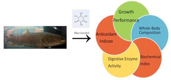
Effects of Myo-Inositol on the Growth Performance, Digestive Enzyme Activity, and Antioxidation of Juvenile Hucho taimen

1
Guizhou Fisheries Research Institute, Guiyang 550025, China
2
Heilongjiang River Fisheries Research Institute, Chinese Academy of Fishery Sciences, Harbin 150070, China
3
School of Fisheries, Nanjing Agricultural University, Wuxi 214081, China
4
School of Life Science, Huzhou University, Huzhou 313000, China
5
Zhejiang Provincial Key Laboratory of Aquatic Resources Conservation and Development, Huzhou University, 759 Erhuan Road (E), Huzhou 313000, China
*
Author to whom correspondence should be addressed.
Fishes 2023, 8(12), 567; https://doi.org/10.3390/fishes8120567
Submission received: 28 September 2023 / Revised: 13 November 2023 / Accepted: 18 November 2023 / Published: 22 November 2023

Abstract

:
Hucho taimen is a cold-water fish with high economic value. Myo-inositol (MI) can accelerate lipid metabolism and promote growth in fish species. The present study aimed to assess the effect of MI on the growth performance, digestive enzyme activity, and antioxidation of juvenile H. taimen. Accordingly, an 8-week feeding trial was conducted. The results demonstrated that increasing MI concentration promoted growth performance in H. taimen. Among the MI concentrations tested, a dose of 328 mg MI/kg corresponded with the lowest feed conversion ratio (FCR) and the highest growth rate. Compared with fish fed a diet of 128 mg MI/kg, the lipase activity in the pyloric caeca significantly increased in fish fed 528 mg MI/kg, while superoxide dismutase (SOD) activity was significantly higher in fish fed 728 mg MI/kg. Consistently, the 128 mg MI/kg diet presented the highest malonaldehyde (MDA) levels. In conclusion, our study revealed that enhanced growth performance, digestive enzyme activity, and antioxidant capacity increased as MI concentration increased. The optimum level of dietary MI in H. taimen was 270–321 mg/kg, based on the FCR and specific growth rate (SGR) on the broken-line regression.
Key Contribution: In this research, analysis of the effects of MI on juvenile H. taimen was carried out. The results showed that growth performance, digestive enzyme activity, and antioxidant capacity were enhanced with the increasing of MI concentration to a specified level. The optimum level of dietary MI in H. taimen was proven to be 270–321 mg/kg; this is the level associated with the lowest FCR and highest SGR on the broken-line regression.

Graphical Abstract

1. Introduction

Hucho taimen belongs to Salmoniformes, Salmonidae, Salmon subfamily. It is a cold-water fish and its growth rate (WG) is slower than most other fish; the optimal temperature for it is 13–16 °C [1]. It is large: its body weight can be up to 50 kg and its body length can reach 1 meter or more. It is a precious fish with delicious, nutritious meat, and it has a high economic value. In recent years, due to overfishing and serious damage to the ecological environment, wild H. taimen resources have plummeted. It is now included in the “Chinese red list of endangered animal species”. At present, it has been successfully domesticated and is promoted for breeding in more than a dozen provinces in China.
Inositol, also known as cyclohexanol, has nine cis-trans isomers. The most common is myo-inositol (MI), which is a type of vitamin B [2] that can promote lipid metabolism, reduce blood lipids, and protect liver health. The main function of MI in aquatic animals is to improve feed utilization, accelerate growth, and promote lipid metabolism in the liver and other tissues. At present, feed company incorporate MI as a nutritional additive in aquatic animal feed.
Symptoms of MI deficiency have been found in young grass carp (Ctenopharyngodon idella) [3], juvenile Chinese mitten crab (Eriocheir sinensis) [4], and juvenile Pacific white shrimp (Litopenaeus vannamei) [5]. Symptoms manifested as anorexia, anemia, poor quality in growth and development, skin erosion, darkened skin color, slow gastric emptying, decreased cholinesterase, and the decreased activity of some transaminases. When Jian carp (Cyprinus carpio var. Jian) were deficient in MI, they suffered from fin erosion, skin bleeding, tail handle exposure (in severe cases), intestinal epithelial edema, and even shedding and necrosis [6,7].
Adding an appropriate level of MI to feed can improve growth performance, as has been demonstrated in Wuchang bream (Megalobrama amblycephala) [8], juvenile sunfish (Morone chrysops × Morone saxatilis) [9], and juvenile Ctenopharyngodon idella [10]. Khosravi found that an appropriate level of MI in feed could significantly promote the growth performance of striped beakfish (Oplegnathus fasciatus); the appropriate supplemental level of MI was found to be 94.3 mg/kg [11]. The growth performance and feed efficiency of olive flounder (Paralichthys olivaceus) were significantly improved by adding 800 mg MI/kg to their diet [12]. In Ctenopharyngodon idella, adding 100–150 mg MI/kg to its whole plant protein source diet was shown to significantly improve its growth performance and feed efficiency [13]. Within the large-scale and high-density culture of Nile tilapia (Oreochromis niloticus), the addition of 0.03% MI to feed was proven to be helpful to the growth and the mobilization of lipid, though the amount of MI added should be controlled strictly [14].
In the research of crustaceans, such as the giant tiger prawn (Penaeus monodon), Chinese white shrimp (Penaeus chinensis), and kuruma shrimp (Penaeus japonicus) [15], it was found that a lack of MI in feed led to a significant decrease in survival rate. Adding the appropriate levels of MI to feed could significantly improve the growth performance of Penaeus monodon, Penaeus chinensis and Penaeus japonicus. The suitable supplemental level of MI in the feed of Penaeus monodon was 3275–3514 mg/kg [16]; the level for Penaeus chinensis was 4000 mg MI/kg [17]. For Eriocheir sinensis, that level was 1613–1707 mg MI/kg [18]. In the study of Litopenaeus vannamei, 600 mg MI/kg was found to be sufficient to meet its needs in terms of body growth and development; the appropriate level of MI significantly improved its resistance to salinity stress [19]. These studies have shown that the level of MI required for aquatic animals to thrive may be related to different feed formulations, growth stages, breeding varieties, feeding habits, and breeding environments and models; however, the regulatory mechanism of MI in growth performance is still not completely clear.
Juvenile H. taimen can accurately assess their own nutritional needs, including determining their MI requirements. In this paper, the MI requirement of juvenile H. taimen was comprehensively evaluated based on growth performance and antioxidant indexes, in order to provide theoretical guidance for the production of H. taimen feed under this lipid level.

2. Materials and Methods

2.1. Experimental Design and Feeding Management

The MI used in this experiment, which was purchased from Sigma, USA, was in powder form and its purity was ≥99%. Six treatment groups were set in the experiment; the MI content in the basic diet was 128 mg/kg without additional MI. The MI supplementation in each group was 0 (G1), 100 (G2), 200 (G3), 400 (G4), 600 (G5), 800 (G6) mg/kg, respectively, and the inositol content values of each group was calculated as 128 (G1), 228 (G2), 328 (G3), 528 (G4), 728 (G5) and 928 (G6) mg/kg, respectively. Feed composition and nutritional level are shown in Table 1. After crushing, the raw materials were mixed evenly according to proportion, granulated (1.0 mm), dried naturally, and stored in a refrigerator at −20 °C.
A total of 540 H. taimen (2.83 ± 0.44 g) were randomly assigned to six groups with three replicates per group and 30 fish per replicate. Body weight and body length were recorded after 7 days of pre-feeding with commercial feed and 24 h of starvation. H. taimen was fed four times a day (07:00, 10:30, 14:00, and 17:00). The daily ration was determined based on the weight of each treatment of fish, which were weighed every 2 weeks and fed at 3–8% of the total weight of each treatment. The feeding trial lasted for 8 weeks [20,21,22,23,24]. The trial employed a flow-through culture system; no aerators were used. The current speed of the flowing water was [(0.3 × 10⁻³)–(0.4 × 10⁻³) m3/h], the water temperature was 9.5–15.2 °C, the dissolved oxygen was kept at more than 8.0 mg/L−1, and the pH of the water was 7.5–7.8.
H. taimen was weighed at the beginning (initial body weight) and at the end (final body weight) of the 8-week feeding trial. The balance (BP221S) used for weighing the fish was made by Sartorius; its maximum range is 210 g and its precision is 0.1 mg. The weight gain, specific growth rate, feed conversion ratio, condition factor, viscerosomatic index, and hepatosmatic index of each treatment group were recorded and analyzed. These parameters were calculated as:
  • Weight gain (WG, %) = (Wf − Wi)/Wi × 100;
  • Specific growth rate (SGR, %/day) = (ln Wf − ln Wi)/days) × 100;
  • Feed conversion ratio (FCR) = Feed consumed (g)/(Wf − Wi);
  • Condition factor (CF, g·cm−3) = Wf × 100/Lf3;
  • Viscerosomatic index (VSI, %) = (Wv/Wf) × 100;
  • Hepatosmatic index (HSI, %) = (Wh/Wf) × 100.
where Wf and Wi are the initial and final body weights, Lf is the initial body length, Wv is the total weight of fish viscera, and Wh is the weight of the fish liver.

2.2. Sample Collection

At the end of the feeding trial, starvation was enforced for 24 h before sampling. Three H. taimen per tank were collected randomly and frozen (−40 °C) for whole-body composition analyses. In addition, three H. taimen were selected per tank and placed on an ice tray for dissection. The liver, pyloric caeca, and intestinal tract were all sampled; the crude enzyme solution of the tissue was extracted in each case and stored at −80 °C for further analysis.

2.3. Whole-Body Composition Analyses

Moisture, crude protein, crude lipid, and ash were measured [25]. The moisture content was measured using the 105 °C drying method. The crude protein was determined by the Kjeldahl method. The crude lipid was analyzed by the Soxhlet extraction method. Last, ash content was determined by using a high-temperature combustion method applied to the fish body.

2.4. Biochemical Analysis

The liver, pyloric caeca, and intestinal tract were sampled. After homogenization, the samples were centrifuged for 10 min at 3500 r/min to obtain the supernatant at 4 °C, then the supernatant was collected. Amylase activity was determined using the iodine-starch colorimetric method. After the color reaction, absorbance was measured at 660 nm. Protease activity was tested by the Folin-phenol method. MDA was determined by the TBA method, and there was a maximum absorption peak at 532 nm. One unit of SOD is the amount of SOD that corresponds to an inhibition rate of 50% in 1 mL of tissue protein in a 1 ml reaction solution. ATP per milligram of tissue protein per hour produces 1 umol of inorganic phosphorus as a unit of Na+,K+-ATPase activity. Amylase, protease, lipase, alkaline phosphatase (AKP), Na+,K+-ATPase, SOD, and MDA were measured by kits that were produced by Nanjing Jiancheng Bioengineering Institute.

2.5. Statistical Analysis

The experimental data were analyzed by one-way ANOVA in SPSS 17.0; p < 0.05 was considered to be statistically significant. Experimental data were expressed as mean ± standard deviation ( X ¯   ± SD) across at least three independent experiments. The optimal MI content for H. taimen was estimated using the broken-line model. The WG and FCR of H. taimen, as well as MI content, were estimated.

3. Results

3.1. Growth Performance

Compared to the control group, other groups significantly increased WG (p < 0.05). The FCR of the G3 group was significantly lower than that of the others (p < 0.05). There was no significant difference in HSI among all groups (p > 0.05). Compared to the control group, the G3 group significantly decreased the CF (p < 0.05) and significantly increased the SGR (p < 0.05); additionally, the G4 group significantly increased the VSI (p < 0.05) (Table 2).
Based on the broken-line model of FCR, SGR, and MI levels, it was concluded that the optimal level for H. taimen was 270–321 mg MI/kg (Figure 1 and Figure 2).

3.2. Whole-Body Composition

No significant difference was found in terms of moisture and crude protein content among all groups (p > 0.05). The whole-body crude lipid contents of treatment groups were all higher than that of the control group, but only that of the G3 group was significantly increased (p < 0.05). Compared to the control group, whole-body ash content was significantly increased in the G3 and G5 groups (p < 0.05) (Table 3).

3.3. Digestive Enzyme Activity

As shown in Table 4, there were no significant differences in the amylase activity of the liver and intestine among all groups (p > 0.05). The MI content in the diet had a significant effect on the amylase activity of the pyloric caeca (p < 0.05); the amylase activity of the G3 group was significantly increased (p < 0.05). There were no significant differences in the protease activity of the liver and pyloric caeca among all groups (p > 0.05). Compared to the control group, the protease activity of the intestine was significantly increased in the G4 and G5 groups (p < 0.05). The lipase activity of the G4 group was higher than that of the control group (p < 0.05); however, there was no significant difference in the lipase activity of the intestine among all groups (p > 0.05) (Table 4).

3.4. Biochemical Index

The AKP and Na+,K+-ATPase activities of the pyloric caeca did not show any significant differences among all groups (p > 0.05). The Na+,K+-ATPase activities of the intestine in the treatment groups were all higher than that of the control group, but only the G4 group was significantly higher (p < 0.05). Compared to the control group, the AKP activities of the intestine were significantly increased in the G3 group, G4 group, and G5 group (p < 0.05) (Table 5).

3.5. Antioxidant Indices

Among the treatment groups, SOD activity was significantly increased in the G5 group (p < 0.05). Compared to the control group, MDA content was decreased in all treatment groups (p < 0.05) (Table 6).

4. Discussion

This study demonstrated that WG was observably increased by increasing MI levels in H. taimen, a result similar to that seen in Penaeus monodon [16], Ctenopharyngodon idella [3], and juvenile Eriocheir sinensis [4]. Our study showed that the CF and FCR decreased with increasing MI levels. A possible reason for this is that MI deficiency affects the synthesis and secretion of low-density lipoprotein. In other words, lipid cannot be transported out of the liver in time, resulting in abnormal accumulation in the liver, which causes lipid deposition in fish bodies which, in turn, results in the increasing of CF [26]. Attaining an appropriate level of MI content could make better use of the lipid in the feed, thereby promoting fish health and improving feed utilization efficiency. Levine found that adding MI could improve the swimming ability of crucian carp (Carassius auratus) [27]. The reason for our results might be that the addition of an appropriate amount of MI to feed improved the utilization rate of lipid in that feed, which promotes growth in H. taimen. A study of Mozambique tilapia (Oreochromis mossambicus) showed that MI could regulate lipid and pyruvic acid metabolism, increasing phospholipid synthesis-related mRNA levels and improving antioxidant properties under long-term salinity stress [18]. In addition, the feeding habits of fish also affect the demand for MI content. Carnivorous fish need high-level lipid in their feed as they need more MI to meet the needs of lipid metabolism. For example, the optimal MI demand of blackhead sea bream (Acanthopagrus schlegelii) was shown to be 2000 mg/kg [28], while the demand of Ctenopharyngodon idella was only 166–214 mg MI/kg [10]. In the liver and kidneys of some aquatic animals, MI can be synthesized with glucose as a substrate through l-inositol-1-phosphate synthase and l-inositol-1-phosphatase. MI synthesis ability is determined by MI synthase activity [29]; moreover, aquatic animals can synthesize a certain amount of MI through intestinal microorganisms, which also affects their MI demands.
This study demonstrated that MI level did not affect whole-body crude protein content in H. taimen, a result similar to that seen in Atlantic salmon (Salmo salar) [30], juvenile Litopenaeus vannamei [5], and Pacific abalone (Haliotis discus hannai Ino) [31], indicating that MI has little effect on digestion and the absorption of protein in H. taimen and that the addition of MI has no significant effect on the protein deposition rate. The contents of ash and crude lipid decreased significantly with the increasing of MI content to a certain extent, which is consistent with the results of a study of juvenile Cyprinus carpio var. Jian [7]. A possible reason for the decreasing of whole-body crude lipid is that an excessive amount of MI promotes lipid decomposition in the liver. The combination of fatty acids and various enzymes on the liver cell membrane improves the lipid exchange capacity of the cell membrane, which is transported to various tissues for utilization, and the lipid content in the body can be effectively reduced; however, no significant differences in the whole-body composition of golden pompano (Trachinotus ovatus) was found between control and treatment groups [32]. The reason for these two differing sets of results could be that the capacity of MI to regulate lipid metabolism in different aquatic animals varies; therefore, the mechanism for the effect of MI on the body composition of aquatic animals requires further study.
The digestive capacity of fish is directly related to the development of digestive organs and digestive enzyme activity [33]. This research demonstrated that digestive enzyme activity increased with the increasing of MI content, which is consistent with previous findings in various species, including juvenile turbot (Scohpthalmus maximus L.) [34], juvenile Cyprinus carpio var. [35], Litopenaeus vannamei [36], and Japanese sturgeon (Acipenser schrenckii) [37]. A possible reason for this finding is that, when MI content is insufficient, the liver and pancreas of fish cannot develop normally; therefore, the digestive enzyme secreted by the pancreas is reduced. On the other hand, in our study, the amount of bile acid secreted by the liver was reduced, which affected the lipase activity of the fish. In addition, MI exists as part of the cell membrane in the form of phosphatidyl-MI. When MI is deficient, the intestinal structure of aquatic animals is affected which, in turn, would also affect digestive enzyme activity. In our study, after the MI was absorbed by H. taimen, it was stored in serum and the liver, and it could improve the activity of cholinesterase, which proved to be conducive to the improvement of amylase activity and WG in individual fish [38]; moreover, by increasing the intake of each fish, MI indirectly stimulated the secretion of digestive enzymes.
The AKP in the intestine participates in the absorption of nutrients such as lipid and glucose, etc. Na+,K+-ATPase activity can reflect intestinal absorption capacity [39]. This study has shown that, as MI content is increased, Na+,K+-ATPase and AKP activities increase, promoting the absorption of nutrients. This finding aligns with the results of similar testing on juvenile Cyprinus carpio var. [6]. A possible reason for the finding is that Na+,K+-ATPase is the main component of the Na+,K+ pump, which constitutes part of the cell membrane; MI is also a component of the membrane. Substances targeting MI on the membrane would, therefore, indirectly affect the activities of Na+,K+-ATPase [40]. The amount and existing state of MI may also affect the activities of Na+,K+-ATPase. Appropriately increasing MI content can help to increase the activities of Na+,K+-ATPase and AKP.
Antioxidant levels can reflect the health status of animals. SOD is an important antioxidant enzyme, which has the effect of mitigating anti-oxidative damage and maintaining cell structure [41]. When there are fewer antioxidant enzymes, the metabolic balance of free radicals will be destroyed, and free oxygen radicals will react with unsaturated fatty acids, resulting in lipid peroxidation and damage to cells and macromoleculars in cells, all of which cause damage to the body. Lipid peroxidation can produce a variety of metabolites, of which MDA is one of the most important. The content of MDA in the liver can reflect the progress of lipid peroxidation; the degree of oxidative damage to cells can also be shown indirectly [42]. In this experiment, when MI content was insufficient, the MDA content increased and the SOD content decreased, which is consistent with the results of a study on juvenile Cyprinus carpio var. [7]. A possible reason for this finding is that insufficient MI content affects the development of the liver, leading to a disorder of lipid metabolism. Another possible reason is that the synthesis and secretion of low-density lipoprotein were affected; because the lipid in the liver could not be transported in time, the lipid did not accumulate normally. This process has also been seen in research on Carassius auratus [43] and juvenile Ctenopharyngodon idella [44]. Increasing MI content to an appropriate level could reduce the production of oxygen free radicals in the body; furthermore, this could promote the elimination of oxygen free radicals which could, in turn, effectively reduce the damage of oxygen free radicals to the body and improve antioxidant capacity, thus enhancing growth performance in H. taimen.

5. Conclusions

When MI content was deficient, the growth performance, digestive enzyme activity, and antioxidant capacity of H. taimen were decreased. The appropriate MI content in feed can improve the WG, digestive enzyme activity, and antioxidant capacity of H. taimen. When the protein content in the feed was 52.3% and the lipid content was 12.4%, the optimal requirement of MI was determined to be 270–321 mg/kg, according to broken-line regression analysis based on the FCR and SGR of H. taimen.

Author Contributions

All authors contributed to the study. M.Z.: experiment execution, formal analysis, investigation, and writing—original draft; X.Y., C.W., B.S., F.Z. and H.X.: methodology, resources, writing–review, and supervision; Q.X.: methodology, supervision, writing—review, project administration, editing, and funding acquisition. All authors commented on previous versions of the manuscript. All authors have read and agreed to the published version of the manuscript.

Funding

This research was funded by the Subsidy project from NSFC of Guizhou Academy of Agricultural Sciences (2021-11); characteristic aquatic industry technology system of Guizhou province (GZCYTX2022-01101).

Institutional Review Board Statement

This study was conducted according to the guidelines of the Declaration of Helsinki, and approved by the Institutional Review Board of Heilongjiang River Fisheries Research Institute, Chinese Academy of Fishery Sciences (code: 20200615, date: 15 June 2020).

Informed Consent Statement

Not applicable.

Data Availability Statement

All data presented in the work are available from the corresponding authors, upon responsible request.

Acknowledgments

The authors are grateful to Yingying Wei, Jinfeng Huang, Liang Luo, and Jinnan Li for their assistance and expertise throughout the study.

Conflicts of Interest

The authors declare no conflict of interest.

References

  1. Zhang, H.; Liu, Y.; Zhao, X.; Wang, C.; Lu, S.; Liu, H.; Xu, Q. Effects of Aloe Powder on growth performance and serum indices of Taimen Hucho taimen. Chin. J. Fish. 2020, 33, 1–6. [Google Scholar]
  2. Wu, Q.; Wang, S.; Zhen, Y.; Wang, T.; Zhang, X.; Sun, Z. Research progress of preparation process and application of inositol. Heilongjiang Anim. Sci. Vet. Med. 2023, 20, 41–46. [Google Scholar]
  3. Li, S.; Jiang, W.; Feng, L.; Liu, Y.; Wu, P.; Jiang, J.; Kuang, S.; Tang, L.; Tang, W.; Zhang, Y.; et al. Dietary myo-inositol deficiency decreased the growth performances and impaired intestinal physical barrier function partly relating to nrf2, jnk, e2f4 and mlck signaling in young grass carp (Ctenopharyngodon idella). Fish Shellfish Immunol. 2017, 67, 475–492. [Google Scholar] [CrossRef] [PubMed]
  4. Bu, X.; Lin, Z.; Liu, S.; Wang, C.; Wang, N.; Lei, Y.; Zhu, J.; Wang, X.; Qin, J.; Chen, L. Effects of myo -inositol on growth performance, body composition, antioxidant status, non-specific immunity and lipid metabolism of juvenile Chinese mitten crab (Eriocheir sinensis). Aquac. Nutr. 2020, 26, 1623–1635. [Google Scholar] [CrossRef]
  5. Li, R.; Kou, S.; Yang, Q.; Tan, B.; Chi, S.; Yi, Y.; Zhu, D. Effects of Myo-Inositol on Growth, Non-Specific Immunity and Intestinal Microflora Composition of Juvenile Litopenaeus Vannamei. Chin. J. Anim. Nutr. 2023, 35, 1182–1194. [Google Scholar]
  6. Feng, L.; Li, J.; Zhou, X.; Liu, Y. Effects of inoistol deficiency on digestive and immune function of juvenile jian carp(Cyprinus carpio var. Jian). Chin. J. Anim. Nutr. 2009, 21, 878–883. [Google Scholar]
  7. Jiang, W.; Liu, Y.; Feng, L.; Zhou, X. Histopathological observation of juvenile Cyprinus carpio var. Jian with myo-inositol deficiency and determinations of antioxidative ability and intestinal microbial population. Vet. Sci. China 2009, 31, 64–69. [Google Scholar]
  8. Cui, H.; Liu, B.; Ge, X.; Xie, J. Effects of dietary inositol on growth performance, physiological and biochemical indexes of serum and fat content in liver and muscle of juvenile Wuchang bream (Megalobrama amblycephala). J. Shanghai Ocean Univ. 2013, 22, 868–875. [Google Scholar]
  9. Deng, D.; Hemre, G.; Wilson, R.P. Juvenile sunshine bass (Morone chrysops × Morone saxatilis) do not require dietary myo-inositol. Aquaculture 2002, 213, 387–393. [Google Scholar] [CrossRef]
  10. Wen, H.; Zhao, Z.; Jiang, M.; Liu, A.; Wu, F.; Liu, W. Dietary myo-inositol requirement for grass carp, Ctenopharyngodon Idella Fingerling. J. Fish. Sci. China 2007, 14, 794–800. [Google Scholar]
  11. Khosravi, S.; Lim, S.; Rahimnejad, S.; Kim, S.; Lee, B.; Kim, K.; Han, H.; Lee, K. Dietary myo-inositol requirement of parrot fish. Oplegnathus fasciatus. Aquaculture 2015, 436, 1–7. [Google Scholar] [CrossRef]
  12. Lee, B.; Lee, K.; Lim, S.; Lee, S. Dietary myo-inositol requirement for olive flounder, Paralichthys olivaceus (Temminch et Schlegel). Aquac. Res. 2009, 40, 83–90. [Google Scholar] [CrossRef]
  13. Lin, K.; Feng, J.; Yang, H.; Chen, Y.; Chen, X.; Luo, H.; Huang, W.; Wang, H.; Luo, L. Effects of inositol supplementation to practical dietary on growth performance, lipid metabolism and antioxidant activity of Cteno pharyngodon Idella. J. Fish. China 2018, 42, 1428–1437. [Google Scholar]
  14. Wu, H.; Tang, Z.; Yang, H.; Luo, Y.; Huang, K.; Gan, X. Effects of dietary inositol on growth, liver and muscle fat content and biochemical indices of blood serum in tilapia (Oreoc hromis niloticus × Oreochromis aureus). J. South. Agric. 2011, 42, 1415–1419. [Google Scholar]
  15. Kanazawa, A.; Teshima, S.; Tanaka, N. Nutritional requirements of prawn V: Requirements for choline and inositol. Mem. Fac. Fish. Kagoshima Univ. 1976, 25, 47–51. [Google Scholar]
  16. Shiau, S.; Su, S. Dietary inositol requirement for juvenile grass shrimp, Penaeus Mondon. Aquac. 2004, 241, 1–8. [Google Scholar] [CrossRef]
  17. Liu, T.; Li, A.; Zhang, J. Studies on vitamin nutrition for the shrimp penaeus chinensis-X: Studies on the choline chloride and inositol requirements in the shrimp Penaeus chinensis. J. Ocean Univ. Qingdao 1993, 23, 71–78. [Google Scholar]
  18. Bu, X.; Zhu, J.; Liu, S.; Wang, C.; Xiao, S.; Lu, M.; Li, E.; Wang, X.; Qin, J.G.; Chen, L. Growth, osmotic response and transcriptome response of the euryhaline teleost, Oreochromis mossambicus fed different myo-inositol levels under long-term salinity stress. Aquaculture 2021, 534, 736294. [Google Scholar] [CrossRef]
  19. Chen, S.; Guo, Y.; Espe, M.; Yang, F.; Fang, W.; Wan, M.; Niu, J.; Liu, Y.; Tian, L. Growth performance, haematological parameters, antioxidant status and salinity stress tolerance of juvenile Pacific white shrimp (Litopenaeus vannamei) fed different levels of dietary myo-inositol. Aquac. Nutr. 2018, 24, 1527–1539. [Google Scholar] [CrossRef]
  20. Zhao, Z.; Zhao, F.; Cairang, Z.; Zhou, Z.; Du, Q.; Wang, J.; Zhao, F.; Wang, Q.; Li, Z.; Zhang, X. Role of Dietary Tea Polyphenols on Growth Performance and Gut Health Benefits in Juvenile Hybrid Sturgeon (Acipenser baerii ♀ × A. schrenckii ♂). Fish Shellfish Immunol. 2023, 139, 108911–108921. [Google Scholar] [CrossRef] [PubMed]
  21. Zhang, Q.; Yang, Q.; Li, M.G.; Li, F.; Qin, M.; Xie, Y.; Xu, J.; Liu, Y.; Tong, T. The Effects of Dietary Fermented Soybean Meal Supplementation on the Growth, Antioxidation, Immunity, and mTOR Signaling Pathway of Juvenile Coho Salmon (Oncorhynchus kisutch). Fishes 2023, 8, 448. [Google Scholar] [CrossRef]
  22. Yuan, H.; Hu, N.; Zheng, Y.; Hou, C.; Tan, B.; Shi, L.; Zhang, S. A Comparison of Three Protein Sources Used in Medium-Sized Litopenaeus vannamei: Effects on Growth, Immunity, Intestinal Digestive Enzyme Activity, and Microbiota Structure. Fishes 2023, 8, 449. [Google Scholar] [CrossRef]
  23. Chu, J.-H.; Weng, T.-S.; Huang, T.-W. The Effects of Replacing Fish Meal Protein with Black Soldier Fly Meal and Sodium Butyrate Supplementation on the Growth Performance, Lipid Peroxidation, and Intestinal Villi Status of Jade Perch, Scortum Barcoo Fingerlings. Fishes 2023, 8, 437. [Google Scholar] [CrossRef]
  24. Yang, Z.; Liu, P.; Kong, Q.; Deng, Y.; Zhang, W.; Xu, G.; Tang, H. Effects of Co-Fermented Feed Using Lactobacillus acidophilus, Limosilactobacillus reuteri and Lactiplantibacillus plantarum on Growth, Antioxidant Capacity, Fatty Acids and Gut Microbiota of Largemouth Bass (Micropterus salmoides). Fishes 2023, 8, 433–450. [Google Scholar] [CrossRef]
  25. Aoac, I. Official Methods of Analysis of AOAC International, 18th ed.; AOAC International: Maryland, MD, USA, 2010. [Google Scholar]
  26. Yin, G.; Zhen, Y.; Wang, T.; Zhang, X.; Sun, Z. The Mechanism of Inositol and Its Application in Animal Production. J. Econ. Anim. 2023, 11, 1–7. [Google Scholar]
  27. Levine, A.; Billington, C. Why do we eat? A neural systems approach. Annu. Rev. Nutr. 1997, 17, 597–619. [Google Scholar] [CrossRef]
  28. Chen, S.; Ji, W.; Lv, Y.; Chang, Q.; Pan, S. Nutrient function of inositol on the growth of black sea bream, juvenile, Sparus Marcocephalus. J. Mar. Sci. 1995, 5, 13–15. [Google Scholar]
  29. Burtle, G.; Lovell, R. Lack of response of channel catfish (Lctalurus punctatus) to dietary myo-inositol. Can. J. Fish. Aquat. Sci. 2011, 46, 218–222. [Google Scholar] [CrossRef]
  30. Waagbø, S.; Lie, R. Effects of inositol supplementation on growth, chemical composition and blood chemistry in Atlantic salmon (Salmo salar L.)fry. Aquac. Nutr. 1998, 4, 53–59. [Google Scholar] [CrossRef]
  31. Mai, K.; Wu, G.; Zhu, W. Abalone, Haloiotis discus hannai Ino, can synthesize myo-inositol de novo to meet physiological needs. Nuturition 2001, 131, 2898–2903. [Google Scholar]
  32. Chen, X.; Wang, J.; Zhao, W. Effects of dietary inositol supplementation on growth, digestive performance, antioxidant capacity, and body composition of golden pompano(Trachinotus ovatus). Front. Physiol. 2022, 13, 850470. [Google Scholar] [CrossRef] [PubMed]
  33. Peres, H.; Lim, C.; Klesius, P. Growth, chemical composition and resistance to Streptococcus iniae challenge of juvenile nile tilapia (Oreochromis niloticus) fed graded levels of dietary inositol. Aquaculture 2004, 235, 423–430. [Google Scholar] [CrossRef]
  34. Wang, Y.; Li, B.; Wang, J.; Wang, C.; Wang, X.; Ma, C.; Wang, S.; Sun, Y.; Hao, T. Dietary Myo-inositol Requirement of Juvenile Turbot(Scophthalmus maximus L.). Chin. J. Anim. Nutr. 2019, 31, 5122–5132. [Google Scholar]
  35. Jiang, W.; Feng, L.; Liu, Y.; Jiang, J.; Zhou, X. Growth, digestive capacity and intestinal microflora of juvenile Jian carp (Cyprinus carpio var. Jian) fed graded levels of dietary inositol. Aquac. Res. 2010, 40, 955–962. [Google Scholar] [CrossRef]
  36. Yu, Q.; Han, F.; Huang, M.; Wang, X.; Qin, J.G.; Chen, L.; Li, E. The effects of dietary myo-inositol on growth and physiological, biochemical, and molecular responses in the Pacific white shrimp (Litopenaeus vannamei). Aquaculture 2023, 568, 739323. [Google Scholar] [CrossRef]
  37. Zhang, S.; Wang, C.; Lu, S.; Liu, S.; Wang, Y.; Liu, H.; Yang, Y.; Xu, Q. Effects of dietary myo-Inositol on digestive physiology of Acipenser Schrenckii. Feed Ind. 2022, 43, 53–59. [Google Scholar]
  38. Yone, Y.; Furuichi, M.; Shitanda, K. Vitamin requirement of the red sea bream: Relationship between inositol requirements and glucose levels in diet. Bull. Jpn. Soc. Sci. Fish. 1971, 37, 149–155. [Google Scholar] [CrossRef]
  39. Lin, Y.; Zhou, X. Dietary glutamine supplementation improves structure and function of intestine of juvenile Jian carp (Cyprinus carpio var. Jian). Aquaculture 2006, 256, 389–394. [Google Scholar]
  40. Fang, Z.; Wang, C.; Wei, H. Effects of mercury and selenium on Na+,K+-ATPase activity in xiphophorus heller heckle. Chin. J. Appl. Environ. Biol. 2006, 12, 220–223. [Google Scholar]
  41. Xu, Q.; Wang, C.; Xu, H.; Zhu, Q.; Yin, J. Effects of glutamine dipeptide on intestine antioxidant, abilities of digestion and absorption of Hucho taimen larvae. J. Fish. Sci. China 2010, 17, 351–356. [Google Scholar]
  42. Ye, J.; Han, Y.; Zhao, J.; Lu, T.; Liu, H.; Yang, Y. Effects of dietary olaquindox on antioxidant enzymes system in hepatopancreas of Cyprinus Carpio. J. Fish. China 2004, 28, 231–235. [Google Scholar]
  43. Liu, Z.; Song, W.; Yu, B. Effects of inositol on tissue lipid and protein oxidation and antioxidant of crucian. China Feed 2019, 12, 39–43. [Google Scholar]
  44. Hu, K.; Li, S.; Feng, L.; Jiang, W.; Wu, P.; Liu, Y.; Jiang, J.; Kuang, S.; Tang, L.; Zhou, X. Protective effect of myo-inositol on oxidative damage of head kidney and spleen in juvenile grass carp (Ctenopharyngodon idella) induced by Aeromonas hydrophila. J. Fish. China 2019, 43, 2256–2267. [Google Scholar]
Figure 1. Effects of MI on the FCR of H. taimen.
Fishes 08 00567 g001
Figure 2. Effects of MI on the SGR of H. taimen.
Fishes 08 00567 g002
Table 1. Ingredients and chemical composition of the basal diets (air-dry basis, %).
IngredientsContent
Casein34.00
Fish meal23.00
Gelatin10.00
Dextrin15.30
Fish oil11.00
Phospholipids2.00
Ca(H2PO4)21.00
CMC2.60
Vitamin premix 10.30
Mineral premix 20.20
Additive 30.60
Total100.00
Nutrient levels
Crude protein52.3
Crude lipid12.4
MI (mg/kg)128
1 The vitamin premix provided the following diet: VK 5 mg·kg−1, VA 15,000 IU·kg−1, VD3 3000 IU·kg−1, VB1 15 mg·kg−1, VB2 30 mg·kg−1, VB6 15 mg·kg−1, VB12 0.5 mg·kg−1, VC 1000 mg·kg−1, VE 60 mg·kg−1, Nicotinic acid 175 mg·kg−1, Folic acid 5 mg·kg−1, Biotin 2.5 mg·kg−1, and Pantothenic acid 50 mg·kg−1. 2 The mineral premix provided the following diet: Fe (as ferrous sulfate) 25 mg·kg−1, Cu (as copper sulfate) 3 mg·kg−1, Mn (as manganese sulfate) 15 mg·kg−1, I (as potassium iodide) 0.6 mg·kg−1, and Zn (as zinc sulfate) 60 mg·kg−1. 3 The additive provided the following diet per kg: Choline chloride 2 g, Antimildew 0.5 g, Magnesia 2 g, Antioxidant 0.5 g, and DMPT 1 g.
Table 2. Effects of different levels of MI on the growth performance of H. taimen.
TreatmentsInitial Weight (g)Final Weigh (g)WG (%)SGR (%)FCRCF (g·cm−3)HSI (%)VSI
(%)
G12.45 ± 0.274.17 ± 0.7670.20 ± 1.91 a0.94 ± 0.11 a1.98 ± 0.20 bc0.60 ± 0.10 b1.03 ± 0.187.11 ± 0.33 a
G22.55 ± 0.144.53 ± 1.0277.65 ± 1.00 b1.03 ± 0.14 ab1.94 ± 0.14 ab0.62 ± 0.03 bc1.12 ± 0.287.34 ± 0.98 ab
G32.75 ± 0.205.02 ± 0.6882.55 ± 2.99 bc1.11± 0.21 b1.89 ± 0.09 a0.58 ± 0.08 a1.61 ± 0.087.88± 0.70 ab
G42.55 ± 0.194.74 ± 0.4186.00 ± 2.16 c1.07 ± 0.12 ab2.00 ± 0.07 bc0.62 ± 0.08 bc1.43 ± 0.159.88 ± 1.00 b
G52.54± 0.214.58± 1.2780.31 ± 1.40 b1.05 ± 0.18 ab2.06 ± 0.15 c0.61 ± 0.07 b1.36 ± 0.047.59 ± 0.32 ab
G62.91± 0.385.14± 1.2576.63 ± 3.51 b1.02 ± 0.09 ab2.06 ± 0.04 c0.63 ± 0.05 bc1.53 ± 0.137.43 ± 1.11 ab
Note: within a column, values ( X ¯   ± SD) with different superscripts are significantly different (p < 0.05). G1, MI 128 mg/kg of feed; G2, MI 228 mg/kg of feed; G3, MI 328 mg/kg of feed; G4, MI 528 mg/kg of feed; G5, MI 728 mg/kg of feed; G6, MI 928 mg/kg of feed.
Table 3. Effects of different levels of MI on the whole-body composition of H. taimen.
TreatmentsMoisture (%)Crude Protein (%)Crude Lipid (%)Ash (%)
G180.19 ± 0.8414.97 ± 1.042.10 ± 0.02 a2.21 ± 0.08 a
G280.74 ± 0.8413.53 ± 1.102.16 ± 0.18 ab2.29 ± 0.16 a
G380.32 ± 0.4013.70 ± 1.422.41 ± 0.19 b2.55 ± 0.51 b
G479.90 ± 0.1214.38 ± 0.552.26 ± 0.02 ab2.33 ± 0.28 a
G580.30 ± 1.9813.12 ± 0.752.28 ± 0.01 ab2.80 ± 0.24 c
G680.49 ± 0.2514.13 ± 0.112.32 ± 0.03 ab2.22 ± 0.36 a
Note: within a column, values ( X ¯   ± SD) with different superscripts are significantly different (p < 0.05). G1, MI 128 mg/kg of feed; G2, MI 228 mg/kg of feed; G3, MI 328 mg/kg of feed; G4, MI 528 mg/kg of feed; G5, MI 728 mg/kg of feed; G6, MI 928 mg/kg of feed.
Table 4. Effects of different levels of MI on the digestive enzyme activity of H. taimen.
TissueEnzymeG1G2G3G4G5G6
LiverAmylase30.37 ± 1.7630.67 ± 4.2132.29 ± 0.6834.85 ± 2.8933.38 ± 3.0630.82 ± 1.77
Protease24.60 ± 2.0430.30 ± 5.5431.55 ± 3.7028.95 ± 1.2627.96 ± 5.2430.31 ± 4.61
Lipase8.54 ± 1.12 b6.44 ± 0.19 a7.61 ± 0.41 ab8.14 ± 0.82 b7.40 ± 0.38 ab8.02 ± 1.52 b
Pyloric caecaAmylase195.98 ± 12.01 a205.04 ± 8.67 ab236.07 ± 21.91 c222.71 ± 5.81 bc224.36 ± 7.13 bc218.60 ± 15.49 abc
Protease98.81 ± 9.59105.28 ± 10.13118.55 ± 14.21120.66 ± 19.28108.24 ± 8.38113.07 ± 4.35
Lipase41.97 ± 0.42 a42.28 ± 1.92 a42.46 ± 1.32 a47.46 ± 2.91 b44.80 ± 4.75 ab41.94 ± 1.29 a
IntestineAmylase161.64 ± 4.44166.58 ± 11.02176.06 ± 14.47164.21 ± 13.04160.95 ± 6.76160.07 ± 4.75
Protease43.66 ± 5.39 a44.98 ± 5.02 a50.87 ± 8.44 ab56.66 ± 2.09 b56.21 ± 1.21 b49.83 ± 3.24 ab
Lipase34.59 ± 6.5134.28 ± 3.5033.38 ± 1.6630.26 ± 2.7029.61 ± 0.9433.18 ± 3.03
Note: within a column, values ( X ¯   ± SD) with different superscripts are significantly different (p < 0.05). G1, MI 128 mg/kg of feed; G2, MI 228 mg/kg of feed; G3, MI 328 mg/kg of feed; G4, MI 528 mg/kg of feed; G5, MI 72 8mg/kg of feed; G6, MI 928 mg/kg of feed.
Table 5. Effects of different levels of MI on the biochemical index of H. taimen.
TissueBiochemical IndexG1G2G3G4G5G6
Pyloric caecaAKP55.89 ± 4.7856.60 ± 6.7769.45 ± 9.5065.55 ± 8.5760.48 ± 2.9161.63 ± 6.93
Na+,K+-ATPase
(μmolpi/gprot/h)
1.32 ± 0.231.23 ± 0.051.25 ± 0.161.39 ± 0.301.21 ± 0.121.18 ± 0.27
IntestineAKP42.16 ± 3.11 a49.58 ± 4.68 ab74.81 ± 11.62 c59.31 ± 8.73 b59.95 ± 8.06 b47.23 ± 8.55 ab
Na+,K+-ATPase
(μmolpi/gprot/h)
0.54 ± 0.09 a0.79 ± 0.05 a0.74 ± 0.05 a1.28 ± 0.27 b0.84 ± 0.05 a0.74 ± 0.12 a
Note: within a column, values ( X ¯   ± SD) with different superscripts are significantly different (p < 0.05). G1, MI 128 mg/kg of feed; G2, MI 228 mg/kg of feed; G3, MI 328 mg/kg of feed; G4, MI 528 mg/kg of feed; G5, MI 728 mg/kg of feed; G6, MI 928 mg/kg of feed. AKP, alkaline phosphatase; Na+,K+-ATPase, Na+,K+-stimulated ATPase.
Table 6. Effects of different levels of MI on the antioxidant indices of H. taimen.
TreatmentsSOD (NU·mgprot−1)MDA(nmol·mgprot−1)
G168.97 ± 8.31 a9.67 ± 0.42 b
G281.57 ± 12.81 ab6.20 ± 0.77 a
G382.26 ± 8.77 ab6.17 ± 1.57 a
G482.89 ± 7.70 ab7.32 ± 1.78 a
G589.59 ± 13.62 b7.47 ± 0.84 a
G676.32 ± 6.66 ab7.37 ± 0.81 a
Note: within a column, values ( X ¯   ± SD) with different superscripts are significantly different (p < 0.05). G1, MI 128 mg/kg of feed; G2, MI 228 mg/kg of feed; G3, MI 328mg/kg of feed; G4, MI 528 mg/kg of feed; G5, MI 728 mg/kg of feed; G6, MI 928 mg/kg of feed. SOD, superoxide dismutase; MDA, malonaldehyde.
Disclaimer/Publisher’s Note: The statements, opinions and data contained in all publications are solely those of the individual author(s) and contributor(s) and not of MDPI and/or the editor(s). MDPI and/or the editor(s) disclaim responsibility for any injury to people or property resulting from any ideas, methods, instructions or products referred to in the content.

