
Isolation and Culture of Primary Human Dental Pulp Cells—A Description of Technical and Methodological Steps to Maximise Predictability and Yield

Abstract
:1. Introduction
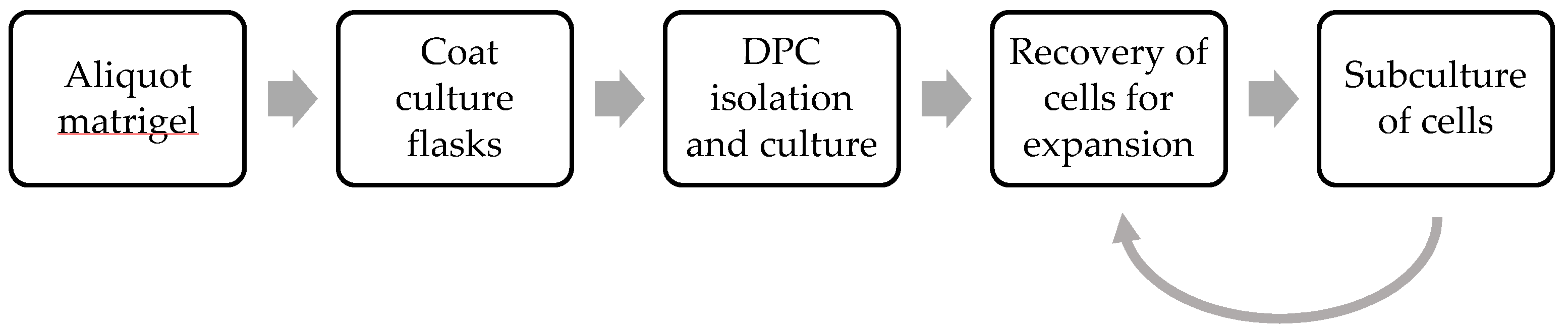
2. Experimental Design
2.1. Materials
- Corning® Matrigel® Basement Membrane Matrix, LDEV-Free (Corning, New York, NY, USA; Cat. No.: 356234).
- Dulbecco’s Modified Eagle Medium (DMEM), containing 2 mM (0.584 g/L) of L-Glutamine (Biowest, Nuaillé, France; Cat. No.: L0104) and supplemented with 1% Penicillin/Streptomycin (100 units/mL of penicillin with 100 μg/mL streptomycin) (Sigma-Aldrich, Arklow, Ireland; Cat. No.: P4333), 2.5 μg/mL Amphotericin B (Sigma-Aldrich, Arklow, Ireland; Cat. No.: A9528).
- Sound third molars, extracted no more than 2 h prior to use.
- Phosphate-buffered saline (PBS), pH 7.5: To be prepared by dissolving PBS tablet (Thermo Fisher Scientific, Dublin, Ireland; Cat. No.: BR0014G) in 500 mL ultrapure water. Autoclave the solution at 121 °C for 15 min and store at room temperature for no longer than 12 months.
- PerioKin mouthwash (0.2% chlorhexidine) (Laboratorios KIN, Barcelona, Spain; Cat. No.: 7153).
- Cell culture medium: DMEM supplemented with 20% (v/v) Foetal Calf Serum (FCS) (Biosera, Labtech International, East Sussex, UK; Cat. No.: FB-1001). Required volumes to be incubated at 37 °C before use in cell culture by placing in a water bath.
- Trypsin-EDTA: 0.25% (w/v) Trypsin and 1 mM EDTA.4Na (Sigma-Aldrich, Arklow, Ireland; Cat. No.: T4049). Required volumes to be incubated at 37 °C in a water bath prior to use.
- Corning® Cell Recovery Solution (Corning, New York, NY, USA; Cat. No.: 354253).
2.2. Equipment
- Ice bucket.
- Pipette tips: 200 µL, 1000 µL (StarLab, Milton Keynes, UK; Cat. No.: S1113-1810, S1111-6811).
- Sterile serological pipettes: 5 mL, 10 mL (Grenier Bio-One, Stonehouse, UK; Cat. No.: 606180, 607180).
- Eppendorf tubes: 0.5 mL (Eppendorf, Hamburg, Germany; Cat. No.: 0030121023).
- Sterile T25 flasks (Sarstedt, Leicester, UK; Cat. No.: 83.3910.302).
- Centrifuge tubes: 50 mL (Abdos Labtech, New Delhi, India; Cat. No.: P10416)
- Water bath (Sub Aqua 12 Plus, Grant Instruments, Royston, UK; Cat. No.: SBB Aqua 12 Plus).
- Water-cooled diamond blade saw (IsoMet Low Speed Saw, Buehler, IL, USA; Cat. No.: 11-1280-170).
- Sterile surgical gauze (Henry Schein, Dublin, Ireland; Cat. No.: 1201516).
- Sterile surgical chisel (Henry Schein, Dublin, Ireland; Cat. No.: 1113696).
- Sterile surgical mallet (Henry Schein, Dublin, Ireland; Cat. No.: 1120746).
- Scalpel (Swann-Morton, Sheffield, UK; Cat. No.: 0503).
- Sterile glass slides (Thermo Fisher Scientific, Dublin, Ireland; Cat. No.: 12373118).
- Centrifuge (Universal 320, Hettich, Tuttlingen, Germany; Cat. No.: 1401).
- Sterile T75 flasks (Sarstedt, Leicester, UK; Cat. No.: 83.3911.302).
- Phase contrast microscope (Zeiss Axiovert 25, Oberkochen, Germany; Cat. No.: 491205 9804)
- Standard 37 °C, 5% CO2 humidified tissue culture incubator (Sanyo, Osaka, Japan; Cat. No.; MCO-18AC(UV)).
3. Procedure
3.1. Aliquoting Matrigel®
 CRITICAL STEP One day prior to aliquoting, store pipette tips, Eppendorf tubes and a tube rack at −80 °C overnight. When handling Matrigel®, all reagents and consumables must be kept on ice. Matrigel® will solidify and adhere to any equipment above 10 °C. Change pipette tips regularly.
- 9.
- To thaw Matrigel®, submerge an unopened vial in ice in ice bucket. Close and seal ice bucket and store at 4 °C overnight.
- 10.
- Determine the protein concentration of the Matrigel® solution as indicated on the specification sheet.
- 11.
- Calculate the volume of Matrigel® corresponding to a 2 mg protein. For example, assuming a solution of Matrigel® with a protein concentration of 8.9 mg/mL, the following applies:Therefore, a 224.7 µL aliquot contains 2 mg protein.
- 12.
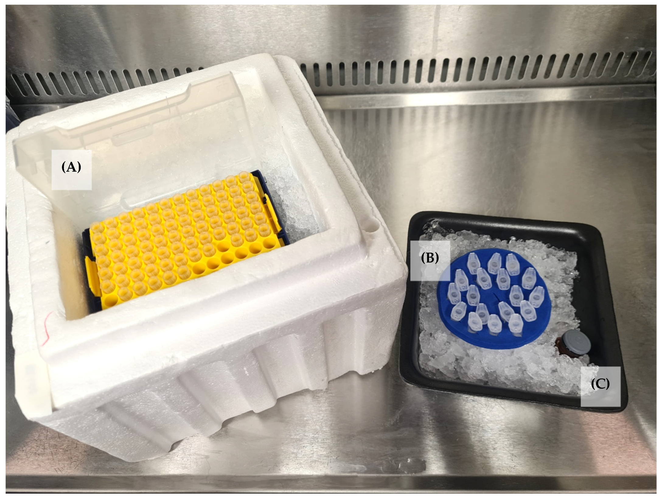
- Place chilled Eppendorf tubes in a chilled tube rack on ice. Place chilled pipette tips on ice (Figure 2).
- 13.
- Swirl Matrigel® to ensure even dispersal.
- 14.
- Using chilled pipette tips, dispense the appropriate volume of Matrigel® corresponding to 2 mg into chilled Eppendorf tubes.
 PAUSE STEP Aliquots can be stored at −20 °C or −70 °C.
3.2. Coating Culture Flasks
- Thaw Matrigel® aliquots on ice.
- Using a sterile cold pipette tip, dilute a 2 mg aliquot of Matrigel® to 1 ml with chilled DMEM for a final concentration of 2 mg/mL (See Note 1, Appendix A).
- Add the Matrigel® solution to a sterile T25 flask and shake gently to ensure even coating.
- Incubate at room temperature in a Class II biosafety cabinet overnight with the lid placed loosely on the flask to allow Matrigel® coating to set.
- Sterilise the Matrigel®-coated flask under UV exposure for 1 h.
- Once set, use a sterile pipette tip to gently remove the top layer of liquid, ensuring not to disturb the Matrigel® layer.
3.3. DPC Isolation
- Transfer extracted sound third molars to 4ml PerioKin mouthwash in a sterile 50 mL centrifuge tube (Figure 3A) (See Note 2, Appendix A).
- Incubate at room temperature for 5 min.
- Transfer molars to 4 mL DMEM in a sterile 50 mL centrifuge tube for short-term storage during pulp extraction.
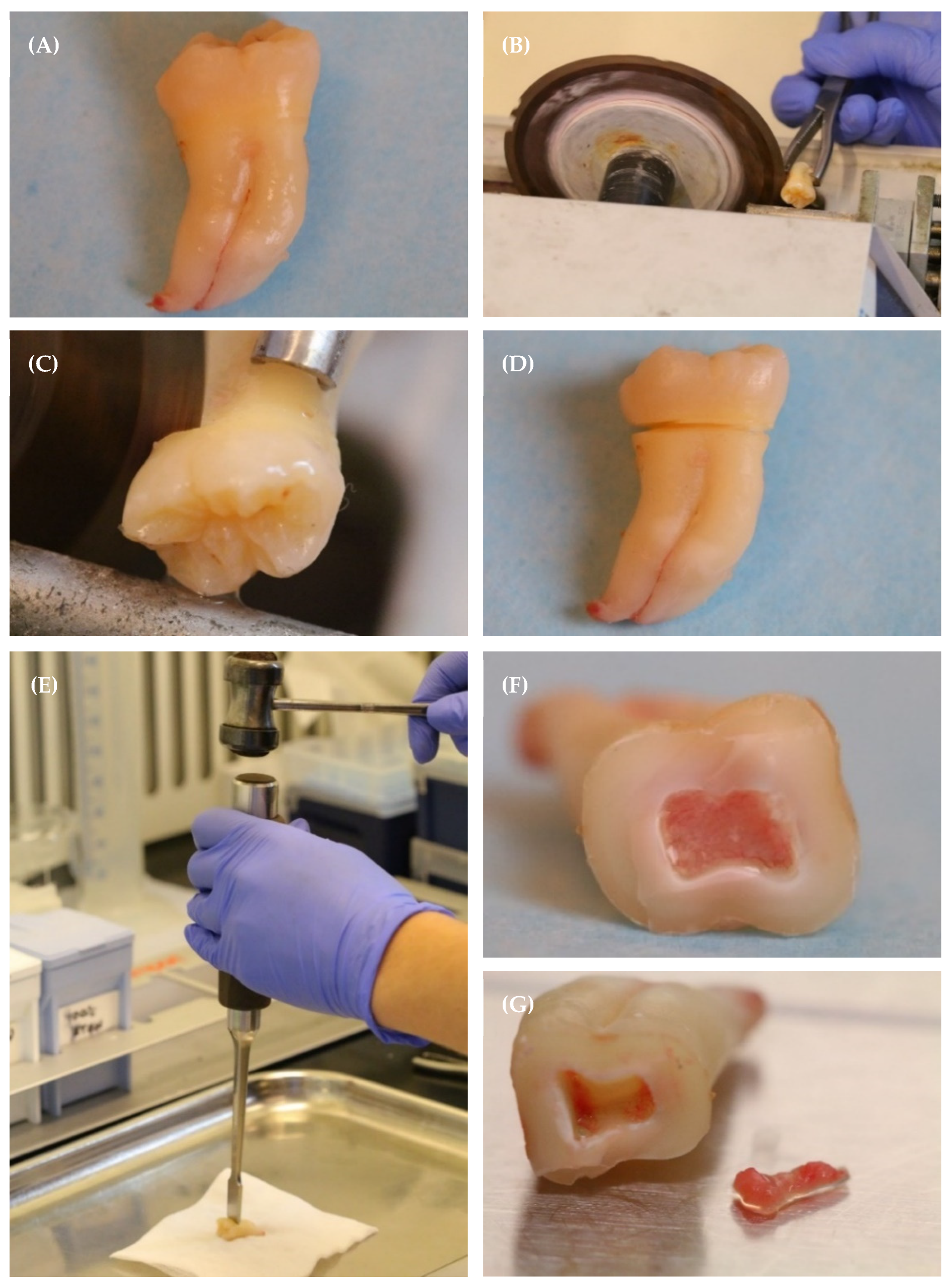
- Using surgical forceps, position the tooth against the edge of the water-cooled saw, with the blade aligned with the cemento-enamel junction (CEJ). (See Notes 3 and 4, Appendix A) (Figure 3B,C).
- Beginning with a low speed and light pressure, gently rotate the tooth to allow the saw to form a shallow groove around the circumference of the CEJ (Figure 3B,C).
- Continue to rotate the tooth to allow the saw to deepen the groove until it is approximately 2 mm deep around the circumference of the CEJ (Figure 3D).
- Place a sterile surgical chisel in the groove and gently tap with a sterile surgical mallet to separate the crown of the tooth from the root and expose the pulp chamber (Figure 3E,F).
- Use a scalpel to gently remove the pulp from the pulp chamber onto a sterile glass slide (Figure 3G). Add 200 µL DMEM to ensure the pulp remains moist.
- 9.
- Use a sterile scalpel to slice the pulp into 5–6 small pieces, approximately 1 mm in size.
- 10.
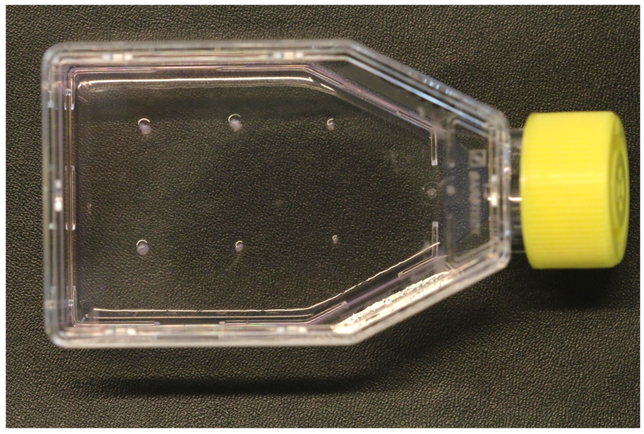
- Use a scalpel to pick up the pulp explants and transfer them to the Matrigel®-coated flask at evenly spaced points (Figure 4). If required, a sterile scalpel may be used to make small grooves in the Matrigel® layer to facilitate initial attachment, taking care not to extend the groove to the plastic base of the flask.
- 11.
- Incubate the flasks in a standard tissue culture incubator for 48 h, at which point 5 mL of cell culture medium can be added. The extended period of time following initial deposition of the explants into the flask until the first medium change is implemented in order to allow the explants to attach to the flask and to reduce the risk of dislodgement.
- 12.
- Change the medium initially after 7 days in culture. Thereafter, the medium may be changed every 3 days. Monitor the cells under phase contrast microscopy for explant outgrowth and signs of contamination or cell death. In the authors’ experience, cell outgrowth was typically observed after approximately 10 days in culture.
- 13.
- Once cellular outgrowth from the explants is established, the explants may be gently removed from the base of the flask with a sterile scalpel. Explants that do not display any signs of cell outgrowth may be discarded, while those that do display cellular outgrowth may be transferred to a sterile Matrigel®-coated T25 flask as outlined in steps 10–11. Explants can be reused in this manner until cell growth is no longer observed. In the authors’ experience, this typically occurred after the explant was reused three times.

3.4. Recovery of Cells from Matrigel® for Further Subculture
![Mps 07 00022 i001]() CRITICAL STEP Use a sterile pipette to aspirate the cell culture medium from the flask, taking care not to disturb the cell monolayer or the Matrigel® coating.
CRITICAL STEP Use a sterile pipette to aspirate the cell culture medium from the flask, taking care not to disturb the cell monolayer or the Matrigel® coating.- Add 2.5 mL pre-chilled Corning® Cell Recovery solution to the flask (See Note 5, Appendix A.
- Use a wide bore pipette tip to pipette the solution up and down and disrupt the Matrigel® matrix.
- Incubate at 4 °C for 20 min (See Note 6, Appendix A)
- Aspirate Corning® Cell Recovery solution, taking care not to aspirate Matrigel® or disturb the cell monolayer.
- Add 1 mL Trypsin-EDTA and gently rock the flask. Aspirate Trypsin-EDTA and add 1 ml fresh Trypsin-EDTA. Incubate the cells at 37 °C, 5% CO2 for 5–10 min prior to adding 1 mL of cell culture medium. A higher volume of cell culture medium may be used if preferred; however, in the authors’ experience, a 1:1 ratio of Trypsin-EDTA and cell culture medium is sufficient for recovery.
- Transfer solution to a sterile 50 mL centrifuge tube and centrifuge at 100× g for 3 min.
- Remove the supernatant and add 1 mL PBS. Centrifuge at 100× g for 3 min.
- Repeat step 8.
- Remove the supernatant and add 1 mL cell culture medium.
- Transfer solution to a sterile T25 flask (passage 1). Incubate at 37 °C, 5% CO2 overnight before topping the medium up to 5 mL (See Note 7, Appendix A).
3.5. Subculture of Confluent Cells
- Aspirate the medium and add 1 mL Trypsin-EDTA. Gently rock the flask and aspirate Trypsin-EDTA prior to adding 1 mL fresh Typsin-EDTA. Incubate the cells at 37 °C, 5% CO2 for 5–10 min prior to adding 1 mL of cell culture medium.
- Transfer the solution to a 50 mL centrifuge tube and centrifuge at 100× g for 3 min.
- Discard the supernatant and resuspend the pellet in 2 mL culture medium. Aliquot the cell suspension into a sterile T75 flask (passage 2) and incubate overnight at 37 °C, 5% CO2. Top the medium up to 10 mL after 24 h (See Notes 7 and 8, Appendix A).
4. Expected Results and Discussion
Author Contributions
Funding
Institutional Review Board Statement
Informed Consent Statement
Data Availability Statement
Acknowledgments
Conflicts of Interest
Appendix A. Notes
- A Matrigel® concentration of 3 mg/mL is recommended by the supplier; however, in the current protocol, other concentrations were tested, including 2 mg/mL, which was found to be sufficient for explant attachment and growth. It should be noted that this concentration results in a fragile gel layer, and care should be taken at all times when handling the flask so as to not disturb the Matrigel® coating.
- Third molars are recommended as they are frequently extracted due to impaction or for orthodontic reasons and are therefore more likely to be free from caries or restorations than other teeth.
- A water-cooled saw is necessary to minimise potential damage caused by high temperatures resulting from frictional heat and speed.
- The CEJ is positioned more coronally on the mesial and distal aspect of a tooth. This anatomical demarcation curves apically on the buccal and lingual aspect of a tooth in a scalloped pattern. The cut should be positioned just beneath the most apical extent of the zenith of the scalloped cemento-enamel junction in order to de-roof the pulp chamber accurately.
- There are a number of methods for depolymerisation of Matrigel®, including digestion with Corning® Cell Recovery Solution or dispase; however, Corning® Cell Recovery Solution is described here as it is recommended by the manufacturer where metabolism assays or RNA extraction are to be carried out.
- The protocol described here is intended for use with primary human dental pulp cells isolated using an explant culture method. Different cells may require different incubation times with Corning® Cell recovery solution.
- Cells are initially seeded in a small quantity of culture medium to encourage settlement and enhance attachment to the base of the flask prior to topping medium up to final volume.
- If required, DPSCs can be selected and isolated from the DPC population using FACS, as previously described [21].
References
- Gronthos, S.; Mankani, M.; Brahim, J.; Robey, P.G.; Shi, S. Postnatal Human Dental Pulp Stem Cells (DPSCs) in Vitro and in Vivo. Proc. Natl. Acad. Sci. USA 2000, 97, 13625–13630. [Google Scholar] [CrossRef]
- Goldberg, M.; Smith, A.J. Cells and Extracellular Matrices of Dentin and Pulp: A Biological Basis for Repair and Tissue Engineering. Crit. Rev. Oral Biol. Med. 2004, 15, 13–27. [Google Scholar] [CrossRef]
- Karaöz, E.; Demircan, P.C.; Saflam, Ö.; Aksoy, A.; Kaymaz, F.; Duruksu, G. Human Dental Pulp Stem Cells Demonstrate Better Neural and Epithelial Stem Cell Properties than Bone Marrow-Derived Mesenchymal Stem Cells. Histochem. Cell Biol. 2011, 136, 455–473. [Google Scholar] [CrossRef]
- Nemeth, C.L.; Janebodin, K.; Yuan, A.E.; Dennis, J.E.; Reyes, M.; Kim, D.H. Enhanced Chondrogenic Differentiation of Dental Pulp Stem Cells Using Nanopatterned PEG-GelMA-HA Hydrogels. Tissue Eng. Part A 2014, 20, 2817–2829. [Google Scholar] [CrossRef]
- Labedz-Maslowska, A.; Bryniarska, N.; Kubiak, A.; Kaczmarzyk, T.; Sekula-Stryjewska, M.; Noga, S.; Boruczkowski, D.; Madeja, Z.; Zuba-Surma, E. Multilineage Differentiation Potential of Human Dental Pulp Stem Cells-Impact of 3D and Hypoxic Environment on Osteogenesis In Vitro. Int. J. Mol. Sci. 2020, 21, 6172. [Google Scholar] [CrossRef]
- Arthur, A.; Rychkov, G.; Shi, S.; Koblar, S.A.; Gronthos, S. Adult Human Dental Pulp Stem Cells Differentiate toward Functionally Active Neurons under Appropriate Environmental Cues. Stem Cells 2008, 26, 1787–1795. [Google Scholar] [CrossRef]
- Sakai, K.; Yamamoto, A.; Matsubara, K.; Nakamura, S.; Naruse, M.; Yamagata, M.; Sakamoto, K.; Tauchi, R.; Wakao, N.; Imagama, S.; et al. Human Dental Pulp-Derived Stem Cells Promote Locomotor Recovery after Complete Transection of the Rat Spinal Cord by Multiple Neuro-Regenerative Mechanisms. J. Clin. Investig. 2012, 122, 80–90. [Google Scholar] [CrossRef]
- Martínez-Sarrà, E.; Montori, S.; Gil-Recio, C.; Núñez-Toldrà, R.; Costamagna, D.; Rotini, A.; Atari, M.; Luttun, A.; Sampaolesi, M. Human Dental Pulp Pluripotent-like Stem Cells Promote Wound Healing and Muscle Regeneration. Stem Cell Res. Ther. 2017, 8, 175. [Google Scholar] [CrossRef]
- Sloan, A.J.; Smith, A.J. Stem Cells and the Dental Pulp: Potential Roles in Dentine Regeneration and Repair. Oral Dis. 2007, 13, 151–157. [Google Scholar] [CrossRef]
- Naz, S.; Khan, F.R.; Zohra, R.R.; Lakhundi, S.S.; Khan, M.S.; Mohammed, N.; Ahmad, T. Isolation and Culture of Dental Pulp Stem Cells from Permanent and Deciduous Teeth. Pak. J. Med. Sci. 2019, 35, 997–1002. [Google Scholar] [CrossRef]
- Sunil, P.M.; Manikandan, R.; Muthumurugan; Yoithapprabhunath, T.R.; Sivakumar, M. Harvesting Dental Stem Cells—Overview. J. Pharm. Bioallied Sci. 2015, 7, S384. [Google Scholar] [CrossRef]
- Huang, H.L.; Hsing, H.W.; Lai, T.C.; Chen, Y.W.; Lee, T.R.; Chan, H.T.; Lyu, P.C.; Wu, C.L.; Lu, Y.C.; Lin, S.T.; et al. Trypsin-Induced Proteome Alteration during Cell Subculture in Mammalian Cells. J. Biomed. Sci. 2010, 17, 36. [Google Scholar] [CrossRef]
- Revel, J.P.; Hoch, P.; Ho, D. Adhesion of Culture Cells to Their Substratum. Exp. Cell Res. 1974, 84, 207–218. [Google Scholar] [CrossRef]
- Tsuji, K.; Ojima, M.; Otabe, K.; Horie, M.; Koga, H.; Sekiya, I.; Muneta, T. Effects of Different Cell-Detaching Methods on the Viability and Cell Surface Antigen Expression of Synovial Mesenchymal Stem Cells. Cell Transplant. 2017, 26, 1089–1102. [Google Scholar] [CrossRef]
- Wu, Y.; Wu, J.; Lee, D.Y.; Yee, A.; Cao, L.; Zhang, Y.; Kiani, C.; Yang, B.B. Versican Protects Cells from Oxidative Stress-Induced Apoptosis. Matrix Biol. 2005, 24, 3–13. [Google Scholar] [CrossRef]
- Huang, G.T.J.; Sonoyama, W.; Chen, J.; Park, S.H. In Vitro Characterization of Human Dental Pulp Cells: Various Isolation Methods and Culturing Environments. Cell Tissue Res. 2006, 324, 225–236. [Google Scholar] [CrossRef]
- Bakopoulou, A.; Leyhausen, G.; Volk, J.; Tsiftsoglou, A.; Garefis, P.; Koidis, P.; Geurtsen, W. Assessment of the Impact of Two Different Isolation Methods on the Osteo/Odontogenic Differentiation Potential of Human Dental Stem Cells Derived from Deciduous Teeth. Calcif. Tissue Int. 2011, 88, 130–141. [Google Scholar] [CrossRef]
- Xu, C.; Inokuma, M.S.; Denham, J.; Golds, K.; Kundu, P.; Gold, J.D.; Carpenter, M.K. Feeder-Free Growth of Undifferentiated Human Embryonic Stem Cells. Nat. Biotechnol. 2001, 19, 971–974. [Google Scholar] [CrossRef]
- Ruocco, G.; Guillermo Diaz Ochoa, J.; Tatullo, M.; Paduano, F.; Marrelli, M.; Codispoti, B.; Shelton, R.M.; Scheven, B.A.; Cooper, P.R. Dental Pulp Stem Cell Mechanoresponsiveness: Effects of Mechanical Stimuli on Dental Pulp Stem Cell Behavior. Front. Physiol. 2018, 9, 1685. [Google Scholar] [CrossRef]
- Luke, A.M.; Patnaik, R.; Kuriadom, S.; Abu-Fanas, S.; Mathew, S.; Shetty, K.P. Human Dental Pulp Stem Cells Differentiation to Neural Cells, Osteocytes and Adipocytes-An in Vitro Study. Heliyon 2020, 6, e03054. [Google Scholar] [CrossRef]
- Tirino, V.; Paino, F.; d’Aquino, R.; Desiderio, V.; de Rosa, A.; Papaccio, G. Methods for the Identification, Characterization and Banking of Human DPSCs: Current Strategies and Perspectives. Stem Cell Rev. Rep. 2011, 7, 608–615. [Google Scholar] [CrossRef] [PubMed]


Disclaimer/Publisher’s Note: The statements, opinions and data contained in all publications are solely those of the individual author(s) and contributor(s) and not of MDPI and/or the editor(s). MDPI and/or the editor(s) disclaim responsibility for any injury to people or property resulting from any ideas, methods, instructions or products referred to in the content. |
© 2024 by the authors. Licensee MDPI, Basel, Switzerland. This article is an open access article distributed under the terms and conditions of the Creative Commons Attribution (CC BY) license (https://creativecommons.org/licenses/by/4.0/).
Share and Cite
Kearney, M.; McReynolds, D.E.; Duncan, H.F. Isolation and Culture of Primary Human Dental Pulp Cells—A Description of Technical and Methodological Steps to Maximise Predictability and Yield. Methods Protoc. 2024, 7, 22. https://doi.org/10.3390/mps7020022
Kearney M, McReynolds DE, Duncan HF. Isolation and Culture of Primary Human Dental Pulp Cells—A Description of Technical and Methodological Steps to Maximise Predictability and Yield. Methods and Protocols. 2024; 7(2):22. https://doi.org/10.3390/mps7020022
Chicago/Turabian StyleKearney, Michaela, David E. McReynolds, and Henry F. Duncan. 2024. "Isolation and Culture of Primary Human Dental Pulp Cells—A Description of Technical and Methodological Steps to Maximise Predictability and Yield" Methods and Protocols 7, no. 2: 22. https://doi.org/10.3390/mps7020022