Next Issue
Volume 2, June
Previous Issue
Volume 1, December


Methods Protoc., Volume 2, Issue 1 (March 2019) – 25 articles

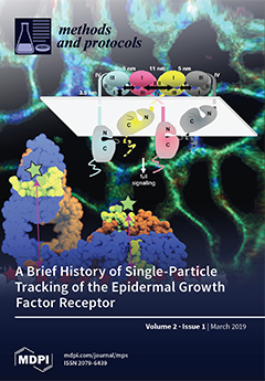
Cover Story (view full-size image): Single-particle tracking has been developed over the past 25 years to investigate molecular dynamics, structure, interactions, and function in the cellular context. We show how single-molecule and ensemble Förster resonance energy transfer (FRET) are used to build a model of epidermal growth factor receptor (EGFR) complexes in cells. EGFRs in cells are labelled with a mixture of donor (green) and acceptor (red) fluorophores. FRET then measures distances between EGFR in complexes. EGFRs are also liganded with EGF labelled with a green fluorescent dye, and the plasma membrane is labelled with a red dye. Ensemble FRET between the dyes measures EGFR-membrane separation, and hence the receptor’s conformation. These data are combined to produce a model for EGFR oligomerization in the cell. Data from Webb et al., Biophys. J. 94, 803 (2008) and Tynan et al., Mol. Cell. Biol. 31, 2241 (2011). View this paper.
  • Issues are regarded as officially published after their release is announced to the table of contents alert mailing list.
  • You may sign up for e-mail alerts to receive table of contents of newly released issues.
  • PDF is the official format for papers published in both, html and pdf forms. To view the papers in pdf format, click on the "PDF Full-text" link, and use the free Adobe Reader to open them.
Order results
Result details
Section
Select all
Export citation of selected articles as:
10 pages, 1513 KiB  
Opinion
Why RGB Imaging Should be Used to Analyze Fusarium Graminearum Growth and Estimate Deoxynivalenol Contamination
by Edgar Cambaza, Shigenobu Koseki and Shuso Kawamura
Methods Protoc. 2019, 2(1), 25; https://doi.org/10.3390/mps2010025 - 18 Mar 2019
Cited by 10 | Viewed by 3267
Abstract
Size-based fungal growth studies are limited because they do not provide information about the mold’s state of maturity, and measurements such as radius and diameter are not practical if the fungus grows irregularly. Furthermore, the current methods used to detect diseases such as [...] Read more.
Size-based fungal growth studies are limited because they do not provide information about the mold’s state of maturity, and measurements such as radius and diameter are not practical if the fungus grows irregularly. Furthermore, the current methods used to detect diseases such as Fusarium head blight (FHB) or mycotoxin contamination are labor-intensive and time consuming. FHB is frequently detected through visual examination and the results can be subjective, depending on the skills and experience of the analyzer. For toxin determination (e.g., deoxynivalenol (DON), the best methods are expensive, not practical for routine. RGB (red, green and blue) imaging analysis is a viable alternative that is inexpensive, easy to use and seemingly better if enhanced with statistical methods. This short communication explains why RGB imaging analysis should be used instead of size-based variables as a tool to measure growth of Fusarium graminearum and DON concentration. Full article
Show Figures

Figure 1

34 pages, 4464 KiB  
Review
A User’s Guide to Cell-Free Protein Synthesis
by Nicole E. Gregorio, Max Z. Levine and Javin P. Oza
Methods Protoc. 2019, 2(1), 24; https://doi.org/10.3390/mps2010024 - 12 Mar 2019
Cited by 138 | Viewed by 27902
Abstract
Cell-free protein synthesis (CFPS) is a platform technology that provides new opportunities for protein expression, metabolic engineering, therapeutic development, education, and more. The advantages of CFPS over in vivo protein expression include its open system, the elimination of reliance on living cells, and [...] Read more.
Cell-free protein synthesis (CFPS) is a platform technology that provides new opportunities for protein expression, metabolic engineering, therapeutic development, education, and more. The advantages of CFPS over in vivo protein expression include its open system, the elimination of reliance on living cells, and the ability to focus all system energy on production of the protein of interest. Over the last 60 years, the CFPS platform has grown and diversified greatly, and it continues to evolve today. Both new applications and new types of extracts based on a variety of organisms are current areas of development. However, new users interested in CFPS may find it challenging to implement a cell-free platform in their laboratory due to the technical and functional considerations involved in choosing and executing a platform that best suits their needs. Here we hope to reduce this barrier to implementing CFPS by clarifying the similarities and differences amongst cell-free platforms, highlighting the various applications that have been accomplished in each of them, and detailing the main methodological and instrumental requirement for their preparation. Additionally, this review will help to contextualize the landscape of work that has been done using CFPS and showcase the diversity of applications that it enables. Full article
(This article belongs to the Special Issue Cell-Free Synthetic Biology)
Show Figures

Graphical abstract

11 pages, 734 KiB  
Protocol
An Evidence-Based Objective Study Protocol for Evaluating Cardiovascular and Cerebrovascular Indices Following Concussion: The Neary Protocol
by J. Patrick Neary, Jyotpal Singh, Scott A. Bishop, Ryan T. Dech, Matthew J. A. Butz and Trevor K. Len
Methods Protoc. 2019, 2(1), 23; https://doi.org/10.3390/mps2010023 - 06 Mar 2019
Cited by 16 | Viewed by 5206
Abstract
Introduction: The prevalence and incidence of sport-related concussion have continued to increase over the past decade, and researchers from various backgrounds strive for evidenced-based clinical assessment and management. When diagnosing and managing a concussion, a battery of tests from several domains (e.g., symptom [...] Read more.
Introduction: The prevalence and incidence of sport-related concussion have continued to increase over the past decade, and researchers from various backgrounds strive for evidenced-based clinical assessment and management. When diagnosing and managing a concussion, a battery of tests from several domains (e.g., symptom reporting, neurocognitive, physiology) must be used. In this study, we propose and develop an objective, evidence-based protocol to assess the pathophysiology of the brain by using non-invasive methods. Methods: Contact sport athletes (n = 300) will be assessed at the beginning of the season in a healthy state to establish baseline values, and then prospectively followed if a mild traumatic brain injury (mTBI) occurs on approximately days 1–2, 3–5, 7–10, 21, 30, and subsequently thereafter, depending on the severity of injury. The protocol includes spontaneous measurements at rest, during head postural change, controlled breathing maneuvers for cerebrovascular reactivity, a neurovascular coupling stimuli, and a baroreflex/autoregulation maneuver. Physiological data collection will include cerebral blood flow velocity, cerebral oxygenation, respiratory gases for end-tidal oxygen and carbon dioxide, finger photoplethysmography for blood pressure, seismocardiography for cardiac mechanics, and electrocardiography. Conclusion, Limitations, and Ethics: The protocol will provide an objective, physiological evidence-based approach in an attempt to better diagnose concussion to aid in return-to-play or -learn. Ethics approval has been granted by the University Research Ethics Board. Full article
Show Figures

Figure 1

13 pages, 1674 KiB  
Protocol
Reverse Genetic Systems for Pseudomonas aeruginosa Leviphages
by Jae-Yeol Lee, Se-Jeong Ahn, Chanseop Park, Hee-Won Bae, Eun Sook Kim and You-Hee Cho
Methods Protoc. 2019, 2(1), 22; https://doi.org/10.3390/mps2010022 - 05 Mar 2019
Cited by 4 | Viewed by 3445
Abstract
Reverse genetic systems for RNA viruses are the platforms to introduce mutations into the RNA genomes and thus have helped understand their life cycle and harness them for human purposes to develop vaccines and delivery systems. These systems are based on the complementary [...] Read more.
Reverse genetic systems for RNA viruses are the platforms to introduce mutations into the RNA genomes and thus have helped understand their life cycle and harness them for human purposes to develop vaccines and delivery systems. These systems are based on the complementary DNA (cDNA) of the RNA viruses, whose transcripts derived from bacterial RNA polymerases act not only as the primary mRNA for phage protein synthesis, but also as the template for phage RNA replicases (aka. RNA-dependent RNA polymerases). Here, we present a protocol optimized for the small RNA phages of Leviviridae (i.e., leviphages) infecting Pseudomonas aeruginosa. This protocol includes three fundamental steps: (i) Creation of a promoter-fused cDNA, (ii) generation of a clone into mini-Tn7-based vector, and (iii) introduction of the clone into non-susceptible hosts. As the representative example, we describe the reverse genetic system for PP7, which infects a set of P. aeruginosa strains such as PAO1. The cDNA was fused to the T7 promoter, which was cloned in mini-Tn7-Gm. This construct was introduced into P. aeruginosa PAK and E. coli HB101. Functional assembly of PP7 phages from the culture supernatants were assessed by plaque formation on PAO1 and the phage particles were observed under transmission microscope. We found that the host cells should be cultured at 30 °C for the maximal phage production (~1012 pfu/mL). The reverse genetic systems will provide a new insight into the life cycle of the RNA phages and help develop engineered variants with new traits for phage applications regarding selective diagnosis and efficient therapy. Full article
Show Figures

Figure 1

15 pages, 374 KiB  
Protocol
School-Based Sexual and Reproductive Health Services for Prevention of Adolescent Pregnancy in the Hoima District, Uganda: Cluster Randomized Controlled Trial
by Gloria Kirungi Kasozi, Julius Kasozi, Frank Pio Kiyingi and Miph Musoke
Methods Protoc. 2019, 2(1), 21; https://doi.org/10.3390/mps2010021 - 04 Mar 2019
Cited by 3 | Viewed by 5659
Abstract
Uganda has persistently had high adolescent pregnancy prevalence; 25% for the last 10 years. This protocol presents the design of a Cluster Randomized Controlled Trial (CRCT) to investigate the effectiveness of School-Based Sexual and Reproductive Health (SBSRH) interventions on prevention of pregnancy among [...] Read more.
Uganda has persistently had high adolescent pregnancy prevalence; 25% for the last 10 years. This protocol presents the design of a Cluster Randomized Controlled Trial (CRCT) to investigate the effectiveness of School-Based Sexual and Reproductive Health (SBSRH) interventions on prevention of pregnancy among school girls aged 15–19 years in the Hoima District, Uganda. 18 secondary schools (clusters) will be selected using cluster sampling and allocated 1:1 into control or intervention group stratified by geographical location. 1080 (60 each cluster) participants/girls aged 15–19 years will be selected using simple random sampling. The intervention group will receive tailored SRH information, in-school medical care and referral over 12 months. The control group will receive no intervention from the research team; however, they can access alternative services elsewhere if they wish. Data will be obtained at baseline, 6 months and 12 months. The outcomes are reduction in occurrence of pregnancy, utilization of SRH services and sexual behavioral change. To our knowledge, this is the first CRCT providing combined SRH interventions for prevention of adolescent pregnancy in Uganda. If effective, it could have great potential in preventing adolescent pregnancy. Trial Registration: Pan African Clinical Trial Registry (PACTR201810882140200) Registered on 16 October 2018. Full article
Show Figures

Figure 1

8 pages, 2305 KiB  
Article
The Inhibition of Radial and Axial Micromovement of Bone Scaffold with Gelfoam® and Titanium Mesh Fixation and Its Effects on Osteointegration
by Jane Kwon, Dong Joon Lee, Mallory Kocher, Yong-Il Kim, Te-Ju Wu, John Whitley and Ching-Chang Ko
Methods Protoc. 2019, 2(1), 20; https://doi.org/10.3390/mps2010020 - 26 Feb 2019
Cited by 3 | Viewed by 2967
Abstract
A major drawback of nanocomposite scaffolds in bone tissue engineering is dimensional shrinkage after the fabrication process. Shrinkage yields gaps between the scaffold and host bone in the defect site and eventually causes failure in osteointegration by micromovement. The present study was conducted [...] Read more.
A major drawback of nanocomposite scaffolds in bone tissue engineering is dimensional shrinkage after the fabrication process. Shrinkage yields gaps between the scaffold and host bone in the defect site and eventually causes failure in osteointegration by micromovement. The present study was conducted using titanium (Ti) mesh and Gelfoam® to prevent radial and axial micromovement, respectively. A critical-sized defect (CSD) was created in the center of the calvarium of Sprague Dawley rats to implant porous polydopamine-laced hydroxyapatite collagen calcium silicate (HCCS-PDA), a novel nanocomposite scaffold. Gelfoam® was applied around the edge of the defect, and then the HCCS-PDA scaffold was inserted in the defect area. Ti mesh was placed between the periosteum and skin right, above the inserted scaffold site. There were two test groups, with a fixture (Gelfoam® and Ti mesh) and without a fixture, each group contained five animals. The rats were sacrificed after three months post-operation. The explanted calvaria underwent micro-CT scanning and a push-out test to quantify osteointegration and mechanical strength between the scaffold and host bone. Histological analysis of undecalcified bone was performed by grinding resin infiltrated calvaria blocks to prepare 10 μm slices. Osteointegration was higher in the group with fixation than without fixation. Movement of the HCCS-PDA scaffold in the gap resulted in diminished osteointegration. With fixation, the movement was inhibited and osteointegration became prominent. Here we present a successful method of preventing axial and radial movement of scaffolds using Gelfoam® and Ti mesh. Applying this fixture, we expect that an HCCS-PDA scaffold can repair CSD more effectively. Full article
Show Figures

Graphical abstract

30 pages, 7215 KiB  
Protocol
Protocol for Construction of Rat Nerve Stimulation Cuff Electrodes
by Manolo U. Rios, Jesse E. Bucksot, Kimiya C. Rahebi, Crystal T. Engineer, Michael P. Kilgard and Seth A. Hays
Methods Protoc. 2019, 2(1), 19; https://doi.org/10.3390/mps2010019 - 15 Feb 2019
Cited by 31 | Viewed by 6504
Abstract
Peripheral nerve stimulation has emerged as a platform therapy to treat a wide range of disorders. Continued development and translation of these strategies requires that researchers have access to reliable, customizable electrodes for nerve stimulation. Here, we detail procedures to build three different [...] Read more.
Peripheral nerve stimulation has emerged as a platform therapy to treat a wide range of disorders. Continued development and translation of these strategies requires that researchers have access to reliable, customizable electrodes for nerve stimulation. Here, we detail procedures to build three different configurations of cuff electrodes with varying numbers and orientations of contacts for nerve stimulation in rats. These designs are built with simple, widely available materials, using platinum–iridium electrodes assembled into polyurethane tubing. Moreover, the designs can easily be customized to increase versatility and individualize for specific stimulation applications. This protocol provides a resource to facilitate the construction and customization of stimulation cuffs to support preclinical nerve stimulation research. Full article
Show Figures

Figure 1

11 pages, 541 KiB  
Review
Campylobacter Phage Isolation and Characterization: What We Have Learned So Far
by Claudia Jäckel, Jens Andre Hammerl and Stefan Hertwig
Methods Protoc. 2019, 2(1), 18; https://doi.org/10.3390/mps2010018 - 15 Feb 2019
Cited by 17 | Viewed by 5351
Abstract
Lytic Campylobacter phages, which can be used to combat this pathogen in animals and on food products, have been studied for more than 30 years. Though, due to some peculiarities of the phages, which hampered their isolation and particularly their molecular analysis for [...] Read more.
Lytic Campylobacter phages, which can be used to combat this pathogen in animals and on food products, have been studied for more than 30 years. Though, due to some peculiarities of the phages, which hampered their isolation and particularly their molecular analysis for a long time, progress in this research field was rather slow. Meanwhile, the situation has changed and much more is known about the biology and genetics of those phages. In this article, we address specific issues that should be considered when Campylobacter phages are studied, starting with the isolation and propagation of the phages and ending with a thorough characterization including whole-genome sequencing. The basis for advice and recommendations given here is a careful review of the scientific literature and experiences that we have had ourselves with Campylobacter phages. Full article
Show Figures

Figure 1

19 pages, 2904 KiB  
Protocol
Efficient Construction and Effective Screening of Synthetic Domain Antibody Libraries
by Arghavan Solemani Zadeh, Alissa Grässer, Heiko Dinter, Maximilian Hermes and Katharina Schindowski
Methods Protoc. 2019, 2(1), 17; https://doi.org/10.3390/mps2010017 - 14 Feb 2019
Cited by 13 | Viewed by 6546
Abstract
Phage display is a powerful technique for drug discovery in biomedical research in particular for antibody libraries. But, several technical challenges are associated with the selection process. For instance, during the panning step, the successful elution of the phages bound to the antigen [...] Read more.
Phage display is a powerful technique for drug discovery in biomedical research in particular for antibody libraries. But, several technical challenges are associated with the selection process. For instance, during the panning step, the successful elution of the phages bound to the antigen is critical in order to avoid losing the most promising binders. Here, we present an efficient protocol to establish, screen and select synthetic libraries of domain antibodies using phage display. We do not only present suitable solutions to the above-mentioned challenges to improve elution by 50-fold, but we also present a step by step in-depth protocol with miniaturized volumes and optimized procedures to save material, costs and time for a successful phage display with domain antibodies. Hence, this protocol improves the selection process for an efficient handling process. The here presented library is based on the variable domain (vNAR) of the naturally occurring novel antibody receptor (IgNAR) from cartilage fishes. Diversity was introduced in the Complementarity-Determining Region 3 (CDR3) of the antigen-binding site with different composition and length. Full article
Show Figures

Graphical abstract

12 pages, 2425 KiB  
Protocol
Efficient Incorporation of Unnatural Amino Acids into Proteins with a Robust Cell-Free System
by Wei Gao, Ning Bu and Yuan Lu
Methods Protoc. 2019, 2(1), 16; https://doi.org/10.3390/mps2010016 - 12 Feb 2019
Cited by 18 | Viewed by 5954
Abstract
Unnatural proteins are crucial biomacromolecules and have been widely applied in fundamental science, novel biopolymer materials, enzymes, and therapeutics. Cell-free protein synthesis (CFPS) system can serve as a robust platform to synthesize unnatural proteins by highly effective site-specific incorporation of unnatural amino acids [...] Read more.
Unnatural proteins are crucial biomacromolecules and have been widely applied in fundamental science, novel biopolymer materials, enzymes, and therapeutics. Cell-free protein synthesis (CFPS) system can serve as a robust platform to synthesize unnatural proteins by highly effective site-specific incorporation of unnatural amino acids (UNAAs), without the limitations of cell membrane permeability and the toxicity of unnatural components. Here, we describe a quick and simple method to synthesize unnatural proteins in CFPS system based on Escherichia coli crude extract, with unnatural orthogonal aminoacyl-tRNA synthetase and suppressor tRNA evolved from Methanocaldococcus jannaschii. The superfolder green fluorescent protein (sfGFP) and p-propargyloxyphenylalanine (pPaF) were used as the model protein and UNAA. The synthesis of unnatural sfGFPs was characterized by microplate spectrophotometer, affinity chromatography, and liquid chromatography-mass spectrometry/mass spectrometry (LC-MS/MS). This protocol provides a detailed procedure guiding how to use the powerful CFPS system to synthesize unnatural proteins on demand. Full article
(This article belongs to the Special Issue Cell-Free Synthetic Biology)
Show Figures

Figure 1

7 pages, 1032 KiB  
Protocol
Extraction of Microbial and Host DNA, RNA, and Proteins from Oak Bark Tissue
by Martin Broberg and James E. McDonald
Methods Protoc. 2019, 2(1), 15; https://doi.org/10.3390/mps2010015 - 05 Feb 2019
Cited by 4 | Viewed by 4476
Abstract
The application of high-throughput nucleic acid and protein sequencing technologies is transforming our understanding of plant microbiomes and their interactions with their hosts in health and disease. However, progress in studying host-microbiome interactions in above-ground compartments of the tree (the phyllosphere) has been [...] Read more.
The application of high-throughput nucleic acid and protein sequencing technologies is transforming our understanding of plant microbiomes and their interactions with their hosts in health and disease. However, progress in studying host-microbiome interactions in above-ground compartments of the tree (the phyllosphere) has been hampered due to high concentrations of phenolic compounds, lignin, and other compounds in tree bark that severely limit the success of DNA, RNA, and protein extraction. Here we present modified sample-preparation and kit-based protocols for the extraction of host and microbiome DNA and RNA from oak (Quercus robus and Quercus petraea) bark tissue for subsequent high-throughput sequencing. In addition, reducing the quantity of bark tissue used for an established protein extraction protocol yielded high quality protein for parallel analysis of the oak-microbiota metaproteome. These procedures demonstrate the successful extraction of nucleic acids and proteins from oak tissue using as little as 50 mg of sample input, producing sufficient quantities for nucleic acid sequencing and protein mass spectrometry of tree stem tissues and their associated microbiota. Full article
Show Figures

Figure 1

13 pages, 2321 KiB  
Technical Note
Air Puff System Fundamentals for Reproducible Eyeblink Conditioning Research
by Frederick Reitz
Methods Protoc. 2019, 2(1), 14; https://doi.org/10.3390/mps2010014 - 02 Feb 2019
Cited by 3 | Viewed by 3640
Abstract
Air puff systems are at once trivially straightforward and dauntingly complex. On the one hand, they are little but a pressure source, valve, and tube connected together. On the other, the air passing through them is a compressible medium, expanding approximately adiabatically while [...] Read more.
Air puff systems are at once trivially straightforward and dauntingly complex. On the one hand, they are little but a pressure source, valve, and tube connected together. On the other, the air passing through them is a compressible medium, expanding approximately adiabatically while travelling at high velocity through a compliant tube, and exiting as a turbulent jet with velocity peak and profile varying non-linearly in its near-field. This complexity puts precise mathematical prediction of puff properties out of reach of most labs. There are, however, a number of phenomena fundamental to air puff system design that are worth understanding to a first order of approximation, or at least qualitatively. Using a simplified, “electronic–hydraulic analogy” model, this paper discusses these phenomena in just enough depth for the reader to confidently specify parts for an air puff delivery system, to measure its key parameters, and/or to describe a given system unambiguously in publications, thus maximizing reproducibility. Full article
Show Figures

Figure 1

14 pages, 5103 KiB  
Protocol
Novel Applications of Lead Acetate and Flow Cytometry Methods for Detection of Sulfur-Containing Molecules
by Evgeniya Anishchenko, Carmela Vigorito, Luigi Mele, Patrizia Lombari, Alessandra F. Perna and Diego Ingrosso
Methods Protoc. 2019, 2(1), 13; https://doi.org/10.3390/mps2010013 - 01 Feb 2019
Cited by 3 | Viewed by 4053
Abstract
Hydrogen sulfide (H2S) is the most recently established gaseous vasodilator, enzymatically produced from cysteine metabolism, involved in a number of pathophysiological processes. However, its accurate detection in vivo is critical due to its volatility and tendency to form sulfane sulfur derivatives, [...] Read more.
Hydrogen sulfide (H2S) is the most recently established gaseous vasodilator, enzymatically produced from cysteine metabolism, involved in a number of pathophysiological processes. However, its accurate detection in vivo is critical due to its volatility and tendency to form sulfane sulfur derivatives, thus limiting the data interpretation of its biological roles. We developed new applications of the simple and rapid method to measure H2S release in cell culture systems, based on the lead acetate strip test. This test, previously prevalently used in microbiology, was compared with the agar trap method, applied, in parallel, on both cell cultures and cell-free samples. Sulfane sulfur represents the major species derived from intracellular H2S. Various fluorescent probes are available for quantitation of H2S derivatives intracellularly. We present here an alternative to the classic imaging method for sulfane sulfur evaluation, running on a flow cytometer, based on SSP4 probe labeling. Flow cytometry turned out to be more direct, fully quantitative and less time-consuming compared to microscopy and more precise with respect to the fluorescence multi-plate reader assay. The new application methods for H2S determination appear to be fully suitable for the analysis of H2S release and sulfane sulfur content in biological samples. Full article
Show Figures

Figure 1

30 pages, 4068 KiB  
Review
A Brief History of Single-Particle Tracking of the Epidermal Growth Factor Receptor
by David T. Clarke and Marisa L. Martin-Fernandez
Methods Protoc. 2019, 2(1), 12; https://doi.org/10.3390/mps2010012 - 30 Jan 2019
Cited by 17 | Viewed by 4285
Abstract
Single-particle tracking (SPT) has been used and developed over the last 25 years as a method to investigate molecular dynamics, structure, interactions, and function in the cellular context. SPT is able to show how fast and how far individual molecules move, identify different [...] Read more.
Single-particle tracking (SPT) has been used and developed over the last 25 years as a method to investigate molecular dynamics, structure, interactions, and function in the cellular context. SPT is able to show how fast and how far individual molecules move, identify different dynamic populations, measure the duration and strength of intermolecular interactions, and map out structures on the nanoscale in cells. In combination with other techniques such as macromolecular crystallography and molecular dynamics simulation, it allows us to build models of complex structures, and develop and test hypotheses of how these complexes perform their biological roles in health as well as in disease states. Here, we use the example of the epidermal growth factor receptor (EGFR), which has been studied extensively by SPT, demonstrating how the method has been used to increase our understanding of the receptor’s organization and function, including its interaction with the plasma membrane, its activation, clustering, and oligomerization, and the role of other receptors and endocytosis. The examples shown demonstrate how SPT might be employed in the investigation of other biomolecules and systems. Full article
(This article belongs to the Special Issue Single-Molecule Techniques)
Show Figures

Figure 1

11 pages, 2236 KiB  
Letter
Large Scale Double-Path Illumination System with Split Field of View for the All-Optical Study of Inter-and Intra-Hemispheric Functional Connectivity on Mice
by Emilia Conti, Anna Letizia Allegra Mascaro and Francesco Saverio Pavone
Methods Protoc. 2019, 2(1), 11; https://doi.org/10.3390/mps2010011 - 29 Jan 2019
Cited by 11 | Viewed by 3889
Abstract
Recent improvements in optical tools that can perturb brain activity and simultaneously reveal the elicited alterations in the associated regions offer an exceptional means to understand and map the connectivity of the brain. In this work, we exploit a combination of recently developed [...] Read more.
Recent improvements in optical tools that can perturb brain activity and simultaneously reveal the elicited alterations in the associated regions offer an exceptional means to understand and map the connectivity of the brain. In this work, we exploit a combination of recently developed optical tools to monitor neural population at the meso-scale level and to mould the cortical patterns of targeted neuronal population. Our goal was to investigate the propagation of neuronal activity over the mouse cortex that is triggered by optogenetic stimulation in the contralateral hemisphere. Towards this aim, we developed a wide-field fluorescence microscope that is characterized by a double illumination path allowing for the optogenetic stimulation of the transfected area in the left hemisphere and the simultaneous recording of cortical activity in the right hemisphere. The microscope was further implemented with a custom shutter in order to split the LED illumination path, resulting in a half-obscured field of view. By avoiding the spectral crosstalk between GCaMP6f and channelrhodopsin 2 (ChR2), this system offered the possibility of simultaneous “pumping and probing” of inter-hemispheric functional connectivity on Thy1-GCaMP6f mice. Full article
Show Figures

Figure 1

3 pages, 139 KiB  
Editorial
Acknowledgement to Reviewers of Methods and Protocols in 2018
by MPs Editorial Office
Methods Protoc. 2019, 2(1), 10; https://doi.org/10.3390/mps2010010 - 18 Jan 2019
Viewed by 1600
Abstract
Rigorous peer-review is the corner-stone of high-quality academic publishing [...] Full article
11 pages, 3362 KiB  
Benchmark
A Computer-Vision-Guided Robot Arm for Automatically Placing Grids in Pioloform Film Preparation
by Markus Peurla, Pekka E. Hänninen and Eeva-Liisa Eskelinen
Methods Protoc. 2019, 2(1), 9; https://doi.org/10.3390/mps2010009 - 17 Jan 2019
Cited by 4 | Viewed by 3515
Abstract
Preparing pioloform/formvar support films on transmission electron microscopy (TEM) grids is a routine laboratory procedure in practically all electron microscopy units. In current practice, these grids are manually placed on the support film one by one using special tweezers, a process requiring a [...] Read more.
Preparing pioloform/formvar support films on transmission electron microscopy (TEM) grids is a routine laboratory procedure in practically all electron microscopy units. In current practice, these grids are manually placed on the support film one by one using special tweezers, a process requiring a steady hand. The work is often ergonomically awkward to continue for a longer period of time. In this article, we describe a low-cost, computer vision-guided robot arm that automatically places the grids on the film. The success rate of the prototype robot is 90%, which is comparable to an experienced laboratory technician. Full article
Show Figures

Graphical abstract

17 pages, 3622 KiB  
Protocol
Fast Proteome Identification and Quantification from Data-Dependent Acquisition–Tandem Mass Spectrometry (DDA MS/MS) Using Free Software Tools
by Jesse G. Meyer
Methods Protoc. 2019, 2(1), 8; https://doi.org/10.3390/mps2010008 - 17 Jan 2019
Cited by 13 | Viewed by 8434
Abstract
The identification of nearly all proteins in a biological system using data-dependent acquisition (DDA) tandem mass spectrometry has become routine for organisms with relatively small genomes such as bacteria and yeast. Still, the quantification of the identified proteins may be a complex process [...] Read more.
The identification of nearly all proteins in a biological system using data-dependent acquisition (DDA) tandem mass spectrometry has become routine for organisms with relatively small genomes such as bacteria and yeast. Still, the quantification of the identified proteins may be a complex process and often requires multiple different software packages. In this protocol, I describe a flexible strategy for the identification and label-free quantification of proteins from bottom-up proteomics experiments. This method can be used to quantify all the detectable proteins in any DDA dataset collected with high-resolution precursor scans and may be used to quantify proteome remodeling in response to drug treatment or a gene knockout. Notably, the method is statistically rigorous, uses the latest and fastest freely-available software, and the entire protocol can be completed in a few hours with a small number of data files from the analysis of yeast. Full article
Show Figures

Graphical abstract

9 pages, 1625 KiB  
Technical Note
A Software Architecture to Mimic a Ventricular Tachycardia in Intact Murine Hearts by Means of an All-Optical Platform
by Francesco Giardini, Valentina Biasci, Marina Scardigli, Francesco S. Pavone, Gil Bub and Leonardo Sacconi
Methods Protoc. 2019, 2(1), 7; https://doi.org/10.3390/mps2010007 - 08 Jan 2019
Cited by 6 | Viewed by 3048
Abstract
Optogenetics is an emerging method that uses light to manipulate electrical activity in excitable cells exploiting the interaction between light and light-sensitive depolarizing ion channels, such as channelrhodopsin-2 (ChR2). Initially used in the neuroscience, it has been adopted in cardiac research where the [...] Read more.
Optogenetics is an emerging method that uses light to manipulate electrical activity in excitable cells exploiting the interaction between light and light-sensitive depolarizing ion channels, such as channelrhodopsin-2 (ChR2). Initially used in the neuroscience, it has been adopted in cardiac research where the expression of ChR2 in cardiac preparations allows optical pacing, resynchronization and defibrillation. Recently, optogenetics has been leveraged to manipulate cardiac electrical activity in the intact heart in real-time. This new approach was applied to simulate a re-entrant circuit across the ventricle. In this technical note, we describe the development and the implementation of a new software package for real-time optogenetic intervention. The package consists of a single LabVIEW program that simultaneously captures images at very high frame rates and delivers precisely timed optogenetic stimuli based on the content of the images. The software implementation guarantees closed-loop optical manipulation at high temporal resolution by processing the raw data in workstation memory. We demonstrate that this strategy allows the simulation of a ventricular tachycardia with high stability and with a negligible loss of data with a temporal resolution of up to 1 ms. Full article
Show Figures

Figure 1

18 pages, 3490 KiB  
Protocol
AFM-Based Force Spectroscopy Guided by Recognition Imaging: A New Mode for Mapping and Studying Interaction Sites at Low Lateral Density
by Melanie Koehler, Anny Fis, Hermann J. Gruber and Peter Hinterdorfer
Methods Protoc. 2019, 2(1), 6; https://doi.org/10.3390/mps2010006 - 08 Jan 2019
Cited by 11 | Viewed by 4015
Abstract
Ligand binding to receptors is one of the most important regulatory elements in biology as it is the initiating step in signaling pathways and cascades. Thus, precisely localizing binding sites and measuring interaction forces between cognate receptor–ligand pairs leads to new insights into [...] Read more.
Ligand binding to receptors is one of the most important regulatory elements in biology as it is the initiating step in signaling pathways and cascades. Thus, precisely localizing binding sites and measuring interaction forces between cognate receptor–ligand pairs leads to new insights into the molecular recognition involved in these processes. Here we present a detailed protocol about applying a technique, which combines atomic force microscopy (AFM)-based recognition imaging and force spectroscopy for studying the interaction between (membrane) receptors and ligands on the single molecule level. This method allows for the selection of a single receptor molecule reconstituted into a supported lipid membrane at low density, with the subsequent quantification of the receptor–ligand unbinding force. Based on AFM tapping mode, a cantilever tip carrying a ligand molecule is oscillated across a membrane. Topography and recognition images of reconstituted receptors are recorded simultaneously by analyzing the downward and upward parts of the oscillation, respectively. Functional receptor molecules are selected from the recognition image with nanometer resolution before the AFM is switched to the force spectroscopy mode, using positional feedback control. The combined mode allows for dynamic force probing on different pre-selected molecules. This strategy results in higher throughput when compared with force mapping. Applied to two different receptor–ligand pairs, we validated the presented new mode. Full article
(This article belongs to the Special Issue Single-Molecule Techniques)
Show Figures

Graphical abstract

4 pages, 498 KiB  
Patent Summary
A Novelty System for Biotization of Plant Microshoots and Collection of Natural Compounds
by Mário Rui da Costa Basílio e Castro, Carla Ragonezi, Paulo Guilherme Leandro de Oliveira and Maria Amely Zavattieri
Methods Protoc. 2019, 2(1), 5; https://doi.org/10.3390/mps2010005 - 07 Jan 2019
Viewed by 2394
Abstract
An in vitro plant microshoot culture system composed of two phases; a liquid phase overlaid by a floating solid phase, which is described in detail herein. This system is designed to enable the extraction of natural compounds released/disseminated into the liquid phase during [...] Read more.
An in vitro plant microshoot culture system composed of two phases; a liquid phase overlaid by a floating solid phase, which is described in detail herein. This system is designed to enable the extraction of natural compounds released/disseminated into the liquid phase during root growth, thus facilitating their processing and biochemical characterization. The solid phase holds the plant afloat and enables the simultaneous culture of a microorganism, yet avoiding its penetration into the liquid phase, where the roots are submerged. Both phases can be independently formulated as required for growth optimization of both organisms. Considering the closed system and known variables described in this patent, applications of the described method include testing with pesticides, herbicides, and other similar products. Full article
Show Figures

Graphical abstract

12 pages, 2956 KiB  
Article
Novel ‘Bacteriospray’ Method Facilitates the Functional Screening of Metagenomic Libraries for Antimicrobial Activity
by Anissa Brahami, Annie Castonguay and Éric Déziel
Methods Protoc. 2019, 2(1), 4; https://doi.org/10.3390/mps2010004 - 07 Jan 2019
Cited by 3 | Viewed by 4267
Abstract
Metagenomic techniques, notably the cloning of environmental DNA (eDNA) into surrogate hosts, have given access to the genome of uncultured bacteria. However, the determination of gene functions based on DNA sequences alone remains a significant challenge. The functional screening of metagenomic libraries represents [...] Read more.
Metagenomic techniques, notably the cloning of environmental DNA (eDNA) into surrogate hosts, have given access to the genome of uncultured bacteria. However, the determination of gene functions based on DNA sequences alone remains a significant challenge. The functional screening of metagenomic libraries represents an interesting approach in the discovery of microbial metabolites. We describe here an optimized screening approach that facilitates the identification of new antimicrobials among large metagenomic libraries. Notably, we report a detailed genomic library construction protocol using Escherichia coli DH10B as a surrogate host, and demonstrate how vector/genomic DNA dephosphorylation, ligase inactivation, dialysis of the ligation product and vector/genomic DNA ratio greatly influence clone recovery. Furthermore, we describe the use of an airbrush device to screen E. coli metagenomic libraries for their antibacterial activity against Staphylococcus aureus, a method we called bacteriospray. This bacterial spraying tool greatly facilitates and improves the functional screening of large genomic libraries, as it conveniently allows the production of a thinner and more uniform layer of target bacteria compared to the commonly used overlay method, resulting in the screening of 5–10 times more clones per agar plate. Using the Burkholderia thailandensis E264 genomic DNA as a proof of concept, four clones out of 70,000 inhibited the growth of S. aureus and were found to each contain a DNA insert. Analysis of these chromosomic fragments revealed genomic regions never previously reported to be responsible for the production of antimicrobials, nor predicted by bioinformatics tools. Full article
Show Figures

Figure 1

5 pages, 2000 KiB  
Protocol
Synthesis of Highly Concentrated Suspensions of Silver Nanoparticles by Two Versions of the Chemical Reduction Method
by Miguel Gakiya-Teruya, Luis Palomino-Marcelo and Juan Carlos F. Rodriguez-Reyes
Methods Protoc. 2019, 2(1), 3; https://doi.org/10.3390/mps2010003 - 24 Dec 2018
Cited by 33 | Viewed by 8651
Abstract
In spite of the widespread use of the chemical reduction method to obtain silver nanoparticles, the nanoparticle yield is often low due to a required addition of small volumes of diluted metal ions to a solution containing a reducer. Higher yields can be [...] Read more.
In spite of the widespread use of the chemical reduction method to obtain silver nanoparticles, the nanoparticle yield is often low due to a required addition of small volumes of diluted metal ions to a solution containing a reducer. Higher yields can be obtained following an alternative method, in which the reducer is added to a greater volume of silver ions in the solution. In this study, protocols for both methods are detailed and compared, using characterization tools such as UV-vis spectrometry, dynamic light scattering (DLS), and zeta potential measurements. By using this alternative method, the amount of silver in the solution is three times greater, and nanoparticles with a narrower size distribution are formed (between 6 and 70 nm in size). In contrast, the regular method produces particles of 3 and 100 nm. Zeta potential measurements indicate that the nanoparticles synthesized with the alternative method will be more stable than those from the regular method. Full article
Show Figures

Graphical abstract

11 pages, 1087 KiB  
Article
Fast Calculation of Computer Generated Holograms for 3D Photostimulation through Compressive-Sensing Gerchberg–Saxton Algorithm
by Paolo Pozzi, Laura Maddalena, Nicolò Ceffa, Oleg Soloviev, Gleb Vdovin, Elizabeth Carroll and Michel Verhaegen
Methods Protoc. 2019, 2(1), 2; https://doi.org/10.3390/mps2010002 - 20 Dec 2018
Cited by 23 | Viewed by 6516
Abstract
The use of spatial light modulators to project computer generated holograms is a common strategy for optogenetic stimulation of multiple structures of interest within a three-dimensional volume. A common requirement when addressing multiple targets sparsely distributed in three dimensions is the generation of [...] Read more.
The use of spatial light modulators to project computer generated holograms is a common strategy for optogenetic stimulation of multiple structures of interest within a three-dimensional volume. A common requirement when addressing multiple targets sparsely distributed in three dimensions is the generation of a points cloud, focusing excitation light in multiple diffraction-limited locations throughout the sample. Calculation of this type of holograms is most commonly performed with either the high-speed, low-performance random superposition algorithm, or the low-speed, high performance Gerchberg–Saxton algorithm. This paper presents a variation of the Gerchberg–Saxton algorithm that, by only performing iterations on a subset of the data, according to compressive sensing principles, is rendered significantly faster while maintaining high quality outputs. The algorithm is presented in high-efficiency and high-uniformity variants. All source code for the method implementation is available as Supplementary Materials and as open-source software. The method was tested computationally against existing algorithms, and the results were confirmed experimentally on a custom setup for in-vivo multiphoton optogenetics. The results clearly show that the proposed method can achieve computational speed performances close to the random superposition algorithm, while retaining the high performance of the Gerchberg–Saxton algorithm, with a minimal hologram quality loss. Full article
Show Figures

Figure 1

10 pages, 372 KiB  
Technical Note
Quantification of Ethanedinitrile in Air Using a New and Accurate Gas Chromatography Method
by Sam E. Brierley, Anthony R. Adlam and Matthew K. D. Hall
Methods Protoc. 2019, 2(1), 1; https://doi.org/10.3390/mps2010001 - 20 Dec 2018
Cited by 3 | Viewed by 2792
Abstract
Compared to previously tested fumigants such as methyl bromide, sulfuryl fluoride and phosphine; ethanedinitrile (EDN) is a new fumigant which is being trialled around the world as a pre-plant soil treatment and as a quarantine and pre-shipment (QPS) treatment of commodities. To collect [...] Read more.
Compared to previously tested fumigants such as methyl bromide, sulfuryl fluoride and phosphine; ethanedinitrile (EDN) is a new fumigant which is being trialled around the world as a pre-plant soil treatment and as a quarantine and pre-shipment (QPS) treatment of commodities. To collect the data necessary to assess the effectiveness of this fumigant, an accurate analytical method is needed across a wide concentration range. We reviewed the methods of detection for EDN described in recently published fumigation studies and have developed and validated a method to quantify EDN in air using a gas chromatograph equipped with a flame ionization detector (GC–FID). Our tested method has a linearity, precision, accuracy, limit of detection (LOD) and limit of quantification (LOQ) of R2 0.9988, 1.36%, 98.8%, 0.750 ppm and 1.073 ppm, respectively. These values were determined using internationally recognised guidelines for the validation of non-standard analytical methods, which means that our method can be applied to the different validation requirements of regulatory agencies and countries. Our method can be used for experimental conditions that require detection at low and high concentrations simultaneously because it is accurate, fast (0.6 min) and repeatable across a concentration range of 1 to 40,000 ppm. This method will help to standardise the quantification of EDN by research groups and facilitate acceptance of data by regulatory organisations around the world. Full article
Show Figures

Figure 1

Previous Issue
Next Issue
Back to TopTop