Article

Effect of Zinc Oxide Nanoparticles on Capsular Gene Expression in Klebsiella pneumoniae Isolated from Clinical Samples

1
College of Biotechnology, Al-Nahrain University, Baghdad 64021, Iraq
2
Department of Chemistry, College of Science, Al-Nahrain University, Baghdad 64021, Iraq
3
Department of Environmental Science, College of Energy and Environmental Science, Al-Karkh University of Science, Baghdad 10081, Iraq
4
Alkarkh Health Directorate, Baghdad 10011, Iraq
5
College of Science, Al-Nahrain University, Baghdad 64021, Iraq
*
Author to whom correspondence should be addressed.
Biomimetics 2022, 7(4), 180; https://doi.org/10.3390/biomimetics7040180
Submission received: 11 September 2022 / Revised: 21 October 2022 / Accepted: 23 October 2022 / Published: 27 October 2022
(This article belongs to the Special Issue Biomimetic Nanotechnology Vol. 3)

Abstract

:
Klebsiella pneumoniae is an opportunistic pathogen with various virulence factors that give it the capability to invade a host. Nevertheless, the treatment of bacterial infection is gradually complicated as the bacteria can develop resistance to antimicrobial agents. As nanotechnology is a prosperous field for researchers, we employed zinc oxide (ZnO) nanoparticles (NPs) on isolates of Klebsiella pneumoniae. Here, we studied the effect of three NP concentrations—0.25, 0.50, and 0.75 mM—on the gene expression of Klebsiella pneumoniae capsules in isolates collected from different samples. After conducting an anti-bacterial test, the highest nine types of bacteria that resisted the antibacterial agent were chosen for further examination. The gene expression of four genes responsible for capsule manufacturing, namely magA, k2A, rmpA, and kfu, were investigated. When the NP concentration was 0.25 mM, the lowest efficiency was obtained. However, when the concentration increased to 0.50 mM, a noticeable effect on gene expression was detected; consequently, at a concentration of 0.75 Mm, the highest impact was achieved and the gene expression was stopped.

1. Introduction

Klebsiella pneumoniae is a broadly dispersed pathogen in healthy individuals’ respiratory, urinary, and gastrointestinal tracts. It is a frequently acquired bacteria in hospitals that causes severe respiratory infections, including pneumonia and other opportunistic illnesses. This type of bacteria can also cause primarily nosocomial and urinary tract infections. However, wound infection, abscesses, sepsis, inflammation, and diarrhea that are associated with K. pneumoniae infections have significant death rates if incorrectly managed. Hence, managing K. pneumoniae infections is challenging [1]. K. pneumoniae has a variety of virulence characteristics that enable it to invade the host, including siderophores, fimbriae, serum resistance, lipopolysaccharide, capsular polysaccharide, and enterotoxin and urea production [2]. However, the fact that this bacteria has resistivity to a vast range of medicines, particularly -lactam antibiotics, is the main reason for its pathogenicity. This is a result of repeated hospital-acquired infections that ultimately forced scientists to consider alternative types of treatments [3]. Isolates of K. pneumoniae produce distinct capsular structures made of intricate acidic polysaccharides. There are significant variations in virulence among the serotypes. The K1 serotype, followed by K2, is thought to be among the most pathogenic since they are connected to extremely invasive illnesses [4]. The serotype-specific genes are largely conserved on each side between the serotypes that make up the capsular polysaccharide (cps) gene cluster synthesis [5]. Using particular primers, PCR was carried out to assess the spread of the genes of regulator of mucoid phenotype A (rmpA), mucoviscosity-associated gene A (magA), iron uptake system (kfu), K2 capsule-associated gene A (k2A), and regulator of mucoid phenotype A (rmpA) in K. pneumoniae. Except for rmpA, which is found in plasmids, all of the target genes are chromosomal [6]. The tendency of bacteria to generate resistance against antimicrobial agents results in a serious challenge to treating bacterial illnesses. To control microbial infection, several novel tactics have been used.
Metal oxide NPs are a group of materials that have recently become highly recognized thanks to their potential in scientific and medical applications. Today’s investigations showed the strong antibacterial potential of carefully designed metal oxide NPs [7]. The antibacterial compositions based on nanoparticles could be used as effective bactericidal substances in contemporary medicine [8]. Because of its distinct chemical, electrical, and optical functions, ZnO, one of many metal oxides, has recently drawn particular attention in medical applications [9]. It was reported that ZnO NPs demonstrated potent antibacterial activity versus many harmful microorganisms [10]. There are two probable pathways connected to bacterial inhibition when treated with ZnO: the eventual penetration of the cell’s envelope and bacterial membrane disruption [11,12].
On the subject of investigating ZnO NPs and antibacterial activity, however, ongoing research is conducted to explain this impact. This method of treatment could decrease the significant harm that results in the patient’s mortality in the absence of exact and prior detection due to the frequent prevalence of Klebsiella spp. and its high virulence. The aim of the recent study is to investigate the effect of ZnO NPs on the capsule gene expression that causes Klebsiella pneumoniae.

2. Materials and Methods

2.1. ZnO NP Synthesis

ZnO NPs were prepared via the direct precipitation approach, employing KOH and Zn(NO₃)₂ as precursors. They were prepared and characterized as mentioned in our previous work [13]. It is good to mention that the NP size was 27.34, as measured by atomic force microscopy (AFM).

2.2. Bacteria Isolation

About 30 strains of Klebsiella pneumoniae were isolated from different hospitals located in Baghdad/Iraq, namely: Al-kindy, Ibn Al-baladi, Al-Wasity, Central Children, and the educational lab at Medical City. The strains were collected using sterile containers and the transport swabs were dampened in normal saline. After being cultured on blood agar and MacConkey plates, all isolates were obtained from the clinical samples. These plates underwent an overnight aerobic incubation at 37 °C before being examined for bacterial growth. In order to distinguish the lactose (pink) and non-lactose (colorless) fermenting bacteria, the pinky and mucous colonies were sub-cultured on MacConkey agar, whereas they appeared pale on blood agar and produced gamma-hemolysis results [14]. The colony morphology, staining reactions, and biochemical tests were used to identify probable isolates, and the outcomes were verified via the API 20E and VITEK 2 systems.

2.3. The Coculture of ZnO NPs and Klebsiella Pneumoniae

Concentrations of 0.25, 0.50, and 0.75 mM of ZnO NPs were set into a 5 mL sterile nutrient broth of pH = 7.4. Then, around 0.1 mL of the standardized bacterial cell suspension was filled and incubated for a whole day at 37 °C.

2.4. Gene Expression

Estimating and analyzing the levels of gene expression for one gene or more relies on the resulting RNA/miRNA concentration after the conversion to cDNA. The processes of total RNA purification, qPCR amplification, and data analysis were included.

2.4.1. Total RNA Purification

RNA was isolated from specimens in accordance with the MagPurix® (New Taipei City, Taiwan) protocol and as illustrated in the following steps:
  • A—Preparation of the sample
The cell culture was obtained by centrifuging at 1.000× g at 4 °C for 5 min. Then, the supernatants were completely removed and resuspended in cell pellets of 220 μL at 4 °C RL lysis buffer. A vortex mixing for 10 s was applied and 200 μL of the sample was taken to the sample tube.
  • B—Purification protocol, MagPurix® series
An appropriate volume of sample was transferred into sample tubes. The protocol barcodes were scanned to select the purification protocol, sample, and elute. Then, the experiment was started by pressing ENTER. The instrument runs the protocol program automatically until the whole process is completed. The purified nucleic acids were stored at 4 °C (short-term, less than 10 days) before performing downstream analysis.

2.4.2. Synthesis of cDNA from RNA Template Protocol

The reaction components (5 μg of template RNA and up to 20 μL of RNase-free water) were added to the RT FDmix (Hexamer). Then, the thermal cycler was programmed as:
25 °C/10 min.
42 °C/30 min.
85 °C/5 min.
4 °C/hold.

2.4.3. Real-Time Quantitative PCR Assays

The table below presents the recommended conditions and component volumes.
  • Reaction conditions
Component20 μL ReactionFinal Conc.
qPCR Master (SYBR)10.0 μL1X
ROX Dye (50X) * (optional)0.4 μL1X
10 μM Forward Primer0.2~2.0 μL0.1~1.0 μM
10 μM Reverse Primer0.2~2.0 μL0.1~1.0 μM
Template DNAVariable≤500 ng/reaction
Water, RNase-Freeup to 20 μLNA
2.
PCR conditions
StepTemp (°C)TimeCycle
Initial Denaturation955 min1
Denature9510~30 s30~40
Anneal55~6810~60 s1
Melting Curve Analysis65~952~5 s/stepCycle
3.
In this study, we used four primers to detect the effect of ZnO NPs on gene expression. Primers were purchased from Bioneer/South Korea and applied in this work; the table below illustrates the primers and the detected target genes in Klebsiella spp. isolates:
No.Target GenePrimerOligo Sequence (5′-3′)Product Size (bp)Ref.
1magAmagA-FGGT GCT CTT TAC ATC ATT GC1283(Turton et al., 2010)
magA-RGCA ATG GCC ATT TGC GTT TGC GTT AG
2k2Ak2A-FCAACCATGGTGGTCGATTAG543(Rivero et al., 2010)
(Doud et al., 2009)
k2A-RTGGTAGCCATATCCCTTTGG
3rmpArmpA-FACT GGG CTA CCT CTG CTT CA536(Nadasy et al., 2007)
(Turton et al., 2010)
rmpA-RCTT GCA TGA GCC ATC TTT CA
4kfukfu-FGAAGTGACGCTGTTTCTGGC797(Yu et al., 2008)
kfu-RTTTCGTGTGGCCAGTGACTC

3. Results and Discussion

3.1. Antibiotic Resistance of K. pneumoniae Isolates

Table 1 shows the 18 different antibiotics that the 30 isolates of K. pneumoniae are noticed to be susceptible to. It was found that one hundred percent of K. pneumoniae isolates showed high resistance to cephalosporin drugs, whereas ampicillin, amoxicillin + clavulanic acid, and piperacillin had resistance rates of 100%, 97%, and 75%, respectively.
The excessive use of antibiotics in healthcare places, particularly with patients receiving many medicines, may result in specific mutations in bacteria and they may become multidrug resistant [15]. In order to avoid these issues, the usage of antibiotics in hospitals must be tracked and controlled. The plasmids that contain resistance genes have a significant function in spreading the multidrug resistance feature among bacteria in addition to mutation [16].

3.2. Impact of ZnO NPs on Capsular Gene Expression

The expression levels of the magA, k2A, kfu, and rmpA genes in Klebsiella pneumoniae isolates were examined using the RTq-PCR method under the influence of various ZnO nanoparticle concentrations. The findings demonstrate that in isolates treated with 0.25, 0.50, and 0.75 mM of ZnO nanoparticles, the transcript levels of the examined genes were reduced.

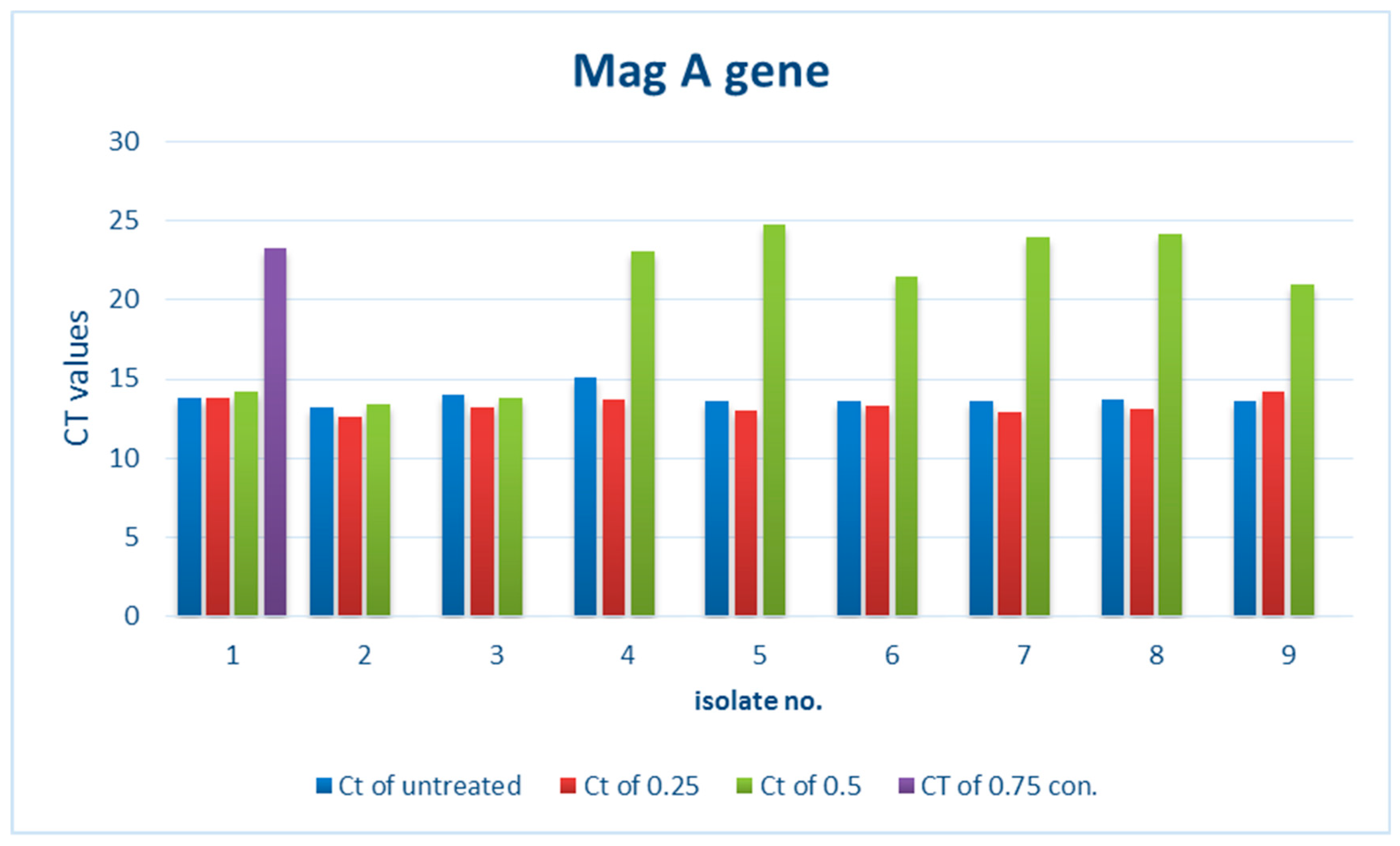
3.2.1. Effect of ZnO NPs on the Rate of magA Gene Expression

There is a noticeable effect on the gene expression levels of magA in Klebsiella pneumoniae isolates, as shown in Table 2 and Figure 1 and Figure 2. Colors in Table 2 represent the curves colors in the Figure 2. At a concentration of 0.25 mM, there was a slight change in the level of expression, and we noticed a reduction in the level of gene expression at a concentration of 0.50 mM. This decrease was higher at a concentration of 0.75 mM and led to a complete stop.

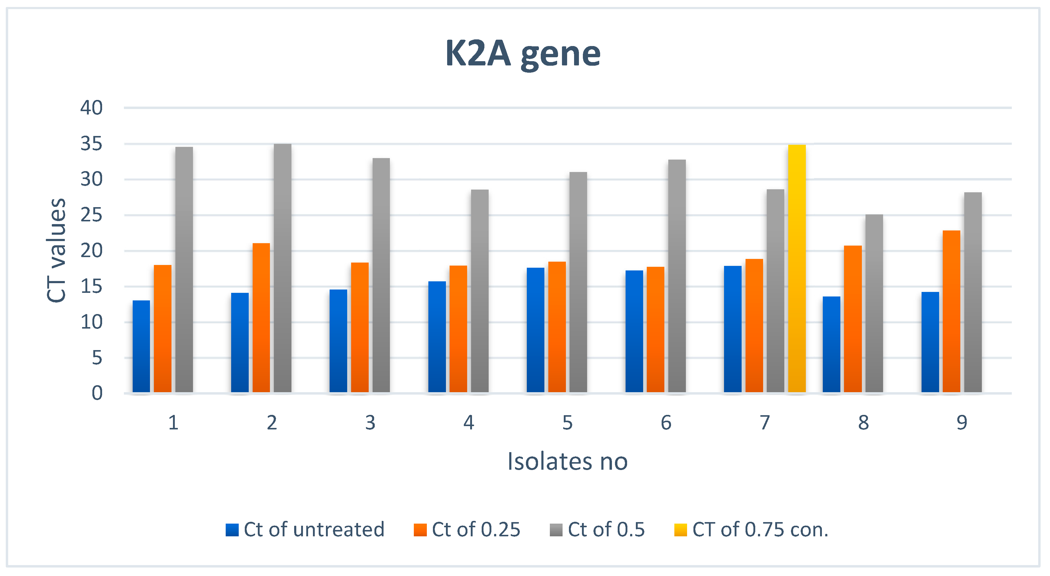
3.2.2. Effect of ZnO NPs on the Rate of k2A Gene Expression

There is a noticeable effect on the gene expression levels of k2A in Klebsiella pneumoniae isolates, as shown in Table 3 and Figure 3 and Figure 4. Colors in the Table 3 represent the curves colors in Figure 4. At a concentration of 0.25 mM, there was a slight change in the level of expression, and at a concentration of 0.50 mM, the level of gene expression began to decrease. However, it completely stopped at a concentration of 0.75 mM.

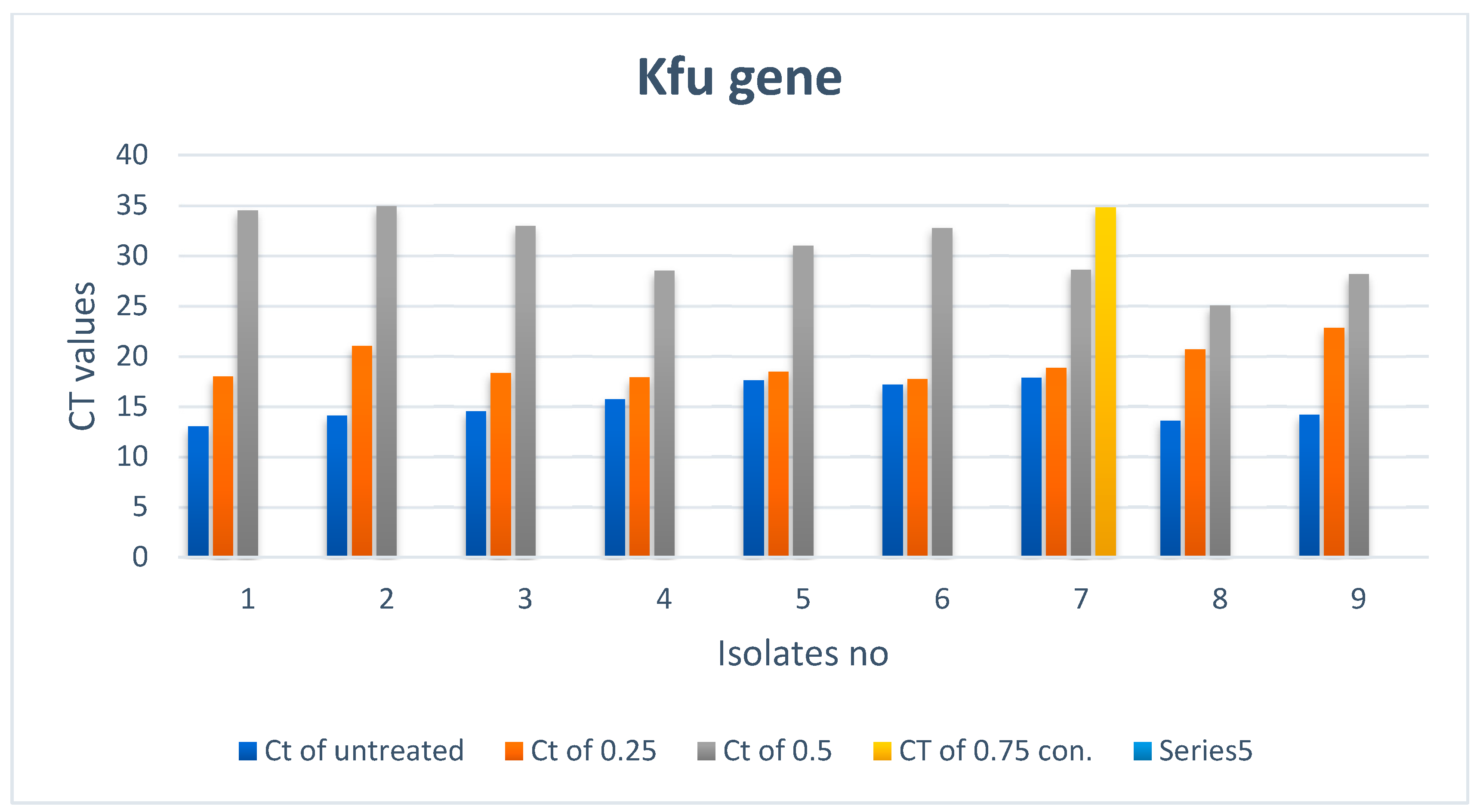
3.2.3. Effect of ZnO NPs on the Rate of kfu Gene Expression

There is a noticeable effect on the kfu gene expression levels in Klebsiella pneumoniae isolates, as shown in Table 4 and Figure 5 and Figure 6. Colors in the Table 4 represent the curves colors in Figure 6. At a concentration of 0.25 mM, there was a slight change in the level of expression, and it was noticed that the level of gene expression began to decrease at a concentration of 0.50 mM. Finally, the gene expression completely stopped at a concentration of 0.75 Mm.

3.2.4. Effect of Zinc Oxide on the Rate of the Gene Expression of rmpA

There is a noticeable effect on the gene expression levels of rmpA in Klebsiella pneumoniae isolates, as shown in Table 5 and Figure 7 and Figure 8. Colors in the Table 5 represent the curves colors in Figure 8. At a concentration of 0.25 mM, there was a slight change in the level of expression. Nevertheless, it is noticed that at a concentration of 0.50 mM, the level of gene expression began to decrease, and finally completely stopped at a concentration of 0.75 mM.

4. Discussion

New, harmful, antibiotic-resistant bacterial strains have emerged because of the indiscriminate utilization of antibiotics in contemporary culture. K. pneumoniae is a significant human pathogen that was recently linked to outbreaks in hospitals. Extended-spectrum b-lactamase (ESBL)-producing K. pneumoniae has been raised as a main and widespread issue because of the usage of prolonged-spectrum cephalosporins [17,18]. Here, new evidence states that an effective bacterial antibiotic in the form of NPs has been presented [19]. This could suggest a solution to the crucial issue of antibiotic resistance while lowering the danger of infections and their associated consequences; these have a serious impact on weak hospital patients. The development of novel and highly powerful bactericidal agents is motivated by the emergence of antibiotic-resistant microorganisms [20]. The current investigation’s findings point out three potential antimicrobial mechanisms for ZnO NP suspensions. (1) A physical mechanism that involves direct attachment to bacteria’s cell walls; (2) a biological mechanism that involves interaction with the components of the cell membrane; and (3) a chemical mechanism that involves the production of active species [21]. The size and concentration of ZnO play an important role in antibacterial activity. H2O2 production mainly depends on the surface area of nano ZnO; larger surface areas and higher concentrations of smaller particles may provide additional antibacterial activity [8]. This is in good agreement with a study on Escherichia coli and Pseudomonas aeruginosa, which were also sensitive to ZnO NPs, observing that NPs ranging from 14 to 25 nm inhibit them. Moreover, ZnO nanoparticles of a size of 22 nm were used against Klebsiella pneumonia and good results were obtained [7].
In general, the main findings imply that ZnO NPs could be employed externally to prevent bacterial infections from spreading. However, enhancing the ZnO NP concentrations slows the reproduction of K. pneumoniae, according to the MIC test and variation in the conventional growth curve. Gram-negative bacteria are found to have a higher negative charge on their cell surfaces than Gram-positive bacteria [22]. In some cases, membrane permeability is the initial stage of bacteria’s resistance to an antibiotic [23]. Marsalek measured the zeta potential of ZnO, where it was +35 mV at a pH of 7. This high positive value works to attract the bacteria due to the difference in charge [24]. Hence, there is a strong antibacterial impact when ZnO NPs exhibit their full potential by directly interfering with the degradation of the LPS membrane. The bacterial attachment is affected by the surface charge and specific surface hydrophobicity; these properties are assumed to be effective on bacterium surface adherence. The impact of the LPS membrane in adherence may be implicated in non-limited physicochemical interactions [25]. By subjecting the bacteria to oxidative stress and preventing bacterial development, ZnO NPs reduce the production of catalase, which is an antioxidant enzyme that shields the bacterium from oxidative stress [26].
On the other hand, several studies indicated ROS formation as the main mechanism for ZnO NP antibacterial activity [27]. The reactive species include superoxide anion (O2), hydrogen peroxide (H2O2), and hydroxide (OH−). The toxicity of these species involves the destruction of cellular components, such as lipids, DNA, and proteins, as a result of their internalization into the bacterial cell membrane. However, the role of ROS in antimicrobial actions has become an issue of debate among researchers in this field [28]. More studies should be done to correlate the role of ROS with ZnO NPs, but our study focuses on the effect of ZnO NPs on the gene expression of the genes responsible for capsular structures.

5. Conclusions

The zinc oxide nanoparticles were found to have antimicrobial activity against Klebsiella pneumoniae. This provides the potential to use this material in therapeutic material (antimicrobial agent) production. The ZnO NPs reduced the gained resistance incidence among pathogens from the misapplication and/or overuse of antibiotics. After investigating different concentrations of the NPs, 0.75 mM was found to be very effective in terminating the bacteria genes.

Author Contributions

N.B.K. conducted the experiments; M.H.R. and E.Y. supervised the work; M.K. revised the manuscript; R.R. and I.S. analyzed the data. All authors have read and agreed to the published version of the manuscript.

Funding

This research received no external funding.

Acknowledgments

The authors would like to thank their affiliations for partially supporting this work.

Conflicts of Interest

The authors declare is no conflict of interest.

References

  1. Chiu, S.; Wu, T.; Chuang, Y.; Lin, J.; Fung, C.; Lu, P.; Wang, J.; Wang, L.; Siu, K.; Yeh, K. National surveillance study on carbapenem Non-susceptible Klebsiella pneumoniae in taiwan: The emergence and rapid dissemination of kpc-2 carbapenemase. PLoS ONE 2013, 8, 1–7. [Google Scholar] [CrossRef] [PubMed]
  2. Aher, T.; Roy, A.; Kumar, P. Molecular detection of virulence genes associated with pathogenicity of Klebsiella spp. isolated from the respiratory tract of apparently healthy as well as sick goats. J. Vet. Med. 2012, 67, 249–252. [Google Scholar]
  3. Dubey, D.; Raza, F.; Sawhney, A.; Pandey, A. Klebsiella pneumoniae renal abscess syndrome: A rare case with metastatic involvement of lungs, eye, and brain. Case Rep. Infect. Dis. 2013, 2013, 1–3. [Google Scholar]
  4. Fang, C.; Lin, Y.; Lin, J.; Chen, T.; Yeh, K.; Chang, F.; Chuang, H.; Wu, H.; Tseng, C.; Siu, L. Klebsiella pneumoniae in Gastrointestinal Tract and Pyogenic Liver Abscess. Emerg. Infect. Dis. 2012, 18, 1322–1325. [Google Scholar]
  5. Doud, M.; Zeppegno, R.; Molina, E.; Miller, N.; Balachandar, D.; Schneper, L.; Poppiti, R.; Mathee, K. A k2A-positive Klebsiella pneumoniae causes liver and brain abscess in a Saint Kitt’s man. Int. J. Med. Sci. 2009, 6, 301–304. [Google Scholar] [CrossRef] [Green Version]
  6. Turton, J.; Perry, C.; Elgohari, S.; Hampton, C. PCR characterization and typing of Klebsiella pneumoniae using capsular type-specific, variable number tandem repeat and virulence gene targets. J. Med. Microbiol. 2010, 59, 541–547. [Google Scholar] [CrossRef] [PubMed] [Green Version]
  7. Azam, A.; Ahmed, A.; Oves, M.; Khan, M.; Memic, A. Size-dependent antimicrobial properties of CuO nanoparticles against Gram-positive and-negative bacterial strains. Int. J. Nanomed. 2012, 7, 3527–3535. [Google Scholar] [CrossRef] [Green Version]
  8. Padmavathy, N.; Vijayaraghavan, R. Enhanced Bioactivity of ZnO Nanoparticles an Antimicrobial Study. Sci. Technol. Adv. Mater. 2008, 9, 035004. [Google Scholar] [CrossRef] [PubMed]
  9. Jiang, J.; Pi, J.; Cai, J. The Advancing of Zinc Oxide Nanoparticles for Biomedical Application. Bioinorg. Chem. Appl. 2018, 2018, 1062562. [Google Scholar] [CrossRef]
  10. Ravishankar Rai, V.; Jamuna Bai, A. Nanoparticles and Their Potential Application as Antimicrobials. In Science against Microbial Pathogens: Communicating Current Research and Technological Advances; Méndez-Vilas, A., Ed.; Microbiology Series Formatex Research Center: Madrid, Spain, 2011; Volume 3, pp. 197–209. [Google Scholar]
  11. Brayner, R.; Ferrari-Iliou, R.; Brivois, N.; Djediat, S.; Benedetti, M.F.; Fievet, F. Toxicological impact studies based on Escherichia coli bacteria in ultrafine ZnO nanoparticles colloidal medium. Nano Lett. 2006, 6, 866–870. [Google Scholar] [CrossRef]
  12. Huang, Y.; Liao, H.; Wu, C.; Peng, H. MrkF is a component of type 3 fimbriae in Klebsiella pneumoniae. J. Res. Microbiol. 2009, 160, 71–79. [Google Scholar] [CrossRef] [PubMed]
  13. Kudaer, N.B.; Yousif, E.; Risan, M.H.; Kadhom, M. Preparation of ZnO Nanoparticles by the Chemical Precipitation Method. J. Serambi Eng. 2022, 7, 3129–3134. [Google Scholar]
  14. Holt, J.G.; Krieg, N.R. Bergey’s Manual of Determinative Bacteriology, 9th ed.; Williams & Wilkins: Baltimore, MD, USA, 1994. [Google Scholar]
  15. Hickling, T.P.; Clark, H.; Malhotra, R.; Sim, R.B. Collectins and theirrole in lung immunity. J. Leukoc. Biol. 2004, 75, 27–33. [Google Scholar] [CrossRef] [PubMed]
  16. Tullus, K. Fecal colonization with P-fimbriated Escherichia coli in newborn children and relation to development of extraintestinal E. coli infections. Acta Paediatr. Scand. 1987, 76, 1–35. [Google Scholar] [CrossRef]
  17. Stock, I.; Wiedemann, B. Natural antibiotic susceptibility of Klebsiella pneumoniae, K. oxytoca, K. planticola, K. ornithinolytica and K. terrigena strains. J. Med. Microbiol. 2001, 50, 396–406. [Google Scholar] [CrossRef] [Green Version]
  18. Schurr, J.R.; Young, E.; Byrne, P. Central role of toll-like receptor 4 signaling and host defense in experimental pneumonia caused by Gram-negative bacteria. Infect. Immun. 2005, 73, 532–545. [Google Scholar] [CrossRef] [Green Version]
  19. Huh, A.J.; Kwon, Y.J. Nanoantibiotics: A new paradigm for treating infectious diseases using nanomaterials in the antibiotics resistant era. J. Control Release 2011, 156, 128–145. [Google Scholar] [CrossRef]
  20. Mohammed, S.I.; Chandrasekaran, N.; Mukherjee, A. Studies on effect of TiO2 nanoparticles on growth and membrane permeability of Escherichia coli, Pseudomonas aeruginosa, and Bacillus subtilis. Curr. Nanosci. 2010, 6, 381–387. [Google Scholar] [CrossRef]
  21. Lingling, Z.; Yunhong, J.; Yulon, D. Investigation into the antibacterial behaviour of suspensions of ZnO nanoparticles (ZnO nanofluids). J. Nanopart. Res. 2007, 9, 479–489. [Google Scholar]
  22. Chung, Y.C.; Su, Y.P.; Chen, C.C. Relationship between antibacterial activity of chitosan and surface characteristics of cell wall. Acta Pharmacol. Sin. 2004, 25, 932–936. [Google Scholar]
  23. Berry, V.; Gole, A.; Kundu, S. Deposition of CTABterminated nanorods on bacteria to form highly conducting hybrid systems. J. Am. Chem. Soc. 2005, 125, 17600–17601. [Google Scholar] [CrossRef] [PubMed]
  24. Marsalek, R. Particle size and zeta potential of ZnO. APCBEE Proc. 2014, 9, 13–17. [Google Scholar] [CrossRef] [Green Version]
  25. Klotz, S. Role of hydrophobic interactions in microbial adhesion to plastics used in medical devices. In Microbial Cell Surface Hydrophobicity; Doyle, R., Rosenberg, M., Eds.; American Society for Microbiology: Washington, DC, USA, 1990; p. 107. [Google Scholar]
  26. Keisari, Y.; Wang, H.; Mesika, A. Surfactant protein D-coated Klebsiella pneumoniae stimulates cytokine production in mononuclear phagocytes. J. Leukoc. Biol. 2001, 70, 135–141. [Google Scholar] [CrossRef] [PubMed]
  27. Jalal, R.; Goharshadi, E.; Abareshi, M.; Moosavi, M.; Yousefi, A.; Nancarrow, P. ZnO nanofluids: Green synthesis, characterization, and antibacterial activity. Mater. Chem. Phys. 2010, 121, 198–201. [Google Scholar] [CrossRef]
  28. Raghupathi, K.; Koodali, R.; Manna, A. Size-Dependent Bacterial Growth Inhibition and Mechanism of Antibacterial Activity of Zinc Oxide Nanoparticles. Langmuir 2011, 27, 4020–4028. [Google Scholar] [CrossRef]
Figure 1. magA gene expression in Klebsiella pneumoniae isolates treated with different concentrations of ZnO NPs.
Biomimetics 07 00180 g001
Figure 2. Real-time PCR results of the magA gene.
Biomimetics 07 00180 g002
Figure 3. k2A gene expression in Klebsiella pneumoniae isolates treated with different concentrations of ZnO NPs.
Biomimetics 07 00180 g003
Figure 4. Real-time PCR results of the k2A gene.
Biomimetics 07 00180 g004
Figure 5. kfu gene expression in Klebsiella pneumoniae isolates treated with different concentrations of ZnO nanoparticles.
Biomimetics 07 00180 g005
Figure 6. Real-time PCR results of the kfu gene.
Biomimetics 07 00180 g006
Figure 7. rmpA gene expression in Klebsiella pneumoniae isolates treated with different concentrations of ZnO NPs.
Biomimetics 07 00180 g007
Figure 8. Real-time PCR results of the rmpA gene.
Biomimetics 07 00180 g008
Table 1. Antibiotic resistance of K. pneumoniae isolates.
AntibioticResistant%Intermediate%Sensitive%
Piperacillin74%4%22%
Cefazolin84%0%16%
Cefoxitin55%0%45%
Ceftazidime63%10%27%
Ceftriaxone100%0%0%
Cefepime40%0%60%
Imipenem21%6%73%
Amikacin10%10%80%
Gentamicin35%5%60%
Ciprofloxacine35%0%65%
Levofloxacin34%0%66%
Tigecyclin0%0%100%
Nitrofurantoin16%42%42%
Trimethoprim75%0%25%
Ampicillin100%0%0%
Amoxicillin97%0%3%
Cephalothin100%0%0%
Cephradine100%0%0%
Table 2. magA gene expression in Klebsiella pneumoniae isolates treated with different concentrations of ZnO NPs *.
Treated Bacteria Isolates with Different con.Untreated
NoCt of con3 (0.75)ColorCt of con2 (0.5)ColorCt of con1 (0.25)ColorCtColor
123.27Biomimetics 07 00180 i00114.24Biomimetics 07 00180 i00213.78Biomimetics 07 00180 i00313.79Biomimetics 07 00180 i004
2Not expressedBiomimetics 07 00180 i00513.43Biomimetics 07 00180 i00612.63Biomimetics 07 00180 i00713.2Biomimetics 07 00180 i008
3Not expressedBiomimetics 07 00180 i00913.84Biomimetics 07 00180 i01013.2Biomimetics 07 00180 i01113.99Biomimetics 07 00180 i012
4Not expressedBiomimetics 07 00180 i01323.01Biomimetics 07 00180 i01413.75Biomimetics 07 00180 i01515.13Biomimetics 07 00180 i016
5Not expressedBiomimetics 07 00180 i01724.75Biomimetics 07 00180 i01813.04Biomimetics 07 00180 i01913.65Biomimetics 07 00180 i020
6Not expressedBiomimetics 07 00180 i02121.42Biomimetics 07 00180 i02213.32Biomimetics 07 00180 i02313.61Biomimetics 07 00180 i024
7Not expressedBiomimetics 07 00180 i02523.96Biomimetics 07 00180 i02612.9Biomimetics 07 00180 i02713.64Biomimetics 07 00180 i028
8Not expressedBiomimetics 07 00180 i02924.15Biomimetics 07 00180 i03013.14Biomimetics 07 00180 i03113.69Biomimetics 07 00180 i032
9Not expressedBiomimetics 07 00180 i03320.99Biomimetics 07 00180 i03414.18Biomimetics 07 00180 i03513.6Biomimetics 07 00180 i036
* CT: cycle of threshold; con: concentration in mM.
Table 3. k2A gene expression in Klebsiella pneumoniae isolates treated with different concentrations of ZnO NPs *.
Treated Bacteria Isolates with Different con.Untreated
Ct of con3 (0.75)ColorCt of con2 (0.5)ColorCt of con1 (0.25)ColorCtColor
1Not expressedBiomimetics 07 00180 i03734.51Biomimetics 07 00180 i03817.98Biomimetics 07 00180 i03913Biomimetics 07 00180 i040
2Not expressedBiomimetics 07 00180 i04134.95Biomimetics 07 00180 i04221.04Biomimetics 07 00180 i04314.09Biomimetics 07 00180 i044
3Not expressedBiomimetics 07 00180 i04532.97Biomimetics 07 00180 i04618.33Biomimetics 07 00180 i04714.53Biomimetics 07 00180 i048
4Not expressedBiomimetics 07 00180 i04928.53Biomimetics 07 00180 i05017.88Biomimetics 07 00180 i05115.7Biomimetics 07 00180 i052
5Not expressedBiomimetics 07 00180 i05331.01Biomimetics 07 00180 i05418.46Biomimetics 07 00180 i05517.58Biomimetics 07 00180 i056
6Not expressedBiomimetics 07 00180 i05732.76Biomimetics 07 00180 i05817.72Biomimetics 07 00180 i05917.19Biomimetics 07 00180 i060
734.8Biomimetics 07 00180 i06128.59Biomimetics 07 00180 i06218.82Biomimetics 07 00180 i06317.86Biomimetics 07 00180 i064
8Not expressedBiomimetics 07 00180 i06525.04Biomimetics 07 00180 i06620.7Biomimetics 07 00180 i06713.58Biomimetics 07 00180 i068
9Not expressedBiomimetics 07 00180 i06928.15Biomimetics 07 00180 i07022.83Biomimetics 07 00180 i07114.19Biomimetics 07 00180 i072
* CT: cycle of threshold; con: concentration in mM.
Table 4. kfu gene expression in Klebsiella pneumoniae isolates treated with different concentrations of ZnO NPs *.
Treated Bacteria Isolates with Different con.Untreated
NoCt of con3 (0.75)ColorCt of con2 (0.5)ColorCt of con1 (0.25)ColorCtColor
1Not expressedBiomimetics 07 00180 i07334.51Biomimetics 07 00180 i07417.98Biomimetics 07 00180 i07513Biomimetics 07 00180 i076
2Not expressedBiomimetics 07 00180 i07734.95Biomimetics 07 00180 i07821.04Biomimetics 07 00180 i07914.09Biomimetics 07 00180 i080
3Not expressedBiomimetics 07 00180 i08132.97Biomimetics 07 00180 i08218.33Biomimetics 07 00180 i08314.53Biomimetics 07 00180 i084
4Not expressedBiomimetics 07 00180 i08528.53Biomimetics 07 00180 i08617.88Biomimetics 07 00180 i08715.7Biomimetics 07 00180 i088
5Not expressedBiomimetics 07 00180 i08931.01Biomimetics 07 00180 i09018.46Biomimetics 07 00180 i09117.58Biomimetics 07 00180 i092
6Not expressedBiomimetics 07 00180 i09332.76Biomimetics 07 00180 i09417.72Biomimetics 07 00180 i09517.19Biomimetics 07 00180 i096
734.8Biomimetics 07 00180 i09728.59Biomimetics 07 00180 i09818.82Biomimetics 07 00180 i09917.86Biomimetics 07 00180 i100
8Not expressedBiomimetics 07 00180 i10125.04Biomimetics 07 00180 i10220.7Biomimetics 07 00180 i10313.58Biomimetics 07 00180 i104
9Not expressedBiomimetics 07 00180 i10528.15Biomimetics 07 00180 i10622.83Biomimetics 07 00180 i10714.19Biomimetics 07 00180 i108
* CT: cycle of threshold; con: concentration in Mm.
Table 5. rmpA gene expression in Klebsiella pneumoniae isolates treated with different concentrations of ZnO NPs *.
Treated Bacteria Isolates with Different con.Untreated
NoCt of con3 (0.75)ColorCt of con2 (0.5)ColorCt of con1 (0.25)ColorCtColor
122.19Biomimetics 07 00180 i10922.18Biomimetics 07 00180 i11020.73Biomimetics 07 00180 i11114.83Biomimetics 07 00180 i112
2Not expressedBiomimetics 07 00180 i11322.63Biomimetics 07 00180 i11418.9Biomimetics 07 00180 i11514.93Biomimetics 07 00180 i116
3Not expressedBiomimetics 07 00180 i11723.22Biomimetics 07 00180 i11819.35Biomimetics 07 00180 i11913.24Biomimetics 07 00180 i120
4Not expressedBiomimetics 07 00180 i12123.24Biomimetics 07 00180 i12220.4Biomimetics 07 00180 i12313.95Biomimetics 07 00180 i124
5Not expressedBiomimetics 07 00180 i12519.36Biomimetics 07 00180 i12619.51Biomimetics 07 00180 i12714.28Biomimetics 07 00180 i128
6Not expressedBiomimetics 07 00180 i12923.39Biomimetics 07 00180 i13019.65Biomimetics 07 00180 i13113.89Biomimetics 07 00180 i132
7Not expressedBiomimetics 07 00180 i13324.29Biomimetics 07 00180 i13419.59Biomimetics 07 00180 i13518.88Biomimetics 07 00180 i136
8Not expressedBiomimetics 07 00180 i13722.48Biomimetics 07 00180 i13819.68Biomimetics 07 00180 i13914.06Biomimetics 07 00180 i140
9Not expressedBiomimetics 07 00180 i14122.82Biomimetics 07 00180 i14220.25Biomimetics 07 00180 i14314.44Biomimetics 07 00180 i144
* CT: cycle of threshold; con: concentration in Mm.
Publisher’s Note: MDPI stays neutral with regard to jurisdictional claims in published maps and institutional affiliations.

