
Magneto-Optics Effects: New Trends and Future Prospects for Technological Developments


Abstract
:
1. Introduction
2. A Brief Review on Magnetoplasmonics
3. A Brief Review on All-Dielectric Magnetophotonics
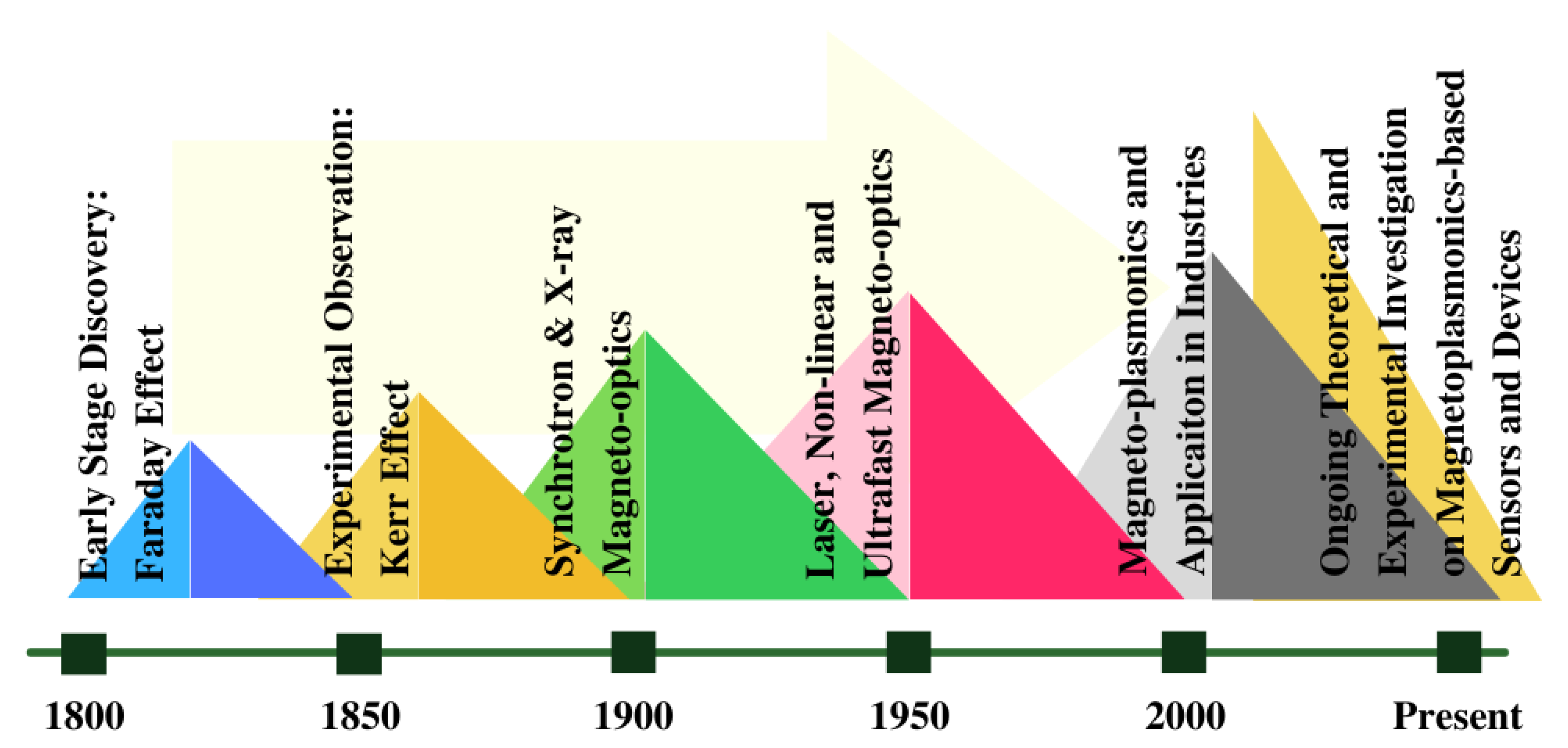
4. A Timeline View of Magneto-Optics and Its Applications
5. Concluding Remarks and Outlook
Author Contributions
Funding
Institutional Review Board Statement
Informed Consent Statement
Data Availability Statement
Acknowledgments
Conflicts of Interest
References
- Faraday, M. I. Experimental researches in electricity—Nineteenth series. Philos. Trans. R. Soc. Lond. 1846, 136, 1–20. [Google Scholar] [CrossRef]
- Kerr, J. XLIII. On rotation of the plane of polarization by reflection from the pole of a magnet. Lond. Edinb. Dublin Philos. Mag. J. Sci. 1877, 3, 321–343. [Google Scholar] [CrossRef]
- Kerr, J. XXIV. On reflection of polarized light from the equatorial surface of a magnet. Lond. Edinb. Dublin Philos. Mag. J. Sci. 1878, 5, 161–177. [Google Scholar] [CrossRef]
- De Hoop, A.T. A reciprocity theorem for the electromagnetic field scattered by an obstacle. Appl. Sci. Res. Sect. B 1960, 8, 135–140. [Google Scholar] [CrossRef]
- Shoji, Y.; Mizumoto, T.; Yokoi, H.; Hsieh, I.W.; Osgood, R.M. Magneto-optical isolator with silicon waveguides fabricated by direct bonding. Appl. Phys. Lett. 2008, 92, 071117. [Google Scholar] [CrossRef]
- Srinivasan, K.; Stadler, B.J.H. Magneto-optical materials and designs for integrated TE- and TM-mode planar waveguide isolators: A review. Opt. Mater. Express 2018, 8, 3307–3318. [Google Scholar] [CrossRef]
- Allwood, D.A.; Xiong, G.; Cooke, M.D.; Cowburn, R.P. Magneto-optical Kerr effect analysis of magnetic nanostructures. J. Phys. D Appl. Phys. 2003, 36, 2175–2182. [Google Scholar] [CrossRef]
- Kirilyuk, A.; Kimel, A.V.; Rasing, T. Ultrafast optical manipulation of magnetic order. RMP 2010, 82, 2731–2784. [Google Scholar] [CrossRef]
- McCord, J. Progress in magnetic domain observation by advanced magneto-optical microscopy. J. Phys. D Appl. Phys. 2015, 48, 333001. [Google Scholar] [CrossRef]
- Higo, T.; Man, H.; Gopman, D.B.; Wu, L.; Koretsune, T.; van’t Erve, O.M.J.; Kabanov, Y.P.; Rees, D.; Li, Y.; Suzuki, M.T.; et al. Large magneto-optical Kerr effect and imaging of magnetic octupole domains in an antiferromagnetic metal. Nat. Photonics 2018, 12, 73–78. [Google Scholar] [CrossRef]
- Tufte, O.N.; Chen, D. Optical techniques for data storage. IEEE Spectr. 1973, 10, 26–32. [Google Scholar] [CrossRef]
- Chen, D.; Zook, J.D. An overview of optical data storage technology. Proc. IEEE 1975, 63, 1207–1230. [Google Scholar] [CrossRef]
- Mansuripur, M. The Physical Principles of Magneto-Optical Recording; Cambridge University Press: Cambridge, UK, 1995. [Google Scholar] [CrossRef]
- Qin, J.; Xia, S.; Yang, W.; Wang, H.; Yan, W.; Yang, Y.; Wei, Z.; Liu, W.; Luo, Y.; Deng, L.; et al. Nanophotonic devices based on magneto-optical materials: Recent developments and applications. Nanophotonics 2022, 11, 2639–2659. [Google Scholar] [CrossRef]
- Armelles, G.; Cebollada, A.; García-Martín, A.; González, M.U. Magnetoplasmonics: Combining magnetic and plasmonic functionalities. Adv. Opt. Mater. 2013, 1, 10–35. [Google Scholar] [CrossRef]
- Sepúlveda, B.; González-Díaz, J.B.; García-Martín, A.; Lechuga, L.M.; Armelles, G. Plasmon-Induced Magneto-Optical Activity in Nanosized Gold Disks. Phys. Rev. Lett. 2010, 104, 147401. [Google Scholar] [CrossRef]
- Du, G.X.; Mori, T.; Saito, S.; Takahashi, M. Shape-enhanced magneto-optical activity: Degree of freedom for active plasmonics. Phys. Rev. B 2010, 82, 161403. [Google Scholar] [CrossRef]
- Brongersma Mark, L.; Shalaev Vladimir, M. The Case for Plasmonics. Science 2010, 328, 440–441. [Google Scholar] [CrossRef]
- Schuller, J.A.; Barnard, E.S.; Cai, W.; Jun, Y.C.; White, J.S.; Brongersma, M.L. Plasmonics for extreme light concentration and manipulation. Nat. Mater. 2010, 9, 193–204. [Google Scholar] [CrossRef]
- Chen, J.; Albella, P.; Pirzadeh, Z.; Alonso-González, P.; Huth, F.; Bonetti, S.; Bonanni, V.; Åkerman, J.; Nogués, J.; Vavassori, P.; et al. Plasmonic Nickel Nanoantennas. Small 2011, 7, 2341–2347. [Google Scholar] [CrossRef] [Green Version]
- Maccaferri, N.; Berger, A.; Bonetti, S.; Bonanni, V.; Kataja, M.; Qin, Q.H.; van Dijken, S.; Pirzadeh, Z.; Dmitriev, A.; Nogués, J.; et al. Tuning the Magneto-Optical Response of Nanosize Ferromagnetic Ni Disks Using the Phase of Localized Plasmons. Phys. Rev. Lett. 2013, 111, 167401. [Google Scholar] [CrossRef]
- Kataja, M.; Hakala, T.K.; Julku, A.; Huttunen, M.J.; van Dijken, S.; Törmä, P. Surface lattice resonances and magneto-optical response in magnetic nanoparticle arrays. Nat. Commun. 2015, 6, 7072. [Google Scholar] [CrossRef]
- Temnov, V.V.; Armelles, G.; Woggon, U.; Guzatov, D.; Cebollada, A.; Garcia-Martin, A.; Garcia-Martin, J.M.; Thomay, T.; Leitenstorfer, A.; Bratschitsch, R. Active magneto-plasmonics in hybrid metal-ferromagnet structures. Nat. Photonics 2010, 4, 107–111. [Google Scholar] [CrossRef]
- Belotelov, V.I.; Akimov, I.A.; Pohl, M.; Kotov, V.A.; Kasture, S.; Vengurlekar, A.S.; Gopal, A.V.; Yakovlev, D.R.; Zvezdin, A.K.; Bayer, M. Enhanced magneto-optical effects in magnetoplasmonic crystals. Nat. Nanotechnol. 2011, 6, 370–376. [Google Scholar] [CrossRef]
- Kreilkamp, L.E.; Belotelov, V.I.; Chin, J.Y.; Neutzner, S.; Dregely, D.; Wehlus, T.; Akimov, I.A.; Bayer, M.; Stritzker, B.; Giessen, H. Waveguide-Plasmon Polaritons Enhance Transverse Magneto-Optical Kerr Effect. Phys. Rev. X 2013, 3, 041019. [Google Scholar] [CrossRef]
- Sepúlveda, B.; Calle, A.; Lechuga, L.M.; Armelles, G. Highly sensitive detection of biomolecules with the magneto-optic surface-plasmon-resonance sensor. Opt. Lett. 2006, 31, 1085–1087. [Google Scholar] [CrossRef]
- Regatos, D.; Sepúlveda, B.; Fariña, D.; Carrascosa, L.G.; Lechuga, L.M. Suitable combination of noble/ferromagnetic metal multilayers for enhanced magneto-plasmonic biosensing. Opt. Express 2011, 19, 8336–8346. [Google Scholar] [CrossRef]
- Manera, M.G.; Ferreiro-Vila, E.; García-Martín, J.M.; Cebollada, A.; García-Martín, A.; Giancane, G.; Valli, L.; Rella, R. Enhanced magneto-optical SPR platform for amine sensing based on Zn porphyrin dimers. Sens. Actuators B Chem. 2013, 182, 232–238. [Google Scholar] [CrossRef]
- Maccaferri, N.; Gregorczyk, K.E.; de Oliveira, T.V.A.G.; Kataja, M.; van Dijken, S.; Pirzadeh, Z.; Dmitriev, A.; Åkerman, J.; Knez, M.; Vavassori, P. Ultrasensitive and label-free molecular-level detection enabled by light phase control in magnetoplasmonic nanoantennas. Nat. Commun. 2015, 6, 6150. [Google Scholar] [CrossRef]
- Firby, C.J.; Elezzabi, A.Y. High-speed nonreciprocal magnetoplasmonic waveguide phase shifter. Optica 2015, 2, 598–606. [Google Scholar] [CrossRef]
- Qin, J.; Deng, L.; Kang, T.; Nie, L.; Feng, H.; Wang, H.; Yang, R.; Liang, X.; Tang, T.; Shen, J.; et al. Switching the Optical Chirality in Magnetoplasmonic Metasurfaces Using Applied Magnetic Fields. ACS Nano 2020, 14, 2808–2816. [Google Scholar] [CrossRef]
- Voronov, A.A.; Karki, D.; Ignatyeva, D.O.; Kozhaev, M.A.; Levy, M.; Belotelov, V.I. Magneto-optics of subwavelength all-dielectric gratings. Opt. Express 2020, 28, 17988–17996. [Google Scholar] [CrossRef] [PubMed]
- Ignatyeva, D.O.; Karki, D.; Voronov, A.A.; Kozhaev, M.A.; Krichevsky, D.M.; Chernov, A.I.; Levy, M.; Belotelov, V.I. All-dielectric magnetic metasurface for advanced light control in dual polarizations combined with high-Q resonances. Nat. Commun. 2020, 11, 5487. [Google Scholar] [CrossRef] [PubMed]
- Carvalho, W.O.F.; Moncada-Villa, E.; Oliveira, O.N.; Mejía-Salazar, J.R. Beyond plasmonic enhancement of the transverse magneto-optical Kerr effect with low-loss high-refractive-index nanostructures. Phys. Rev. B 2021, 103, 075412. [Google Scholar] [CrossRef]
- Carvalho, W.O.F.; Mejía-Salazar, J.R. All-dielectric magnetophotonic gratings for maximum TMOKE enhancement. Phys. Chem. Chem. Phys. 2022, 24, 5431–5436. [Google Scholar] [CrossRef]
- Fakhrul, T.; Tazlaru, S.; Beran, L.; Zhang, Y.; Veis, M.; Ross, C.A. Magneto-Optical Bi:YIG Films with High Figure of Merit for Nonreciprocal Photonics. Adv. Opt. Mater. 2019, 7, 1900056. [Google Scholar] [CrossRef]
- Fakhrul, T.; Tazlaru, S.; Khurana, B.; Beran, L.; Bauer, J.; Vančík, M.; Marchese, A.; Tsotsos, E.; Kučera, M.; Zhang, Y.; et al. High Figure of Merit Magneto-Optical Ce- and Bi-Substituted Terbium Iron Garnet Films Integrated on Si. Adv. Opt. Mater. 2021, 9, 2100512. [Google Scholar] [CrossRef]
- Mizumoto, T.; Baets, R.; Bowers, J.E. Optical nonreciprocal devices for silicon photonics using wafer-bonded magneto-optical garnet materials. MRS Bull. 2018, 43, 419–424. [Google Scholar] [CrossRef]
- Pintus, P.; Huang, D.; Morton, P.A.; Shoji, Y.; Mizumoto, T.; Bowers, J.E. Broadband TE Optical Isolators and Circulators in Silicon Photonics Through Ce:YIG Bonding. J. Light. Technol. 2019, 37, 1463–1473. [Google Scholar] [CrossRef]
- Zhang, Y.; Du, Q.; Wang, C.; Fakhrul, T.; Liu, S.; Deng, L.; Huang, D.; Pintus, P.; Bowers, J.; Ross, C.A.; et al. Monolithic integration of broadband optical isolators for polarization-diverse silicon photonics. Optica 2019, 6, 473–478. [Google Scholar] [CrossRef]
- Murai, T.; Shoji, Y.; Nishiyama, N.; Mizumoto, T. Nonvolatile magneto-optical switches integrated with a magnet stripe array. Opt. Express 2020, 28, 31675–31685. [Google Scholar] [CrossRef]
- Carvalho, W.O.F.; Mejía-Salazar, J.R. Magneto-optical micro-ring resonators for dynamic tuning of add/drop channels in dense wavelength division multiplexing applications. Opt. Lett. 2021, 46, 2396–2399. [Google Scholar] [CrossRef] [PubMed]
- Liu, S.; Shoji, Y.; Mizumoto, T. TE-mode magneto-optical isolator based on an asymmetric microring resonator under a unidirectional magnetic field. Opt. Express 2022, 30, 9934–9943. [Google Scholar] [CrossRef] [PubMed]
- Kolmychek, I.A.; Pomozov, A.R.; Leontiev, A.P.; Napolskii, K.S.; Murzina, T.V. Magneto-optical effects in hyperbolic metamaterials. Opt. Lett. 2018, 43, 3917–3920. [Google Scholar] [CrossRef] [PubMed]
- Fan, B.; Nasir, M.E.; Nicholls, L.H.; Zayats, A.V.; Podolskiy, V.A. Magneto-Optical Metamaterials: Nonreciprocal Transmission and Faraday Effect Enhancement. Adv. Opt. Mater. 2019, 7, 1801420. [Google Scholar] [CrossRef]
- Wang, X.; Wang, H.; Jian, J.; Rutherford, B.X.; Gao, X.; Xu, X.; Zhang, X.; Wang, H. Metal-Free Oxide-Nitride Heterostructure as a Tunable Hyperbolic Metamaterial Platform. Nano Lett. 2020, 20, 6614–6622. [Google Scholar] [CrossRef]
- Moncada-Villa, E.; Oliveira, O.N.; Mejía-Salazar, J.R. Uniaxial ε-near-zero metamaterials for giant enhancement of the transverse magneto-optical Kerr effect. Phys. Rev. B 2020, 102, 165304. [Google Scholar] [CrossRef]
- Wang, X.; Jian, J.; Wang, H.; Liu, J.; Pachaury, Y.; Lu, P.; Rutherford, B.X.; Gao, X.; Xu, X.; El-Azab, A.; et al. Nitride-Oxide-Metal Heterostructure with Self-Assembled Core-Shell Nanopillar Arrays: Effect of Ordering on Magneto-Optical Properties. Small 2021, 17, 2007222. [Google Scholar] [CrossRef]
- Pershan, P.S. Magneto-Optical Effects. J. Appl. Phys. 1967, 38, 1482–1490. [Google Scholar] [CrossRef]
- Chen, D. Magneto-Optic Principles, Materials And Applications. In Proceedings of the Electro-Optics Principles and Applications, Boston, MA, USA, 1–2 January 1973; Volume 0038. [Google Scholar] [CrossRef]
- Mansuripur, M. The Faraday Effect. Opt. Photon. News 1999, 10, 32–36. [Google Scholar] [CrossRef]
- Qiu, Z.Q.; Bader, S.D. Surface magneto-optic Kerr effect. Rev. Sci. Instrum. 2000, 71, 1243–1255. [Google Scholar] [CrossRef]
- Floess, D.; Giessen, H. Nonreciprocal hybrid magnetoplasmonics. Rep. Prog. Phys. 2018, 81, 116401. [Google Scholar] [CrossRef] [PubMed]
- Maccaferri, N.; Zubritskaya, I.; Razdolski, I.; Chioar, I.A.; Belotelov, V.; Kapaklis, V.; Oppeneer, P.M.; Dmitriev, A. Nanoscale magnetophotonics. J. Appl. Phys. 2020, 127, 080903. [Google Scholar] [CrossRef]
- Wood, R.W. XLII. On a remarkable case of uneven distribution of light in a diffraction grating spectrum. Lond. Edinb. Dublin Philos. Mag. J. Sci. 1902, 4, 396–402. [Google Scholar] [CrossRef]
- Rayleigh, L. III. Note on the remarkable case of diffraction spectra described by Prof. Wood. Lond. Edinb. Dublin Philos. Mag. J. Sci. 1907, 14, 60–65. [Google Scholar] [CrossRef]
- Rayleigh, L. On the dynamical theory of gratings. Proc. R. Soc. Lond. Ser. A Contain. Pap. Math. Phys. Character 1907, 79, 399–416. [Google Scholar] [CrossRef]
- Fano, U. The Theory of Anomalous Diffraction Gratings and of Quasi-Stationary Waves on Metallic Surfaces (Sommerfeld’s Waves). J. Opt. Soc. Am. 1941, 31, 213–222. [Google Scholar] [CrossRef]
- Bohm, D.; Pines, D. A Collective Description of Electron Interactions. I. Magnetic Interactions. Phys. Rev. 1951, 82, 625–634. [Google Scholar] [CrossRef]
- Pines, D.; Bohm, D. A Collective Description of Electron Interactions: II. Collective vs Individual Particle Aspects of the Interactions. Phys. Rev. 1952, 85, 338–353. [Google Scholar] [CrossRef]
- Pines, D. A Collective Description of Electron Interactions: IV. Electron Interaction in Metals. Phys. Rev. 1953, 92, 626–636. [Google Scholar] [CrossRef]
- Bohm, D.; Pines, D. A Collective Description of Electron Interactions: III. Coulomb Interactions in a Degenerate Electron Gas. Phys. Rev. 1953, 92, 609–625. [Google Scholar] [CrossRef]
- Ritchie, R.H. Plasma Losses by Fast Electrons in Thin Films. Phys. Rev. 1957, 106, 874–881. [Google Scholar] [CrossRef]
- Powell, C.J.; Swan, J.B. Origin of the Characteristic Electron Energy Losses in Aluminum. Phys. Rev. 1959, 115, 869–875. [Google Scholar] [CrossRef]
- Kretschmann, E.; Raether, H. Notizen: Radiative Decay of Non Radiative Surface Plasmons Excited by Light. Z. Für Naturforschung A 1968, 23, 2135–2136. [Google Scholar] [CrossRef]
- Otto, A. Excitation of nonradiative surface plasma waves in silver by the method of frustrated total reflection. Z. Für Phys. A Hadron. Nucl. 1968, 216, 398–410. [Google Scholar] [CrossRef]
- Stockman, M.I. Nanoplasmonics: Past, present, and glimpse into future. Opt. Express 2011, 19, 22029–22106. [Google Scholar] [CrossRef]
- Maxwell-Garnett, J.C. XII. Colours in metal glasses and in metallic films. Philos. Trans. R. Soc. Lond. Ser. A Contain. Pap. Math. Phys. Character 1904, 203, 385–420. [Google Scholar] [CrossRef]
- Mie, G. Beiträge zur Optik trüber Medien, speziell kolloidaler Metallösungen. Ann. Phys. 1908, 330, 377–445. [Google Scholar] [CrossRef]
- Rizal, C.; Manera, M.G.; Ignatyeva, D.O.; Mejía-Salazar, J.R.; Rella, R.; Belotelov, V.I.; Pineider, F.; Maccaferri, N. Magnetophotonics for sensing and magnetometry toward industrial applications. J. Appl. Phys. 2021, 130, 230901. [Google Scholar] [CrossRef]
- Nylander, C.; Liedberg, B.; Lind, T. Gas detection by means of surface plasmon resonance. Sens. Actuators 1982, 3, 79–88. [Google Scholar] [CrossRef]
- Liedberg, B.; Nylander, C.; Lunström, I. Surface plasmon resonance for gas detection and biosensing. Sens. Actuators 1983, 4, 299–304. [Google Scholar] [CrossRef]
- Mejía-Salazar, J.R.; Oliveira, O.N. Plasmonic Biosensing. Chem. Rev. 2018, 118, 10617–10625. [Google Scholar] [CrossRef] [PubMed]
- Guo, J.; Zhu, Z.; Deng, W. Small-angle measurement based on surface-plasmon resonance and the use of magneto-optical modulation. Appl. Opt. 1999, 38, 6550–6555. [Google Scholar] [CrossRef] [PubMed]
- David, S.; Polonschii, C.; Luculescu, C.; Gheorghiu, M.; Gáspár, S.; Gheorghiu, E. Magneto-plasmonic biosensor with enhanced analytical response and stability. Biosens. Bioelectron. 2015, 63, 525–532. [Google Scholar] [CrossRef]
- Diaz-Valencia, B.F.; Mejía-Salazar, J.R.; Oliveira, O.N.; Porras-Montenegro, N.; Albella, P. Enhanced Transverse Magneto-Optical Kerr Effect in Magnetoplasmonic Crystals for the Design of Highly Sensitive Plasmonic (Bio)sensing Platforms. ACS Omega 2017, 2, 7682–7685. [Google Scholar] [CrossRef]
- Tran, V.T.; Kim, J.; Tufa, L.T.; Oh, S.; Kwon, J.; Lee, J. Magnetoplasmonic Nanomaterials for Biosensing/Imaging and in Vitro/in Vivo Biousability. Anal. Chem. 2018, 90, 225–239. [Google Scholar] [CrossRef] [PubMed]
- Abujetas, D.R.; de Sousa, N.; García-Martín, A.; Llorens, J.M.; Sánchez-Gil, J.A. Active angular tuning and switching of Brewster quasi bound states in the continuum in magneto-optic metasurfaces. Nanophotonics 2021, 10, 4223–4232. [Google Scholar] [CrossRef]
- Kausaite-Minkstimiene, A.; Popov, A.; Ramanaviciene, A. Surface Plasmon Resonance Immunosensor with Antibody-Functionalized Magnetoplasmonic Nanoparticles for Ultrasensitive Quantification of the CD5 Biomarker. ACS Appl. Mater. Interfaces 2022, 14, 20720–20728. [Google Scholar] [CrossRef]
- Tsakmakidis, K. Non-reciprocal plasmonics. Nat. Mater. 2013, 12, 378. [Google Scholar] [CrossRef]
- Shastri, K.; Abdelrahman, M.I.; Monticone, F. Nonreciprocal and Topological Plasmonics. Photonics 2021, 8, 133. [Google Scholar] [CrossRef]
- Wang, J.; Shi, Q.Y.; Liu, Y.J.; Dong, H.Y.; Fung, K.H.; Dong, Z.G. Stopping surface magneto-plasmons by non-reciprocal graded waveguides. Phys. Lett. A 2021, 398, 127279. [Google Scholar] [CrossRef]
- Shimizu, H.; Zayets, V. Plasmonic isolator for photonic integrated circuits. MRS Bull. 2018, 43, 425–429. [Google Scholar] [CrossRef]
- Armelles, G.; Caballero, B.; Prieto, P.; García, F.; Cebollada, A.; González, M.U.; García-Martin, A. Magnetic field modulation of chirooptical effects in magnetoplasmonic structures. Nanoscale 2014, 6, 3737–3741. [Google Scholar] [CrossRef] [PubMed]
- Yannopapas, V. Magnetochirality in hierarchical magnetoplasmonic clusters. Solid State Commun. 2015, 217, 47–52. [Google Scholar] [CrossRef]
- Feng, H.Y.; de Dios, C.; García, F.; Cebollada, A.; Armelles, G. Analysis and magnetic modulation of chiro-optical properties in anisotropic chiral and magneto-chiral plasmonic systems. Opt. Express 2017, 25, 31045–31055. [Google Scholar] [CrossRef]
- Wu, X.; Hao, C.; Xu, L.; Kuang, H.; Xu, C. Chiromagnetic Plasmonic Nanoassemblies with Magnetic Field Modulated Chiral Activity. Small 2020, 16, 1905734. [Google Scholar] [CrossRef]
- Wang, N.; Ding, L.; Wang, W. Magnetoplasmonic coupling in graphene nanodisk dimers: An extended coupled-dipole model for circularly polarized states. Phys. Rev. B 2022, 105, 235435. [Google Scholar] [CrossRef]
- Nguyen, H.Q.; Hwang, D.; Park, S.; Nguyen, M.C.T.; Kang, S.S.; Tran, V.T.; Lee, J. One-Pot Synthesis of Magnetoplasmonic Au@FexOy Nanowires: Bioinspired Bouligand Chiral Stack. ACS Nano 2022, 16, 5795–5806. [Google Scholar] [CrossRef]
- Baron Kelvin, W.T. Baltimore Lectures on Molecular Dynamics and the Wave Theory of Light; Cambridge University Press: Cambridge, UK, 2010. [Google Scholar] [CrossRef] [Green Version]
- Barron, L. Chirality and Life. In Strategies of Life Detection; Botta, O., Bada, J.L., Gomez-Elvira, J., Javaux, E., Selsis, F., Summons, R., Eds.; Springer: Boston, MA, USA, 2008; pp. 187–201. [Google Scholar] [CrossRef]
- Inaki, M.; Liu, J.; Matsuno, K. Cell chirality: Its origin and roles in left-right asymmetric development. Philos. Trans. R. Soc. B Biol. Sci. 2016, 371, 20150403. [Google Scholar] [CrossRef]
- McConathy, J.; Owens, M.J. Stereochemistry in Drug Action. Prim. Care Companion J. Clin. Psychiatry 2003, 5, 70–73. [Google Scholar] [CrossRef]
- Singh, K.; Shakya, P.; Kumar, A.H.S.; Alok, S.; Kamal, M.; Singh, S.P. Stereochemistry and its role in drug design. Int. J. Pharm. Sci. Res. 2014, 5, 4644–4659. [Google Scholar]
- Du, X.; Zhou, J.; Wang, J.; Zhou, R.; Xu, B. Chirality Controls Reaction-Diffusion of Nanoparticles for Inhibiting Cancer Cells. ChemNanoMat 2017, 3, 17–21. [Google Scholar] [CrossRef] [PubMed]
- Nguyen, L.A.; He, H.; Pham-Huy, C. Chiral drugs: An overview. Int. J. Biomed. Sci. IJBS 2006, 2, 85–100. [Google Scholar] [PubMed]
- Smith, S.W. Chiral Toxicology: It’s the Same Thing…Only Different. Toxicol. Sci. 2009, 110, 4–30. [Google Scholar] [CrossRef] [PubMed]
- Tokunaga, E.; Yamamoto, T.; Ito, E.; Shibata, N. Understanding the Thalidomide Chirality in Biological Processes by the Self-disproportionation of Enantiomers. Sci. Rep. 2018, 8, 17131. [Google Scholar] [CrossRef]
- Rodger, A. Circular Dichroism. In Encyclopedia of Analytical Chemistry; John Wiley & Sons, Ltd.: Hoboken, NJ, USA, 2014; pp. 1–34. [Google Scholar] [CrossRef]
- Liu, Z.; Du, Y.; Feng, Z. Enantioseparation of drugs by capillary electrochromatography using a stationary phase covalently modified with graphene oxide. Microchim. Acta 2017, 184, 583–593. [Google Scholar] [CrossRef]
- Gwak, J.; Park, S.J.; Choi, H.Y.; Lee, J.H.; Jeong, K.J.; Lee, D.; Tran, V.T.; Son, K.S.; Lee, J. Plasmonic Enhancement of Chiroptical Property in Enantiomers Using a Helical Array of Magnetoplasmonic Nanoparticles for Ultrasensitive Chiral Recognition. ACS Appl. Mater. Interfaces 2021, 13, 46886–46893. [Google Scholar] [CrossRef]
- Atzori, M.; Rikken, G.L.J.A.; Train, C. Magneto-Chiral Dichroism: A Playground for Molecular Chemists. Chem. Eur. J. 2020, 26, 9784–9791. [Google Scholar] [CrossRef]
- Petrucci, G.; Gabbani, A.; Faniayeu, I.; Pedrueza-Villalmanzo, E.; Cucinotta, G.; Atzori, M.; Dmitriev, A.; Pineider, F. Macroscopic magneto-chiroptical metasurfaces. Appl. Phys. Lett. 2021, 118, 251108. [Google Scholar] [CrossRef]
- Li, C.; Li, S.; Zhao, J.; Sun, M.; Wang, W.; Lu, M.; Qu, A.; Hao, C.; Chen, C.; Xu, C.; et al. Ultrasmall Magneto-chiral Cobalt Hydroxide Nanoparticles Enable Dynamic Detection of Reactive Oxygen Species in Vivo. J. Am. Chem. Soc. 2022, 144, 1580–1588. [Google Scholar] [CrossRef]
- Zubritskaya, I.; Maccaferri, N.; Inchausti Ezeiza, X.; Vavassori, P.; Dmitriev, A. Magnetic Control of the Chiroptical Plasmonic Surfaces. Nano Lett. 2018, 18, 302–307. [Google Scholar] [CrossRef]
- Girón-Sedas, J.A.; Mejía-Salazar, J.R.; Granada, J.C.; Oliveira, O.N. Repulsion of polarized particles near a magneto-optical metamaterial. Phys. Rev. B 2016, 94, 245430. [Google Scholar] [CrossRef]
- Maccaferri, N.; Vavassori, P.; Garoli, D. Magnetic control of particle trapping in a hybrid plasmonic nanopore. Appl. Phys. Lett. 2021, 118, 193102. [Google Scholar] [CrossRef]
- Biagioni, P.; Huang, J.S.; Hecht, B. Nanoantennas for visible and infrared radiation. Rep. Prog. Phys. 2012, 75, 024402. [Google Scholar] [CrossRef] [PubMed]
- Boriskina, S.V.; Cooper, T.A.; Zeng, L.; Ni, G.; Tong, J.K.; Tsurimaki, Y.; Huang, Y.; Meroueh, L.; Mahan, G.; Chen, G. Losses in plasmonics: From mitigating energy dissipation to embracing loss-enabled functionalities. Adv. Opt. Photonics 2017, 9, 775–827. [Google Scholar] [CrossRef]
- Hernández-Sarria, J.J.; Oliveira, O.N.; Mejía-Salazar, J.R. Toward Lossless Infrared Optical Trapping of Small Nanoparticles Using Nonradiative Anapole Modes. Phys. Rev. Lett. 2021, 127, 186803. [Google Scholar] [CrossRef]
- Decker, M.; Staude, I. Resonant dielectric nanostructures: A low-loss platform for functional nanophotonics. J. Opt. 2016, 18, 103001. [Google Scholar] [CrossRef]
- Kuznetsov Arseniy, I.; Miroshnichenko Andrey, E.; Brongersma Mark, L.; Kivshar Yuri, S.; Boris, L. Optically resonant dielectric nanostructures. Science 2016, 354, aag2472. [Google Scholar] [CrossRef]
- Huang, T.; Wang, B.; Zhang, W.; Zhao, C. Ultracompact Energy Transfer in Anapole-based Metachains. Nano Lett. 2021, 21, 6102–6110. [Google Scholar] [CrossRef]
- Bi, L.; Hu, J.; Jiang, P.; Kim, H.S.; Kim, D.H.; Onbasli, M.C.; Dionne, G.F.; Ross, C.A. Magneto-Optical Thin Films for On-Chip Monolithic Integration of Non-Reciprocal Photonic Devices. Materials 2013, 6, 5094–5117. [Google Scholar] [CrossRef]
- Onbasli, M.C.; Goto, T.; Sun, X.; Huynh, N.; Ross, C.A. Integration of bulk-quality thin film magneto-optical cerium-doped yttrium iron garnet on silicon nitride photonic substrates. Opt. Express 2014, 22, 25183–25192. [Google Scholar] [CrossRef]
- Onbasli, M.C.; Beran, L.; Zahradník, M.; Kučera, M.; Antoš, R.; Mistrík, J.; Dionne, G.F.; Veis, M.; Ross, C.A. Optical and magneto-optical behavior of Cerium Yttrium Iron Garnet thin films at wavelengths of 200–1770 nm. Sci. Rep. 2016, 6, 23640. [Google Scholar] [CrossRef] [PubMed]
- Permin, D.A.; Novikova, A.V.; Koshkin, V.A.; Balabanov, S.S.; Snetkov, I.L.; Palashov, O.V.; Smetanina, K.E. Fabrication and Magneto-Optical Properties of Yb2O3 Based Ceramics. Magnetochemistry 2020, 6, 63. [Google Scholar] [CrossRef]
- Aung, Y.L.; Ikesue, A.; Yasuhara, R.; Iwamoto, Y. Magneto-optical Dy2O3 ceramics with optical grade. Opt. Lett. 2020, 45, 4615–4617. [Google Scholar] [CrossRef]
- Dai, J.; Li, J. Promising magneto-optical ceramics for high power Faraday isolators. Scr. Mater. 2018, 155, 78–84. [Google Scholar] [CrossRef]
- Ikesue, A.; Aung, Y.L. Magneto-Optic Transparent Ceramics. In Processing of Ceramics; John Wiley & Sons, Ltd.: Hoboken, NJ, USA, 2021; Chapter 4; pp. 143–185. [Google Scholar] [CrossRef]
- Murai, T.; shoji, Y.; Mizumoto, T. Light-induced thermomagnetic recording of thin-film magnet CoFeB on silicon waveguide for on-chip magneto-optical memory. Opt. Express 2022, 30, 18054–18065. [Google Scholar] [CrossRef]
- Rostova, E.; Ben Adiba, C.; Dietler, G.; Sekatskii, S.K. Kinetics of Antibody Binding to Membranes of Living Bacteria Measured by a Photonic Crystal-Based Biosensor. Biosensors 2016, 6, 52. [Google Scholar] [CrossRef]
- Borovkova, O.V.; Ignatyeva, D.O.; Sekatskii, S.K.; Karabchevsky, A.; Belotelov, V.I. High-Q surface electromagnetic wave resonance excitation in magnetophotonic crystals for supersensitive detection of weak light absorption in the near-infrared. Photon. Res. 2020, 8, 57–64. [Google Scholar] [CrossRef]
- Chernov, A.I.; Kozhaev, M.A.; Ignatyeva, D.O.; Beginin, E.N.; Sadovnikov, A.V.; Voronov, A.A.; Karki, D.; Levy, M.; Belotelov, V.I. All-Dielectric Nanophotonics Enables Tunable Excitation of the Exchange Spin Waves. Nano Lett. 2020, 20, 5259–5266. [Google Scholar] [CrossRef]
- Zimnyakova, P.E.; Ignatyeva, D.O.; Karki, D.; Voronov, A.A.; Shaposhnikov, A.N.; Berzhansky, V.N.; Levy, M.; Belotelov, V.I. Two-dimensional array of iron-garnet nanocylinders supporting localized and lattice modes for the broadband boosted magneto-optics. Nanophotonics 2022, 11, 119–127. [Google Scholar] [CrossRef]
- Gerasimov, M.V.; Logunov, M.V.; Spirin, A.V.; Nozdrin, Y.N.; Tokman, I.D. Time evolution of domain-wall motion induced by nanosecond laser pulses. Phys. Rev. B 2016, 94, 014434. [Google Scholar] [CrossRef]
- Fanciulli, M.; Bresteau, D.; Vimal, M.; Luttmann, M.; Sacchi, M.; Ruchon, T. Electromagnetic theory of helicoidal dichroism in reflection from magnetic structures. Phys. Rev. A 2021, 103, 013501. [Google Scholar] [CrossRef]
- Berger, A.; Alcaraz de la Osa, R.; Suszka, A.K.; Pancaldi, M.; Saiz, J.M.; Moreno, F.; Oepen, H.P.; Vavassori, P. Enhanced Magneto-Optical Edge Excitation in Nanoscale Magnetic Disks. Phys. Rev. Lett. 2015, 115, 187403. [Google Scholar] [CrossRef] [PubMed]
- Manera, M.G.; Colombelli, A.; Taurino, A.; Martin, A.G.; Rella, R. Magneto-Optical properties of noble-metal nanostructures: Functional nanomaterials for bio sensing. Sci. Rep. 2018, 8, 12640. [Google Scholar] [CrossRef] [PubMed]
- Nedoliuk, I.O.; Hu, S.; Geim, A.K.; Kuzmenko, A.B. Colossal infrared and terahertz magneto-optical activity in a two-dimensional Dirac material. Nat. Nanotechnol. 2019, 14, 756–761. [Google Scholar] [CrossRef]
- Zdoroveyshchev, A.V.; Vikhrova, O.V.; Demina, P.B.; Dorokhin, M.V.; Kudrin, A.V.; Temiryazev, A.G.; Temiryazeva, M.P. Magneto-Optical and Micromagnetic Properties of Ferromagnet/Heavy Metal Thin Film Structures. Int. J. Nanosci. 2019, 18, 1940019. [Google Scholar] [CrossRef]
- Siegrist, F.; Gessner, J.A.; Ossiander, M.; Denker, C.; Chang, Y.P.; Schröder, M.C.; Guggenmos, A.; Cui, Y.; Walowski, J.; Martens, U.; et al. Light-wave dynamic control of magnetism. Nature 2019, 571, 240–244. [Google Scholar] [CrossRef]
- Novikov, I.A.; Kiryanov, M.A.; Nurgalieva, P.K.; Frolov, A.Y.; Popov, V.V.; Dolgova, T.V.; Fedyanin, A.A. Ultrafast Magneto-Optics in Nickel Magnetoplasmonic Crystals. Nano Lett. 2020, 20, 8615–8619. [Google Scholar] [CrossRef]
- Möller, C.; Probst, H.; Otto, J.; Stroh, K.; Mahn, C.; Steil, S.; Moshnyaga, V.; Jansen, G.S.M.; Steil, D.; Mathias, S. Ultrafast element-resolved magneto-optics using a fiber-laser-driven extreme ultraviolet light source. Rev. Sci. Instrum. 2021, 92, 065107. [Google Scholar] [CrossRef]
- Ando, Y.; Ogawa, K.; Hashimoto, S. Clinical application of a magneto-optical disk image filing system/image save and carry (ISAC) system. Comput. Methods Programs Biomed. 1991, 36, 125–129. [Google Scholar] [CrossRef]
- McBirney, S.; Chen, D.; Scholtz, A.; Ameri, H.; Armani, A.M. Rapid Diagnostic for Point-of-Care Malaria Screening. ACS Sens. 2018, 3, 1264–1270. [Google Scholar] [CrossRef]
- Moncada-Villa, E.; Fernández-Hurtado, V.; García-Vidal, F.J.; García-Martín, A.; Cuevas, J.C. Magnetic field control of near-field radiative heat transfer and the realization of highly tunable hyperbolic thermal emitters. Phys. Rev. B 2015, 92, 125418. [Google Scholar] [CrossRef]
- Moncada-Villa, E.; Cuevas, J.C. Magnetic field effects in the near-field radiative heat transfer between planar structures. Phys. Rev. B 2020, 101, 085411. [Google Scholar] [CrossRef]
- Abrantes, P.P.; Bastos, G.; Szilard, D.; Farina, C.; Rosa, F.S.S. Tuning resonance energy transfer with magneto-optical properties of graphene. Phys. Rev. B 2021, 103, 174421. [Google Scholar] [CrossRef]
- Zayets, V.; Ando, K. Magneto-Optical Devices for Optical Integrated Circuits. In Frontiers in Guided Wave Optics and Optoelectronics; Pal, B., Ed.; IntechOpen: Rijeka, Croatia, 2010; Chapter 16. [Google Scholar] [CrossRef]
- Protopopov, V.; Protopopov, V. Magneto-Optics. In Practical Opto-Electronics: An Illustrated Guide for the Laboratory; Springer International Publishing: Cham, Switzerland, 2014; pp. 231–252. [Google Scholar] [CrossRef]
- Vlasov, V.S.; Lomonosov, A.M.; Golov, A.V.; Kotov, L.N.; Besse, V.; Alekhin, A.; Kuzmin, D.A.; Bychkov, I.V.; Temnov, V.V. Magnetization switching in bistable nanomagnets by picosecond pulses of surface acoustic waves. Phys. Rev. B 2020, 101, 024425. [Google Scholar] [CrossRef]
- Feng, W.; Hanke, J.P.; Zhou, X.; Guo, G.Y.; Blügel, S.; Mokrousov, Y.; Yao, Y. Topological magneto-optical effects and their quantization in noncoplanar antiferromagnets. Nat. Commun. 2020, 11, 118. [Google Scholar] [CrossRef]
- Rizal, C. Magneto-Optic-Plasmonic sensors with improved performance. IEEE Trans. Magn. 2021, 57, 4000405. [Google Scholar] [CrossRef]
- Faraday, M. V. Experimental researches in electricity. Philos. Trans. R. Soc. Lond. 1832, 122, 125–162. [Google Scholar]
- Hare, R. LXXIV. Second letter to Prof. Faraday. Lond. Edinb. Dublin Philos. Mag. J. Sci. 1841, 18, 465–477. [Google Scholar] [CrossRef]
- Weinberger, P. John Kerr and his effects found in 1877 and 1878. Philos. Mag. Lett. 2008, 88, 897–907. [Google Scholar] [CrossRef]
- Starke, K.; Heigl, F.; Prieto, J.; Krupin, O.; Vollmer, A.; Reichardt, G.; Senf, F.; Follath, R.; Brookes, N.; Kaindl, G. X-ray Magneto-Optics. In Advances in Solid State Physics; Springer: Berlin/Heidelberg, Germany, 2001; pp. 161–170. [Google Scholar]
- Lalieu, M.L.; Koopmans, B. Magneto-optics and Laser-Induced Dynamics of Metallic Thin Films. In Handbook of Magnetism and Magnetic Materials; Springer: Berlin/Heidelberg, Germany, 2021; pp. 477–547. [Google Scholar]
- Gabbani, A.; Petrucci, G.; Pineider, F. Magneto-optical methods for magnetoplasmonics in noble metal nanostructures. J. Appl. Phys. 2021, 129, 211101. [Google Scholar] [CrossRef]
- Rizal, C.; Belotelov, V.; Ignatyeva, D.; Zvezdin, A.K.; Pisana, S. Surface plasmon resonance (SPR) to magneto-optic SPR. Condens. Matter 2019, 4, 50. [Google Scholar] [CrossRef]
- Del Tedesco, A.; Piotto, V.; Sponchia, G.; Hossain, K.; Litti, L.; Peddis, D.; Scarso, A.; Meneghetti, M.; Benedetti, A.; Riello, P. Zirconia-Based Magnetoplasmonic Nanocomposites: A New Nanotool for Magnetic-Guided Separations with SERS Identification. ACS Appl. Nano Mater. 2020, 3, 1232–1241. [Google Scholar] [CrossRef]
- Mejía-Salazar, J.R.; Rodrigues Cruz, K.; Materón Vásques, E.M.; Novais de Oliveira, O., Jr. Microfluidic Point-of-Care Devices: New Trends and Future Prospects for eHealth Diagnostics. Sensors 2020, 20, 1951. [Google Scholar] [CrossRef] [PubMed]
- Kalabina, N.A.; Ksenevich, T.I.; Beloglazov, A.A.; Nikitin, P.I. Pesticide sensing by surface-plasmon resonance. In Optical Sensors for Environmental and Chemical Process Monitoring; SPIE: Bellingham, WA, USA, 1995; Volume 2367, pp. 126–132. [Google Scholar]
- Bernieri, T.; Rodrigues, D.; Barbosa, I.R.; Ardenghi, P.G.; da Silva, L.B. Occupational exposure to pesticides and thyroid function in Brazilian soybean farmers. Chemosphere 2019, 218, 425–429. [Google Scholar] [CrossRef]
- Prado, E.; Colas, F.; Laurent, S.; Tardivel, M.; Evrard, J.; Forest, B.; Bocher, A.; Rouxel, J. Toward a SPR imaging in situ system to detect marine biotoxin. In Proceedings of the Biophotonics in Point-of-Care, Online, 6–10 April 2020; Volume 11361, pp. 15–26. [Google Scholar]
- Pal, A.; Bisht, S.; Sharma, A.; Panwar, B.; Bhayana, D.A.; Sharma, S.; Srivastava, S. Prism based surface plasmon resonance biosensor for biomedical applications. In ICOL-2019; Springer: Berlin/Heidelberg, Germany, 2021; pp. 437–440. [Google Scholar]
- Wang, W.; Thiemann, S.; Chen, Q. Utility of SPR technology in biotherapeutic development: Qualification for intended use. Anal. Biochem. 2022, 654, 114804. [Google Scholar] [CrossRef]
- Ignatov, A.I.; Merzlikin, A.M. Two optical sensing elements for H2O and NO2 gas sensing based on the single plasmonic–photonic crystal slab. Adv. Opt. Technol. 2020, 9, 203–208. [Google Scholar] [CrossRef]
- Abedin, S.; Kenison, J.; Vargas, C.; Potma, E.O. Sensing biomolecular interactions by the luminescence of a planar gold film. Anal. Chem. 2019, 91, 15883–15889. [Google Scholar] [CrossRef]
- Moura, C.; André, I.; Neves, F.; Pereira, M.; Almeida, M.; Oliveira, M.; Bento, M.; Taborda, N.; Cruz, R.; Cordeiro, R.; et al. LxUs. 2021. Available online: https://www.sensus.org/team-result-documents-2021 (accessed on 15 July 2022).
- Rizal, C. Magneto-optic SPR-based Biosensors. In Proceedings of the 2020 Photonics North (PN), Niagara Falls, ON, Canada, 26–28 May 2020; p. 1. [Google Scholar]


Publisher’s Note: MDPI stays neutral with regard to jurisdictional claims in published maps and institutional affiliations. |
© 2022 by the authors. Licensee MDPI, Basel, Switzerland. This article is an open access article distributed under the terms and conditions of the Creative Commons Attribution (CC BY) license (https://creativecommons.org/licenses/by/4.0/).
Share and Cite
Rizal, C.; Shimizu, H.; Mejía-Salazar, J.R. Magneto-Optics Effects: New Trends and Future Prospects for Technological Developments. Magnetochemistry 2022, 8, 94. https://doi.org/10.3390/magnetochemistry8090094
Rizal C, Shimizu H, Mejía-Salazar JR. Magneto-Optics Effects: New Trends and Future Prospects for Technological Developments. Magnetochemistry. 2022; 8(9):94. https://doi.org/10.3390/magnetochemistry8090094
Chicago/Turabian StyleRizal, Conrad, Hiromasa Shimizu, and Jorge Ricardo Mejía-Salazar. 2022. "Magneto-Optics Effects: New Trends and Future Prospects for Technological Developments" Magnetochemistry 8, no. 9: 94. https://doi.org/10.3390/magnetochemistry8090094