
Biological Activity of Combretum erythrophyllum: Antioxidant, Apoptotic, and Cytotoxic Activity of the Leaf and Stembark Extract
 ,
,  ,
,  and
and
Abstract
:1. Introduction
2. Materials and Methods
2.1. Generation of Crude Extract
2.1.1. Plant Collection
2.1.2. Extraction
2.2. Antioxidant Assays
2.2.1. Extract Concentrations
2.2.2. DPPH (2,2-diphenyl-1-picryl-hydrazyl-hydrate) Scavenging Activity
2.2.3. Ferric (Fe3+) Reducing Antioxidant Power
2.2.4. Total Phenolic Content
2.2.5. Total Flavonoid Content
2.3. Cytotoxicity Assay
2.3.1. Preparation of Extracts: Concentrations
2.3.2. MTT (3-(4,5-dimethylthiazol-2-yl)-2,5-diphenyltetrazolium bromide) Assay
2.4. Apoptosis Assay
2.5. Statistical Analysis
3. Results and Discussion
3.1. Antioxidant Assays
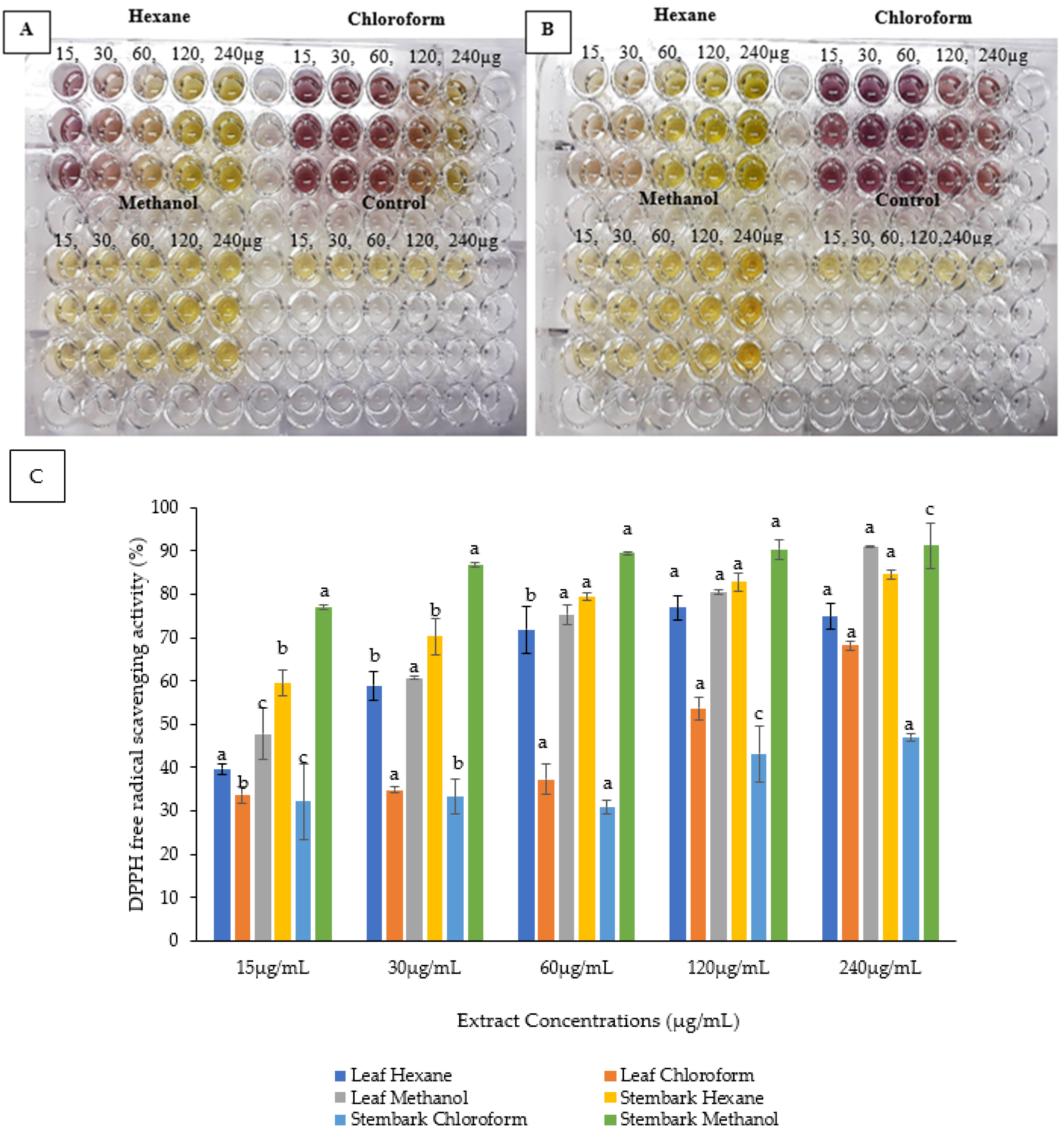
3.1.1. Assay of 2,2-diphenyl-1-picryl-hydrazyl-hydrate (DPPH)
3.1.2. FRAP (Ferric Reducing Antioxidant Power) Assay
3.2. Total Phenolic Content
3.3. Total Flavonoid Content
3.4. Cytotoxicity
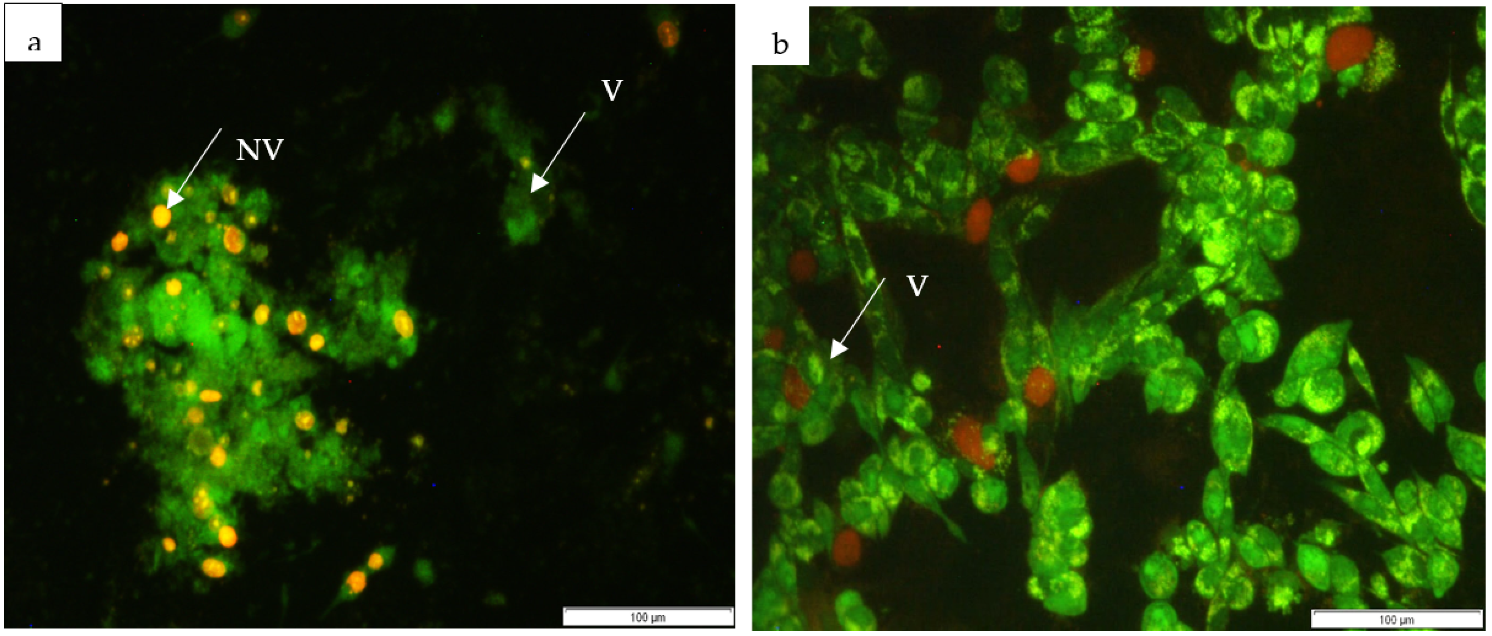
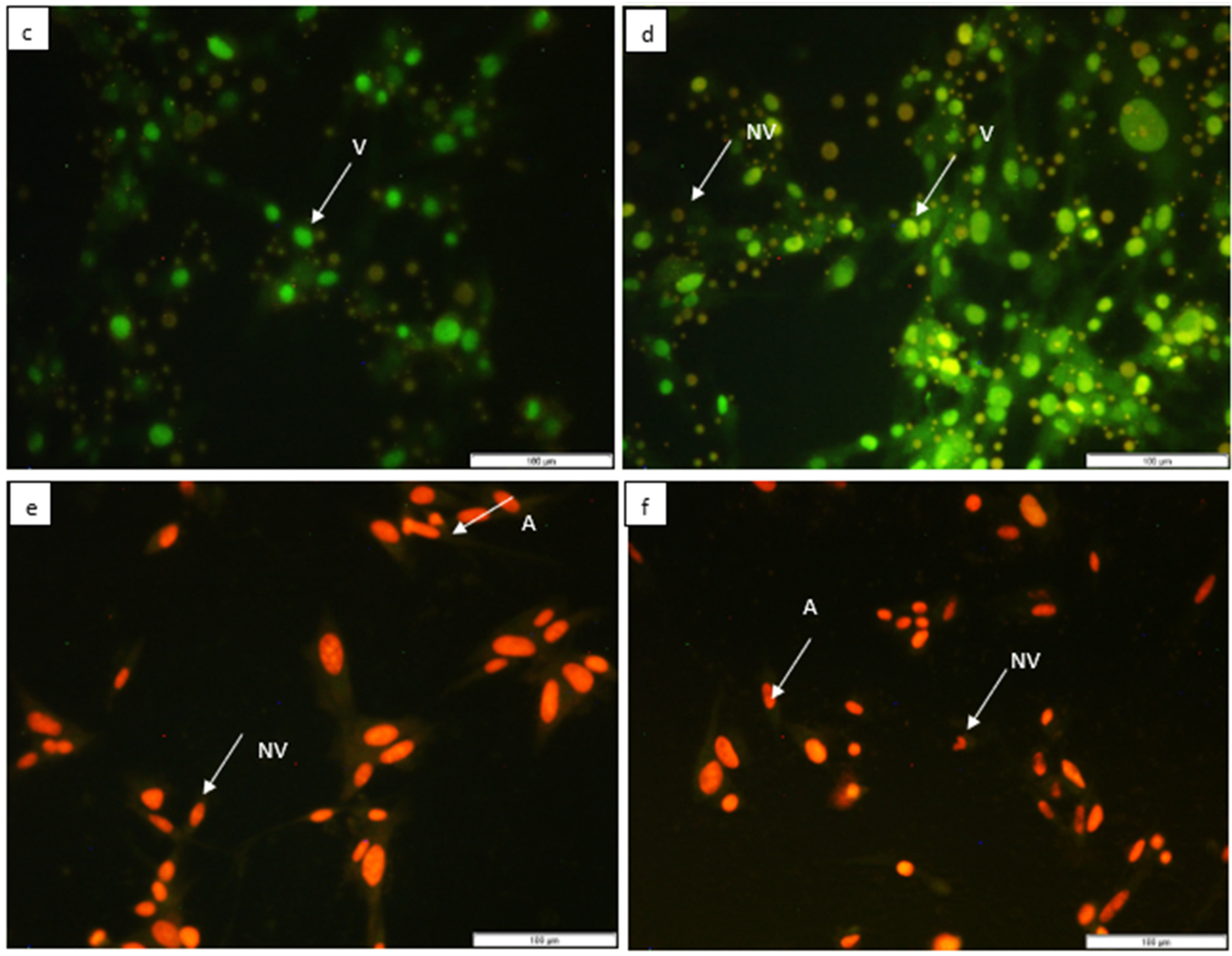
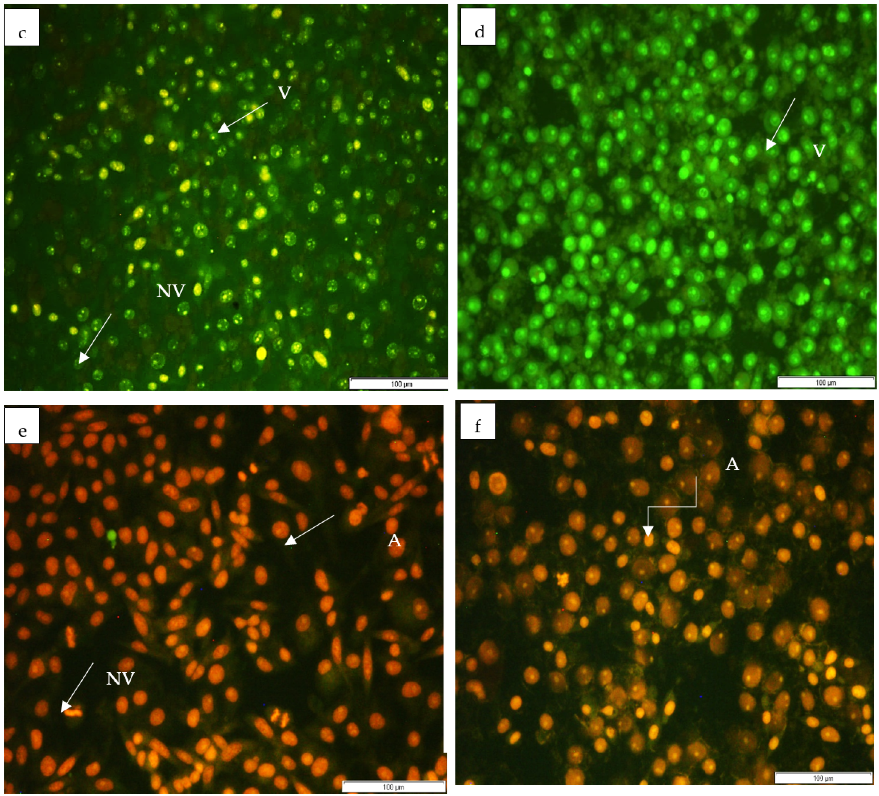
3.5. Apoptosis
4. Conclusions
Author Contributions
Funding
Institutional Review Board Statement
Informed Consent Statement
Data Availability Statement
Acknowledgments
Conflicts of Interest
References
- Soyemi, T. Neglect of common infectious disease outbreaks during the COVID-19 pandemic: An impending crisis in Nigeria? Afr. J. Clin. Exp. Microbiol. 2021, 22, 113–116. [Google Scholar] [CrossRef]
- Sharaf, M.H.; El-Sherbiny, G.M.; Moghannem, S.A.; Abdelmonem, M.; Elsehemy, I.A.; Metwaly, A.M.; Kalaba, M.H. New combination approaches to combat methicillin-resistant Staphylococcus aureus (MRSA). Sci. Rep. 2021, 11, 4240. [Google Scholar] [CrossRef] [PubMed]
- More, M.P.; Pardeshi, S.R.; Pardeshi, C.; Sonawane, G.A.; Shinde, M.N.; Deshmukh, P.K.; Naik, J.B.; Kulkarni, A.D. Recent advances in phytochemical based Nano-formulation for drug resistant Cancer. Med. Drug Discov. 2021, 10, 100082. [Google Scholar] [CrossRef]
- Hussain, Y.; Islam, L.; Khan, H.; Filosa, R.; Aschner, M.; Javed, S. Curcumin-cisplatin chemotherapy: A novel strategy in promoting chemotherapy efficacy and reducing side effects. Phytother. Res. 2021, 35, 6514–6529. [Google Scholar] [CrossRef]
- Sundaram, M.K.; Silas, S.; Hussain, A. Combinational therapy using chemotherapeutic agents and dietary bioactive compounds: A pragmatic approach to cancer treatment. In Treating Endocrine and Metabolic Disorders with Herbal Medicines; IGI Global: Hershey, PA, USA, 2021; pp. 188–214. [Google Scholar]
- Zheng, R.R.; Zhao, L.P.; Liu, L.S.; Deng, F.A.; Chen, X.Y.; Jiang, X.Y.; Wang, C.; Yu, X.Y.; Cheng, H.; Li, S.Y. Self-delivery nanomedicine to overcome drug resistance for synergistic chemotherapy. Biomater. Sci. 2021, 9, 3445–3452. [Google Scholar] [CrossRef]
- Tagde, P.; Tagde, P.; Tagde, S.; Bhattacharya, T.; Garg, V.; Akter, R.; Rahman, M.H.; Najda, A.; Albadrani, G.M.; Sayed, A.A.; et al. Natural bioactive molecules: An alternative approach to the treatment and control of glioblastoma multiforme. Biomed. Pharmacother. 2021, 141, 111928. [Google Scholar] [CrossRef]
- Kayser, O. Ethnobotany and medicinal plant biotechnology: From tradition to modern aspects of drug development. Planta Med. 2018, 84, 834–838. [Google Scholar] [CrossRef] [PubMed] [Green Version]
- Burman, S.; Bhattacharya, K.; Mukherjee, D.; Chandra, G. Antibacterial efficacy of leaf extracts of Combretum album Pers. against some pathogenic bacteria. BMC Complement. Altern. Med. 2018, 18, 213. [Google Scholar] [CrossRef] [Green Version]
- Thorat, B. Chemical extraction and biomedical importance of secondary organic metabolites from plants—A review. J. Biomed. Ther. Sci. 2018, 5, 9–42. [Google Scholar]
- Martini, N.; Eloff, J.N. The preliminary isolation of several antibacterial components from Combretum erythrophyllum (Combretaceae). J. Ethnopharmacol. 1998, 62, 255–263. [Google Scholar] [CrossRef]
- Singh, S.; Gupta, A.; Verma, S. In vitro antioxidant activities of two medicinal plants on the basis of dpph free radical scavenging activity. Ann. Rom. Soc. Cell Biol. 2021, 25, 4807–4811. [Google Scholar]
- Phuyal, N.; Jha, P.K.; Raturi, P.P.; Rajbhandary, S. Total phenolic, flavonoid contents, and antioxidant activities of fruit, seed, and bark extracts of Zanthoxylum armatum DC. Sci. World J. 2020, 2020, 8780704. [Google Scholar] [CrossRef] [PubMed] [Green Version]
- Bantho, S.; Naidoo, Y.; Dewir, Y.H.; Bantho, A.; Murthy, H.N. Chemical Composition of Combretum erythrophyllum Leaf and Stem Bark Extracts. Horticulturae 2022, 8, 755. [Google Scholar] [CrossRef]
- Van der Goot, A.J.; Pelgrom, P.J.; Berghout, J.A.; Geerts, M.E.; Jankowiak, L.; Hardt, N.A.; Keijer, J.; Schutyser, M.A.; Nikiforidis, C.V.; Boom, R.M. Concepts for further sustainable production of foods. J. Food Eng. 2016, 168, 42–51. [Google Scholar] [CrossRef]
- Bantho, S.; Naidoo, Y.; Dewir, Y.H. The secretory scales of Combretum erythrophyllum (Combretaceae): Micromorphology, ultrastructure and histochemistry. S. Afr. J. Bot. 2020, 131, 104–117. [Google Scholar] [CrossRef]
- Kitts, D.D.; Wijewickreme, A.N.; Hu, C. Antioxidant properties of a North American ginseng extract. Mol. Cell Biochem. 2000, 203, 1–10. [Google Scholar] [CrossRef]
- Annadurai, P.; Annadurai, V.; Yongkun, M.; Pugazhendhi, A.; Dhandayuthapani, K. Phytochemical composition, antioxidant and antimicrobial activities of Plecospermum spinosum Trecul. Process Biochem. 2021, 100, 107–116. [Google Scholar] [CrossRef]
- Akwu, N.A.; Naidoo, Y.; Singh, M.; Nundkumar, N.; Lin, J. Phytochemical screening, in vitro evaluation of the antimicrobial, antioxidant and cytotoxicity potentials of Grewia lasiocarpa E. Mey. ex Harv. S. Afr. J. Bot. 2019, 123, 180–192. [Google Scholar] [CrossRef]
- Neri, T.S.; Silva, K.W.L.; Maior, L.P.S.; Oliveira-Silva, S.K.; Azevedo, P.V.M.; Gomes, D.C.S.; Souza, M.A.; Pavão, J.M.S.J.; Costa, J.G.; Cunha, A.L.; et al. Phytochemical characterization, antioxidant potential and antibacterial activity of the Croton argyrophylloides Muell. Arg. (Euphorbiaceae). Braz. J. Biol. 2021, 83, e236649. [Google Scholar] [CrossRef] [PubMed]
- Hamza, R.Z.; Al-Bogami, N.M.; Mansour, A.A.; El-Megharbel, S.M. Possible antioxidant and antidiabetic effects of Combretum Molle extract in a diabetes mellitus experimental model in male rats. Nat. Prod. Commun. 2021, 16, 1934578X211043964. [Google Scholar]
- Sousa, H.G.; Uchôa, V.T.; Cavalcanti, S.M.G.; de Almeida, P.M.; Chaves, M.H.; Lima Neto, J.D.S.; Nunes, P.H.M.; da Costa Júnior, J.S.; Rai, M.; Do Carmo, I.S.; et al. Phytochemical screening, phenolic and flavonoid contents, antioxidant and cytogenotoxicity activities of Combretum leprosum Mart.(Combretaceae). J. Toxicol. Environ. Health Part A 2021, 84, 399–417. [Google Scholar] [CrossRef] [PubMed]
- Coulidiati, T.H.; Millogo-Kone, H.; Lamien-Meda, A.; Lamien, C.E.; Lompo, M.; Kiendrebeogo, M.; Bakasso, S.; Yougbaré-Ziébrou, M.; Millogo-Rasolodimby, J.; Nacoulma, O.G. Antioxidant and antibacterial activities of Combretum nioroense Aubrév. ex Keay (Combretaceae). Pak. J. Biol. Sci. 2009, 12, 264–269. [Google Scholar] [CrossRef] [PubMed] [Green Version]
- Masoko, P.; Eloff, J.N. Screening of twenty-four South African Combretum and six Terminalia species (Combretaceae) for antioxidant activities. Afr. J. Tradit. Complement. Altern. Med. 2007, 4, 231–239. [Google Scholar] [CrossRef] [Green Version]
- Burman, S.; Chandra, G. Phytochemical screening and in-vitro antibacterial and DPPH free radical scavenging activities of methanol extract of root of Combretum album Pers. Plant Sci. Today 2021, 8, 820–829. [Google Scholar] [CrossRef]
- Daniel, A.O.; Temikotan, T. Antioxidant and Radical Scavenging of Piliostigma reticulatum using FRAP and DPPH. J. Pharm. Res. Dev. 2021, 2, 1–6. [Google Scholar]
- Ghissing, U.; Goswami, A.; Mitra, A. Temporal accumulation of pigments during colour transformation from white to red in Combretum indicum (L.) DeFilipps (syn. Quisqualis indica L.) flowers. Nat. Prod. Res. 2021, 37, 529–533. [Google Scholar] [CrossRef]
- Ibrahim, A.; Onyike, E.; Nok, A.J.; Umar, I.A. Combined effect on antioxidant properties of Gymnema Sylvestre and Combretum Micranthum leaf extracts and the relationship to hypoglycemia. Eur. Sci. J. 2017, 13, 266–281. [Google Scholar]
- Ladekan-Yayi, E.C.; Toklo, P.M.; Dah-Nouvlessounon, D.; Assogba, M.F.; Wouamba, S.C.N.; Tchegnitegni, B.T.; Alowanou, G.G.; Baba-Moussa, L.; Hounzangbe-Adote, S.; Lenta, B.N.; et al. Anthelmintic and antimicrobial activities of tannin extracts of Mitragyna inermis (Willd.) Kuntze (Rubiaceae) and Combretum glutinosum Perr. ex DC (Combretaceae). Am. J. Appl. Chem. 2021, 9, 145–153. [Google Scholar] [CrossRef]
- Garcia-Macias, P.; Ordidge, M.; Vysini, E.; Waroonphan, S.; Battey, N.H.; Gordon, M.H. Changes in the flavonoid and phenolic acid contents and antioxidant activity of red leaf lettuce (Lollo rosso) due to cultivation under plastic films varying in ultraviolet transparency. J. Agric. Food Chem. 2007, 55, 10168–10172. [Google Scholar] [CrossRef]
- Do, Q.D.; Angkawijaya, A.E.; Tran-Nguyen, P.L.; Huynh, L.H.; Soetaredjo, F.E.; Ismadji, S.; Ju, Y.H. Effect of extraction solvent on total phenol content, total flavonoid content, and antioxidant activity of Limnophila aromatica. J. Food Drug Anal. 2014, 22, 296–302. [Google Scholar] [CrossRef] [Green Version]
- Gupta, S.; Jain, R.; Kachhwaha, S.; Kothari, S.L. Nutritional and medicinal applications of Moringa oleifera Lam. Review of current status and future possibilities. J. Herb. Med. 2018, 11, 1–11. [Google Scholar] [CrossRef]
- Williamson, A.; Day, A.J.; Plumb, G.W.; Couteau, D. Human metabolic pathways of dietary flavonoids and cinnamates. Biochem. Soc. Trans. 2000, 28, 16–22. [Google Scholar] [CrossRef]
- da Silva, A.W.; Ferreira, M.K.A.; Pereira, L.R.; Rebouças, E.L.; Coutinho, M.R.; Dos, J.; Lima, R.; Guedes, M.I.F.; Bandeira, P.N.; Magalhães, F.E.A.; et al. Combretum lanceolatum extract reverses anxiety and seizure behavior in adult zebrafish through GABAergic neurotransmission: An in vivo and in silico study. J. Biomol. Struct. Dyn. 2021, 40, 9801–9814. [Google Scholar] [CrossRef] [PubMed]
- Mashi, R.L.; Abu, M.S.; Jalo, A.; Lawal, J.Y. Antioxidant and angiotensin converting enzyme inhibitory activity of fractionated extract of Combretum Micranthum leaves. J. Biol. Sci. 2022, 22, 57–64. [Google Scholar] [CrossRef]
- Forid, M.; Rahman, M.; Aluwi, M.F.F.M.; Uddin, M.; Roy, T.G.; Mohanta, M.C.; Huq, A.K.M.; Amiruddin Zakaria, Z. Pharmacoinformatics and UPLC-QTOF/ESI-MS-Based Phytochemical Screening of Combretum indicum against Oxidative Stress and Alloxan-Induced Diabetes in Long–Evans Rats. Molecules 2021, 26, 4634. [Google Scholar] [CrossRef] [PubMed]
- Nascimento, G.G.F.; Locatelli, C.F.; Paulo, C.F.; Silva, G. The antimicrobial activity of plant extract and phytochemical on antibiotic resistant bacteria. Braz. J. Microbiol. 2000, 31, 247–256. [Google Scholar] [CrossRef]
- Karim, N.; Khan, I.; Khan, H.; Ayub, B.; Abdel-Halim, H.; Gavande, N. Anxiolytic potential of natural flavonoids. SM J. Steroids Horm. 2018, 1, 1001–1010. [Google Scholar]
- Fernández, J.; Silván, B.; Entrialgo-Cadierno, R.; Villar, C.J.; Capasso, R.; Uranga, J.A.; Lombó, F.; Abalo, R. Antiproliferative and palliative activity of flavonoids in colourectal cancer. Biomed. Pharmacother. 2021, 143, 112241. [Google Scholar] [CrossRef] [PubMed]
- Wu, Y.; Xu, L.; Liu, X.; Hasan, K.F.; Li, H.; Zhou, S.; Zhang, Q.; Zhou, Y. Effect of thermosonication treatment on blueberry juice quality: Total phenolics, flavonoids, anthocyanin, and antioxidant activity. LWT 2021, 150, 112021. [Google Scholar] [CrossRef]
- Wang, T.Y.; Li, Q.; Bi, K.S. Bioactive flavonoids in medicinal plants: Structure, activity and biological fate. Asian J. Pharm. Sci. 2018, 13, 12–23. [Google Scholar] [CrossRef]
- Yang, B.; Liu, H.; Yang, J.; Gupta, V.K.; Jiang, Y. New insights on bioactivities and biosynthesis of flavonoid glycosides. Trends Food Sci. Technol. 2018, 79, 116–124. [Google Scholar] [CrossRef]
- Rogers, C.B.; Verotta, L. Chemistry and Biological Properties of the African Combretaceae; UZ Publications: Harare, Zimbabwe, 1996. [Google Scholar]
- Martini, N.; Katerere, D.R.P.; Eloff, J.N. Biological activity of five antibacterial flavonoids isolated from Combretum erythrophyllum (Combretaceae). J. Ethnopharmacol. 2004, 93, 207–212. [Google Scholar] [CrossRef]
- Bhatnagar, S.; Sahoo, S.; Mohapatra, K.A.; Behera, D.R. Phytochemical analysis, antioxidant and cytotoxic activity of medicinal plant Combretum roxburghii (Family, Combretaceae). Int. J. Drug Dev. Res. 2012, 4, 193–202. [Google Scholar]
- Cutler, G.; Nettleton, J.; Ross, J.; Harnack, L.; Jacobs, R.D., Jr.; Scrafford, C.; Barraj, L.; Mink, P.; Robien, K. Dietary flavonoid intake and risk of cancer in postmenopausal women: The Iowa Women’s Health Study. Int. J. Cancer 2008, 123, 664–671. [Google Scholar] [CrossRef] [Green Version]
- Alabsi, A.M.; Lim, K.L.; Paterson, I.C.; Ali-Saeed, R.; Muharram, B.A. Cell cycle arrest and apoptosis induction via modulation of mitochondrial integrity by Bcl-2 family members and caspase dependence in Dracaena cinnabari-treated H400 human oral squamous cell carcinoma. BioMed Res. Int. 2016, 2016, 4904016. [Google Scholar] [CrossRef] [PubMed] [Green Version]
- Wende, M.; Sithole, S.; Fru, C.G.; Stevens, M.Y.; Mukanganyama, S. The effects of combining cancer drugs with compounds isolated from Combretum zeyheri Sond. and Combretum platypetalum Welw. ex MA Lawson (Combretaceae) on the Viability of Jurkat T Cells and HL-60 Cells. BioMed Res. Int. 2021, 2021, 6049728. [Google Scholar] [CrossRef] [PubMed]
- Nguyen, H.H.; Nguyen, T.P.; Trung, N.T.; Phan, C.T.D.; Mai, D.T.; Sichaem, J.; Nguyen, N.H.; Tran, C.L.; Duong, T.H. Two new cycloartanes from the leaves of Combretum quadrangulare growing in Vietnam and their biological activities. Arab. J. Chem. 2021, 14, 103189. [Google Scholar] [CrossRef]
- Gade, I.S.; Richard, T.S.; Chadeneau, C.; Seite, P.; Vannier, B.; Atchade, A.D.T.; Etet, P.F.S.; Talla, E.; Kamdje, A.H.N.; Muller, J.M. Anticancer activity of Combretum fragrans F. Hoffm on glioblastoma and prostate cancer cell lines. Asian Pac. J. Cancer Prev. 2021, 22, 1087. [Google Scholar] [CrossRef]
- Maphutha, J.; Twilley, D.; Lall, N. Inhibition of phosphatidylinositol 3-kinase (PI3K) enzyme and human skin carcinoma cell growth by Combretum apiculatum Sond. S. Afr. J. Bot. 2021, 140, 95–102. [Google Scholar] [CrossRef]
- Al-Nasser, M.M.; Al-Dosari, M.S.; Parvez, M.K.; Al-Anazi, M.R.; Alkahtane, A.A.; Alothaid, H.; Alahmari, A.; Alarifi, S.; Albasher, G.; Almeer, R.; et al. The potential effects of Indigofera coerulea extract on THP-1 human cell line. J. King Saud Univ.-Sci. 2021, 33, 101446. [Google Scholar] [CrossRef]
- Ali, S.; Khan, M.R.; Batool, R.; Shah, S.A.; Iqbal, J.; Abbasi, B.A.; Yaseen, T.; Zahra, N.; Aldhahrani, A.; Althobaiti, F. Characterization and phytochemical constituents of Periploca hydaspidis Falc crude extract and its anticancer activities. Saudi J. Biol. Sci. 2021, 28, 5500–5517. [Google Scholar] [CrossRef] [PubMed]
- Basu, A. The interplay between apoptosis and cellular senescence: Bcl-2 family proteins as targets for cancer therapy. Pharmacol. Ther. 2021, 230, 107943. [Google Scholar] [CrossRef]
- Thapa, S.; Rather, R.A.; Singh, S.K.; Bhagat, M. Insights into the Role of Defective Apoptosis in Cancer Pathogenesis and Therapy; Intech Open: London, UK, 2021; pp. 1–6. [Google Scholar]
- Safarzadeh, E.; Sandoghchian Shotorbani, S.; Baradaran, B. Herbal medicine as inducers of apoptosis in cancer treatment. Adv. Pharm. Bull. 2014, 4 (Suppl. 1), 421–427. [Google Scholar] [CrossRef] [PubMed]
- Anitha, J.; Selvakumar, R.; Hema, S.; Murugan, K.; Premkumar, T. Facile Green Synthesis of Nano-Sized ZnO Using Leaf Extract of Morinda tinctoria: MCF-7 cell cycle arrest, antiproliferation, and apoptosis studies. J. Ind. Eng. Chem. 2021, 105, 520–529. [Google Scholar] [CrossRef]
- Silva, F.C.O.; de Menezes, J.E.S.A.; Ferreira, M.K.A.; da Silva, A.W.; Holanda, C.L.A.; dos Reis Lima, J.; Campos, A.R.; Evaristo, F.F.V.; Teixeira, E.H.; Magalhães, F.E.A.; et al. Antinociceptive activity of 3β-6β-16β-trihydroxylup-20 (29)-ene triterpene isolated from Combretum leprosum leaves in adult zebrafish (Danio rerio). Biochem. Biophys. Res. Commun. 2020, 533, 362–367. [Google Scholar] [CrossRef] [PubMed]


| Extracts | IC50 (µg/mL) | |
|---|---|---|
| Leaves | Stembark | |
| Hexane | 31.25 a | 13.56 b |
| Chloroform | 120.71 a | 26.41 c |
| Methanol | 5.23 b | 4.29 c |
| Extracts | IC50 (µg/mL) | |
|---|---|---|
| Leaves | Stembark | |
| Hexane | >1000 b | 250.51 c |
| Chloroform | 593.44 c | >1000 b |
| Methanol | <1 a | <1 a |
| Extracts | Total Phenolic Content (mg/GAE/g) | |
|---|---|---|
| Leaves | Stembark | |
| Hexane | 163.13 ± 2.22 | 253.13 ± 1.73 |
| Chloroform | 178.54 ± 1.63 | 119.79 ± 1.90 |
| Methanol | 713.13 ± 0.5 | 1341.05 ± 4.4 |
| Extracts | Total Flavonoid Content (mg/QE/g) | |
|---|---|---|
| Leaves | Stembark | |
| Hexane | 16.99 ± 0.26 | 1.73 ± 0.46 |
| Chloroform | 22.50 ± 0.21 | 5.94 ± 1.96 |
| Methanol | 39.42 ± 1.86 | 37.05 ± 17.8 |
| Cell Lines | Extracts | Cytotoxicity (µg/mL) | |
|---|---|---|---|
| Leaves | Stembark | ||
| HEK293 | Hexane | 90.89 | 78.40 |
| Chloroform | 70.25 | 53.37 | |
| Methanol | 59.01 | 18.30 | |
| HeLa | Hexane | 97.15 | 73.04 |
| Chloroform | 72.34 | 50.92 | |
| Methanol | 54.53 | 39.23 | |
| MCF7 | Hexane | 202.88 | 227.38 |
| Chloroform | 1083.11 | 371.86 | |
| Methanol | 198.87 | 204.77 | |
Disclaimer/Publisher’s Note: The statements, opinions and data contained in all publications are solely those of the individual author(s) and contributor(s) and not of MDPI and/or the editor(s). MDPI and/or the editor(s) disclaim responsibility for any injury to people or property resulting from any ideas, methods, instructions or products referred to in the content. |
© 2023 by the authors. Licensee MDPI, Basel, Switzerland. This article is an open access article distributed under the terms and conditions of the Creative Commons Attribution (CC BY) license (https://creativecommons.org/licenses/by/4.0/).
Share and Cite
Bantho, S.; Naidoo, Y.; Dewir, Y.H.; Singh, M.; Bantho, A. Biological Activity of Combretum erythrophyllum: Antioxidant, Apoptotic, and Cytotoxic Activity of the Leaf and Stembark Extract. Horticulturae 2023, 9, 305. https://doi.org/10.3390/horticulturae9030305
Bantho S, Naidoo Y, Dewir YH, Singh M, Bantho A. Biological Activity of Combretum erythrophyllum: Antioxidant, Apoptotic, and Cytotoxic Activity of the Leaf and Stembark Extract. Horticulturae. 2023; 9(3):305. https://doi.org/10.3390/horticulturae9030305
Chicago/Turabian StyleBantho, Sahejna, Yougasphree Naidoo, Yaser Hassan Dewir, Moganavelli Singh, and Ayuvna Bantho. 2023. "Biological Activity of Combretum erythrophyllum: Antioxidant, Apoptotic, and Cytotoxic Activity of the Leaf and Stembark Extract" Horticulturae 9, no. 3: 305. https://doi.org/10.3390/horticulturae9030305