Article

The Endophytic Entomopathogenic Fungus Beauveria bassiana Alleviates Adverse Effects of Salt Stress in Potato Plants

by
Oksana G. Tomilova
1,2,*,
Natalia A. Kryukova
1,
Marina V. Efimova
3,
Liliya V. Kolomeichuk
3,
Irina S. Kovtun
3 and
Victor V. Glupov
1
1
Institute of Systematics and Ecology of Animals, SB RAS, Frunze Str. 11, 630091 Novosibirsk, Russia
2
All-Russian Institute of Plant Protection, Podbel’skogo 3, 196608 St. Petersburg, Russia
3
Department of Plant Physiology, Biotechnology and Bioinformatics, National Research Tomsk State University, Lenin Ave., 36, 634050 Tomsk, Russia
*
Author to whom correspondence should be addressed.
Horticulturae 2023, 9(10), 1140; https://doi.org/10.3390/horticulturae9101140
Submission received: 12 September 2023 / Revised: 9 October 2023 / Accepted: 14 October 2023 / Published: 16 October 2023
(This article belongs to the Topic Tolerance to Drought and Salt Stress in Plants)

Abstract

:
The considerable decrease in crop productivity associated with the expansion of saline soils is an acute problem in agriculture. Endophytic fungi positively affect plant fitness under salinity conditions. The effects of potato inoculation with the conidia of the Beauveria bassiana strain Sar-31 on growth (the weight of fresh and dry biomass, shoot and root length, numbers of stolons and leaves, and the leaf surface) and physiological indices (the concentration of pigments, free proline and malondialdehyde, and antioxidant enzymes’ activity) were evaluated under moderate chloride salinity (100 mM). The results indicated that the plant’s association with the fungus mitigated the negative impact of salinity probably because of the activation of antioxidant enzymes and accumulation of free proline in potato tissues. Moreover, under the influence of B. bassiana Sar-31, the number of stolons significantly increased, which is one of the main characteristics of potato as an agricultural crop. Thus, Sar-31 may be a promising candidate for further investigation of its ability to stimulate growth and increase the stress tolerance of potato plants.

1. Introduction

Salinity is the dominant stress factor limiting the growth and productivity of agricultural crops, which is especially true under the conditions of a dry climate. Over 800 million hectares of soil are currently affected by salinity, and this problem is continuing to worsen [1,2]. The most widespread type of salinization is that caused by sodium chloride; its effect on plants is the most harmful [3]. Soils are regarded as saline if their electrical conductivity is above 4 dS/m, i.e., equivalent to a ≥40 mM solution of NaCl [4].
Intense salinization affects all major physiological processes in plants. The adverse action of the increased salt content is based on a disturbance of osmotic and ionic homeostasis in plants’ tissues as well as the toxicity of inorganic ions to cellular metabolism [5,6]. These processes are accompanied by the formation of excessive amounts of reactive oxygen species (ROS) causing oxidative stress [7,8]. In cells and tissues, an excess of ROS leads to enzyme inactivation, protein and nucleic acid damage, and the dysfunction of membranes. Additionally, the development of osmotic and oxidative stress harms the photosynthetic apparatus, thus leading to impaired crop production [9].
Potato is one of the main food crops cultivated all over the world [10]. Wild species of potato plants are relatively resistant to salinization [11], while modern potato cultivars, which are a product of long-term selection, are much more susceptible to the action of salinity [12,13]. The production and productivity of potato are affected by salt stress [14]. Therefore, searching for new, effective ways to overcome the effects of salt stress on potato plants is a relevant and topical task.
Researchers are trying to solve the problem of crop salt sensitivity by different methods, including modern methods of selection [3,15,16], the treatment of plants with exogenous phytohormones [17,18,19,20], and the colonization of plants by endophytic microorganisms contributing to plant growth under salinity conditions [21,22,23]. The latter approach has high potential because it includes a wide range of promising species of bacteria and fungi for study.
Endophytic fungi associated with plants have been shown to diminish the abrogative action of salt stress through the activation of systemic resistance, increased levels of useful metabolites, the activation of the antioxidant system, and the modulation of plant growth phytohormones. Plants’ colonization by endophytic fungi is accompanied by the active absorption of nutrients and maintenance of ionic (Na+ and K+) homeostasis [24]. Among endophytic fungi, new species from various taxonomic and trophic groups are continually being discovered and reisolated [25,26,27,28]. In recent years, entomopathogenic fungi, primarily from the genera Metarhizium and Beauveria, have been explored as potential endophytes (reviews: [29,30,31]). It has been experimentally proven that plant endophytic colonization by entomopathogenic fungi negatively affects the development and number of phytophages and leads to the stimulation of plant growth, a decrease in their damage from phytopathogens, a change in immune status, and an increase in plant productivity [32,33,34,35,36]. For Solanaceae plants, similar effects have been registered in conjunction with the application of entomopathogenic endophytes [37,38,39,40,41]. Nonetheless, only a limited number of articles show an increase in resistance to osmotic stress, including salinity and drought stress, in plants colonized by the entomopathogenic endophytes Metarhizium and Beauveria [42,43,44,45,46,47]. For instance, in the work of Khan et al. [42], endophytic colonization by Metarhizium anisopliae improved the growth parameters of soybean under chloride salinity. The ability of the M. pinghaense isolate AAUBC-M26 to reduce salt stress (up to 50 mM NaCl) in tomato plants under nursery and pot culture conditions was evaluated in a study by Chaudhary et al. [43]. An increase in plants’ drought tolerance as a result of endophytic colonization by Beauveria bassiana is demonstrated in the works of Dara, Kuzhuppillymyal-Prabhakarankutty, and Gana [44,45,46]. The mitigation of salt stress by B. bassiana has been shown by Akter et al. when treating rice seeds [47]. Considering the above findings, it can be hypothesized that B. bassiana will also be conducive to the development of salt tolerance in potato plants.
Previously, we have investigated the effects of different concentrations of NaCl (in the range from 50 to 150 mM) on the growth and physiological parameters of mid-ripening potato varieties [48]. It was found in our study that plants of the Lugovskoy variety actively respond to moderate chloride salinity (100 mM) with a significant decrease in plant weight and in the number of stolons, the accumulation of proline, and an increase in the level of malondialdehyde (MDA). The impact of endophytic colonization by B. bassiana and Metarhizium robertsii on growth parameters has also been estimated in this variety [49]. It was found in that report that both fungi successfully colonize plants under hydroponic conditions. The initial stage of colonization by fungi was accompanied by placing a moderate level of stress on the plants, whereas inoculation with B. bassiana conidia was tolerated by the plants much better. Preliminary experiments allowed us to choose an optimal laboratory model for the estimation of the influence of an entomopathogenic endophyte on potato’s susceptibility to salinity by an express test (the duration of the experiments was 7 days). The aim of the present study was to investigate the effect of inoculation with the conidia of B. bassiana on the growth and physiological parameters of potato plants grown in the presence of moderately intense chloride salinity under hydroponic conditions.

2. Materials and Methods

2.1. A Fungal Isolate and Its Cultivation

In the experiments, a strain of the entomopathogenic fungus B. bassiana (Sar-31) was used from the collection of microorganisms at the Institute of Systematics and Ecology of Animals (the Siberian Branch of the Russian Academy of Sciences). The species was identified using a translation elongation factor (EF1α) sequence [50] (accession number MZ564259). When compared to GenBank sequences, 100% identity was found for B. bassiana entries. Other species of this genus showed lower identity indices, thus confirming the species’ identification as B. bassiana. The culture was incubated at 26 °C for 10 days in the dark on Sabouraud dextrose agar supplemented with 0.25% yeast extract (SDAY). An aqueous Tween 80 solution (0.03%) was used to prepare conidial mass, and the concentration of conidia was scored by means of a Neubauer hemocytometer (Fristaden Lab, Chicago, IL, USA). The liquid medium used for the plant growth was supplemented with the conidial suspension at the final concentration of 106 conidia/mL.

2.2. Plant Cultivation and Experimental Design

We used hydroponic culture of potato plants (Solanum tuberosum L.) cv. Lugovskoy (identifier 8,301,891) propagated in vitro. Potato plantlets regenerated from aseptic stem-node potato meristems were cultured on half-strength Murashige–Skoog agar medium (0.5 MS) for 30 days. For 2 weeks, the seedlings were adapted to the liquid 0.5 MS medium in a controlled climate chamber using L36 W/77 Fluora luminescent lamps (Osram, Germany; 100 μM photons m−2 s−1) with day/night temperatures of 23 ± 0.5/20 ± 0.5 °C and a 16 h/8 h day/night photoperiod. The seedlings were cultivated in the liquid medium for 6 weeks before they were transferred either to the pure medium serving as the control or to the medium supplemented with the fungal conidia as described above. After 2 days, salt (100 mM NaCl) was added to the medium to induce salinity stress. Thus, the plants were grown in the presence of fungal conidia for 7 days and in the presence of salinity for 5 days. The following growth parameters of potato plants were measured in each treatment group: shoot and root length, shoot and root fresh and dry weight, and numbers of leaves and stolons. A part of the plant material was used for analysis of physiological and biochemical parameters. The above period is sufficient to obtain an adequate early plant response. A longer experiment may lead to contamination of the nutrient medium with foreign microorganisms.

2.3. Plant Colonization Assay

Plating of plant tissues in media was used to assess the frequency of plant colonization by entomopathogenic fungi. Plant organs (roots, stems, and leaves) presterilized with hypochlorite and ethanol [51] were imprinted [52] and planted on modified Sabouraud agar (10 g/L peptone, 40 g/L anhydrous D-glucose, 20 g/L agar, 1 g/L yeast extract, 0.35 g/L cetyl trimethyl ammonium bromide, 0.05 g/L cycloheximide, 0.05 g/L tetracycline, and 0.6 g/L streptomycin) in 90 mm Petri dishes (one plant per Petri dish) and incubated for 10 days at 24 °C. The imprints from each plant were carried out in separate dishes. Samples in whose prints fungal growth was found were excluded from the analysis. The level of colonization was determined as a percentage of Beauveria-positive organs. Twenty plants from each treatment group were analyzed.

2.4. Plant Growth Parameters and Leaf Contents of Photosynthetic Pigments

The main potato plant growth parameters—the lengths of roots and shoots and the total leaf surface area—were determined and expressed in centimeters. The gravimetric method was utilized for measuring fresh and dry biomass of the plant material. To estimate the weight of fresh plant biomass (FW), plants were dried in advance with filter paper, and FW was determined to within 1 mg. Dry weight (DW) was determined after the material was fixed at 90 °C and dried at 70 °C until constant weight was reached. To calculate the water content (WC), the following formula was used:
WC = [(FWDW)/FW] × 100
The photosynthetic pigment content was evaluated spectrophotometrically in fresh leaves from middle nodes of the plants according to the method presented in our previous work [49]. Optical density (D) of a leaf ethanol extract was measured at wavelengths of 470, 664, 648, and 720 nm on a spectrophotometer (Genesys 10S UV-Vis, Thermo Scientific, Waltham, MA, USA); 96% ethanol served as a control. Pigment concentration (C) was determined [53] via the following equations:
Cchla = 13.36 × D664 − 5.19 × D648
Cchlb = 27.43 × D648 − 8.12 × D664
Cchla+b = 5.24 × D664 + 22.24 × D648
Ccarot = (1000 × D470 × 2.13 × Cchla − 97.64 × Cchlb)/209
To calculate the amount of pigment per unit of sample weight (A), the formula
A = C × V/(1000 × n)
was employed, where C is the concentration, V is the volume of the extract, and n is the sample’s weight (g).

2.5. Lipid Peroxidation

The extent of lipid peroxidation was assessed using the method of Buege and Aust [54]. Formation of a colored product (thiobarbituric acid with thiobarbituric acid reactive substances (TBARS)) was evaluated at two wavelengths, 532 and 600 nm, on a spectrophotometer (Genesys 10S UV-Vis, Thermo Scientific, USA). TBARS were quantified in 100 mg of the plant material (roots, stems, and leaves).
The TBARS content was calculated via the following formula:
C = (E532E600) × V1 × 2/(156 × V2 × m)
where C is TBARS concentration in μg/g FW; E is optical density of the solution at wavelengths of 532 and 600 nm; V1 is the volume of thiobarbituric acid, mL; V2 is supernatant volume, mL; and m is the sample’s weight, g.

2.6. Activity of Antioxidant Enzymes in the Leaves

Activities of total superoxide dismutase (SOD) and peroxidase (POX) were estimated in crude extracts of leaf tissues. The activities of the enzymes were determined according to our previous work [49]. The riboflavin/nitro blue tetrazolium (B2/NBT) method was used to determine the total SOD activity. This technique was adapted from Beauchamp and Fridovich [55]. Absorbance was measured spectrophotometrically (Genesys 10S UV-Vis, Thermo Scientific, USA) at 560 nm. During the reaction, the total SOD activity was calculated in units of transmittance (∆A) of the incubation mixture per minute per milligram of protein.
The activity of peroxidase was measured with H2O2 and guaiacol (Fluka, Buchs, Switzerland) according to Shevyakova et al. [56]. Absorbance was measured spectrophotometrically (Genesys 10S UV-Vis, Thermo Scientific, USA) at 470 nm. POX activity was calculated in mM guaiacol/([mg protein]·min). The protein content of the samples was determined using the method of Esen [57]. Bovine serum albumin was used to create a calibration curve.

2.7. The Free-Proline Content of Plants

In roots, stems, and leaves, free-proline concentration was estimated according to Bates et al. [58]. The proline content was determined according to a calibration curve constructed using a set of standard proline solutions in 3% sulfosalicylic acid. Optical density was measured using a spectrophotometer (Genesys 10S UV-Vis Thermo Electron) at 520 nm. The following formula was used to determine the concentration of proline in the samples:
C = E × k × (V/m × 1000)
where C is the concentration of proline, μM/g FW; E is optical density; k is a coefficient calculated from the calibration curve; V is the final volume of extract, mL; and m is the weight of the sample, g.

2.8. Statistical Analyses

The assessment of the water content and fresh and dry weights as well as the assays of enzymatic activity were performed on 20 plants in each treatment group. Organ length and morphometric parameters were measured in 50 plants in each treatment group. Levels of pigments, proline, and TBARS were quantified using 10 plants per treatment group. The experiments were conducted twice. Data were analyzed in STATISTICA 8.0 software (StatSoft Inc., Tulsa, OK, USA). Normally distributed data were subjected to two-way analysis of variance (ANOVA) followed by a post hoc test (Fisher’s least significant difference (LSD) test).
Data are presented as the mean and standard error (SE). The chi-square test was performed to evaluate differences in the fungal colonization among the plants. Data are presented as percentages.

3. Results

3.1. Plant Colonization by the Fungus

It was found that background chloride salinity under hydroponic conditions does not affect the extent of colonization of potato plants by B. bassiana. After 7 days of plant growth, both in the medium containing conidia and the medium containing conidia + salt, there was colonization by fungi (Figure 1). After each treatment, 90–95% of the roots and stems and 5–20% of the leaves were colonized by the entomopathogenic fungus, and the difference between these treatment groups was not significant (χ2 ≥ 0.34, df = 1, p ≤ 0.58; Figure 1b). In the control plants, the growth of the fungus was not detectable (Figure 1a). It is important to note that on the seventh day of the experiment, the proportion of germinated conidia on the MS medium was only 4–7%.

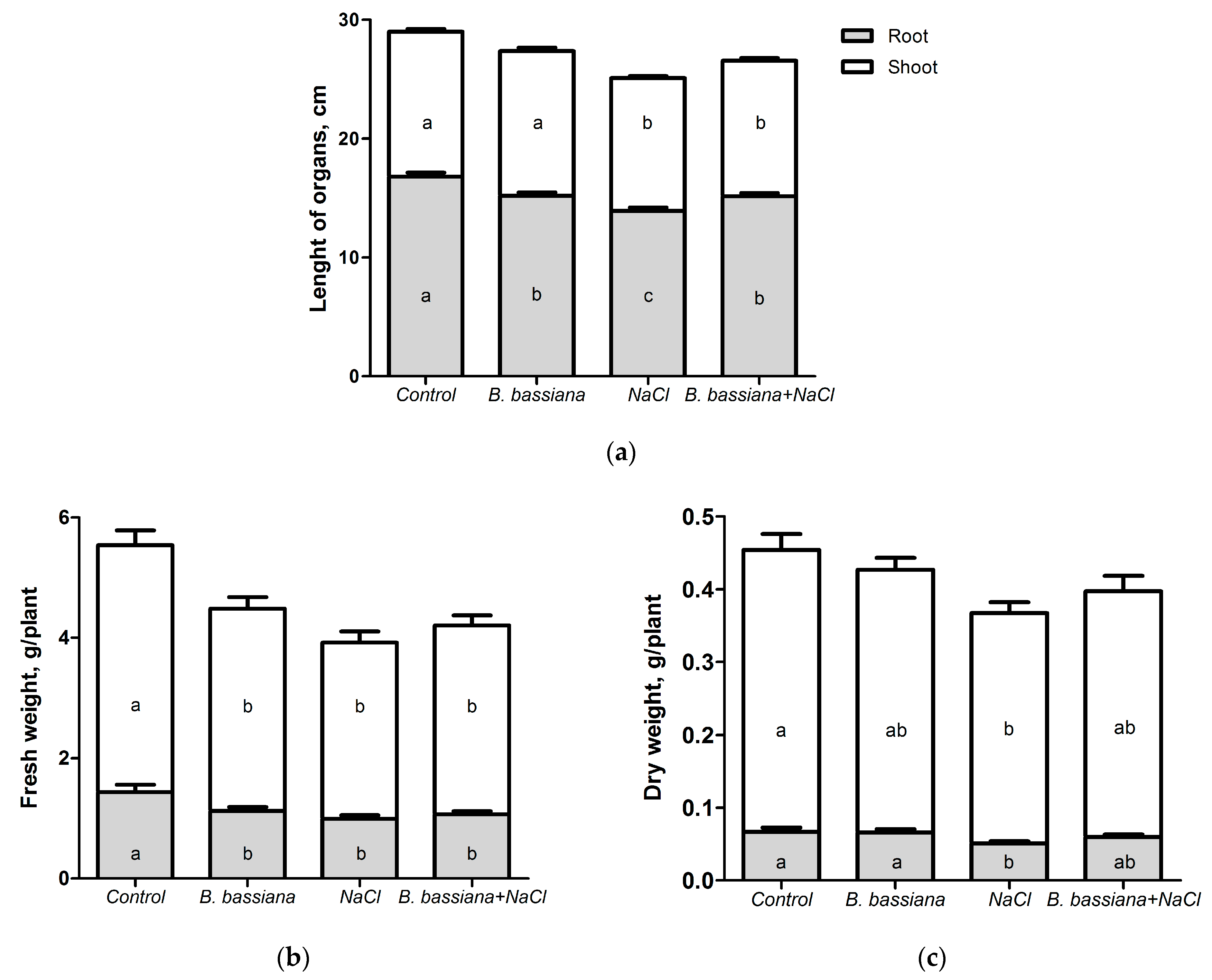
3.2. Effects of B. bassiana and Salt on Plant Growth

The 1-week cultivation of the potato plants both with the fungus conidia and salt affected several parameters of potato plant growth (Figure 2 and Figure 3). The strongest inhibition of growth characteristics was noted in the group treated with NaCl alone. Plants colonized by the fungus were more tolerant to the salt stress. This finding was clearly evidenced by such parameters as the length of the roots and the dry weight of plants (Figure 3a,c). Under salinity stress, the length of the roots was significantly smaller in comparison to the combined treatment with B. bassiana and salt (p = 0.003, Figure 3a). Inoculation with the conidia of B. bassiana did not significantly influence the dry weight of the aboveground and underground plant organs in comparison to that of the control (p = 0.08), whereas the application of salt alone caused it to decrease in comparison with the control and fungus treatment groups, respectively (p ≤ 0.017; Figure 3c). At the same time, in all treatment groups, the fresh weight of the aboveground and underground plants’ organs was significantly lower in comparison with that of the control (p ≤ 0.019; Figure 3b). A statistically significant decrease in water content was noted in the shoots and roots under the influence of salt (F1,122 ≥ 9.30, p ≤ 0.003) as well as in the roots under the influence of B. bassiana (F1,122 = 20.00, p < 0.001, Figure 4), which was most likely the cause of the equalization of the dry weight between the control and inoculated plants.
Most of the quantitative parameters were also significantly different from the control, with the strongest inhibition being caused by exposure to salt alone (F1,396 ≥ 9.03, p ≤ 0.003; Table 1). The interaction of factors led to a significant reduction in the leaf surface area (F1,396 = 8.56, p = 0.004). Meanwhile, a notable rise in the number of stolons was noted due to the effect of the B. bassiana conidia (F1,396 = 4.21, p = 0.04). Significant differences in the number of stolons from all other treatment groups were seen in the plants inoculated with B. bassiana alone (p ≤ 0.02). In the meantime, the numbers of stolons and leaves remained at the control level when the fungus was applied simultaneously with salinity (p ≥ 0.08, Table 1).

3.3. Effects of B. bassiana and Salt on the Photosynthetic Pigment Content

Short-term exposure to the B. bassiana conidia and/or salt did not induce statistically significant changes in the levels of the photosynthetic pigments in potato plants compared to those of the control plants (Table 2). Nonetheless, even minor changes in the pigment profiles led to a significant decline in the chlorophyll a/b ratio under the influence of B. bassiana (F1,76 = 7.54, p = 0.008).

3.4. The Impact of B. bassiana and Salt on Lipid Peroxidation

It was found that in plants exposed to both B. bassiana and NaCl, TBARS are upregulated (Figure 5). Under the influence of B. bassiana, the level of TBARS in the potato roots rose by 20–40%, although the differences were not significant (p ≥ 0.127 as compared to the control). Salinity was the main statistically significant factor that affected the level of MDA in the stems and leaves (F1,75 ≥ 4.361, p ≤ 0.040). In the potato stems, the TBARS level increased 1.6-fold under salinization (p = 0.007 as compared to the control). In the leaves, B. bassiana caused a 1.40-fold significant reduction in the TBARS content (p = 0.007 as compared to NaCl).

3.5. Effects of B. bassiana and Salt on the Activity of Antioxidant Enzymes

In the leaves of S. tuberosum, either endophytic colonization by B. bassiana or salinization enhanced the activity of the antioxidant enzymes (Figure 6). For instance, the total SOD level after inoculation with the B. bassiana conidia was significantly higher than that in the control (1.54-fold higher; p = 0.036; Figure 6a), although the influence of B. bassiana as a factor was statistically insignificant (F1,75 = 1.51, p = 0.224). The peroxidase activity in the leaves was the highest under the combined influence of the fungus and salt and was significantly increased as compared to that of the control (1.59 times) and the “B. bassiana alone” group (1.55-fold; p ≤ 0.031; Figure 6b). Nevertheless, only the NaCl factor had a significant effect on the POX activity: F1,75 = 5.18, p = 0.026.

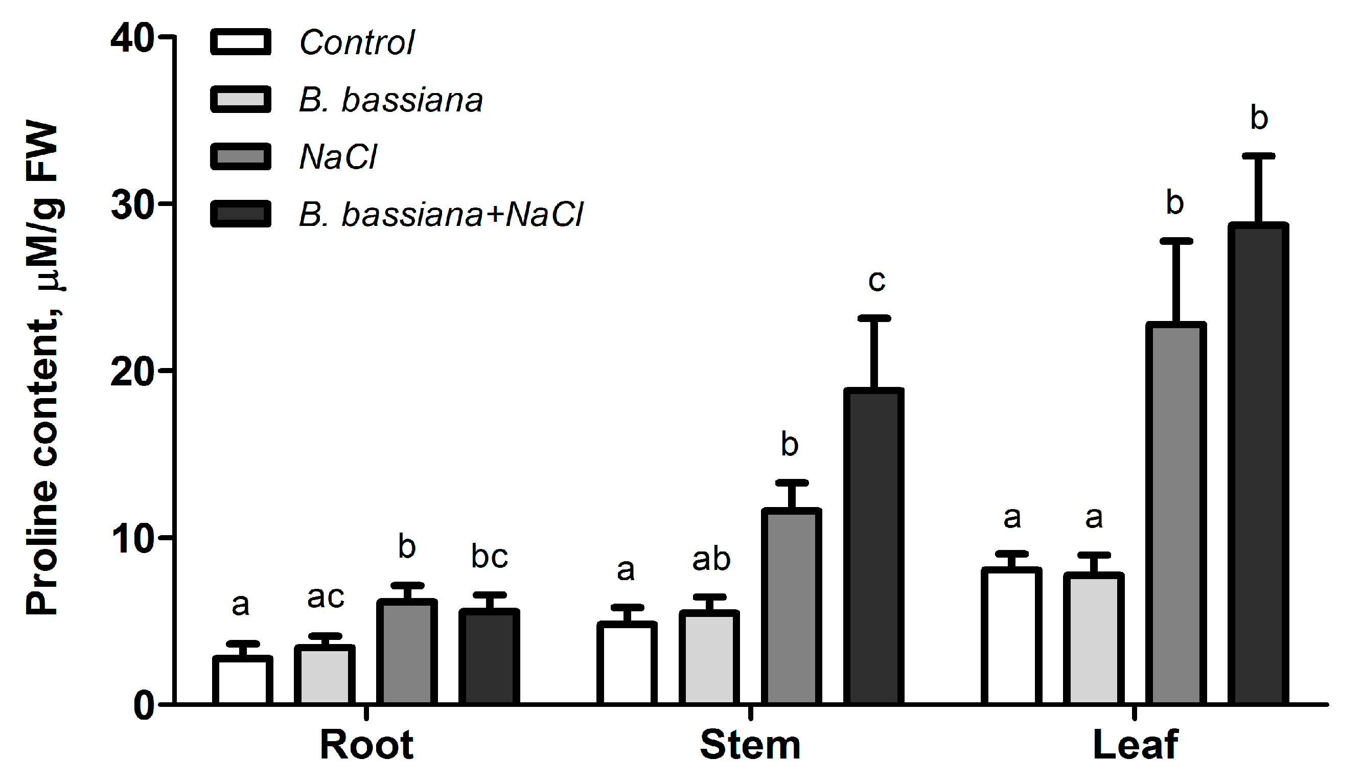
3.6. Effects of B. bassiana and Salt on Proline Accumulation in Different Parts of Plants

It was found that in the presence of B. bassiana, the level of free proline was about the same as in the control (p ≥ 0.635), but there was a significant upregulation of proline in all the organs of the potato plants under salinity stress (2.2–2.8 times; p ≤ 0.040) and especially with the joint application of B. bassiana and salt (2.0–3.9 times; p ≤ 0.036; Figure 7). A statistically significant impact of salinity on the proline level was detectable in all the organs of the potato plants (NaCl: F1,73 ≥ 9.41, p ≤ 0.003).

4. Discussion

On the basis of our results, we can say that salinity did not affect the colonization of potato plants by B. bassiana. Over a 7-day period, either with the soil inoculation with pure fungi conidia or in combination with salt, the colonization of roots and stems reached high values (90–95%). It is known that fungi, just like plants, experience osmotic stress under elevated levels of salinity. Nonetheless, B. bassiana shows a high degree of tolerance to salinity. For example, Cazorla Perfetti et al. [59] have demonstrated that the in vitro germination of B. bassiana conidia is inhibited only at a salinity level exceeding 7% NaCl [59]. It should be noted that in our experiments, we chose a salt concentration of 100 mM, corresponding to 0.6%, which is well below the threshold values. The strain tested in our experiments was able to actively develop in vitro at a salt concentration of 50–150 mM. The salt did not reduce the fungal colonies’ radial growth in the medium. On the other hand, the salt induced a significant increase in growth as compared to the growth on the salt-free medium (Figure A1 (Appendix A)).
There are many papers testifying to the benefit of avirulent strains of endophytic fungi from the genera Alternaria, Aspergillus, Trichoderma, Candida, Stemphylium, Penicillium and Piriformospora for the growth of various crops (soybean, corn, rice, and basil) under salt stress [60,61,62,63,64], and the same is true for tomato, which belongs to the same botanical family as potato [65,66,67,68,69]. In addition, a number of authors have demonstrated the positive effect of inoculation with entomopathogenic fungi on plant growth parameters under high-salinity conditions [42,43,47,70]. For instance, articles by Khan et al. have shown an improvement of growth parameters under salinity stress in soybean plants inoculated with M. anisopliae LHL07 [42] and cucumber plants inoculated with Paecilomyces formosus LHL10 [70] compared with those of control plants. Chaudhary et al. [43] and Akter et al. [47] have registered significant attenuation of the destructive effect of salinity stress on plants due to their inoculation with the conidia of entomopathogenic fungi (M. pinghaense AAUBC-M26 and B. bassiana BeauA1). Nevertheless, the growth stimulation by endophytic fungi under salt stress was detectable only after prolonged exposure to these factors. As a rule, researchers apply salt 1–3 weeks after a plant’s inoculation with the fungus, and the duration of the salinization is at least 2 weeks [42,47,62,63,64,68,69]. In our relatively short experiment, we noticed an inhibition of the growth of potato plants under chloride salinization and as a result of colonization by B. bassiana. The strongest inhibition of potato plant growth was caused by NaCl salinization alone. The low salt tolerance of the Lugovskoy potato variety has been reported earlier, in an experiment with different concentrations of NaCl (50 to 150 mM) [48,71]. The inhibitory effect of exposure to B. bassiana was less pronounced and was reflected only in a reduction in the leaf surface area. For some parameters, it also significantly mitigated the negative impact of salinization (the plant dry mass and root length). Phenotypically, plants inoculated with the fungus and grown either with an elevated level of salinity or without it did not differ from each other. In contrast, plants treated with salt alone were significantly shorter than the control and the fungus-inoculated plants (Figure 2). It should be noted that such obvious differences in plant development were obtained in a very short period; this finding once again highlights the active involvement of B. bassiana in the formation of salt tolerance in potato. In a previous work, we showed that a short-term delay in potato plant development is mediated by the activation of the defense system at the initial stage of colonization by entomopathogenic fungi (B. bassiana or M. robertsii) [49]. Partially confirming our results, the investigation by Proietti et al. [72] indicates an effect on tomato plants at early stages of colonization by B. bassiana (5–7 days). These researchers discovered that the activation of molecular pathways associated with protein and/or amino acid metabolism and the biosynthesis of energy compounds occur later (12–19 days after colonization) and lead to the stimulation of plant growth and development [72]. In our experiment, the quantitative parameters (the numbers of stolons and leaves) of the plants colonized by B. bassiana under salinity stress were similar to the control values (see Table 1). The application of the fungus alone significantly raised the number of stolons. This parameter is very relevant because it directly affects the tuber formation in new crops. Our results are consistent with those of numerous reports on the growth-stimulating effects of inoculation with endophytic entomopathogenic fungi, as demonstrated on a wide range of plants from different botanical families and has been extensively documented in recent reviews [29,30,31,52,73,74].
Photosynthetic activity and changes in concentrations of photosynthetic pigments in plants are often used to measure tolerance to abiotic stress. Numerous studies suggest that elevated levels of salinity exert an inhibitory action on photosynthesis [75,76,77,78,79,80,81]. Salt stress may affect chloroplast structures, thereby possibly lowering the chlorophyll content, resulting in a reduced photosynthetic rate [75,78,82,83]. In plants affected by salt stress, a decrease in the chlorophyll level is considered a typical symptom of the oxidative stress associated with the inhibition of chlorophyll synthesis and degradation by chlorophyllase [84,85]. Furthermore, salinity affects chlorophyll a more negatively than chlorophyll b [86,87]. Under our experimental conditions, relevant changes in the concentrations of photosynthetic pigments were not detectable. Probably, this finding is due to the briefness of the exposure to salt as well as the activation of antioxidant systems (enzymatic and nonenzymatic). Only a slight rise in the chlorophyll b content in the presence of the B. bassiana conidia under salinity stress was noted. A similar effect has been detected by Abdelaziz et al. [68]. They state that tomato colonization by Piriformospora indica raises the level of chlorophyll b under salt stress. It should be pointed out that under salt stress, the prolonged interaction of plants with endophytes causes an alignment of physiological and biochemical parameters, including the biosynthesis of photosynthetic pigments, thus increasing plant biomass [47,61,63,64].
In halophytic plants adapted to growth and survival in saline habitats, a set of adaptations related to selective ion uptake, Na+ compartmentalization, ion homeostasis, the production of protective metabolites, the formation of salt glands and salt bladders, and improvements in antioxidant properties have emerged during their evolution [88,89,90]. The vast majority of crops, including potato, are glycophytes, i.e., salt-sensitive plants. Glycophytes have a relatively limited ability to adapt to salinity, mainly by the activation of stress-induced protective systems [3]. The de novo synthesis of organic osmolytes and their accumulation in plant tissues, the modulation of levels of endogenous phytohormones, and the upregulation of antioxidant enzymes (e.g., superoxide dismutase, peroxidase, and catalase) are consequences of this activation [47,90,91]. A widespread marker of oxidative stress during exposure to a biotic or abiotic stressor is MDA: the end product of the lipid oxidation of cell membranes by free ROS. In the present investigation, we quantified MDA in roots, leaves, and stems. A significant increase in the MDA level was observed in potato stems in the presence of NaCl salinity. At the same time, in the leaves of the plants colonized by the fungus, an enhancement in the SOD activity was accompanied by a statistically significant decline in the MDA concentration in comparison to that of the control; in the leaves of the plants exposed to B. bassiana and salt, the POX activity was also accompanied by a statistically significant decrease in the MDA level. The high activity of the antioxidant system and consequent downregulation of MDA indicate the absence of a pathological imbalance within the system of stress-induced protective mechanisms. An increase in the activity of antioxidant enzymes has been documented in most papers on the adaptogenic properties of endophytic fungi in relation to plants grown under salinity stress [60,62,63,64,67,68] and after the application of B. bassiana [46,47,92]. In our experiments, there were no significant changes in the MDA concentration of the roots, regardless of the presence or absence of salt and/or the endophyte. There were no significant changes in the MDA levels within the roots, regardless of the presence or absence of salt and/or the endophyte. It is known that under salt stress, roots can quickly respond and adapt more readily by activating secretion functions (e.g., [93,94,95]).
One of important factors of plants’ salt tolerance is proline. Typically, proline accumulates in large quantities in response to drought or elevated salinity. This amino acid is a well-known osmoprotectant. At the same time, it acts as a molecular chaperone, contributing to the stabilization of subcellular structures and the neutralization of free radicals [96,97]. In our experiment, the quantity of free proline was not different between the potato plants colonized by the fungus and the control potato plants. On the other hand, in the presence of elevated salinity levels, this amino acid accumulated in all the analyzed plant organs, and the highest level of free proline was detected during the joint application of the fungal conidia and salt. The accumulation of proline may be triggered by the diminished water content of plant tissues owing to salinity stress and fungal inoculation (Figure 4). This finding is consistent with data from other studies on the alleviating effect of endophytic fungi or bacteria on salt stress [68,98,99].
Some researchers attribute the improved salt tolerance of plants inoculated with endophytic fungi to an increase in the concentrations of phenols and flavonoids [43,61,62,64] as well as to changes in the levels of endogenous phytohormones [60,62,63,67,68]. Accordingly, our further studies will address the role of phytohormones and phenylpropanoid pathway products in the development of salt tolerance in potato plants under the influence of B. bassiana.

5. Conclusions

Our results support the hypothesis that potato plant colonization by the entomopathogenic fungus B. bassiana can improve salt tolerance. Despite mild stress caused by the initial stage of endophytic colonization, the plants showed better tolerance to stress induced by chloride salinization. At least two aspects of salt tolerance were detected in potato plants owing to the fungal colonization: (1) the enhanced activity of antioxidant enzymes; and (2) the accumulation of free proline in tissues. Considering that the mechanisms improving plant stress tolerance are based on common principles and often act additively or synergistically, the potential use of B. bassiana as a potato endophyte has encouraging prospects.

Author Contributions

Conceptualization, O.G.T.; methodology, M.V.E.; validation, O.G.T., M.V.E. and V.V.G.; formal analysis, O.G.T., I.S.K. and L.V.K.; investigation, I.S.K. and L.V.K.; resources, M.V.E. and V.V.G.; data curation, O.G.T., L.V.K. and I.S.K.; writing—original draft preparation, O.G.T. and N.A.K.; writing—review and editing, O.G.T., N.A.K., M.V.E. and V.V.G.; visualization, O.G.T. and N.A.K.; supervision, V.V.G.; project administration, V.V.G.; funding acquisition, V.V.G. All authors have read and agreed to the published version of the manuscript.

Funding

This research was funded by the Russian Science Foundation (RSF), grant number 19-14-00138.

Data Availability Statement

The authors will make results available if requested.

Acknowledgments

We are grateful to Y.S. Tokarev, V.Y. Kryukov, and G.R. Lednev for their consultations regarding our work.

Conflicts of Interest

The authors declare no conflict of interest.

Appendix A

Figure A1. Radial growth of B. bassiana (strain Sar-31) on a PDA medium supplemented with NaCl. Fungus conidia were grown on the PDA medium. To assess the osmotolerance of the fungus, at 4 days postplanting, 1 cm blocks were cut out and placed in the center of Petri dishes containing an “osmotic stress” medium (PDA with NaCl at 50, 100, or 150 mM) in four replicates. After 10 and 20 days of cultivation of the fungus in the “osmotic stress” medium, colonies’ diameters were measured. (a) Radial growth of B. bassiana colonies on the PDA medium supplemented with different concentrations of NaCl (photo at 10 days). (b) The diameter of fungal colonies on the “osmotic stress” medium after 10 and 20 days. Identical letters denote insignificant differences between variants (one-way ANOVA, Fisher LSD test, p > 0.05).
Horticulturae 09 01140 g0a1

References

  1. Liang, W.; Ma, X.; Wan, P.; Liu, L. Plant salt-tolerance mechanism: A review. Biochem. Biophys. Res. Commun. 2018, 495, 286–291. [Google Scholar] [CrossRef] [PubMed]
  2. Yang, Y.; Guo, Y. Elucidating the molecular mechanisms mediating plant salt-stress responses. New Phytol. 2018, 217, 523–539. [Google Scholar] [CrossRef] [PubMed]
  3. Munns, R.; Tester, M. Mechanisms of salinity tolerance. Annu. Rev. Plant Biol. 2008, 59, 651–681. [Google Scholar] [CrossRef] [PubMed]
  4. Shahid, S.A.; Rahman, K. Soil salinity development, classification, assessment, and management in irrigated agriculture. In Handbook of Plant and Crop Stress; Pessarakli, M., Ed.; CRC Press, Taylor and Francis Group: Boca Raton, FL, USA, 2011; pp. 23–38. [Google Scholar]
  5. Zhang, J.L.; Shi, H. Physiological and molecular mechanisms of plant salt tolerance. Photosynth. Res. 2013, 115, 1–22. [Google Scholar] [CrossRef]
  6. Gupta, B.; Huang, B. Mechanism of salinity tolerance in plants: Physiological, biochemical, and molecular characterization. Int. J. Genom. 2014, 2014, 701596. [Google Scholar] [CrossRef]
  7. Chawla, S.; Jain, S.; Jain, V.J. Salinity induced oxidative stress and antioxidant system in salt-tolerant and saltsensitive cultivars of rice (Oryza sativa L.). Plant Biochem. Biotechnol. 2013, 22, 27–34. [Google Scholar] [CrossRef]
  8. Schmitt, F.J.; Renger, G.; Friedrich, T.; Kreslavski, V.D.; Zharmukhamedov, S.K.; Los, D.A.; Kuznetsov, V.V.; Allakhverdiev, S.I. Reactive oxygen species: Re-evaluation of generation, monitoring and role in stress-signaling in phototrophic organisms. Biochim. Biophys. Acta 2014, 1837, 835–848. [Google Scholar] [CrossRef]
  9. Gao, H.-J.; Yang, H.-Y.; Bai, J.-P.; Liang, X.-Y.; Lou, Y.; Zhang, J.-L.; Wang, D.; Zhang, J.-L.; Niu, S.-Q.; Chen, Y.-L. Ultrastructural and physiological responses of potato (Solanum tuberosum L.) plantlets to gradient saline stress. Front. Plant Sci. 2015, 5, 787. [Google Scholar] [CrossRef]
  10. FAO Statistical Yearbook. World Food and Agricultural. Rome: Food and Agricultural Organization for the United Nations, 2022. [Electronic Resource]. Available online: https://www.fao.org/documents/card/en/c/cc2211en (accessed on 15 July 2023).
  11. Jaarsma, R.; de Boer, A.H. Salinity tolerance of two potato cultivars (Solanum tuberosum) correlates with differences in vacuolar transport activity. Front. Plant Sci. 2018, 9, 737. [Google Scholar] [CrossRef]
  12. Jaarsma, R.; de Vries, R.S.M.; de Boer, A.H. Effect of salt stress on growth, Na+ accumulation and proline metabolism in potato (Solanum tuberosum) cultivars. PLoS ONE 2013, 8, e60183. [Google Scholar] [CrossRef]
  13. Faried, H.F.; Ayyub, C.M.; Amjad, M.; Ahmed, R. Salinity impacts ionic, physiological and biochemical attributes in potato. Pak. J. Agric. Sci. 2016, 53, 17–25. [Google Scholar] [CrossRef]
  14. Chourasia, K.N.; Lal, M.K.; Tiwari, R.K.; Dev, D.; Kardile, H.B.; Patil, V.U.; Kumar, A.; Vanishree, G.; Kumar, D.; Bhardwaj, V.; et al. Salinity stress in potato: Understanding physiological, biochemical and molecular responses. Life 2021, 11, 545. [Google Scholar] [CrossRef] [PubMed]
  15. Schubert, S.; Neubert, A.; Schierholt, A.; Sümer, A.; Zörb, C. Development of salt-resistant maize hybrids: The combination of physiological strategies using conventional breeding methods. Plant Sci. 2009, 177, 196–202. [Google Scholar] [CrossRef]
  16. Afzal, M.; Hindawi, S.E.S.; Alghamdi, S.S.; Migdadi, H.H.; Khan, M.A.; Hasnain, M.U.; Arslan, M.; Habib ur Rahman, M.; Sohaib, M. Potential breeding strategies for improving salt tolerance in crop plants. J. Plant Growth Regul. 2023, 42, 3365–3387. [Google Scholar] [CrossRef]
  17. Vikram; Pooja; Sharma, J.; Sharma, A. Role of brassinosteroids in plants responses to salinity stress: A review. JANS 2022, 14, 582–599. [Google Scholar] [CrossRef]
  18. Efimova, M.V.; Khripach, V.A.; Boyko, E.V.; Malofii, M.K.; Kolomeichuk, L.V.; Murgan, O.K.; Vidershpun, A.N.; Mukhamatdinova, E.A.; Kuznetsov, V.V. The priming of potato plants induced by brassinosteroids reduces oxidative stress and increases salt tolerance. Dokl. Biol. Sci. 2018, 478, 33–36. [Google Scholar] [CrossRef] [PubMed]
  19. Han, Y.; Yang, R.; Zhang, X.; Wang, Q.; Wang, B.; Zheng, X.; Li, Y.; Prusky, D.; Bi, Y. Brassinosteroid accelerates wound healing of potato tubers by activation of reactive oxygen metabolism and phenylpropanoid metabolism. Foods 2022, 11, 906. [Google Scholar] [CrossRef]
  20. Hu, Y.; Xia, S.; Su, Y.; Wang, H.; Luo, W.; Su, S.; Xiao, L. Brassinolide increases potato root growth in vitro in a dose-dependent way and alleviates salinity stress. Biomed Res. Int. 2016, 2016, 8231873. [Google Scholar] [CrossRef]
  21. Dodd, I.C.; Perez-Alfocea, F. Microbial amelioration of crop salinity stress. J. Exp. Bot. 2012, 63, 3415–3428. [Google Scholar] [CrossRef]
  22. Zhang, Y.; Yu, X.; Zhang, W.; Lang, D.; Zhang, X.; Cui, G.; Zhang, X. Interactions between endophytes and plants: Beneficial effect of endophytes to ameliorate biotic and abiotic stresses in plants. J. Plant Biol. 2019, 62, 1–13. [Google Scholar] [CrossRef]
  23. Kamran, M.; Imran, Q.M.; Ahmed, M.B.; Falak, N.; Khatoon, A.; Yun, B.-W. Endophyte-mediated stress tolerance in plants: A sustainable strategy to enhance resilience and assist crop improvement. Cells 2022, 11, 3292. [Google Scholar] [CrossRef] [PubMed]
  24. Gupta, S.; Schillaci, M.; Walker, R.; Smith, P.M.C.; Watt, M.; Roessner, U. Alleviation of salinity stress in plants by endophytic plant-fungal symbiosis: Current knowledge, perspectives and future directions. Plant Soil 2021, 461, 219–244. [Google Scholar] [CrossRef]
  25. Zheng, H.; Qiao, M.; Lv, Y.; Du, X.; Zhang, K.-Q.; Yu, Z. New Species of Trichoderma isolated as endophytes and saprobes from southwest China. J. Fungi 2021, 7, 467. [Google Scholar] [CrossRef] [PubMed]
  26. Manjunatha, N.; Manjunatha, N.; Li, H.; Sivasithamparam, K.; Jones, M.G.K.; Edwards, I.; Wylie, S.J.; Agarrwal, R. Fungal endophytes from salt-adapted plants confer salt tolerance and promote growth in wheat (Triticum aestivum L.) at early seedling stage. Microbiology 2022, 168, 001225. [Google Scholar] [CrossRef]
  27. Bouzouina, M.; Kouadria, R.; Lotmani, B. Fungal endophytes alleviate salt stress in wheat in terms of growth, ion homeostasis and osmoregulation. J. Appl. Microbiol. 2021, 130, 913–925. [Google Scholar] [CrossRef]
  28. Hosseyni Moghaddam, M.S.; Safaie, N.; Soltani, J.; Hagh-Doust, N. Desert-adapted fungal endophytes induce salinity and drought stress resistance in model crops. Plant Physiol. Biochem. 2021, 160, 225–238. [Google Scholar] [CrossRef]
  29. Bamisile, B.S.; Dash, C.K.; Akutse, K.S.; Keppanan, R.; Wang, L. Fungal endophytes: Beyond herbivore management. Front. Microbiol. 2018, 9, 544. [Google Scholar] [CrossRef]
  30. Vega, F.E. The use of fungal entomopathogens as endophytes in biological control: A review. Mycologia 2018, 110, 4–30. [Google Scholar] [CrossRef]
  31. Jaber, L.R.; Ownley, B.H. Can we use entomopathogenic fungi as endophytes for dual biological control of insect pests and plant pathogens? Biol. Control 2018, 116, 36–45. [Google Scholar] [CrossRef]
  32. Liao, X.; Lovett, B.; Fang, W.; St Leger, R.J. Metarhizium robertsii produces indole-3-acetic acid, which promotes root growth in Arabidopsis and enhances virulence to insects. Microbiology 2017, 163, 980–991. [Google Scholar] [CrossRef]
  33. Lozano-Tovar, M.D.; Garrido-Jurado, I.; Quesada-Moraga, E.; Raya-Ortega, M.C.; Trapero-Casas, A. Metarhizium brunneum and Beauveria bassiana release secondary metabolites with antagonistic activity against Verticillium dahliae and Phytophthora megasperma olive pathogens. Crop Prot. 2017, 100, 186–195. [Google Scholar] [CrossRef]
  34. Jaber, L.R.; Araj, S.E. Interactions among endophytic fungal entomopathogens (Ascomycota: Hypocreales), the green peach aphid Myzus persicae Sulzer (Homoptera: Aphididae), and the aphid endoparasitoid Aphidius colemani Viereck (Hymenoptera: Braconidae). Biol. Control 2018, 116, 53–61. [Google Scholar] [CrossRef]
  35. Raad, M.; Glare, T.R.; Brochero, H.L.; Müller, C.; Rostás, M. Transcriptional reprogramming of Arabidopsis thaliana defence pathways by the entomopathogen Beauveria bassiana correlates with resistance against a fungal pathogen but not against insects hormones, plant–microbe interaction, Plutella xylostella, Myzus persicae, Sclerotinia sclerotiorum. Front. Microbiol. 2019, 10, 615. [Google Scholar] [CrossRef]
  36. Mantzoukas, S.; Eliopoulos, P.A. Endophytic entomopathogenic fungi: A valuable biological control tool against plant pests. Appl. Sci. 2020, 10, 360. [Google Scholar] [CrossRef]
  37. Krell, V.; Unger, S.; Jakobs-Schoenwandt, D.; Patel, A.V. Endophytic Metarhizium brunneum mitigates nutrient deficits in potato and improves plant productivity and vitality. Fungal Ecol. 2018, 34, 43–49. [Google Scholar] [CrossRef]
  38. Kryukov, V.Y.; Kabilov, M.R.; Smirnova, N.; Tomilova, O.G.; Tyurin, M.V.; Akhanaev, Y.B.; Polenogova, O.V.; Danilov, V.P.; Zhangissina, S.K.; Alikina, T.; et al. Bacterial decomposition of insects post-Metarhizium infection: Possible influence on plant growth. Fungal Biol. 2019, 123, 927–935. [Google Scholar] [CrossRef]
  39. Tomilova, O.G.; Shaldyaeva, E.M.; Kryukova, N.A.; Pilipova, Y.V.; Schmidt, N.S.; Danilov, V.P.; Kryukov, V.Y.; Glupov, V.V. Entomopathogenic fungi decrease Rhizoctonia disease in potato in field conditions. PeerJ 2020, 16, e9895. [Google Scholar] [CrossRef]
  40. Barra-Bucarei, L.; González, M.G.; Iglesias, A.F.; Aguayo, G.S.; Peñalosa, M.G.; Vera, P.V. Beauveria bassiana multifunction as an endophyte: Growth promotion and biologic control of Trialeurodes vaporariorum, (Westwood) (Hemiptera: Aleyrodidae) in tomato. Insects 2020, 11, 591. [Google Scholar] [CrossRef]
  41. Barra-Bucarei, L.; Iglesias, A.F.; González, M.G.; Aguayo, G.S.; Carrasco-Fernández, J.; Castro, J.F.; Campos, J.O. Antifungal activity of Beauveria bassiana endophyte against Botrytis cinerea in two Solanaceae crops. Microorganisms 2020, 8, 65. [Google Scholar] [CrossRef]
  42. Khan, A.L.; Hamayun, M.; Khan, S.A.; Kang, S.-M.; Shinwari, Z.K.; Kamran, M.; ur Rehman, S.; Kim, J.-G.; Lee, I.-J. Pure culture of Metarhizium anisopliae LHL07 reprograms soybean to higher growth and mitigates salt stress. World J. Microbiol. Biotechnol. 2012, 28, 1483–1494. [Google Scholar] [CrossRef]
  43. Chaudhary, P.J.; Raghunandan, B.L.; Patel, H.K.; Mehta, P.V.; Patel, N.B.; Sonth, B.; Dave, A.; Bagul, S.Y.; Divya, M.; Jain, D.; et al. Plant growth-promoting potential of entomopathogenic fungus Metarhizium pinghaense AAUBC-M26 under elevated salt stress in tomato. Agronomy 2023, 13, 1577. [Google Scholar] [CrossRef]
  44. Kuzhuppillymyal-Prabhakarankutty, L.; Tamez-Guerra, P.; Gomez-Flores, R.; Rodriguez-Padilla, M.C.; Ek-Ramos, M.J. Endophytic Beauveria bassiana promotes drought tolerance and early flowering in corn. World J. Microbiol. Biotechnol. 2020, 36, 47. [Google Scholar] [CrossRef] [PubMed]
  45. Dara, S.; Dara, S.; Dara, S. Impact of entomopathogenic fungi on the growth, development, and health of cabbage growing under water stress. Am. J. Plant Sci. 2017, 8, 1224–1233. [Google Scholar] [CrossRef]
  46. Gana, L.P.; Etsassala, N.G.E.R.; Nchu, F. Interactive effects of water deficiency and endophytic Beauveria bassiana on plant growth, nutrient uptake, secondary metabolite contents, and antioxidant activity of Allium cepa L. J. Fungi 2022, 8, 874. [Google Scholar] [CrossRef] [PubMed]
  47. Akter, T.; Mimma, A.A.; Haque, M.A.; Hossain, M.M.; Ghosh, T.K.; Zinan, N.; Chowdhury, M.Z.H.; Islam, S.M.N. Seed priming with Beauveria bassiana improves growth and salt stress response in rice. Environ. Exp. Bot. 2023, 213, 105427. [Google Scholar] [CrossRef]
  48. Danilova, E.D.; Medvedeva, Y.V.; Efimova, M.V. The effect of chloride salinity on growth and physiological processes in mid-ripening varieties of Solanum tuberosum plants. Vestnik Tomskogo gosudarstvennogo universiteta. Biologiya Tomsk State University J. Biol. 2018, 44, 158–171. [Google Scholar] [CrossRef]
  49. Tomilova, O.G.; Kryukova, N.A.; Efimova, M.V.; Kovtun, I.S.; Kolomeichuk, L.V.; Kryukov, V.Y.; Glupov, V.V. Early physiological response of potato plants to entomopathogenic fungi under hydroponic conditions. Horticulturae 2021, 7, 217. [Google Scholar] [CrossRef]
  50. Lednev, G.; Levchenko, M.; Kazartsev, I. Members of the genus Beauveria associated with natural populations of locusts in southern european Russia. Diversity 2023, 15, 930. [Google Scholar] [CrossRef]
  51. Posada, F.; Aime, M.C.; Peterson, S.W.; Rehner, S.A.; Vega, F.E. Inoculation of coffee plants with the fungal entomopathogen Beauveria bassiana (Ascomycota: Hypocreales). Mycol. Res. 2007, 111, 748–757. [Google Scholar] [CrossRef]
  52. McKinnon, A.C.; Saari, S.; Moran-Diez, M.E.; Meyling, N.V.; Raad, M.; Glare, T.R. Beauveria bassiana as an endophyte: A critical review on associated methodology and biocontrol potential. BioControl 2017, 62, 1–17. [Google Scholar] [CrossRef]
  53. Lichtenthaler, H.K. Chlorophylls and carotenoids: Pigments of photosynthetic biomembranes. Methods Enzymol. 1987, 148, 350–382. [Google Scholar] [CrossRef]
  54. Buege, J.A.; Aust, S.D. Microsomal lipid peroxidation. Methods Enzymol. 1978, 52, 302–310. [Google Scholar] [CrossRef] [PubMed]
  55. Beauchamp, C.; Fridovich, I. Superoxide dismutase: Improved assays and an assay applicable to acrylamide gels. Anal. Biochem. 1971, 44, 276–287. [Google Scholar] [CrossRef]
  56. Shevyakova, N.I.; Stetsenko, L.A.; Meshcheryakov, A.B.; Kuznetsov, V.V. The activity of the peroxidase system in the course of stress-induced CAM development. Russian J. Plant Physiol. 2002, 49, 598–604. [Google Scholar] [CrossRef]
  57. Esen, A. A simple method for quantitative, semiquantitative, and qualitive assay of protein. Anal. Biochem. 1978, 89, 264–327. [Google Scholar] [CrossRef]
  58. Bates, L.S.; Waldren, R.P.; Teare, I.D. Rapid determination of free proline for water-stress studies. Plant Soil 1973, 39, 205–207. [Google Scholar] [CrossRef]
  59. Cazorla Perfetti, D.J.; Morales Moreno, P.; Acosta Quintero, M.E. Effects of thermal, saline and pH gradients on in vitro germination of a native isolate of Beauveria bassiana (Balsamo) Vuillemin, pathogenic to Rhodnius prolixus and Triatoma maculata. Rev. Cient. 2007, 17, 627–631. [Google Scholar]
  60. Lubna; Asaf, S.; Hamayun, M.; Khan, A.L.; Waqas, M.; Khan, M.A.; Jan, R.; Lee, I.-J.; Hussain, A. Salt tolerance of Glycine max L induced by endophytic fungus Aspergillus flavus CSH1, via regulating its endogenous hormones and antioxidative system. Plant Physiol. Biochem. 2018, 128, 13–23. [Google Scholar] [CrossRef]
  61. Rouphael, Y.; Colla, G.; Giordano, M.; Raimondi, G.; Pannico, A.; Di Stasio, E.; Cardarelli, M.; Bonini, P.; De Pascale, S. Endophytic fungi induce salt stress tolerance in greenhouse-grown basil. Acta Hortic. 2020, 1268, 125–132. [Google Scholar] [CrossRef]
  62. Ali, R.; Gul, H.; Rauf, M.; Arif, M.; Hamayun, M.; Husna; Khilji, S.A.; Ud-Din, A.; Sajid, Z.A.; Lee, I.J. Growth-promoting endophytic fungus (Stemphylium lycopersici) ameliorates salt stress tolerance in maize by balancing ionic and metabolic status. Front. Plant Sci. 2022, 13, 890565. [Google Scholar] [CrossRef]
  63. Siddiqui, Z.S.; Wei, X.; Umar, M.; Abideen, Z.; Zulfiqar, F.; Chen, J.; Hanif, A.; Dawar, S.; Dias, D.A.; Yasmeen, R. Scrutinizing the application of saline endophyte to enhance salt tolerance in rice and maize plants. Front. Plant Sci. 2022, 12, 770084. [Google Scholar] [CrossRef] [PubMed]
  64. Jan, F.G.; Hamayun, M.; Hussain, A.; Jan, G.; Ali, S.; Khan, S.A.; Lee, I.-J. Endophytic Candida membranifaciens from Euphorbia milii L. alleviate salt stress damages in maize. Agronomy 2022, 12, 2263. [Google Scholar] [CrossRef]
  65. Morsy, M.; Cleckler, B.; Armuelles-Millican, H. Fungal endophytes promote tomato growth and enhance drought and salt tolerance. Plants 2020, 9, 877. [Google Scholar] [CrossRef] [PubMed]
  66. Azad, K.; Kaminskyj, S. A fungal endophyte strategy for mitigating the effect of salt and drought stress on plant growth. Symbiosis 2016, 1, 73–78. [Google Scholar] [CrossRef]
  67. Khan, A.L.; Waqas, M.; Khan, A.R.; Hussain, J.; Kang, S.-M.; Gilani, S.A.; Lee, I.-J. Fungal endophyte Penicillium janthinellum LK5 improves growth of ABA-deficient tomato under salinity. World J. Microbiol. Biotechnol. 2013, 29, 2133–2144. [Google Scholar] [CrossRef] [PubMed]
  68. Abdelaziz, M.E.; Abdelsattar, M.; Abdeldaym, E.A.; Atia, M.A.M.; Mahmoud, A.W.M.; Saad, M.M.; Hirt, H. Piriformospora indica alters Na+/K+ homeostasis, antioxidant enzymes and LeNHX1 expression of greenhouse tomato grown under salt stress. Sci. Hortic. 2019, 256, 108532. [Google Scholar] [CrossRef]
  69. Ghorbani, A.; Razavi, S.M.; Ghasemi Omran, V.O.; Pirdashti, H. Piriformospora indica inoculation alleviates the adverse effect of NaCl stress on growth, gas exchange and chlorophyll fluorescence in tomato (Solanum lycopersicum L.). Plant Biol. 2018, 20, 729–736. [Google Scholar] [CrossRef]
  70. Khan, A.L.; Hamayun, M.; Kang, S.M.; Kim, Y.H.; Jung, H.Y.; Lee, J.H.; Lee, I.J. Endophytic fungal association via gibberellins and indole acetic acid can improve plant growth under abiotic stress: An example of Paecilomyces formosus LHL10. BMC Microbiol. 2012, 12, 3. [Google Scholar] [CrossRef]
  71. Efimova, M.V.; Kolomeichuk, L.V.; Boyko, E.V.; Malofii, M.K.; Vidershpan, A.N.; Plyusnin, I.N.; Golovatskaya, I.F.; Murgan, O.K.; Kuznetsov, V.V. Physiological mechanisms of Solanum tuberosum L. plants’ tolerance to chloride salinity. Russ. J. Plant Physiol. 2018, 65, 394–403. [Google Scholar] [CrossRef]
  72. Proietti, S.; Falconieri, G.S.; Bertini, L.; Pascale, A.; Bizzarri, E.; Morales-Sanfrutos, J.; Sabidó, E.; Ruocco, M.; Monti, M.M.; Russo, A. Beauveria bassiana rewires molecular mechanisms related to growth and defense in tomato. J. Exp. Bot. 2023, 74, 4225–4243. [Google Scholar] [CrossRef]
  73. Quesada Moraga, E. Entomopathogenic fungi as endophytes: Their broader contribution to IPM and crop production. Biocontrol Sci. Technol. 2020, 30, 864–877. [Google Scholar] [CrossRef]
  74. Bamisile, B.S.; Siddiqui, J.A.; Akutse, K.S.; Ramos Aguila, L.C.; Xu, Y. General limitations to endophytic entomopathogenic fungi use as plant growth promoters, pests and pathogens biocontrol agents. Plants 2021, 10, 2119. [Google Scholar] [CrossRef]
  75. Sudhir, P.; Murthy, S. Effects of salt stress on basic processes of photosynthesis. Photosynthetica 2004, 42, 481–486. [Google Scholar] [CrossRef]
  76. Chaves, M.M.; Flexas, J.; Pinheiro, C. Photosynthesis under drought and salt stress: Regulation mechanisms from whole plant to cell. Ann. Bot. 2009, 103, 551–560. [Google Scholar] [CrossRef] [PubMed]
  77. Ben Ahmed, C.; Magdich, S.; Ben Rouina, B.; Sensoy, S.; Boukhris, M.; Ben Abdullah, F. Exogenous proline effects on water relations and ions contents in leaves and roots of young olive. Amino Acids 2011, 40, 565–573. [Google Scholar] [CrossRef]
  78. Ma, Q.; Yue, L.J.; Zhang, J.L.; Wu, G.Q.; Bao, A.K.; Wang, S.M. Sodium chloride improves photosynthesis and water status in the succulent xerophytes Zygophyllum xanthoxylum. Tree Physiol. 2012, 32, 4–13. [Google Scholar] [CrossRef]
  79. Wani, A.S.; Ahmad, A.; Hayat, S.; Tahir, I. Is foliar spray of proline sufficient for mitigation of salt stress in Brassica juncea cultivars? Environ. Sci. Pollut. Res. 2016, 23, 13413–13423. [Google Scholar] [CrossRef]
  80. Hnilickova, H.; Kraus, K.; Vachova, P.; Hnilicka, F. Salinity stress affects photosynthesis, malondialdehyde formation, and proline content in Portulaca oleracea L. Plants 2021, 10, 845. [Google Scholar] [CrossRef]
  81. Kolomeichuk, L.V.; Efimova, M.V.; Zlobin, I.E.; Kreslavski, V.D.; Murgan, O.K.; Kovtun, I.S.; Khripach, V.A.; Kuznetsov, V.V.; Allakhverdiev, S.I. 24-Epibrassinolide alleviates the toxic effects of NaCl on photosynthetic processes in potato plants. Photosynth. Res. 2020, 146, 151–163. [Google Scholar] [CrossRef]
  82. Wydrzynski, T.J. Water splitting by photosystem II—Where do we go from here? Photosynth. Res. 2008, 98, 43–51. [Google Scholar] [CrossRef]
  83. Efimova, M.V.; Danilova, E.D.; Zlobin, I.E.; Kolomeichuk, L.V.; Murgan, O.K.; Boyko, E.V.; Kuznetsov, V.V. Priming potato plants with melatonin protects stolon formation under delayed salt stress by maintaining the photochemical function of photosystem II, ionic homeostasis and activating the antioxidant system. Int. J. Mol. Sci. 2023, 24, 6134. [Google Scholar] [CrossRef] [PubMed]
  84. Santos, C.V. Regulation of chlorophyll biosynthesis and degradation by salt stress in sunflower leaves. Sci. Hortic. 2004, 103, 93–99. [Google Scholar] [CrossRef]
  85. Elsheery, N.I.; Cao, K.F. Gas exchange, chlorophyll fluorescence, and osmotic adjustment in two mango cultivars under drought stress. Acta Physiol. Plant. 2008, 30, 769–777. [Google Scholar] [CrossRef]
  86. Jamil, M.; Bashir, S.; Anwar, S.; Bibi, S.; Bangash, A.; Ullah, F.; Rha, E.S. Effect of salinity on physiological and biochemical characteristics of different varieties of rice. Pak. J. Bot. 2012, 44, 7–13. [Google Scholar]
  87. Sonone, M.; Mane, A.; Sawardekar, S.; Kunkerkar, R. Consequences of salt stress on chlorophyll pigments of rice genotypes. Pharma Innov. 2023, 12, 3272–3275. [Google Scholar]
  88. Flowers, T.J.; Colmer, T.D. Plant salt tolerance: Adaptations in halophytes. Ann. Bot. 2015, 115, 327–331. [Google Scholar] [CrossRef] [PubMed]
  89. Chen, M.; Yang, Z.; Liu, J.; Zhu, T.; Wei, X.; Fan, H.; Wang, B. Adaptation mechanism of salt excluders under saline conditions and its applications. Int. J. Mol. Sci. 2018, 19, 3668. [Google Scholar] [CrossRef]
  90. Rahman, M.M.; Mostofa, M.G.; Keya, S.S.; Siddiqui, M.N.; Ansary, M.M.U.; Das, A.K.; Rahman, M.A.; Tran, L.S.-P. Adaptive mechanisms of halophytes and their potential in improving salinity tolerance in plants. Int. J. Mol. Sci. 2021, 22, 10733. [Google Scholar] [CrossRef]
  91. Carillo, P.; Annunziata, M.G.; Pontecorvo, G.; Fuggi, A.; Woodrow, P. Salinity stress and salt tolerance. In Abiotic Stress in Plants–Mechanisms and Adaptations; Shanker, A., Venkateswarlu, B., Eds.; InTechOpen: Rijeka, Croatia, 2011; Volume 1, pp. 21–38. [Google Scholar] [CrossRef]
  92. Nchu, F.; Macuphe, N.; Rhoda, I.; Niekerk, L.-A.; Basson, G.; Keyster, M.; Etsassala, N.G.E.R. Endophytic Beauveria bassiana induces oxidative stress and enhances the growth of Fusarium oxysporum-infected tomato plants. Plants 2022, 11, 3182. [Google Scholar] [CrossRef]
  93. Rahnama, A.; Fakhri, S.; Meskarbashee, M. Root growth and architecture responses of bread wheat cultivars to salinity stress crop. Agron. J. 2019, 111, 2991–2998. [Google Scholar] [CrossRef]
  94. Shelden, M.C.; Dias, D.A.; Jayasinghe, N.S.; Bacic, A.; Roessner, U. Root spatial metabolite profiling of two genotypes of barley (Hordeum vulgare L.) reveals differences in response to short-term salt stress. J. Exp. Bot. 2016, 67, 3731–3745. [Google Scholar] [CrossRef]
  95. Robin, A.H.K.; Matthew, C.; Uddin, M.J.; Bayazid, K.N. Salinity-induced reduction in root surface area and changes in major root and shoot traits at the phytomer level in wheat. J. Exp. Bot. 2016, 67, 3719–3729. [Google Scholar] [CrossRef] [PubMed]
  96. Verslues, P.E.; Sharma, S. Proline metabolism and its implications for plant-environment interaction. Arab. Book 2010, 8, e0240. [Google Scholar] [CrossRef] [PubMed]
  97. Kaur, G.; Asthir, B. Proline: A key player in plant abiotic stress tolerance. Biol. Plant. 2015, 59, 609–619. [Google Scholar] [CrossRef]
  98. García, J.E.; Maroniche, G.; Creus, C.; Suárez-Rodríguez, R.; Ramirez-Trujillo, J.A.; Groppa, M.D. In vitro PGPR properties and osmotic tolerance of different Azospirillum native strains and their effects on growth of maize under drought stress. Microbiol. Res. 2017, 202, 21–29. [Google Scholar] [CrossRef] [PubMed]
  99. Fan, D.; Subramanian, S.; Smith, D.L. Plant endophytes promote growth and alleviate salt stress in Arabidopsis thaliana. Sci. Rep. 2020, 10, 12740. [Google Scholar] [CrossRef] [PubMed]
Figure 1. Colonization of S. tuberosum organs by B. bassiana. Plants were grown in a nutrient medium (0.5 MS) supplemented with both entomopathogenic fungus B. bassiana conidia (strain Sar-31, final concentration in the medium: 1 × 10⁶ spores/mL) and salt (NaCl, 100 mM). (a) Fungal reisolation from sterilized plant organs (roots, stems, and leaves) in the Sabouraud modified medium; (b) the percentage of plant organs colonized by the fungus. The colonization was estimated on 20 plants in each treatment group (each plant was a biological replicate). The two groups inoculated with B. bassiana did not significantly differ in the extent of the plants’ organ colonization (χ2 ≥ 0.34, df = 1, p ≤ 0.58).
Horticulturae 09 01140 g001
Figure 2. S. tuberosum plants grown in a nutrient medium (0.5 MS) with the addition of entomopathogenic fungus B. bassiana Sar-31 conidia (106 spores/mL) and/or salt (NaCl; 100 mM). Exposure duration: the fungus, 7 days; salt, 5 days. The scale bar is presented in millimeters.
Horticulturae 09 01140 g002
Figure 3. Effects of inoculation with B. bassiana and/or of salt on plant growth: length (a) and fresh (b) and dry (c) weights. Values are the mean ± SE. Identical letters within a row indicate insignificant differences between treatment groups (two-way ANOVA, Fisher LSD test, p > 0.05).
Horticulturae 09 01140 g003
Figure 4. Effects of inoculation with B. bassiana and/or of salt on the water content (%). Values are means ± SE. Identical letters within a row denote insignificant differences between treatment groups (two-way ANOVA, Fisher LSD test, p > 0.05).
Horticulturae 09 01140 g004
Figure 5. Effects of inoculation with B. bassiana and/or of salt on the TBARS content in plant organs. Twenty plants were used in each treatment group. Presented values are means ± SE. Identical letters denote insignificant differences between treatment groups within each organ (two-way ANOVA, Fisher LSD test, p > 0.05).
Horticulturae 09 01140 g005
Figure 6. Effects of inoculation with B. bassiana and/or of salt on the activity of antioxidant enzymes in the leaves of S. tuberosum. (a) SOD activity; and (b) POX activity. Twenty plants were subjected to each treatment. Identical letters indicate insignificant differences between treatment groups (two-way ANOVA, Fisher LSD test, p > 0.05).
Horticulturae 09 01140 g006
Figure 7. Effects of inoculation with B. bassiana and/or of salt on the free-proline content of plant organs. Twenty plants were subjected to each treatment. Identical letters denote insignificant differences between treatment groups within each organ (two-way ANOVA, Fisher LSD test, p > 0.05).
Horticulturae 09 01140 g007
Table 1. Effects of inoculation with B. bassiana and/or of salt on biometrics and leaf area of S. tuberosum (100 plants per treatment group). Values are means ± SE.
TreatmentNumber of Stolons, pcs.Number of Leaves, pcs.Leaf Surface Area, cm2
Control2.82 ± 0.17 a6.96 ± 0.12 ab71.66 ± 2.49 a
B. bassiana3.37 ± 0.17 b7.02 ± 0.17 b59.62 ± 2.23 b
NaCl2.29 ± 0.16 c6.52 ± 0.13 c53.85 ± 2.47 b
B. bassiana + NaCl2.43 ± 0.17 ac6.61 ± 0.14 ac55.55 ± 2.08 b
Identical letters in the columns indicate insignificant differences between treatment groups (two-way ANOVA, Fisher LSD test, p > 0.05).
Table 2. Effects of inoculation with B. bassiana and/or of salt on concentrations of photosynthetic pigments and pigment ratios in potato leaves. Presented values are means ± SE.
ControlB. bassianaNaClB. bassiana
+ NaCl
Chlorophyll a (mg/g FW)2.11 ± 0.06 a2.04 ± 0.09 a2.01 ± 0.11 a2.04 ± 0.10 a
Chlorophyll b (mg/g FW)0.51 ± 0.03 ab0.52 ± 0.03 ab0.48 ± 0.02 a0.58 ± 0.04 b
Carotenoids (mg/g FW)0.53 ± 0.02 a0.51± 0.09 a0.50± 0.03 a0.51± 0.03 a
Total chlorophyll (mg/g FW)2.62 ± 0.08 a2.56 ± 0.11 a2.48 ± 0.13 a2.61 ± 0.13 a
Total pigments (mg/g FW)3.15 ± 0.10 a3.06 ± 0.14 a2.99 ± 0.16 a3.12 ± 0.15 a
Chlorophyll/carotenoids ratio4.96 ± 0.10 a5.09 ± 0.09 a4.96 ± 0.08 a5.17 ± 0.17 a
Chlorophyll a/b ratio4.27 ± 0.14 a4.07 ± 0.13 a4.19 ± 0.08 a3.67 ± 0.16 b
Identical letters within a row signify insignificant differences between treatment groups (two-way ANOVA, Fisher LSD test, p > 0.05).
Disclaimer/Publisher’s Note: The statements, opinions and data contained in all publications are solely those of the individual author(s) and contributor(s) and not of MDPI and/or the editor(s). MDPI and/or the editor(s) disclaim responsibility for any injury to people or property resulting from any ideas, methods, instructions or products referred to in the content.

