Article

The Effects of UV-C Irradiation and Low Temperature Treatment on Microbial Growth and Oxidative Damage in Fresh-Cut Bitter Gourd (Momordica charantia L.)

1
Master Program in Global Agriculture Technology and Genomic Science, National Taiwan University, Taipei 10617, Taiwan
2
Department of Horticulture and Landscape Architecture, National Taiwan University, Taipei 10617, Taiwan
3
Graduate Institute of Biotechnology, Chinese Culture University, Taipei 11114, Taiwan
*
Author to whom correspondence should be addressed.
Horticulturae 2023, 9(10), 1068; https://doi.org/10.3390/horticulturae9101068
Submission received: 30 August 2023 / Revised: 21 September 2023 / Accepted: 21 September 2023 / Published: 23 September 2023
(This article belongs to the Special Issue Postharvest Biology and Molecular Research of Horticulture Crops)

Abstract

:
Fresh-cut fruits and vegetables are convenient and retain maximum nutrients. However, even minimal processing accelerates product deterioration and reduces food safety due to microbial infection. In this study, the effects of UV-C irradiation, low temperature treatment, and their combination on the microbial risk of fresh-cut bitter gourd were evaluated. Firstly, next-generation sequencing technology was utilized to identify microorganisms on the surface of fresh-cut bitter gourd after 12 h of exposure to room temperature, and a total of 34 bacterial species were identified. Subsequently, fresh-cut bitter gourd treated with UV-C or/and 4 °C and then kept at room temperature for 6 h was assessed for its viable bacterial count. The results showed that both 0.5 and 1.5 kJ·m−2 UV-C irradiation significantly inhibited microbial growth compared to 4 °C and the no treatment control. Meanwhile, no significant differences were observed between UV-C and the combined treatments. Lower doses of UV-C irradiation reduced hydrogen peroxide and malondialdehyde content, increased the proline level, and improved the activities of antioxidant enzymes such as superoxide dismutase, ascorbate peroxidase, catalase, and critical enzymes involved in the phenylpropanoid pathway, such as phenylalanine ammonia-lyase and polyphenol oxidase. This suggests that UV-C irradiation alone can effectively reduce bacterial contamination in fresh-cut bitter gourd to an acceptable level.

Graphical Abstract

1. Introduction

As one of the most important food crops in tropical countries, bitter gourd (Momordica charantia L.) is a widely grown and consumed vegetable in Asia, East Africa, India, and South America [1]. This vegetable is rich in fiber, calcium, potassium, iron, and vitamins C and A and possesses hypoglycemic properties for diabetes treatment [1]. In response to modern society’s fast-paced lifestyle, there has been a growing market trend for fresh-cut and ready-to-eat (RTE) produce. This market has grown by more than 30% in the last decade, due to the convenience, high utilization, and nutrition retention that these products provide [2]. Recently, RTE fresh-cut bitter gourd became popular [3] and captured consumers’ attention in school and corporate cafeterias, convenience stores, hotels, and other food service establishments. However, like other popular minimally processed fruits and vegetables, fresh-cut bitter gourd is susceptible to faster deterioration and reduced food safety due to microbial infection [4]. Therefore, a more efficient and safe control decontamination method must be applied to RTE fresh-cut vegetables to ensure the safety of consumers.
The increase in the consumption of fresh-cut produce has been correlated with a rise in the number of foodborne disease outbreaks [5]. Recently, there have been several reported foodborne illness outbreaks associated with fresh-cut fruits and vegetables in Korea due to the infection of the pathogenic strains of Escherichia coli and Staphylococcus aureus [6]. In addition, specific pathogenic strains of E. coli have been identified in the United States and Europe, causing foodborne illness outbreaks at homes and restaurants due to the consumption of fresh-cut fruits and vegetables [7,8,9]. Furthermore, S. aureus and pathogenic E. coli have been consistently detected at high levels in agricultural products and RTE fresh-cut vegetables [10,11].
Sodium hypochlorite (NaClO) was frequently employed as a critical control point (CCP) during the washing and disinfection of fresh-cut vegetables [12]. However, these chemicals can produce carcinogenic chlorinated compounds that may pose a risk to human health. While research on fresh-cut bitter gourd is limited, previous studies have utilized preservation techniques such as washing sanitizer (100 ppm NaOCl and 2% CaCl2) [3] and active modified atmospheric packaging (MAP) [13] to delay microbial contamination and prolong shelf life. Nonetheless, the use of chemical reagents may alter the taste and consumer acceptance of the product. To address these concerns, UV-C irradiation and low temperature treatment have been suggested as simple, safe, environmentally friendly, and effective treatments for reducing pathogenic microorganisms in fresh-cut vegetables.
The US Food and Drug Administration had recognized UV-C irradiation (200–280 nm) as a safe and effective treatment for food products [14]. This method has been extensively studied for its ability to inhibit microbial growth, delay ripening, extend shelf life, and maintain overall quality in fresh-cut products [15,16,17,18]. UV-C irradiation controlled microbial growth by damaging microorganism DNA and inducing resistance against phytopathogens in fruits and vegetables [19,20]. It has been shown to reduce pathogenic bacteria on fresh-cut lettuce and strawberry [21], delay senescence in Cucurbita pepo tissues [22], and suppress populations of pathogenic E. coli and S. aureus in fresh-cut cabbage, carrots, celery, and paprika [23].
Low-temperature treatment is a widely used method for preserving the quality of fresh-cut fruits and vegetables (FFVs) by reducing respiration rates, retarding microbial growth, and delaying deterioration. Fresh-cut items should be stored at 0–5 °C to maintain their quality, safety, and shelf life [24]. A study by Lee et al. [23] found that storage at 5 °C after UV-C treatment resulted in a lower survival rate of pathogenic E. coli and enterotoxin A-producing S. aureus on fresh-cut carrot, celery, cabbage, and paprika compared to storage at 15 °C for 7 d. This suggests that low temperatures play an important role in inhibiting pathogen growth in fresh-cut vegetables.
There is a lack of information on the presence of spoilage and pathogenic microorganisms on fresh-cut bitter gourd. This information is critical for developing food safety policies and preventing foodborne illness outbreaks. Therefore, the main objective of this study was to identify and quantify the microorganisms that grew on the surface of fresh-cut bitter gourd and determine the effects of UV-C irradiation or/and low-temperature treatments on the microbial populations. Understanding these effects could help optimize resources by reducing microbial risk and inducing natural resistance in fresh-cut vegetables in a more sustainable way than chemical-based crop protection strategies. Hence, the impacts of the best treatment on the activity of defense-related enzymes such as phenylalanine ammonia-lyase (PAL), polyphenol oxidase (PPO), superoxide dismutase (SOD), ascorbate peroxidase (APX), and catalase (CAT), as well as defense-related compounds malondialdehyde (MDA) and proline, were also investigated.

2. Materials and Methods

2.1. Fruit Materials and Fresh-Cut Processing

White jade bitter gourd fruits (Momordica charantia L.) were obtained from a local supermarket in Taipei, Taiwan, and immediately brought to the Laboratory in the Transgenic Greenhouse, Department of Horticulture and Landscape Architecture, National Taiwan University (NTU) for processing. The fruits used in the experiments were selected for uniformity and free from defects. Prior to processing, the facility and equipment were thoroughly sanitized. To remove any dirt or soil attachments from the fruit, it was washed with water and air-dried for 30 min. The fresh-cut fruit was prepared by slicing two distal parts off with sharp stainless-steel knives, removing the inner part in the seed cavity, and cutting into slices approximately 2 cm thick along the longitudinal axis. After that, fresh-cut bitter gourd weighing 5 g (for protein extraction) and 10 g (for microbial count analysis) was placed on a plastic cling wrap for treatment application and microbiota analysis.

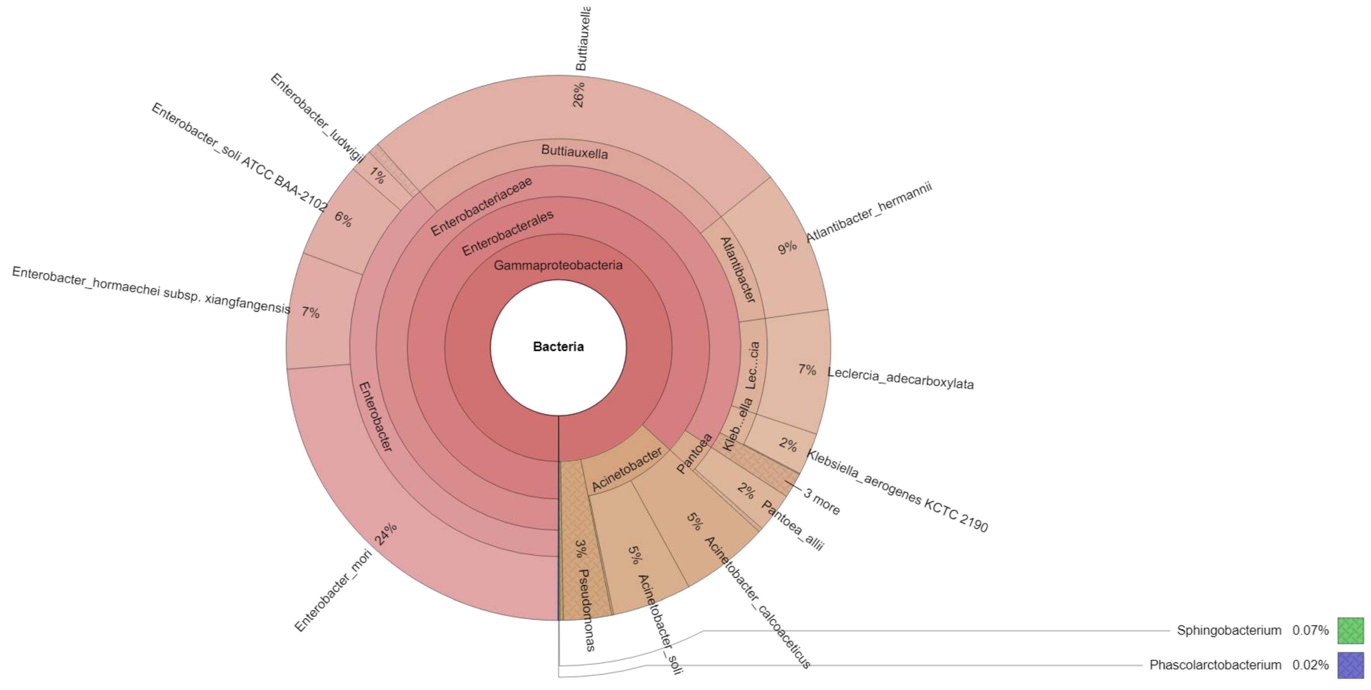
2.2. Microbiota Analysis

To separate and concentrate the pathogens from the surface of fresh-cut bitter gourd, the procedure used by Tatsika et al. [25] was followed with minor modifications. The fruit tissues weighed 25 g, were placed in a tray lined with aluminum foil and covered with plastic wrap, and then kept at room temperature for 12 h. Following that, the fruit tissue slices were collected and transferred into a 250 mL beaker containing 60 mL of phosphate-buffered saline (PBS) (pH 7.4), then sonicated for 10 min. After sonication, the supernatant solution was transferred into new centrifuge tubes. To increase the likelihood of recovering a microbial pellet, the collected supernatant solution was centrifuged at 9500 rpm for 20 min. The recovered pellet was transferred to a microfuge tube and centrifuged at 12,000 rpm for 10 min at 4 °C then sealed with parafilm and stored at −20 °C until use. Mixed microbial DNAs extracted from the recovered pellets were used to amplify full-length 16S rRNA gene and the amplicons were constructed into the SMRTbell library and conducted for sequencing by Illumina MiSeq (paired-end 300 bp). The experiment was performed twice. In the data analysis stage, CCS reads were subjected to processing with DADA2 to obtain single-nucleotide amplicons, followed by taxonomy annotation based on information retrieved from the NCBI database. Alpha and beta diversity analyses are employed to evaluate species complexity within and among samples, respectively.

2.3. Treatments Application

To determine the best treatment for microbial population reduction, 10 g (for microbial count analysis) and 5 g (for protein/RNA extraction) of fresh-cut bitter gourd samples were treated with UV-C and 4 °C, respectively, with three replications.

2.3.1. UV-C Treatment

To conduct UV-C irradiation treatment, a closed chamber (76 cm (W) × 58 cm (L) × 66 cm (H)) was evenly installed, with eight germicidal tubes on the top and bottom at 30 cm intervals (Philips S1-P, Yangyang Technology, Miaoli County, Taiwan) with a total power of 80 W and an intensity of 50 W·m−2. The chamber was lined with aluminum foil on the inside to ensure even light distribution. Before use, the UV-C chamber was turned on for 15 min to stabilize the UV-C dose. Fresh-cut bitter gourd with a constant weight of 5 g and 10 g in plastic film (for RNA/protein extraction and microbial count analysis) was placed at 30 cm distance from the light source. Fruit tissue samples were irradiated with germicidal lamps from the upper and lower surfaces. The first group was exposed to UV-C for a total dose of 0.5 kJ·m2 (7 min, 51 s), the second for 1.5 kJ·m2 (23 min, 34 s), and the third for 3 kJ·m2 (47 min, 7 s). The samples without UV-C treatment were served as the control group. After treatment, samples kept at room temperature (22 °C) for 0, 0.5, 1, 3, and 6 h (supposed shelf life) were collected, frozen in liquid nitrogen, and then stored at −80 °C until used for RNA and protein extraction. On the other hand, samples after treatment were kept at room temperature (22 °C) for 0 and 6 h, respectively, and processed for microbial count analysis. The experiment was performed twice with three replications.

2.3.2. Low Temperature

To determine the effect of temperature on the microbial quality of fresh-cut bitter gourd, 10 g (for microbial count analysis) and 5 g (for protein/RNA extraction) samples were incubated at 4 and 22 °C, respectively, for 3, 6, and 12 h. Treated and control samples kept at room temperature (22 °C) for 0 and 6 h after treatment were collected and analyzed for microbial count. On the other hand, samples kept at room temperature (22 °C) for 0, 0.5, 1, 3, and 6 h after treatment were collected, frozen in liquid N2, and then stored at −80 °C for gene expression analysis and enzyme activity assay. The experiment was executed twice with three replications.

2.3.3. UV-C Combined with Low Temperature

This experiment was conducted using a similar UV-C treatment procedure as described in this study. Freshly cut bitter gourd was treated with 0.5, 1.5, and 3.0 kJ·m2, respectively, while samples without UV-C treatment were used as the control. Following that, both treated and untreated samples were immediately incubated at 4 °C for 3 h. The microbial population reduction was assessed at 0 and 6 h after treatment. Meanwhile, samples after treatment were kept at room temperature (22 °C) for 0, 0.5, 1, 3, and 6 h, collected, frozen in liquid nitrogen, and then stored at −80 °C for further analysis. This experiment was carried out twice with three replications.

2.4. Microbiological Load Determinations

To investigate the effect of each treatment for reducing the microbial population on fresh-cut bitter gourd, viable cell count was measured using the aerobic plate count (APC) method. After treatments, 10 g of fresh-cut bitter gourd samples were placed in a tray lined with aluminum foil and covered with plastic wrap, then kept at room temperature (22 °C) for 6 h. Following that, fruit tissue slices were collected at 0 and 6 h after treatments and transferred to a 100 mL beaker containing 10 mL of PBS, which was then properly covered with plastic film and sonicated for 10 min. A ten-fold serial dilution of the microbial suspension for microbial counting was performed. One mL samples (inoculum) from different dilutions (stock solution, 10−1 to 10−5) were inoculated onto the 3M™ Petrifilm Aerobic Count Plates. Then, Petrifilm plates were incubated at 35 °C for 48 h. Finally, red-colored colonies were counted and calculated as colony-forming units CFU·g1. The results are the average of three replicates ± standard error.

2.5. Determination of Reactive Oxygen Species (ROS) Production

2.5.1. H2O2 Content Analysis

The content of H2O2 was determined spectrophotometrically by reacting it with potassium iodide (KI), following the protocol of Jungle et al. [26], with minor modifications. A mass of 0.45 g of tissue powder was homogenized with a solution containing 0.75 mL of 0.1% trichloroacetic acid (TCA), 0.75 mL of 10 mM potassium phosphate buffer (pH 8), and 1.5 mL of 1 M KI (w/v in fresh, double-distilled water). A control was prepared for each sample by substituting KI with water to account for tissue coloration background. Precautions were taken to protect samples and solutions from light exposure. The homogenate was centrifuged at 12,000× g for 15 min at 4 °C and the reaction mixture was incubated at room temperature (22 °C) for 20 min. Absorbance was measured at 350 nm and a calibration curve obtained using H2O2 standard solutions prepared in 0.1% TCA was used for quantification. The concentration of H2O2 was calculated using a standard curve generated (y = 0.0661 + 0.0036, R2 = 0.995) with known concentrations of H2O2 and expressed as nmol·g−1 fresh weight (FW). The results are the average of three replicates ± standard error.

2.5.2. Analysis of O2•− and H2O2 and Scavenging Activity

The O2•− radical scavenging activity in UV-C-treated fresh-cut bitter gourd was measured using a modified version of the protocol described by Mathur et al. [27]. One gram of fresh-cut bitter gourd samples was ground using a mortar and pestle and then homogenized in 3 mL of PBS (pH 7.4). The suspension was then centrifuged at 13,200× g for 15 min at 4 °C. The supernatant was collected and used to determine the O2•− scavenging activity. To do this, 1 mL of the supernatant was mixed with 3 mL of phosphate-buffered solution (pH 7.4) containing 1.3 μM riboflavin, 0.02 M methionine, and 5.1 μM NBT. The reaction solution was then exposed to 28 W fluorescent lamps for 20 min and the absorbance was measured at 560 nm using a spectrophotometer. Ascorbic acid was used as a positive control and the reaction mixture without any samples was used as a negative control. The O2•− radical scavenging activity (%) was calculated using the following formula:
O2•− scavenging ability of samples (%) = [(A0 − As)/A0] × 100
where A0 = absorbance of positive control, As = absorbance of sample. Decreased absorbance of the reaction mixture indicated increased O2•− scavenging activity.
To gather sufficient data to prove the presence of ROS such as H2O2, we also determined its scavenging activity in UV-C-treated and untreated fresh-cut bitter gourd tissues. The method for determining the ability of UV-C irradiated fresh-cut bitter gourd to scavenge H2O2 was based on the protocol of Ruch et al. [28], as described by Hassan et al. [29]. A solution of 40 mM H2O2 was prepared in PBS (pH 7.4), and an extract of the fresh-cut bitter gourd was prepared at a concentration of 1 mg·mL1 in sterilized distilled water. Subsequently, 1 mL of the extract was mixed with 3 mL of phosphate buffer and 1 mL of the 40 mM H2O2 solution. The mixture was incubated for 10 min at room temperature (22 °C) before measuring the absorbance of H2O2 at 230 nm against a blank solution containing only phosphate buffer without H2O2. The results are the average of three replicates ± standard error. The percentage scavenging of H2O2 was determined as follows:
H2O2 scavenging ability of samples (%) = [(Ab − As)/Ab)] × 100
where Ab is the absorbance of the control, and As is the absorbance of the sample.

2.6. Analysis of Antioxidant Enzymes Activities

To determine the effect of UV-C treatment on the activity of enzymes involved in ROS metabolism, we examined SOD, APX, and CAT activity in RTE bitter gourd samples kept at room temperature at 0, 1, 3, and 6 h after UV-C treatment.
To determine SOD activity, we used the procedures of Kant and Turan [30]. In a sterile mortar and pestle, 0.2 g of treated and untreated RTE bitter gourd samples were ground to powder using liquid nitrogen. Then, the powder was homogenized in 1.5 mL of 50 mM sodium phosphate buffer (pH 7.5), which contained 1% insoluble PVP, 0.1 mM EDTA, and 1 mM PMSF. After that, the homogenate was centrifuged at 13,200× g for 20 min at 4 °C. All steps in preparation were carried out at 0–4 °C. The collected supernatant solution was used as the enzyme extract for the activity assay. The reaction system contained 50 mM phosphate buffer (pH 7.8), 1 mL extract, 13 mM methionine, 75 μM NBT, 0.1 mM EDTA, and 2 μM riboflavin, with a total reaction volume of 1.5 mL. Riboflavin was added last, and the tubes were placed 20 cm below the light source of a 28-watt fluorescent lamp. The reaction ran for 15 min. The reaction was stopped by switching off the light and placing the tubes in the dark. A non-irradiated reaction mixture that was run in parallel and did not develop color served as a blank. The reaction mixture lacking enzymes developed the maximum color (control). The inhibition percentage at 560 nm was plotted as a function of the volume of enzyme extract in the reaction mixture. The results are the average of three replicates ± standard error. The readings at 560 nm were measured using a spectrophotometer, and one unit of enzyme activity was defined as a 50% inhibition of NBT photochemical reduction per minute, described as U·g1.
The protocol used by Nakano and Asada [31] was adopted to measure the activity of APX enzyme on irradiated and non-irradiated fresh-cut bitter gourd samples. Samples (0.2 g FW) were ground to powder and homogenized in 1.5 mL of 50 mM sodium phosphate buffer (pH 6.8). The homogenate was centrifuged at 12,000× g for 20 min at 4 °C. The supernatant was then collected and used as the enzyme extract. A total of 100 μL of the extract was reacted with a mixture of 1 mL 150 mM potassium phosphate buffer (pH 7.0), 1 mL 1.5 mM ascorbate, 0.4 mL 0.75 mM EDTA, and 0.5 mL 6 mM H2O2, for a total reaction volume of 3 mL. The absorbance at 290 nm was read by a spectrophotometer for 1 min. The results are the average of three replicates ± standard error. One unit is defined as 1 μM of ascorbate consumed per minute. APX activity (units·g1) = A290/2.8 (K, mM1 cm1) × 3 (reaction volume) × 40 (dilution rate)/min/g FW.
The method of Kato and Shimizu [32] was used to determine the activity of CAT. Treated and untreated fresh-cut bitter gourd samples weighing 0.2 g were ground to a fine powder and homogenized with 1.5 mL of 50 mM sodium phosphate buffer (pH 6.8). Homogenates were centrifuged at 4 °C for 20 min at 12,000× g. After centrifugation, the supernatant was collected and used as an enzyme extract. In a total volume of 3 mL, 0.2 mL of extracts were combined with 2.7 mL of 0.1 M sodium phosphate buffer (pH 7.0) and 0.1 mL 1 M H2O2. An absorbance at 240 nm is measured for one minute by a spectrophotometer. The results are the average of three replicates ± standard error. In CAT activity, 1 unit equals 1 nmol H2O2 consumed per minute. CAT activity (Units·g1) = A240/40 (K, mM1 cm1) × 3 (reaction volume) × 20 (dilute rate)/min/g FW.

2.7. Determination of Defense-Related Compound Content

2.7.1. MDA Content Analysis

MDA content was determined using the method described by Promyou and Supapvanich [33], with minor modifications. In a sterile mortar and pestle, 1 g of treated and untreated samples were ground to powder using liquid nitrogen. The fruit tissue powder was homogenized in 4 mL of 10% (w/v) trichloroacetic acid and centrifuged at 10,000× g for 10 min. After centrifugation, 1 mL of supernatant was reacted with 2 mL of 15% TCA containing 0.5% thiobarbituric acid. The reaction mixture was incubated for 20 min at 95 °C (boiling water bath). The results are the average of three replicates ± standard error. After that, the sample was immediately placed in an ice bath for 10 min to stop the reaction. The absorbance at 532 (OD532) and 600 (OD600) nm wavelengths of the sample was measured and calculated as follows:
MDA content (nmol MDA·g−1 FW) = [(OD532 − OD600) × A × V]/(a × E × W)
A = Total volume of reaction solution and enzyme extract (mL). V = Total volume of buffer used for enzyme extract (mL). a = Volume of the enzyme extract used (mL). W = Fresh weight of the sample (g). E = The molar extinction coefficient of MDA (155 mM−1·cm−1).

2.7.2. Proline Content Analysis

The proline content was determined in accordance with Bates et al. [34]. Tissues of irradiated and non-irradiated samples weighing 0.5 g were homogenized in 10 mL of 3% sulfosalicylic acid and centrifuged at 10,000× g for 15 min. After centrifugation, supernatant was collected. Two mL of supernatant was added to two mL acid ninhydrin, two mL glacial acetic acid, and two mL 6 N phosphoric acid. The mixture was incubated for one hour at 95 °C (boiling water bath). Then, the reaction mixture was extracted with four mL toluene, and then mixed with a vortex. The chromophore containing toluene was carefully collected from the aqueous phase, warmed to room temperature, and the absorbance reads at 520 nm used toluene as a blank. The results are the average of three replicates ± standard error. The proline concentration was determined from an obtained standard curve (y = 0.0596x + 0.0266, R2 = 0.9937) and calculated on a fresh weight basis as follows:
µmoles proline·g−1 of fresh weight material = [(µg proline·mL−1 × mL toluene)/115.5 µg·µmole−1]/[(g sample)/5].

2.8. Analysis of PAL Activity

PAL activity was measured with the procedure of Li et al. [18], with minor modifications. A sample of 1 g was ground into powder using liquid nitrogen and a sterile mortar and pestle. The powder was homogenized with 2.5 mL 0.1 M Na2B4O7·10H2O buffer (pH 8.8), which contained 5 mM 2-mercaptoethanol and 2 mM EDTA, for 20 min at 4 °C. The suspension was centrifuged at 13,200× g for 15 min at 4 °C. The recovered supernatant was used to assay PAL activity. Following that, a total reaction volume of 2.5 mL containing 1.9 mL 0.1 M Na2B4O7·10H2O buffer (pH 8.8), 0.1 mL of extract, and 0.5 mL of 20 mM L-phenylalanine was mixed. After that, each tube was incubated at 37 °C for 1 h. The control was the system without L-phenylalanine, and the absence of extraction in the mixture was the blank. Finally, 0.1 mL 6 mM HCl was added to stop the reaction. One unit of PAL was defined as an increase of 0.1 in absorbance per gram of FW. The results are the average of three replicates ± standard error.

2.9. Analysis of PPO Activity

The activity of PPO was assayed based on the protocol of Moreno et al. [35]. Fresh-cut bitter gourds samples (1 g) were ground with a mortar and pestle and then homogenized in 3 mL 0.1 M phosphate buffer (pH 6.0) containing 10 g·L1 PVPP and 10 μL 0.1% (v/v) Triton X-100 for 15 min at 4 °C. The suspension was centrifuged at 13,200× g for 15 min at 4 °C. After that, the supernatant was collected for the determination of PPO activity. The reaction mixture for PPO determination contained 0.1 M phosphate buffer (pH 6.0), 0.3 mL of extract, and 0.2 mL of 0.1 M catechol in a total volume of 3 mL. The reaction mixture was incubated for 2 h at 35 °C, and then the enzymatic activity was determined spectrophotometrically at 410 nm. One unit of PPO activity (U) was defined as an increase of 0.001 units of absorbance per min at 410. The results are the average of three replicates ± standard error.

2.10. Statistical Analysis
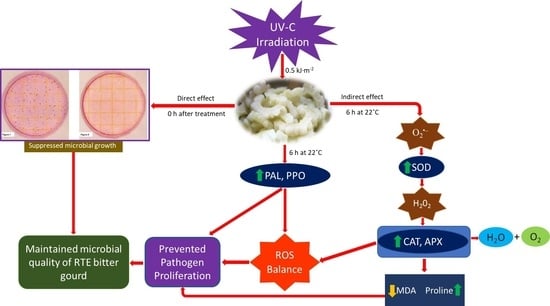
Data analysis was conducted using the statistical package SPSS software (IBM Corporation). Two-way analysis of variance (ANOVA) with shelf life and treatment as factors and Tukey’s range tests at p < 0.05 were performed to analyze the data, which was presented as the mean ± standard error (SE) of three replicates. Significance was considered when p < 0.05.

3. Results

3.1. Microorganisms Grew on the Surface of Fresh-Cut Bitter Gourd

As the first step to reduce the microbial risk, this study utilized next-generation sequencing (NGS) technology to determine the composition of the microbial community on the surface of fresh-cut bitter gourd. The findings revealed that 34 bacterial species had been identified (Table 1), with Buttiauxella izardii, Enterobacter mori, and Atlantibacter hermannii emerging as dominant, with 26%, 24%, and 9% species richness and abundance, respectively (Figure 1). Meanwhile, highly important and emerging human pathogen bacterial species such as Klebsiella pneumoniae, Klebsiella aerogenes, Pseudomonas monteilii, Enterobacter cloacae, Leclercia adecarboxylata, and Cedecea lapagei were also identified at low levels.

3.2. Effect of UV-C Irradiation Treatment on the Total Viable Bacterial Count of Fresh-Cut Bitter Gourd

The bactericidal effects of UV-C irradiation on fresh-cut bitter gourd after exposure to room temperature for 6 h are shown in Table 2. In general, the total viable bacterial count in fresh-cut bitter gourd treated with UV-C was significantly lower than that in the control throughout the shelf life. Immediately after treatment, treated samples with 0.5, 1.5, and 3.0 kJ·m−2 had 1.33 × 10², 1.26 × 10², and 1.07 × 10² CFU·g−1, respectively, compared to the control with 4.03 × 10³ CFU·g−1. Six hours after treatment, the results indicated that the 0.5 and 1.5 kJ·m−2 UV-C irradiation suppressed microbial growth compared to the control (Table 2). In particular, the total bacterial count at 6 h after UV-C treatment in fresh-cut bitter gourd was found to be 100-fold lower than that in the control. There was no significant difference in reducing the bacterial population of bitter gourd between 0.5 and 1.5 kJ·m−2. It is also worth noting that exposing the samples to the higher dose of 3.0 kJ·m−2 resulted in a 10-fold higher total count of bacteria compared to the lower doses of 0.5 and 1.5 kJ·m−2, which exceeded the bacterial load limit based on EU Food Safety Regulations.

3.3. The Effect of Low-Temperature Treatment on the Total Viable Bacterial Count of Fresh-Cut Bitter Gourd

To determine the effects of temperature on the microbial quality of fresh-cut bitter gourd, samples were incubated at 4 °C and 22 °C for 3, 6, and 12 h, respectively. The bacterial population on fresh-cut bitter gourd treated at 4 °C or room temperature is depicted in Table 3. The data demonstrated a general trend in which the sample incubated at 4 °C, one of the most used refrigeration temperatures, showed slower bacterial growth than the other storage temperature. In low-temperature treatments, samples incubated at 4 °C for 3 h had the slowest bacterial growth as 1.47 × 10² CFU·g−1, while a higher number up to a maximum of 7.05 × 10⁵ CFU·g−1 of bacteria were found at 22 °C.

3.4. The Effect of UV-C Followed by Low-Temperature Treatment on the Bacterial Population of Fresh-Cut Bitter Gourd

The bacterial population on fresh-cut bitter gourd treated with UV-C irradiation followed by 4 °C after exposure to room temperature is shown in Table 4. Compared with the control, UV-C combined with low-temperature treatments significantly reduced the initial number of bacterial colonies in fresh-cut bitter gourd after 6 h of exposure to room temperature. In particular, fresh-cut bitter gourd treated with UV-C of 1.5 and 0.5 kJ·m−2 incubated at 4 °C had the lowest number of colonies after 6 h of exposure to room temperature. There was no significant difference (p < 0.01) between two-dose treatments.

3.5. The Effect of UV-C on the Production of ROS in Fresh-Cut Bitter Gourd

3.5.1. H2O2 Content in RTE Fresh-Cut Bitter Gourd

Figure 2A shows the H2O2 levels in both UV-C-treated and untreated samples. The data indicated that the H2O2 levels increased over time for both groups. However, samples treated with UV-C irradiation had lower H2O2 levels than the control group. A significant decrease in H2O2 was observed 1 h after treatment with 0.5 and 1.5 kJ·m−2 of UV-C irradiation, with a 1.8 and 1.6-fold decrease, respectively, compared to the control and a higher dose of 3.0 kJ·m−2. This trend continued until 3 h, with a 2.13 and 2.23-fold decrease, and remained stable at 6 h with a 2.08 and 2.0-fold decrease. No significant difference was observed between the H2O2 production of 0.5 and 1.5 kJ·m−2 UV-C irradiation doses.

3.5.2. O2•− and H2O2 Scavenging Activity in UV-C-Treated RTE Fresh-Cut Bitter Gourd

Figure 2B demonstrates the capability of UV-C-treated fresh-cut bitter gourd to scavenge O2•−. In general, UV-C-treated samples showed a higher scavenging ability than the control during the shelf life period. A 27% higher O2•− inhibition percentage was recorded for 0.5 and 1.5 kJ·m−2 at 3 h after treatment compared to the control and 3.0 kJ·m−2. At the end of the shelf life period, the scavenging ability of lower doses of UV-C (0.5 and 1.5 kJ·m−2) increased to 56%, against the control and highest dose of 3.0 kJ·m−2. Consistently, there was no significant difference in terms of the O2•− scavenging ability between 0.5 and 1.5 kJ·m−2 of UV-C.
Meanwhile, the ability of UV-C-treated fresh-cut bitter gourd to scavenge H2O2 is shown in Figure 2C. Overall, UV-C-treated samples exhibited a superior H2O2 scavenging ability compared to the control over the shelf life period. Our results indicated that, at 1 h, the samples treated with UV-C irradiation at doses of 0.5 and 1.5 kJ·m−2 had the highest H2O2 scavenging ability, with 1.38 and 1.36-fold increases, respectively, compared to the control and a higher dose of 3.0 kJ·m−2. This trend of an increased H2O2 scavenging capability at lower UV-C doses was evident at 3 h and remained steady 6 h after treatment, with a 1.7-fold increase against the control and highest dose. The H2O2 scavenging ability of the control was observed to decline at the end of shelf life. Consistently, there was no significant difference in terms of the scavenging ability of lower doses (0.5 and 1.5 kJ·m−2) of UV-C were recorded.

3.6. The Effect of UV-C Irradiation on the Activity of Antioxidant Enzymes of Fresh-Cut Bitter Gourd

In this study, UV-C treatment effectively improved the enzyme activities of SOD, APX, and CAT involved in the ROS metabolism of fresh-cut bitter gourd (Figure 3).
As shown in Figure 3A, the SOD activity displayed a rising trend during shelf life. Compared with control, the SOD activity significantly increased immediately after treatment with UV-C irradiation and remained at a higher level after 6 h of exposure to room temperature. No significant difference in SOD activity was found between 0.5, 1.5, and 3.0 kJ·m−2 at each exposure time. Remarkably, UV-C of 0.5 kJ·m−2 recorded the highest level of SOD activity, reaching a maximum value of 1.75 U·g−1 at the end of the shelf life.
In the case of APX activity, an increasing trend of data was recorded for both the control and treated samples throughout the shelf life (Figure 3B). A significant increase was observed at 1, 3, and 6 h of exposure to room temperature after treatment. Our results have shown that the application of 0.5 and 1.5 kJ·m−2 significantly enhanced the activity of the APX enzyme with 63.64 and 60.35 U·g−1 at the end of the shelf life compared to the control and higher dose of 3.0 kJ·m−2 with 41.85 and 45.78 U·g−1. No significant difference between 0.5 and 1.5 kJ·m−2 was detected. However, at 6 h, a UV-C dose of 0.5 kJ·m−2 reached the highest value of 63.64 U·g−1.
Based on the analysis of CAT activity, both non-irradiated and irradiated samples showed an upward trend over the entire shelf life (Figure 3C). UV-C did not have an effect on the samples during the first hour of shelf life. However, after 3 h of CAT activity in irradiated samples significantly increased and remained stable at the end of shelf life. Notably, UV-C at 0.5 and 1.5 kJ·m−2 displayed very superior activity compared to the control and 3.0 kJ·m−2 in the whole shelf life. Like SOD and APX, CAT activity between 0.5 and 1.5 kJ·m−2 had no significant difference throughout the shelf life. Consistently, the UV-C of 0.5 kJ·m−2 produced the highest CAT activity up to 1.70 U·g−1.

3.7. The Effect of UV-C on the Defense-Related Compounds of Fresh-Cut Bitter Gourd

3.7.1. MDA Content

MDA is the main product of cell membrane lipid peroxidation. The MDA content steadily increased throughout the shelf life in both the control and treated samples (Figure 4A). It was found that the MDA content in the control samples increased more rapidly than in the UV-C-treated fresh-cut bitter gourd. In particular, the 0.5 and 1.5 kJ·m−2 of UV-C irradiation significantly reduced the MDA levels immediately after treatment and until the end of the shelf life compared to the higher dose of 3.0 kJ·m−2 and the control. The initial (0 h) and final (6 h) MDA contents at all the tested doses of UV-C (0, 0.5, 1.5, and 3.0 3.0 kJ·m−2) were 4.68, 2.32, 2.61, and 4.78 nmol·g−1 and 10.73, 5.60, 5.67, and 8.2 nmol·g−1, respectively. There was no significant difference in the MDA content between 0.5 and 1.5 kJ·m−2 of UV-C irradiation. Still, the shortest exposure to UV-C (0.5 kJ·m−2) suppressed MDA accumulation more effectively in freshly cut bitter gourd.

3.7.2. Proline Content

Proline is frequently used as an indicator for cell membrane degradation or oxidative stress. The proline content increased similarly to MDA during the entire shelf life (Figure 4B). However, the proline content of fresh-cut bitter gourd treated with UV-C irradiation was elevated with the prolonged shelf life. During the first hour of shelf life, UV-C had no positive effect on the proline values in the samples. After exposure to UV-C light at doses of 0.5 and 1.5 kJ·m−2, the proline levels increased significantly after 3 and 6 h of shelf life, reaching levels of 2.15 and 2.0 and 2.82 and 2.77 μM·g−1 FW, respectively. In contrast, a higher UV dose of 3.0 kJ·m−2 resulted in lower proline levels of 1.55 and 2.24 μM·g−1 FW, while the control group had the lowest levels of 1.04 and 1.72 μM·g−1 FW. Even though there was no significant difference in proline promotion between 0.5 and 1.5 kJ·m−2 over the shelf life, the lowest dose of UV-C (0.5 kJ·m−2) produced the highest level of proline compounds.

3.8. The Effect of UV-C on the Activity of Enzymes Involved in the Phenylpropanoid Pathway of Fresh-Cut Bitter Gourd

3.8.1. Phenylalanine Ammonia-Lyase (PAL)

The PAL in treated tissues increased initially and reached a peak value at 1 h and then decreased slightly in the subsequent period, while the control showed a steady but slow increase during the whole shelf life (Figure 5A). In the UV-C dose of 0.5 and 1.5 kJ·m−2 treatments, the activity level of PAL was elevated to the maximum values of 2.07 and 1.96 U·g−1 after 1 h of exposure to room temperature as compared to control and 3.0 kJ·m−2, with 0.74 and 1.51 U·g−1, respectively. At the end of the shelf life, PAL activity was significantly accelerated by UV-C treatment, which brought 2.73, 3.1, and 2.01-fold increases for 0.5, 1.5, and 3.0 kJ·m−2, respectively, compared to the control with a 1.55-fold increase. In this experiment, no significant difference was observed between short UV-C exposure (0.5 and 1.5 kJ·m−2) on the PAL activity of the samples. Still, the shortest exposure to UV-C (0.5 kJ·m−2) was found to improve the activity of this enzyme.

3.8.2. Polyphenol Oxidase (PPO)

The PPO activity presented a rising trend during shelf life (Figure 5B). It ranged from 17.33, 35, 35.66, and 33.33 U·g−1 to 37.33, 64, 60, and 49.66 U·g−1 in the control and 0.5, 1.5, and 3.0 kJ·m−2 in the UV-C-treated tissues, respectively. The activity of this enzyme increased sharply on samples treated with 0.5 and 1.5 kJ·m−2 UV-C irradiation than the control and 3.0 kJ·m−2. Changes in PPO activity in tissues irradiated by 0.5 and 1.5 kJ·m2 UV-C increased 53.46 and 47.2% compared to the control at the end of the shelf life. Meanwhile, no significant differences between 0.5 and 1.5 kJ·m−2 UV-C were detected. Interestingly, 0.5 kJ·m−2 showed superior enzyme values.

4. Discussion

In a typical restaurant buffet set-up, RTE fresh-cut fruits and vegetables are often prepared, served, or kept on the shelves (in the buffet food container) at room temperature with air conditioning immediately after being cut or sliced. Usually, this practice can result in microorganisms attaching to the surface of the produce during the shelf life period. However, to our knowledge, there are no published data or information on the presence of microorganisms on the surface of RTE fresh-cut bitter gourd fruit after cutting. In this study, we used molecular-based techniques to uncover the microbiota in RTE fresh-cut bitter gourd. Our findings revealed that, while the most dominant detected bacterial species were not classified as human pathogenic microorganisms, our data also showed the presence of commonly found foodborne pathogens in RTE fresh-cut fruits and vegetables. For instance, Klebsiella pneumoniae and other pathogenic Klebsiella species were also detected in fresh-cut fruit and vegetable salads served in restaurants in Taif City, Kingdom of Saudi Arabia [36]. Similarly, Klebsiella pneumoniae was identified in salad vegetables such as lettuce, parsley, cucumber, tomato, and carrot from hypermarkets and wet markets in Malaysia [37]. In a different scenario, Pseudomonas species, including Pseudomanas aeruginosa and Pseudomanas monteilii, were reportedly detected in raw cucumber and zucchini collected from the La Rioja region in Spain [38]. The detection of these pathogens can largely be attributed to the destruction of surface cells and exposure of the cytoplasm during the processing of fresh-cut produce, providing microorganisms with a richer source of nutrients than intact produce [39]. Although the analysis indicated that their numbers were low, these bacterial species still threaten human health if they are not contained within safe limits. These microorganisms can cause severe and often fatal infections, such as bloodstream infections and pneumonia [40]. Interestingly, the bacterium Cedecea lapagei was also identified in fresh-cut bitter gourd. This emerging pathogen has been found to cause serohematogenous bullae infections in humans [41]. Identifying Cedecea lapagei in this study provides valuable information, as this pathogen, which is currently under surveillance, has yet to be reported in either whole or fresh-cut fruits and vegetables. The microorganisms identified in this study compromise food safety and contribute significantly to the spoilage of fresh-cut products [42]. The identification of important and emerging pathogens with their compositions on RTE fresh-cut bitter gourd in this study is a crucial step in developing approaches to inhibit and control these organisms. Furthermore, this information is valuable in determining which sanitizing methods or practices should be implemented for fresh-cut fruits and vegetables to ensure their safety and quality.
UV-C irradiation was an effective tool for inhibiting microbial growth and improving the safety of fresh-cut produce [16,18,35,43]. In this study, UV-C alone was also the most effective treatment for decontaminating RTE fresh-cut bitter gourd compared to low temperature treatment and was more practical to use than UV-C combined with low temperature treatment. The bacterial population in the UV-C alone treated samples after 6 h of exposure to room temperature was below 6 × 10³ CFU·g−1, demonstrating that the microbial quality met the acceptable standards set by EU food safety regulation (No. 2073/2005). Low-temperature treatment slowed down the growth of bacteria until 3 h; however, when the samples were removed from refrigeration during this time, the number of bacteria grew faster and exceeded the microbial load limit. On the other hand, the combination of UV-C and low temperature suppressed the microbial growth within the safe range at the end of the shelf life (6 h); however, the effect of this combined treatment did not outperform UV-C alone statistically. The responses of vegetables to UV-C mainly depend on the radiation dose and intensity of the commodity considered [44]. Previous studies have shown that a higher dose of 3.0 kJ·m−2 and above was more effective in reducing the microbial populations in fresh-cut strawberries [18], carambola [35], watermelon [43], and RTE salad [45]. Conversely, our results clearly showed that the shortest UV-C dose of 0.5 kJ·m−2 had a superior effect on inhibiting bacterial growth compared to the higher dose of 3.0 kJ·m−2 after 6 h of exposure to room temperature. Similarly, 0.5 kJ·m−2 has shown a significant reduction of decay caused by Penicillium sp. in star ruby grapefruit without causing damage to the tissues [46]. Similar results were also observed by Manzacco et al. [47], who found that short UV-C light (0.2 kJ·m−2) decreased the microbial growth of pineapple sticks during storage. The data obtained from our study confirmed the decontaminating efficacy of UV-C irradiation by controlling microbial growth through damaging microorganism DNA and inducing resistance against phytopathogens in fruits and vegetables [19,20]. The higher dose of 3.0 kJ·m−2 in this study might cause damage to the fruit tissue, which then creates an opening for microorganisms to grow and spread further. These results suggested that fresh-cut bitter gourd should be treated with a dose of 0.5 to 1.5 kJ·m−2 to preserve the microbial quality for 6 h at room temperature.
In fresh-cut fruit and vegetable processing, cutting is necessary to divide the intact products into smaller pieces. Usually, this operation inevitably results in wounding stress on the tissue, which can hasten deterioration processes and negatively impact the shelf life of fresh-cut produce [48]. In this study, we evaluated the effect of the best treatment (UV-C irradiation) on oxidative stress indicators such as H2O2, MDA, and proline. In addition to this, we also assessed the ability of UV-C treatment to scavenge or counter oxidative stress in fresh-cut bitter gourd by determining the antioxidant enzyme activity as the activity of the enzymes involved in the phenylpropanoid pathway.
Our data demonstrate that the UV-C irradiation (0.5 and 1.5 kJ·m−2) significantly reduced the content of H2O2 in fresh-cut bitter gourd throughout the end of the shelf life period. These findings were supported by the data that we gathered from the scavenging activity of O2•− in UV-C-treated fresh-cut bitter gourd, wherein samples with UV-C treatment of 0.5 to 1.5 kJ·m−2 were effective in scavenging O2•−, as indicated by the 74.22 and 74.94% increases compared to the control samples at the end of the shelf life period. O2•− is a primary biological source of ROS [49]; however, the O2•− itself is not a particularly strong oxidant; it can react with other molecules to produce more powerful and dangerous reactive oxygen species such as 1O2, H2O2, and OH [50]. In this case, our results might contribute to the lower conversion of O2•− into H2O2. Additional experimental results from this study further confirmed the scavenging effect of UV-C treatment on H2O2 in fresh-cut bitter gourd, wherein we observed that the greatest H2O2 scavenging ability was exhibited by samples treated with lower doses of UV-C of 0.5 and 1.5 kJ·m−2. This observation of reduced or low levels of H2O2 is consistent with the findings reported in several previous studies published, such as in fresh-cut apples [51], fresh-cut Huangguan pear [52], lettuce [53], banana fruit peel [54], peach [55], and apricot fruit [56]. A transient increase or balance level of H2O2 might act as a signaling molecule to activate stress-related protective mechanisms [54]. The low levels of H2O2 were believed to have antimicrobial properties that can have toxic effects on microorganisms [48]. These results suggest that lower doses of UV-C could regulate H2O2 levels, thereby alleviating oxidative damage and activating protective mechanisms against pathogen invasion.
To deal with oxidative stress caused by the excessive accumulation of ROS such as O2•− and H2O2, plants possess ROS scavenging systems wherein CAT, SOD, and APX are considered critical enzymes in this system and are extremely important in improving plant antioxidant activity [57]. In our experiment, UV-C (0.5 and 1.5 kJ·m−2)-treated fresh-cut bitter gourd tissues significantly exhibited higher SOD, APX, and CAT activities in the entire shelf life compared to the control. Thus, the excess O2•− could be efficiently converted to H2O2 by SOD, and the H2O2 was then degraded into H2O by CAT and APX, as in Zhang et al. [57]. Also, researchers recently found that UV-C effectively promoted antioxidant enzyme activities in fresh-cut fruits and vegetables [57,58,59], implying that UV-C treatment could preserve high antioxidant enzyme activity while avoiding excessive ROS production. This finding was consistent with the observation of Jia et al. [60] that the ROS scavenging enzymes in jujube fruit were accelerated by UV-C irradiation treatment. Similar results were also reported in fresh-cut strawberries, potato tubers, and pears treated with UV-C [18,59,61]. In our case, the continuous increase in SOD and APX activity and the elevated levels of CAT in irradiated fresh-cut bitter gourd tissues prevented the excessive accumulation of H2O2. On the other hand, exposure to UV radiation at a high dose (3.0 kJ·m−2) or for extended periods (longer than 47 min) might cause cellular stress and damage, which could explain the observed reduction in antioxidant enzyme activity. Therefore, it can be concluded that short UV-C (0.5 and 1.5 kJ·m−2) exposure has a regulatory effect on ROS levels by enhancing the activity of antioxidant enzymes.
The enhancement of antioxidant enzyme activity, including SOD, APX, and CAT, in UV-C-treated fresh-cut potato tubers corresponded with a reduction in lipid peroxidation and MDA content. This facilitated the maintenance of cell membrane integrity and supported normal cellular metabolism [61]. In this study, it was observed that applying a lower dose of UV-C radiation resulted in a significant reduction in the MDA content of RTE fresh-cut bitter gourd. A UV-C dose of 0.5 kJ·m−2 reduced MDA content by 56.41 and 37.91%, respectively, compared to a control and a higher dose of 3.0 kJ·m−2 at the end of the shelf life. The monitoring of MDA levels indicates the degree to which membrane lipid peroxidation has occurred and the integrity of cellular membranes [62]. The excessive production of MDA can further damage cell membranes and accelerate the browning process in fruits and vegetables [63], providing a suitable environment for pathogen attack. Such a response could indicate that the lower doses of UV-C irradiation reduced membrane injury. Our results are consistent with the findings of Artes and Allende [64], who reported that the synthesis of defense-related compounds can be triggered by UV-C irradiation when applied at low doses for short periods. A similar reduction in MDA content was also observed in potato tubers treated with lower-dose UV-C irradiation [61,65]. In contrast, MDA content was higher in whole or intact bitter gourd fruits treated with 4.0 kJ·m−2 of UV-C than in untreated tissues because high doses might induce cell damage, which occurs due to impairment of the cell defense mechanism [19]. This indicated that the higher dose (3.0 kJ·m−2) of UV-C applied in this study might increase the production of lipid peroxidation, thus increasing membrane damage. Furthermore, low levels of MDA could play a positive role in plant defense and development by activating specific regulatory genes and protecting against oxidative stress [62]. These results indicated that a lower dose (0.5 kJ·m−2) of UV-C irradiation could effectively protect cell membranes from ROS attack and thus maintain cell membrane integrity.
Another important defense-related compound that is naturally secreted in plants is proline. Our data showed a significant increase in the proline content in fresh-cut bitter gourd fruit tissue exposed to lower doses of UV-C irradiation compared with the control and the higher dose. The proline content increased with prolonged shelf life, with the highest levels observed in UV-C-treated samples (0.5 and 1.5 kJ·m−2) on the sixth hour of exposure to room temperature, which resulted in a 1.5-fold increase compared to the control. A similar phenomenon was observed in bell peppers, where a significant increase in proline levels was observed as storage duration increased [66]. This indicated that short UV-C irradiation induced the accumulation of proline in fresh-cut bitter gourd fruit tissues. The proline accumulation in the plant cell was vital for maintaining cellular homeostasis, water uptake, osmotic adjustment, and redox balance to protect the cell structure from oxidative damage [67]. Moreover, the synthesis of proline removed excess H+ ions generated in response to UV irradiation and positively reduced damage caused by UV-C and UV-B radiation [68]. Therefore, the application of lower UV-C doses (0.5 and 1.5 kJ·m−2) increased proline levels in fresh-cut bitter gourd, which helped to maintain redox balance (ROS homeostasis) by scavenging ROS, thereby preserving the cell membrane structure and integrity of fresh-cut bitter gourd.
The phenylpropanoid metabolism is a critical pathway for plants to produce secondary metabolites. Exposure to UV-C radiation has been shown to stimulate the phenylpropanoid pathway in various fruits. This was mainly due to the induction of phenylalanine ammonia-lyase (PAL), a key enzyme in this pathway [18]. The induction of PAL was found to be directly related to the production and accumulation of phenolics and flavonoids [18,69], which are important secondary metabolites in the phenylpropanoid pathway. They can eliminate reactive oxygen species (ROS) and increase plant antioxidant capacity [59]. Furthermore, these compounds that are synthesized by the phenylpropanoid pathway protect plants against pathogens through the reinforcement of plant cell walls, the direct inhibition of growth, and/or the inactivation of enzymes that contribute to tissue maceration [70]. In our study, UV-C significantly increased PAL activity at all tested doses at 1 h and towards the end of the shelf life compared to the control. Notably, samples treated with 0.5 and 1.5 kJ·m−2 showed superior PAL activity and reached a peak value at 1 h with a 2-fold increase than the control. Similar phenomena were recorded as far as the elevation of PAL activity by UV-C is concerned in fresh-cut strawberries [18], fresh-cut broccoli [71], and peaches [55]. This could be potentially explained by the membrane damage caused by long-time exposure to UV-C. UV-C stimulation of PAL activity implies activating some defense mechanism in fruit tissues. Therefore, these results suggested that the UV-C treatment activated the phenylpropanoid pathway through the induction of a key enzyme (PAL), thus promoting the accumulation of phenolics as well as enhancing antioxidant capacity and preventing microbe invasion at the wound site by forming a mechanical barrier in fresh-cut bitter gourd tissues.
The indirect role of phenolic compounds in fruit protection might be attributed to their synthesis and accumulation following UV-C irradiation. These compounds act as natural substrates for polyphenol oxidase (PPO), whose reaction products, quinones, have been suggested to possess bactericidal and fungicidal properties in plant defense [72]. This was confirmed by the high bacterial resistance in transgenic tomato plants over-expressing PPO [73]. Similarly, UV-C-treated strawberry fruits escalated the activity of PPO from 10 h post-treatment until 48 h of storage at 20 °C, which contributes to the resistance against Botrytis cinerea [20]. To our knowledge, there is no available data on the role of PPO in pathogen defense in fresh-cut fruits and vegetables. In our case, the induction of PPO enzyme activity by UV-C (0.5 and 1.5 kJ·m−2) was evident throughout the shelf life, reaching its peak value after 6 h. This suggested that the increase in PPO activity after 0.5 and 1.5 kJ·m−2 of UV-C exposure may protect against pathogen invasion via the production of quinones in fresh-cut bitter gourd fruit tissues.

5. Conclusions

In conclusion, this study revealed the presence of nonpathogenic, opportunistic, and emerging human pathogens in fresh-cut bitter gourd through NGS. Interestingly, UV-C alone (0.5 and 1.5 kJ·m−2) was found to be a more effective treatment for decontaminating fresh-cut bitter gourd compared to low temperature treatment and was more practical to use than UV-C combined with low temperature treatment. In addition, UV-C irradiation enhanced the ROS scavenging system, as indicated by the low production of O2•− and H2O2, increased SOD, APX, and CAT activities, as well as elevated levels of proline and reduced MDA content. This response demonstrates that UV-C (optimal condition) can maintain the balance levels of ROS, thereby alleviating oxidative damage and preserving the cell membrane structure and integrity of fresh-cut bitter gourd. Moreover, UV-C treatment activated the phenylpropanoid pathway by inducing PAL and PPO activity, which are involved in the defense mechanism against pathogens. This suggests that 0.5 kJ·m−2 UV-C can be used effectively to maintain microbial safety and extend the shelf life of fresh-cut bitter gourd.

Author Contributions

Conceptualization, Y.-Y.D.; methodology, Y.-Y.D. and P.-L.H.; software, J.L.B.; validation, Y.-Y.D. and P.-L.H.; formal analysis, J.L.B.; investigation, J.L.B.; resources, Y.-Y.D. and P.-L.H.; data curation, Y.-Y.D. and P.-L.H.; writing—original draft preparation, J.L.B.; writing—review and editing, Y.-Y.D.; visualization, J.L.B.; supervision, Y.-Y.D. and P.-L.H.; project administration, Y.-Y.D. and P.-L.H.; funding acquisition, Y.-Y.D. All authors have read and agreed to the published version of the manuscript.

Funding

This research was funded by grants 111AS-1.4.1-ST-a7 and 112AS-1.4.1-ST-a8 supported by the Council of Agriculture, Republic of China.

Data Availability Statement

Data sharing is not applicable to this article.

Acknowledgments

We thank Southeast Asian Regional Center for Graduate Study and Research in Agriculture (SEARCA) for providing John Louie Baligad with scholarship throughout the journey of his master’s degree.

Conflicts of Interest

The authors declare no conflict of interest.

References

  1. Lucas, E.A.; Dumancas, G.G.; Smith, B.J.; Clarke, S.L.; Arjmandi, B.H. Health benefits of bitter melon (Momordica charantia). In Bioactive Foods in Promoting Health, 1st ed.; Watson, R.R., Preedy, V.R., Eds.; Academic Press: London, UK, 2010; pp. 525–549. [Google Scholar] [CrossRef]
  2. Pan, Y.; Chen, L.; Pang, L.; Chen, X.; Jia, X.; Li, X. Ultrasound treatment inhibits browning and improves antioxidant capacity of fresh-cut sweet potato during cold storage. Rsc Adv. 2020, 10, 9193–9202. [Google Scholar] [CrossRef]
  3. Srilatha, V.; Reddy, K.; Reddy, R.; Anitha, T.; Mamatha, N.C.; Kumari, P.L. Chemical interventions for extending shelf life of minimally processed bitter gourd (Momordica charantia L.). Pharma Innov. 2021, 10, 746–753. [Google Scholar] [CrossRef]
  4. Wang, L.; Li, Q.; Cao, J.; Cai, T.; Jiang, W. Keeping quality of fresh-cut bitter gourd (Momordica charantia L.) at low temperature of storage. J. Food Process. Preserv. 2007, 31, 571–582. [Google Scholar] [CrossRef]
  5. Olaimat, A.N.; Holley, R.A. Factors influencing the microbial safety of fresh produce: A review. Food Microbiol. 2012, 32, 1–19. [Google Scholar] [CrossRef]
  6. Ministry of Food and Drug Safety (MFDS). Statistics for Foodborne Illness Outbreaks. Available online: https://www.foodsafetykorea.go.kr/portal/healthyfoodlife/foodPoisoningStat.do?menu_no=3724&menu_grp=MENU_NEW02 (accessed on 7 May 2020).
  7. Centers for Disease Control and Prevention (CDC). National Outbreak Reporting System (NORS). Available online: https://www.cdc.gov/norsdashboard/ (accessed on 15 June 2020).
  8. European Food Safety Authority (EFSA). Shiga toxin-producing E. coli (STEC) O104:H4 2011 Outbreaks in Europe: Taking Stock. EFSA J. 2011, 9, 2390. [Google Scholar] [CrossRef]
  9. King, L.A.; Nogareda, F.; Weill, F.X.; Mariani-Kurkdjian, P.; Loukiadis, E.; Gault, G.; de Valk, H. Outbreak of Shiga toxin–producing Escherichia coli O104: H4 associated with organic fenugreek sprouts, France, June 2011. Clin. Infect. Dis. 2012, 54, 1588–1594. [Google Scholar] [CrossRef] [PubMed]
  10. Castro-Rosas, J.; Cerna-Cortés, J.F.; Méndez-Reyes, E.; Lopez-Hernandez, D.; Gómez-Aldapa, C.A.; Estrada-Garcia, T. Presence of faecal coliforms, Escherichia coli and diarrheagenic E. coli pathotypes in ready-to-eat salads, from an area where crops are irrigated with untreated sewage water. Int. J. Food Microbiol. 2012, 156, 176–180. [Google Scholar] [CrossRef] [PubMed]
  11. Faour-Klingbeil, D.; Murtada, M.; Kuri, V.; Todd, E.C. Understanding the routes of contamination of ready-to-eat vegetables in the Middle East. Food Control 2016, 62, 125–133. [Google Scholar] [CrossRef]
  12. Lee, H.H.; Hong, S.I.; Kim, D. Microbial reduction efficacy of various disinfection treatments on fresh-cut cabbage. Food Sci. Nutr. 2014, 2, 585–590. [Google Scholar] [CrossRef] [PubMed]
  13. Preetha, P.; Deepa, J.; Venilla, P. Influence of temperature on the respiration rate of fresh-cut bitter gourd. Trends Biosci. 2014, 7, 3189–3192. [Google Scholar]
  14. Hu, X.; Chen, Y.; Wu, X.; Liu, W.; Jing, X.; Liu, Y.; Qin, W. Combination of calcium lactate impregnation with UV-C irradiation maintains quality and improves antioxidant capacity of fresh-cut kiwifruit slices. Food Chem. X 2022, 14, 100329. [Google Scholar] [CrossRef] [PubMed]
  15. Gonzalez-Aguilar, G.A.; Villegas-Ochoa, M.A.; Martinez-Tellez, M.A.; Gardea, A.A.; Ayala-Zavala, J.F. Improving antioxidant capacity of fresh-cut mangoes treated with UV-C. J. Food Sci. 2007, 72, S197–S202. [Google Scholar] [CrossRef] [PubMed]
  16. Han, C.; Zhen, W.; Chen, Q.; Fu, M. UV-C irradiation inhibits surface discoloration and delays quality degradation of fresh-cut stem lettuce. LWT-Food Sci. Technol. 2021, 147, 111533. [Google Scholar] [CrossRef]
  17. Liu, X.; Zhang, A.; Zhao, J.; Shang, J.; Zhu, Z.; Wu, X.; Zha, D. Transcriptome profiling reveals potential genes involved in browning of fresh-cut eggplant (Solanum melongena L.). Sci. Rep. 2021, 11, 16081. [Google Scholar] [CrossRef] [PubMed]
  18. Li, M.; Li, X.; Han, C.; Ji, N.; Jin, P.; Zheng, Y. UV-C treatment maintains quality and enhances antioxidant capacity of fresh-cut strawberries. Postharvest Biol. Technol. 2019, 156, 110945. [Google Scholar] [CrossRef]
  19. Prajapati, U.; Asrey, R.; Varghese, E.; Singh, A.K.; Singh, M.P. Effects of postharvest ultraviolet-C treatment on shelf-life and quality of bitter gourd fruit during storage. Food Packag. Shelf Life 2021, 28, 100665. [Google Scholar] [CrossRef]
  20. Pombo, M.A.; Rosli, H.G.; Martínez, G.A.; Civello, P.M. UV-C treatment affects the expression and activity of defense genes in strawberry fruit (Fragaria × ananassa, Duch.). Postharvest Biol. Technol. 2011, 59, 94–102. [Google Scholar] [CrossRef]
  21. Birmpa, A.; Sfika, V.; Vantarakis, A. Ultraviolet light and ultrasound as non-thermal treatments for the inactivation of microorganisms in fresh ready-to-eat foods. Int. J. Food Microbiol. 2013, 167, 96–102. [Google Scholar] [CrossRef]
  22. Erkan, M.; Wang, C.Y.; Krizek, D.T. UV-C irradiation reduces microbial populations and deterioration in Cucurbita pepo fruit tissue. Environ. Exp. Bot. 2001, 45, 1–9. [Google Scholar] [CrossRef]
  23. Lee, J.Y.; Yang, S.Y.; Yoon, K.S. Control measures of pathogenic microorganisms and shelf-life extension of fresh-cut vegetables. Foods 2021, 10, 655. [Google Scholar] [CrossRef]
  24. Cantwell, M.I.; Suslow, T.V. Postharvest handling systems: Fresh-cut fruits and vegetables. In Postharvest Technology of Horticultural Crops; Adel, K., Ed.; University of California Agriculture and Nature Resources Publication 3311: Oakland, CA, USA, 2002; pp. 445–463. [Google Scholar]
  25. Tatsika, S.; Karamanoli, K.; Karayanni, H.; Genitsaris, S. Metagenomic characterization of bacterial communities on ready-to-eat vegetables and effects of household washing on their diversity and composition. Pathogens 2019, 8, 37. [Google Scholar] [CrossRef] [PubMed]
  26. Junglee, S.; Urban, L.; Sallanon, H.; Lopez-Lauri, F. Optimized assay for hydrogen peroxide determination in plant tissue using potassium iodide. Am. J. Anal. Chem. 2014, 5, 730. [Google Scholar] [CrossRef]
  27. Mathur, A.; Verma, S.K.; Purohit, R.; Gupta, V.; Dua, V.K.; Prasad, G.B.K.S.; Mathur, D.; Singh, S.K.; Singh, S. Evaluation of in vitro antimicrobial and antioxidant activities of peel and pulp of some citrus fruits. J. Biotechnol. Biother. 2011, 1, 1–17. [Google Scholar]
  28. Ruch, R.J.; Cheng, S.J.; Klaunig, J.E. Prevention of cytotoxicity and inhibition of intercellular communication by antioxidant catechins isolated from Chinese green tea. Carcinogenesis 1989, 10, 1003–1008. [Google Scholar] [CrossRef] [PubMed]
  29. Hassan, A.B.; Al Maiman, S.A.; Elkhatim, K.A.S.; Elbadr, N.A.; Alsulaim, S.; Osman, M.A.; Ahmed, I.A.M. Effect of UV-C radiation treatment on microbial load and antioxidant capacity in hot pepper, fennel and coriander. LWT-Food Sci. Technol. 2020, 134, 109946. [Google Scholar] [CrossRef]
  30. Kant, C.; Turan, M. Hydrogel substrate alleviates salt stress with increased antioxidant enzymes activity of bean (Phaseolus vulgaris L.) under salinity stress. Afr. J. Agric. Res. 2011, 6, 715–724. [Google Scholar]
  31. Nakano, Y.; Asada, K. Hydrogen peroxide is scavenged by ascorbate-specific peroxidase in spinach chloroplasts. Plant Cell Physiol. 1981, 22, 867–880. [Google Scholar] [CrossRef]
  32. Kato, M.; Shimizu, S. Chlorophyll metabolism in higher plants VI. Involvement of peroxidase in chlorophyll degradation. Plant Cell Physiol. 1985, 26, 1291–1301. [Google Scholar] [CrossRef]
  33. Promyou, S.; Supapvanich, S. Combinative effect of salicylic acid immersion and UV-C illumination on chilling injury-related factors of longan (Dimocarpus longan Lour.) fruit. Int. J. Fruit Sci. 2020, 20, 133–148. [Google Scholar] [CrossRef]
  34. Bates, L.S.; Waldren, R.A.; Teare, I.D. Rapid determination of free proline for water-stress studies. Plant Soil 1973, 39, 205–207. [Google Scholar] [CrossRef]
  35. Moreno, C.; Andrade-Cuvi, M.J.; Zaro, M.J.; Darre, M.; Vicente, A.R.; Concellón, A. Short UV-C treatment prevents browning and extends the shelf-life of fresh-cut carambola. J. Food Qual. 2017, 2017, 2548791. [Google Scholar] [CrossRef]
  36. Abu-Zaid, A.A.; Sehrawy, M.H.; Mahmoud, H.; Nemari, A.H. Detection of Klebsiella pneumonia in raw food and their antibiotic resistance. Adv. Environ. Biol. 2016, 10, 80–92. [Google Scholar]
  37. Puspanadan, S.; Afsah-Hejri, L.; Loo, Y.Y.; Nillian, E.; Kuan, C.H.; Goh, S.G.; Chang, W.S.; Lye, Y.L.; John, Y.H.T.; Rukayadi, Y.; et al. Detection of Klebsiella pneumoniae in raw vegetables using most probable number-polymerase chain reaction (MPN-PCR). Int. Food Res. J. 2012, 19, 1757. [Google Scholar]
  38. Ruiz-Roldán, L.; Rojo-Bezares, B.; Lozano, C.; López, M.; Chichón, G.; Torres, C.; Sáenz, Y. Occurrence of Pseudomonas spp. in raw vegetables: Molecular and phenotypical analysis of their antimicrobial resistance and virulence-related traits. Int. J. Mol. Sci. 2021, 22, 12626. [Google Scholar] [CrossRef] [PubMed]
  39. Barry-Ryan, C.; Pacussi, J.M.; O’beirne, D. Quality of shredded carrots as affected by packaging film and storage temperature. J. Food Sci. 2000, 65, 726–730. [Google Scholar] [CrossRef]
  40. World Health Organization. Prioritization of Pathogens to Guide Discovery, Research and Development of New Antibiotics for Drug-Resistant Bacterial Infections, Including Tuberculosis (No. WHO/EMP/IAU/2017.12). Available online: https://www.who.int/publications/i/item/WHO-EMP-IAU-2017.12 (accessed on 5 May 2023).
  41. Chavez Herrera, V.R.; Rosas De Silva, M.F.; Orendain Alcaraz, H.; Ceja Espiritu, G.; Carrazco Peña, K.; Melnikov, V. Death related to Cedecea lapagei in a soft tissue bullae infection: A case report. J. Med. Case Rep. 2018, 12, 328. [Google Scholar] [CrossRef]
  42. Huang, H.; Ge, Z.; Limwachiranon, J.; Li, L.; Li, W.; Luo, Z. UV-C treatment affects browning and starch metabolism of minimally processed lily bulb. Postharvest Biol. Technol. 2017, 128, 105–111. [Google Scholar] [CrossRef]
  43. Fonseca, J.M.; Rushing, J.W. Effect of ultraviolet-C light on quality and microbial population of fresh-cut watermelon. Postharvest Biol. Technol. 2006, 40, 256–261. [Google Scholar] [CrossRef]
  44. Cote, S.; Rodoni, L.; Miceli, E.; Concellón, A.; Civello, P.M.; Vicente, A.R. Effect of radiation intensity on the outcome of postharvest UV-C treatments. Postharvest Biol. Technol. 2013, 83, 83–89. [Google Scholar] [CrossRef]
  45. Chun, H.H.; Kim, J.Y.; Song, K.B. Inactivation of foodborne pathogens in ready-to-eat salad using UV-C irradiation. Food Sci. Biotechnol. 2010, 19, 547–551. [Google Scholar] [CrossRef]
  46. D’hallewin, G.; Schirra, M.; Pala, M.; Ben-Yehoshua, S. Ultraviolet C irradiation at 0.5 kJ/m−2 reduces decay without causing damage or affecting postharvest quality of star ruby grapefruit (C. paradisi Macf.). J. Agric. Food Chem. 2000, 48, 4571–4575. [Google Scholar] [CrossRef] [PubMed]
  47. Manzocco, L.; Plazzotta, S.; Maifreni, M.; Calligaris, S.; Anese, M.; Nicoli, M.C. Impact of UV-C light on storage quality of fresh-cut pineapple in two different packages. LWT-Food Sci. Technol. 2016, 65, 1138–1143. [Google Scholar] [CrossRef]
  48. Fan, L.; Song, J. Microbial quality assessment methods for fresh-cut fruits and vegetables. Stewart Postharvest Rev. 2008, 4, 10. [Google Scholar]
  49. Apel, K.; Hirt, H. Reactive oxygen species: Metabolism, oxidative stress, and signal transduction. Annu. Rev. Plant Biol. 2004, 55, 373–399. [Google Scholar] [CrossRef] [PubMed]
  50. Das, K.; Roychoudhury, A. Reactive oxygen species (ROS) and response of antioxidants as ROS-scavengers during environmental stress in plants. Front. Environ. Sci. 2014, 2, 53. [Google Scholar] [CrossRef]
  51. Zhou, Q.; Chen, C.; Zhou, F.; Hu, W.; Zhao, L.; Xu, Y. Correlation between enzymatic browning inhibition by UV-C treatment and reactive oxygen species metabolism of fresh-cut apples. Shipin Kexue/Food Sci. 2019, 40, 102–109. [Google Scholar]
  52. Chen, C.; Jiang, A.; Liu, C.H.; Zhao, Q.Q.; Zhang, Y.H.; Hu, W.Z. Effect of UV-C on the browning of fresh-cut Huangguan pear. Sci. Agric. Sin. 2020, 53, 5081–5090. [Google Scholar]
  53. Ouhibi, C.; Attia, H.; Nicot, P.; Lecompte, F.; Vidal, V.; Lachaâl, M.; Urban, L.; Aarrouf, J. Effects of nitrogen supply and of UV-C irradiation on the susceptibility of Lactuca sativa L to Botrytis cinerea and Sclerotinia minor. Plant Soil 2015, 393, 35–46. [Google Scholar] [CrossRef]
  54. Pongprasert, N.; Sekozawa, Y.; Sugaya, S.; Gemma, H. The role and mode of action of UV-C hormesis in reducing cellular oxidative stress and the consequential chilling injury of banana fruit peel. Int. Food Res. J. 2011, 18, 741–749. [Google Scholar]
  55. Yang, Z.; Cao, S.; Su, X.; Jiang, Y. Respiratory activity and mitochondrial membrane associated with fruit senescence in postharvest peaches in response to UV-C treatment. Food Chem. 2014, 161, 16–21. [Google Scholar] [CrossRef]
  56. Lv, Y.; Fu, A.; Song, X.; Wang, Y.; Chen, G.; Jiang, Y. 1-Methylcyclopropene and UV-C treatment effect on storage quality and antioxidant activity of ‘Xiaobai’ apricot fruit. Foods 2023, 12, 1296. [Google Scholar] [CrossRef] [PubMed]
  57. Zhang, H.; Liu, F.; Wang, J.; Yang, Q.; Wang, P.; Zhao, H.; Wang, J.; Wang, C.; Xu, X. Salicylic acid inhibits the postharvest decay of goji berry (Lycium barbarum L.) by modulating the antioxidant system and phenylpropanoid metabolites. Postharvest Biol. Technol. 2021, 178, 111558. [Google Scholar] [CrossRef]
  58. Jin, P.; Wang, H.; Zhang, Y.; Huang, Y.; Wang, L.; Zheng, Y. UV-C enhances resistance against gray mold decay caused by Botrytis cinerea in strawberry fruit. Sci. Hortic. 2017, 225, 106–111. [Google Scholar] [CrossRef]
  59. Sun, T.; Ouyang, H.; Sun, P.; Zhang, W.; Wang, Y.; Cheng, S.; Chen, G. Postharvest UV-C irradiation inhibits blackhead disease by inducing disease resistance and reducing mycotoxin production in ‘Korla’ fragrant pear (Pyrus sinkiangensis). Int. J. Food Microbiol. 2022, 362, 109485. [Google Scholar] [CrossRef] [PubMed]
  60. Jia, L.; Li, Y.; Liu, G.; He, J. UV-C delays senescence in ‘Lingwu long’ jujube fruit by regulating ROS and phenylpropanoid metabolism. Plant Physiol. Biochem. 2023, 194, 383–393. [Google Scholar] [CrossRef]
  61. Zhang, X.; Zheng, X.; Han, Y.; Yang, R.; Wang, Q.; Gong, D.; Li, Y.; Prusky, D.; Bi, Y. UV-C irradiation maintains cell membrane integrity at wounds of potato tubers during healing by regulating ROS homeostasis and increasing antioxidant activity. Postharvest Biol. Technol. 2023, 199, 112308. [Google Scholar] [CrossRef]
  62. Morales, M.; Munné-Bosch, S. Malondialdehyde: Facts and artifacts. Plant Physiol. 2019, 180, 1246–1250. [Google Scholar] [CrossRef]
  63. Tao, D.; Wang, J.; Zhang, L.; Jiang, Y.; Lv, M. 1-Methylcyclopropene alleviates peel browning of ‘Nanguo’ pears by regulating energy, antioxidant and lipid metabolisms after long term refrigeration. Sci. Hortic. 2019, 247, 254–263. [Google Scholar] [CrossRef]
  64. Artés, F.; Allende, A. Processing lines and alternative preservation techniques to prolong the shelf-life of minimally fresh processed leafy vegetables. Eur. J. Hortic. Sci. 2005, 70, 231–245. [Google Scholar]
  65. Lin, Q.; Xie, Y.; Liu, W.; Zhang, J.; Cheng, S.; Xie, X.; Wang, Z. UV-C treatment on physiological response of potato (Solanum tuberosum L.) during low temperature storage. J. Food Sci. Technol. 2017, 54, 55–61. [Google Scholar] [CrossRef]
  66. Patel, N.; Gantait, S.; Panigrahi, J. Extension of postharvest shelf-life in green bell pepper (Capsicum annuum L.) using exogenous application of polyamines (spermidine and putrescine). Food Chem. 2019, 275, 681–687. [Google Scholar] [CrossRef] [PubMed]
  67. Ghosh, U.K.; Islam, M.N.; Siddiqui, M.N.; Cao, X.; Khan, M.A.R. Proline, a multifaceted signalling molecule in plant responses to abiotic stress: Understanding the physiological mechanisms. Plant Biol. 2022, 24, 227–239. [Google Scholar] [CrossRef] [PubMed]
  68. Mahdavian, K.; Ghorbanli, M.; Kalantaraki, K. The effects of ultraviolet radiation on the contents of chlorophyll, flavonoid, anthocyanin and proline in Capsicum annuum L. Turk. J. Bot. 2008, 32, 25–33. [Google Scholar]
  69. Wei, Y.; Zhou, D.; Peng, J.; Pan, L.; Tu, K. Hot air treatment induces disease resistance through activating the phenylpropanoid metabolism in cherry tomato fruit. J. Agric. Food Chem. 2017, 65, 8003–8010. [Google Scholar] [CrossRef]
  70. Treutter, D. Significance of flavonoids in plant resistance and enhancement of their biosynthesis. Plant Biol. 2005, 7, 581–591. [Google Scholar] [CrossRef]
  71. Lemoine, M.L.; Civello, P.M.; Chaves, A.R.; Martínez, G.A. Influence of a combined hot air and UV-C treatment on quality parameters of fresh-cut broccoli florets at 0 °C. Int. J. Food Sience Technol. 2010, 45, 1212–1218. [Google Scholar] [CrossRef]
  72. Yoruk, R.; Marshall, M.R. Physicochemical properties and function of plant polyphenol oxidase: A review. J. Food Biochem. 2003, 27, 361–422. [Google Scholar] [CrossRef]
  73. Li, L.; Steffens, J.C. Overexpression of polyphenol oxidase in transgenic tomato plants results in enhanced bacterial disease resistance. Planta 2002, 215, 239–247. [Google Scholar] [CrossRef]
Figure 1. Pie chart of species richness and abundance of microorganisms grew on the surface of fresh-cut bitter gourd after exposure to room temperature (22 °C) for 12 h.
Horticulturae 09 01068 g001
Figure 2. Effect of UV-C treatments on the production of ROS in fresh-cut bitter gourd after exposure to room temperature. (A) Hydrogen peroxide content (H2O2); (B) superoxide anion (O2•−); and (C) H2O2 scavenging activity. The results are the average of three replicates ± standard error. Bars with different lower-case letters indicate a significant effect of UV-C irradiation within the same shelf life, and other upper-case letters indicate a significant effect on shelf life within the same UV-C dose (p < 0.05; Tukey’s HSD).
Horticulturae 09 01068 g002
Figure 3. Effect of UV-C treatments on the activity of antioxidant enzymes in fresh-cut bitter gourd activities after room temperature exposure. (A) superoxide dismutase (SOD); (B) ascorbate peroxidase (APX); and (C) catalase (CAT). The results are the average of three replicates ± standard error. Bars that have different lower-case letters indicate a significant effect of UV-C irradiation within the same shelf life, and different upper-case letters indicate a significant effect on shelf life within the same UV-C dose (p < 0.05; Tukey’s HSD).
Horticulturae 09 01068 g003
Figure 4. Effect of UV-C treatments on the defense-related compounds in fresh-cut bitter gourd after exposure to room temperature (22 °C). (A) Malondialdehyde (MDA) and (B) proline contents. The results are the average of three replicates ± standard error. Bars that have different lower-case letters indicate a significant effect of UV-C irradiation within the same shelf life, and different upper-case letters indicate a significant effect on shelf life within the same UV-C dose (p < 0.05; Tukey’s HSD).
Horticulturae 09 01068 g004
Figure 5. Effect of UV-C treatments on the activity of enzymes involved in phenylpropanoid pathway in fresh-cut bitter gourd after exposure to room temperature. (A) Phenylalanine ammonia-lyase (PAL) and (B) polyphenol oxidase (PPO). The results are the average of three replicates ± standard error. Bars that have different lower-case letters indicate a significant effect of UV-C irradiation within the same exposure time, and different upper-case letters indicate a significant effect on shelf life within the same UV-C dose (p < 0.05; Tukey’s HSD).
Horticulturae 09 01068 g005
Table 1. Identified microorganisms grew on the surface of fresh-cut bitter gourd after exposure to room temperature (22 °C) for 12 h through full-length 16S rRNA analysis.
ReadsFamilyBacterial Isolates (Genus/Species) *
8SphingobacteriaceaeSphingobacterium multivorum
2AcidaminococcaceaePhascolarctobacterium faecium
14XanthomonadaceaeXanthomonas hortorum pv. Gardneri
10XanthomonadaceaeStenotrophomonas maltophilia
9MoraxellaceaeAcinetobacter radioresistens
4MoraxellaceaeAcinetobacter proteolyticus
560MoraxellaceaeAcinetobacter soli
624MoraxellaceaeAcinetobacter calcoaceticus
12PseudomonadaceaePseudomonas psychrotolerans
20PseudomonadaceaePseudomonas straminea
13PseudomonadaceaePseudomonas argentinensis
115PseudomonadaceaePseudomonas parafulva NBRC 16,636 = DSM 17004
37PseudomonadaceaePseudomonas fulva
24PseudomonadaceaePseudomonas monteilii
2PseudomonadaceaePseudomonas japonica NBRC 103,040 = DSM 22348
73PseudomonadaceaePseudomonas plecoglossicida
44PseudomonadaceaePseudomonas taiwanensis DSM 21245
11ErwiniaceaePantoea eucrina
18ErwiniaceaePantoea ananatis
282ErwiniaceaePantoea allii
25EnterobacteriaceaeCronobacter sakazakii
23EnterobacteriaceaeCronobacter turicensis z3032
1017EnterobacteriaceaeAtlantibacter hermannii
824EnterobacteriaceaeEnterobacter hormaechei subsp. xiangfangensis
24EnterobacteriaceaeCitrobacter cronae
79EnterobacteriaceaeEnterobacter cloacae subsp. Dissolvens
7EnterobacteriaceaeKlebsiella pneumoniae
163EnterobacteriaceaeEnterobacter ludwigii
106EnterobacteriaceaeCedecea lapagei
683EnterobacteriaceaeEnterobacter soli ATCC BAA-2102
297EnterobacteriaceaeKlebsiella aerogenes KCTC 2190
2826EnterobacteriaceaeEnterobacter mori
879EnterobacteriaceaeLeclercia adecarboxylata
3069EnterobacteriaceaeButtiauxella izardii
* Taxonomically annotated species name of amplicon sequences based on information retrieved from the NCBI.
Table 2. Bacterial population on UV-C-treated fresh-cut bitter gourd after exposure to room temperature (22 °C).
Dose
(kJ·m−2)
Viable Bacterial Count after Exposure to Room Temperature (CFU·g−1)
0 h6 h
04.03 × 10³ ± 0.15 a6.12 × 105 ± 0.15 a
0.51.33 × 102 ± 0.11 b6.75 × 103 ± 0.07 c
1.51.26 × 102 ± 0.15 b6.21 × 103 ± 0.11 c
3.01.07 × 102 ± 0.06 b4.11 × 104 ± 0.03 b
The data represent the average of 3 replications. Different letters at the same column indicate significant differences among treatments for each exposure time. The difference among means ± standard error was determined by using Tukey’s range tests at p < 0.05.
Table 3. Bacterial population on low-temperature-treated fresh-cut bitter gourd after exposure to room temperature (22 °C).
TreatmentViable Bacterial Count after Exposure to Room Temperature
TemperatureIncubation Time(CFU·g−1)
(°C)(h)0 h6 h
2203.47 × 103 ± 0.30 c1.07 × 105 ± 0.05 a
36.33 × 10³ ± 1.53 c1.38 × 105 ± 0.74 a
61.08 × 10⁵ ± 0.01 a1.39 × 105 ± 0.09 a
121.69 × 10⁵ ± 0.03 a7.05 × 105 ± 0.08 a
431.47 × 10² ± 0.15 d1.08 × 104 ± 0.21 b
69.20 × 10³ ± 0.25 c2.42 × 104 ± 0.08 b
126.13 × 10⁴ ± 0.55 b1.37 × 105 ± 0.04 a
The data represent the average of 3 replications. Different letters at the same column indicate significant differences among treatments for each exposure time. The difference among means ± standard error was determined by using Tukey’s range tests at p < 0.05.
Table 4. Bacterial population on UV-C followed by low-temperature-treated fresh-cut bitter gourd (4 °C for 3 h) after exposure to room temperature (22 °C).
Dose
(kJ·m−2)
Viable Bacterial Count after Exposure to Room Temperature (CFU·g−1)
0 h6 h
07.11 × 10³ ± 0.31 a7.74 × 104 ± 0.33 a
0.52.15 × 102 ± 0.5 b6.20 × 103 ± 0.23 b
1.51.88 × 102 ± 0.23 b5.84 × 103 ± 0.21 b
The data represent the average of 6 repeats. Different letters at the same column indicate significant differences among treatments for each exposure time. The difference among means ± standard error was determined by using Tukey’s range tests at p < 0.05.
Disclaimer/Publisher’s Note: The statements, opinions and data contained in all publications are solely those of the individual author(s) and contributor(s) and not of MDPI and/or the editor(s). MDPI and/or the editor(s) disclaim responsibility for any injury to people or property resulting from any ideas, methods, instructions or products referred to in the content.

