Article

Antimicrobial Multiresistant Phenotypes of Genetically Diverse Pseudomonas spp. Isolates Associated with Tomato Plants in Chilean Orchards

1
Instituto de Nutrición y Tecnología de los Alimentos, Universidad de Chile, Avenida El Líbano 5524, Santiago 7830490, Chile
2
Facultad de Química y Biología, Departamento de Biología, Universidad de Santiago de Chile, Santiago 9170022, Chile
3
Millennium Institute for Integrative Biology (iBio), Santiago 7500574, Chile
4
Facultad de Ciencias Agronómicas, Universidad de Chile, Avenida Santa Rosa 11315, Santiago 8820808, Chile
5
Vitalab Chile, Parcela San Eduardo s/n, Camino Santa Cecilia, El Monte 9810000, Chile
*
Author to whom correspondence should be addressed.
Horticulturae 2022, 8(8), 750; https://doi.org/10.3390/horticulturae8080750
Submission received: 13 June 2022 / Revised: 22 July 2022 / Accepted: 22 July 2022 / Published: 19 August 2022

Abstract

:
Tomatoes are susceptible to bacterial diseases, mainly related to some Pseudomonas syringae pathovars. Many Pseudomonas species are considered innocuous, but some have shown the ability to opportunistically infect tomato plants. Antimicrobial compounds have been used to control pathogenic organisms, and this can lead to environmental selection of phenotypically resistant bacteria. We assessed the diversity of Pseudomonas species associated with tomato plants from Chilean orchards and analyzed antimicrobial resistance among the isolated strains. A total of 64 Pseudomonas isolates (P. syringae, P. viridiflava, P. fluorescens, P. koreensis, P. gessardii, and P. azotoformans) were evaluated for their phenotypic resistance to seven antimicrobial compounds, including copper, streptomycin, and five other antibiotics typically not used in agriculture. The results showed that 95%, 86%, 70%, 53%, 45%, and 1.6% of the isolates were resistant to rifampin, ampicillin, copper, chloramphenicol, streptomycin, and tetracycline, respectively, with no isolates being resistant to gentamicin. A total of 96.9% of Pseudomonas isolates exhibited a multiresistant phenotype to at least two of the antimicrobials tested. The most frequent multiresistance phenotype was Cu-Str-Amp-Cm-Rif (23.4%). The presence of Pseudomonas strains tolerant to conventional bactericides, metals, and other antimicrobials makes these bacteria an emerging threat to the agriculture industry and to human health.

1. Introduction

The tomato (Solanum lycopersicum L.) is the most cultivated vegetable worldwide, covering five million hectares, with production of over 180 million tons in 2018. In Chile, the tomato crop has the third largest planted area, exceeding 15,000 hectares, with 951,000 tons produced per year [1]. The Chilean tomato industry has grown approximately 25% in the last decade. However, a range of factors limit this growth, such as diseases of viral, fungal, or bacterial origin, with the latter being responsible for considerable annual production losses [2].
Tomato plants harbor diverse bacterial communities in their rhizosphere, phyllosphere, and endosphere that play crucial roles in plant health and growth. In a previous study of the microbiome associated with tomato plants, Ottesen et al. (2013) [3] showed that the most frequently observed bacterial taxa across the plant phyllosphere (leaves, flowers, and stems) are those belonging to the genera Pseudomonas and Xanthomonas [3]. Another study of tomato plant bacterial communities revealed the prevalence of Pseudomonas and Acinetobacter in the rhizosphere; and Acinetobacter, Enterobacter, and Pseudomonas in the endosphere, phyllosphere, and rhizosphere (roots, stems, and leaves) [4]. Additionally, a study on the leaf-associated microbiomes of grafted tomato plants showed that the genera Sphingomonas, Methylobacterium, and Pseudomonas were often seen across the rootstock (rhizosphere) of the varieties examined [5]. Thus, multiple studies have shown that the genus Pseudomonas is common in the microbiomes of tomato plants.
Currently, there are 247 recognized species of the genus Pseudomonas [6]. Many of these are distributed worldwide both in the soil and on the aerial surfaces of plants (epiphytes). Pseudomonas spp. are aerobic, non-spore-forming, Gram-negative, rod-shaped bacteria with one or more polar flagella. Some Pseudomonas species are pathogenic, with the nonpathogenic ones acting as commensals with no known effect on their host plant. However, other species, such as P. fluorescens, P. mohnii, and P. plecoglossicida, establish a mutualistic relationship in which the host provides nutrients and refuge, and the bacteria promote plant growth and enhance resistance against insects, metals, and pathogens [4]. Among the pathogenic Pseudomonas associated with tomato plants, P. syringae has been studied most due to its high destructive potential. However, other species of the genus Pseudomonas present in the soil or phyllosphere are considered pathogenic, such as Pseudomonas corrugata and Pseudomonas viridiflava [7,8], while still others, generally considered innocuous, can be opportunistically involved in tomato diseases, such as some strains of Pseudomonas fluorescens [9,10].
Since the development of intensive agricultural production, the agri-food industry has benefitted from the availability of antimicrobial compounds for food and animal production and crop protection. Agricultural antimicrobials such as copper and streptomycin have been used for decades to control infections by phytopathogens [11,12]. However, the improper and excessive use of chemical control has fostered the selection of bacterial strains that are resistant to bactericidal compounds, diminishing the success of efforts to control phytopathogens. This has led to increased doses or frequency of agrochemical applications, thus contributing the increase in levels of resistance in both pathogenic and other environmental bacteria (understood as bacteria present in the soil, water, air, and sediments covering the planet, including animals and plants that inhabit these areas) [11]. Since the mid-1980s, there has been an increase in the number of reports on copper tolerance in a wide range of bacteria important to the agricultural environment, including pathogenic and environmental strains such as P. syringae, P. fluorescens, and P. putida [11,13,14,15]. Additionally, the prolonged use of the antibiotic streptomycin in agriculture has given rise to streptomycin resistance in plant pathogens. Bacterial strains of P. syringae pv. lachrymans, P. syringae pv. papulans, P. syringae pv. syringae, and P. syringae pv. Actinidae, as well Erwinia amylovora, Erwinia carotovora, and Xanthomonas campestris, were resistant to streptomycin and have been isolated in North and South America [12,16,17].
Currently, bacterial resistance to copper and streptomycin in agricultural environments is an increasingly common phenomenon that has been described in fruit trees and annual crops [11]. This situation is exacerbated by evidence suggesting the transfer of resistance genes between bacteria [17,18]. Resistance genes are transferred to new hosts by horizontal gene transfer, conferring multiresistance to phytopathogenic bacteria [19]. Moreover, several studies support the idea that environmental bacteria, including those associated to edible plants, play a role as vector of antimicrobial resistance genes (ARGs), mainly via their mobilization trough the food chain [20]. An example of this is the presence of antimicrobial resistant bacteria in fresh fruits and vegetables (including tomato) for human consumption [21,22,23]. Members of genera such as Enterobacter, Acinetobacter, Pseudomonas, Staphylococcus, Burkholderia, Serratia, Stenotrophomonas, and Bacillus often associated with crops, may harbor antibiotic-resistance genes [24]. Moreover, the study of Sun et al. (2021) in fresh tomatoes ready for human consumption in the Chinese market, detected a total of 191 ARGs and 10 mobile genetic elements (MGEs) on fresh tomato surfaces [23]. Their results indicated that fifteen bacterial families might be the potential hosts of ARGs. These results are a call to pay more attention to ecological environment impacts of ARGs and ARB on the surfaces of vegetable or fruit.
Many nurseries alternate the spraying of copper and streptomycin or combine them to reduce the emergence of resistant strains and avoid copper phytotoxicity, but this has been insufficiently effective due to the selection of multiresistant bacterial strains [25,26]. Currently, concern is growing with regard to the selection of bacteria resistant to both antibiotics and metals (such as copper) due to their excessive agricultural use [25,26]. It has been shown that the presence of high concentrations of metals favors the co-selection of resistance to antibiotics [26]. An example of this is the case described in nurseries of the Pacific Northwest of United States, where research detected that 24% of P. syringae pvs. isolates were resistant to both copper and streptomycin and presented the characteristic genes of resistance to these compounds [27].
Although, the excessive use of agrochemicals has been pointed out by different authors as the main cause of resistance development, other possible sources of antibiotic/metal resistance need to be considered. Several studies demonstrate that plant disease control agents such as antibiotics or copper, also affect the native phyllosphere and soil microflora and further indicated that antibiotic resistance genes can be selected in epiphytic bacteria in antibiotic-sprayed plant habitats and could provide a route of acquisition by plant pathogens [12]. Moreover, it must be considered the effect of environment contamination with antimicrobials, for example, as a consequence of the use of manure in soil or reuse of wastewater for irrigation. Different studies argued that bacteria exposed to human or animal-derived biological contamination may susceptible to acquire resistance genetic determinants under selective pressure [24] and must be considered in the evaluation of risk factors contributing to the global spread of antimicrobial resistance [20].
Hwang et al. (2005) focused on Pseudomonas syringae isolated from different crops including tomato, between years 1935–1998 and from different countries including USA, UK, Japan, Canada, Switzerland, Zimbabwe, Ethiopia, Yugoslavia, Greece. Overall, the results show, that from a total of 95 strains, 75%, 58%, 38%, 16%, 8%, and 1% showed resistant phenotypes against copper, ampicillin, chloramphenicol, rifampicin, streptomycin, and kanamycin–tetracycline, respectively [28]. This demonstrated that the problem of antimicrobial resistance is present worldwide even in plant-associated bacteria and since at least 20 years ago. Recent studies have confirmed that this is a problem common for different bacterial species; for example, Escherichia coli strains isolated from the jalapeño pepper, tomato, and cantaloupe farm environments in Northeast Mexico presented resistance to more than five antibiotics [29]. Finally, in relation to the seriousness of the antimicrobial resistance problem in agricultural bacteria compared with human pathogens, we share the opinion of Scaccia et al. (2021) that, under the One-Health concept (humans, animals, and environment), the environment contamination with antibiotic-resistant bacteria cannot be dissociated from its potential transmission to humans [24]. Thus, we consider that the presence of environmental bacteria resistant to antibiotics should be considered a risk as serious as antibiotic resistance in human pathogens.
In this context, agricultural environments are considered an important reservoir of antimicrobial resistance genes in nature and a potential source for antimicrobial resistance in bacteria, including those pathogenic for animals and humans. Hence, the growing concern regarding the accumulation of antimicrobial resistance genes in agricultural ecosystems (soil, water, plant remains, composted plant tissue, and crops, among others) and their potential spread to other environments [30].
Earlier studies have shown that antimicrobial resistance is present in agricultural environments of Chile [31,32,33]. However, there are a limited number of studies on this topic in bacteria associated with these agricultural crops, with this study being the first associated with tomato crops in agricultural environments of our country. Thus, the objective of this study was to evaluate the phenotypic resistance to agrochemicals and to antimicrobials used in other fields, such as human medicine in bacterial strains associated with tomato crops, especially strains of the genus Pseudomonas, including both pathogenic and environmental strains that may act as reservoirs of resistance genes in Chilean orchards.
The antimicrobials analyzed in this study were as follows: copper, a metal that is cytotoxic at high concentrations due to the generation of reactive intermediaries that can cause DNA damage, degrade lipids, and disrupt normal protein function leading to cell death [34]; streptomycin, an aminoglycoside antibiotic commonly used in agriculture with bactericidal interference on protein synthesis by inactivation of the 30S ribosome; ampicillin, which belongs to the penicillin group of beta-lactam antibiotics that inactivate penicillin-binding proteins, thereby inhibiting cell wall biosynthesis; rifampin, of the rifamycin class of antibiotics, which target the subunit of RNA polymerase II, thereby inhibiting transcription initiation; chloramphenicol, a broad spectrum antibiotic that binds to the 70S ribosome and inhibits the peptidyl transferase reaction during translation; gentamycin, an aminoglycoside antibiotic which inhibits protein synthesis by targeting the 30S ribosome; and tetracycline, which inhibits chain elongation during protein synthesis by blocking aminoacyl tRNA binding at the A site [28]. These antibiotics were chosen due to their use in agriculture (streptomycin and gentamycin) or in human/veterinary medicine (ampicillin, chloramphenicol, rifampin, and tetracycline) [35,36,37,38]. We chose to work with antibiotics belonging to different classes with varied mechanisms of action. Additionally, we considered information from previous studies reporting environmental or plant/human pathogenic Pseudomonas resistance to antimicrobials [28,35,37].

2. Materials and Methods

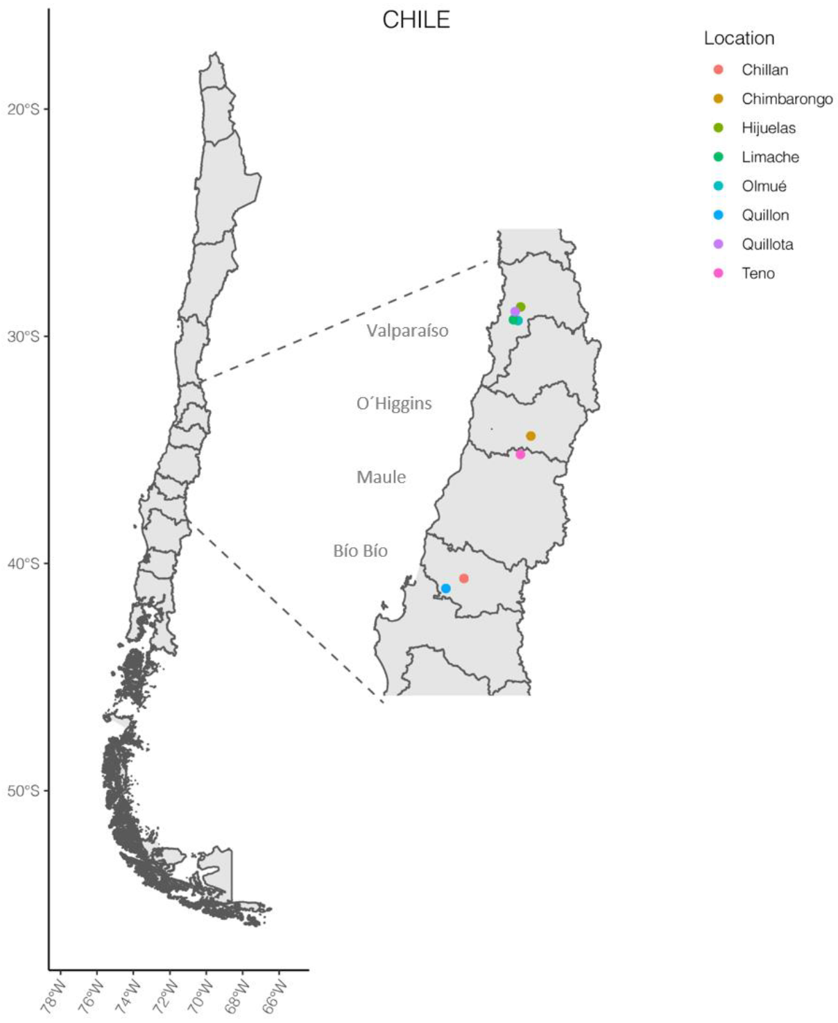
2.1. Bacterial Isolation and Culture Conditions

A total of 58 strains of Pseudomonas spp. were isolated between April and December 2018, and six additional isolates were obtained from samples taken in November 2020 and March 2021, providing a total of 64 isolates included in this study. Unless otherwise specified, bacterial isolates were recovered from the vegetable tissue of tomato plants cultivated in greenhouses, open fields, and nurseries found throughout four regions of the Central Zone of Chile: Valparaíso, O’Higgins, Maule, and Bío Bío (Figure 1). These regions were chosen, as they are the most important to produce tomatoes in Chile, equivalent to more than 75% of the national productive surface [1]. Several studies have shown the ability of Pseudomonads to survive in the environment outside their host plants, even in non-agricultural habitats [39]. For these reasons, samples of irrigation water, soil and other vegetables grown in the same agricultural environment were included for their analysis. Details of the origin of each strain are described in Table 1.
Before processing for bacterial isolation, vegetal tissue was surface disinfected by immersion for 2 min in 5% sodium hypochlorite solution and then immersed for 5 min in sterile distilled water.
To obtain isolates, the plant samples were processed as described below: from each sample, a pool of 10 leaves (or pieces of fruit) was collected and macerated. For the maceration process, 3–5 circular pieces (approximately 1 cm in diameter) of leaf tissue or one fruit piece (approximately 1 cm3) was mixed with 300 µL of buffered physiological water (BPW buffer) (0.14 M NaCl, 2.6 mM NaH2PO4 x 2H2O, and 7.5 mM Na2HPO4 x 12H2O). Bacterial isolation was conducted under sterile conditions by inoculating 50 µL of supernatant from each resulting mash onto solid King’s B medium (KB) (2% protease peptone, 0.15% K2PO4, 0.15% MgSO2 x 7H2O, 1% glycerol, and 1.5% agar, pH 7) [40] and incubated at 28 °C for a period of 24 to 72 h. The initial selection of bacterial isolates was made based on the macroscopic characteristics of the colonies, such as color, shape, brightness, and mucosity. Then the genus of each sampled isolate was confirmed by sequencing the 16S rRNA genes and comparing them with sequences available in the GenBank database. The isolated strains were cultivated in solid KB medium or nutrient broth (NB) (meat extract 0.3%, peptone 0.5%) and incubated at 28 °C. For long-term maintenance, the isolated strains were cryopreserved and stored at −80 °C in nutrient broth with glycerol in a 1:1 ratio.

2.2. Molecular Characterization of Bacterial Isolates

For the molecular characterization of the isolated strains, 16S rRNA gene sequencing and multilocus sequence analysis (MLSA) were conducted, as described below. The primers used in the molecular characterization protocols are listed in Table 2.

2.3. Amplification, Sequencing and Analysis of the 16S rRNA Gene

The genomic DNA of bacterial isolates was extracted by using the Wizard® Genomic DNA Purification Kit (Promega, WI, USA) according to the manufacturer’s instructions. PCR amplification of the 16S rRNA gene was conducted by using established primers 27F and 1492R (Table 2) [41]. PCR was prepared according to the GoTaq® DNA polymerase (Promega, WI, USA) standard protocol. Amplification conditions consisted of initial denaturation for 3 min at 95 °C, followed by thirty cycles of 30 s at 95 °C, 1 min at 58 °C, and 1 min at 72 °C, plus a final extension of 7 min at 72 °C. The PCR product obtained was checked by electrophoresis in an 8% polyacrylamide gel, along with a 100 bp DNA ladder (Promega, WI, USA), following the standard running protocol (200 V, 20 min) [42]. Subsequently, the amplicons were purified and sequenced by an external service at Macrogen Inc. (Seoul, Korea). All sequences were edited and compiled by using Geneious Prime® 2021.1.1 (Biomatters Ltd., Auckland, New Zealand). Partial 16S sequences of 763 bp were obtained for each isolate. Each sequence was compared against the nonredundant nucleotide database of the NCBI (National Center for Biotechnology Information), using the BLAST tool of Geneious software. Pseudomonas species sequences among the first hits with at least 99% identity were considered for selection of reference sequences in later analyses.

2.4. Housekeeping Gene Sequencing and Multilocus Sequence Analysis (MLSA)

Multilocus sequence analysis was performed for the Pseudomonas isolates by PCR amplification and sequencing. Four housekeeping genes were sequenced: those encoding sigma Factor 70 (rpoD), aconitate hydratase B (acnB), citrate synthase (cts), and phosphoglucoisomerase (pgi) [43]. In addition, 16S rDNA sequences were also considered in the MLSA analysis since their usefulness in this type of analysis was previously demonstrated [44,45,46]. To further analyze the genetic variability of the 64 isolated strains of Pseudomonas spp., four loci corresponding to acnB, cts, pgi, and rpoD housekeeping genes were analyzed in addition to the 16S rDNA sequences [44,47,48]. Each gene from the 64 different isolates was PCR-amplified and sequenced by using specific primers. Representative sequences from the genus Pseudomonas were included as references for comparative analysis, using only sequences from complete sequenced genomes (Table 3) [47,48]. Sequences from the corresponding genes of E. coli strain K-12 were included as an outgroup.
Genomic DNA was prepared by using the Wizard® Genomic DNA Purification Kit protocol (Promega) according to the manufacturer’s instructions. PCR amplification was performed on 250 ng of template DNA by using an Agilent thermal cycler. PCR was prepared according to the GoTaq® DNA polymerase (Promega) standard protocol with nucleotide concentrations of 200 µM each and primer concentrations of 1 µM. The amplification conditions consisted of initial denaturation for 3 min at 95 °C, followed by thirty cycles of amplification, with template denaturation at 94 °C for 2 min, the proper annealing temperature (Table 2) for 1 min, and extension at 72 °C for 1 min. A final extension step of 10 min at 72 °C was performed. The correct amplification and size of the amplicons were confirmed through electrophoresis in polyacrylamide gels, as described above.
The amplicons of each gene were sequenced by Macrogen, Inc. (Seoul, Korea). To establish genetic relatedness, the sequences were subjected to sequence pair distance and phylogenetic analysis, along with ten reference sequences (Table 3).
Nucleotide sequences were edited, assembled, aligned, trimmed, and compiled by using Geneious Prime® 2021.1.1 (Biomatters Ltd., Auckland, New Zealand). Since the amount of data obtained was different for each strain, all sequences were trimmed to include only those regions for which we had data for all strains. Sequences from each locus were aligned by using Geneious alignment default parameters and were trimmed to the minimal shared length. For the multilocus sequence analysis, gene sequences were concatenated and aligned according to the alphabetical order of the genes, ending in a sequence of 3095 bp (bp 1 to 768 for 16S rDNA, 769 to 1411 for acn, 1412 to 1938 for cts, 1939 to 2591 for pgi, and 2592 to 3095 for rpoD).
Finally, for genetic relationship analyses, neighbor-joining (NJ) trees were generated in MEGA X software by using the Tamura–Nei evolutionary model with gamma correction and 1000 bootstrap replicates for all sequences [43,47,48,49].

2.5. Copper Tolerance

To evaluate whether the Pseudomonas strains isolated from Chilean fields presented a copper-tolerant phenotype, the minimum inhibitory concentration (MIC) of copper was found for each strain [28,31,50]. Briefly, different bacterial isolates were cultured in liquid medium CYEG (Casitone 1.7%, yeast extract 0.35%, and glycerol 2%) supplemented with increasing concentrations of copper sulfate (considering 1 mM CuSO4 x 5H2O = 63.5 μg/mL Cu2+). The copper concentrations measured were 0, 8, 16, 32, 64, 80, 100, 160, and 200 μg/mL Cu2+, using an incubation temperature of 28 °C. Each isolate was analyzed in triplicate, and bacterial growth was determined by spectrophotometry, measuring the optical density of the culture at 600 nm wavelength (OD600) after 0, 24, 48, and 72 h of incubation. Each assay was started (0 h) with a fresh inoculum of the tested bacterial isolate, fixing an initial OD600 at 0.1. MIC was defined as the point where the OD600 of the bacterial culture at 24, 48, and 72 h was equal to (indicating no growth) or less (indicating possible cell death or lysis) tan OD600 at 0 h.
Pseudomonas isolates were considered copper tolerant when the minimum inhibitory concentration (MIC) was equal to or greater than 64 µg/mL [51].

2.6. Streptomycin Susceptibility

The minimum inhibitory concentration (MIC) of streptomycin was found based on the method described by Valenzuela et al. (2019) [52]. Briefly, bacterial isolates were grown overnight in LB broth (Luria-Bertani; 1% tryptone, 0.5% yeast extract, and 1% NaCl) at 28 °C, with constant agitation. Optical density was measured in a spectrophotometer, as described above, and all cultures were diluted in LB broth to adjust the OD600 to 0.3. Subsequently, 10 µL of each culture was plated onto CYEG agar (Casitone 1.7%, yeast extract 0.35%, glycerol 2%, and agar 12%) supplemented with streptomycin sulfate (Phytotechnology Labs) at concentrations of 0, 2, 4, 6, 8, 10, 12, 15, 20, 50, 100, 250, and 500 µg/mL. Three replicates were conducted for each concentration. Inoculated plates were incubated at 28 °C for 48–72 h. Experiments were performed in duplicate. The MIC of streptomycin was defined as the lowest concentration of streptomycin sulfate at which bacterial growth was inhibited after 48 h of incubation at 28 °C. Pseudomonas isolates with MICs ≥ 50 µg/mL were considered streptomycin resistant [28,52,53].

2.7. Antimicrobial Susceptibility Test (AST)

The in vitro susceptibility of bacterial isolates to five antibiotics used in human medicine was evaluated by using the disc diffusion method. Commercial discs for antibiogram (Thermo Scientific™, Waltham, MA USA, Oxoid™, Basingstokec, UK) with rifampin (5 µg), ampicillin (10 µg), gentamicin (10 µg), chloramphenicol (30 µg), and tetracycline (30 µg) were used according to the manufacturer’s recommendations. Briefly, a fresh inoculum of each strain was diluted in a sterile saline solution (0.85% NaCl), and the OD600 was adjusted to 0.08–0.1 (equivalent to 0.5 on the McFarland turbidity scale). The medium chosen for the test was Mueller–Hinton agar (Thermo Scientific™ Oxoid™) suitable for AST, and the agar plates were poured on leveled surfaces to ensure a uniform depth of agar (~4 mm in depth). Using a sterile technique, the bacteria were inoculated into the Petri dish by spreading a homogeneous layer over the entire surface of the plate. Five antibiogram disks were applied over the inoculated agar, and plates were incubated for 16–18 h at 30 °C. At the end of the culture, the diameters of the zones of inhibition were measured to the nearest whole millimeter, as recommended by the National Committee for Clinical Laboratory Standards (NCCLS) [54]. E. coli ATCC 25922 was used for quality control. Each experiment was repeated twice. The sensitivity of the bacteria to antibiotics was determined according to the specifications of the Clinical and Laboratory Standards Institute (CLSI) [54,55,56,57] and was defined as follows: susceptible (S), a status in which the growth of the bacteria is inhibited in vitro by the applied concentration of the antibiotic and the antibiotic dose is therapeutically effective; moderately susceptible (MS), bacterial response to the drug is lower, and the drug may fail to achieve a therapeutic response; or resistant (R), bacteria are not responsive to the given antibiotic, thus clearly indicating therapeutic failure.
For representation of antimicrobial susceptibility of all studied strains, a heat map was generated by using R software (https://www.R-project.org/) (accessed on 15 November 2021) (R Core Team, 2008), the “heatmap” package stats 3.6.0, and plotted by using the “ggplot2” package.

3. Results

3.1. Isolation and Identification of Pseudomonas from Orchards

The first choice of the bacterial isolates obtained was made based on the macroscopic characteristics of the colonies, such as color, shape, shine, and mucosity. Using these criteria, 298 isolates were obtained. Bacterial colonies that presented characteristics such as the classical morphology of Pseudomonads in KB medium (mucoid, spherical/convex colonies with a defined smooth border, shiny surface, and a fluorescent yellow color) were selected for identification by 16S rRNA sequencing. For sixty-four isolates, the greatest identity matched with representatives of the genus Pseudomonas (Table 4). Among them, 19 isolates were most similar to P. syringae, 7 to P. koreensis, and 6 to P. viridiflava. Additionally, P. gessardii, P. azotoformans, P. fluorescens, and P. marginalis appeared in the first matches of a few isolates. Sequences of 17 isolates matched with at least two sequences among the following species: P. fluorescens, P. koreensis, P. paralactis, P. putida, P. gessardii, P. reinekei, P. brenneri, P. punonensis, P. argentinensis, and unidentified Pseudomonas sp. (Table 4). These 64 isolates identified as Pseudomonas spp. (Table 1 and Table 4) were selected for further analysis.

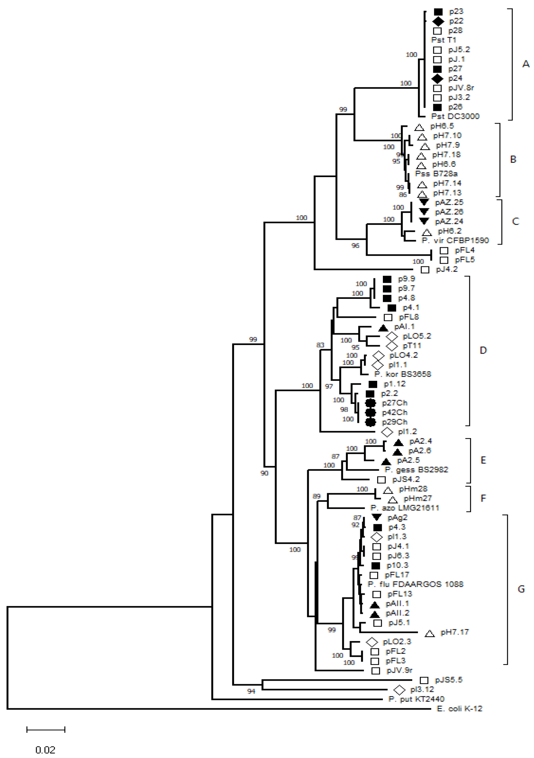
3.2. Genetic Diversity of Pseudomonas spp. Isolates

Reference Pseudomonas species were selected according to the results of the 16S rDNA sequence analysis (Table 4). To study the genetic relationships among the selected strains, NJ trees were constructed based on the concatenation of all five genes (Figure 2) or single genes (Supplementary Figures S1–S5).
Figure 2 shows that 57 strains were grouped into seven genetic lineages (Figure 2, groups A–G), with identity ranging between 95.2 and 100% inside each group. The other seven isolates (pl3.12, pJS5.5, pJV.9r, pl1.2, pJ4.2, pFL5, and pFL4) had identity values lower than 95% (cutoff) when compared with the strains included in the seven groups. Thus, these isolates were set aside and were not included as part of a particular group. The identity among the 64 isolates ranged from 82.2 to 100%. The lowest identity percentage (82.2%) was seen when comparing sequences from the pJ4.2 and pJS5.5 strains. Groups A, B, and C were genetically closer to the reference sequences of P. syringae pv. tomato, P. syringae pv. syringae, and P. viridiflava, respectively, which all belong to the P. syringae complex [48]. Groups D, E, F, and G were closely related to the reference sequences of P. koreensis, P. gessardii, P. azotoformans, and P. fluorescens, respectively.
Group A was composed of 10 isolates that were retrieved from the Valparaíso region. Isolates from various locations (Quillota, Hijuelas, and Limache) clustered together, with identities ranging from 99.1 to 100%. Group B contained seven isolates from Quillón (Bío Bío region) grouped with the P. syringae pv. syringae B728a reference strain. In this group, identity percentages ranged from 98.9 to 100%, with pH6.5 and pH7.9 being the most dissimilar isolates.
Four isolates from the Maule and Bío Bío regions clustered with the P. viridiflava CFBP1590 reference strain to form group C, with identity percentages from 98.7 to 100%. Group D, one of the largest groups (along with group G), consisted of 15 isolates from the Valparaíso, O’Higgins, and Bío Bío regions grouped with the P. koreensis BS3658 reference strain. The identity percentage varied from 95.2 to 100%. Strains isolated from geographically distant points were closely related, as was the case for p2.2 and p27Ch (99.7% identical) isolated from Hijuelas and Chillan locations, respectively. Group E included four isolates from the Valparaíso region and the P. gessardii BS2982 reference strain. Inside this group, identity percentages ranged from 95.8 to 99.7%. Group F was the smallest, including only two isolates (from the same location, Quillón, Bío Bío region) whose sequences were 99.7% identical and clustered with the P. azotoformans reference strain. Finally, group G included 15 isolates and showed identity percentages ranging from 95 to 99.9%. This group included strains from six different orchards distributed in the Valparaíso, O´Higgins, Maule, and Bío Bío regions. Strains in this group were closely related to the P. fluorescens FDAARGOS 1088 reference strain.
Seven isolates could not be clustered in the NJ tree, namely pl1.2, pJ4.2, pFL4, pFL5, pJV.9r, pJS5.5, and pl3.12. For isolate pl1.2, the maximum identity was with pFL8 (95.3%), but with the rest of group D, the identity values were lower than 95%. A similar situation occurred with pJ4.2, pFL4, and pFL5 isolates, which were genetically closer to strains in group C with maximum identity values of 93.8% and 94.3%, when comparing pJ4.2 with pAZ.24/pAZ.25/pAZ.25 and pFL4/pFL5 with all the group C isolates, respectively. Isolate pJV.9r was more closely related to isolates belonging to groups F and G, with identity percentages of 95.2% and 95% when compared with pj5.1 (group G) and pHm28 (group F), respectively. Interestingly, isolates pJS5.5 and pl3.12 were not genetically closer to any of the formed groups, with maximum identity values of 86.2 to 87%, respectively, when compared with the rest of the isolates. This is consistent with their 16S rDNA sequences, which showed identity with other Pseudomonas species not being used as references in the MLSA analysis.
When individual genes were used to compare the isolates (Supplementary Figures S1–S5), similar clustering was seen in the respective dendrograms. The most conserved gene was 16S rDNA, with identity values over 95.4% (Supplementary Figure S1). The compositions of groups A, B, C, and D were conserved in all the constructed dendrograms, with the exception of the 16S rDNA-based analysis, where the major cluster included MLSA groups A and B, which were indistinguishable. Greater sequence variability was seen in the pgi and rpoD genes, mainly among the strains of groups D and G. In the NJ tree based on the cts gene (Supplementary Figure S3), only five groups were clearly distinguished. In this case, MLSA groups E, F, and G (Figure 2) clustered together, except for the pA2.4 and pA2.6 isolates, which were not grouped with the others. Additionally, the position of isolates pl3.12 and pJS5.5 in the tree differed with respect to the MLSA analysis. Furthermore, different results were seen for isolates pl1.2, pJ4.2, pFL4, pFL5, pJV.9r, pJS5.5, and pl3.12 (which were excluded from groups A–G based on the MLSA analysis). Meanwhile, pJS5.5 and pl3.12 were consistently excluded from the clusters; however, this was not the case for pl1.2, pJ4.2, pFL4, pFL5, and pJV.9r. In the cts- and pgi-based analyses, strain pl1.2 was included in group D with the reference strain P. koorensis BS3658. Isolate pJ4.2 was included in group C (including the P. viridiflava CFBP1590 reference strain) when analyses were based on either the pgi or rpoD genes. Otherwise, only pFL4 and pFL5 were included in group C in the analysis based on the cts gene. Isolate pJV.9r showed variable clustering depending on the gene analyzed; for example, in the acn-based tree, this strain was included in group E, but in the NJ tree based on the rpoD gene, this strain was more closely related to the strains of group F. Thus, pJV.9r did not show a clear genetic filiation with the other analyzed isolates. Finally, it should be noted that clustering based only on the rpoD gene best reflected the genetic relationship obtained through the analysis based on the five genes together (MLSA) (Figure 2 and Supplementary Figure S5).

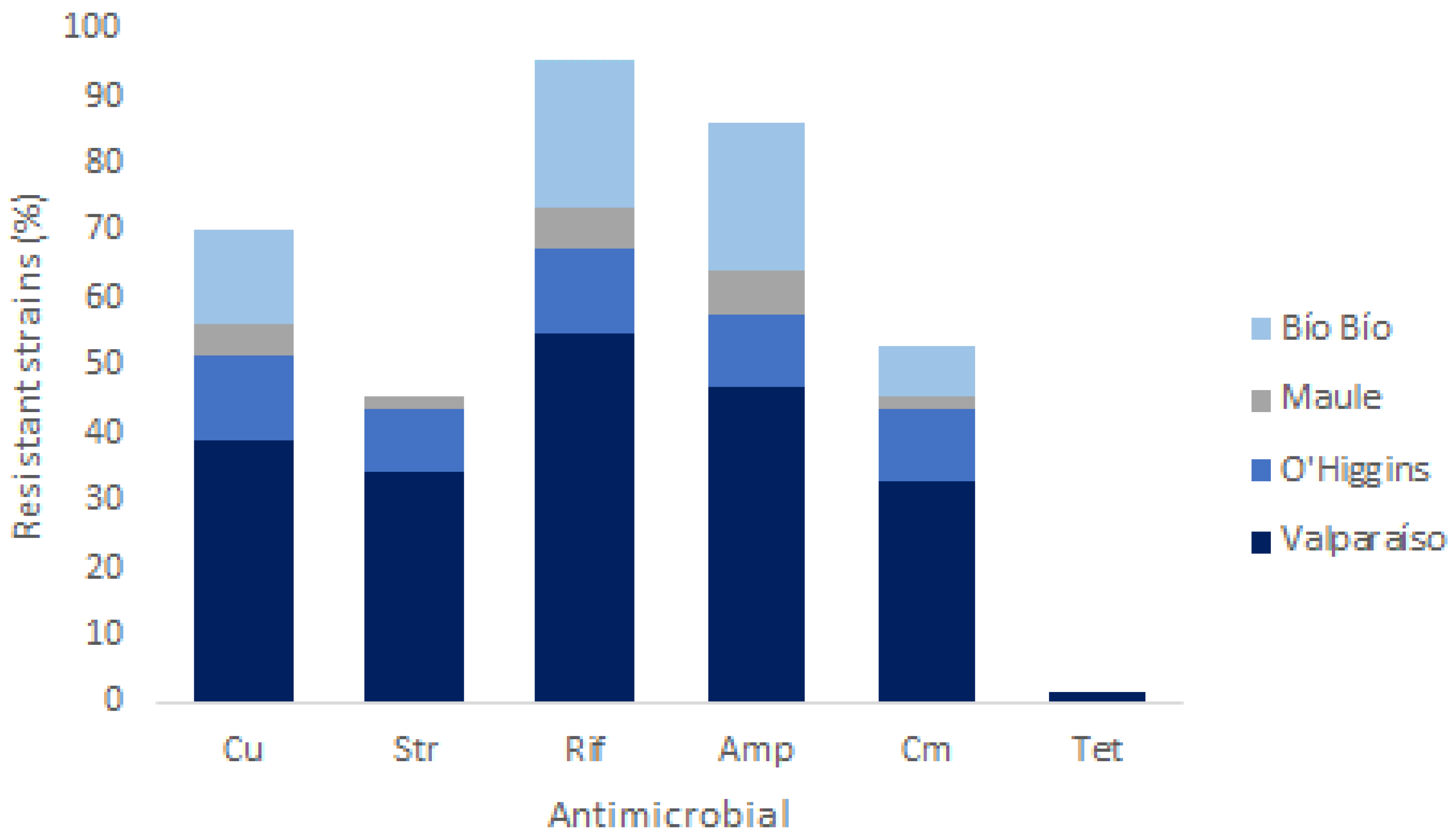
3.3. Phenotypic Tolerance to Copper and Antibiotics

Since copper-based antimicrobials, together with the antibiotic streptomycin, have been the main bacterial control strategy for decades in agricultural crops [11,16,53], the susceptibility of the 64 isolates to these compounds was first evaluated. For this, the minimum inhibitory concentration (MIC) for each isolate was determined in the presence of variable concentrations of copper (Cu) or streptomycin (str). The details of the MIC obtained in each case are shown in Table 5. The results indicated that 70.1% of the isolates showed tolerance to copper, with MIC values greater than 64 µg/mL, and 45.3% were resistant to streptomycin (MIC values ≥ 50 µg/mL). Of the 29 isolates considered resistant to streptomycin, 16 had a highly resistant phenotype with MIC values above 200 µg/mL [52]. Among the 45 copper-tolerant isolates, 24 were also resistant to streptomycin (37.5% of the total). Resistant isolates to one or both antimicrobials typically used in agriculture (copper and streptomycin) were retrieved from all sampled locations (Figure 3). Only 14 isolates were susceptible to both copper and streptomycin, with representatives from the Valparaíso, Maule, O’Higgins, and Bío Bío regions (Table 5).
In addition to streptomycin, we wanted to assess whether the isolates showed resistance to other antibiotics used in human or animal medicine. This could represent a significant health risk, considering the possible transfer of genetic resistance determinants between bacterial isolates, including those pathogenic to humans or animals. For this, an AST using the disc diffusion method was conducted with the antibiotics ampicillin (amp), rifampicin (rif), gentamicin (gn), chloramphenicol (cm), and tetracycline (tet). The results showed that 95%, 86%, 53%, and 1.6% of the isolates could be considered resistant to rifampin, ampicillin, chloramphenicol, and tetracycline, respectively, with no isolates being resistant to gentamicin (Table 5 and Figure 3). Some isolates were considered moderately susceptible (MS), representing a few cases (one to seven) for each antibiotic (Table 5). Resistance to five of the seven antimicrobials evaluated was present in all regions of isolation, except for tetracycline, with only one resistant isolate isolated from the Valparaíso region, and for streptomycin, with no resistant isolates from the Bío Bío region (Figure 3).
To facilitate the analysis, bacterial isolates were grouped in a heatmap according to their antimicrobial resistance (Figure 4). The isolation region and the group to which they belong, based on the MLSA analysis, are also displayed. Overall, the results show that 96.9% of the isolates are tolerant or resistant to a combination of at least two of the antimicrobials evaluated, with only two isolates being resistant to a single antimicrobial. This is the case for the isolates pJS5.5 and AI.1, which showed resistance to copper and rifampin, respectively. Considering resistance characteristics, we found 17 phenotypes among the 64 isolates, ranging from resistance to a single antimicrobial to resistance to five of them (Figure 4). Among all the resistance phenotypes, multi-resistance to Cu-Str-Amp-Cm-Rif was the most frequent (23.4%), followed by the Cu-Amp-Cm-Rif and Cu-Amp-Rif resistance phenotypes, each representing 12.5% of the total isolates (Figure 4).
According to resistance phenotype, isolates were placed into four clusters (Clusters I–IV) (Figure 4). Cluster I contained twenty isolates, 75% resistant to rifampin and ampicillin, with 40% of isolates also being copper tolerant. Only two isolates (pJS5.5 and pAI.1) with resistance to a single antimicrobial (rif) were found in this group. In Cluster II, all the isolates were resistant to chloramphenicol and to ampicillin. Of the 34 isolates in this group, 52.9% were resistant to five antibiotics, and 44.1% were resistant to five antimicrobials (Cu + antibiotics). Isolate JV.9r constituted Cluster III, showing resistance to five antimicrobials (Cu, str, amp, rif, and tet), being the unique isolate resistant to tetracycline. Finally, Cluster IV was composed of three (33.3%) isolates sharing the resistance phenotype to streptomycin, rifampin, ampicillin, and copper. Among them, seven (77.8%) showed resistance to three antimicrobials (Cu, str, and rif). No isolates had resistance to chloramphenicol, gentamicin, and tetracycline.
From a genetic perspective, Cluster I contained all isolates from groups B and C classified according to MLSA. Cluster II contained a major portion of groups D and G, and Cluster IV contained mainly isolates from group A. Additionally, half of the isolates considered highly resistant to streptomycin were grouped in Cluster IV, all of which belonged to group A (MLSA), except for isolate pA2.6 (Figure 4).
All the studied isolates showed resistance to at least one of the tested antimicrobials, regardless of geographic location or sample type of origin, thus suggesting no relation between the spectrum of antimicrobial resistance and the geographic origin of the isolates (Figure 3 and Figure 4).

4. Discussion

We analyzed the diversity of Pseudomonas species associated with tomato plants from Chilean orchards. Pseudomonas isolates were found in 92% of the orchards that were sampled (14 orchards in 8 locations) and were present in both healthy and diseased plants, representing 21.5% of the identified isolates. These results are consistent with previous studies, showing that the genus Pseudomonas has been consistently associated with tomato plants [3,4,5]. Indeed, Dong et al. (2019) reported that the genus Pseudomonas represents 36.76%, 25%, and 13% of the genera associated with the rhizosphere, root endophytes, and leaf endophytes of tomato plants, respectively [4].
Previous studies of Pseudomonas species associated with tomato plants in Chile have mainly focused on phytopathogenic Pseudomonas, such as P. syringae, P. viridiflava, P. corrugate, and P. mediterranea, as etiological agents of bacterial speck, stem necrosis, and pith necrosis of tomato [58]. In this study, phytopathogenic species were isolated from both healthy and diseased plants, consistent with the endophytic character of these phytopathogens; hence, isolates from five locations were identified as P. syringae pv. tomato, P. syringae pv. syringae, or P. viridiflava (Figure 2, MLSA groups A–C), all members of the P. syringae complex.
Members of the P. fluorescens group, such as P. fluorescens, P. koreensis, P. gessardii, and P. azotoformans, usually described as plant growth-promoting rhizobacteria (PGPR), were also obtained, with P. fluorescens and P. koreensis present in seven of the eight sampled locations. Different authors have described these species as being associated with plants or agricultural environments [59,60,61], so we expected to find them in association with tomato crops in Chile, but the distribution or proportion of the varied species found was previously unknown.
Although P. fluorescens has been studied primarily for its biostimulant properties and its potential as a biocontroller [5,62], its ability to emerge as an opportunistic pathogen has been reported in recent years [9], so its presence in tomato plants with symptoms of disease, as is the case of pAII.1, pAII.2, pJ5.1, pJ4.1, and pJ6.3 isolates (Figure 2, group G), must be carefully analyzed. Our identification of isolates as P. koreensis (Figure 2, group D) is similar, since some were obtained from plants with symptoms of illness. However, to our knowledge, there are no reports of P. koreensis with pathogenic characteristics [61].
Earlier MLSAs of Pseudomonas isolates, using the same or some of the genes analyzed in this study, have shown variable results regarding the resolution at the species level or the usefulness of individual genes in predicting genetic parentage. However, in this work, the use of five genes together allowed a greater resolution of the clustering of isolates in the NJ analysis, where each group was mainly associated with a reference isolate representative of a particular species. The same was not seen when analyzing the 16S gene by itself, where the high similarity between all the isolates studied did not allow adequate resolution, even though the genetic region used contained the gene fragment suggested by Singh et al., in studying the phylogenetic relationship of Pseudomonas species [46]. For example, in analyzing this gene, it was found that the isolates associated with the species P. azotoformans, P. gessardii, and P. fluorescens could not be clearly distinguished from each other. This would be expected since these species are defined as being closely related, even being grouped together in the past within the so-called “P. fluorescens group” [45]. The analysis of the individual genes showed that the result obtained from the individual analysis of the rpoD gene was the one that best represented the clustering results obtained by MLSA. This agrees with what was seen by Oueslati et al., who used the rpoD, 16S, and gyrB genes to study the phylogenetic relationship of Pseudomonas syringae isolates [63]. Other authors have proposed that the cts gene alone can accurately predict the phylogenetic grouping for the P. syringae group [47]. However, in this study, the results of the analysis based only on the cts gene were the most dissimilar from those of the MLSA. This may be because the isolates we studied included diverse species, not only P. syringae. In this regard, the most conserved groups both in the MLSA and analysis of individual genes were those that were genetically related to Pseudomonas species of the syringae complex (syringae and viridiflava), thus confirming that these markers are useful for the phylogenetic analysis of these species [43,47].
It is worth mentioning that there are no earlier studies that analyzed the diversity of Pseudomonas by using the five housekeeping genes used in this study on isolates of Chilean origin. Generally, previous studies were conducted primarily with isolates from Europe and North America [64], making these results the first to focus on Pseudomonas spp. associated with tomato crops in Chile. We believe it imperative to continue genetic analysis in this region since important genetic differences have been reported among isolates from different geographical origins. For example, in the case of P. syringae pv. tomato, differences in type III effector genetic markers have been associated with host-range determination of different strains [64,65].
The intensive use of agrochemicals to control different Pseudomonas species has promoted the selection of resistant bacteria. Furthermore, isolates resistant and multiresistant to these treatments have been increasingly reported [11,28]. This study was the first associated with tomato crops to evaluate the phenotypic and tolerance to antimicrobial compounds in agricultural environments in Chile. Samples of the same orchard in different seasons or years were not taken, so reliable conclusions related to bacterial community changes in different seasons or how it is antimicrobial resistance would be affected depending on the season cannot be drawn and require future studies.
The results showed variable resistance phenotypes among the Pseudomonas isolates, but all 64 isolates showed resistance to at least one of the antimicrobials evaluated. In general, according to the MLSA, the results suggest no relationship between antimicrobial resistance and the genetic group to which the isolates belonged. The resistance of the studied isolates to copper and streptomycin is not surprising, since this characteristic is widely reported in different bacterial species associated with agricultural crops, including P. syringae and P. fluorescens, among others [11,14,17,28].
With ampicillin, the high percentage of resistant isolates has a precedent in the study conducted by Hwang et al. [28], where the author proved that resistance to this antibiotic seems to be an ancestral trait in the P. syringae group. Additionally, Armalyté et al. described a high frequency of ampicillin-resistant Pseudomonas isolated from agricultural soil [37]. Ampicillin, a beta-lactam antibiotic, is affected by β-lactamases. Among them, extended spectrum β-lactamases (ESBLs) are enzymes of Gram-negative bacteria conferring resistance against β-lactam antibiotics. ESBL producing Gram-negative bacteria have been reported worldwide, and resistance genes of the ESBL type are mostly plasmid associated and therefore can spread among different bacteria by horizontal transfer, leading to widespread resistance among different bacterial species [66]. In concordance, Igbinosa et al., detected the blaTEM antibiotic resistance gene (β-lactamase conferring ampicillin resistance) in 12.5% of P. putida, 57.14% of P. fluorescens, 100% of P. aeruginosa, and 40% in other Pseudomonas species isolated from environmental water samples [35].
The same has not been observed with the antibiotic rifampicin, for which contradictory results have been reported. In the study of Hwang et al., only 16 out of 60 Pseudomonas isolates were shown to be phenotypically resistant to rifampin [28], but a different result was observed by Igbinosa et al., where Pseudomonas isolates from two locations showed 100% resistance to rifampin [35]. Rifampin resistance is generally mediated by rifampin ADP-ribosyltransferases or by a mutation in the subunit of RNA polymerase, which has been considered unusual among environmental bacteria [37,67]. However, our results showed a high frequency of rifampin-resistant phenotypes among Pseudomonas isolates, similar to that observed by Igbinosa et al. [35], a scenario that is consistent with a generalized increase in resistance to different antibiotics in environmental bacteria worldwide.
In the context of antibiotic resistance, it has been determined that P. aeruginosa, which has been extensively studied due to its effect on human health, possesses intrinsic resistance to some of these compounds, including ampicillin, chloramphenicol, and tetracyclines [68]. However, apart from the high frequency of resistance to ampicillin, these “intrinsic” resistance characteristics have not been observed in environmental or agricultural crop-associated Pseudomonas isolates in this or in previous studies [28,63,69]. Although resistance to these antimicrobials cannot be considered an intrinsic characteristic, some Pseudomonas isolates with phenotypes resistant to ampicillin, chloramphenicol, and tetracycline have been found in this study and previously [28,37,63,69]. In addition to ampicillin resistance, a chloramphenicol resistance phenotype was also seen with high frequency among the Pseudomonas isolates. While ampicillin resistance is often associated with the presence of beta-lactamases, chloramphenicol resistance is most often due to the presence of chloramphenicol acetyltransferases (CATs). However, in both cases, the participation of efflux pumps or membrane-associated transporters that could confer multidrug resistance has been described [36,37]. This is the case for resistance–nodulation–cell division (RND) superfamily exporters, which play a major role in drug expulsion in P. aeruginosa but have also been reported in several environmental Pseudomonas spp. [37]. In the case of tetracycline, resistance is most often due to the acquisition of new genes that code for energy-dependent efflux of tetracyclines, proteins that protect bacterial ribosomes from the action of tetracyclines, or enzymatically inactivate tetracyclines [36]. Despite a great diversity of genetic determinants conferring tetracycline resistance among several bacterial species, in this study, only one isolate showed resistance to this antibiotic.
Regarding gentamicin, the absence of resistance among the isolates studied agrees with earlier studies where it was seen that resistance to this antibiotic is not a widespread characteristic in the genus Pseudomonas [28,63]. This can be explained since, in contrast to streptomycin, gentamicin binds to multiple sites on ribosomes; thus, several mutations in bacterial chromosome would be needed to generate spontaneous mutants resistant to gentamicin [38]. However, Pseudomonas is considered among the most competent of bacteria regarding DNA uptake, imparting it a high possibility of picking up genes from the environment, which may explain the development of several new resistance genes [35]. Moreover, genetic determinants of antimicrobial resistance are often associated with plasmids, transposons, or gene cassettes, and also may include integrative and conjugative elements [11,18,36]. Additionally, antibiotic resistance may be a function of more than one gene or even a combination of genetic and environmental factors, and a resistant phenotype can sometimes be observed without detecting a specific genetic determinant [35].
Aside from resistance to a single antimicrobial agent, the most challenging public health problem is the increasing number of multiresistant bacteria. In this study, antimicrobial resistance analyses showed that 96.9% of the isolates were phenotypically resistant to a combination of at least two of the antimicrobials assessed. Among all the resistance phenotypes, multiresistance to Cu-Str-Amp-Cm-Rif was the most frequent (23.4%). Currently, there is growing concern about the co-selection of bacteria resistance to antibiotics and metals (such as copper) due to the excessive use of these compounds in agriculture [25,26]. It has been shown that the presence of high concentrations of metals favors the co-selection of resistance to antibiotics [26,70], generating co-resistance (presence of different determinants of resistance in the same genetic element) and cross-resistance, where the same genetic determinant confers resistance to antibiotics and metals [25]. In this context, bacterial efflux pumps can capture and extrude many structurally diverse antibiotics, in addition to nonantibiotic compounds, such as metal ions. In Gram-negative bacteria, the multidrug-resistant phenotype is largely conferred by resistance–nodulation–cell division superfamily (RND) efflux systems contributing to the intrinsic resistance of different Pseudomonas species [71]. Thus, this type of efflux pump probably plays a role in the multiple resistance of the Pseudomonas isolates studied, but more genomic and functional studies are needed to corroborate this hypothesis.
Faced with the imminent problem of antibiotic resistance, it is necessary to develop, implement, or reinforce alternative control measures. In this context, reinforcement of preventive control of bacteriosis would allow for us to reduce the use of bactericides. In the nursery, it is critical to use bacteria-free seeds and test seedlings before delivery to growers. At the producer level, it is opportune to consider carrying out crop rotation, the cleaning of greenhouse structures, the elimination of plant residues from previous production, the use of resistant varieties, the reduction of relative humidity inside the greenhouse, and use of ozonated or ultrafiltered water for irrigation as principal preventive management [72]. Moreover, the use of non-conventional control strategies could be implemented, such as biocontrol or phage therapy [73], to reduce the use of agrochemicals. Finally, it is considered of great importance to establish a program of antimicrobial resistance surveillance as a critical step within risk-assessment schemes and for detecting new trends and emerging threats [74].
Although copper or streptomycin resistance has been identified in several phytopathogenic bacteria [11,53], we are aware of few studies reporting resistance in environmental bacteria that are considered to be nonpathogenic either to both antimicrobials (Cu and Str) or in combination with antibiotics for nonagricultural use. The results of this study show that there are multiresistant environmental Pseudomonas in Chile, probably the result of intensive agrochemical use. Although the genetic basis of resistant phenotypes was not addressed in this study, the results establish a precedent for concern, where environmental bacteria frequently associated with agricultural crops can constitute a natural reservoir of resistance genes, not only for phytopathogens but also for human pathogens [30], and may further threaten public health and ecological security via horizontal gene transfer [11,19,70,75]. Since horizontal gene transfer (HGT) is one of the mechanisms that contributes the most to the spread of resistance determinants, recent studies demonstrating HGT events between endophytic and epiphytic bacteria associated with plants are of importance, considering that endophytic bacteria associated with edible plants could easily enter in contact with human associated bacteria trough the food-chain [76,77]. Thus, the presence of Pseudomonas isolates that are resistant to conventional bactericides and other antimicrobials makes these bacteria an emerging threat to the agriculture industry and to human health.

5. Conclusions

Numerous Pseudomonas spp. associated with tomato crops in Chile were found in this study. The isolated Pseudomonas spp. belonged to distinct species based on multilocus sequence analysis (MLSA) and showed diverse antimicrobial response patterns, with most of the isolates showing resistance to at least two of the tested antimicrobial agents. Antimicrobial resistance dissemination among bacterial populations is an increasing challenge worldwide. Antibiotic resistance in Pseudomonas isolates, mainly P. syringae, P. viridiflava, and P. fluorescens species, recovered from vegetal tissue is of particular concern because these Pseudomonas spp. are considered phytopathogens or opportunistic pathogens. Additionally, multiresistant environmental Pseudomonas could be a vehicle for the transmission of resistance genes to human pathogens. For all of these reasons, phytopathogenic bacteria should be at the center of antimicrobial resistance studies, and surveillance throughout the agricultural environment is needed to detect emerging multiresistant phenotypes.

Supplementary Materials

The following are available online at https://www.mdpi.com/article/10.3390/horticulturae8080750/s1, Figure S1. Neighbor-Joining tree based on 16S rRNA gene sequences data set. The length of the 692 compared sequences is 768 b. Bootstrap scores greater than 80 are given at each node. E. coli K-12 693 strain was used as an outgroup. The evolutionary distances were computed using the Tamura-Nei 694 method and are in the units of the number of base substitutions per site. The rate variation among 695 sites was modeled with a gamma distribution (shape parameter = 2), Figure S2. Neighbor-Joining tree based on acn gene rDNA sequences data set. The length of the compared 698 sequences is 642 b. Bootstrap scores greater than 80 are given at each node. E. coli K-12 strain was used as an 699 outgroup. The evolutionary distances were computed using the Tamura-Nei method and are in the units of the 700 number of base substitutions per site. The rate variation among sites was modeled with a gamma distribution 701 (shape parameter = 2), Figure S3. Neighbor-Joining tree based on cts gene rDNA sequences data set. The length of the 704 compared sequences is 526 bp. Bootstrap scores greater than 80 are given at each node. E. coli K-12 705 strain was used as an outgroup. The evolutionary distances were computed using the Tamura-Nei 706 method and are in the units of the number of base substitutions per site. The rate variaon among 707 sites was modeled with a gamma distribution (shape parameter = 2), Figure S4. Neighbor-Joining tree based on pgi gene rDNA sequences data set. The length of the compared se- 710 quences is 652 b. Bootstrap scores greater than 80 are given at each node. E. coli K-12 strain was used as an 711 outgroup. The evolutionary distances were computed using the Tamura-Nei method and are in the units of the 712 number of base substitutions per site. The rate variation among sites was modeled with a gamma distribution 713 (shape parameter = 2), Figure S5. Neighbor-Joining tree based on rpoD gene sequences data set. The length of the compared sequences 822 is 503 b. Bootstrap scores greater than 80 are given at each node. E. coli K-12 strain was used as an outgroup. 823 The evolutionary distances were computed using the Tamura-Nei method and are in the units of the number 824 of base substitutions per site. The rate variation among sites was modeled with a gamma distribution (shape 825 parameter = 2).

Author Contributions

Conceptualization, P.C. and G.H.; methodology, J.P.R.-G., V.R.-M., F.V. and D.S.M.; software, P.V., J.P.R.-G. and J.B.; validation, J.R.; writing—original draft preparation, P.C.; writing—review and editing, G.H., A.Z. and N.F.; visualization and investigation, C.I.-D.; formal analysis, F.V. and E.G.; supervision, G.H. and P.C.; project administration, P.C.; resources, A.Z. and N.F. All authors have read and agreed to the published version of the manuscript.

Funding

This work was funding by Fondecyt Postdoctorado Nº 3180500 headed by Pamela Córdova. Fondecyt Postdoctorado Nº 3170806. CONICYT, PAI/Concurso Nacional Inserción de Capital Humano Avanzado en la Academia Convocatoria año 2017 Nº PAI79170055. Vicerrectoría de Investigación y Desarrollo (VID) de la Universidad de Chile, código Nº UI-038/19.

Institutional Review Board Statement

Not applicable.

Informed Consent Statement

Not applicable.

Data Availability Statement

The data presented in this study are available upon request from the corresponding author.

Acknowledgments

The authors thank Rodrigo Rojas for providing the E. coli ATCC 25922 strain that was used in this work.

Conflicts of Interest

The authors declare no conflict of interest.

References

  1. Muñoz, V.M. Boletín de Hortalizas. Diciembre 2021 [En Linea]. Available online: https://app.powerbi.com/view?r=eyJrIjoiOGQ1OTI0YzMtYzUzOC00OWM5LWFiMzEtMTQxMGNmOTEyYzU3IiwidCI6IjMzYjdmNzA3LTZlNmYtNDJkMi04ZDZmLTk4YmZmOWZiNWZhMCIsImMiOjR9 (accessed on 10 July 2022).
  2. Basim, H.; Basim, E.; Yilmaz, S.; Dickstein, E.R.; Jones, J.B. An Outbreak of Bacterial Speck Caused by Pseudomonas syringae pv. tomato on Tomato Transplants Grown in Commercial Seedling Companies Located in the Western Mediterranean Region of Turkey. Plant Dis. 2004, 88, 1050. [Google Scholar] [CrossRef] [PubMed]
  3. Ottesen, A.R.; Peña, A.G.; White, J.R.; Pettengill, J.B.; Li, C.; Allard, S.; Rideout, S.; Allard, M.; Hill, T.; Evans, P.; et al. Baseline survey of the anatomical microbial ecology of an important food plant: Solanum lycopersicum (tomato). BMC Microbiol. 2013, 13, 114. [Google Scholar] [CrossRef] [PubMed]
  4. Dong, C.-J.; Wang, L.-L.; Li, Q.; Shang, Q.-M. Bacterial communities in the rhizosphere, phyllosphere and endosphere of tomato plants. PLoS ONE 2019, 14, e0223847. [Google Scholar] [CrossRef]
  5. Toju, H.; Okayasu, K.; Notaguchi, M. Leaf-associated microbiomes of grafted tomato plants. Sci. Rep. 2019, 9, 1787. [Google Scholar] [CrossRef] [PubMed]
  6. Parte, A.C. LPSN—List of prokaryotic names with standing in nomenclature (Bacterio.Net), 20 years on. Int. J. Syst. Evol. Microbiol. 2018, 68, 1825–1829. [Google Scholar] [CrossRef]
  7. Catara, V. Pseudomonas corrugata: Plant pathogen and/or biological resource? Mol. Plant Pathol. 2007, 8, 233–244. [Google Scholar] [CrossRef]
  8. Jones, J.B. Pseudomonas viridiflava: Causal Agent of Bacterial Leaf Blight of Tomato. Plant Dis. 1984, 68, 341–342. [Google Scholar] [CrossRef]
  9. Dimartino, M.; Panebianco, S.; Vitale, A.; Castello, I.; Leonardi, C.; Cirvilleri, G.; Polizzi, G. Occurrence and Patho-genicity of Pseudomonas fluorescens and P. putida on tomato plants in Italy. J. Plant Pathol. 2011, 93, 79–87. [Google Scholar] [CrossRef]
  10. Pekhtereva, E.; Kornev, K.; Matveeva, E.; Polityko, V.; Budenkov, N.; Ignatov, A.; Schaad, N. Pith necrosis of tomato in Russia. Acta Hortic. 2009, 808, 251–254. [Google Scholar] [CrossRef]
  11. Lamichhane, J.R.; Osdaghi, E.; Behlau, F.; Köhl, J.; Jones, J.B.; Aubertot, J.-N. Thirteen decades of antimicrobial copper compounds applied in agriculture. A review. Agron. Sustain. Dev. 2018, 38, 28. [Google Scholar] [CrossRef]
  12. Sundin, G.W.; Wang, N. Antibiotic Resistance in Plant-Pathogenic Bacteria. Annu. Rev. Phytopathol. 2018, 56, 161–180. [Google Scholar] [CrossRef]
  13. Cooksey, D.A.; Azad, H.R. Accumulation of Copper and Other Metals by Copper-Resistant Plant-Pathogenic and Saprophytic Pseudomonads. Appl. Environ. Microbiol. 1992, 58, 274–278. [Google Scholar] [CrossRef]
  14. Yang, C.H.; Menge, J.A.; Cooksey, D.A. Role of copper resistance in competitive survival of Pseudomonas fluorescens in soil. Appl. Environ. Microbiol. 1993, 59, 580–584. [Google Scholar] [CrossRef]
  15. Griffin, K.; Gambley, C.; Brown, P.; Li, Y. Copper-tolerance in Pseudomonas syringae pv. tomato and Xanthomonas spp. and the control of diseases associated with these pathogens in tomato and pepper. A systematic literature review. Crop Prot. 2017, 96, 144–150. [Google Scholar] [CrossRef]
  16. Cameron, A.; Sarojini, V. Pseudomonas syringae pv actinidiae: Chemical control, resistance mechanisms and possible alternatives. Plant Pathol. 2013, 63, 1–11. [Google Scholar] [CrossRef]
  17. Xu, Y.; Luo, Q.-Q.; Zhou, M.-G. Identification and Characterization of Integron-Mediated Antibiotic Resistance in the Phytopathogen Xanthomonas oryzae pv. oryzae. PLoS ONE 2013, 8, e55962. [Google Scholar] [CrossRef]
  18. Cooksey, D.A. Molecular mechanisms of copper resistance and accumulation in bacteria. FEMS Microbiol. Rev. 1994, 14, 381–386. [Google Scholar] [CrossRef]
  19. Walsh, C.A. Molecular mechanisms that confer antibacterial drug resistance. Nature 2000, 406, 775–781. [Google Scholar] [CrossRef]
  20. Thanner, S.; Drissner, D.; Walsh, F. Antimicrobial Resistance in Agriculture. mBio 2016, 7, e02227-15. [Google Scholar] [CrossRef]
  21. Ruimy, R.; Brisabois, A.; Bernede, C.; Skurnik, D.; Barnat, S.; Arlet, G.; Momcilovic, S.; Elbaz, S.; Moury, F.; Vibet, M.-A.; et al. Organic and conventional fruits and vegetables contain equivalent counts of Gram-negative bacteria expressing resistance to antibacterial agents. Environ. Microbiol. 2010, 12, 608–615. [Google Scholar] [CrossRef]
  22. Allydice-Francis, K.; Brown, P.D. Diversity of Antimicrobial Resistance and Virulence Determinants in Pseudomonas aeruginosa Associated with Fresh Vegetables. Int. J. Microbiol. 2012, 2012, 426241. [Google Scholar] [CrossRef]
  23. Sun, Y.; Guo, G.; Tian, F.; Chen, H.; Liu, W.; Li, M.; Wang, S. Antibiotic resistance genes and bacterial community on the surfaces of five cultivars of fresh tomatoes. Ecotoxicology 2020, 30, 1550–1558. [Google Scholar] [CrossRef]
  24. Scaccia, N.; Vaz-Moreira, I.; Manaia, C.M. The risk of transmitting antibiotic resistance through endophytic bacteria. Trends Plant Sci. 2021, 26, 1213–1226. [Google Scholar] [CrossRef]
  25. Baker-Austin, C.; Wright, M.S.; Stepanauskas, R.; McArthur, J. Co-selection of antibiotic and metal resistance. Trends Microbiol. 2006, 14, 176–182. [Google Scholar] [CrossRef]
  26. Seiler, C.; Berendonk, T.U. Heavy metal driven co-selection of antibiotic resistance in soil and water bodies impacted by agriculture and aquaculture. Front. Microbiol. 2012, 3, 399. [Google Scholar] [CrossRef]
  27. Scheck, H.J.; Pscheidt, J.W.; Moore, L.W. Copper and Streptomycin Resistance in Strains of Pseudomonas syringae from Pacific Northwest Nurseries. Plant Dis. 1996, 80, 1034–1039. [Google Scholar] [CrossRef]
  28. Hwang, M.S.H.; Morgan, R.L.; Sarkar, S.F.; Wang, P.W.; Guttman, D.S. Phylogenetic Characterization of Virulence and Resistance Phenotypes of Pseudomonas syringae. Appl. Environ. Microbiol. 2005, 71, 5182–5191. [Google Scholar] [CrossRef] [PubMed]
  29. Corzo-Ariyama, H.A.; García-Heredia, A.; Heredia, N.; García, S.; León, J.; Jaykus, L.; Solís-Soto, L. Phylogroups, pathotypes, biofilm formation and antimicrobial resistance of Escherichia coli isolates in farms and packing facilities of tomato, jalapeño pepper and cantaloupe from Northern Mexico. Int. J. Food Microbiol. 2018, 290, 96–104. [Google Scholar] [CrossRef] [PubMed]
  30. Berg, J.; Tom-Petersen, A.; Nybroe, O. Copper amendment of agricultural soil selects for bacterial antibiotic resistance in the field. Lett. Appl. Microbiol. 2004, 40, 146–151. [Google Scholar] [CrossRef] [PubMed]
  31. Esterio, M.; Pérez, L.; Auger, J. Analysis of genetic diversity of Xanthomonas arboricola pv juglandis strains, in Chile. Fitopatología 2007, 43, 110–121. [Google Scholar]
  32. Altimira, F.; Yáñez, C.; Bravo, G.; González, M.; Rojas, L.A.; Seeger, M. Characterization of copper-resistant bacteria and bacterial communities from copper-polluted agricultural soils of central Chile. BMC Microbiol. 2012, 12, 193. [Google Scholar] [CrossRef]
  33. Méndez, V.; Fuentes, S.; Morgante, V.; Hernández, M.; González, M.; Moore, E.; Seeger, M. Novel hydrocarbon clastic metal-tolerant Acinetobacter and Pseudomonas strains from Aconcagua River oil-polluted soil. J. Soil Sci. Plant Nutr. 2017, 17, 1074–1087. [Google Scholar] [CrossRef]
  34. Arguello, J.M.; Raimunda, D.; Padilla-Benavides, T. Mechanisms of copper homeostasis in bacteria. Front. Cell. Infect. Microbiol. 2013, 3, 73. [Google Scholar] [CrossRef]
  35. Igbinosa, I.H.; Nwodo, U.U.; Sosa, A.; Tom, M.; Okoh, A.I. Commensal Pseudomonas Species Isolated from Wastewater and Freshwater Milieus in the Eastern Cape Province, South Africa, as Reservoir of Antibiotic Resistant Determinants. Int. J. Environ. Res. Public Health 2012, 9, 2537–2549. [Google Scholar] [CrossRef]
  36. Roberts, M.C.; Schwarz, S. Tetracycline and Phenicol Resistance Genes and Mechanisms: Importance for Agriculture, the Environment, and Humans. J. Environ. Qual. 2016, 45, 576–592. [Google Scholar] [CrossRef]
  37. Armalytė, J.; Skerniškytė, J.; Bakienė, E.; Krasauskas, R.; Šiugždinienė, R.; Kareivienė, V.; Kerzienė, S.; Klimienė, I.; Sužiedėlienė, E.; Ružauskas, M. Microbial Diversity and Antimicrobial Resistance Profile in Microbiota From Soils of Conventional and Organic Farming Systems. Front. Microbiol. 2019, 10, 892. [Google Scholar] [CrossRef]
  38. Stockwell, V.; Duffy, B. Use of antibiotics in plant agriculture. Rev. Sci. Et Technol. De L’oie 2012, 31, 199–210. [Google Scholar] [CrossRef]
  39. Monteil, C.L.; Cai, R.; Liu, H.; Llontop, M.E.M.; Leman, S.; Studholme, D.J.; Morris, C.E.; Vinatzer, B.A. Nonagricultural reservoirs contribute to emergence and evolution of Pseudomonas syringae crop pathogens. New Phytol. 2013, 199, 800–811. [Google Scholar] [CrossRef]
  40. King, E.O.; Ward, M.K.; Raney, D.E. Two simple media for the demonstration of pyocyanin and fluorescin. J. Lab. Clin. Med. 1954, 44, 301–307. [Google Scholar]
  41. Frank, J.A.; Reich, C.I.; Sharma, S.; Weisbaum, J.S.; Wilson, B.A.; Olsen, G.J. Critical Evaluation of Two Primers Commonly Used for Amplification of Bacterial 16S rRNA Genes. Appl. Environ. Microbiol. 2008, 74, 2461–2470. [Google Scholar] [CrossRef]
  42. Sambrook, J.; Russell, D.W. Molecular Cloning: A Laboratory Manual (3-Volume Set); Molecular Cloning: A Laboratory Manual; Cold Spring Harbor Laboratory Press: Cold Spring Harbor, NY, USA, 2001. [Google Scholar]
  43. Sarkar, S.F.; Guttman, D.S. Evolution of the Core Genome of Pseudomonas syringae, a Highly Clonal, Endemic Plant Pathogen. Appl. Environ. Microbiol. 2004, 70, 1999–2012. [Google Scholar] [CrossRef] [PubMed]
  44. Yamamoto, S.; Harayama, S. Phylogenetic relationships of Pseudomonas putida strains deduced from the nucleotide sequences of gyrB, rpoD and 16S rRNA genes. Int. J. Syst. Bacteriol. 1998, 48, 813–819. [Google Scholar] [CrossRef] [PubMed]
  45. Anzai, Y.; Kim, H.; Park, J.-Y.; Wakabayashi, H.; Oyaizu, H. Phylogenetic affiliation of the Pseudomonads based on 16S rRNA sequence. Int. J. Syst. Evol. Microbiol. 2000, 50, 1563–1589. [Google Scholar] [CrossRef] [PubMed]
  46. Singh, V.; Kumar, A.; Kumar, S. Phylogenetic analysis on 16s ribosomal DNA of Pseudomonas strains from clinical cases of animals. Progress. Res.-Int. J. 2018, 13, 92–95. [Google Scholar]
  47. Berge, O.; Monteil, C.; Bartoli, C.; Chandeysson, C.; Guilbaud, C.; Sands, D.C.; Morris, C.E. A User’s Guide to a Data Base of the Diversity of Pseudomonas syringae and Its Application to Classifying Strains in This Phylogenetic Complex. PLoS ONE 2014, 9, e105547. [Google Scholar] [CrossRef]
  48. Flores, O.; Prince, C.; Nuñez, M.; Vallejos, A.; Mardones, C.; Yáñez, C.; Besoain, X.; Bastías, R. Genetic and Phenotypic Characterization of Indole-Producing Isolates of Pseudomonas syringae pv. actinidiae Obtained From Chilean Kiwifruit Orchards. Front. Microbiol. 2018, 9, 1907. [Google Scholar] [CrossRef]
  49. van de Peer, Y.; Salemi, M. Phylogenetic Inference Based on Distance Methods. In The Phylogenetic Handbook; Cambridge University Press: Cambridge, UK, 2012. [Google Scholar]
  50. Abbasi, P.A.; Khabbaz, S.E.; Weselowski, B.; Zhang, L. Occurrence of copper-resistant strains and a shift in Xanthomonas spp. causing tomato bacterial spot in Ontario. Can. J. Microbiol. 2015, 61, 753–761. [Google Scholar] [CrossRef]
  51. Nakajima, M.; Goto, M.; Hibi, T. Similarity between Copper Resistance Genes from Pseudomonas syringae pv. actinidiae and P. syringae pv. tomato. J. Gen. Plant Pathol. 2002, 68, 68–74. [Google Scholar] [CrossRef]
  52. Valenzuela, M.; Méndez, V.; Montenegro, I.; Besoain, X.; Seeger, M. Streptomycin resistance in Clavibacter michiganensis subsp. michiganensis strains from Chile is related to an rpsL gene mutation. Plant Pathol. 2018, 68, 426–433. [Google Scholar] [CrossRef]
  53. Sundin, G.W.; Bender, C.L. Ecological and genetic analysis of copper and streptomycin resistance in Pseudomonas syringae pv. syringae. Appl. Environ. Microbiol. 1993, 59, 1018–1024. [Google Scholar] [CrossRef]
  54. Clinical and Laboratory Standards Institute. Performance Standards for Antimicrobial Susceptibility Testing; Twenty-Second Informational Supplement; Scientific Research: Wuhan, China, 2012; Volume 32. [Google Scholar]
  55. Jorgensen, J.H.; Ferraro, M.J. Antimicrobial Susceptibility Testing: A Review of General Principles and Contemporary Practices overview of commonly used susceptibility testing methods. Clin. Infect. Dis. 2009, 49, 1749–1755. [Google Scholar] [CrossRef]
  56. Khezri, S.; Rahimian, H.; Ahangaran, A.; Mohammadi, M. Comparisons of Iranian strains of Pseudomonas syringae pv syringae from various hosts with different methods. Int. J. Agric. Biol. 2010, 12, 106–110. [Google Scholar]
  57. Sarker, M.R.; Islam, K.N.; Huri, H.Z.; Rahman, M.; Imam, H.; Hosen, B.; Mohammad, N.; Sarker, Z.I. Studies of the Impact of Occupational Exposure of Pharmaceutical Workers on the Development of Antimicrobial Drug Resistance. J. Occup. Health 2014, 56, 260–270. [Google Scholar] [CrossRef]
  58. Estay, P.; Bruna, A. Insectos, Ácaros y Enfermedades Asociadas al Tomate en Chile; Colección Libros INIA; Instituto de Investigaciones Agropecuarias: Santiago, Chile, 2002; Volume 7. [Google Scholar]
  59. Garrido-Sanz, D.; Arrebola, E.; Martínez-Granero, F.; García-Méndez, S.; Muriel, C.; Blanco-Romero, E.; Martín, M.; Rivilla, R.; Redondo-Nieto, M. Classification of Isolates from the Pseudomonas fluorescens Complex into Phylogenomic Groups Based in Group-Specific Markers. Front. Microbiol. 2017, 8, 413. [Google Scholar] [CrossRef]
  60. Haque, M.; Mosharaf, K.; Khatun, M.; Haque, A.; Biswas, S.; Islam, S.; Islam, M.; Shozib, H.B.; Miah, M.U.; Molla, A.H.; et al. Biofilm Producing Rhizobacteria with Multiple Plant Growth-Promoting Traits Promote Growth of Tomato Under Water-Deficit Stress. Front. Microbiol. 2020, 11, 542053. [Google Scholar] [CrossRef]
  61. Guo, Q.; Sun, Y.; Shi, M.; Han, X.; Jing, Y.; Li, Y.; Li, H. Pseudomonas koreensis promotes tomato growth and shows potential to induce stress tolerance via auxin and polyphenol-related pathways. Plant Soil 2021, 462, 141–158. [Google Scholar] [CrossRef]
  62. Couillerot, O.; Prigent-Combaret, C.; Caballero-Mellado, J.; Moënne-Loccoz, Y. Pseudomonas fluorescens and closely-related fluorescent pseudomonads as biocontrol agents of soil-borne phytopathogens. Lett. Appl. Microbiol. 2009, 48, 505–512. [Google Scholar] [CrossRef]
  63. Oueslati, M.; Mulet, M.; Zouaoui, M.; Chandeysson, C.; Lalucat, J.; Hajlaoui, M.R.; Berge, O.; García-Valdés, E.; Sadfi-Zouaoui, N. Diversity of pathogenic Pseudomonas isolated from citrus in Tunisia. AMB Express 2020, 10, 198. [Google Scholar] [CrossRef]
  64. Cai, R.; Lewis, J.; Yan, S.; Liu, H.; Clarke, C.R.; Campanile, F.; Almeida, N.; Studholme, D.; Lindeberg, M.; Schneider, D.; et al. The Plant Pathogen Pseudomonas syringae pv. tomato Is Genetically Monomorphic and under Strong Selection to Evade Tomato Immunity. PLOS Pathog. 2011, 7, e1002130. [Google Scholar] [CrossRef]
  65. Almeida, N.F.; Yan, S.; Lindeberg, M.; Studholme, D.J.; Schneider, D.J.; Condon, B.; Liu, H.; Viana, C.J.; Warren, A.; Evans, C.; et al. A Draft genome sequence of Pseudomonas syringae pv tomato T1 reveals a type iii effector repertoire significantly divergent from that of Pseudomonas syringae pv tomato DC3000. Mol. Plant-Microbe Interact. MPMI 2009, 22, 52–62. [Google Scholar] [CrossRef]
  66. Economou, V.; Gousia, P. Agriculture and food animals as a source of antimicrobial-resistant bacteria. Infect. Drug Resist. 2015, 8, 49–61. [Google Scholar] [CrossRef]
  67. Compeau’t Boutros, G.; Al-Achi, B.J.; Platsouka, E.; Levy, S.B. Survival of rifampin-resistant mutants of Pseudomonas fluorescens and Pseudomonas putida in soil systems. Appl. Environ. Microbiol. 1988, 54, 2432–2438. [Google Scholar] [CrossRef]
  68. Leclercq, R.; Cantón, R.; Brown, D.F.J.; Giske, C.G.; Heisig, P.; MacGowan, A.P.; Mouton, J.W.; Nordmann, P.; Rodloff, A.C.; Rossolini, G.M.; et al. EUCAST expert rules in antimicrobial susceptibility testing. Clin. Microbiol. Infect. 2013, 19, 141–160. [Google Scholar] [CrossRef]
  69. Estepa, V.; Rojo-Bezares, B.; Torres, C.; Sáenz, Y. Genetic Lineages and Antimicrobial Resistance in Pseudomonas spp. Isolates Recovered from Food Samples. Foodborne Pathog. Dis. 2015, 12, 486–491. [Google Scholar] [CrossRef]
  70. Zhou, Y.; Xu, Y.-B.; Xu, J.-X.; Zhang, X.-H.; Xu, S.-H.; Du, Q.-P. Combined Toxic Effects of Heavy Metals and Antibiotics on a Pseudomonas fluorescens Strain ZY2 Isolated from Swine Wastewater. Int. J. Mol. Sci. 2015, 16, 2839–2850. [Google Scholar] [CrossRef]
  71. Quintieri, L.; Fanelli, F.; Caputo, L. Antibiotic Resistant Pseudomonas Spp. Spoilers in Fresh Dairy Products: An Underestimated Risk and the Control Strategies. Foods 2019, 8, 372. [Google Scholar] [CrossRef]
  72. Mann, A.; Nehra, K.; Rana, J.; Dahiya, T. Antibiotic resistance in agriculture: Perspectives on upcoming strategies to overcome upsurge in resistance. Curr. Res. Microb. Sci. 2021, 2, 100030. [Google Scholar] [CrossRef]
  73. Buttimer, C.; McAuliffe, O.; Ross, R.P.; Hill, C.; O’Mahony, J.; Coffey, A. Bacteriophages and Bacterial Plant Diseases. Front. Microbiol. 2017, 8, 34. [Google Scholar] [CrossRef]
  74. Oniciuc, E.A.; Likotrafiti, E.; Alvarez-Molina, A.; Prieto, M.; Santos, J.A.; Alvarez-Ordóñez, A. The Present and Future of Whole Genome Sequencing (WGS) and Whole Metagenome Sequencing (WMS) for Surveillance of Antimicrobial Resistant Microorganisms and Antimicrobial Resistance Genes across the Food Chain. Genes 2018, 9, 268. [Google Scholar] [CrossRef]
  75. Bender, C.L.; Cooksey, D.A. Indigenous plasmids in Pseudomonas syringae pv. tomato: Conjugative transfer and role in copper resistance. J. Bacteriol. 1986, 165, 534–541. [Google Scholar] [CrossRef]
  76. Nongkhlaw, F.M.W.; Joshi, S.R. Horizontal Gene Transfer of the Non-ribosomal Peptide Synthetase Gene Among Endophytic and Epiphytic Bacteria Associated with Ethnomedicinal Plants. Curr. Microbiol. 2016, 72, 1–11. [Google Scholar] [CrossRef] [PubMed]
  77. Karmakar, R.; Bindiya, S.; Hariprasad, P. Convergent evolution in bacteria from multiple origins under antibiotic and heavy metal stress, and endophytic conditions of host plant. Sci. Total Environ. 2019, 650, 858–867. [Google Scholar] [CrossRef] [PubMed]
Figure 1. Geographical distribution map of sampling locations for Pseudomonas isolates in Chile. On the right, the central zone of the country is enlarged, and the four regions (Valparaíso, O´Higgins, Maule, and Bío Bío) show where sampled tomato-producing orchards were found. Colored points show locations of the sampled orchards inside each region.
Horticulturae 08 00750 g001
Figure 2. Neighbor-joining tree of the MLSA dataset. The total length of the compared sequences is 3095 bp in the final dataset. The seven major groups discussed in the text are labeled (A–G). Bootstrap scores greater than 80 are given at each node. E. coli K-12 (genome accession number U00096) strain was used as outgroup. The evolutionary distances were computed by using the Tamura–Nei method and are in the units of the number of base substitutions per site. The rate variation among sites was modeled with a gamma distribution (shape parameter = 2). The location of the isolates is represented with different markers next to each strain name as follow: empty square, Limache (Valparaíso); filled square, Hijuelas (Valparaíso); empty diamond, Chimbarongo (O´Higgins); filled diamond, Quillota (Valparaíso); empty triangle, Quillón (Bío Bío); filled triangle, Olmué (Valparaíso); filled inverted triangle, Teno (Maule); filled circle, Chillán (Bío Bío). Representative sequences from the genus Pseudomonas included as references are listed below, along with their geographic origins in parenthesis and accession numbers in square brackets: P. azotoformans LMG21611 (Japan) [LT629702], P. fluorescens FDAARGOS 1088 (Germany) [NZ_CP068151.1], P. gessardii BS2982 (France) [FNKR01000003], P. koreensis BS3658 (Republic of Korea) [LT629687.1], P. putida KT2440 (Japan) [NC_002947.4], P. syringae pv. syringae B728a (USA) [NC_007005.1], P. syrigae pv. tomato DC3000 (United Kingdom) [NC_004578.1] and T1 (Canada) [NZ_ABSM00000000], and P. viridiflava CFBP 1590 (France) [NZ_LT855380.1]. Abbreviations: P. azotoformans (P. azo), P. fluorescens (P. flu), P. gessardii (P. gess), P. koreensis (P. kor), P. putida (P. put), P. syringae pv. syringae (Pss), and P. syrigae pv. tomato (Pst). P. viridiflava (P. vir).
Horticulturae 08 00750 g002
Figure 3. Presence and distribution of antimicrobial resistant Pseudomonas isolates from different Chilean regions. Data are expressed as percentage of total isolates. Antimicrobial are abbreviated as follows: Cu, copper; Str, streptomycin; Rif, rifampin; Amp, ampicillin; Cm, chloramphenicol; Tet, tetracycline.
Horticulturae 08 00750 g003
Figure 4. Heat map representation of Pseudomonas isolates antimicrobial susceptibility. The vertical axis contains the bacterial isolates which were clustered according to their susceptibility to the different antimicrobials (Clusters I–IV). In the case of Cu and streptomycin, both dark and light blue colored squares represent resistant phenotypes (with different MIC). For remaining antimicrobials, only dark blue represents resistant phenotype. Asterisks (*) highlight isolates classified as highly resistant to streptomycin (MIC ≥ 200 µg/mL). Isolation location and the genetic cluster to which they belong according to the MLSA analysis are also displayed.
Horticulturae 08 00750 g004
Table 1. Bacterial species most closely related to Pseudomonas spp. isolates based on identity of 16S rRNA gene sequences.
StrainSample OriginPlant Health
Appearance 2,3
Cultivation ModalityRegión 1/LocationIsolation Date
pA2.4tomato plantwith signs of illnessgreenhouseV/OlmuéAugust 2018
pA2.5tomato plantwith signs of illnessgreenhouseV/OlmuéAugust 2018
pA2.6tomato plantwith signs of illnessgreenhouseV/OlmuéAugust 2018
pAI.1tomato plantwith signs of illnessgreenhouseV/OlmuéAugust 2018
pAII.1tomato plantwith signs of illnessgreenhouseV/OlmuéAugust 2018
pAII.2tomato plantwith signs of illnessgreenhouseV/OlmuéAugust 2018
pFL13tomato plant debrisrecently deadgreenhouseV/LimacheJuly 2018
pFL17tomato plant debrisrecently deadgreenhouseV/LimacheJuly 2018
pFL2tomato planthealthygreenhouseV/LimacheJuly 2018
pFL3tomato planthealthygreenhouseV/LimacheJuly 2018
pFL4tomato planthealthygreenhouseV/LimacheJuly 2018
pFL5tomato planthealthygreenhouseV/LimacheJuly 2018
pFL8tomato planthealthygreenhouseV/LimacheJuly 2018
pJV.8rtomato plantwith signs of illnessgreenhouseV/LimacheAugust 2018
pJV.9rtomato plantwith signs of illnessgreenhouseV/LimacheAugust 2018
pJ.1tomato plantwith signs of illnessgreenhouseV/LimacheAugust 2018
pJ3.2tomato plantwith signs of illnessgreenhouseV/LimacheAugust 2018
pJ4.1tomato plantwith signs of illnessgreenhouseV/LimacheAugust 2018
pJ5.1tomato plantwith signs of illnessgreenhouseV/LimacheAugust 2018
pJ5.2tomato plantwith signs of illnessgreenhouseV/LimacheAugust 2018
pJ4.2tomato plantwith signs of illnessgreenhouseV/LimacheAugust 2018
pJ6.3tomato plantwith signs of illnessgreenhouseV/LimacheAugust 2018
p1.12tomato seedlinghealthynurseryV/HijuelasAugust 2018
p10.3tomato seedlinghealthynurseryV/HijuelasAugust 2018
p2.2tomato seedlinghealthynurseryV/HijuelasAugust 2018
p4.1tomato seedlinghealthynurseryV/HijuelasAugust 2018
p4.8tomato seedlinghealthynurseryV/HijuelasAugust 2018
p9.7tomato seedlinghealthynurseryV/HijuelasAugust 2018
p9.9tomato seedlinghealthynurseryV/HijuelasAugust 2018
p4.3tomato seedlingwith signs of illnessnurseryV/HijuelasAugust 2018
pAZ.24tomato planthealthyopen fieldVII/TenoDecember 2018
pAZ.25tomato planthealthyopen fieldVII/TenoDecember 2018
pAZ.26tomato planthealthyopen fieldVII/TenoDecember 2018
p27Chtomato plantwith signs of illnessopen fieldVIII/ChillánApril 2018
p29Chtomato plantwith signs of illnessopen fieldVIII/ChillánApril 2018
p42Chtomato plantwith signs of illnessopen fieldVIII/ChillánApril 2018
pH7.13tomato planthealthygreenhouseVIII/QuillónDecember 2018
pH7.17tomato planthealthygreenhouseVIII/QuillónDecember 2018
pH6.6tomato planthealthygreenhouseVIII/QuillónDecember 2018
pH7.10tomato planthealthygreenhouseVIII/QuillónDecember 2018
pHm27tomato planthealthygreenhouseVIII/QuillónDecember 2018
pH7.14tomato planthealthygreenhouseVIII/QuillónDecember 2018
pHm28tomato planthealthygreenhouseVIII/QuillónDecember 2018
pH7.18tomato planthealthygreenhouseVIII/QuillónDecember 2018
pH7.9tomato planthealthygreenhouseVIII/QuillónDecember 2018
pH6.2tomato planthealthygreenhouseVIII/QuillónDecember 2018
pH6.5tomato planthealthygreenhouseVIII/QuillónDecember 2018
p22tomato seedlingwith signs of illnessnurseryV/QuillotaNovember 2020
p23tomato seedlingwith signs of illnessnurseryV/HijuelasNovember 2020
p24tomato seedlingwith signs of illnessnurseryV/QuillotaNovember 2020
p26tomato seedlingwith signs of illnessnurseryV/HijuelasMarch 2021
p27tomato seedlingwith signs of illnessnurseryV/HijuelasMarch 2021
p28tomato seedlingwith signs of illnessnurseryV/LimacheMarch 2021
pT11tomato seedlingwith signs of illnessnurseryVI/ChimbarongoAugust 2018
pLO2.3tobacco seedlingwith signs of illnessnurseryVI/ChimbarongoOctober 2018
pLO4.2tobacco seedlingwith signs of illnessnurseryVI/ChimbarongoOctober 2018
pLO5.2cucumber seedlinghealthynurseryVI/ChimbarongoOctober 2018
pl1.1lettuce seedlinghealthynurseryVI/ChimbarongoAugust 2018
pl3.12lettuce seedlinghealthynurseryVI/ChimbarongoAugust 2018
pl1.2lettuce seedlingwith signs of illnessnurseryVI/ChimbarongoAugust 2018
pl1.3lettuce seedlingwith signs of illnessnurseryVI/ChimbarongoAugust 2018
pJS4.2soil-greenhouseV/LimacheAugust 2018
pJS5.5soil-greenhouseV/LimacheAugust 2018
pAg2irrigation water-greenhouseVII/TenoOctober 2018
1 V, Valparaiso; VI, O’Higgins; VII, Maule; VIII, Bío Bío. 2 With signs of illness: dark necrotic spots (with or without a chlorotic halo) on leaves or stems, on plants that were still green. 3 Recently dead = in the final stage of the crop (prior to being discarded), corresponding to the state known as senescence.
Table 2. Primers used in this study.
Primer NameSequence (5′-3′)Target GeneTm (°C) 1
27FAGAGTTTGATCMTGGCTCAG16SrRNA58
1492RTACGGYTACCTTGTTACGACTT
cts-FpAGTTGATCATCGAGGGCGCWGCCcts56
cts-RpTGATCGGTTTGATCTCGCACGG
acn-FpACATCCCGCTGCACGCYCTGGCCacn60
acn-RpGTGGTGTCCTGGGAACCGACGGTG
pgi-FpTGCAGGACTTCAGCATGCGCGAAGCpgi60
pgi-RpCGAGCCGCCCTGSGCCAGGTACCAG
rpoD-FpAAGGCGARATCGAAATCGCCAAGCGrpoD63
rpoD-RpsGGAACWKGCGCAGGAAGTCGGCACG
1 Temperature of annealing used for PCR amplification conditions with each primer pair.
Table 3. Information details of strains used as reference for both 16SrRNA and MLSA analysis (complete genomes).
SpeciesStrain NameCountryAccess Number
Pseudomonas fluorescensFDAARGOS 1088GermanyNZ_CP068151.1
Pseudomonas syringae pv. SyringaeB728aUSANC_007005.1
Pseudomonas syringae pv. tomatoDC3000UKNC_004578.1
Pseudomonas syringae pv. tomatoT1CanadaNZ_ABSM00000000
Pseudomonas viridiflavaCFBP 1590FranceNZ_LT855380.1
Pseudomonas putidaKT2440JapanNC_002947.4
Pseudomonas koreensisBS3658KoreaLT629687.1
Pseudomonas gessardiiBS2982FranceFNKR01000003
Pseudomonas azotoformansLMG21611JapanLT629702
Escherichia coliK-12USAU00096
Table 4. Bacterial species most closely related to Pseudomonas spp. isolates based on identity of 16S rRNA gene sequences.
IsolatesSpecies
Identification
Identity (%)GenBank Accession Number 1
Isolates matching with single Pseudomonas speciesp22, p23, p24, p26, p27, p28, pJV.8r, pJ.1, pJ3.2, pJ5.1, pJ5.2, pH6.5, pH6.6, pH7.9, pH7.10, pH7.13, pH7.14, pH7.17, pH7.18Pseudomonas syringae99.5–100MW138064
pAZ.24, pAZ.25, pAZ.26, pH6.2, pFL4, pFL5Pseudomonas viridiflava100OK091003
p27Ch, p29Ch, p42Ch, pLO4.2, pLO5.2, p1.12, p2.2Pseudomonas koreensis99.9–100MZ707723
pJ6.3, p10.3, pLO2.3Pseudomonas azotoformans100MW221357
pA2.4, pA2.5, pA2.6Pseudomonas gessardii100MT889683
pHm27Pseudomonas putida100MZ209185
pJV.9rPseudomonas marginalis100OK086045
pHm28Pseudomonas fluorescens99.3KJ590506
Isolates matching with at least two Pseudomonas speciespFL2, pFL3, pFL13, pFL17, pJ4.2, pl1.3, pAII.1, pAII.2Pseudomonas paralactis100MZ674188
Pseudomonas putida100MZ497031
Pseudomonas gessardii100MZ452411
pFL8, p4.8, p9.7, p9.9Pseudomonas fluorescens100MZ503692
Pseudomonas koreensis100MW524107
pAI.1, pT11Pseudomonas putida99.9OK083428
Pseudomonas koreensis99.9MZ965051
Pseudomonas fluorescens99.9MZ914652
pJS5.5Pseudomonas punonensis99.9NR_109583
Pseudomonas argentinensis99.9MZ853950
Pseudomonas sp.99.9MW033798
pl1.1Pseudomonas sp.100MT354167
Pseudomonas koreensis100MK790616
pJS4.2Pseudomonas brenneri100MZ914419
Pseudomonas fluorescens100MW295497
Isolates matching with undefined Pseudomonas speciespl3.12Pseudomonas sp.100MZ825296
pl1.2Pseudomonas sp.100MZ758888
p4.3Pseudomonas sp.100MW930799
pAg2Pseudomonas sp.100JN899567
pJ4.1Pseudomonas sp.99.1MW089209
1 Accession number of the first match is reported. More than one sequence is only specified in cases where different species were found in the first positions with the same identity percentage.
Table 5. Antimicrobial susceptibility of the Pseudomonas isolates.
MIC (µg/mL)AST Test 1
IsolateCuStreptomycinAmpicillinChloramphenicolGentamicinTetracyclineRifampin
pA2.46415RRSSR
pA2.5324RSMSSR
pA2.664250MSSSSR
pAI.1326SSSSR
pAII.132100RRSSR
pAII.26450RRSSR
pFL1332100RRSSR
pFL1764250RRSSR
pFL2100500RRSSR
pFL380500RRSSR
pFL4324RSSSR
pFL5324RMSSSR
pFL83215RRSMSR
pJV.8r32100MSSSMSR
pJV.9r100250RMSSRR
pJ.16450SMSSSR
pJ3.26415RRSSR
pJ4.16450RRSSR
pJ5.132100RRSSR
pJ5.264250RSSSR
pJ4.2100250RRSSR
pJ6.380100RRSSR
p1.128012RRSSMS
p10.364250RRSSR
p2.28015RRSSR
p4.13212RRSSR
p4.86415RRSSR
p9.73215RRSMSR
p9.93215RRSSR
p4.3644RSSSMS
pAZ.24644RMSSSR
pAZ.25322RMSSSR
pAZ.266415RMSSSR
p27Ch3215RRSSR
p29Ch3215RRSSR
p42Ch6415RRSMSR
pH7.136415RSSSR
pH7.171002RSSSR
pH6.6324RMSSSR
pH7.10806RSSSR
pHm2710015RRSSR
pH7.141002RSSSR
pHm288015RRSSR
pH7.181004RSSSR
pH7.9804RSSSR
pH6.2322RSSSR
pH6.53210RSSSR
p2264250MSSSSR
p2364250RSSSR
p2432500RSSSR
p2664250MSSSSR
p2764250MSSSSR
p2864250RSSSR
pT1180500RRSMSR
pLO2.380100RRSSR
pLO4.2100100RRSSR
pLO5.26420RRSSR
pl1.1100500RRSSR
pl3.12642MSSSSR
pl1.264100RRSSR
pl1.364100RRSSR
pJS4.26420RRSSR
pJS5.56415MSSSSS
pAg264100RRSSR
1 Susceptibility to antimicrobials not used in agriculture was determined by the disc diffusion method. S, susceptible; MS, moderately susceptible; R, resistant
Publisher’s Note: MDPI stays neutral with regard to jurisdictional claims in published maps and institutional affiliations.

