Article

Morphological and Molecular Characterization of Prevalent Plant-Parasitic Nematodes from Turfgrasses in Guangdong, China

1
Innovative Institute for Plant Health, College of Agriculture and Biology, Zhongkai University of Agriculture and Engineering, Guangzhou 510225, China
2
Pee Dee Research and Education Center, Department of Plant and Environmental Sciences, Clemson University, 2200 Pocket Road, Florence, SC 29506, USA
3
Department of Plant Pathology, North Carolina State University, Raleigh, NC 27695-7613, USA
4
Syngenta Professional Solutions, 410 S Swing Road, Greensboro, NC 27409, USA
*
Author to whom correspondence should be addressed.
Horticulturae 2022, 8(7), 611; https://doi.org/10.3390/horticulturae8070611
Submission received: 11 May 2022 / Revised: 27 June 2022 / Accepted: 30 June 2022 / Published: 6 July 2022
(This article belongs to the Section Floriculture, Nursery and Landscape, and Turf)

Abstract

:
The turfgrass industry has undergone a rapid development in Guangdong province, China, which has the largest number of golf courses in the country. Recent surveys of turfgrasses in the province revealed five plant-parasitic nematodes that are prevalent: Helicotylenchus dihystera, Mesocriconema xenoplax, Meloidogyne graminis, Hemicriconemoides rosae and Tylenchorhynchus leviterminalis. The most prevalent species are M. xenoplax and M. graminis, found in 60.6% and 27.3% of locations, respectively. These five species are morphologically and morphometrically described. Molecular characterization and phylogenetic analyses using 18S rRNA and the D2-D3 expansion segments of 28S rRNA sequences are provided. This is the first report on molecular characterization and phylogenetic relationships of plant-parasitic nematodes associated with turfgrasses in Guangdong, China. This work was a first step for future study including pathogenicity assay, relationship examination with other pathogens and development of control measures of these turf nematodes to provide more precise and effective management options to turf superintendents.

1. Introduction

With rapid urbanization and growing demand for high-quality life, the turfgrass industry has undergone a rapid expansion in Guangdong province, China. In 2019, Guangdong province ranked first in the country for green space, with an estimated 502,400 hectares [1]. Guangdong has always been at the forefront of golf course development in China. It currently has 97 golf courses [2]. These golf courses are mainly distributed throughout the economically developed Pearl River Delta region, including Guangzhou, Zhongshan, Foshan, Dongguan, Huizhou, Zhuhai and Shenzhen. To maintain a high-quality turfgrass, especially putting greens, turfgrass needs to be intensely managed with pesticides, fertilizer and irrigation to prevent diseases and pest insects, and to minimize deleterious effects due to extremes in environmental conditions [3]. However, due to economic pressures, restrictions on the application of pesticides and damage by pests, it is often difficult for management inputs to maintain a desirable turfgrass surface.
Of the pests that impact turfgrass, plant-parasitic nematodes are an important pathogen; however, they are often overlooked because their microscopic size makes it difficult to see them with the naked eye and because the symptoms they cause are similar to those caused by drought stress, nutrient deficiency and fungal root diseases, making it hard to diagnose them. Furthermore, there are no turfgrass cultivars resistant to nematodes at the present time, and few effective measures can be taken to manage the nematodes in turfgrass once their infestation is established. Soil fumigation is effective for controlling nematodes before planting, but it can’t be used easily once turf is established, and some effective fumigants, for example, Dichloropropene, were restricted due to expensive cost (about $16,000 for 35 acres of fairway) and their being potentially phytotoxic to turfgrass [4]. Therefore, nematodes have become a serious problem in turfgrass worldwide, especially in subtropical and tropical regions like Guangdong province. Nematode problems highlight the need for a greater understanding of nematodes infecting turfgrasses; this includes species identification so that targeted and sustainable management strategies can be developed.
Several surveys of nematodes associated with turfgrasses have been conducted in the USA and other countries, and have shown a broad diversity with different countries and regions [5,6,7,8,9,10,11]. In Florida, USA, common genera of nematodes that damage turf include ectoparasitic Belonolaimus, Trichodorus, Nanidorus, Helicotylenchus and Mesocriconema, and endoparasitic Hoplolaimus and Meloidogyne [5]. A total of 29 species belonging to 22 genera in 15 families in North Carolina (NC) and South Carolina (SC), USA [10]; 28 species/taxa belonging to 16 genera and 12 families in Korea [6]; 52 different species/taxa belonging to 23 genera and 9 families in Belgium [8]; and 9 genera of plant-parasitic nematodes associated with turfgrass in southern Ontario, Canada [9] were reported. The nematodes associated with turfgrasses in NC and SC were molecularly characterized using 18S rDNA sequences [12]. Meloidogyne graminis (Sledge & Golden) Whitehead was reported to be occurring on golf greens of Yangjing and Zhuhai, Guangdong province [13]. However, an extensive survey of plant-parasitic nematodes associated with turfgrasses in Guangdong, China is needed. The objectives of this work were to: (i) identify the nematode species associated with turfgrasses in Guangdong province; (ii) characterize the most common species using morphological, morphometric and molecular methods; and (iii) analyze phylogenetic relationships among these nematode species using sequences of the 18S nuclear ribosomal RNA and the D2-D3 expansion segments of the 28S nuclear ribosomal RNA gene.

2. Material and Methods

2.1. Soil Sampling

During 2016–2020, samples for extracting nematodes were collected four times a year at a total of 33 locations of planted turfgrasses in Guangdong province (23.1317 °N, 113.2663 °E), China. Soil samples were collected from the root zone of bermudagrass (Cynodon dactylon (L.) Pers.) at 19 golf courses and 14 other locations in Guangdong province, China (sampling information shown in Table 1). Each sample consisted of 12 soil cores (1.5 cm diam. × 20 cm deep) sampled at roughly equal intervals in a zig-zag pattern across an area of 500 m2 or less. Soil samples were combined for bulk sample and placed in sealed plastic bags. The sealed plastic bags were placed in sample boxes and stored at 4 °C before analysis to minimize changes in nematode populations.

2.2. Morphological Characterization

Nematodes were extracted from soil samples using the rapid centrifugal-flotation method [14]. Specimens were heat-killed, fixed in 3% formaldehyde and processed to glycerin using the formalin–glycerin method [15]. Measurements were performed with the aid of a camera lucida and a stage micrometer. The morphometric data were processed using Excel software [16]. Photomicrographs were taken with a Leica video camera (DFC490) attached via a C-mount Adapter fitted on a Leica microscope (DM4000B) and edited using Adobe Photoshop CS6.
The abbreviations and their definitions for the de Man’s ratios and other indices used in tables are as follows:
n = number of specimens on which measurements are based
L = overall body length
V = % distance of vulva from anterior relative to body length
a = body length/greatest body diameter
b = body length/distance from anterior to esophago-intestinal valve
c = body length/tail length
c′ = tail length/tail diameter at anus or cloaca
VA = distance from vulva to anus
VBD = diameter of body at vulva
PUS = postuterine sac
MB = % distance of center of the middle esophageal bulb from anterior relative to esophageal length
T = % distance of testis relative to body length
R = ring number of body cuticle
Rs = ring number from anterior to base of stylet base
Rex = ring number from anterior to excretory pore
Roes = ring number from anterior to base of esophageal glands
Rv = ring number from vulva to tail tip
Ran = ring number from anus to tail tip
Rvan = ring number between vulva and anus

2.3. Molecular Characterization

One male or female was hand-picked and placed into 50 µL of worm lysis buffer (WLB) containing Proteinase K for DNA extraction [17]. DNA samples were stored at −20 °C until used as a PCR template.
The primers for small subunit (SSU) 18S amplification and DNA sequencing were forward primer 18S965 (5′ GGCGATCAGATACCGCCCTAGTT 3′) and reverse primer 18S1573R (5′ TACAAAGGGCAGGGACGTAAT 3′) [18]. Primers for large subunit (LSU) 28S amplification and DNA sequencing were forward primer D2A (5′ ACAAGTACCGTGAGGGAAAGTTG 3′) and reverse primer D3B (5′ TGCGAAGGAACCAGCTACTA 3′) [19].
PCR reactions (25 µL) were performed using Dream Taq Green PCR Master Mix DNA polymerase (Thermo Fisher Scientific [China] Co. Ltd., Shanghai, China) according to the manufacturer’s protocol. The thermal cycler program for PCR was as follows: denaturation at 95 °C for 5 min, followed by 35 cycles of denaturation at 94 °C for 30 s, annealing at 55 °C for 45 s, and extension at 72 °C for 2 min. A final extension was performed at 72 °C for 10 min [20]. PCR products were cleaned using ExoSap-IT (Affymetrix Inc., Santa Clara, CA, USA) according to the manufacturer’s protocol. PCR products were sequenced by Guangzhou Tianyihuiyuan Gene Science & Technology Co., Ltd., Guangzhou, China, using an ABI PRISM 3730 sequencing system.
The rDNA SSU and LSU sequences from this project were deposited in GenBank under the accession numbers presented in Table 2 and compared with other nematode species in GenBank using the BLAST homology search program. The most similar sequences were downloaded for phylogenetic analysis. DNA sequences were aligned using Mega5.05 [21]. The model of base substitution in the SSU and LSU sets were evaluated using MODELTEST version 3.06 [22]. The Akaike-supported model, the proportion of invariable sites and the gamma distribution shape parameters and substitution rates were used in phylogenetic analyses. Bayesian analysis was performed to confirm the tree topology for each gene separately using MrBayes 3.1.0 [23] running the chain for 1 × 10⁶ generations and setting the ‘burn-in’ at 1000. The MCMC (Markov chain Monte Carlo) methods within a Bayesian framework were employed to estimate the posterior probabilities of the phylogenetic trees [24] using the 50% majority-rule.

3. Results

3.1. Nematode Species

The prevalent plant-parasitic nematodes found among the 33 sampling locations were Helicotylenchus dihystera (Cobb), Sher, 1966; Mesocriconema xenoplax (Raski), Loof & De Grisse, 1989; Meloidogyne graminis (Sledge & Golden), Whitehead, 1968; Hemicriconemoides rosae, Rathour, Sharma, Singh & Ganguly, 2003; and Tylenchorhynchus leviterminalis, Siddiqi, Mukherjee & Dasgupta, 1982 (Table 3). Mesocriconema xenoplax was the most prevalent, with a detection rate (percentage of locations) of 60.6%, followed by Meloidogyne graminis with 27.3%, and other species with around 20% each.

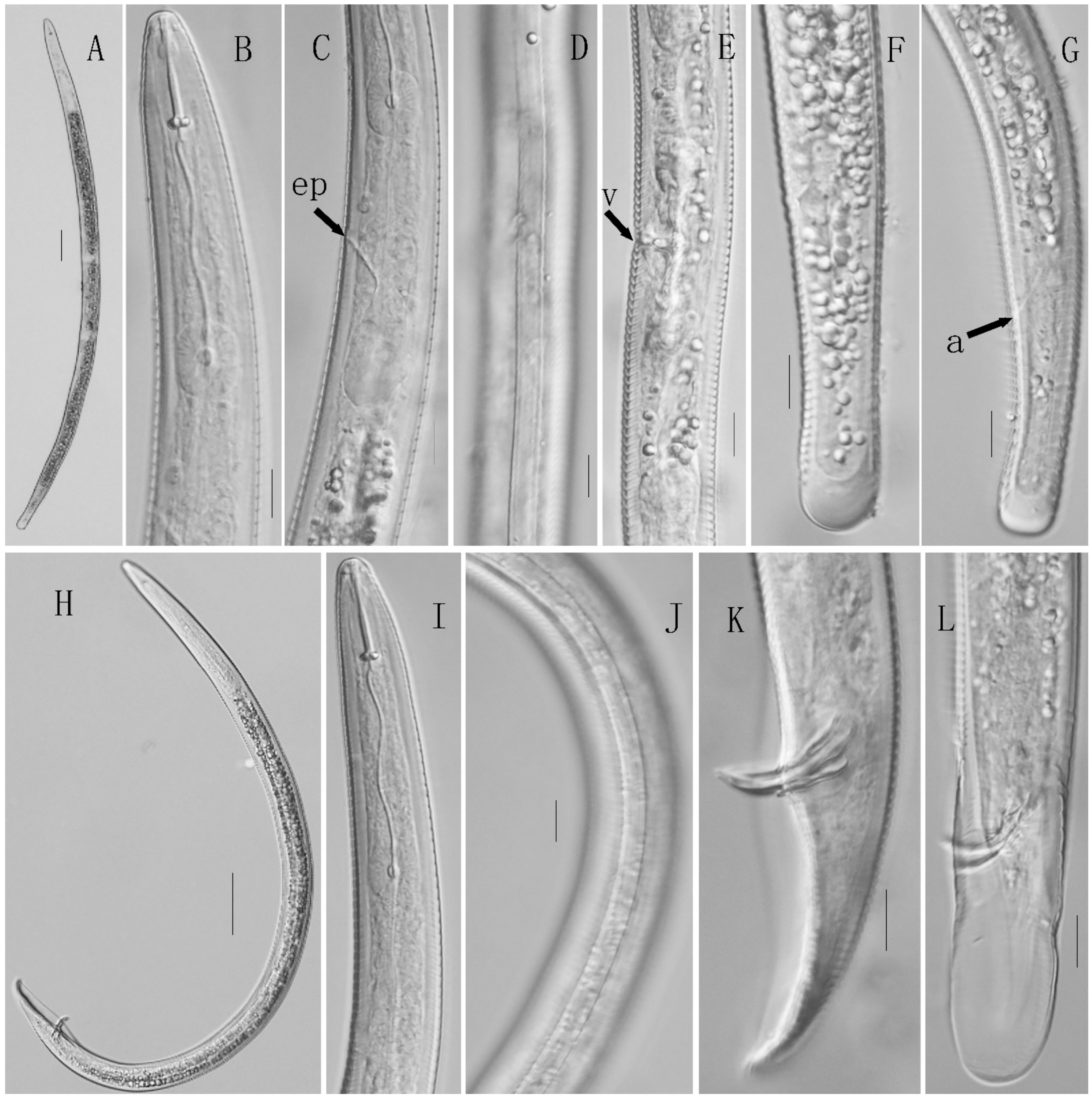
3.2. Morphological Description

Because the SSU and LSU sequences aligned by ClustalW from the populations from different locations for each species in the present study were identical, they are considered as one species in the following description.

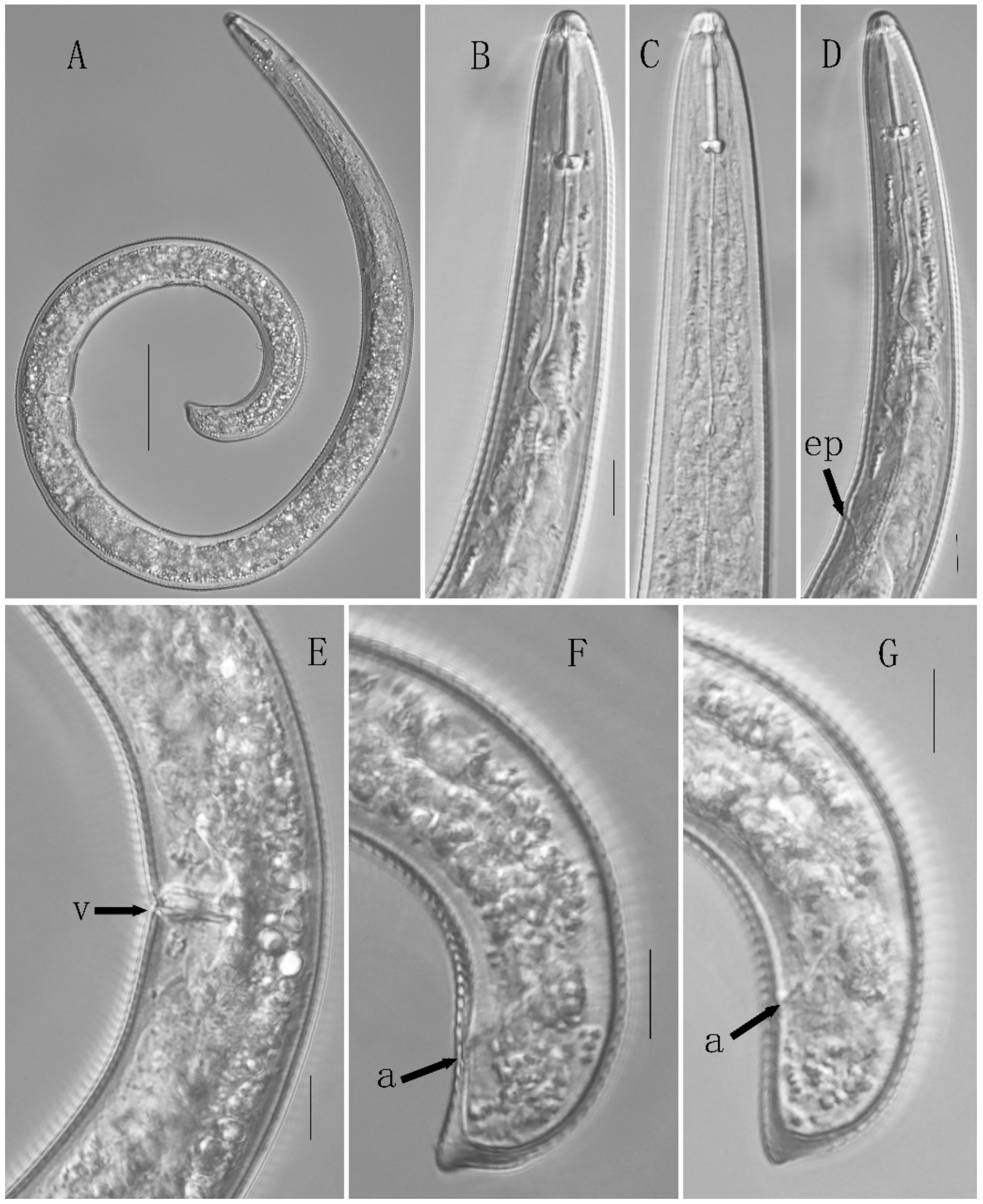
3.2.1. Description of Helicotylenchus dihystera

Female: Body spiral-shaped when heat-killed and distinctly annulated. Four incisures visible in lateral field with light microscopy. Stylet well-developed with rounded stylet knobs. Orifice of dorsal esophageal gland at about half stylet length behind stylet base. Lip hemispherical-shaped with lip rings. Median esophageal bulb oval with a well-developed valve. Excretory pore located at the anterior level of the esophageal gland. Hemizonion indistinct. The posterior esophageal glands overlapping intestines. Ovaries paired, outstretched, with oocytes in single row. Spermatheca without sperms. Vulva transverse without vaginal membrane. Tail ventrally curved, dorsally convex-conoid to a narrow terminus which may form a slight projection (Figure 1).
Male: Not observed.
Morphometrics: See Table 4.
Remarks: Helicotylenchus dihystera was first described from soil around sugarcane roots (Saccharum officinarum L.) in Harwood, Australia. It is distributed worldwide, occurring in 3 countries in North America, 18 in Central America and Caribbean, 8 in South America, 8 in Oceania, 17 in Europe, 30 in Africa and 28 in Asia [25]. It is present in 7 provinces in China [25]. It has a broad host range including sugarcane, potato (Solanum tuberosum L.), banana (Musa spp. L.), rice (Oryza sativa L.), tea (Camellia sinensis (L.) Kuntze), avocado (Persea americana Mill.), coffee (Coffea arabica L.), maize (Zea mays L.), beans (Phaseolus vulgaris L.), wheat (Triticum aestivum L.), rye (Secale cereale L.), oat (Avena sativa L.), sorghum (Sorghum bicolor (L.) Moench) and turfgrass [26,27,28]. Turfgrass hosts include Kentucky bluegrass (Poa pratensis L.) [29], bermudagrass (Cynodon dactylon (L.) Pers.), bentgrass (Agrostis stolonifera L.) and zoysiagrass (Zoysia japonica Steud.) [10,11,28]. In this study, H. dihystera was found in Zhuhai Cuihu Golf, Guangzhou Lihu Golf, South China Botanical Garden, Guangzhou Haizhuhu Park, Xiaogang Park and Yingzhou Ecological Park. The morphometric data and morphological characteristics of this present species match with American populations given by Zeng et al. [10].

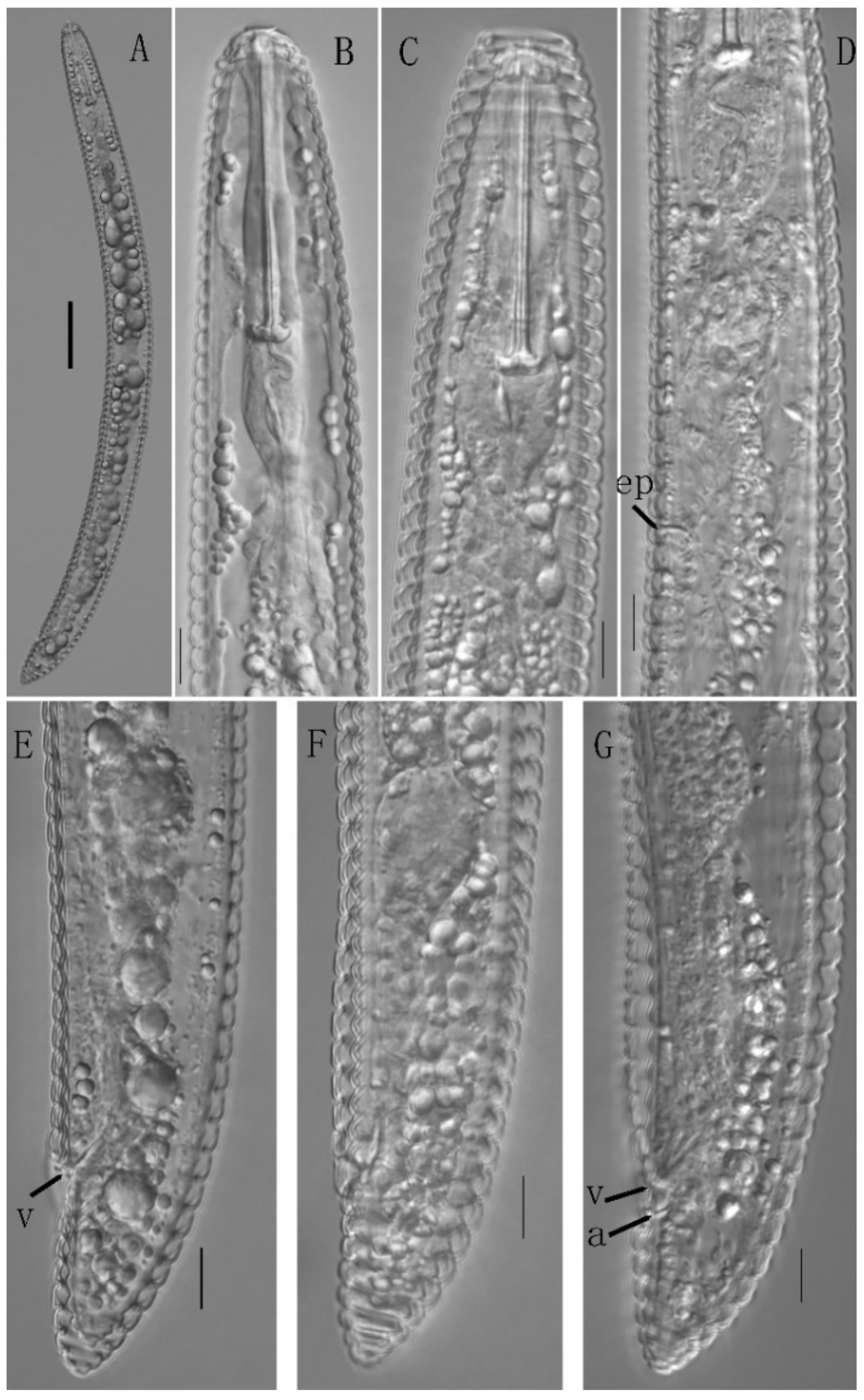
3.2.2. Description of Hemicriconemoides rosae

Female: Body slightly ventrally curved when heat-killed. Cuticular sheath and rounded annuli distinct. Lateral field absent and without anastomoses. Lip region not set off, rounded with a prominent labial disc and two annuli. Cephalic framework well-developed. Stylet strong, long, with well-developed, anchor-shaped knobs. Procorpus and metacorpus amalgamated. Median esophageal bulb oblong, with well-developed valve. Isthmus narrow followed by distinct basal bulb, becoming vase-shaped. Excretory pore posterior to basal bulb end, 28–31 annuli from anterior end. Vulva with a membranous sheath, without vulval flap. Vagina distinct, sigmoid. Ovary monodelphic, prodelphic, outstretched with oocytes in a single row. Spermatheca well-developed, oblong to ovoid-shaped, without sperms. Anus visible. Tail dorsally convex-conoid, with a bluntly rounded or pointed tip (Figure 2).
Male: Not observed.
Morphometrics: See Table 5.
Remarks: Hemicriconemoides rosae was first collected from the rhizosphere of rose (Rosa indica L.) in the Bareilly district, Uttar Pradesh, India, and was originally described by Rathour et al. [30]. It was collected from sugarcane and redescribed by Khan et al. [31]. Hemicriconemoides rosae is known to occur only in rose and sugarcane in India and Pilea cadierei Gagnep. & Guill. in China. In this study, it was found in Guangzhou Lihu Golf, Culture Park, People Park, Sand River Golf, Shenzhen Juhao Golf and Dongguan Zhongxin Golf. Both morphology and morphometrics match the original description [30]. This is the first report of H. rosae on turfgrasses.

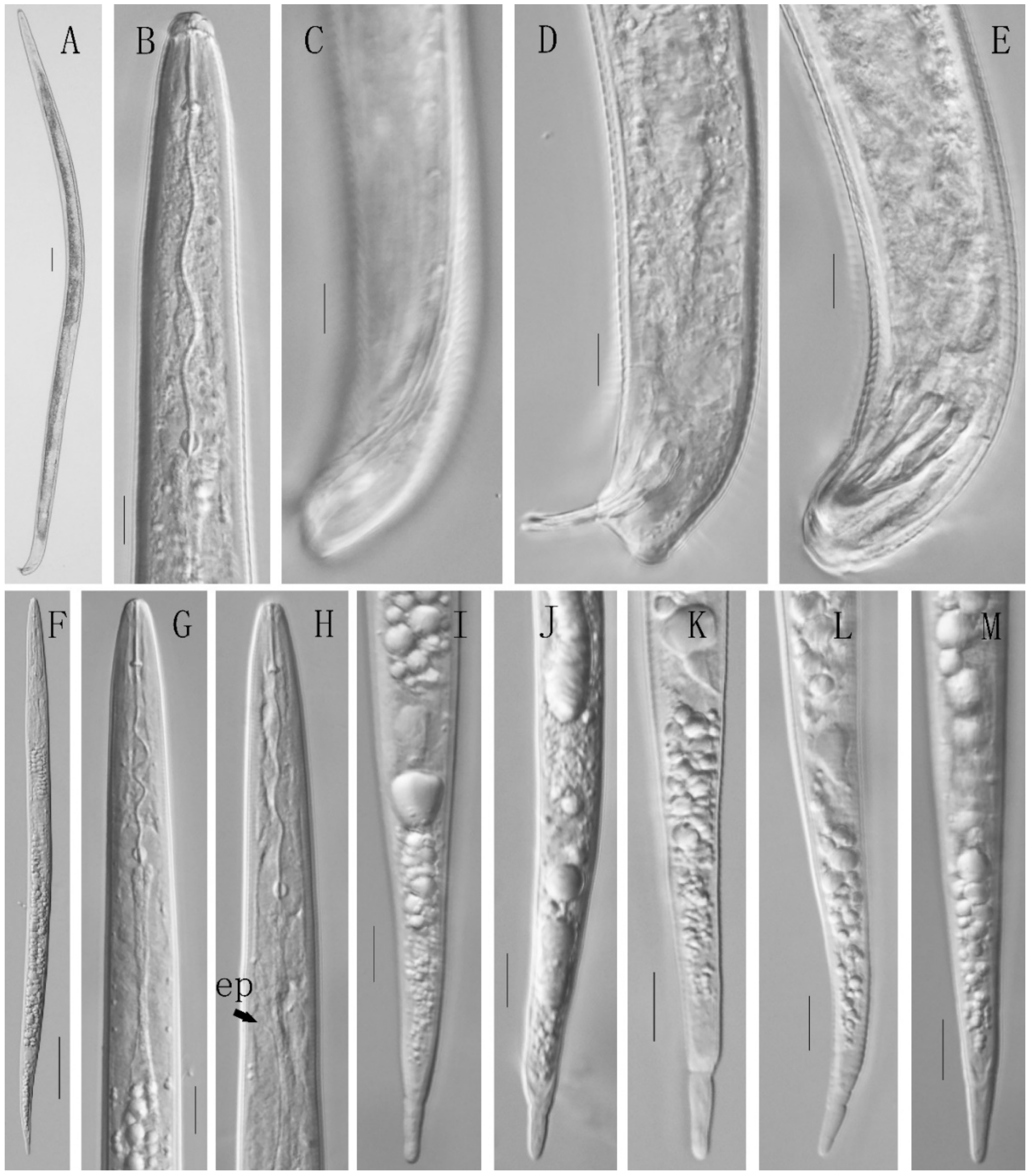
3.2.3. Description of Meloidogyne graminis

Second-stage juvenile: Body cylindrical, tapering to the posterior end. Head without distinct cephalic framework and annuli. No obvious contraction between the head and the body. Four incisures visible in lateral field. Stylet small with rounded knobs. Median esophageal bulb elongated with well-developed valves. Tail with a distinct, relatively long hyaline terminus and rounded tip (Figure 3).
Male: Body long and vermiform-shaped. Cuticle striated. Four incisures visible in lateral field. Head with well-developed cephalic framework, without annuli. No obvious contraction exists between the head and the body. Stylet robust with distinct rounded knobs. Median esophageal bulb oval with a well-developed valve. Spicules paired, separated, slightly curved ventrally, with a slender gubernaculum, near tail terminus. Tail short, with rounded tip (Figure 3).
Female: Not observed.
Morphometrics: See Table 6.
Remarks: Meloidogyne graminis was first described from grass in Florida by Sledge [32] and has been reported in other states in USA [10,11,33,34,35]. It has also been recorded in Venezuela [36], Brazil [37], Germany [38], the Netherlands [39], China [13] and India [40]. Turfgrass hosts of M. graminis include bermudagrass (Cynodon spp.), St. Augustinegrass (Stenotaphrum secundatum (Walter) Kuntze), zoysiagrass (Zoysia spp.), centipedegrass (Eremochloa ophiuroides (Munro) Hack.), seashore paspalum (Paspalum vaginatum Swartz), bentgrass (Agrostis spp.), and bluegrass (Poa spp.) [10,11,34,41,42]. In this study, M. graminis was found in turfgrass in Zhuhai Cuihu Golf, Guangzhou Lihu Golf, Shunde Junan Golf, Shenzhen Juhao Golf, Dongguan Zhongxin Golf, Changan Golf, Huizhou Taojing Golf, Longgang Golf and China Zhongshan Hot Spring Golf, Guangdong province, China. Both morphology and morphometrics match the description of other population [10].

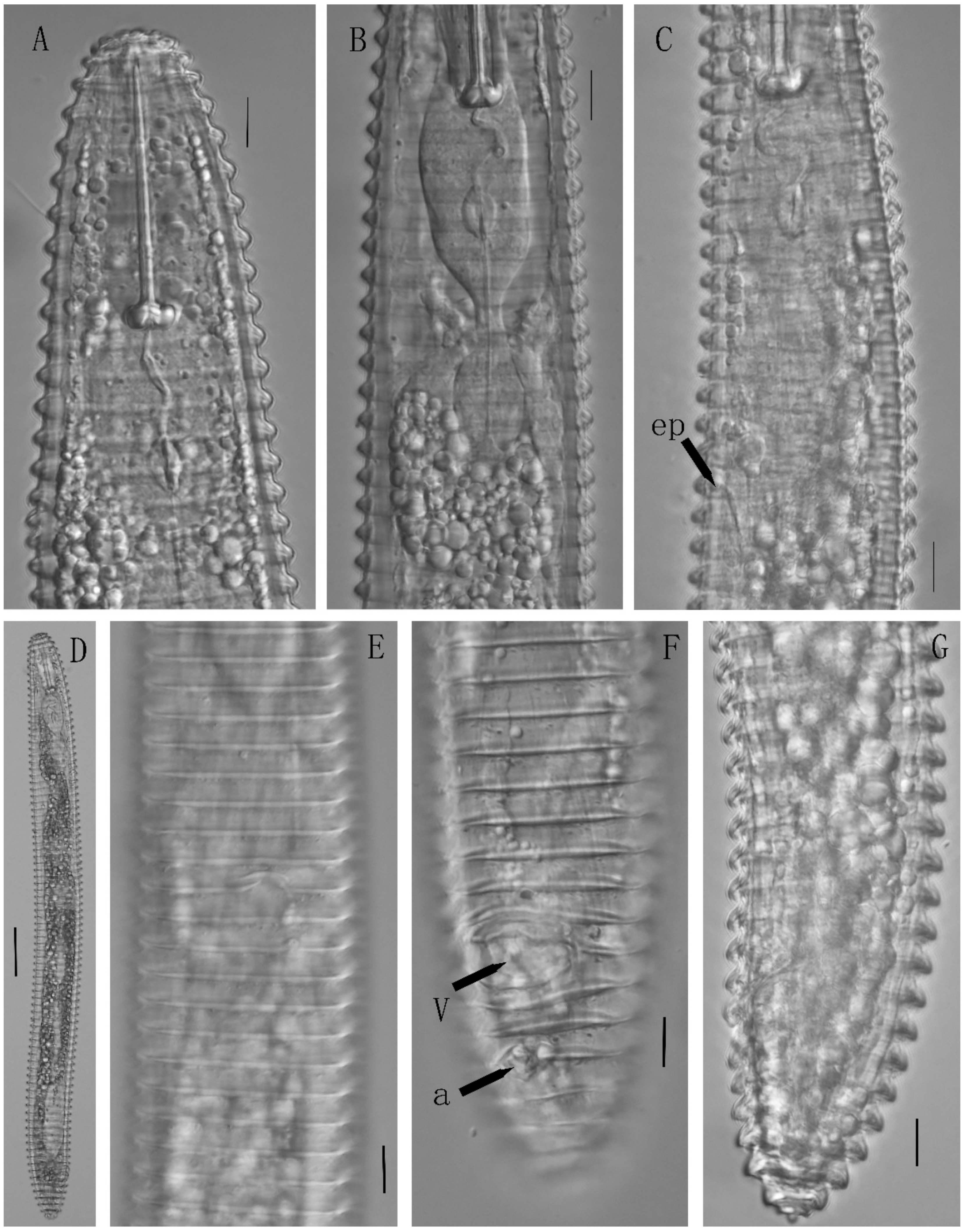
3.2.4. Description of Microcinema xenoplax

Female: Body slightly curved ventrally when heat-killed. Body rings distinct with smooth margins. Anastomoses rare. Head broad, first annule entire or emarginated laterally. Submedian lobes well-developed. Lip region conspicuous, elevated. Four labial plates distinct, well-separated. Stylet strong and long with anchor-shaped knobs. Esophagus typical Criconematid esophagus. Excretory pore located near the esophageal basal bulb. Vulva distinctly open with lips separated, two protruberances bearing anterior lip visible in ventral view. Vagina always sigmoid in lateral view. Ovary monodelphic, prodelphic. Tail broadly rounded to conoid, terminus button-shaped (Figure 4).
Male: Not observed.
Morphometrics: See Table 7.
Remarks: Mesocriconema xenoplax was first documented from grapevines (Vitis vinifera L. var. sultanina) in California [43]. It has been recorded in North America [44], South America [45], Europe [46,47], South Africa [48], Australia [49], New Zealand [50], China [51], India [52], Japan [53] and Iran [54]. Turfgrass hosts of M. xenoplax include tall fescue (Festuca arundinacea J. C. D. von Schreber) [55] and bermudagrass, creeping bentgrass and zoysiagrass [10,11]. In this study, M. xenoplax was detected in 18 golf courses and 2 parks (Table 3). Both morphology and morphometrics fit the description of other population [10]. This was the most widely distributed nematode species on turfgrasses in Guangdong province, China.

3.2.5. Description of Tylenchorhynchus leviterminalis

Female: Body ventrally curved to open C-shaped when heat-killed. Cuticle annuli distinct, 1.8–2.0 µm wide at mid-body, 2.0–2.3 pm at the tail region. Lateral field with four incisures. Lip region not offset, hemispherical. Cephalic framework slightly sclerotized. Stylet with rounded knobs. Median bulb well-developed, rounded to slightly ovate. Secretory-excretory pore (ep) near anterior end of saccate basal bulb, hemizonid two annuli anterior to ep. Ovaries outstretched. Vagina straight. Spermatheca rounded with sperms. Tail distinctly clavate with 18–22 annuli and with large hemispherical smooth hyaline portion (Figure 5).
Male: Similar to female in general. Testis single, outstretched. Spicules slightly curved ventrally, paired, separate. Gubernaculum well developed. Tail conoid and pointed. Bursa enveloping tail (Figure 5).
Morphometrics: See Table 8.
Remarks: Tylenchorhynchus leviterminalis was first described from banana, mango (Mangifera indica L.) and jackfruit (Artocarpus heterophyllus Lam.) in India [56]. It was reported from banana (Musa spp.) in Sistan and Baluchestan province; from grasses at the College of Agriculture (Badjgah Region), Shiraz University, Fars province, Iran [57,58]; from banana, sugarcane and bamboo (Bambusa spp.) in Taiwan [59]; from strawberry (Fragaria ananassa Duch.) in China mainland [60]; and from sugarcane in Japan [61]. In this study, T. leviterminalis was found in turfgrass samples from Liuhuahu Park, Liwanhu Park, Zhuhai Cuihu Golf, Guangzhou Jiulonghu Golf, Yuntai Park, Shamian Park and South China Botanical Garden, Guangdong province, China. Both morphology and morphometrics fit the description of other populations [56]. This is the first record of T. leviterminalis on turfgrasses C. dactylon, E. indica, P. repens and Z. tenuifolia.

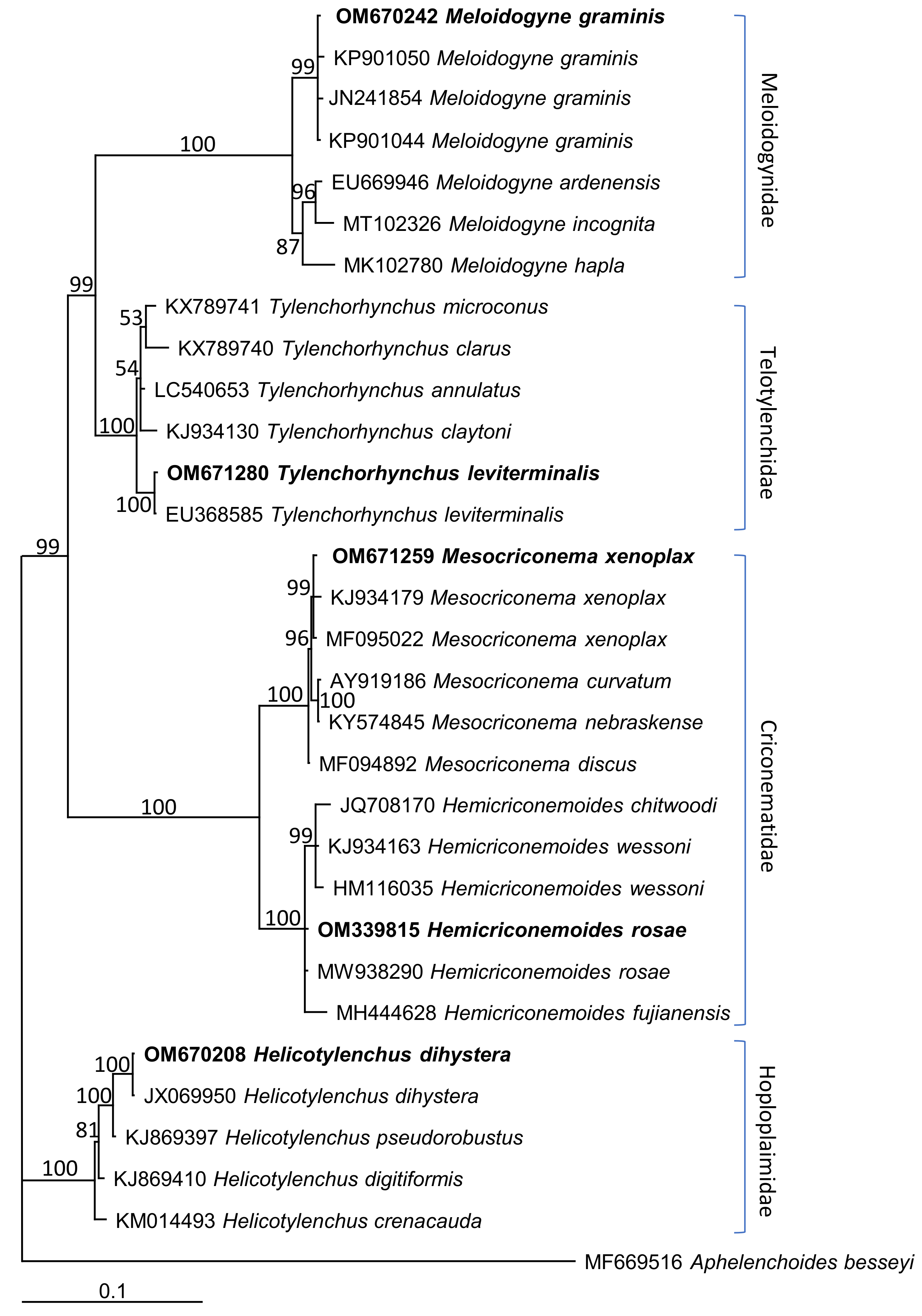
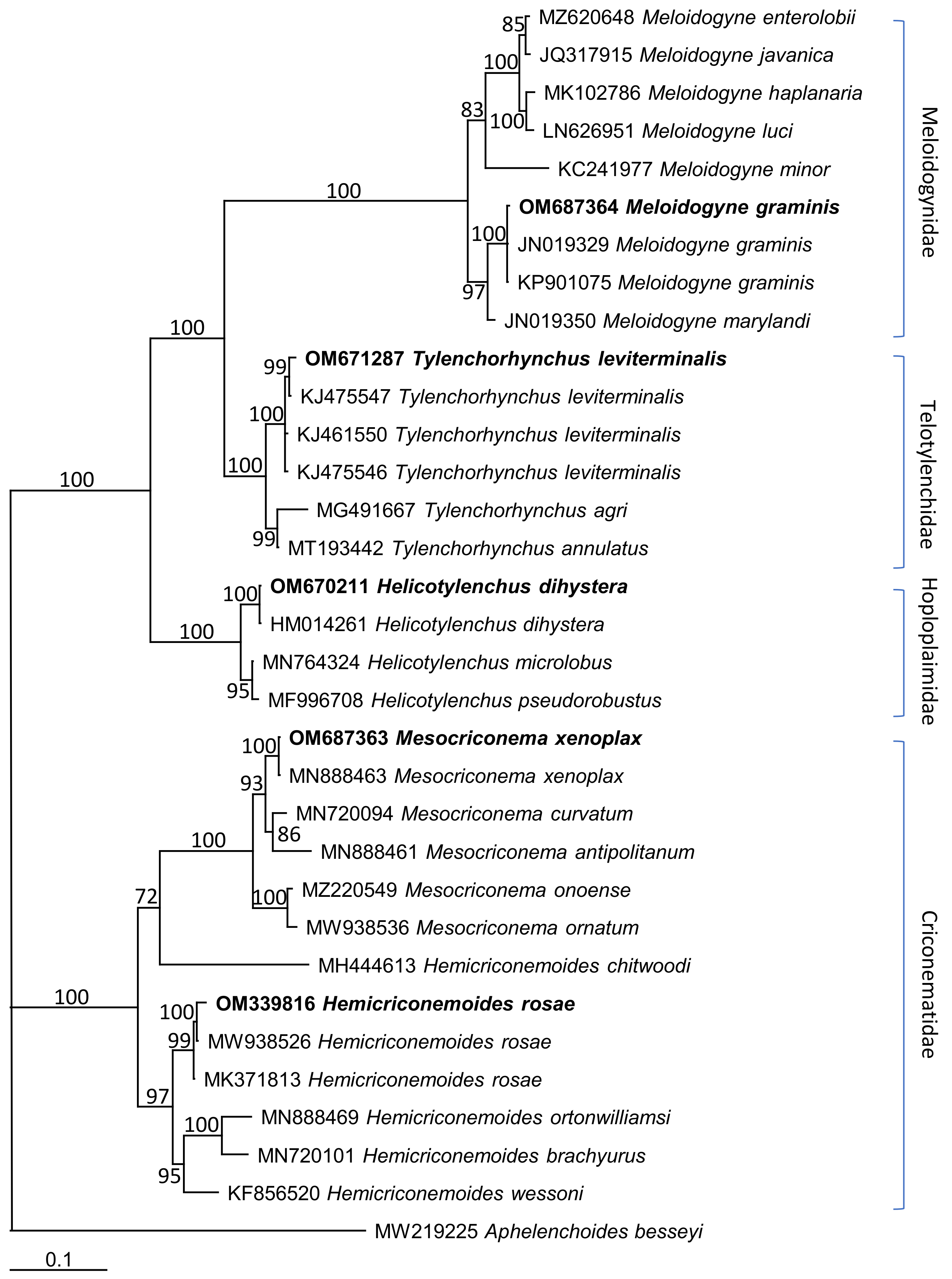
3.3. Molecular Characterization and Phylogenetic Relationships

A 602-bp 18S rDNA and a 574-bp 28S D2-D3 expansion segment of H. dihystera in this study were amplified and sequenced. A BLASTN search of this species matches well with its corresponding species. From 18S sequence, the study H. dihystera and a population of H. dihystera (JX069950) in GenBank yielded 602 total characters with 99.83% identity; intraspecific sequence variation for H. dihystera was 0.17% (1 nucleotide, nt). The study H. dihystera shared 581 (581/582 = 99.83%), 579 (579/582 = 99.48%) and 579 (579/582 = 99.48%) identical nucleotides with H. pseudorobustus (KJ869397), H. digitiformis (KJ869410) and H. crenacauda (KM014493), respectively. From the 28S sequence, alignment of the study H. dihystera with another population of H. dihystera (HM014261) in GenBank revealed 99.83% identity; intraspecific sequence variation for H. dihystera was 0.17% (1 nt). The study H. dihystera shared 556 (556/575 = 96.70%) identical nucleotides with H. microlobus (MN764324), H. pseudorobustus (MF996708).
A 606-bp 18S rDNA and a 696-bp 28S D2-D3 expansion segment of H. rosae in this study were amplified and sequenced. A BLASTN search of this species matches well with its corresponding species. From 18S sequence, the study H. rosae and a population of H. rosae (MW938290) in GenBank yielded 606 total characters with 99.83% identity; intraspecific sequence variation for H. rosae was 0.17% (1 nt). It shared 601 (601/606 = 99.17%), 595 (595/598 = 99.50%), 604 (604/606 = 99.67%) and 518 (518/524 = 98.85%) identical nucleotides with H. fujianensis (MH444628), H. wessoni (HM116035), H. wessoni (KJ934163) and H. chitwoodi (JQ708170), respectively. From the 28S sequence, alignment of the study H. rosae with two other populations of H. rosae (MW938526 and MK371813) in GenBank revealed 99.57% identity; intraspecific sequence variation for H. rosae was 0.43% (3 nt). It shared 600 (600/641 = 93.60%), 637 (637/697 = 91.39%) and 634 (634/696 = 91.09%) identical nucleotides with H. wessoni (KF856520), H. wessoni (HM116035), H. ortonwilliamsi (MN888469) and H. brachyurus (MN720101), respectively.
A 606-bp 18S rDNA and a 682-bp 28S D2-D3 expansion segment of M. graminis in this study were amplified and sequenced. A BLASTN search of this species matches well with its corresponding species. From 18S sequence, the study M. graminis and three populations of M. graminis (KP901050, KP901044 and JN241854) in GenBank yielded 606 total characters with 99.51–99.83% identities; intraspecific sequence variations for M. graminis were 0.33–0.49% (2–3 nt). It shared 598 (598/606 = 98.68%), 596 (596/607 = 98.19%) and 596 (596/607 = 98.19%) identical nucleotides with M. ardenensis (EU669946), M. incognita (MT102326) and M. hapla (MK102780), respectively. From the 28S sequence, alignment of the study M. graminis with other two populations of M. graminis (JN019329 and KP901075) in GenBank revealed 99.71% identity; intraspecific sequence variation for M. graminis was 0.29% (2 nt). It shared 662 (662/683 = 96.93%), 620 (620/692 = 89.60%), 618 (618/684 = 90.35%), 620 (620/684 = 90.64%), 609 (609/667 = 91.30%) and 623 (623/684 = 91.08%) identical nucleotides with M. marylandi (JN019350), M. minor (KC241977), M. luci (LN626951) M. haplanaria (MK102786), M. javanica (JQ317915) and M. enterolobii (MZ602648), respectively.
A 633-bp 18S rDNA and a 718-bp 28S D2-D3 expansion segment of M. xenoplax in this study were amplified and sequenced. A BLASTN search of this species matches well with its corresponding species. From 18S sequence, the study M. xenoplax and a population of M. xenoplax (MF095022) in GenBank yielded 633 total characters with 99.84% identity; intraspecific sequence variation for M. xenoplax was 0.16% (1nt). It shared 628 (628/634 = 99.05%), 628 (628/634 = 99.05%) and 629 (629/634 = 99.21%) identical nucleotides with M. curvatum (AY919186), M. nebraskense (KY574845) and M. discus (MF094892), respectively. From the 28S sequence, alignment of the study M. xenoplax with one other population of M. xenoplax (MN888463) in GenBank revealed 99.72% identity; intraspecific sequence variation for H. rosae was 0.28% (2 nt). It shared 639 (639/663 = 96.38%), 669 (669/709 = 94.36%), 658 (658/703 = 93.60%) and 650 (650/697 = 93.26%) identical nucleotides with M. curvatum (MN720094), M. antipolitanum (MN888461), M. onoense (MZ220549) and M. ornatum (MW938536), respectively.
A 602-bp 18S rDNA and a 704-bp 28S D2-D3 expansion segment of T. leviterminalis in this study were amplified and sequenced. A BLASTN search of this species matches well with its corresponding species. From 18S sequence, the study T. leviterminalis and a population of T. leviterminalis (EU368585) in GenBank yielded 602 total characters with 99.83% identity; intraspecific sequence variation for T. leviterminalis was 0.17% (1 nt). It shared 582 (582/591 = 98.48%), 577 (577/591 = 97.63%), 500 (500/506 = 98.81%) and 577 (577/592 = 97.47%) identical nucleotides with T. microconus (KX789741), T. clarus (KX789740), T. annulatus (LC540653) and T. claytoni (KJ934130), respectively. From the 28S sequence, alignment of the study T. leviterminalis with three other populations of T. leviterminalis (KJ475547, KJ461550 and KJ475546) in GenBank revealed 99.57–99.86% identities; intraspecific sequence variations for T. leviterminalis were 0.14–0.43% (1–3 nt). It shared 682 (682/706 = 96.60%) and 682 (682/705 = 96.74%) identical nucleotides with T. agri (MG491667) and T. annulatus (MT193442), respectively.
Phylogenetic analyses of the partial 18S and 28S D2-D3 were performed to examine the relationships among the most common species from this study and related species from Genbank. The dendrogram inferred from 18S rDNA sequences (Figure 6) using Aphelenchoides besseyi Christie, 1942 as an outgroup demonstrates: (i) four monophyletic clades with 100% posterior probability (pp) corresponding to the Meloidogynidae, Telotylenchidae, Criconematidae and Hoplolaimidae families; (ii) with 100% pp, M. graminis from this study is grouped into the Meloidogynidae-clade with six other species/populations of Meloidogyne, T. leviterminalis into the Telotylenchidae-clade with five other species/populations of Tylenchorynchus, both M. xenoplax and H. rosae into the Criconematidae-clade with five other species/populations of their respective genera, and H. dihystera into the Hoplolaimidae-clade with four other species/populations of Helicotylenchus from GenBank; (iii) species belonging to the Meloidogynidae and Telotylenchidae are grouped in a well-supported (pp = 99%) monophyletic clade. The Meloidogynidae, Telotylenchidae and Criconematidae also form a monophyletic clade with 99% pp.
The tree inferred from D2-D3 expansion segments of 28S rDNA (Figure 7) using Aphelenchoides besseyi Christie, 1942 as an outgroup demonstrates: (i) four distinct clades are the same as those identified by 18S rDNA sequences; (ii) the families Meloidogynidae and Telotylenchidae are in a highly-supported (pp = 100%) monophyletic clade, which together with the Hoplolaimidae forms an additional clade (pp = 100%); species/populations belonging to the genera Mesocriconema and Hemicriconemoides are in a well-supported (pp = 100%) monophyletic clade; (iii) Meloidogyne graminis from this study is clustered in a monophyletic clade with two other populations of M. graminis (JN019329 and KP901075) with 100% pp, T. leviterminalis is in a well-supported (pp = 100%) monophyletic clade with three other populations of T. leviterminalis (KJ475547, KJ461550 and KJ475546), H. dihystera is in a monophyletic clade with one other population of H. dihystera (HM014261) with 100% pp, M. xenoplax is in a monophyletic clade with one other population of M. xenoplax (MN888463) with 100% pp, and H. rosae is in a highly-supported (pp = 99%) monophyletic clade with two other populations of H. rosae (MW938526 and MK371813).

4. Discussion

Prior to this work, there have been several research papers illustrating a broad diversity of nematodes associated with turfgrasses in the United States [5,10,11,28,62], Canada [7,63,64], Argentina [65], Germany, Sweden, Norway [66,67], Belgium [8], Israel [68] and Korea [6]. Previous research showed that the species of Hemicriconemoides and Tylenchorynchus associated with turfgrasses include H. chitwoodi Esser, 1960 and H. wessoni Chitwood & Birchfield, 1957 in Cynodon dactylon [10,11]; H. brachyurus (Loos, 1949) Chitwood & Birchfield, 1957 in Poa pratensis and Zoysia japonica [6]; T. claytoni Steiner, 1937 in P. pratensis and Z. japonica [6,10,11]; T. dubius in Agrostis stolonifera and P. pratensis [69]; T. annulatus in Z. japonica; and T. thermophilus in Z. japonica and P. pratensis [6]. The work presented here provides new records of H. rosae and T. leviterminalis associated with turfgrasses.
The genus Hemicriconemoides Chitwood & Birchfield, 1957 morphologically differ from other genera within Criconematidae by possessing a loosened outer cuticular sheath covering the cuticle of their body; in females, the sheath is attached to the main body at the head and vulva. This genus is represented by 52 species [70]. The species H. rosae in this study morphologically agreed with the populations described by Rathour et al. [30] and Khan et al. [31]. Its status as a distinct species is corroborated by molecular sequences of 18S and 28S D2-D3 (Figure 6 and Figure 7). Hemicriconemoides rosae was first found around the rhizosphere of rose (Rosa indica) [30]. It was also documented as occurring in sugarcane [31] and Pilea cadierei [71]. In the present study, it was extracted from rhizosphere soils of multiple turfgrasses including Cynodon dactylon, Eleusine indica, Panicum repens and Sporobolus indicus, thus extending associated plant range of H. rosae.
The genus Tylenchorhynchus was established by Cobb (1913) for T. cylindricus found in southern California [72]. This genus contains 111 species that parasitize a wide variety of plants [73]. Handoo [73] defined the valid and most significant differentiating characters and prepared a key and a compendium containing morphometric and related details of these valid species. Siddiqi et al. [56] first described T. leviterminalis from banana, mango and jackfruit in India. The morphological and morphometric characters of the studied T. leviterminalis fit the original description [56]. Its presence as a distinct species is supported by molecular sequences of 18S and 28S D2-D3 (Figure 6 and Figure 7). Some hosts of T. leviterminalis were reported, including banana (Musa spp.) [55], sugarcane [57,59], bamboo (Bambusa spp.) [57] and strawberry (Fragaria ananassa) [60]. In the present study, T. leviterminalis was first reported on turfgrasses C. dactylon, E. indica, P. repens and Z. tenuifolia.
The genus Helicotylenchus Steiner, 1945 belongs to the family Hoplolaimidae. There are over 200 species within the genus [74]. Morphological identification of Helicotylenchus species is often difficult because of high intra- and interspecific variability and a lot of poorly described species [75,76]. Application of non-morphological characters such as DNA sequences can help to confirm classical morphology-based identifications and resolve some of the problems experienced in the identification of Helicotylenchus species [76]. Helicotylenchus dihystera is the type species of the genus. The morphometric data and morphological characteristics of H. dihystera in this study match with other populations given by Zeng et al. [10] and Siddiqi [27]. Phylogenetic analysis inferred from sequences of 18S and 28S D2-D3 segment support the presence of H. dihystera (Figure 6 and Figure 7).
The genus Mesocriconema Andrássy, 1965, belonging to the family Criconematidae, is characterized by having thick, rounded, protruding, retrorse cuticular annulations. This genus contains 90 species [77], some of which are morphologically very close to each other. Molecular and morphological analyses of species within Mesocriconem, from North America, were used to differentiate formally described members of the genus as well as lineages lacking a formal description [78,79]. Powers et al. [79] distinguished 24 haplotype groups by using COI sequences. In this study, molecular and morphological analyses were employed to determine the species M. xenoplax; both morphology and morphometrics of M. xenopax matched the description by Zeng et al. [10], and its presence as a distinct species is supported by molecular sequences of 18S and 28S D2-D3 (Figure 6 and Figure 7). Mesocriconema xenoplax has a wide host range. Rootstocks of most species in the genus Prunus L. support populations of M. xenoplax. It also infects various other fruit trees such as grapes [80]. The turfgrass hosts of M. xenoplax include tall fescue (Festuca arundinacea J. C. D. von Schreber) [55] and bermudagrass, creeping bentgrass and zoysiagrass [10,11]. In this study, M. xenoplax was detected in the rhizosphere soils from C. dactylon, P. repens, S. indicus and Z. tenuifolia. Pathogenicity examinations are needed for these turfgrasses in the future.
Root-knot nematodes (Meloidogyne spp.) have been one of the most prevalent species on turfgrasses worldwide [6,8,10,11,40,63,64]. So far, reported Meloidogyne species associated with turfgrasses include M. incognita (Kofoid & White) Chitwood, 1949 [65], M. graminis, M. graminicola Golden & Birchfield, 1965 [6], M. marylandi Jepson & Golden, 1987 [6,62], M. minor Karssen et al., 2004 [63] and M. naasi Franklin, 1965 [8,10]. Zeng et al. [10,11] and Sánchez-Arce et al. [7] showed that M. graminis was one of the most prevalent species in golf course turfgrasses. Sánchez-Arce et al. [7] and Zhuo et al. [13] reported that M. graminis was associated with bermudagrass. Zeng et al. [10,11] showed that M. graminis was found in two grass species (bermudagrass and zoysiagrass). In the present study, M. graminis was also detected in bermudagrass from 9 golf courses. Our report, along with previous publications, shows a strong association of M. graminis and bermudagrass, but more extensive sampling is needed. Meloidogyne minor was previously associated with yellow patch disease on Agrostis stolonifera var. stolonifera L. on golf greens [63]. Meloidogyne spp. have variations in pathogenicity, but a recent survey in SC indicated severe damage caused by M. graminis on golf course turfgrasses, particularly bermudagrass [81]. Although the pathogenicity of some Meloidogyne species to turfgrasses still needs to be further examined, root-knot nematodes are undoubtedly worthy of attention in the future.

5. Conclusions

Morphologically, this study clarified the identity of the most common plant-parasitic nematodes on golf course turf in Guangdong. Molecularly, it characterized these species using sequences of the 18S nuclear ribosomal RNA (rRNA) and the D2-D3 expansion segments of the 28S rRNA gene, showing little intraspecific variation of the sequences with respective corresponding species from GenBank. Phylogenetically, it investigated the phylogenetic relationships among these plant-parasitic nematode species based on sequences of the 18S rRNA and the D2-D3 expansion segments of the 28S rRNA gene, revealing correct phylogenetic placements of the five species in this study. Both sequences of the 18S rRNA and the D2-D3 expansion segments of the 28S rRNA gene verified morphological-based identification of these species in the present study. Five nematode species are described, including H. dihystera, H. rosae, M. graminis, M. xenoplax and T. leviterminalis, with new records of H. rosae, and T. leviterminalis associated with turfgrass. This work was a first step for future study including pathogenicity assay, relationship examination with other pathogens and development of control measures of these turf nematodes to provide more precise and effective management options to turf superintendents. This is the first report on molecular characterization and phylogenetic relationships of plant-parasitic nematodes associated with turfgrasses in Guangdong, China.

Author Contributions

Conceptualization, Y.Z.; Methodology, Y.Z., X.C. and Y.N.; Formal Analysis, Y.Z., X.C., Y.N. and C.Z.; Investigation, Y.Z., X.C., Y.N. and C.Z.; Data Curation, Y.Z. and J.R.; Supervision, J.R.; writing the paper—original draft preparation, Y.Z.; writing the paper—review & editing, J.K., L.T. and J.R. All authors have read and agreed to the published version of the manuscript.

Funding

This work was supported by a grant from Natural Science Foundation of Guangdong Province, China (S2013010016516, RMB 50,000).

Institutional Review Board Statement

Not applicable.

Informed Consent Statement

Not applicable.

Data Availability Statement

Not applicable.

Acknowledgments

We thank Natural Science Foundation of Guangdong Province for funding support and golf course superintendents for assistance with sample collection.

Conflicts of Interest

The authors declare no conflict of interest.

References

  1. Chinese National Bureau of Statistics. The Top Ten Provinces in the Country in Terms of Urban Green Area Rankings. Available online: https://www.ixigua.com/6921648720102556164?wid_try=1 (accessed on 25 January 2021).
  2. Hong’e, M. Golf Courses Proliferate in China, Defying Gov’t Ban. Available online: https://lawrencesolomon.files.wordpress.com/2011/10/prolif.pdf (accessed on 22 May 2011).
  3. Walker, N.R.; Goad, C.L.; Zhang, H.; Martin, D.L. Factors associated with populations of plant-parasitic nematodes in bentgrass putting greens in Oklahoma. Plant Dis. 2002, 86, 764–768. [Google Scholar] [CrossRef] [PubMed] [Green Version]
  4. Jo, Y.K.; Starr, J.L. Nematodes in Texas Golf Course. Available online: https://cdn-ext.agnet.tamu.edu/wp-content/uploads/2018/09/E-294-nematodes-in-texas-golf-courses.pdf (accessed on 30 September 2018).
  5. Crow, W. Plant parasitic nematodes on golf course turf. Pest Manag. 2005, 12, 10–15. [Google Scholar] [CrossRef] [Green Version]
  6. Mwamula, A.O.; Lee, D.W. Occurrence of plant-parasitic nematodes of turfgrass in Korea. Plant Pathol. J. 2021, 37, 446–454. [Google Scholar] [CrossRef] [PubMed]
  7. Sánchez-Arce, O.; Palacios-Espinosa, A.; Carillo-Fasio, J.; Hernandez-Montiel, L.G.; Hernández-Rubio, J.S.; Romero, M. Diversity and distribution of plant-parasitic nematodes on golf courses in localities of Baja California Sur, Mexico. Rev. Fac. Agron. La Univ. Del Zulia 2021, 38, 651–660. [Google Scholar]
  8. Vandenbossche, B.; Viaene, N.; De Sutter, N.; Maes, M.; Kareeen, G.; Bert, W. Diversity and incidence of plant-parasitic nematodes in Belgian turf grass. Nematology 2011, 13, 245–256. [Google Scholar] [CrossRef]
  9. Yu, Q.; Potter, J.W.; Gilby, G. Plant-parasitic nematodes associated with turfgrass in golf courses in southern Ontario. Can. J. Plant Pathol. 1998, 20, 304–307. [Google Scholar] [CrossRef]
  10. Zeng, Y.; Ye, W.; Tredway, L.; Martin, S.; Martin, M. Taxonomy and morphology of plant-parasitic nematodes associated with turfgrasses in North and South Carolina, USA. Zootaxa 2012, 3452, 1–46. [Google Scholar] [CrossRef] [Green Version]
  11. Zeng, Y.; Ye, W.; Martin, S.; Martin, M.; Tredway, L. Diversity and occurrence of nematodes associated with turfgrasses in North and South Carolina, USA. J. Nematol. 2012, 44, 345–355. [Google Scholar]
  12. Zeng, Y.; Ye, W.; Kerns, J.; Tredway, L.; Martin, S.; Martin, M. Molecular characterization and phylogenetic relationships of plant-parasitic nematodes associated with turfgrasses in North Carolina and South Carolina, United States. Plant Dis. 2015, 99, 982–993. [Google Scholar] [CrossRef] [Green Version]
  13. Zhuo, K.; Hu, M.; Wang, H.; Tang, Z.; Shao, X.; Liao, J. Identification of Meloidogyne graminis on golf greens. Acta Pratacul. Sin. 2011, 20, 253–256. (In Chinese) [Google Scholar]
  14. Jenkins, W.R. A rapid centrifugal-flotation technique for separating nematodes from soil. Plant Dis. Rep. 1964, 48, 692. [Google Scholar]
  15. Hooper, D.J. Handling, fixing, staining and mounting nematodes. In Laboratory Methods for Work with Plant and Soil Nematodes, 5th ed.; Her Majesty’s Stationery Office: London, UK, 1970; pp. 39–54. [Google Scholar]
  16. Ye, W. Applying Microsoft Works spread sheet in statistics for morphometric data of nematode identification. Afro-Asian J. Nematol. 1996, 6, 203–211. [Google Scholar]
  17. Williams, B.D.; Schrank, B.; Huynh, C.; Shownkeen, R.; Waterston, R.H. A genetic mapping system in Caenorhabditis elegans based on polymorphic sequence—Tagged sites. Genetics 1992, 131, 609–624. [Google Scholar] [CrossRef] [PubMed]
  18. Mullin, P.G.; Harris, T.S.; Powers, T.O. Phylogenetic relationships of Nygolaimina and Dorylaimina (Nematoda: Dorylaimida) inferred from small subunit ribosomal DNA sequences. Nematology 2005, 7, 59–79. [Google Scholar] [CrossRef]
  19. Nunn, G.B. Nematode Molecular Evolution. Ph.D. Dissertation, University of Nottingham, Nottingham, UK, 1992. [Google Scholar]
  20. Ye, W.; Giblin-Davis, R.M.; Braasch, H.; Morris, K.; Thomas, W.K. Phylogenetic relationships among Bursaphelenchus species (Nematoda: Parasitaphelenchidae) inferred from nuclear ribosomal and mitochondrial DNA sequence data. Mol. Phylogenetics Evol. 2007, 43, 1185–1197. [Google Scholar] [CrossRef]
  21. Tamura, K.; Peterson, D.; Peterson, N.; Stecher, G.; Nei, M.; Kumar, S. MEGA5: Molecular evolutionary genetic analysis using maximum likelihood, evolutionary distance, and maximum parsimony methods. Mol. Biol. Evol. 2011, 28, 2731–2739. [Google Scholar] [CrossRef] [Green Version]
  22. Posada, D.; Crandall, K.A. Modeltest: Testing the model of DNA substitution. Bioinformatics 1998, 14, 817–818. [Google Scholar] [CrossRef] [Green Version]
  23. Huelsenbeck, J.P.; Ronquist, F. Mr Bayes: Bayesian inference of phylogenetic trees. Bioinformatics 2001, 17, 1754–1755. [Google Scholar] [CrossRef] [Green Version]
  24. Larget, B.; Simon, D.L. Markov chain Monte Carlo algorithms for the Bayesian analysis of phylogenetic trees. Mol. Biol. Evol. 1999, 16, 750–759. [Google Scholar] [CrossRef]
  25. CABI. Helicotylenchus dihystera. [Distribution map]. In Distribution Maps of Plant Diseases (April), 1st ed.; CABI: Wallingford, UK, 2010. [Google Scholar]
  26. Kinloch, R.A. Florida field crops as hosts of Helicotylenchus dihystera. Nematropica 1971, 1, 38–39. [Google Scholar]
  27. Siddiqi, M.R. Helicotylenchus dihystera; Commonwealth Institute of Helminthology Descriptions of Plant-Parasitic Nematodes, Commonwealth Agricultural Bureaux: St. Albans, UK, 1972. [Google Scholar]
  28. Lucas, L.T.; Blake, C.T.; Barker, K.R. Nematodes associated with bentgrass and bermudagrass golf greens in North Carolina. Plant Dis. Rep. 1974, 58, 822–824. [Google Scholar]
  29. Sumner, D.R. Nematodes in bluegrass. Plant Dis. Rep. 1967, 51, 457–460. [Google Scholar]
  30. Rathour, K.S.; Sharma, S.; Singh, K.; Ganguly, S. Three new species of Hemicriconemoides (Nematoda: Tylenchida: Criconematina) associated with ornamental plants in Bareilly district of Uttar Pradesh, India. Indian J. Nematol. 2003, 33, 160–166. [Google Scholar]
  31. Khan, M.; Phani, V.; Chauhan, K.; Somvanshi, V.S.; Pervez, R.; Walia, R.K. Redescription and molecular characterisation of Hemicriconemoides rosae Rathour, Sharma, Singh & Ganguly, 2003 from rhizosphere of sugarcane in India. Nematology 2019, 21, 767–778. [Google Scholar]
  32. Sledge, E.B. Preliminary report on a Meloidogyne sp. parasite of grass in Florida. Plant Dis. Rep. 1962, 46, 52–54. [Google Scholar]
  33. Dickson, O.J. Some observations on Hypsoperine graminis in Kansas. Plant Dis. Rep. 1966, 50, 396–398. [Google Scholar]
  34. Grisham, M.P.; Dale, J.L.; Riggs, R.D. Meloidogyne graminis and Meloidogyne spp. on zoysia; infection, reproduction, disease development, and control. Phytopathology 1974, 64, 1485–1489. [Google Scholar] [CrossRef]
  35. McClure, M.A.; Nischwitz, C.; Skantar, A.M.; Schmitt, M.E.; Subbotin, S.A. Root-knot nematodes in golf greens of the western United States. Plant Dis. 2012, 96, 635–647. [Google Scholar] [CrossRef] [Green Version]
  36. Perichi, G. First report of Meloidogyne graminis (Nematoda: Tylenchida) in Venezuela. Fitopatol. Venez. 2006, 19, 17–18. [Google Scholar]
  37. Oliveira, S.A.; Oliveira, C.M.G.; Maleita, C.M.N.; Silva, M.F.A.; Abrantes, I.M.O.; Wilcken, S.R.S. First report of Meloidogyne graminis on golf courses turfgrass in Brazil. PLoS ONE 2018, 13, e0192397. [Google Scholar]
  38. Sturhan, D. Outdoor occurrence of Meloidogyne species in western Germany. Nachr. Dtsch. Pflanzenschutzd. 1976, 288, 113–117. [Google Scholar]
  39. Wesemael, W.; Viaene, N.; Moens, M. Root-knot nematodes (Meloidogyne spp.) in Europe. Nematology 2011, 13, 3–16. [Google Scholar] [CrossRef]
  40. Kaul, V.K.; Chhabra, H.K. A new record of Meloidogyne graminis on Raya and occurrence of Meloidogyne spp. in Ludhiana, Punjab, India. J. Oilseeds Res. 1988, 5, 200–202. [Google Scholar]
  41. Murray, J.J.; Poole, T.E.; Ostazeski, S.A. Techniques for determining reproduction of Meloidogyne graminis on zoysiagrass and bermudagrass. Plant Dis. 1986, 70, 559–560. [Google Scholar] [CrossRef] [Green Version]
  42. Sledge, E.B.; Golden, A.M. Hypsoperine graminis (Nematode: Heteroderidae), a new genus and species of plant parasitic nematode. Proc. Helminthol. Soc. Wash. 1964, 31, 83–88. [Google Scholar]
  43. Raski, D.J. On the morphology of Criconemoides Taylor 1936, with descriptions of six new species. Proc. Helminthol. Soc. Wash. 1952, 19, 85–99. [Google Scholar]
  44. Nyczepir, A.P.; Bertrand, P.F.; Miller, R.W.; Motsinger, R.E. Incidence of Criconemella spp. and peach orchard histories in short life and non-short life sites in Georgia and South Carolina. Plant Dis. 1985, 69, 874–877. [Google Scholar] [CrossRef]
  45. Aballay, E.; Persson, P.; Martensson, A. Plant-parasitic nematodes in Chilean vineyards. Nematropica 2009, 39, 85–97. [Google Scholar]
  46. De Abrantes, I.M.O.; Vieira dos Santos, M.C.; da Conceição, I.L.P.M.; de Santos, M.S.N.A.; Vovlas, N. Root-knot and other plant-parasitic nematodes associated with fig trees in Portugal. Nematol. Medit. 2008, 36, 131–136. [Google Scholar]
  47. Karanastasi, E.; Handoo, Z.A.; Tzortzakakis, E.A. First report of Mesocriconema xenoplax (Nematoda: Criconematidae) in Greece and first record of Viburnum sp. as a possible host for this ring nematode. Helminthologia 2008, 45, 103–105. [Google Scholar] [CrossRef] [Green Version]
  48. Van den Berg, E. Studies on some Criconematoidea (Nematoda) from South Africa with a description of Ogma rhombosquamatum (Mehta & Raski, 1971) Andrassy, 1979. Phytophylactica 1980, 12, 15–23. [Google Scholar]
  49. Stirling, G.R. Distribution of plant parasitic nematodes in South Australian vineyards. Aust. J. Exp. Agric. Animal Hus. 1976, 16, 588–591. [Google Scholar] [CrossRef]
  50. Loof, P.A.A.; Wouts, W.M.; Yeates, G.W. Criconematidae (Nematoda: Tylenchida) from the New Zealand region: Genera Mesocriconema, Criconema, Discocriconemella, and Hemicriconemoides. N. Z. J. Zool. 1997, 24, 123–151. [Google Scholar] [CrossRef]
  51. Xie, Z.; Yang, Q.; Cheng, J.; Zhang, S. Eight species of nematodes parasitized at the roots of rice. J. Fujian Agric. Forest. Univ. (Nat. Sci. Ed.) 2007, 36, 20–24. [Google Scholar]
  52. Gupta, N.K.; Gupta, A.K. On some plant parasitic nematodes of the genus Macroposthonia De Man, 1880 (Medinematidae: Criconematoidea) from India. Rev. Iber. Parasitol. 1981, 41, 25–41. [Google Scholar]
  53. Orton Williams, K.J. Macroposthonia xenoplax; Commonwealth Institute of Helminthology Descriptions of Plant-Parasitic Nematodes, Commonwealth Agricultural Bureaux: St. Albans, UK, 1972. [Google Scholar]
  54. Deimi, A.M.; Chitambar, J.J.; Maafi, Z.T. Nematodes associated with flowering ornamental plants in Mahallat, Iran. Nematol. Medit. 2008, 36, 115–123. [Google Scholar]
  55. Nyczepir, A.P. Host suitability of an endophyte-friendly tall fescue grass to Mesocriconema xenoplax and Pratylenchus vulnus. Nematropica 2011, 41, 45–51. [Google Scholar]
  56. Siddiqi, M.R.; Mukherjee, B.; Dasgupta, M.K. Tylenchorhynchus microconus n. sp., T. crassicaudatus leviterminalis n. subsp. and T. coffeae Siddiqi & Basir, 1959 (Nematoda: Tylenchida). Syst. Parasitol. 1982, 4, 257–262. [Google Scholar]
  57. Tanha Maafi, Z.; Amani, M.; Ebrahimi, N. Plant parasitic nematodes of banana plantations in Systan and Baluchestan province. In Proceedings of the 17th Iranian Plant Protection Congress, Karadj, Iran, 2–5 September 2006; 330p. [Google Scholar]
  58. Karani, S.H.M.; Kashi, L.; Ghaderi, R.; Karegar, A. Five Species of Tylenchidae and Dolichodoridae (Nematoda: Tylenchoidea) from Iran. J. Agric. Sci. Technol. 2015, 15, 227–240. [Google Scholar]
  59. Chen, D.Y.; Ni, H.F.; Yen, J.H.; Tsay, T.T. Identification of stunt nematode Tylenchorhynchus annulatus and a new recorded Tylenchorhynchus leviterminalis (Nematoda: Belonolaimidae) in Taiwan. Plant Pathol. Bull. 2006, 15, 251–262, (In Chinese with English Abstract). [Google Scholar]
  60. Vovlas, N.; Cheng, H. Morpho-anatomy of Tylenchorhynchus leviterminalis from the People’s Republic of China. Nematol. Medit. 1988, 16, 149–152. [Google Scholar]
  61. Talavera, M.; Watanabe, T.; Mizukubo, T. Description of Tylenchorhynchus shimizui n. sp. from Paraguay and notes on T. leviterminalis Siddiqi, Mukherjee & Dasgupta from Japan (Nematoda: Tylenchida: Telotylenchidae). Syst. Parasitol. 2002, 51, 171–177. [Google Scholar] [PubMed]
  62. Mitkowski, N.A. First report of Subanguina radicicola, the root gall nematode infecting Poa annua putting greens in Washington State. Plant Dis. 2007, 91, 905. [Google Scholar] [CrossRef]
  63. Karssen, G.; Bolk, R.J.; Vanaelst, A.C.; Vanden Beld, I.; Kox, L.F.F.; Korthals, G.; Molendijk, L.; Zijlstra, C.; Van Hoof, R.; Cook, R. Description of Meloidogyne minor n. sp. (Nematoda: Meloidogynidae), a root-knot nematode associated with yellow patch disease in golf courses. Nematology 2004, 6, 59–72. [Google Scholar] [CrossRef]
  64. Simard, L.; Bélair, G.; Miller, S. First report of Longidorus breviannulatus associated with damage on creeping bentgrass golf greens in Québec, Canada. Plant Dis. 2009, 93, 846–847. [Google Scholar] [CrossRef] [PubMed]
  65. Echeverría, M.M.; Chaves, E.J. Identification of Meloidogyne naasi Franklin, 1965 from Argentina. Nematologica 1998, 44, 219–220. [Google Scholar]
  66. Magnusson, C.; Hammeraas, B. Nematoder i grasbaner. Växtskyddsnotiser 1997, 61, 121–132. [Google Scholar]
  67. Knuth, P. Schaden in einem Sportsrasen durch das Würzengallenälchen Meloidogyne naasi in Baden-Württemberg. Nachr. Des Dtsch. Pflanzenschutzd. 1998, 12, 305–307. [Google Scholar]
  68. Oka, Y.; Karssen, G.; Mor, M. First report of the root-knot nematode Meloidogyne marylandi on turfgrasses in Israel. Plant Dis. 2004, 88, 309. [Google Scholar] [CrossRef]
  69. Laughlin, C.W.; Vargas, J.M., Jr. Pathogenic potential of Tylenchorhynchus dubius on selected turfgrass. J. Nematol. 1972, 4, 277–280. [Google Scholar]
  70. Geraert, E. The Criconematidae of the World. In Identification of the Family Criconematidae (Nematoda); Academia Press: Ghent, Belgium, 2010; 615p. [Google Scholar]
  71. Zhao, X. Identification and Analysis of Molecular Phylogeny of Criconematid Species in the Southern China. Master’s Thesis, Zhongkai University of Agriculture and Engineering, Guangzhou, China, 2021. (In Chinese). [Google Scholar]
  72. Cobb, N.A. New nematode genera found inhabiting fresh-water and non-brackish soils. J. Wash. Acad. Sci. 1913, 3, 432–445. [Google Scholar] [CrossRef]
  73. Handoo, Z.A. A Key and diagnostic compendium to the species of the genus Tylenchorhynchus Cobb, 1913 (Nematoda: Belonolaimidae). J. Nematol. 2000, 32, 20–34. [Google Scholar] [PubMed]
  74. Uzma, I.; Nasira, K.; Firoza, K.; Shahina, F. Review of the genus Helicotylenchus Steiner, 1945 (Nematoda: Hoplolaimidae) with updated diagnostic compendium. Pakistan. J. Nematol. 2015, 33, 115–160. [Google Scholar]
  75. Fortuner, R.; Maggenti, A.R.; Whittaker, L.M. Morphometrical variability in Helicotylenchus Steiner, 1945. 4: Study of field populations of H. pseudorobustus and related species. Rev. Nematol. 1984, 7, 121–135. [Google Scholar]
  76. Subbotin, S.A.; Inserra, R.N.; Marias, M.; Mullin, P.; Powers, T.O.; Roberts, P.A.; Van Den Berg, E.; Yates, G.W.; Baldwin, J.G. Diversity and phylogenetic relationships within the spiral nematodes of Helicotylenchus Steiner, 1945 (Tylenchida: Hoplolaimidae) as inferred from analysis of the D2-D3 expansion segments of 28S rRNA gene sequences. Nematology 2011, 13, 333–345. [Google Scholar]
  77. Brzeski, M.W.; Loof, P.A.A.; Choi, Y.E. Compendium of the genus Mesocriconema Andrássy, 1965 (Nematoda: Criconematidae). Nematology 2002, 4, 341–360. [Google Scholar]
  78. Powers, T.O.; Bernard, E.C.; Harris, T.; Higgins, R.; Olson, M.; Lodema, M.; Mullin, P.; Sutton, L.; Powers, K.S. COI haplotype groups in Mesocriconema (Nematoda: Criconematidae) and their morphospecies associations. Zootaxa 2014, 3827, 101–146. [Google Scholar] [CrossRef] [Green Version]
  79. Powers, T.O.; Mullin, P.; Higgins, R.; Harris, T.; Powers, K.S. Description of Mesocriconema ericaceum n. sp. (Nematoda: Criconematidae) and notes on other nematode species discovered in an ericaceous heath bald community in Great Smoky Mountains National Park, USA. Nematology 2016, 18, 879–903. [Google Scholar] [CrossRef] [Green Version]
  80. Pinkerton, J.N.; Vasconcelos, M.C.; Sampaio, T.L.; Shaffer, R.G. Reaction of grape rootstocks to ring nematode Mesocriconema xenoplax. Am. J. Enol. Vitic. 2005, 56, 377–385. [Google Scholar]
  81. Zeng, Y.; Roberts, J. Interactions of Root-Knot Nematodes (Meloidogyne spp.) with Mini-Ring Disease (Rhizoctonia zeae); Pee Dee Research and Education Center, Clemson University: Florence, SC, USA, 2022; manuscript in preparation. [Google Scholar]
Figure 1. Adult female of Helicotylenchus dihystera in lateral view. (A): Entire body; (B,C): Anterior body; (D): Anterior body (ep arrow refers to excretory pore); (E): Vulva (v arrow); (F,G): Tails (a arrow refers to anus). (Scale bars: (A) = 50 μm; (BG) = 10 μm).
Horticulturae 08 00611 g001
Figure 2. Adult female of Hemicriconemoides rosae. (A): Entire body; (B,C): Anterior body; (D): Excretory pore (ep arrow); (EG): Tails (a refers to anus, v refers to vulva) (Scale bars: (A) = 50 μm; (BG) = 10 μm).
Horticulturae 08 00611 g002
Figure 3. Adult male (AE) and juvenile (FM) of Meloidogyne graminis. (A): Entire male body; (B): Anterior male body; (C): Lateral field; (D,E): Male tails; (F): Entire juvenile body; (G): Anterior juvenile body; (H): Excretory pore (ep arrow); (IM): Tails (Scale bars: (A,F) = 50 μm; (BE,GM) = 10 μm).
Horticulturae 08 00611 g003
Figure 4. Adult female of Mesocriconema xenoplax. (A): Anterior body; (B): Esophagus; (C): Excretory pore (ep arrow); (D): Entire body; (E): Rings (annules); (F): Vulva (v arrow) and anus (a arrow); (G): Tail (Scale bars: (D) = 50 μm; (AC,EG) = 10 μm).
Horticulturae 08 00611 g004
Figure 5. Adult male and female of Tylenchorhynchus leviterminalis. (A): Entire female body; (B): Anterior female body; (C): Excretory pore (ep arrow); (D): Female lateral field; (E): Reproductive system (arrow v refers to vulva); (F,G): female tails (arrow a refers to anus); (H): Entire male body; (I): Anterior male body; (J): Male lateral field; (K): Male tail (lateral view); (L): Male tail (subventral view) (Scale bars: (A,H) = 50 μm; (BG,IL) = 10 μm).
Horticulturae 08 00611 g005
Figure 6. The 10,001st Bayesian tree inferred from 18S under the GTR + I + G model (lnL = 6925.1914; freqA = 0.2438; freqC = 0.2192; freqG = 0.2803; freqT = 0.2568; R(a) = 1.2950; R(b) = 2.2321; R(c) = 1.0279; R(d) = 0.7318; R(e) = 4.2831; R(f) = 1; Pinvar = 0.3455; Shape = 0.6090). Posterior probability values exceeding 50% are given on appropriate clades. The new species is in bold font.
Horticulturae 08 00611 g006
Figure 7. The 10,001st Bayesian tree inferred from D2-D3 under the GTR+I+G model (lnL = 5789.4619; freqA = 0.1932; freqC = 0.2222; freqG = 0.3323; freqT = 0.2523; R(a) = 0.6368; R(b) = 2.4422; R(c) = 1.4785; R(d) = 0.3450; R(e) = 3.8018; R(f) = 1; Pinvar = 0.2939; Shape = 1.3309). Posterior probability values exceeding 50% are given on appropriate clades. The new species is in bold font.
Horticulturae 08 00611 g007
Table 1. Sampling locations and turf species.
LocationsTurf SpeciesLocationsTurf Species
Zhuhai Cuihu GolfCynodon dactylon (L.) Pers.Sand River GolfCynodon dactylon
Zhuhai Jinwan GolfCynodon dactylonGuangzhou Martyrs’ CemeteryZoysia tenuifolia Willd. ex Trin.
Guangzhou Xiancun International GolfCynodon dactylonSun Yat-sen UniversityEleusine indica (L.) Gaertn.
Sporobolus indicus (L.) R. Br.
Guangzhou Lihu GolfCynodon dactylonGuangzhou Baiyun MountainPanicum repens L.
Zoysia tenuifolia
Guangzhou Fengshen Golf Cynodon dactylonGuangzhou Haizhuhu ParkZoysia tenuifolia
Guangzhou Jiulonghu Golf Cynodon dactylonGuangzhou Liwanhu ParkPanicum repens
Guangzhou Nanhu GolfCynodon dactylonSouth China Botanical GardenCynodon dactylon
Guangzhou Luhu GolfCynodon dactylonGuangzhou Liuhuahu ParkEleusine indica
Guangzhou Nansha Golf Cynodon dactylonGuangzhou People ParkPanicum repens
Guangzhou Lianhuashan GolfCynodon dactylonGuangzhou Tianhe ParkSporobolus indicus
Shunde Junan GolfCynodon dactylonGuangzhou Shamian ParkPanicum repens
Nanhai Taoyuan GolfCynodon dactylonGuangzhou Culture ParkEleusine indica
Sporobolus indicus
Dongguan Zhongxin GolfCynodon dactylonGuangzhou Xiaogang ParkPanicum repens
Zoysia tenuifolia
Dongguan Changan GolfCynodon dactylonGuangzhou Yingzhou Ecological ParkCynodon dactylon
Shenzhen Juhao GolfCynodon dactylonGuangzhou Yuntai ParkZoysia tenuifolia
Shenzhen Longgang GolfCynodon dactylon
China Zhongshan Hot Spring GolfCynodon dactylon
Huizhou Taojing GolfCynodon dactylon
Table 2. Nematode species and accession numbers in the GenBank database.
Nematode SpeciesAccession Numbers
18S28S
Helicotylenchus dihystera (Cobb) SherOM670208OM670211
Hemicriconemoides rosae Rathour, Sharma, Singh & GangulyOM339815OM339816
Meloidogyne graminisOM670242OM687364
Mesocriconema xenoplax (Raski) Loof & De GrisseOM671259OM687363
Tylenchorhynchus leviterminalis Siddiqi, Mukherjee & DasguptaOM671280OM671287
Table 3. Prevalent species of plant-parasitic nematodes in soil samples from rhizosphere of turf grasses in Guangdong, China.
Parasitic NematodesLocationsDetection Rates * (%)
Helicotylenchus dihysteraZhuhai Cuihu Golf, Guangzhou Lihu Golf, South China Botanical Garden, Guangzhou Haizhuhu Park, Xiaogang Park, Yingzhou Ecological Park18.2
Hemicriconemoides rosaeGuangzhou Lihu Golf, Culture Park, People Park, Sand River Golf, Shenzhen Juhao Golf, Dongguan Zhongxin Golf18.2
Meloidogyne graminisZhuhai Cuihu Golf, Guangzhou Lihu Golf, Shunde Junan Golf, Shenzhen Juhao Golf, Dongguan Zhongxin Golf, Changan Golf, Huizhou Taojing Golf, Longgang Golf, China Zhongshan Hot Spring Golf27.3
Mesocriconema xenoplaxZhuhai Cuihu Golf, Zhuhai Jinwan Golf, Guangzhou Xiancun International Golf, Lihu Golf, Fengshen Golf, Jiulonghu Golf, Nanhu Golf, Luhu Golf, Nansha Golf, Lianhuashan Golf, Shunde Junan Golf, Nanhai Taoyuan Golf, Dongguan Zhongxin Golf, Dongguan Changan Golf, Shenzhen Juhao Golf, Longgang Golf, China Zhongshan Hot Spring Golf, Huizhou Taojing Golf, Tianhe Park, Guangzhou Baiyun Mountain Park60.6
Tylenchorynchus leviterminalisLiuhuahu Park, Liwanhu Park, Zhuhai Cuihu Golf, Guangzhou Jiulonghu Golf, Yuntai Park, Shamian Park, South China Botanical Garden21.2
* refer to the proportion of the locations in which the nematode tests positive.
Table 4. Morphometrics of females of Helicotylenchus dihystera populations mounted in formalin–glycerin in this study compared to those reported by Zeng et al. [10]. All measurements in μm and in the format: mean ± s.d. (Range).
CharacterGuangzhou
Lihu Golf
Zhuhai
Cuihu Golf
Guangzhou Haizhuhu ParkSouth China Botanical GardenGuangzhou
Xiaogang Park
Zeng et al. [10]
H. dihysteraH. dihysteraH. dihysteraH. dihysteraH. dihysteraH. dihystera
n10 ♀♀10 ♀♀10 ♀♀10 ♀♀10 ♀♀15 ♀♀
L650.0 ± 32.1
(610.0–699.0)
649.9 ± 28.9
(621.2–695.8)
682.4 ±89.7
(562.3–828.2)
648.6 ± 66.8
(527.2–762.8)
663.5 ± 36.6
(583.5–662.6)
649.8 ± 27.9
(620.2–693.3)
a23.8 ± 2.9
(21.3–26.0)
22.9 ± 1.6
(19.2–24.3)
24.3 ± 2.4
(21.9–30.8)
25.7 ± 2.1
(21.5–28.8)
24.5 ± 3.5
(20.0–29.3)
21.9 ± 1.4
(19.8–23.6)
b4.5 ± 0.6
(3.8–5.3)
4.5 ± 0.4
(4.0–5.0)
4.4 ± 0.8
(4.1–6.7)
5.1± 0.8
(4.8 -6.8)
5.0 ± 0.6
(4.3–6.9)
4.9 ± 0.2
(4.7–5.2)
c38.3 ± 4.8
(35.3–46.5)
37.8 ± 1.9
(34.2–44.6)
36.7 ± 6.3
(29.9–46.9)
38.5 ± 4.3
(33.5–46.6)
31.9 ±3.8
(27.5–36.3)
36.1 ± 0.9
(35.2–37.2)
C′1.1 ± 0.1
(1.0–1.2)
1.1 ± 0.1
(1.0–1.2)
1.4 ± 0.2
(1.2–1.6)
1.2 ± 0.2
(1.0–1.4)
1.2 ±0.1
(1.0–1.3)
1.1 ± 0.1
(1.0–1.2)
V62.6 ± 0.9
(61.0–63.5)
61.9 ± 1.5
(60.8–63.5)
64.5 ± 1.0
(63.0–65.5)
63.5 ± 1.2
(61.3–65.9)
63.1 ± 2.5
(59.6–67.2)
61.7 ± 1.7
(58.8–63.2)
Stylet length24.2 ± 1.5
(23.0–26.0)
23.6 ± 0.5
(22.5–24.0)
24.5 ± 1.2
(23.2–26.4)
24.0 ± 1.0
(23.0 – 25.8)
24.6 ±2.8
(19.9–26.8)
23.7 ± 0.4
(23.0–24.0)
Excretory pore
from anterior end
106.7 ± 3.1
(102.0–109.8)
108.0 ± 6.7
(101.5–116.5)
111.0 ± 1.0
(110.0–112.0)
105.3 ± 12.3
(93.8–136.2)
109.3 ± 6.3
(101.9–116.5)
109.0 ± 5.7
(101.0–116.6)
Table 5. Morphometrics of females of Hemicriconemoides rosae populations mounted in formalin–glycerin. All measurements in μm and in the format: mean ± s.d. (Range).
CharacterBermuda Grass Population
n10 ♀♀
L477.7 ± 22.6
(434.3–509.1)
a14.8 ± 1.4
(11.2–16.2)
b4.8 ± 0.3
(4.5–5.4)
c25.0 ± 7.8
(19.9–38.7)
c′1.0 ± 0.2
(0.8–1.2)
V93.5 ± 0.4
(92.8–94.3)
Body diameter32.6 ± 4.1
(28.5–43.5)
Stylet length52.2 ± 1.4
(49.9–54.8)
Pharynx length100.5 ± 4.9
(91.5–107.7)
Anal body diameter19.9 ± 2.5
(16.2–22.7)
Tail length20.5 ± 5.3
(12.6–25.5)
Excretory pore from anterior end135.0 ± 9.0
(125.4–144.6)
R103.8 ± 3.9
(97.0–110.0)
Rs13.6 ± 1.0
(12.0–15.0)
Roes23.8 ± 1.6
(21.0–26.0)
Rex30.0 ± 1.1
(28.0–31.0)
Rv8.8 ± 0.8
(8.0–10.0)
Ran5.6 ± 1.1
(4.0–7.0)
Rvan3.2 ± 1.3
(2.0–5.0)
Table 6. Morphometrics of second-stage juveniles of Meloidogyne graminis populations mounted in formalin–glycerin in this study compared to those reported by Zeng et al. [10]. All measurements in μm and in the format: mean ± s.d. (Range).
CharacterZhuhai Cuihu GolfGuangzhou Lihu GolfShunde Junan GolfZeng et al. [10]
M. graminisM. graminisM. graminisM. graminis
n15151515
L410.6 ±19.8
(393.4–440.0)
392.5 ±16.9
(378.0–422.0)
401.9 ± 13.4
(374.6–430.6)
392.4 ± 16.8
(368.2–420.2)
a28.0 ± 1.3
(26.5–30.0)
26.9 ± 1.7
(22.0 -29.5)
27.0 ± 1.1
(26.5–30.0)
24.9 ± 1.8
(22.7–27.9)
b4.2 ± 0.3
(3.8–4.5)
4.3 ± 0.2
(4.0–4.7)
4.2 ± 0.4
(4.0 – 4.6)
4.3 ± 0.2
(4.1–4.7)
c6.7 ± 0.3
(6.3–7.8)
6.6 ± 0.5
(6.0–7.0)
6.7 ± 0.4
(6.0–7.5)
6.5 ± 0.4
(6.0–6.9)
c′5.7 ± 0.3
(5.5–6.4)
5.5 ± 0.5
(4.3–6.5)
5.6 ± 0.3
(4.5 -6.5)
5.5 ± 0.6
(4.8–6.4)
Body width14.5 ± 0.6
(13.0–16.2)
15.0 ± 1.2
(13.5–17.0)
15.0 ± 0.4
(14.5–16.0)
15.9 ± 1.3
(13.9–17.7)
Stylet length11.8 ± 0.4
(11.0–12.5)
11.5 ± 0.2
(11.0–12.0)
11.0 ± 0.5
(10.0–11.8)
11.7 ± 0.4
(11.0–12.0)
Body diam. at stylet basal knob9.5 ± 0.5
(9.0–10.0)
9.4 ± 0.3
(9.0–10.0)
9.6 ± 0.5
(9.0–11.0)
9.7 ± 0.3
(9.3–10.0)
Pharynx length
(Head to metacarpus base)
96.0 ± 7.0
(90.0–108.5)
90.5 ± 5.0
(81.5–96.0)
91.8 ± 4.5
(84.0–96.0)
90.7 ± 5.2
(81.3–96.2)
Anal body width10.8± 0.5
(9.5–11.5)
11.0± 0.8
(10.0–12.5)
10.6 ± 0.4
(8.5–11.8)
11.2 ± 0.9
(10.0–12.6)
Tail length62.0 ± 5.0
(56.0–69.0)
61.0 ± 3.5
(54.0–64.5)
61.5 ± 1.6
(58.0–67.0)
61.0 ± 3.8
(53.9–64.5)
Hyaline tail part11.5 ± 1.0
(10.5–13.5)
11.2 ± 0.6
(10.0–12.5)
11.5 ± 1.9
(10.0–12.5)
11.5 ± 0.8
(10.2–12.6)
Lip width5.6 ± 0.5
(5.0–6.5)
5.5 ± 0.2
(5.0–5.7)
5.5 ± 0.6
(5.0–5.8)
5.4 ± 0.2
(5.1–5.6)
Lip height2.5 ± 0.2
(2.3–2.8)
2.2 ± 0.1
(2.2–2.3)
2.4 ± 0.2
(2.2–2.6)
2.2 ± 0.1
(2.2–2.3)
Metacorpus length10.0 ± 0.3
(9.5–10.5)
10.0 ± 0.5
(9.0–10.7)
10.0 ± 0.2
(9.5–10.5)
10.0 ± 0.7
(9.1–10.8)
Metacorpus width7.6 ± 0.5
(7.0–8.5)
7.7 ± 0.3
(7.0–8.5)
7.5 ± 0.2
(7.0–8.0)
7.8 ± 0.5
(7.1–8.3)
LipL/W2.2 ± 0.2
(2.0- 2.5)
2.4 ± 0.1
(2.2–2.5)
2.3 ± 0.3
(2.1–2.5)
2.4 ± 0.1
(2.3–2.5)
Metacorpus L/W1.3 ± 0.1
(1.3–1.4)
1.3 ± 0.0
(1.2–1.3)
1.3 ± 0.1
(1.3–1.4)
1.3 ± 0.0
(1.2–1.3
H% tail23.0 ± 3.5
(18.0–29.0)
20.5 ± 2.0
(17.5–22.9)
21.0 ± 2.0
(18.5–25.5)
18.9 ± 2.0
(16.7–21.8)
Table 7. Morphometrics of females of Mesocriconema xenoplax populations mounted in formalin–glycerin in this study compared to those reported by Zeng et al. [10]. All measurements in μm and in the format: mean ± s.d. (Range).
CharacterGuangzhou Lihu GolfZhuhai Cuihu GolfGuangzhou Baiyun Mountain ParkGuangzhou Tianhe ParkZeng et al. [10]
M. xenoplaxM. xenoplaxM. xenoplaxM. xenoplaxM. xenoplax
n10 ♀♀10 ♀♀10 ♀♀10 ♀♀15 ♀♀
L553.9 ± 28.0
(502.9–603.5)
542.6 ± 49.5
(455.8–622.0)
444.4 ±51.2
(371.3–578.2)
386.6 ± 53.8
(304.2–503.8)
532.8 ± 44.7
(472.7–632.2)
a13.0 ± 0.6
(12.4–13.6)
13.2 ± 0.7
(12.5–13.5)
9.8 ± 1.4
(8.9–12.8)
9.7 ± 1.1
(8.5–12.6)
12.6 ± 0.9
(11.3–14.1)
b4.5 ± 0.2
(4.0–5.2)
4.6 ± 0.2
(4.3–5.0)
4.0 ± 0.4
(3.3–4.9)
4.2± 0.5
(3.3–4.8)
4.7 ± 0.3
(4.2–5.2)
c31.0 ± 4.8
(25.5–41.0)
30.5 ± 3.5
(24.5–36.3)
23.2 ± 2.6
(20.8–25.3)
21.5 ± 2.1
(18.0–23.9)
29.1 ± 3.4
(23.9–35.3)
c’0.8 ± 0.1
(0.7–0.9)
0.7 ± 0.1
(0.6–0.8)
0.7 ± 0.2
(0.6–0.9)
0.7 ± 0.1
(0.6–0.8)
0.8 ± 0.1
(0.7–0.9)
V92.5 ± 0.5
(91.5–93.7)
92.3 ± 0.4
(91.5–93.5)
93.7 ± 0.6
(91.9–94.6)
92.5 ± 0.7
(91.5–94.6)
92.3 ± 0.4
(91.7–92.9)
Stylet length56.5 ± 1.9
(56.5–61.5)
55.3 ± 1.5
(52.5–58.8)
54.0 ± 2.3
(52.8–57.6)
52.0 ± 2.1
(60.0–80.0)
55.3 ± 1.6
(52.3–58.2)
Pharynx length
(Head to metacarpus base)
119.3 ± 7.4
(105.4–132.0)
118.0 ± 6.0
(108.5–128.9)
110.5 ± 9.5
(98.8–132.6)
95.0 ± 5.0
(87.8–180.6)
114.2 ± 5.1
(109.6–122.9)
Excretory pore from anterior end136.7 ± 7.5
(130.6–150.0)
133.8 ± 9.2
(128.5–141.6)
130.5 ± 4.7
(126.3–141.4)
132.8 ± 5.5
(127.9–140.0)
127.7 ± 5.0
(120.6–133.8)
R100.8 ± 3.3
(87.0–108.0)
101.9 ± 3.1
(97.0–109.0)
87.0 ± 3.0
(84.0–92.0)
96.0 ± 13.0
(82.0–110.0)
102.8 ± 4.2
(96.0–112.0)
Rs13.0 ± 0.5
(12.0–14.0)
13.5 ± 0.4
(13.0–14.5)
13.0 ± 1.0
(12.0–14.0)
15.0 ± 1.0
(14.0–16.0)
13.4 ± 0.5
(13.0–14.0)
Roes25.0 ± 1.5
(21.0–27.0)
24.5 ± 0.9
(21.0–25.0)
23.0 ± 2.0
(21.0–26.0)
26.0 ± 3.0
(22.0–29.0)
25.5 ± 0.9
(23.0–26.0)
Rex29.0 ± 0.7
(26.0–33.0)
29.0 ± 1.5
(25.0–32.0)
24.0 ± 2.0
(22.0–26.0)
27.0 ± 2.0
(25.0–30.0)
28.2 ± 1.3
(25.0–30.0)
Rv8.0 ± 0.9
(6.0–10.0)
8.5 ± 0.7
(7.0–11.0)
7.0 ± 1.0
(6.0–8.0)
7.0 ± 1.0
(6.0–8.0)
8.6 ± 0.7
(8.0–10.0)
Ran4.5 ± 1.0
(4.0–7.0)
4.8 ± 0.5
(4.0–8.0)
4.0 ± 1.0
(3.0–5.0)
6.0 ± 1.0
(5.0–7.0)
4.3 ± 0.5
(4.0–5.0)
Rvan4.0 ± 0.8
(2.0–5.0)
4.3 ± 0.5
(3.0–5.0)
2.5 ± 0.2
(2.0–3.0)
2.3 ± 0.5
(2.0–3.0)
4.3 ± 0.6
(3.0–5.0)
Table 8. Morphometrics of Tylenchorynchus leviterminalis populations mounted in formalin–glycerin. All measurements in μm and in the format: mean ± s.d. (Range).
CharacterLiuhuahu ParkLiwanhu Park
T. leviterminalisT. leviterminalis
n10 ♀♀10 ♂♂10 ♀♀10 ♂♂
L642.4 ± 17.7
(620.3–661.2)
628.6 ± 32.2
(560.2–662.2)
667.5 ± 36.6
(606.5–722.0)
687.5 ± 36.0
(620.5–791.0)
a29.3 ± 1.4
(27.9–30.0)
29.7 ± 2.8
(27.5–34.8)
27.5 ± 2.5
(25.0–31.0)
29.5 ± 2.6
(26.5–34.0)
b4.5 ± 0.3
(4.1–5.0)
4.0 ± 0.8
(3.0–5.1)
4.8 ± 0.2
(4.4–5.3)
5.5 ± 0.6
(4.9–6.5)
c14.0 ± 1.1
(12.9–16.0)
13.5 ± 0.9
(12.5–14.6)
15.0 ± 0.8
(13.5–16.3)
17.0 ± 2.2
(13.1–21.3)
c′3.5 ± 0.5
(2.8–4.3)
2.8 ± 0.2
(2.3–3.2)
2.6 ±0.3
(2.1–3.2)
2.2 ± 0.3
(1.6–3.1)
V55.5 ± 0.5
(54.0–56.5)
-
-
54.5 ± 1.5
(51.0–56.2)
-
-
Stylet length22.5 ± 0.5
(21.0–23.0)
21.0 ± 6.0
(20.2–21.8)
20.6 ± 0.8
(19.0–21.8)
19.6 ± 0.8
(18.8–21.6)
MB48.3 ± 1.8
(46.4–51.3)
47.5 ± 2.2
(45.1–50.1)
48.6 ± 1.6
(46.8–50.4)
50.6 ± 3.2
(47.1–53.8)
Excretory pore from anterior end101.0 ± 6.0
(95.0–110.5)
-
-
109.4 ± 6.6
(102.0–125.5)
-
-
Tail annules16.0 ± 3.0
(12.0–20.0)
-
-
17.0 ± 2.0
(15.0–21.0)
-
-
T-
-
56.0 ± 3.5
(52.0–60.0)
-
-
50.5 ± 5.5
(44.0–57.5)
Spicule length-
-
23.3 ± 0.8
(21.5–24.6)
-
-
22.3 ± 1.8
(20.5–25.6)
Publisher’s Note: MDPI stays neutral with regard to jurisdictional claims in published maps and institutional affiliations.

