Article

Foliar Application of Selenium under Nano Silicon on Artemisia annua: Effects on Yield, Antioxidant Status, Essential Oil, Artemisinin Content and Mineral Composition

1
Federal Scientific Vegetable Center, 143072 Moscow, Russia
2
Nikitsky Botanic Gardens, National Scientific Center of RAS, 298648 Yalta, Russia
3
Pyatigorsk Medical and Pharmaceutical Institute, A Branch of Volgograd State Medical University Ministry of Health of the Russian Federation, Kalinina 11, 357532 Pyatigorsk, Russia
4
A. Baikov Institute of Metallurgy and Material Science, Leninsky pr. 49, 119334 Moscow, Russia
5
Institute of Living Systems, Immanuel Kant Baltic Federal University, 236040 Kaliningrad, Russia
6
Department of Horticulture, Faculty of Biotechnology and Horticulture, University of Agriculture, 31-120 Krakow, Poland
7
Department of Agricultural Sciences, University of Naples Federico II, 80055 Portici, Naples, Italy
*
Author to whom correspondence should be addressed.
Horticulturae 2022, 8(7), 597; https://doi.org/10.3390/horticulturae8070597
Submission received: 20 May 2022 / Revised: 27 June 2022 / Accepted: 30 June 2022 / Published: 2 July 2022
(This article belongs to the Special Issue Vegetable Biofortification: Strategies, Benefits and Challenges)

Abstract

:
The unique biological properties of A. annua have stimulated the research on its cultivation in different regions of the world. In this study, the effect of the Se and nano-Si supply on the yield, biochemical characteristics and mineral content of A. annua was investigated. Growth stimulation and a significant increase in the antioxidant status were recorded under Se and nano-Si foliar application. A decrease in the number of essential oil components and significant changes in the essential oil amount and composition led to significant phenophase shifts: nano-Si significantly stimulated eucalyptol and artemisia ketone accumulation and decreased germacrene D production, whereas Se demonstrated the opposite effect. A joint Se and nano-Si supply significantly decreased the camphor content and increased artemisia ketone and artemisinin levels by 1.3–1.5 times. Se/Si supplementation affected the macro- and microelements content, causing either a redistribution of leaves/stems elements (Al, Li and Zn) or a significant decrease in Ca, Mg, K, B, Cu, Fe and Mn concentrations in leaves, with no signs in growth inhibition or a decrease in the photosynthetic pigments content. The biofortification of A. annua with Se singly or in combination with nano-Si resulted in the synthesis of products with a Se content of as much as approximately 16% of the daily adequate Se consumption level (ACL) when using 5 g day−1 as a spice, or 36% of ACL when using 50 mL of tea infusion (1:2, v/w). The results indicated a high possibility of Se and nano-Si application toward the regulation of A. annua growth, biochemical characteristics (including essential oil and artemisinin) and mineral content.

1. Introduction

Among wormwood species, Artemisia annua L. attracts special attention due to its effectiveness against malaria, cancer [1], inflammation [2] and oxidative stresses [3,4]. This plant is rich with terpenoids; phenolic compounds, such as coumarins and flavonoids [5,6]; and lipopolysaccharides [2], and is considered to be the only plant source of artemisinin—the main sesquiterpene with a powerful antimalarial effect [7,8]. This sesquiterpene lactone is very effective against chloroquine-resistant strains of P. falciparum malaria and also against various types of cancers and tumors; some viruses, including SARS-CoV-2; tuberculosis; and many other parasites, including Schistosoma sp., Leishmania sp., Borrelia burgdorferi and Trypanosoma sp. [9]. Artemisinin and its derivatives are released by A. annua within the glandular trichomes on the surface of the leaves, stems and flowers [8,10]. A. annua inhabits China, Australia, Argentina, Brazil, Bulgaria, France, Hungary, Italy, Spain, Romania, the United States, Mozambique, Altay and Buryatia [11,12], and is rarely found in European part of Russia, though, according to current and future climate scenarios, mid-latitudes in western and central Europe, southeastern Asia and North and South America are potentially suitable for A. annua growth and development [13]. However, to date, no information exists about the possibilities of plant production in the middle part of European Russia.
The wide biological activity of A. annua relates to a synergetic effect between the main components of its biologically active compounds, such as artemisinin, polyphenols and essential oil [14,15]. Nevertheless, to date, most of the investigations has been devoted to the improvement of artemisinin content, either via the utilization of different fertilizers [15], AMF [16] or various chemical elements supplementation: Mn and Zn [17], P [16], Na [18] and the ionic form of Si [19]. On the other hand, no attempts have been carried out regarding the evaluation of the Se supply efficiency with and without nano Si treatment. Such an approach may provide additional benefits in A. annua cultivation, as moderate doses of Se reportedly improve the plant yield and antioxidants accumulation [20] and modulate the hormonal status [21]. The latter phenomenon, along with the Si supply, results in an increase in trichomes development [22], i.e., the place of artemisinin production. Furthermore, along with the known beneficial effect of Se on the yield and nutritional quality of agricultural crops [20], the biofortification of A. annua may produce new challenges in human protection against oncological and viral diseases because Se was proven to be effective against cancer [20] and SARS-CoV-2 [23]. Therefore, the aim of the present study was the evaluation of the Se and nano-Si effect on the growth and biochemical characteristics of A. annua L. grown in the northern biosphere.

2. Material and Methods

2.1. Growing Conditions and Experimental Protocol

Research was conducted in 2020–2021, from April to September, at the experimental fields of the Federal Scientific Vegetable Center, Moscow region, Russia (55°39.51′ N, 37°12.23′ E), in a loam sod podzolic soil with the following initial soil characteristics: pH 6.2, 2.12% organic matter, 1.32 mg-eq/100 g hydrolytic acidity, 18.5 mg kg−1 mineral nitrogen, 21.3 mg kg−1 ammonium nitrogen, sum of absorbed bases as much as 93.6%, 402 mg kg−1 mobile phosphorous, 198 mg kg−1 exchangeable potassium, 1 mg kg−1 S, 10.95 mg kg−1 Ca, 2.05 mg kg−1 Zn, 0.86 mg kg−1 B, 220 µg kg−1 d.w. Se, 7.65 mg kg−1 Ni, 0.22 mg kg−1 Cd, 1.6 mg kg−1 As, 12.85 mg kg−1 Pb. The values of mean temperature and relative humidity during the vegetation period are presented in Table 1. The photoperiod duration (day length, hours) was 17.20–17.33 in June and 17.26–17.00 in July.
Sowing was practiced on 1–3 May. In mid-June, plants were supplied with Se and/or Si using foliar treatment with sodium selenate solution (50 mg L−1), colloidal solution of Si nanoparticles (13 mg L−1) and their joint application. Control plants were sprayed with water. The treatments were repeated on 10 August. Three replicates were used in this study, with each bed covering 1 m2. To exclude the interference of other factors, no fertilizers were applied during the experiment. The results were expressed as means of the two-year data.

2.2. Colloidal Solution of Silicon Nanoparticles

Colloidal solution of Si nanoparticles was obtained by pulsed laser ablation in a liquid. The irradiation was achieved by using a pulsed nanosecond Nd:YAG laser with a wavelength λ = 1064 nm. The laser pulse length was 12 ns and pulse frequency 1 Hz. Rated energy in the pulse was 2.5 J. As a target, special-purity-grade single-crystalline silicon was sprayed. The target was immersed in a static glass cell with 250 mL of deionized water. The laser beam was focused on target inside the cell by lens. The target was irradiated for 30 min without stirring.
Determination of nano-Si concentration in the colloidal solutions was achieved using ICP-AES with ULTIMA 2 (Horiba Jobin Yvon, Longjumeau, France) spectrometer described in study by Golubkina et al. [24].
Nano-Si particles’ size was measured by dynamic light scattering (DLS) method using Photocor Compact Z (Photocor, Tallinn, Estonia) laser analyzer (λ = 589 nm, laser rated-power output 32 mW).
The concentration of nano-Si in the colloidal solution was equal to 14 mg L−1.

2.3. Sample Preparation

After harvesting in the middle of September and removal of soil particles from roots and stems, leaves, stems and roots of at least 10 plants were separated and homogenized. Fresh leaves homogenates were used for the determination of nitrates, ascorbic acid and photosynthetic pigments. The remaining parts of plants were dried at 70 °C to constant weight and homogenized, and the resulting powders were used for the determination of total polyphenols content (TP), total antioxidant activity (AOA), proline, malonic dialdehyde (MDA) and mineral composition.

2.4. Dry Matter

The dry matter was assessed gravimetrically by drying the samples in an oven at 70 °C until constant weight.

2.5. Nitrates

Nitrates were assessed using ion-selective electrode with ionomer Expert-001 (Econix Inc., Moscow, Russia) according to [24].

2.6. Elemental Composition

Al, As, B, Ca, Cd, Co, Cr, Cu, Fe, Hg, K, Li, Mg, Mn, Na, Ni, P, Pb, Si, Sn, Sr, V and Zn contents in dried homogenized leaves were assessed using ICP-MS with quadruple mass-spectrometer Nexion 300D (Perkin Elmer Inc., Shelton, CT, USA) according to Golubkina et al. [24].
Trace levels of Hg and Sn in samples were not taken into account and, accordingly, they are not included in the tables.

2.7. Ascorbic Acid

The ascorbic acid content was determined by visual titration of leaf and stem extracts in 3% trichloracetic acid with sodium 2.6-dichlorophenol indophenolate solution (Tillman’s reagent) [25]. Roots were not taken into consideration due to low ascorbic acid content.

2.8. Photosynthetic Pigments

Photosynthetic pigments were measured using 96% ethanolic extracts of A. annua leaves according to Lichtenthaler [26].

2.9. Total Polyphenols (TP)

Total polyphenols were determined in 70% ethanol extracts of dried samples using the Folin–Ciocalteu colorimetric method as previously described [27]. As an external standard, 0.02% gallic acid was used. The results were expressed as mg of gallic acid equivalent per g of dry weight (mg GAE g−1 d.w).

2.10. Antioxidant Activity (AOA)

The antioxidant activity of wormwood roots, stems and leaves was assessed on ethanolic extracts of dry samples, produced as described in Section 2.9 using a redox titration method [27]. The values were expressed in mg gallic acid equivalents (mg GAE g−1 d.w.).

2.11. Artemisinin

Artemisinin was determined using capillary electrophoresis according to [28] with a small modification. A. annua homogenate (4.00 ± 0.01 g with particle size less than 1 mm) was extracted with 25 mL of chloroform stirred for 48 h. The mixture was filtered and the solvent was removed on a rotary evaporator. The residue was dissolved in 10 mL of 70% ethanol, filtered again and used for the analysis. The study was carried out on a Capel-105M capillary electrophoresis system (OJSC Lumex-Marketing, Saint Petersburg, Russia) with quartz capillary parameters: diameter 75 μm, ratio of total capillary length to effective length 50/60 cm. The quartz capillary was preliminarily washed successively with distilled water, 1 M sodium hydroxide solution, distilled water, 1 M hydrochloric acid solution, distilled water and buffer solution.
The resulting solutions were filtered through a Vladipor membrane filter of the MFAS-B-4 type with disk diameter 25 mm and then centrifuged at 8000 rpm for 5 min. A 2 mL aliquot of ethanol extract was placed in a volumetric flask; the volume was adjusted to 25 mL with a 0.1 M solution of sodium hydroxide, and light absorption at 292 nm was determined using spectrophotometer UNICO (Dayton, NJ, USA).
Analysis conditions: sample injection was carried out hydro dynamically at 150 mbars; detection at a wavelength of 291 nm; voltage +20 kV; temperature in the capillary 20 °C; electrolyte: 10 mM borate buffer pH 9.2; analysis time—15 min.
Electrophoregrams were processed using the Elforan program (version 3.2.5; Lumex, Saint Petersburg). Artemisinin content was determined using standard curve built with 0.05–0.6 mg mL−1 artemisinin (Sigma-Aldrich, Buchs, Switzerland).

2.12. Proline

Proline concentration was determined according to [29] with slight modification. A total of 0.05 g of dry homogenized A. annua leaves was homogenized with 10 mL of 3% sulfur salicylic acid in a mortar. The mixture was filtered. One milliliter of the resulting filtrate, one milliliter of ninhydrin reagent and one milliliter of acetic acid were heated at 95 °C for 1 h. Proline concentration was evaluated using absorption value of the reaction mixture at 505 nm and a calibration curve with 5 different proline (Merck, Altoona, PA, USA) concentrations.

2.13. Malonic Dialdehyde

Approximately 100 mg of dried homogenized leaves was heated at 95 °C for 30 min with 5 mL of 0.5% tiobarbituric acid containing 10% of trichloroacetic acid. The resulting solutions after cooling were filtered, and absorption at 532 nm was determined. Calculation of MDA concentration was achieved using extinction value equal to 155 [30].

2.14. Selenium

Selenium was analyzed using the microfluorimetric method according to Alfthan [30]. The precision of the results was verified using a reference standard of Se-fortified chervil stem powder in each determination, with a Se concentration of 1865 μg·Kg−1 (Federal Scientific Vegetable Center, Moscow, Russia).

2.15. Essential Oil

Dry A. annua leaves/florets powder (50 g) was hydro-distilled in a Ginsberg-type apparatus for 2 h, and then percentage and yield of essential oils were calculated. The essential oils were dried over anhydrous sodium sulfate, stored in a dark glass vials and kept at 4 °C [31]. Composition of essential oil was investigated using gas-chromatograph “Chromatec-Kristall 5000.2” (Chromatec Inc., Ioshkar-Ola, Russia) with mass-spectrographic detector. Volatile components were separated with a capillary column CR-5ms (5% phenylmethyl-polysiloxane, 0.25 mm × 30 m; 0.25 µm film thicknesses). The temperature of injector and transfer line were set to 250 and 300, respectively. The oven was heated to 75 °C, subsequently 4.0 °C min−1, and up to 240 °C; evaporator temperature −250 °C. The following conditions were adopted: split ratio 1:25, at flow 1.1 mL min–1, with helium as carrier gas and injection volume of 1 mL of essential oil diluted in dichloromethane (1:300 v/v). The components of the essential oils were identified by comparison of their retention indices relative to (C8-C30) n-alkanes (Sigma-Aldrich, Buchs, Switzerland) and Supelco analytical standards (Sigma-Aldrich, St. Louis, MO, USA) and via comparison of their mass-spectra with those of NIST 14 mass spectra collection. (National Institute of Standards and Technologies, Gaithersburg, MD, USA).

2.16. Statistical Analysis

Data were processed by analysis of variance, and mean separations were performed through the Duncan’s multiple range test, with reference to 0.05 probability level, using SPSS software version 21 (Armonk, NY, USA).

3. Results and Discussion

3.1. Morphological Characteristics

The growth stimulation effect of nano-Si, Se and, especially the joint application of nano-Si + Se, resulted in a significant increase in the leaves and stems weight compared to control plants (Table 2). All treatments significantly increased the leaves number by 40%, while the nano-Si + Se supply provided a 1.9 times increase in leaves length. On the contrary, no statistically significant changes in plant height were recorded.
The duration of phenological phases was similar both for the control and Se/Si-treated plants. Despite the fact that the most intensive growth period of A. annua (15 July–15 August) was in accordance with the literature data [9], the cool September typical for the region investigated impeded plants to reach full maturity, so that, at harvest (mid-September), all plants were in the phenological phase between budding and the beginning of flowering.
A growth stimulation effect of Se applied at low doses has been reported in many plant species [20], and the present results are in agreement with these observations. A similar beneficial effect of low doses of nano-Si was manifested earlier only in chervil plants [24]. Nevertheless, these plants differ greatly in their response to the joint application of Se and nano-Si. The data presented in Table 2 indicate that the growth stimulation effect of joint Se and nano-Si application on A. annua is twice higher than in the case of separate Se and Si utilization, whereas no differences were found in chervil in similar conditions [24]. The phenomenon may be connected with genetic peculiarities of the above plants and, in particular, with significant differences in the duration of the vegetation period.

3.2. Biochemical Characteristics

The effect of Si/Se foliar application on the dry matter, nitrates and total dissolved solids (TDS) content in A. annua plants demonstrated moderate changes in most cases. Indeed, according to Table 3 data, the applied treatments did not change the dry matter content in leaves, stems and roots of plants, with a mean roots:stems:leaves dry matter ratio equal to 1.23:1.05:1.00.
According to Table 3 data, neither the separate nor joint application of Se and nano-Si affect nitrate and total dissolved solids (TDS) levels in A. annua leaves (p > 0.05). On the contrary, the same parameters when referring to stems showed statistically significant decreased values under a separate Se and Si supply, though, at a small magnitude. Apparently, the effect of the sodium selenate foliar supply on the nitrates and TDS content in plant leaves is species-specific, as a 1.5-fold leaf TDS increase and 5-fold nitrate decrease have been reported previously in Indian mustard due to selenate biofortification [32].

3.3. Photosynthetic Pigments

Selenium is known to participate in chlorophyll biosynthesis [33], the phenomenon indicated also on A. annua (Table 4). In the latter case, we recorded a 20% increase in the chlorophyll a content and a tendency for the values to increase in the case of chlorophyll b and carotene due to Se application. On the other hand, nano-Si affected A. annua differently, because it significantly decreased the chlorophyll accumulation by 28%, but did not influence the carotene content. The latter phenomenon is opposite to that described in chervil plants treated with similar doses of nano-Si and Se [24] and in other Se-fortified plant species [34,35]. A more valuable carotene improvement was recorded for the joint application of Se and nano-Si, providing a 23% increase in the parameter. These facts prove the significance of the effect of genetic factors on the plant’s response to the Se and Si supply.

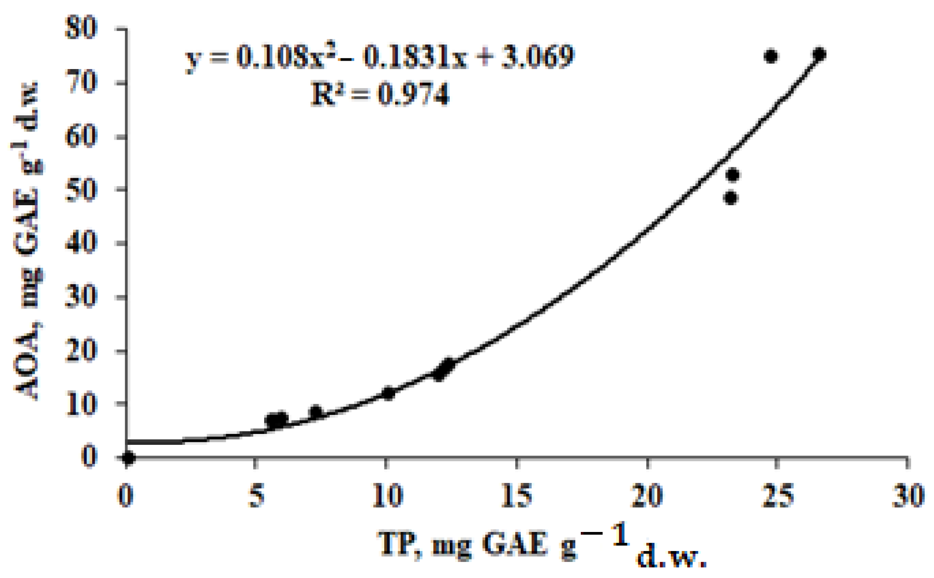
3.4. Antioxidant Status

The improvement of the plant antioxidant status due to nano-Si application is one of the mechanisms for plant protection against oxidant stresses [34], though extremely little information is available about the nano-Si effect on the plants’ antioxidant status in ordinary conditions of vegetation without significant stress factors. In case of A. annua, both the separate and joint application of Se and Si significantly improved ascorbic acid (AA) accumulation (Table 5). In this respect, in similar conditions, these elements did not cause any effect on ascorbic acid accumulation in chervil [24].
A positive effect of Si/Se application on ascorbic acid accumulation seems to be one of the most significant effects for the A. annua antioxidant status, with the mean AA increasing by a factor of 1.36. Regarding the total antioxidant activity (AOA), a statistically significant improvement of this parameter was registered for nano-Si (1.52) and Se (1.54 times) treatments. On the contrary, the joint application of Se + nano-Si did not change AOA and total phenolics (TP) levels in leaves. The mean AOA leaves/stems/roots ratio reached 6.86:1.72:1 for control plants; 10.88:2.21:1- for nano-Si; 8.85:1.95:1 for Se; and 7.03:2.09:1 for the joint application of Se and nano-Si. Accordingly, the distribution of phenolics (TP) between leaves, stems and roots reached 4.07:1.77:1 (control); 4.41:2.21:1 (nano-Si); 3:1.67:1 (Se); and 3.88:2:1 (Se + nano-Si).
The close relationship between AOA and TP parameters (Figure 1) indicates a nonlinear character of the curve with a higher variability of AOA in leaves, indicating that the leaves AOA is affected not only by the TP content but also by other antioxidants and essential oil that are particularly known to accumulate predominantly in leaves and florets [36].
At the same time, the non-statistically-significant changes in the proline and malonic dialdehyde (MDA) content in A. annua treated with Se and/or nano-Si indicate a low level of oxidant stress during vegetation. The exception is the high concentration of proline in plant leaves under the joint application of Se and nano-Si, which suggests an induction of plant tolerance. According to [37], high concentrations (150 mg L−1) of nano-Si mitigates lipid peroxidation and boosts osmoprotection. Taking into account the low concentrations of Se and especially Si in the present investigation, these factors supposedly provided only a low effect on MDA and proline changes.

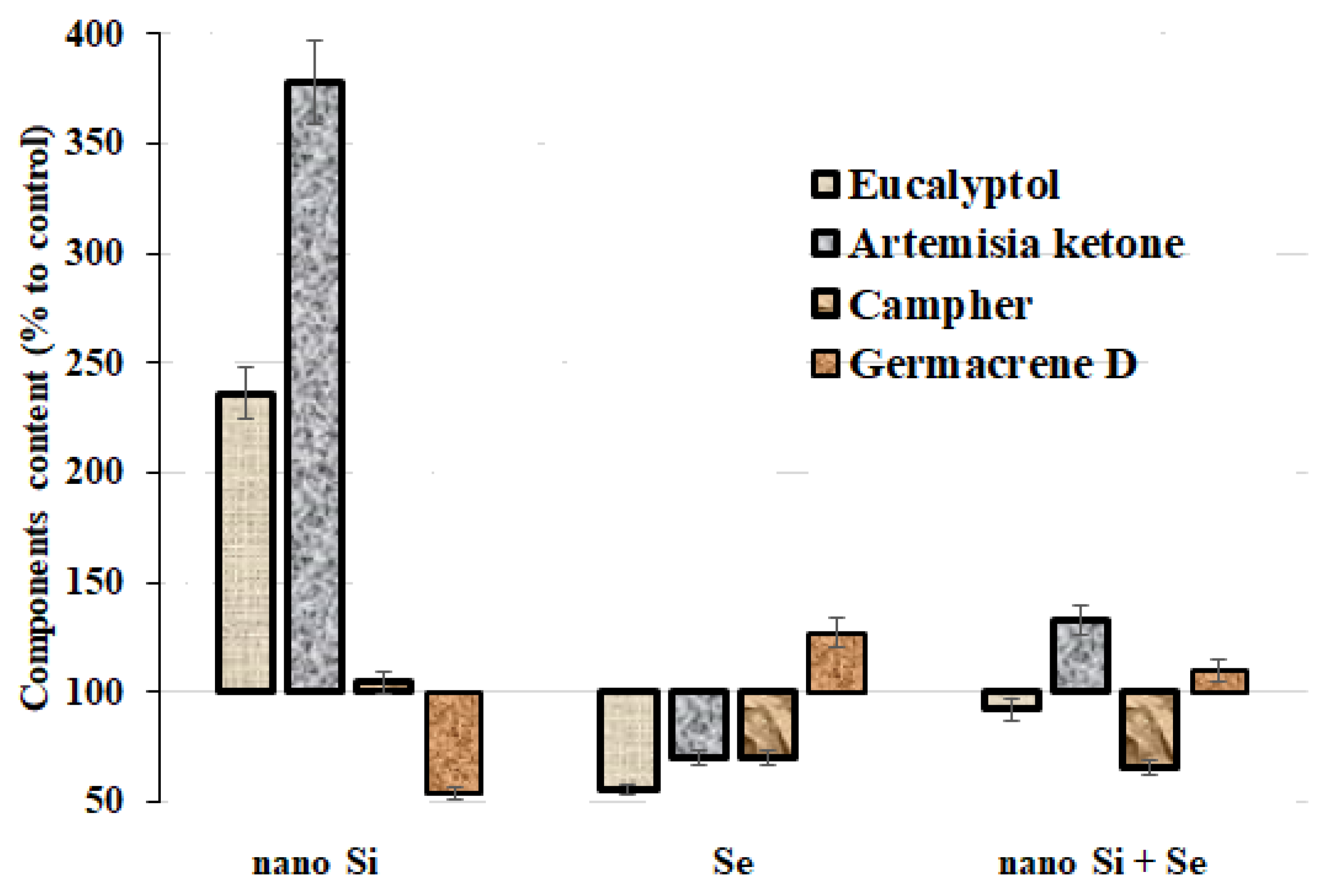
3.5. Essential Oil Accumulation

The essential oil chemical profile is generally influenced by the harvesting season, fertilizer and the pH of soils, the choice and stage of drying conditions, the geographic location, chemotype or subspecies, and the choice of the part plant or genotype or extraction method.
It is evident that the geographic location greatly influences the type and concentration of the main constituents. Other factors that determine the chemical composition and thus the biological activity are the chemical and biological treatment.
Eucalyptol, artemisia ketone, camphor and germacrene D are known to be the main components of A. annua essential oil, with the concentration being in the range of 4.8–31.5%, 2.2–68.5%, 1.9–48.0%, and 10.9–21.2%, respectively [38].
All of these chemicals demonstrate high antioxidant, anti-inflammatory, anti-viral, anti-bacterial and analgesic properties [39,40].
Essential oil accumulation is known to be directly connected with the antioxidant activity of Artemisia leaves [41]. Up to date, rather scant data are available on the effect of nano-Si supplementation on essential oil accumulation in aromatic plants: lemongrass Cymbopogon flexuosus [37], Mentha piperita [42] and coriander Coriandrum Sativum L. [43] (concentrations applied were within 42–150 mg L−1). In addition, separate data indicate the possibility of essential oil synthesis stimulation as a result of sodium selenate foliar application [44].
In general, biological effects of A. annua essential oil are the result of a synergism of all molecules contained in an essential oil, even if it is possible that the activity of the main components is modulated by other minor molecules, but the activity of the isolated constituents is also remarkable [38].
The essential oil content and number of constituents in A. annua plants greatly depend on the plant ontogenetic conditions [45,46]. Significant changes in the essential oil yield and its components number as a result of the single and joint application of nano-Si and Se indicate phenophase shifts, which are directly connected with a significant increase in the biomass of Se/Si-treated plants (Table 2 and Table 6, Figure 2). Nano-Si foliar application seems to be the greatest effector of these parameters. Table 6 and Figure 2 data indicate that, under these conditions, the number of essential oil components decreased significantly by 1.6 times and was accompanied by a significant increase in eucalyptol by 2.36 times, and in artemisia ketone by 3.78 times, whereas the germacrene D content decreased by 1.84 times. Whether the phenomenon relates to the decreased accumulation of photosynthetic pigments in plants treated with nano-Si needs further investigation.
The separate and joint nano-Si of Se application significantly decreased the camphor content by 1.4–1.5 times. Besides camphor, eucalyptol and artemisia ketone demonstrated a significant decrease under a single supply of sodium selenate.
A significant decrease in the number of oxidized forms of essential oil due to the Se supply may be connected with antioxidant properties of this element (the examples are camphor, pinocarvon, artemisia ketone and eucalyptol). On the contrary, Se seems to stimulate the production of carbohydrates, such as β-Selinene, humulene, (E)- β-famesene, β-caryophyllene, germacrene D and camphene (see supplement, Table S1). However, it should be indicated that there is at least one exception: caryophyllene and caryophyllene oxide both demonstrated a significantly increased concentration as a result of Se and Se + nano-Si treatment.
Several studies have permitted the conclusion that the A. annua crop could be harvested much before the onset of flowering in order to obtain high yields of artemisinin, and the crop must be allowed to attain maturity to obtain high yields of the essential oil [8,10]. Finally, the planting date and harvest time can influence the maximum concentration of the produced essential oil [10]. In the present work, at harvesting, A. annua plants were only at the stage of blooming and the beginning of flowering, which is not optimal for essential oil production.

3.6. Artemisinin Accumulation

In these conditions, a low content of artemisinin was recorded in A. annua leaves/florets (Figure 3) compared to plants grown in a warm climate. Usually, the artemisinin content in A. annua has a wide concentration range (0.01–0.45%) [9] depending on the habitat, climate [47,48], soil and plant characteristics [49,50,51], time of sowing, planting and harvesting [52] and plant genetic potential [9,13]. A lack of significant stress factors, which are known to stimulate the production of artemisinin [53,54,55,56], relatively low temperature and high humidity reduced artemisinin production in conditions of the Moscow region. Despite nano-Si being known to stimulate glandular trichomes development (the place of artemisinin biosynthesis) [49], no beneficial effect of Si nanoparticles was recorded in the present work. The latter fact may be attributed to the low concentration of Si nanoparticles, which was 10 times lower than that described in the literature [9]. Nevertheless, a significant beneficial effect on artemisinin accumulation was revealed under the joint application of Se and nano-Si (Figure 4).

3.7. Mineral Composition

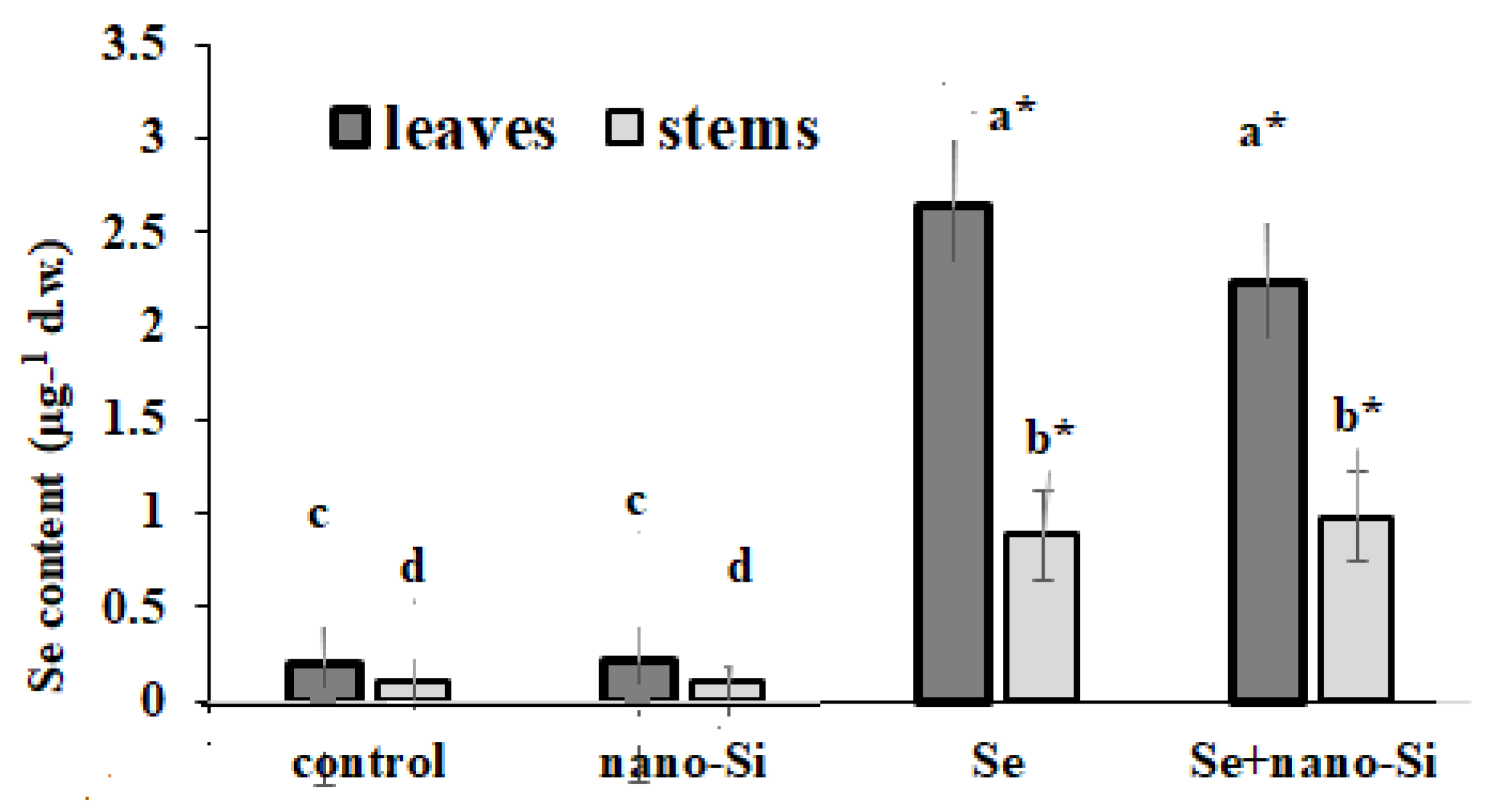
3.7.1. Se Accumulation

According to literature data, nano-Si does not significantly affect Se accumulation in plants [24]. The results of the present research regarding A. annua Se biofortification are in accordance with bibliographic data indicating significantly higher Se concentrations in leaves compared to stems (Figure 4). Furthermore, the differences between Se levels in plants treated separately or jointly with Se and nano-Si were not statistically significant.
From a practical point of view, Se concentrations in A. annua plants arising from Se biofortification may provide up to 11 µg of Se per 5 g of dry leaves weight, which corresponds to 16% of the adequate Se consumption level in the case of plant utilization as a spice. In the case of A. annua tea application, 50 mL of water extract (1:1) [57] provides approximately 75% (52.4 µg Se) of the daily adequate Se consumption level, taking into account that approximately 50% of Se in the plant is represented by water-soluble forms of this element.

3.7.2. Other Elements

The singular and joint plant supplementation with Se and Si greatly changed macro- and microelements accumulation, causing either a redistribution of elements between leaves/stems or a decrease/rise in the accumulation levels.
Data presented in Table 7, Table 8 and Table 9 show a specific peculiarity of A. annua to significantly decrease the levels of Ca and Mg as a result of the separate and joint Se/Si supply, as well as a decrease in Ca in the case of Se and Se + Si supplementation. Despite the essentiality of these macro-elements in plants, no signs of any growth inhibition and photosynthetic pigments level decrease were recorded. On the contrary, Se solely or in combination with Si stimulated the biosynthesis of chlorophyll and carotene in A. annua leaves and did not cause any significant changes in proline and MDA levels, which are indicators of the environmental stress degree (Table 4 and Table 5). The statistically significant decrease in chlorophyll concentration was recorded only under the Si supply, but, in these conditions, only slight changes in the mineral composition of plants were recorded (Table 7, Table 8 and Table 9).
Earlier investigations on spinach [21] confirmed the possibility of this phenomenon. Taking into account that a significant decrease in Ca, K and Mg under the selenite and selenate supply was recorded only in male spinach forms, it may be supposed that a similar phenomenon in A. annua plants may be connected with hormonal regulation, though this topic needs further investigation.
Furthermore, Se treatment also significantly reduced the accumulation of B, Cu, Fe, Mn and Zn in A. annua leaves. The suggestion of Łukaszewicz et al. [58] that the similar microelement (Fe, Zn, Mn, B) decrease in pea due to Se+6/Se+4 application is connected with dehydration associated with the membrane damage of roots does not seem applicable to A. annua plants, as no growth inhibition or photosynthetic pigment decrease was recorded. The effect is certainly species-dependent, as no significant differences were recorded in chervil grown and treated in similar way [24].
The matter of the Se/Si effect on essential macro- and trace elements accumulation has been poorly discussed in literature, and the experimental data often demonstrate controversial results. Indeed, the foliar biofortification of A. cepa with sodium selenate in the Chechen republic conditions did not cause any statistically significant changes in macroelements, whereas, among microelements, the B content increased as a result of the Se supply [59]. Maize seedlings obtained in hydroponics showed a Ca and P increase and significant decrease in K content as a result of the selenite supply [60,61,62,63]. No significant changes in macro and essential microelements accumulation were recorded in chickpea treated with sodium selenate [61]. In chervil, the Se supply decreased only the content of Na [24].
Regarding heavy metals, the singular or joint application of Si and Se resulted in a significant decrease in leaf As, Ca, Pb and Sr content, whereas the joint application of Se and Si caused the addition of other heavy metals to this list: Cr, Ni and V. These results are in agreement with the known antagonistic relationship between heavy metals and Se [62,63] and the protective effect of nano-Si against the former [64,65].
In the present work, a valuable redistribution of Li, Zn and Al between leaves and stems of A. annua as a result of Se/Si treatment was revealed for the first time (Table 8 and Table 9). Indeed, a two-fold increase in leaf Al and Li levels as a result of the foliar Se+6 supply was accompanied by a corresponding significant decrease in stem Al/Li concentration. The joint application of Se and Si resulted in a significant decrease in the Zn level in the leaves and its increase in stems.
In general, the leaves/stems distribution of elements in A. annua plants indicates the predominant accumulation of Mg, P, V, B, Cu, Fe, Mn, Mo and Se in leaves and Al, Cd, Cr, Pb and Sr in stems (Table 6, Table 7 and Table 8), where only trace levels of B were found.
In the present investigation, low doses of nano-Si were not sufficient to elicit a statistically significant increase in Si concentration, both in the stems and leaves of A. annua, characterized by a high biomass reflecting significant biological dilution. In this respect, it should be highlighted that the beneficial effect of a low dose of nano-Si on the plant yield and antioxidant status is associated with a new phenomenon of nanoparticles application.

4. Conclusions

The results of the present experiment indicate typical peculiarities of A. annua Se biofortification under a nano-Si supply: a growth stimulation effect and increase in the antioxidant status of plants, with a significant reduction in the content of essential oil and number of its components, as well as profound qualitative and quantitative changes in the oil composition. Neither Se nor nano-Si in a singular application changed the artemisinin levels, whereas the joint supply of Se and nano-Si to plants significantly increased the artemisinin level by 1.4 times. The effect of Al, Li and Zn redistribution between leaves and stems, due to Se/Si supplementation and the anomalous decrease in Ca, Mg, K, B, Cu, Fe, Mn and Zn levels in leaves, as a result of the Se treatment not accompanied by growth inhibition or a significant decrease in the photosynthetic pigments content, needs further investigations in order to reveal the mechanism of the phenomenon.

Supplementary Materials

The following are available online at https://www.mdpi.com/article/10.3390/horticulturae8070597/s1. Table S1: Effect of Se and nano-Si supply on A. annua essential oil composition.

Author Contributions

Experiment conceptualization: N.G., L.L., L.S. and G.C.; silicon nanoparticles production and characteristics provision: M.F. and A.A.; field experiment management and determinations: N.G. and L.L.; biochemical analysis: N.G. and L.S.; essential oil determination and components identification: L.L. and O.S.; artemisinin determination: D.K. and E.G.; data statistical processing: N.G., L.S., A.S. and G.C.; data interpretation and draft and final-version manuscript writing: N.G., L.L, O.S., L.S., M.F., D.K., E.G., A.S. and G.C. All authors have read and agreed to the published version of the manuscript.

Funding

This research did not receive any grants from public, commercial or not-for-profit agencies.

Institutional Review Board Statement

Not applicable.

Informed Consent Statement

Not applicable.

Data Availability Statement

Not applicable.

Conflicts of Interest

The authors declare no conflict of interest.

References

  1. Ho, W.E.; Peh, H.Y.; Chan, T.K.; Wong, W.S. Artemisinins: Pharmacological actions beyond anti-malarial. Pharmacol. Ther. 2014, 142, 126–139. [Google Scholar] [CrossRef] [PubMed]
  2. Hunt, S.; Yoshida, M.; Davis, C.E.; Greenhill, N.S.; Davis, P.F. An extract of the medicinal plant Artemisia annua modulates production of inflammatory markers in activated neutrophils. J. Inflamm. Res. 2015, 8, 9–14. [Google Scholar] [CrossRef] [PubMed] [Green Version]
  3. Sadiq, A.; Hayat, M.Q.; Ashraf, M. Ethnopharmacology of Artemisia annua L.: A Review. In Artemisia Annua—Pharmacology and Biotechnology; Aftab, T., Ferreira, J., Khan, M., Naeem, M., Eds.; Springer: Berlin/Heidelberg, Germany, 2014. [Google Scholar]
  4. Oh, S.-I.; Kim, J.-S.; Kim, C.-K.; Yi, S.S.; Kim, S.-J.; Park, S.-K. Artemisia annua increases resistance to heat and oxidative stresses, but has no effect on lifespan in Caenorhabditis elegans. Food Sci. Technol. 2016, 36, 356–361. [Google Scholar] [CrossRef] [Green Version]
  5. Fatima, K.; Abbas, S.R.; Zia, M.; Sabir, S.M.; Khan, R.T.; Khan, A.A.; Hassan, Z.; Zaman, R. Induction of secondary metabolites on nanoparticles stress in callus culture of Artemisia annua L. Braz. J. Biol. 2021, 81, 474–483. [Google Scholar] [CrossRef]
  6. Abate, G.; Zhang, L.; Pucci, M.; Morbini, G.; Sweeney, E.M.; Maccarinelli, G.; Ribaudo, G.; Gianoncelli, A.; Uberti, D.; Memo, M.; et al. Phytochemical Analysis and Anti-Inflammatory Activity of Different Ethanolic Phyto-Extracts of Artemisia annua L. Biomolecules 2021, 11, 975. [Google Scholar] [CrossRef]
  7. Salehi, M.; Karimzadeh, G.; Naghavi, M.R.; Badi, H.N.; Monfared, S.R. Expression of key genes affecting artemisinin content in five Artemisia species. Sci. Rep. 2018, 8, 12659. [Google Scholar] [CrossRef]
  8. Lu, F.; He, X.L.; Culleton, R.; Cao, J. A brief history of artemisinin: Modes of action and mechanisms of resistance. Chin. J. Nat. Med. 2019, 17, 331–336. [Google Scholar] [CrossRef]
  9. Wani, K.I.; Choudhary, S.; Zehra, A.; Naeem, M.; Weathers, P.; Aftab, T. Enhancing artemisinin content in and delivery from Artemisia annua: A review of alternative, classical, and transgenic approaches. Planta 2021, 254, 29. [Google Scholar] [CrossRef]
  10. Li, J.; Zhou, B. Biological Actions of Artemisinin: Insights from Medicinal Chemistry Studies. Molecules 2010, 15, 1378–1397. [Google Scholar] [CrossRef]
  11. Klayman, D.L. Artemisia annua: From weed to respectable antimalarial plant. In Human Medicinal Agents from Plants; Kinghom, A.D., Balandrin, M.F., Eds.; American Chemical Society Symposium Series; ACS Publications: Washington, DC, USA, 1993; pp. 242–255. [Google Scholar]
  12. Dite, Z.; Suvada, R.; Toth, T.; Jun, P.E.; Pis, V.; Dite, D. Current Condition of Pannonic Salt Steppes at Their Distribution Limit: What Do Indicator Species Reveal about Habitat Quality? Plants 2021, 10, 530. [Google Scholar] [CrossRef]
  13. Wang, D.; Shi, C.; Alamgir, K.; Pan, L.; Zhu, Y.; Yang, X. Global assessment of the distribution and conservation status of key medicinal plants (Artemisia annua L.): The roles of climate and anthropogenic activities. Sci. Total Environ. 2022, 821, 153378. [Google Scholar] [CrossRef]
  14. Septembre-Malaterre, A.; Rakoto, M.L.; Marodon, C.; Bedoui, Y.; Nakab, J.; Simon, E.; Hoarau, L.; Savriama, S.; Strasberg, D.; Guiraud, P.; et al. Artemisia annua, a Traditional Plant Brought to Light. Int. J. Mol. Sci. 2020, 21, 4986. [Google Scholar] [CrossRef] [PubMed]
  15. Namuli, A.; Bazira, J.; Casim, T.U.; Engeu, P.O. A review of various efforts to increase artemisinin and other antimalarial compounds in Artemisia annua L plant. Cogent Biol. 2018, 4, 1513312. [Google Scholar] [CrossRef]
  16. Todeschini, V.; Anastasia, F.; Massa, N.; Marsano, F.; Cesaro, P.; Bona, E.; Gamalero, E.; Oddi, L.; Lingua, G. Impact of Phosphatic Nutrition on Growth Parameters and Artemisinin Production in Artemisia annua Plants Inoculated or Not with Funneliformis mosseae. Life 2022, 12, 497. [Google Scholar] [CrossRef] [PubMed]
  17. Wu, Y.; Li, L.; Peng, M.; Wu, X.; Li, F.; Wang, Z. Effects of zinc manganese and boron on artemisinin and yields of Artemisia annua Zhongguo Zhong Yao Za Zhi Zhongguo Zhongyao Zazhi. China J. Chin. Mater. Medica 2010, 5, 275–278. [Google Scholar]
  18. Aftab, T.; Khan, M.M.A.; Idrees, M.; Naeem, M.; Hashmi, N. Moinuddin Effect of Salt Stress on Growth, Membrane Damage, Antioxidant Metabolism and Artemisinin Accumulation in Artemisia annua L. Plant Stress 2010, 4, 3643. [Google Scholar]
  19. Rostkowska, C.; Mota, C.; Oliveira, T.C.; Santiago, F.M.; Oliveira, L.A.; Korndörfer, G.H.; Lana, R.M.Q.; Rossi, M.; Mogueira, N.L.; Simonnet, X.; et al. Si-Accumulation in Artemisia annua Glandular Trichomes Increases Artemisinin Concentration, but Does Not Interfere in the Impairment of Toxoplasma gondii Growth. Front. Plant Sci. 2016, 7, 1430. [Google Scholar] [CrossRef] [Green Version]
  20. Golubkina, N.A.; Kharchenko, V.A.; Caruso, G. Selenium: Prospects of functional food production with high antioxidant activity. In Chapter 7 in Reference Series in Phyto-Chemistry. Plant Antioxidants and Health; Ekiert, H., Ramawat, K.G., Arora, J., Eds.; Springer: Berlin/Heidelberg, Germany, 2021; pp. 149–176. [Google Scholar]
  21. Golubkina, N.A.; Kosheleva, O.V.; Krivenkov, L.V.; Dobrutskaya, H.G.; Nadezhkin, S.M.; Caruso, G. Intersexual differences in plant growth, yield, mineral composition and antioxidants of spinach (Spinacia oleracea L.) as affected by selenium form. Sci. Hortic. 2017, 225, 350–358. [Google Scholar] [CrossRef]
  22. Golubkina, N.; Zayachkovsky, V.; Sheshnitsan, S.; Skrypnik, L.; Smirnova, T.; Antoshkina, M.; Fedotov, M.; Caruso, G. Prospects of garlic extracts, selenium and silicon application for plants protection against herbivory. Review. Agriculture 2022, 12, 64. [Google Scholar] [CrossRef]
  23. Zhang, J.; Taylor, E.W.; Bennett, K.; Saad, R.; Rayman, M.P. Association between regional selenium status and reported outcome of COVID-19 cases in China. Am. J. Clin. Nutr. 2020, 111, 1297–1299. [Google Scholar] [CrossRef]
  24. Golubkina, N.; Moldovan, A.; Fedotov, M.; Kekina, H.; Kharchenko, V.; Folmanis, G.; Alpatov, A.; Caruso, G. Iodine and Selenium Biofortification of Chervil Plants Treated with Silicon Nanoparticles. Plants 2021, 10, 2528. [Google Scholar] [CrossRef] [PubMed]
  25. Association Official Analytical Chemists (AOAC). The Official Methods of Analysis of AOAC International; 22 Vitamin C.; AOAC: Rockville, MD, USA, 2012. [Google Scholar]
  26. Lichtenthaler, H.K. Chlorophylls and carotenoids: Pigments of photosynthetic biomembranes. Methods Enzymol. 1987, 148, 350–382. [Google Scholar]
  27. Golubkina, N.A.; Kekina, H.G.; Molchanova, A.V.; Antoshkina, M.S.; Nadezhkin, S.M.; Soldatenko, A.V. Plants Antioxidants and Methods of Their Determination; INFRA-M: Moscow, Russia, 2020. [Google Scholar] [CrossRef]
  28. Numonov, S.; Sharopov, F.; Salimov, A.; Sukhrobov, P.; Atolikshoeva, S.; Safarzoda, R.; Habasi, M.; Aisa, H.A. Assessment of Artemisinin Contents in Selected Artemisia Species from Tajikistan (Central Asia). Medicines 2019, 6, 23. [Google Scholar] [CrossRef] [PubMed] [Green Version]
  29. Ouertani, R.N.; Abid, G.; Karmous, C.; Chikha, M.B.; Boudaya, O.; Mahmoudi, H.; Mejri, S.; Jansen, K.; Ghorbel, A. Evaluating the contribution of osmotic and oxidative stress components on barley growth under salt stress. AoB Plants 2021, 1, plab034. [Google Scholar] [CrossRef]
  30. Alfthan, G.V. A micromethod for the determination of selenium in tissues and biological fluids by single-test-tube fluorimetry. Anal. Chim. Acta 1984, 165, 187–194. [Google Scholar] [CrossRef]
  31. Heath, R.L.; Parker, L. Photoperoxidation in isolated chloroplasts. I. Kinetics and stoichiometry of fatty acid peroxidation. Arch. Biochem. Biophys. 1968, 125, 189–198. [Google Scholar] [CrossRef]
  32. Golubkina, N.; Kekina, H.; Caruso, G. Yield, Quality and Antioxidant Properties of Indian Mustard (Brassica juncea L.) in Response to Foliar Biofortification with Selenium and Iodine. Plants 2018, 7, 80. [Google Scholar] [CrossRef] [Green Version]
  33. Sali, A.; Zeka, D.; Fetahu, S.; Rusinovci, I.; Kaul, H.-P. Selenium supply affects chlorophyll concentration and biomass production of maize (Zea mays L.). Die Bodenkult. J. Land Manag. Food Environ. 2008, 69, 249–255. [Google Scholar] [CrossRef] [Green Version]
  34. Rajput, V.D.; Minkina, T.; Feizi, M.; Kumari, A.; Khan, M.; Mandzhieva, S.; Sushkova, S.; El-Ramady, H.; Verma, K.K.; Singh, A.; et al. Effects of Silicon and Silicon-Based Nanoparticles on Rhizosphere Microbiome, Plant Stress and Growth. Biology 2021, 10, 791. [Google Scholar] [CrossRef]
  35. Siddiqui, H.; Ahmed, K.B.M.; Sami, F.; Hayat, S. Silicon nanoparticles and plants: Current knowledge and future perspectives. In Sustainable Agriculture Reviews 41: Nanotechnology for Plant Growth and Development; Hayat, S., Pichtel, J., Faizan, M., Fariduddin, Q., Eds.; Springer International Publishing: Cham, Switzerland, 2020; pp. 129–142. [Google Scholar]
  36. Tzenkova, R.; Kamenarska, Z.; Draganov, A.; Atanassov, A. Composition of A. annua essential oil obtained from species growing wild in Bulgaria. Biotechnol. Biotechnol. Equip. 2010, 24, 1833–1835. [Google Scholar] [CrossRef] [Green Version]
  37. Mukarram, M.; Khan, M.M.A.; Corpas, F.J. Silicon nanoparticles elicit an increase in lemongrass (Cymbopogon flexuosus (Steud.) Wats) agronomic parameters with a higher essential oil yield. J. Hazard. Mater. 2021, 412, 125254. [Google Scholar] [CrossRef] [PubMed]
  38. Bilia, A.R.; Santomauro, F.; Sacco, C.; Bergonzi, M.C.; Donato, R. Essential oil of Artemisia annua L.: An extraordinary component with numerous antimicrobial properties. Evid.-Based Complement. Altern. Med. 2014, 2014, 159819. [Google Scholar] [CrossRef] [Green Version]
  39. Verdian-rizi, M.R.; Sadat-Ebrahimi, E.; Hadjiakhoondi, A.; Fazeli, M.R.; Pirali Hamedani, M. Chemical Composition and Antimicrobial Activity of Artemisia annua L. Essential Oil from Iran. J. Med. Plants 2008, 7, 58–62. [Google Scholar]
  40. Juteau, F.; Masotti, V.; Bessière, J.M.; Dherbomez, M.; Viano, J. Antibacterial and antioxidant activities of Artemisia annua essential oil. Fitoterapia 2002, 73, 532–535. [Google Scholar] [CrossRef]
  41. Golubkina, N.; Logvinenko, L.; Molchanova, A.; Caruso, G. Genetic and Environmental Influence on Macro- and Microelement Accumulation in Plants of Artemisia Species. In Chapter 17 in Plants Micronutrients. Deficiency and Toxicity Management; Aftab, T., Hakeem, K.R., Eds.; Springer: Cham, Switzerland, 2020; pp. 389–416. [Google Scholar]
  42. Ahmad, B.; Khan, M.M.A.; Jaleel, H.; Shabbir, A.; Sadiq, Y.; Uddin, M. Silicon nanoparticles mediated increase in glandular trichomes and regulation of photosynthetic and quality attributes in Mentha piperita L. J. Plant Growth Regul. 2020, 39, 346–357. [Google Scholar] [CrossRef]
  43. Afshari, M.; Pazoki, A.; Sadeghipour, O. Foliar-applied Silicon and its Nanoparticles Stimulate Physio-chemical Changes to Improve Growth, Yield and Active Constituents of Coriander (Coriandrum Sativum L.) Essential Oil Under Different Irrigation Regimes. Silicon 2021, 13, 4177–4188. [Google Scholar] [CrossRef]
  44. Skrypnik, L.; Novikova, A.; Tokupova, E. Improvement of Phenolic Compounds, Essential Oil Content and Antioxidant Properties of Sweet Basil (Ocimum basilicum L.) Depending on Type and Concentration of Selenium Application. Plants 2019, 8, 458. [Google Scholar] [CrossRef] [PubMed] [Green Version]
  45. Mohammadreza, V. Variation in the essential oil composition of Artemisia annua L. of different growth stages cultivated in Iran. Afr. J. Plant Sci. 2008, 2, 16–18. [Google Scholar]
  46. Coşge, B.; Kiralan, M.; Yaman, C. The Effect of Different Harvest Stages on Chemical Composition and Antioxidant Capacity of Essential Oil from Artemisia annua L. J. Agric. Sci. 2015, 21, 71–77. [Google Scholar] [CrossRef] [Green Version]
  47. Delabays, N.; Simonne, X.; Gaudin, M. The Genetics of Artemisinin Content in Artemisia annua L. and the Breeding of High Yielding Cultivars. Curr. Med. Chem. 2002, 8, 1795–1801. [Google Scholar] [CrossRef] [PubMed]
  48. Brown, G.D. The biosynthesis of artemisinin (qinghaosu) and the phytochemistry of Artemisia annua L. (qinghao). Molecules 2010, 15, 7603–7698. [Google Scholar] [CrossRef] [PubMed] [Green Version]
  49. Domokos, E.; Jakab-Farkas, L.; Darko, B.; Biro-Janka, B.; Mara, G.; Albert, C.; Balog, A. Increase in Artemisia annua plant biomass artemisinin content and guaiacol peroxidase activity using the arbuscular mycorrhizal fungus Rhizophagus irregularis. Front. Plant Sci. 2018, 9, 478. [Google Scholar] [CrossRef] [PubMed] [Green Version]
  50. Huang, L.; Xie, C.; Duan, B.; Chen, S. Mapping the potential distribution of high artemisinin-yielding Artemisia annua L. (Qinghao) in China with a geographic information system. Chin. Med. 2010, 5, 18. [Google Scholar] [CrossRef] [PubMed] [Green Version]
  51. Widiyastuti, Y.; Subositi, D. Photoperiod effect on the growth and artemisinin content of Artemisia Annua grown in tropical region. In Proceedings of the AIP Conference Proceedings, Yogyakarta, Indonesia, 19–20 October 2018; Volume 2099, p. 020027. [Google Scholar] [CrossRef]
  52. Verma, R.K.; Chauhan, A.; Verma, R.S.; Gupta, A. Influence of planting date on growth, artemisinin yield, seed and oil yield of Artemisia annua L. under temperate climatic conditions. Ind. Crops Prod. 2011, 34, 860–864. [Google Scholar] [CrossRef]
  53. Marchese, J.A.; Ferreira, J.F.S.; Rehder, V.L.G.; Rodrigues, O. Water deficit effect on the accumulation of biomass and artemisinin in annual wormwood (Artemisia annua L., Asteraceae). Braz. J. Plant Physiol. 2010, 22, 1–9. [Google Scholar] [CrossRef] [Green Version]
  54. Marchese, J.A.; Rehder, V.L.G. The influence of temperature in the production of artemisinin in Artemisia annua L. Braz. J. Med. Plant 2001, 4, 89–93. [Google Scholar]
  55. Marchese, J.A.; Rehder, V.L.G.; Casiraghi, V.; Tedesco, A.C.; Lira, R. Flowering in plants of Artemisia annua L. submitted to different conditions of photoperiod and temperature. Acta Hortic. 2002, 569, 275–280. [Google Scholar] [CrossRef]
  56. Ferreira, J.F.S. Nutrient deficiency in the production of artemisinin, dihydroartemisinic acid, and artemisinic acid in Artemisia annua L. J. Agric. Food Chem. 2007, 55, 1686–1694. [Google Scholar] [CrossRef] [PubMed]
  57. Mueller, M.S.; Karhagomba, I.B.; Hirt, H.M.; Wemakor, E. The potential of Artemisia annua L. as a locally produced remedy for malaria in the tropics: Agricultural, chemical and clinical aspects. J. Ethnopharmacol. 2000, 73, 487–493. [Google Scholar] [CrossRef]
  58. Łukaszewicz, S.; Politycka, B.; Smoleń, S. Effects of selenium on the content of essential micronutrients and their translocation in garden pea. J. Elem. 2018, 23, 1307–1317. [Google Scholar] [CrossRef]
  59. Golubkina, N.; Amagova, Z.; Matsadze, V.; Zamana, S.; Tallarita, A.; Caruso, G. Effects of arbuscular mycorrhizal fungi on yield, biochemical characteristics and elemental composition of garlic and onion under selenium supply. Plants 2020, 9, 84. [Google Scholar] [CrossRef] [PubMed] [Green Version]
  60. Hawrylak-Nowak, B. Effect of selenium on selected macronutrients in maize plants. J. Elem. 2008, 13, 513–519. [Google Scholar]
  61. Golubkina, N.; Gomez, L.D.; Kekina, H.; Cozzolino, E.; Simister, R.; Tallarita, A.; Torino, V.; Koshevarov, A.; Cuciniello, A.; Maiello, R.; et al. Joint Selenium–Iodine Supply and Arbuscular Mycorrhizal Fungi Inoculation Affect Yield and Quality of Chickpea Seeds and Residual Biomass. Plants 2020, 9, 804. [Google Scholar] [CrossRef] [PubMed]
  62. Hasanuzzaman, M.; Nahar, K.; García-Caparrós, P.; Parvin, K.; Zulfiqar, F.; Ahmed, N.; Fujita, M. Selenium Supplementation and Crop Plant Tolerance to Metal/Metalloid Toxicity. Front. Plant Sci. 2022, 12, 792770. [Google Scholar] [CrossRef] [PubMed]
  63. Zhou, P.; Adeel, M.; Shakoor, N.; Guo, M.; Hao, Y.; Azeem, I.; Li, M.; Liu, M.; Rui, Y. Application of Nanoparticles Alleviates Heavy Metals Stress and Promotes Plant Growth: An Overview. Nanomaterials 2021, 11, 26. [Google Scholar] [CrossRef] [PubMed]
  64. Khan, I.; Awan, S.A.; Rizwan, M.; Ali, S.; Hassan, M.J.; Brestic, M.; Zhang, X.; Huang, L. Effects of silicon on heavy metal uptake at the soil-plant interphase: A review. Ecotoxicol. Environ. Saf. 2021, 222, 112510. [Google Scholar] [CrossRef] [PubMed]
  65. Zhou, X.; Yang, J.; Kronzucker, H.J.; Shi, W. Selenium Biofortification and Interaction With Other Elements in Plants: A Review. Front. Plant Sci. 2020, 11, 586421. [Google Scholar] [CrossRef] [PubMed]
Figure 1. Correlation between AOA and TP in A. annua plants (r = 0.987, p < 0.001).
Horticulturae 08 00597 g001
Figure 2. Effect of separate and joint application of nano-Si and Se on essential oil content in Artemisia annua, grown in Moscow region.
Horticulturae 08 00597 g002
Figure 3. Effect of nano-Si and sodium selenate on artemisinin content. nts). Values with the same letter do not differ statistically according to Duncan test at p < 0.05.
Horticulturae 08 00597 g003
Figure 4. Effect of Se and nano-Si on leaves/stems Se distribution (*: values have been halved). Values with the same letters do not differ statistically according to Duncan’s test at p < 0.05.
Horticulturae 08 00597 g004
Table 1. Mean temperature and humidity in 2020 and 2021.
Month20202021
Mean Temperature °CHumidity %Mean Temperature °CHumidity %
May13.56613.566
June21.07317.269
July23.8174.020.172
August17.87217.875
September11.98011.980
Table 2. Morphological characteristics of A. annua plants subjected to sodium selenate and nano-Si (M ± SD).
ControlSiSeSi + Se
Plant height (cm)123 ± 15 a131 ± 17 a127 ± 16 a127 ± 15 a
Leaf length (cm)24 ± 4 b25 ± 3 b27 ± 4 b45 ± 7 a
Number of leaves14 ± 1 b19 ± 2 a19 ± 2 a20 ± 3 a
Leaf weight (g)26.03 ± 2.05 c50.08 ± 5.10 b52.63 ± 5.05 b115.14 ± 9.71 a
Stem weight (g)32.3 ± 2.78 c41.94 ± 3.98 b39.05 ± 3.04 b76.79 ± 8.02 a
Leaf weight (% of plant weight)44.6 ± 4.0 b56.4 ± 4.2 a57.4 ± 4.1 a60.0 ± 5.0 a
Along each line, the values with the same letters do not differ statistically according to Duncan’s test at p < 0.05.
Table 3. Dry matter, nitrates and total dissolved solids (TDS) accumulation in A. annua under Se and/or Si supply (M ± SD).
ParameterPlant PartControlNano-SiSeSe + Nano-Si
Dry matter (%)Leaves13.0 ± 1.0 a15.0 ± 1.2 a13.7 ± 1.1 a13.3 ± 1.0 a
Stems13.4 ± 1.1 a15.6 ± 1.3 a14.5 ± 1.3 a14.3 ± 1.2 a
Roots16.6 ± 1.4 a17.3 ± 1.5 a17.5 ± 1.5 a16.3 ± 1.4 a
Nitrates
(mg g−1 d.w.)
Leaves3.00 ± 0.25 a3.04 ± 0.22 a2.90 ± 0.21 a2.76 ± 0.21 a
Stems5.38 ± 0.48 a4.34 ± 0.35 b3.93 ± 0.37 b4.69 ± 0.37 ab
Stems/leaves1.79 ± 0.03 a1.43 ± 0.03 b1.36 ± 0.02 b1.70 ± 0.04 a
TDS (mg g−1 d.w.)Leaves67.7 ± 6.1 a67.2 ± 6.1 a73.1 ± 6.9 a67.1 ± 6.1 a
Stems127.7 ± 11.1 a97.0 ± 8.8 b89.4 ± 8.2 b105.5 ± 9.0 ab
Stems/leaves1.89 ± 0.06 a1.44 ± 0.06 bc1.22 ± 0.05 c1.57 ± 0.04 b
TDS: total dissolved solids. Along each line, the values with the same letters do not differ statistically according to Duncan’s test at p < 0.05.
Table 4. Photosynthetic pigment content in A. annua leaves as affected by nano-Si and/or sodium selenate (M ± SD).
ParameterControlNano-SiSeSe + Nano-Si
Chl a (mg g−1 f.w.)1.32 ± 0.11 b1.01 ± 0.09 c1.66 ± 0.13 a1.45 ± 0.12 ab
Chl b (mg g−1 f.w.)3.48 ± 0.30 a2.73 ± 0.21 b3.89 ± 0.32 a3.61 ± 0.31 a
Total chl (mg g−1 f.w.)4.80 ± 0.42 a3.74 ± 0.33 b5.55 ± 0.50 a5.06 ± 0.45 a
Carotene (mg g−1 f.w.)0.56 ± 0.04 b0.49 ± 0.03 b0.68 ± 0.06 a0.70 ± 0.06 a
Chl b/Chl a2.64 ± 0.02 a2.70 ± 0.02 a2.34 ± 0.03 a2.49 ± 0.02 a
Carotene /total chl (%)11.9 ± 0.01 a13.1 ± 0.01 a12.3 ± 0.01 a13.8 ± 0.02 a
Chl: chlorophyll. Along each line, values with the same letters do not differ statistically according to Duncan’s test at p < 0.05.
Table 5. Antioxidant status of A. annua L. at flowering stage (M ± SD).
TissueControlSiSeSe + Si
AA (mg 100 g−1 f.w.)Leaves74.2 ± 7.1 b106.0 ± 8.5 a90.6 ± 8.5 a106.3 ± 8.2 a
AOA (mg GAE g−1 d.w.)Leaves48.7 ± 4.2 b75.1 ± 7.0 a75.2 ± 7.1 a52.7 ± 5.1 b
Stems12.2 ± 1.2 b17.4 ± 1.6 a16.6 ± 1.6 a15.7 ± 1.5 a
Roots7.1 ± 0.7 a6.9 ± 0.6 a8.5 ± 0.7 a7.5 ± 0.7 a
TP (mg GAE g−1 d.w.)Leaves23.2 ± 2.1 a24.7 ± 2.2 a26.6 ± 2.3 a23.3 ± 2.1 a
Stems10.1 ± 1.0 a12.4 ± 1.2 a12.2 ± 1.1 a12.0 ± 1.1 a
Roots5.7 ± 0.5 b5.6 ± 0.5 b7.3 ± 0.7 a6.0 ± 0.6 ab
MDA (mM g−1 d.w.)Leaves0.38 ± 0.04 a0.31 ± 0.03 a0.35 ± 0.03 a0.36 ± 0.03 a
Proline (mg g−1 d.w.)Leaves8.14 ± 0.81 ab8.21 ± 0.81 ab7.67 ± 0.78 b9.41 ± 0.91 a
Artemisinin (mg kg−1 d.w.)Leaves3.85 ± 0.17 b3.94 ± 0.11 b3.68 ± 0.14 b5.42 ± 0.25 a
AA: ascorbic acid; AOA: total antioxidant activity; TP: total phenolics; MDA: malonic dialdehyde. Along each line, values with the same letters do not differ significantly according to Duncan’s test at p < 0.05.
Table 6. Yield and composition of A. annua essential oil under Se and/or Si supply (M ± SD).
ParameterControlSiSeSi + Se
Essential oil yield (%)0.20 ± 0.01 a0.15 ± 0.01 b0.15 ± 0.01 b0.10 ± 0.01 c
Number of components81 ± 2 a50 ± 1 c71 ± 1 b72 ± 2 b
The Main Oil Components, %
Eucalyptol3.32 ± 0.30 b7.85 ± 0.75 a 1.83 ± 0.16 c 3.06 ± 0.29 b
Artemisia ketone6.28 ± 0.63 c23.73 ± 2.33 a 4.39 ± 0.41 d 8.36 ± 0.82 b
Camphor32.83 ± 2.90 a34.35 ± 3.04 a 23.02 ± 2.04 b 21.61 ± 2.10 b
Germacrene D9.25 ± 0.91 b5.03 ± 0.50 c11.77 ± 1.13 a 10.17 ± 1.02 ab
Along each line, values with the same letters do not differ statistically according to Duncan’s test at p < 0.05.
Table 7. Macroelements in A. annua leaves and stems as affected by nano-Si, sodium selenate and nano Si + Se (M ± SD; mg kg−1 d.w.).
ElementControlSiSeSe + Si
Leaves
Ca10,778 ± 1057 a8678 ± 812 b6789 ± 628 c8253 ± 800 bc
K75,681 ± 7405 a71,512 ± 6998 a45,027 ± 4220 b52,833 ± 5179 b
Mg3718 ± 357 a2810 ± 277 b2743 ± 268 b2679 ± 260 b
Na514 ± 50 b438 ± 44 b491 ± 48 bc677 ± 67 a
P6177 ± 602 a5172 ± 511 ab5400 ± 535 a5150 ± 506 ab
Stems
Ca9328 ± 901 a7006 ± 688 b6821 ± 632 b8044 ± 795 a
K78,648 ± 7004 a72,945 ± 6987 a69,363 ± 6123 a75,420 ± 7224 a
Mg1512 ± 150 a1495 ± 145 a1129 ± 110 b1327 ± 133 a
Na312 ± 29 a314 ± 30 a381 ± 34 a315 ± 30 a
P4799 ± 456 a3391 ± 329 b4571 ± 446 a3817 ± 377 b
Along each line, values with the same letters do not differ statistically according to Duncan’s test at p < 0.05.
Table 8. Heavy metals, As and Al in A. annua leaves as affected by nano-Si, sodium selenate and nano Si + Se (M ± SD; mg kg−1 d.w.).
ElementControlSiSeSe + Si
Leaves
Al61.3 ± 6.1 b57.2 ± 5.7 b128.0 ± 12.8 a41.1 ± 4.0 c
As0.15 ± 0.01 a0.15 ± 0.01 a0.09 ± 0.01 b0.08 ± 0.01 b
Cd0.46 ± 0.04 a0.29 ± 0.03 b0.25 ± 0.02 b0.20 ± 0.02 c
Cr0.42 ± 0.04 b0.46 ± 0.05 b0.62 ± 0.06 a0.32 ± 0.03 c
Ni1.49 ± 0.15 a1.17 ± 0.11 b1.45 ± 0.14 a0.91 ± 0.09 b
Pb0.66 ± 0.06 a0.78 ± 0.07 a0.39 ± 0.04 c0.53 ± 0.05 b
Sr13.10 ± 1.30 a11.8 ± 1.10 ab9.68 ± 0.92 c10.82 ± 1.03 bc
V0.68 ± 0.06 a0.75 ± 0.07 a0.77 ± 0.07 a0.42 ± 0.04 b
Stems
Al104.0 ± 10.4 a55.3 ± 5.5 b47.4 ± 4.7 b45.1 ± 4.5 b
As0.06 ± 0.01 a0.04 ± 0.01 b0.03 ± 0.001 c0.03 ± 0.001 c
Cd0.82 ± 0.08 a0.87 ± 0.08 a0.87 ± 0.09 a0.73 ± 0.07 a
Cr1.45 ± 0.14 b2.35 ± 0.22 a1.44 ± 0.14 b2.99 ± 0.30 a
Ni1.62 ± 0.16 a1.74 ± 0.17 a1.23 ± 0.12 b1.59 ± 0.15 ab
Pb3.36 ± 0.33 a1.21 ± 0.12 b0.83 ± 0.08 c0.98 ± 0.10 bc
Sr28.17 ± 2.80 a23.53 ± 2.35 a25.94 ± 2.45 a25.91 ± 2.51 a
V0.32 ± 0.03 a0.23 ± 0.02 b0.14 ± 0.01 c0.15 ± 0.01 c
Along each line, values with the same letters do not differ statistically according to Duncan’s test at p < 0.05.
Table 9. Microelements content in A. annua leaves as affected by nano-Si, sodium selenate and nano Si + Se (M ± SD; mg kg−1 d.w.).
ElementControlSiSeSe + Si
Leaves
B54.81 ± 5.46 a37.77 ± 3.80 b31.95 ± 3.20 bc29.69 ± 3.00 c
Co0.12 ± 0.01 de0.14 ± 0.01 cd0.10 ± 0.01 e0.06 ± 0.01 f
Cu20.17 ± 2.02 a19.76 ± 2.00 a16.41 ± 1.62 b16.18 ± 1.60 b
Fe277.0 ± 27.5 a240.0 ± 24.1 ab216.0 ± 21.3 b148.0 ± 14.7 c
Li0.06 ± 0.01 d0.16 ± 0.02 a0.14 ± 0.01 b0.08 ± 0.01 c
Mn40.60 ± 4.03 a30.01 ± 3.00 b25.72 ± 2.55 c22.19 ± 2.19 c
Mo1.22 ± 0.12 b2.18 ± 0.22 a1.18 ± 0.11 b1.26 ± 0.12 b
Si10.39 ± 1.04 a10.11 ± 1.00 a10.41 ± 1.02 a9.32 ± 0.92 a
Zn111.0 ± 11.2 a114.0 ± 11.3 a83.1 ± 8.3 b68.1 ± 6.8 c
Stems
B<0.021 a<0.021 a<0.021 a<0.021 a
Co0.23 ± 0.02 a0.15 ± 0.01 bc0.14 ± 0.01 c0.17 ± 0.02 b
Cu11.34 ± 1.13 a9.07 ± 0.90 a11.03 ± 0.119.38 ± 0.09 a
Fe170.0 ± 17.0 a103.0 ± 10.1 b77.7 ± 7.7 c87.8± 8.7 bc
Li0.15 ± 0.01 a0.15 ± 0.02 a0.09 ± 0.01 b0.10 ± 0.01 b
Mn24.09 ± 2.40 a21.84 ± 2.12 a17.45 ± 1.73 b16.73 ± 1.64 b
Mo0.86 ± 0.08 b1.21 ± 0.12 a0.99 ± 0.10 ab1.04 ± 0.10 ab
Si9.14 ± 0.91 a8.04 ± 0.80 a6.64 ± 0.65 b7.85 ± 0.78 ab
Zn96.6 ± 9.6 b107.0 ± 10.0 b106.0 ± 10.0 b159.0 ± 15.0 a
Along each line, values with the same letters do not differ statistically according to Duncan’s test at p < 0.05.
Publisher’s Note: MDPI stays neutral with regard to jurisdictional claims in published maps and institutional affiliations.

