Article

Identification of Two Diamondback Moth Parasitoids, Diadegma fenestrale and Diadegma semiclausum, Using LAMP for Application in Biological Control

1
Agriculture and Life Science Research Institute, Kangwon National University, Chuncheon 24341, Korea
2
Highland Agriculture Research Institute, National Institute of Crop Science, Rural Development Administration, Pyeongchang 25342, Korea
3
World Vegetable Center, Tainan 74151, Taiwan
4
Program of Applied Biology, Division of Bio-Resource Sciences, College of Agriculture and Life Science, Kangwon National University, Chuncheon 24341, Korea
*
Author to whom correspondence should be addressed.
Horticulturae 2022, 8(5), 366; https://doi.org/10.3390/horticulturae8050366
Submission received: 28 February 2022 / Revised: 21 April 2022 / Accepted: 21 April 2022 / Published: 22 April 2022
(This article belongs to the Special Issue Implementation of IPM Measures in Vegetable Cropping Systems)

Abstract

:
The diamondback moth, Plutella xylostella L., is a lepidopteran pest that damages various vegetable plants belonging to the genus Brassica worldwide. Various biological controls, such as parasitoid wasps, have been used to control this pest. Among these, Diadegma semiclausum and Diadegma fenestrale are widely used globally. In field-based biological control research, the investigation of the population dynamics of parasitoids and the rate of parasitism within the pest population is very important. However, achieving profundity in research is difficult when morphologically similar species coexist in the field. The morphological characteristics of D. semiclausum and D. fenestrale are very similar, and they both parasitize P. xylostella larvae. Therefore, to accurately identify these species, in this study, we developed a molecular diagnostic method by using loop-mediated isothermal amplification (LAMP). The mitochondrial genome of D. fenestrale and partial nucleotide sequences, including the ITS region of D. semiclausum, were analyzed for use as species diagnosis markers. The results showed that the homology of D. fenestrale to D. semiclausum was 94%, due to the excessively low homology of the D loop, but the actual homology was higher than 94%, particularly in the coding region. D. fenestrale species-specific primers for LAMP were designed based on the region encoding COX3, and the optimal diagnostic reaction condition for the four primers (F3, B3, FIP, and BIP) was 63 °C for 35 min. A species-specific primer capable of classifying D. semiclausum was developed based on the ITS2 region, and the optimal reaction condition for diagnosis was 63 °C for 40 min. Under optimal conditions for both species, upon addition of the loop primer LB, the reaction efficiency increased, and the reaction time was shortened by more than 5 min. The diagnostic limit concentration was up to 10 pg under both optimal conditions; therefore, it was possible to detect even very low concentrations. For both species, diagnosis was possible by using LAMP assay with a DNA-releasing technique, without a DNA extraction process, and by incubating a tissue sample or the homogenized whole body at 95 °C for 5 min. In the case of D. fenestrale, it was possible to diagnose the parasitoid in P. xylostella larvae. Therefore, the developed LAMP diagnostic method can be used in a variety of ways to determine whether P. xylostella has been parasitized in the process of field research and mass breeding, and to accurately distinguish the species that are parasitic to P. xylostella larvae. This LAMP-based diagnostic method can be applied to identify various parasitoids that are used for the biological control of P. xylostella.

1. Introduction

The diamondback moth (DBM), Plutella xylostella L. (Lepidoptera: Plutellidae), is one of the most destructive pests of cruciferous crops worldwide. Because of the use of massive insecticide-based controls, DBM has become resistant to a high number of insecticides, as well as commercial products of Bacillus thuringiensis Berliner [1]. Even though indigenous natural enemies have failed to satisfactorily control of P. xylostella, there are more than 90 species of parasitoids that attack P. xylostella [2,3]. The two koinobiont endoparasitoid species of P. xylostella in Korea are the specialist Diadegma semiclausum (Hellen) and the generalist Diadegma fenestrale (Holmgren) (Hymenoptera: Ichneumonidae). Moreover, laboratory crosses between the arrhenotokous D. semiclausum (Ds) and D. fenestrale (Df) have successfully produced offspring possessing morphological features that are characteristic of each parent [4]. Furthermore, Ds has been shown to cross with native Japanese Df in the laboratory [5]; however, this generation does not undergo crossbreeding.
The specialist Ds is thought to have originated in Eurasia [6] and is an effective solitary larval endoparasitoid of P. xylostella. In Taiwan, the rate of parasitism by Ds is more than 70% in highland areas (>1600 m above sea level), substantially reducing crop losses by DBM [1]. The generalist Df is polyphagous and attacks several phylogenetically unrelated herbivore species [7]. P. xylostella is a much more suitable host for Ds than for Df, which has a very broad host range and attacks other more suitable host species in the field [8,9].
The two species are morphologically very similar [8,9,10], and it is difficult to determine whether DBM has been parasitized until Diadegma species emerge from the host. Moreover, the coexistence of the two species is observed in various regions, such as Europe, Australia, Asia, and other countries [11], thus indicating that there is a probability that these two species parasitize the same population of DBM simultaneously.
The difficulty of distinguishing between these closely related species when they coexist in a host makes it difficult to accurately investigate and monitor density fluctuation after mass-breeding and releasing a specific species into the field. To date, even after conducting studies to confirm the density change of parasitoids in the field after releasing mass-bred individuals of Cotesia glomerata, which is a parasitoid of P. xylostella, the problem of co-occurrence of closely related species has not been completely addressed [12]. Given these problems of field experiments, the diagnosis of species is very important for mass breeding of Df and for accurate study of density fluctuations in the fields. However, studies on species-specific molecular markers for these species are limited. Most existing molecular species diagnostic methods involve the comparison of mitochondrial (mt) CO1 sequences or PCR [13,14]. As in the case of Df and Ds, there are more cases where related species have the same mtCO1 partial sequence [15,16]. Sequencing, PCR-based diagnosis, or species diagnosis methods require samples to be analyzed in a laboratory, which is an inconvenient and time-consuming process. In contrast, loop-mediated isothermal amplification (LAMP) has been verified as a method that has high utility in the field and is easy [17]; therefore, in this study, we developed a LAMP diagnostic method to determine whether DBM has been parasitized in the process of field research and mass breeding. Moreover, we aimed to accurately diagnose Diadegma species that have parasitized DBM larvae, using a species-specific molecular marker.

2. Materials and Methods

2.1. Sample Collection and mt Genome Sequencing

Df was initially collected from parasitized Phthorimaea operculella larvae infesting a potato cultivation field in Jeju, Korea in May 2009 [10]. Ds, donated by the Asian Vegetable Research and Development Center (AVRDC), Taiwan, in 2001 [18], was examined for use as a biological control agent for DBM. The species were maintained at the Highland Agriculture Research Institute. About six generations of replicates were obtained each year. Df was reared on P. operculella and DBM, and Ds was reared on DBM as a host in plastic cages (30 cm, cube shape) under the conditions of 25 ± 2 °C, 16 L:8 D photoperiod, and 50–70% relative humidity.
The genomic DNA (gDNA) of eight individuals of (four lab strains, two Jeju populations of Df were collected in 2016 and 2018, respectively) Df and Ds was directly extracted with DNAzol (Molecular Research Center, Cincinnati, OH, USA) and quantified by using a Nanodrop (NanoDrop Technologies, Wilmington, DE, USA). Various primers (Table 1) were used with the individual Df and Ds gDNA as templates in 20 μL PCR reaction containing 1 U TOYOBO KOD—FX TaqTM (Toyobo Life Science, Osaka, Japan), 2 × PCR buffer for KOD FX Neo (with 15 mM MgCl2), 0.2 mM each dNTP, 0.5 μM each primer, and 100 ng gDNA [13]. The PCR products were directly sequenced (chromatogram) to verify any nucleotide polymorphism, and no intraspecific variations were found within replications. The MiSeq platform was used for mt genome sequencing, and more than 3 Gb was sequenced (PHYZEN, Seoul, Korea). For verification, the GridION Oxford nanopore platform was also used (PHYZEN), and to assemble these data, the CLC Assembly Cell package (version 4.2.1) was used. After trimming the raw data by using the CLC quality trim (version 4.21), assembly was performed by using the CLC de novo assembler with dnaLCW. Assembled sequences were confirmed by BLASTZ [19], the GeSeq program was used for annotation [20], and the results were manually checked based on alignment with the mt genomes of other species, using molecular evolutionary genetics analysis (MEGA) software version 11 [21].

2.2. Diagnostic LAMP and PCR

A molecular phylogenetic analysis of mt genomes was performed by using the maximum likelihood method with bootstrapping, conducted in MEGA 11 [21]. The mt genome sequences of other related species were used in the in-house database and downloaded from GenBank and NCBI. For comparative analysis, mt genomes were aligned by using mVISTA [22,23]. Based on the alignment of the Df and Ds results, partial sequences were realigned for LAMP primer design, using PrimerExplorer V5 (https://primerexplorer.jp/e/, (accessed on 26 February 2022)).
We utilized three different targets (mt genome, ITS1, and ITS2) for designing the LAMP primers (Table 1). A WarmStart® LAMP Kit (New England Biolabs, Ipswich, UK) was used for the LAMP assay. LAMP was performed according to the manufacturer’s guidelines in a 25 μL reaction mixture. LAMP amplification was carried out by using an Applied Biosystems ProFlex PCR system (Thermo Fisher Scientific, Waltham, MA, USA). To optimize the reaction temperature, the LAMP assay was performed at 61, 63, and 65 °C for 40 min with four primers (F3, B3, FIP, and BIP). The efficiency of the loop primer was checked in the presence of additional loop primer(s) at 63 °C for 40 min. The detection limit of gDNA was also tested at 63 °C for 40 min, using four primers. The DNA-release technique was applied under the same conditions as LAMP (63 °C for 40 min with four primers). However, the reaction time was applied variably depending on the degree of the amplification reaction.

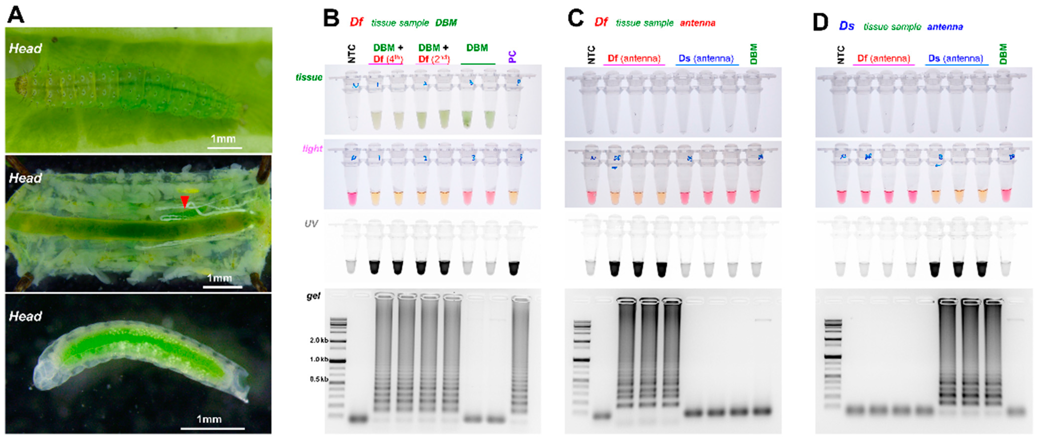
2.3. LAMP Amplification Using Biological Sample

To verify the possibility of diagnosis in a biological sample (feasible diagnosis of Df-parasitized DBM), at least 30 DBMs that hatched in five days (early 3rd instar) were placed in an open-type cylindrical plastic cage (diameter of 20 cm, and length of 30 cm) per repetition, and 10 males and 10 females of Df adults were placed together for one day to induce spawning (parasitic). After 24 h, the Df adults were removed, and the growth period in the DBM was monitored after inoculation. Kimchi cabbage was provided throughout the DBM larval period. The inoculation experiments were performed according to a previously published study [24]. After inoculation, three or more DBMs were dissected daily to confirm the development of Df. Parasitism and survival rate of Df in the DBM host were observed after dissection under a stereomicroscope (Leica M205C), photographed, and analyzed with a flexacam c1 equipped Leica Application Suite (LAS) X (Leica, Wetzlar, Germany) on a daily basis.

3. Results

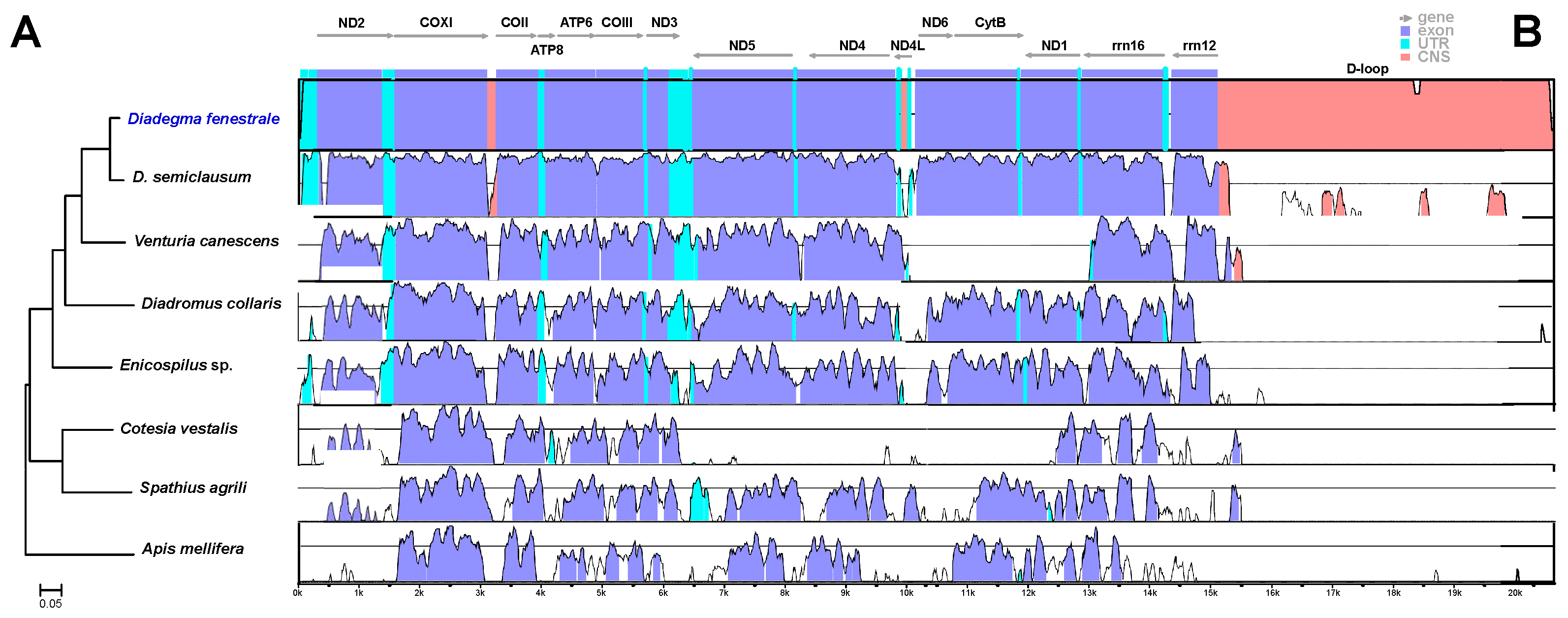
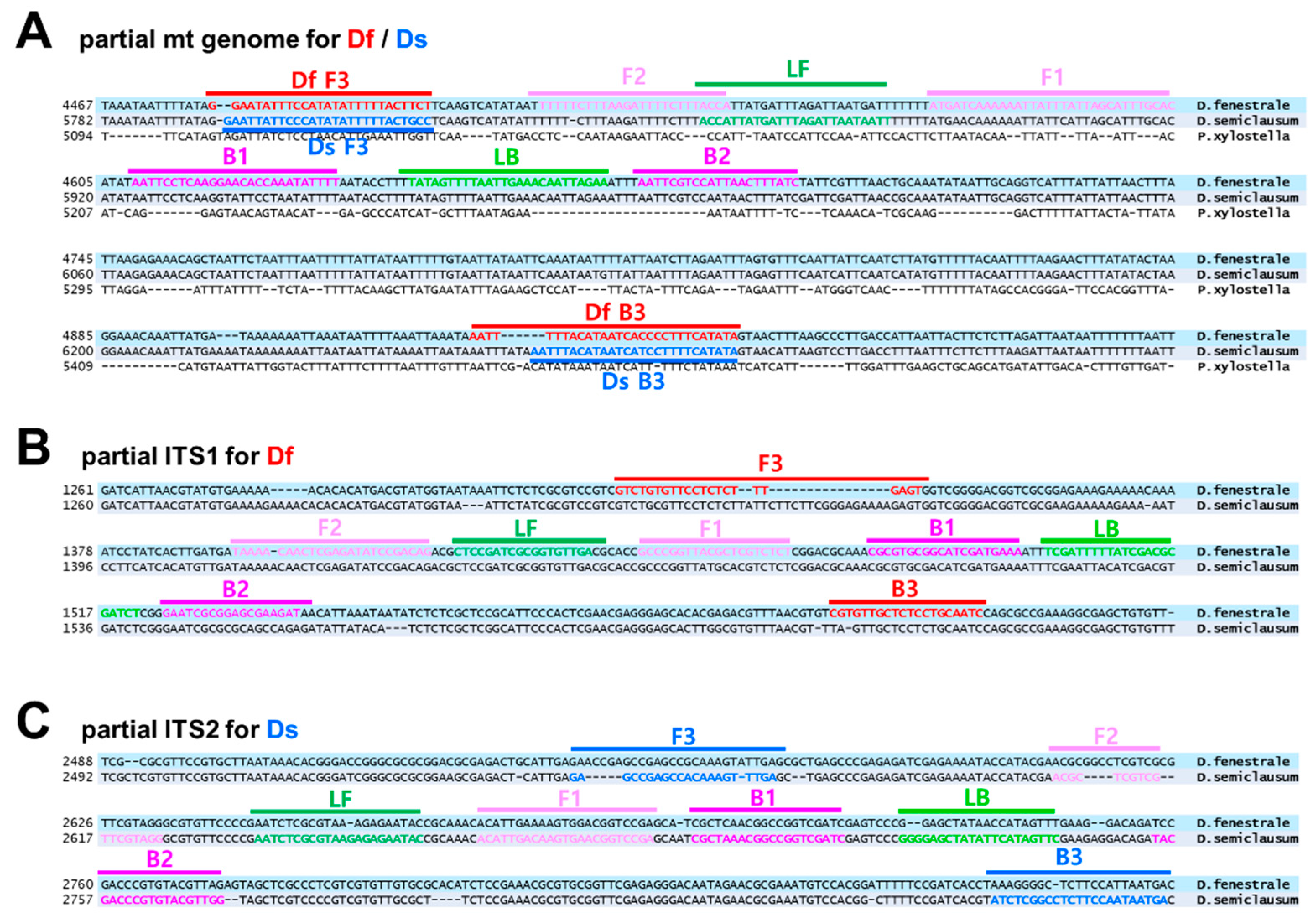
3.1. mt Genome Sequencing, Primer Design, and Selection

Miseq was used to assemble the mt genome of Df after trimming from approximately 3.4 Gb (11,424,866 reads) of nucleotide sequences. However, three N gaps were observed. For gap filling, 5.0 Gb (1,518,192 reads) sequences were obtained by using the GridION Oxford Nanopore platform. Finally, 20,849 bp of the mt genome was assembled (GenBank accession number MN599978). The mt genome included thirteen protein-coding genes: NADH dehydrogenase components (complex I, ND), cytochrome oxidase subunits (complex VI, COX), cytochrome oxidase b (CYPB), two ATP synthases, two ribosomal RNA genes, and twenty-two transfer RNAs (Supplementary Figure S1).
The phylogenetic relationship between the mt genomes of eight species (including Enicospilus sp.) was examined (Figure 1A) to confirm a Df-specific nucleotide sequence. The phylogenetic relationships were mostly similar to the megaBLAST results. Based on the mVISTA alignment results, the homology of the entire mt genome between Df and Ds was 94% (Figure 1B). However, this result was obtained because of the excessively low homology of the D-loop and the higher similarity of the partial nucleotide sequence particularly the protein coding region. There was a difference in some specific parts of the sequence that are AT rich. Generally, it is really hard to design primers for AT-rich regions. Therefore, we excluded these regions from designing the F3 and B3 diagnostic primers.
In the mt genome, the F3 and B3 primers were designed based on the Df- and DS-specific sequences in the region encoding the CoX3 gene, and the rest of the primers were designed for universal use (Figure 2A). In contrast, in ITS1 and ITS2, each diagnostic primer of F3 and B3 was designed based on the Df-specific and Ds-specific sequences, respectively (Figure 2B,C). Because the specific primer regions are different in ITS1 and ITS2, the regions of the inner primers and loop primers are also different. The designed F3 and B3 primers were validated by conventional PCR (Supplementary Figure S2). Each PCR product was confirmed by sequencing.

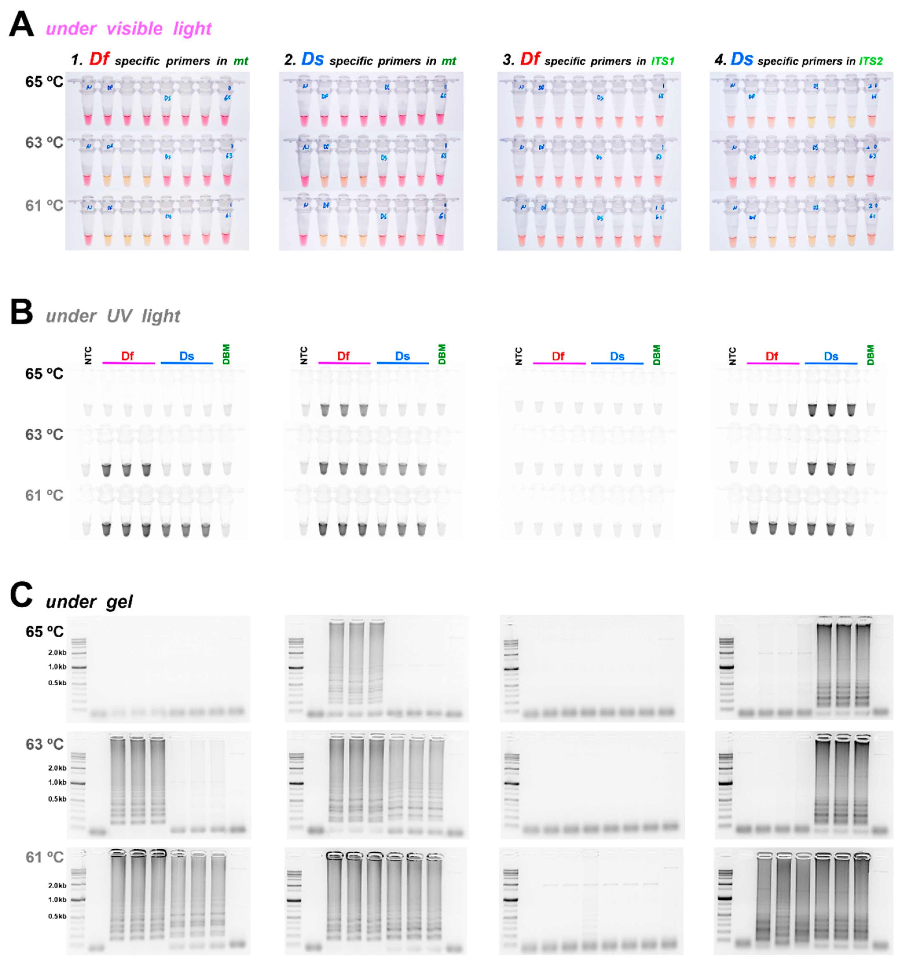
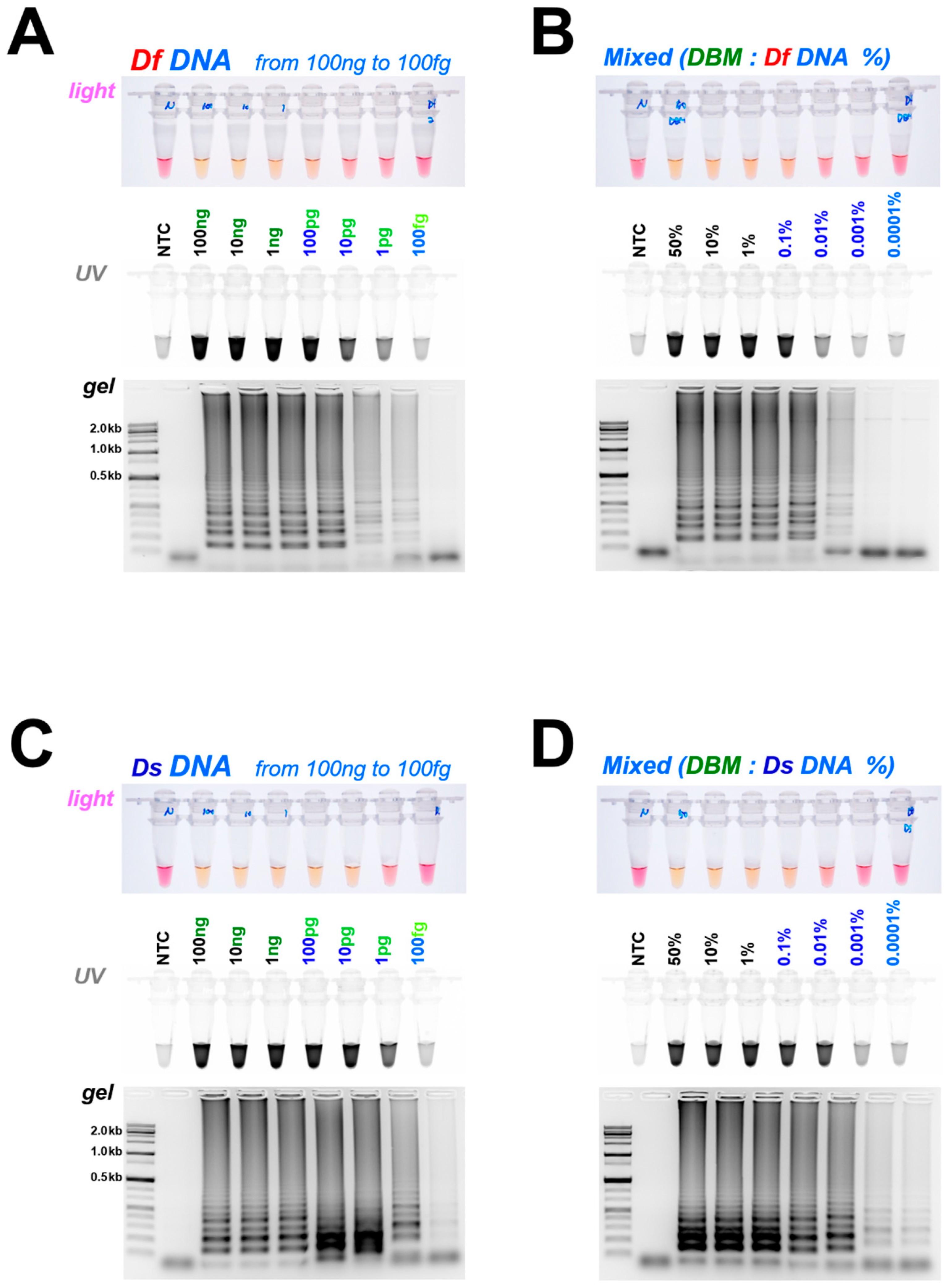
3.2. Diagnostic LAMP and PCR

As previously reported, the sensitivity of LAMP may vary depending on temperature and reaction time [25]. Therefore, the reaction was performed for 40 min under the three conditions of 65, 63, and 61 °C to find the optimal reaction temperature for the four designed primer sets (Figure 3). The results are summarized below in Table 2.
The reaction efficiency of the Ds diagnostic primer set for the mt region was higher than that of the Df primer set for diagnosis, and this was confirmed in repeated experiments; therefore, further experiments were not performed.
The Ds diagnostic primer set for the ITS2 region was significantly stable and yielded remarkable results at 63 and 65 °C. Therefore, subsequent experiment was performed at 63 °C, using the primer set based on mt for diagnosing Df and the primer set based on ITS2 for diagnosing Ds.

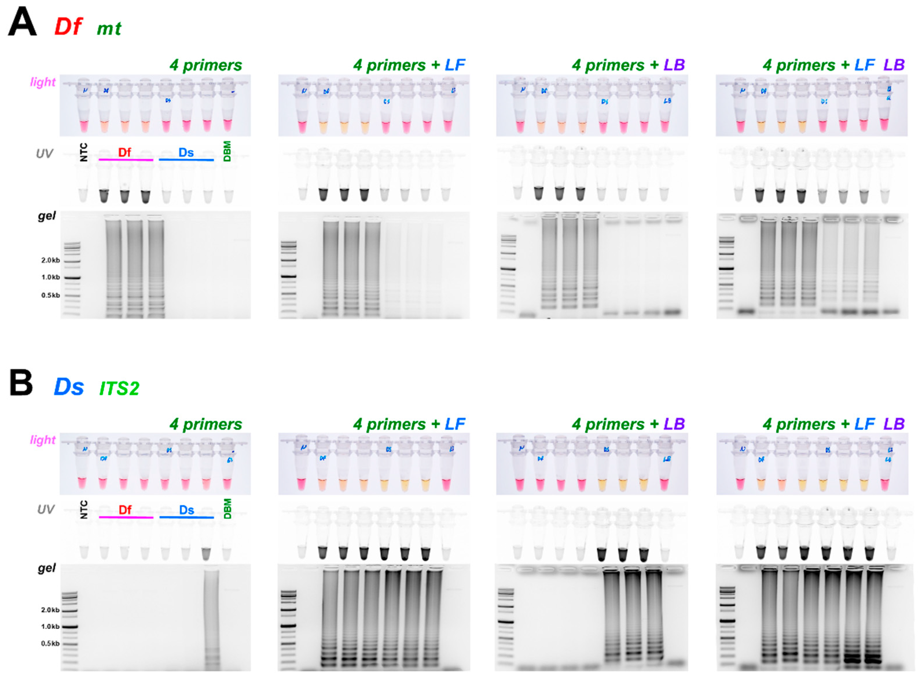
3.3. Comparison of Loop Primer Effectiveness

In addition to the four essential primers selected (F3, B3, FIP, and BIP), we determined whether the additional loop primers LB and LF, used individually or simultaneously, could improve the reaction’s efficiency and minimize false-positive reactions (Figure 4). In the case of Df, it was confirmed that a reaction time of 35 min was sufficient when using the four basic primers (F3, B3, FIP, and BIP); however, a false-positive reaction occurred after 5 min (i.e., in total 40 min) (Figure 3C). There was almost no non-specific reaction when the loop primer LB was added, but a partial nonspecific reaction occurred when the loop primer LF was added. In particular, a higher degree of non-specific reactions occurred (Figure 4A) when the two loop primers (LF and LB) were added simultaneously. Therefore, the optimal condition to diagnose Df was considered to be a reaction conducted at 63 °C for 35 min, using only the four essential primers or the four primers along with the loop primer LB to increase the efficiency.
In the case of Ds, the required reaction time was longer than 35 min, which was sufficient for Df, and this difference in the requirements was confirmed in repeated experiments using only the four essential primers (Figure 4B). However, the reaction and reaction times were stable when the loop primer LB was added. Non-specific reactions were observed upon the addition of LF alone or the two loop primers (LF and LB simultaneously). Therefore, we confirmed that using only four essential primers or adding only loop primer LB to the four primers at 63 °C for 35 min is the optimal condition to diagnose Ds.
The diagnostic limit concentration of Df and Ds DNA was 10–100 pg with visual observation and 1 pg with UV-light observation (Figure 5A,C). To determine the diagnosis limit when Df or Ds parasitized DBM, we calculated the amount of Df or Ds gDNA that could be detected when mixed with DBM gDNA in varying ratios of 50% (Df or Ds gDNA 50 ng + DBM gDNA 50 ng), 10%, and 0.1–0.0001% (Df or Ds gDNA 100 fg; sequentially serially diluted), with the total DNA being 100 ng. As a result, the diagnostic limit concentration was found to be approximately 10-fold higher than that when parasitoid DNA was tested alone. In other words, it was approximately 100 pg under visual observation and approximately 10 pg under UV light. Certainly, a faint band could be verified by gel electrophoresis, but, in general, for the LAMP reaction, we confirmed the reaction results by visual observation and UV light as a standard, and not by gel electrophoresis.
Even if larvae are observed externally by visual observation or under a microscope, it is impossible to determine whether the parasitoid is parasitic to the DBM without dissection. Although parasitism was verified through dissection, it was difficult to diagnose the parasitoid species because of the morphological similarity of the Df and Ds parasitoid larvae (Figure 6A). Therefore, to investigate the parasitism in DBM larvae, the second and fourth instars of Df (parasitized and non-parasitized larvae) were homogenized with sterile water and reacted at 95 °C for 5 min (Figure 6B). After spinning down the reaction product, 2 µL of the supernatant was reacted with the LAMP reaction solution. As a result, it was confirmed that Df parasitized the DBM (Figure 6B). In addition, part of the parasitoid tissue (Df or Ds antenna) was reacted with 30 µL of sterile water at 95 °C for 5 min, without a separate DNA extraction process, and then 2 µL of the supernatant was reacted with the LAMP reaction solution containing each species-specific primer set. Using this method, the species could be clearly diagnosed.

4. Discussion

Diadegma sp. has superior parasitic efficiency and is widely distributed worldwide. Previously, a study was conducted to identify Diadegma parasitoids as biocontrol agents against P. xylostella in various regions [1,3,6]. Different species within this genus show species specificity, as with the two Diadegma species discussed in this study.
As indicated in Figure 1, to design Df and Ds species-specific primers, the mt genome information of some well-known insects from GenBank was used. Even though it is not shown in Figure 1, the reliability was high, because more than 20 types of mt genome sequences were compared with those of the parasitoids, in addition to those of six other species. However, in the case of species-specific primers based on ITS1 and ITS2 regions, there is limited information about related species that can be compared. Therefore, if the nucleotide sequence of the relevant region is the same or highly similar, other species may be misdiagnosed. The sequences of the ITS1 and ITS2 regions are fairly species-specific sequences that can be utilized to effectively diagnose the two species.
Before applying the four primer sets based on mt and ITS regions to LAMP, we verified the utility of the species-specific diagnostic primer sets F3 and B3. The PCR analysis revealed that all F3 and B3 primer sets can be used for diagnosis (Supplementary Figure S2). However, non-specific reactions were observed, or the amplification efficiency was lowered according to various annealing temperature conditions. Sensitivity depending on the reaction temperature was also confirmed in LAMP, and for this reason, an experiment was conducted to confirm the optimal temperature of LAMP. As a result, the four sets of Df- and Ds-specific primers based on mt and ITS, a temperature of 63 °C, and a reaction time of approximately 5 min provided the most reliable results, depending on the presence or absence of a loop primer. In addition, the diagnostic efficiency clearly differed depending on the position of the loop primer. For example, in the mt-based Df primer set, a non-specific reaction was confirmed when LF was added, and when LB was added, the difference in amplification efficiency was not significant compared with that using the four essential primer sets (Figure 4A). In contrast, when adding LB, the diagnostic efficiency was significantly improved in the assay using the ITS-based Ds primer set, compared with that obtained with only the four essential primer sets (Figure 4B). This result was comparable to that obtained when LAMP was used previously to diagnose species and insecticide resistance of Lepidoptera [26], and it is considered to be a phenomenon of various conditions, such as loop primer position and GC% of amplicon interlock [27]. Finally, when using the mt-based diagnostic primer set (F3, B3, FIP, BIP, and LB) for Df and the ITS2-based diagnostic primer set for Ds, both sets could diagnose the parasitoid species within 35 min at 63 °C.
Since there was no Ds in the mass-reared population, the possibility of parasitism in DBM was not verified; however, we could confirm Df parasitism in DBM (Figure 6). In both Df and Ds, the larval stage comprises four instars, and the size rapidly increases during the fourth instar period [23]. Therefore, in addition to the early second and fourth instar experiments (Figure 6), we examined the possibility of diagnosis at the third instar stage, and all diagnoses were feasible (data not shown). Considering that the weight of the egg and first instar is 1/2 to 1/3 of the second instar, therefore, we did not apply the egg and first instar; instead, we replaced them with the diagnostic limit concentration (Figure 5). Although there was a difference between replicates, the parasitic rate under laboratory conditions was 70–90%, which was almost the same as the previously reported parasitic rate [21].
In this experiment, since the species-specific primers were designed by using strains that were bred for a long time in the laboratory, we cannot ensure that this diagnostic method will be precisely applicable to all Df and Ds populations. However, this study may consider being of sufficient value on how this molecular diagnostic can be applied to biological control research using parasitoids.
In a similar previous study, the exact parasitism rate was investigated by dissecting each species. However, herein, it was possible to investigate the parasitism rate within an hour after collection, using the molecular diagnostic method developed in this study. Moreover, our method addresses the problems outlined in the introduction, in that it makes it possible to investigate the parasitism rate for a specific species, as well as to accurately investigate the species, even in a situation where related species coexist in the field experiments. Furthermore, because Df is a generalist with various hosts, it can be used in the field for host range research.

Supplementary Materials

The following supporting information can be downloaded from https://www.mdpi.com/article/10.3390/horticulturae8050366/s1. Figure S1. Organization of the mitochondrial genome of the Korean population of Diadegma fenestrale (MN599978). ND: NADH dehydrogenase component (complex I) is shown in yellow. COX: cytochrome oxidase subunit (complex VI) is shown in pink. ATP synthase is shown in green. CYPB: cytochrome oxidase B, shown in purple. Ribosomal RNA genes are shown in red, and tRNA genes are shown in blue. The noncoding regions are not colored. Figure S2. Conventional polymerase chain reaction (PCR) to distinguish Df and Ds, using specific primers in the mt genome, ITS1, and ITS2. PCR with loop-mediated isothermal amplification (LAMP) external primer set (A) based on mt genome at three temperature conditions (62, 63, and 64 °C) and (B) based on ITS1 and ITS2 at three temperature conditions (63, 65, and 67 °C). PCR was performed under the following thermal conditions: initial denaturation at 95 °C for 1 min, followed by 35 cycles of 95 °C for 15 s, various annealing temperatures for 15 s, and 68 °C for 15 s, and final extension for 1 min at 68 °C.

Author Contributions

Conceptualization, methodology, investigation, data curation, writing original draft preparation, writing review and editing, and visualization, H.N. and J.K.; software, validation, formal analysis, and supervision, J.K.; resources, H.N., M.K., S.R. and J.K. All authors have read and agreed to the published version of the manuscript.

Funding

This research was funded by a Research Grant for New Faculty from Kangwon National University (Project No. D1001849), Republic of Korea, and Basic Science Research Program through the National Research Foundation of Korea (NRF) funded by the Ministry of Education (NRF-2021R1A6A1A03044242).

Institutional Review Board Statement

Not applicable.

Informed Consent Statement

Not applicable.

Acknowledgments

We are grateful to Sunghee Lee, Eunjoo Hong, and Hongju Moon for supporting DBM and parasitoid collection and rearing.

Conflicts of Interest

The authors declare no conflict of interest.

References

  1. Talekar, N.; Shelton, A. Biology, ecology, and management of the diamondback moth. Annu. Rev. Entomol. 1993, 38, 275–301. [Google Scholar] [CrossRef]
  2. Kok, L.T. Natural enemies of insect pests in the cruciferous crop ecosystem. Recent Res. Dev. Environ. Biol. 2004, 1, 31–42. [Google Scholar]
  3. Rowell, B.; Bunsong, N.; Satthaporn, K.; Phithamma, S.; Doungsa-Ard, C. Hymenopteran parasitoids of Diamondback moth (Lepidoptera: Ypeunomutidae) in northern Thailand. J. Econ. Entomol. 2005, 98, 449–456. [Google Scholar] [CrossRef] [PubMed]
  4. Hardy, J. Plutella maculipennis, Curt., its natural and biological Control in England. Bull. Entomol. Res. 1938, 29, 343–372. [Google Scholar] [CrossRef]
  5. Davies, A.P.; Takashino, K.; Watanabe, M.; Miura, K. Parental genetic traits in offspring from inter-specific crosses between introduced and indigenous Diadegma Foerster (Hymenoptera: Ichneumonidae): Possible effects on conservation genetics. Appl. Entomol. Zool. 2009, 44, 535–554. [Google Scholar] [CrossRef] [Green Version]
  6. Fitton, M.; Walker, A. Hymenopterous parasitoids associated with diamondback moth: Taxonomic dilemma. In Proceedings of the Second International Workshop, Tainan, Taiwan, 10–14 December 1990; Talekar, N.S., Ed.; Asian Vegetable Research and Development Center: Shanhua, Taiwan, 1992. AVRDC Publication No. 92–368. pp. 225–232. [Google Scholar]
  7. Thompson, W.R. A Catalogue of the Parasites and Predators of Insect Pests. Section 2, Host Parasite Catalogue. In Part 4, Hosts of Hymenoptera (Ichneumonidae); Commonwealth Institute of Biological Control: Ottawa, ON, Canada, 1957. [Google Scholar]
  8. Azidah, A.A.; Fitton, M.G.; Quicke, D.L.J. Identification of the Diadegma species (Hymenoptera: Ichneumonidae, Campopleginae) attacking the diamondback moth, Plutella xylostella, (Lepidoptera: Plutellidae). Bull. Entomol. Res. 2000, 90, 375–389. [Google Scholar] [CrossRef]
  9. Wagener, B.; Reineke, A.; Löhr, B.; Zebitz, C.P.W. Phylogenetic study of Diadegma species (Hymenoptera: Ichneumonidae) inferred from analysis of mitochondrial and nuclear DNA sequences. Biol. Control 2006, 37, 131–140. [Google Scholar] [CrossRef]
  10. Choi, J.; Kim, J.; Kwon, M.; Lee, J. Description of the Diadegma fenestrale (Hymenoptera:Ichneumonidae: Campopleginae) Attacking the Potato Tuber Moth, Phthorimaea operculella (Lep.: Gelechiidae) New to Korea. Anim. Syst. Evol. Divers. 2013, 29, 70–73. [Google Scholar] [CrossRef] [Green Version]
  11. CABI Invasive Species Compendium. Available online: https://www.cabi.org/isc/search/index (accessed on 26 February 2022).
  12. Kwon, M.; Kim, J.; Hong, E.; Lee, Y. Ecological characteristics of Cotesia glomerata L. (Hymenoptera: Braconidae) and its parasitism rates for diamondback moth (Plutella xylostella L.) in a kimchi cabbage field in the Korean highland area. Korean J. Appl. Entomol. 2019, 58, 355–362. [Google Scholar] [CrossRef]
  13. Folmer, O.; Black, M.; Hoeh, W.; Lutz, R.; Vrijenhoek, R. DNA primers for amplification of mitochondrial cytochrome c oxidase subunit I from diverse metazoan invertebrates. Mol. Mar. Biol. Biotechnol. 1994, 3, 294–299. [Google Scholar]
  14. Juric, I.; Salzburger, W.; Balmer, O. Spread and global population structure of the diamondback moth, Plutella xylostella (Lepidoptera: Plutellidae), and its larval parasitoids, Diadegma semiclausum and Diadegma fenestrale (Hymenoptera: Ichneumonidae), based on mtDNA. Bull. Enotomol. Res. 2017, 107, 155–164. [Google Scholar] [CrossRef] [PubMed] [Green Version]
  15. Yara, K. Identification of Torymus sinensis and T. beneficus (Hymenoptera: Torymidae), introduced and indigenous parasitoids of the chestnut gall wasp Dryocosmus kuriphilus (Hymenoptera: Cynipidae), using the ribosomal ITS2 region. Biol. Control 2006, 36, 15–21. [Google Scholar] [CrossRef]
  16. Wacławik, B.; Nugnes, F.; Bernardo, U.; Gebiola, M.; Przybycień, M.; Lachowska-Cierlik, D. An integrative revision of the subgenus Liophloeodes (Coleoptera: Curculionidae: Entiminae: Polydrusini): Taxonomic, systematic, biogeographic and evolutionary insights. Arthropod Syst. Phylogeny 2021, 79, 419–441. [Google Scholar]
  17. Notomi, T.; Okayama, H.; Masubuchi, H.; Yonekawa, T.; Watanabe, K.; Amino, N.; Hase, T. Loop-mediated isothermal amplification of DNA. Nucleic Acids Res. 2000, 28, e63. [Google Scholar] [CrossRef] [PubMed] [Green Version]
  18. Kwon, M.; Park, K.; Kwon, H. Developmental characteristics of Diadegma semiclausum Hellen (Hymenoptera: Ichneumonidae), a larval parasitoid of Plutella xylostella L. (Lepidoptera: Yponomeutidae). J. Asia Pac. Entomol. 2003, 6, 105–110. [Google Scholar] [CrossRef]
  19. Schwartz, S.; Kent, W.J.; Smit, A.; Zhang, Z.; Baertsch, R.; Hardison, R.C.; Haussler, D.; Miller, W. Human-mouse alignments with BLASTZ. Genome Res. 2003, 13, 103–107. [Google Scholar] [CrossRef] [Green Version]
  20. Tillich, M.; Lehwark, P.; Pellizzer, T.; Ulbricht-Jones, E.S.; Fischer, A.; Bock, R.; Greiner, S. GeSeq-versatile and accurate annotation of organelle genomes. Nucleic Acids Res. 2017, 45, W6–W11. [Google Scholar] [CrossRef]
  21. Tamura, K.; Stecher, G.; Kumar, S. MEGA11: Molecular Evolutionary Genetics Analysis version 11. Mol. Biol. Evol. 2021, 38, 3022–3027. [Google Scholar] [CrossRef]
  22. Frazer, K.A.; Pachter, L.; Poliakov, A.; Rubin, E.M.; Dubchak, I. VISTA: Computational tools for comparative genomics. Nucleic Acids Res. 2004, 32, W273–W279. [Google Scholar] [CrossRef]
  23. Mayor, C.; Brudno, M.; Schwartz, J.R.; Poliakov, A.; Rubin, E.M.; Frazer, K.A.; Patcher, L.S.; Dubchak, I. VISTA: Visualizing global DNA sequence alignments of arbitrary length. Bioinformatics 2000, 16, 1046–1047. [Google Scholar] [CrossRef] [Green Version]
  24. Kim, J.I.; Kwon, M.; Lee, S.H.; Kim, Y. Parasitism and survival rate of Diadegma fenestrale (Hymenoptera: Ichneumonidae) and DfIV gene expression patterns in two lepidopteran hosts. Biochem. Biophys. Res. Commun. 2015, 459, 579–584. [Google Scholar] [CrossRef] [PubMed]
  25. Kim, J.; Nam, H.Y.; Kwon, M.; Kim, H.J.; Yi, H.J.; Haenniger, S.; Unbehend, M.; Heckel, D.G. Development of a simple and accurate molecular tool for Spodoptera frugiperda species identification using LAMP. Pest Manag. Sci. 2021, 77, 3145–3153. [Google Scholar] [CrossRef] [PubMed]
  26. Kim, J.; Nam, H.Y.; Kwon, M.; Choi, J.H.; Cho, S.R.; Kim, G.H. Novel diamide resistance-linked mutation in Korean Spodop-tera exigua and a LAMP assay based on a mutation-associated intronic InDel. J. Pest Sci. 2021, 94, 1017–1029. [Google Scholar] [CrossRef]
  27. Nagamine, K.; Hase, T.; Notomi, T. Accelerated reaction by loop-mediated isothermal amplification using loop primers. Mol. Cell. Probe 2002, 16, 223–229. [Google Scholar] [CrossRef] [PubMed]
Figure 1. Comparison of entire mitochondrial genomes of Df and other Hymenoptera species. (A) Phylogeny inferred by using maximum likelihood with 1000 bootstrap repeats under MEGA11. The percentage of trees in which the associated taxa are clustered together is shown next to the branches. The tree is drawn to scale, with branch lengths measured in the number of substitutions per site. (B) Schematic diagram of genes and their flanking regions showing sequence diversity in mVISTA. Partial mitochondrial genome sequences of parasitoids and additional Hymenoptera species were realigned for primer design with those of the target species, Df and Ds (Figure 2A). Df, Diadegma fenestrale; Ds, Diadegma semiclausum; UTR, untranslated region; D-loop, displacement loop. D. semiclausum EU871947, Venturia canescens FJ478176, Diadromus collaris JX131613, Enicospilus sp. FJ478177, Cotesia vestalis FJ154897, Spathius agrili FJ387020, and Apis mellifera KY926884 sequence information was downloaded from GenBank.
Horticulturae 08 00366 g001
Figure 2. Location of primers and primer binding regions on the partial sequences of mitochondrial genomes from Df and Ds, and additional DBM. Three sequences are aligned in (A), and two sequences are aligned in (B,C); these were used to pick species-specific regions for primer design. F3 and B3 are the main diagnostic primers. The inner primer, FIP, consists of F1c (complementary sequences of F1) and F2. The other inner primer, BIP, is composed of B1 and B2c (complementary sequences of B2). The four essential LAMP primers (F3, FIP, BIP, and B3) generate a dumbbell structure, and the two loop primers, LF and LB, accelerate the LAMP reaction. Df, Diadegma fenestrale; Ds, Diadegma semiclausum; DBM, diamondback moth; LAMP, loop-mediated isothermal amplification.
Horticulturae 08 00366 g002
Figure 3. The sensitivity of the LAMP assay, using specific primers for mt genome, ITS1, and ITS2 regions under three temperature conditions (65, 63, and 61 °C) for 40 min for Df and Ds detected under (A) visible light, (B) ultraviolet light with SYBR Green, and (C) gel electrophoresis. The original pink color of the reaction mixture turned yellow in a positive reaction when the product was formed but remained pink in negative reactions. ITS, internal transcribed spacer; mt, mitochondrial; NTC, no template control; Df, Diadegma fenestrale; Ds, Diadegma semiclausum; DBM, diamondback moth; LAMP, loop-mediated isothermal amplification.
Horticulturae 08 00366 g003
Figure 4. LAMP assay results with four primers (F3, B3, FIP, and BIP) and additional loop primers, loop backward (LB) and loop forward (LF), of (A) mt-based Df and (B) ITS-based Ds. The LAMP assay was performed at 63 °C for 35 min under visible light, ultraviolet light with SYBR Green, and gel electrophoresis. The abbreviations are the same as those described in Figure 3. Df, Diadegma fenestrale; Ds, Diadegma semiclausum; LAMP, loop-mediated isothermal amplification.
Horticulturae 08 00366 g004
Figure 5. Identification of the detection limit of genomic DNA in the LAMP assay of (A) Df and (C) Ds from 100 ng to 100 fg under visible light, ultraviolet light with SYBR Green, and gel electrophoresis. LAMP assay with varying ratios (0.0001–50%) of (B) Df and (D) Ds gDNA mixed with DBM gDNA to a total of 100 ng. All LAMP assay results were generated with incubation at 63 °C for 35 min, with five primers (F3, B3, FIP, BIP, and LB). The abbreviations are the same as described in Figure 3.
Horticulturae 08 00366 g005
Figure 6. (A) Dissection of Plutella xylostella. The red triangle indicates the larvae of the parasitoid. (B,C) Representative results of LAMP assay with the DNA release technique from (B) insect tissue (parasitic and non-parasitic larvae), (C) antenna of Df, and (D) antenna of Ds. The whole larval tissue of DBM with or without Df and partial fragment of the antenna were incubated at 95 °C for 5 min and used as a template. LAMP assay results were generated with incubation at 63 °C for 35 min, with five primers (F3, B3, FIP, BIP, and LB), under visible light, ultraviolet light with SYBR Green, and gel electrophoresis. Df, Diadegma fenestrale; Ds, Diadegma semiclausum; DBM, diamondback moth; LAMP, loop-mediated isothermal amplification; NTC, no template control; PC, positive control.
Horticulturae 08 00366 g006
Table 1. Primers used in this study for the identification of the two Diadegma parasitoid species by LAMP and PCR.
PrimersSequence (5′->3′)
For identifying D. fenestrale by using primers for the mitochondrial region
Df-F3GGGAATATTTCCATATATTTTTACTTCT
Df-B3TATATGAAAGGGGTGATTATGTAAAAATT
Dfs-FIPGTGCAAATGCTAATAAATAATTTTTTGATCATTTTTTCTTTAAGATTTTCTTTACCA
Dfs-BIPAATTCCTCAAGGAACACCAAATATTTTGATAAAGTTAATGGACGAATT
Df-LFAATCATTAATCTAAATCATAATGGT
Df-LBTATAGTTTTAATTGAAACAATTAGAA
For identifying D. semiclausum by using primers for the mitochondrial region
Ds-F3GAATTATTCCCATATATTTTTACTGCC
Ds-B3GCTATATGAAAAGGATGATTATGTAAATT
For identifying D. fenestrale by using primers for the internal transcribed spacer (ITS) 1 region
Df_ITS1-F3GTCTGTGTTCCTCTCTTTGAGT
Df_ITS1-B3GATTGCAGGAGAGCAACACG
Df_ITS1-FIPAGAGACGAGCGTAACCGGGCTAAAACAACTCGAGATATCCGACAG
Df_ITS1-BIPCGCGTGCGGCATCGATGAAAATCTTCGCTCCGCGATTC
For identifying D. semiclausum by using primers for the internal transcribed spacer (ITS) 2 region
Ds_ITS2-F3GAGCCGAGCCACAAAGTTTGA
Ds_ITS2-B3TCATTATTGGAAGAGGCCGAGAT
Ds_ITS2-FIPTCGGACCGTTCACTTGTCAATGTACGCTCGTCGTTCGTAGG
Ds_ITS2-BIPCGCTAAACGGCCGGTCGATCCCAACGTACACGGGTCGTA
Ds_ITS2-LFGTATTCTCTCTTACGCGAGATT
Ds_ITS2-LBGGGGAGCTATATTCATAGTTC
For PCR
LCO1490GGTCAACAAATCATAAAGATATTGG
HCO2198TAAACTTCAGGCTGACCAAAAAATCA
18S_rRNA-FGGCAAGTCTGGTGCCAGCA
18S_rRNA-RGGTGTGTACAAAGGGCAGGGAC
18S_rRNA-R-FGTCCCTGCCCTTTGTACACAC
28S_rRNA-RCCACCCACTTAGAGCTGCACT
Wasp_ITS2RATATGCTTAAATTCAGCGGG
Wasp_ITS2R2CGCCTGCTCTGAGGTCGTT
G’s at the 5′ end depicted in pink were added to adjust the primer melting temperature.
Table 2. Summarized amplification efficiency depending on the incubation condition of LAMP.
Primer SetIncubation Condition
65 °C, 40 min63 °C, 40 min61 °C, 40 min
Df (mt)Not amplifiedGoodFalse positive
Ds (mt)GoodFalse positiveFalse positive
Df (ITS1)Not amplifiedNot amplifiedNot amplified
Ds (ITS2)GoodGoodFalse positive
Publisher’s Note: MDPI stays neutral with regard to jurisdictional claims in published maps and institutional affiliations.

