Article

Microbiome and Metagenome Analysis Reveals Huanglongbing Affects the Abundance of Citrus Rhizosphere Bacteria Associated with Resistance and Energy Metabolism

1
Key Laboratory of Horticultural Plant Biology (Ministry of Education), College of Horticulture and Forestry Sciences, Huazhong Agricultural University, Wuhan 430070, China
2
Guangxi Academy of Specialty Crops/Guangxi Citrus Breeding and Cultivation Research Center of Engineering Technology, Guilin 541004, China
*
Author to whom correspondence should be addressed.
Horticulturae 2021, 7(6), 151; https://doi.org/10.3390/horticulturae7060151
Submission received: 12 April 2021 / Revised: 18 May 2021 / Accepted: 19 May 2021 / Published: 13 June 2021
(This article belongs to the Special Issue Citrus Rhizosphere Microbiome)

Abstract

:
The plant rhizosphere microbiome is known to play a vital role in plant health by competing with pathogens or inducing plant resistance. This study aims to investigate rhizosphere microorganisms responsive to a devastating citrus disease caused by ‘Candidatus Liberibacter asiaticus’ (CLas) infection, by using 16S rRNA sequencing and metagenome technologies. The results show that 30 rhizosphere and 14 root bacterial genera were significantly affected by CLas infection, including 9 plant resistance-associated bacterial genera. Among these, Amycolatopsis, Sphingopyxis, Chryseobacterium, Flavobacterium, Ralstonia, Stenotrophomonas, Duganella, and Streptacidiphilus were considerably enriched in CLas-infected roots, while Rhizobium was significantly decreased. Metagenome analysis revealed that the abundance of genes involved in carbohydrate metabolism, such as glycolysis, starch and sucrose metabolism, amino sugar and nucleotide sugar metabolism, was significantly reduced in the CLas-infected citrus rhizosphere microbial community. Likewise, the abundance of genes involved in phosphoinositide signaling and phosphoinositide metabolism, which play important roles in energy metabolism (such as carbohydrate metabolism and lipid metabolism), was also decreased in the CLas-infected samples. Taken together, our results indicate that CLas infection could affect the resistance potential and energy metabolism of the citrus rhizosphere microbial community, which may help us to understand the rhizosphere responses to plant disease and thus facilitate the development and application of antagonistic microorganism products in citrus industry.

1. Introduction

Huanglongbing (HLB) is a devastating disease of citrus plants caused by phloem bacteria (Candidatus liberobacter asiaticus, CLas), which is known as “citrus greening“ [1]. With the development of the global citrus industry, HLB is widely spread around the world, especially in Southeast Asia, southern Africa, South America, and North America [2,3,4,5], and has caused huge economic losses to the global citrus industry [6]. CLas is mainly transmitted to citrus plants through Asian citrus psyllids [7]. Once CLas enters the plant phloem, it can multiply and spread to all tissues containing the phloem [8], and the colonization and dispersion of CLas could be affected by a variety of environmental factors, such as temperature and solar radiation [9].
HLB can adversely affect the root development and health as well as root-associated microbial community structure. At the late stages of the HLB disease, plant carbohydrate metabolism is decreased, and root growth is seriously affected due to the starch transport and accumulation being significantly reduced in roots [10,11]. Roots are the primary place where plants interact with soil microorganisms [12]; the rhizosphere microbiota has been reported to have a profound effect on plant growth and development by providing nutrients for plants such as nitrogen fixation or phosphorus dissolution, regulating plant growth through the production or degradation of plant hormones [13]. The rhizosphere microbiota is also important for plant health, as it could compete with pathogens or induce plant resistance [14].
The rhizosphere microbiome upon HLB infection has been investigated in recent years. By using 16S rRNA Gene Clone Library Sequencing technology, Trivedi et al. (2010, 2012) found that Clas infection significantly impacted the abundance of bacterial phyla in the citrus root microbial community [15,16], and the abundance of genes involved in key biological processes such as nitrogen cycling, carbon fixation, phosphorus utilization, and metal homeostasis was significantly decreased in the CLas-infected citrus rhizosphere microbiome compared to the healthy control. With the development of high-throughput sequencing technology, metagenomics was recently used to investigate microbiome changes upon CLas infection [17], and it was revealed that the rhizosphere-to-rhizoplane enrichment process of the citrus root-associated microbiome was impaired by reducing the relative abundance of most rhizoplane-enriched bacterial genera. However, it is worth mentioning that, due to the interference of genome from the host plant, the metagenomic technology could not detect the root endophytic microbiome yet. To this end, here we employed both 16S high-throughput sequencing and metagenomics technologies to investigate the rhizosphere as well as root endophytic bacteria communities upon CLas infection, in order to provide a more comprehensive picture for understanding the effect of HLB on the root-associated microbiome.

2. Materials and Methods

2.1. Sample Collection and Processing

Three healthy and three CLas-infected 20-year-old Newhall navel orange (Citrus sinesis Osb. Newhall) plants grafted on Poncirus trifoliata L. Raf. rootstock were cultured in a net room in Ganzhou city (25°46′34.85″ N, 114°50′58.54″ E). The healthy and CLas-infected plants were identified based on visual symptoms and PCR detection using both leaf and root samples (Supplementary Figure S1). The root samples (roots with approximately 5 cm-thick adjacent soil layers) from four corners of each tree were pooled together as a sample. The loosely attached soil on the roots was removed with gentle shaking. Then, the roots were placed in pre-cooled phosphate-buffered saline (PBS) buffer and the rhizosphere soil was isolated by ultra-sonication as previously described [18]. The roots were sonicated twice for 20 s each (time interval 5 s) using a sonication bath, and the clean roots were stored at −80 °C until use. The resulting PBS solution without roots was centrifugated at 4000× g for 5 min at 4 °C, and the rhizosphere soil was collected and stored at −80 °C until use.

2.2. DNA Extraction and CLas Detection

In this study, a modified CTAB (cetyltrimethylammonium bromide) method [19] was used to extract the total DNA from citrus root and leaf samples. PCR detection of 16S rRNA genes of CLas was conducted by using primer pairs P400F/R (P400F: GCGTTCATGTAGAAGTTGTG, P400R: CCTACAGGTGGCTGACTCAT). Total DNA was extracted from rhizosphere soil samples using the Fast DNA SPIN Kit (MP Biomedicals LLC in 29525 Fountain Pkwy, Solon, OH 44139, USA).

2.3. Metagenome and 16S rRNA Libraries Construction and High-Throughput Sequencing

High-quality genomic DNA of root and rhizosphere soils was selected to construct libraries. For genomics DNA, we used a fusion primer with dual index and adapters for PCR, and fragments that were too short were removed by Ampure beads. In this case, only the qualified library can be used for sequencing. Finally, the high-throughput sequencing of 16S rRNA libraries was performed on the Illumina Hiseq 2000 platform of BGI Research Institute (Shenzhen, China).
We used Covaris Ultrasonic Processor to randomly shear DNA of rhizosphere samples into approximately 350 bp fragments, and then created a library by PCR. The library was subjected to quality control and quantification by Agilent 2100 Bioanalyzer (Agilent, 5301 Stevens Creek Blvd. Santa Clara, CA 95051 United States) and ABI Step One Plus Real-Time PCR System (ABI, Marlborough, MA, USA). Then, the high-throughput sequencing of metagenome was performed on the Illumina Hiseq 2000 platform of BGI Research Institute (Shenzhen, China).
The raw sequencing reads were deposited in the NCBI (National Center for Biotechnology Information) Bioproject database under the accession number PRJNA721043.

2.4. Bioinformatic Analyses

The 16S sequencing raw data were filtered to eliminate the adapter pollution and low quality to obtain clean reads, then paired-end reads with overlap were merged into tags. All the tags were clustered to OTU (Operational Taxonomic Unit) at 97% sequence similarity through USEARCH (v7.0.1090) software [20]. Taxonomic ranks were assigned to OTU representative sequences using Ribosomal Database Project (RDP) Classifier v.2.2. Alpha diversity, beta diversity and the different species screenings were analyzed based on OTU and taxonomic ranks. After removing the unqualified sequences from the original metagenomic data, we performed a de novo assembly using SOAPdenovo2 software [21]. The assembly sequences were further assembled into longer contigs using Rabbit software [22]. All contigs were used to predict open reading frames through MetaGeneMark (http://exon.gatech.edu/GeneMark/metagenome/Prediction/, version 2.10, On 8 December 2017) software [23], and non-redundant sequences were obtained after clustering sequences by Cd-hit software with a similarity of more than 95% [24].
We used Pathoscope (version v1.0) software [25] to redistribute all the non-redundant sequences to the most homologous genes and to calculate the relative abundance of each gene for each sample. All predicted genes were then blasted with functionally annotated databases (including nr, KEGG, eggNOG, and CAZy), and the most similar protein sequence was used to predict gene function annotation. After annotating OTUs (based on 16s rRNA) and genes (Metagenome), the α diversity index (including Shannon index, Chao1 index and Ace index) of each sample was calculated by Mothur software according to the relative proportion of OTUs or genes. Dilution curves of all samples were made by R software (version 2.15.3) based on the observed species parameter.
To study the impact of CLas infection on bacterial community, we used principal component analysis (PCA) and principal coordinate analysis (PCoA) to perform correlation analysis of diseased and healthy samples. PCA analysis was conducted with R software based on OTUs (or genes). PCoA analysis was achieved by calculating the unrestricted UniFrac Distance Metrics among different samples to obtain the distance matrix, using QIIME (v1.80) software [26]. Significance analysis was performed using Meta stats software [27] (http://metastats.cbcb.umd.edu/, On 8 December 2017) and R software with Wilcox test [28]. The relative abundance of bacterial genes in each sample was calculated with Pathscope software, and the differentially abundance genes between the healthy and CLas-infected samples were identified by NOISeq software with Log2 FC ≥ 1 or ≤−1 and r ≥ 0.8 [29]. Functional enrichment analysis of differentially abundant genes was conducted by blasting the Kyoto Encyclopedia of Genes and Genomes (KEGG) database.

3. Results

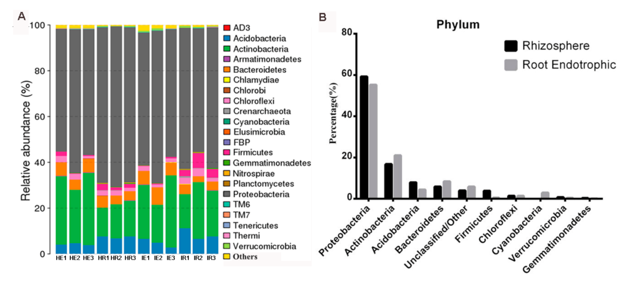
3.1. Taxonomic Features of the Citrus Rhizosphere and Root Microbiome

Three CLas-infected and three healthy Newhall navel orange (Citrus sinesis L. Osbeck) plants in one net house located in Ganzhou city were used as materials in this study. PCR detection of 16S rRNA genes of CLas by using primer pairs P400F/R confirmed that the CLas did exist in the leaves and roots of CLas-infected samples whereas it was absent in healthy samples (Supplementary Figure S1). Then, the citrus fine root and rhizosphere soil samples from CLas-infected and healthy plants were subject to DNA extraction and subsequent 16S rRNA amplification by Illumina sequencing. A total of 1,010,116 valid tags were produced. All the filtered tags were clustered into operational taxonomic units (OTUs) at 97% similarity, resulting in 2464 bacterial OTUs. Then, by classifying and annotating these OTUs, we identified a total of 16 bacterial phyla (Supplementary Table S1). Among them, Proteobacteria, Actinobacteria, and Acidobacteria are the most abundant species and comprise 54.91% of the bacterial species (Figure 1A, Supplementary Table S1). In addition, we analyzed the abundance of CLas in each sample and found that the CLas only existed in the CLas-infected citrus root endophytes sample (Supplementary Figure S2).
To analyze the difference in the phylum level of citrus root and rhizosphere bacterial communities, we counted the distribution of citrus root endophytes and rhizosphere bacterial communities at the phylum level, and analyzed the significance of the differences. The abundance of Cyanobacteria in the citrus roots (2.89%) is significantly higher than that in the rhizosphere (0.00%). The abundances of Acidobacteria, Gemmatimonadetes, and Firmicutes in the rhizosphere soil (7.90%, 0.28%, and 3.83%) were significantly higher than those in the roots (4.32%, 0.07%, and 0.44%, respectively). It is worth noting that the relative abundance of dominant bacteria phyla such as Proteobacteria, Actinobacteria, Acidobacteria, and Bacteroides in citrus roots and rhizosphere soil is the same (Figure 1B).

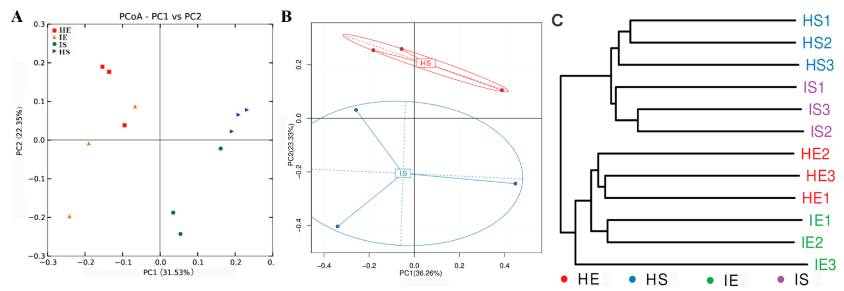
3.2. HLB Alters the Structural Diversity of Citrus Rhizosphere and Root Microbiome

To investigate the impact of CLas infection on the abundance and diversity of citrus endophytic and rhizosphere bacterial communities, we calculated all samples’ alpha diversity index based on OTU numbers. No difference was found in community richness and community diversity between the rhizosphere and endophytic root microbiome (Supplementary Table S2), suggesting that CLas did not actually affect the overall structure of the root-associated microbiome. PCoA analysis based on Unweighted Unifrac distance revealed that the healthy rhizosphere (HS) and CLas-infected rhizosphere samples (IS) were well separated (Figure 2A), whereas the healthy (HE) and CLas-infected root endophytes (IE) samples could not (Figure 2A). Metagenomic sequencing was also used to identify the community structure and function of the citrus rhizosphere microbiome and their changes after CLas infection. PCA analysis based on the number of genes measured in metagenomics showed that healthy or CLas-infected rhizosphere samples were separated (Figure 2B). The impact on the rhizosphere microbial is more significant than that of the endophytes. Further cluster analysis based on the unweighted pair group method with arithmetic mean (UPGMA showed that root endophytes and rhizosphere samples were divided into two groups, the healthy and CLas-infected samples were also clearly separated in either root endophytes or rhizosphere samples (Figure 2C).

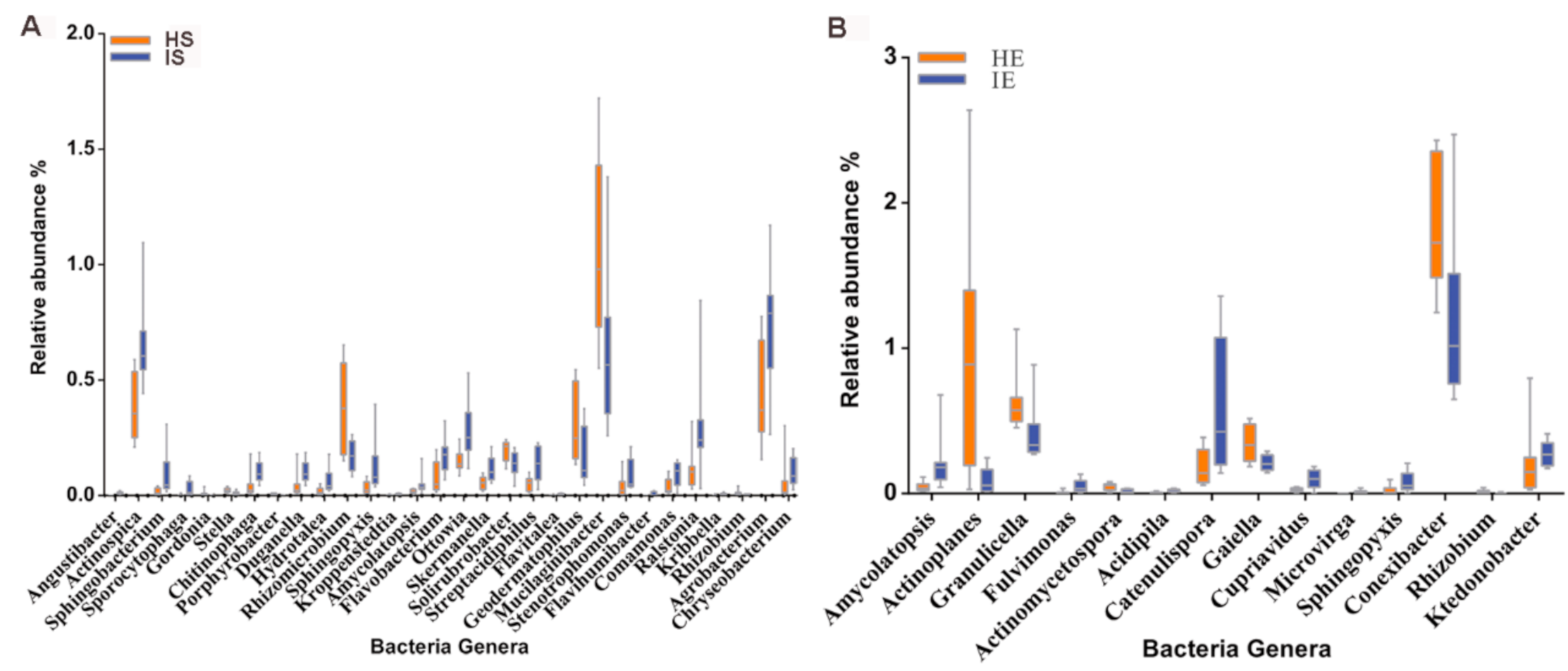
3.3. CLas Alters the Relative Abundance of Some Bacteria Genera

The above results show that, though the CLas infection did not substantially alter the global pattern of root-associated microbiome, it significantly changed the abundance for some bacterial genera. Therefore, we further investigated the effect of CLas infection on the abundance of root endophytic and rhizosphere microbes at genus level. We found the relative abundances of 30 bacterial genera in the rhizosphere (Figure 3A, Supplementary Table S3) and 14 bacterial genera in root endophytes (Figure 3B, Supplementary Table S4) were significantly affected by CLas infection. It is worth noting that, for genera in both the root endophyte and rhizosphere, the abundance of Amycolatopsis, Sphingopyxis, and Rhizobium were significantly affected by CLas infection, among which the abundance of Rhizobium was decreased whereas that of Amycolatopsis and Sphingopyxis was increased (Figure 3B).

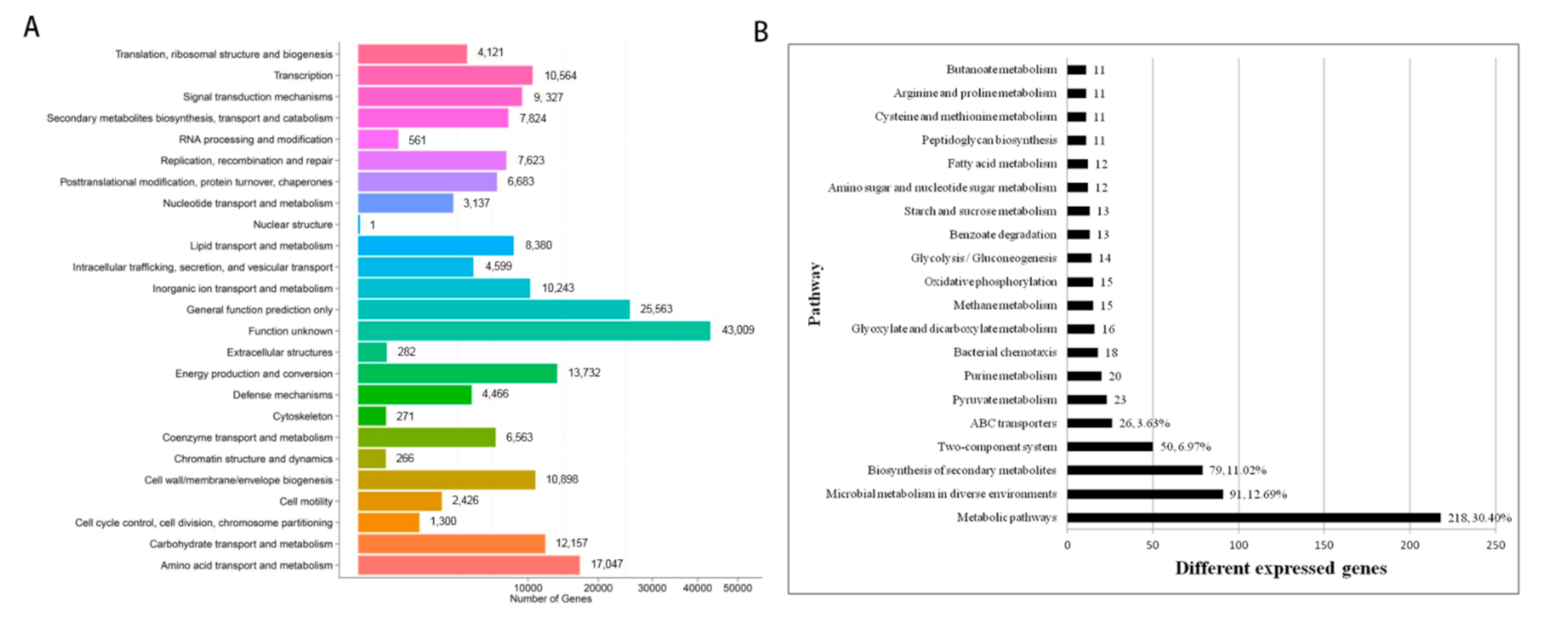
3.4. Effects of CLas Infection on Gene Abundances of Citrus Rhizosphere Bacteria

To explore the genetic composition and function of the citrus rhizosphere microbial community, we blasted the genes identified by metagenomics with the eggNOG database sequences. A total of 211,088 genes were annotated. Among these, amino acid transport and metabolism (17,047 genes, 8.08%), energy production and conversion (13,732 genes, 6.51%), and carbohydrate transport and metabolism (12,157 genes, 5.76%) are the top three functional classifications (Figure 4A).
To identify the impact of CLas infection on rhizosphere bacterial genetic composition, we used NOISeq (Log2 Fold Change, Log2 FC ≥ 1 or ≤ −1, r ≥ 0.8) to analyze the difference in the abundance of genes in the rhizosphere bacteria of healthy and CLas-infected citrus. We then obtained 1225 genes differential in abundance, of which 1088 genes decreased while 137 genes increased in the CLas-infected rhizosphere (Supplementary Figure S3, Supplementary Table S5). A total of 1103 differential genes were annotated into 143 functional pathways by blasting the Kyoto Encyclopedia of Genes and Genomes (KEGG) database (Supplementary Table S6). Metabolic pathways (218 genes, 30.40%), microbial metabolism in diverse environments (91 genes, 12.69%), and biosynthesis of secondary metabolites (79 genes, 11.02%) are the top three functional pathways (Figure 4B).
Notably, we found that 12 of the top 20 pathways are related to energy metabolism and transportation (Figure 4B). Among them, glycolysis, starch and sucrose metabolism and amino sugar and nucleotide sugar metabolism of the citrus rhizosphere microbial community were significantly reduced in the CLas-infected samples as compared with the healthy controls. Enrichment analysis of functional pathways revealed that seven metabolic pathways (bacterial invasion of epithelial cells, phosphatidylinositol signaling system, spliceosome, apoptosis, secondary bile acid biosynthesis, secondary bile acid biosynthesis, inositol phosphate metabolism, and dioxin degradation) were significantly enriched in the CLas-infected samples (p < 0.05) (Supplementary Table S7). The phosphatidylinositol signaling system and inositol phosphate metabolism are known to play important roles in lipid metabolism and signal transduction [30]. Interestingly, as many as 12 genes involved in the phosphatidylinositol signaling system and inositol phosphate metabolism are affected by CLas infection, with 10 downregulated and 2 upregulated (Figure 4B, Supplementary Table S7).

4. Discussion

In the present study, the endophyte and rhizosphere microbial communities of the citrus root system were analyzed by 16S specific segment high-throughput sequencing. A total of 2464 bacterial OTUs were identified, which greatly exceeded the bacterial numbers detected in citrus rhizosphere or root endophytes in previous studies [15,16,17,31]. On one hand, the result may be due to the rapid development of high-throughput sequencing technologies in recent years, which thus improved the detection accuracy of microorganisms; on the other hand, it may be due to the fact that our root samples are from an old citrus orchard (20 years), which may have formed a stable and richer microbial community.
We identified 16 bacterial phyla in citrus root endophytes and rhizosphere, of which Proteobacteria, Actinobacteria, and Acidobacteria were the top three most abundant phyla, which is in line with previous studies [32,33,34]. This indicates that these three bacterial phyla may play important roles in the environmental acclimation in citrus roots, and possibly their abundance may not be easily affected by geographical location and citrus variety. Similar to previous studies performed in citrus [17], Arabidopsis [35], corn [36], and tomato [37], we found that the plant root endophytes and rhizosphere always possessed similar dominant bacterial phyla, although the abundance and diversity of microorganisms in the rhizosphere is higher than that of endophytes. The results suggest that the endophytic bacteria are mainly from the rhizosphere, and that plant roots could smartly select specific or beneficial bacteria to enter the root system according to their own needs and gradually form a stable bacterial community.

4.1. CLas Infection Affects Citrus Root and Rhizosphere Bacteria Associated with Resistance

Pathogen attack could affect the community structure of root microorganisms [38,39]. As expected, 30 bacterial genera in the rhizosphere and 14 in endophytes were significantly affected by CLas infection. Previously, Zhang et al. (2017) found that 84 rhizosphere bacteria genera were significantly affected [17], and Emily et al. (2019) found that 8 rhizosphere bacterial genera and 13 endophytes bacterial genera were significantly affected by CLas infection [31]. Interestingly, only the rhizobium genus was significantly affected by CLas infection in all the three studies (Supplementary Table S8), with a significant reduction in abundance. This indicates that the rhizobium genus plays a conservative role in roots responsive to CLas infection, and the rhizobium residence is not affected by citrus varieties and growth environment. The rhizobium has been reported to inhibit the immunity triggered by microbe-associated molecular pattern (MAMP) in Arabidopsis, tomato, corn, and soybeans, thereby facilitating pathogen infection in plants [40]. We speculate that the CLas infection activated citrus root immunity system and thus may be harmful for the survival of rhizobium.
We found that CLas infection significantly increased the abundance of Amycolatopsis and Sphingopyxis in both root endophytes and the rhizosphere. The Amycolatopsis is a high GC-content bacterium belonging to Pseudonocardiaceae, which can produce epoxyquinomicin C and vancomycin to help plants resist pathogen invasion [41], suggesting that the bacterium might be also involved in the root resistance to CLas infection. The depletion of carbohydrates in HLB-infected citrus roots often causes root death and releases a large amount of aromatic compounds such as recalcitrant lignin [42]. Correspondingly, the abundance of Sphingopyxis, which could degrade lignin [43], was significantly increased upon CLas infection. Therefore, we hypothesized that CLas infection might affect the microbial community structure through altering the abundance and composition of root exudates such as aromatic compounds.
We also found that another six bacteria genera were significantly increased by CLas infection, including Chryseobacterium, Flavobacterium, Ralstonia, Stenotrophomonas, Duganella, and Streptacidiphilus. These bacteria genera were reported as being closely associated with disease resistance in plants [44]. For example, Chryseobacterium can inhibit fungal colonization and arrest hyphal expansion growth, while Flavobacterium can suppress disease. The activation of these bacteria implied that the citrus root could recruit disease resistance associated microorganisms to resist CLas.

4.2. CLas Infection Impairs the Energy Metabolism of Citrus Rhizosphere Microorganisms

Our metagenomic analysis revealed that more than 20% of the detected genes of the citrus rhizosphere microbiome were associated with energy metabolism-related function, such as amino acid transport and metabolism, energy production and preservation, carbohydrate transport and metabolism. This indicates the importance of energy metabolism-associated genes in the biological activities of citrus rhizosphere microbial community. Notably, as many as 74% of the differential genes in response to CLas infection were involved in metabolism-related pathways (Supplementary Table S8), indicating that the HLB had a significant impact on the metabolic function of the citrus rhizosphere microbial community. We discovered that the gene abundance of the metabolic pathways such as glycolysis, starch and sucrose metabolism and amino sugar and nucleic acid sugar metabolism was significantly reduced in CLas-infected citrus rhizosphere samples. Similar results were also found in previous studies that the abundance of genes involved in carbon cycling was significantly reduced in citrus rhizosphere under CLas infection [16,17]. It has been reported that CLas infection could block citrus phloem sieve and then reduce the transport of photosynthesis products to citrus root system [10,45]. Our result implies that CLas infection might affect the metabolic function of citrus rhizosphere bacterial community by reducing the carbohydrate secretion from plants to rhizosphere.

5. Conclusions

In conclusion, our study provided a comprehensive analysis of the citrus root microbial community and its response to CLas infection. Some rhizosphere microorganisms associated with resistance and energy metabolism were found in response to CLas infection in citrus roots, which may provide clues for the biological control of citrus CLas, and thus is of great significance to the sustainable development of the citrus industry.

Supplementary Materials

The following are available online at https://www.mdpi.com/article/10.3390/horticulturae7060151/s1. Figure S1: Detection of CLas in root and leaf samples. Agarose gel electrophoresis of PCR products amplified using primers A2-J5 targeting 16S rRNA gene of CLas. Figure S2: The abundance of CLas in each healthy and CLas-infected sample. Calculate P (p-value) by Wilcox.test. Figure S3: Number of rhizosphere bacterial genes whose abundance were significantly affected by CLas-infection. Table S1: The distribution of bacteria reads and generated OTUs at phylum level through blasting against the Greengene database. Table S2: The genetic diversity (α) of bacteria communities identified in root endophytic compartment and rhizosphere of healthy and HLB infected citrus. (Data are means ± SD). Table S3: The wilcox.test analysis of the different genera of rhizosphere in healthy and CLas infected roots. Table S4: The wilcox.test analysis of the different genera of endophytic bacteria in healthy and CLas infected roots. Table S5: The genes with different abundance between healthy and HLB-infected citrus rhizosphere metagenome. Table S6: The KEGG functional classification of differentially expressed genes in the metagenome of citrus rhizosphere under CLas infection Pathway. Table S7: The KEGG functional enrichment analysis of the genes with different abundance between healthy and HLB infected citrus rhizosphere metagenome. Table S8: A comparison of three similar studies (This research, Zhang et al., 2017 and Emily et al.) on microbes affected by the disease.

Author Contributions

Conceptualization, H.L., F.S. and Z.P.; Methodology, H.L. and F.S.; Software, H.L. and F.S.; Validation, Z.P. and F.S., Formal analysis, H.L. and F.S.; Investigation, H.L., F.S. and X.W.; Resources, X.W. and C.D.; Data curation, H.L.; Writing—original draft preparation, H.L.; Writing—review and editing, Z.P., Q.X. and S.P.; Visualization, H.L. and F.S.; Supervision, Z.P., Q.X. and S.P.; Project administration, Z.P., Q.X. and S.P. All authors have read and agreed to the published version of the manuscript.

Funding

This research was funded by Ntional Key R & D Programmes of China, grant number 2018YFD0201506, 2020YFD1000102 and 2018YFD1000103; Guangxi Science and Technology Major Project grant number GK AA18118046-6; Supported by China Agriculture Research System of MOF and MARA; Supported by the Fundamental Research Funds for the Central Universities, Program No. 2662018JC039.

Institutional Review Board Statement

Not applicable.

Informed Consent Statement

Not applicable.

Data Availability Statement

The raw sequencing reads were deposited in the NCBI (National Center for Biotechnology Information) Bioproject database under the accession number PRJNA721043.

Acknowledgments

Sincere thanks to Xia Changxiu from Ganzhou Citrus Science Research Institute for providing plant materials.

Conflicts of Interest

All the authors declare no conflict of interest.

References

  1. Bové, J.M. Invited review. Huanglongbing: A destructive, newly-emerging, century-old disease of citrus. J. Plant Pathol. 2006, 88, 7–37. [Google Scholar]
  2. Lee, H.A. The relation of stocks to mottled leaf of citrus trees. Philipp. J. Sci. 1921, 18, 85–95. [Google Scholar]
  3. Obergolzer, P.C.J. Greening disease of sweet orange in South Africa. Int. Organ. Citrus Virol. Conf. Proc. 1965, 3, 213–219. [Google Scholar]
  4. Halbert, S. The discovery of huanglongbing in Florida. In Proceedings of the International Citrus Canker and Huanglongbing Research Workshop, Orlando, FL, USA, 7–11 November 2015. [Google Scholar]
  5. Texeira, D.D.C.; Ayres, J.; Kitajima, E.; Danet, L.; Jagoueix-Eveillard, S.; Saillard, C.; Bové, J. First report of a huanglongbing-like disease of citrus in São Paulo state, Brazil and association of a new Liberibacter species, ‘Candidatus Liberibacter americanus’, with the disease. Plant Dis. 2005, 89, 107. [Google Scholar] [CrossRef]
  6. Grafton-Cardwell, E.E.; Stelinski, L.L.; Stansly, P.A. Biology and management of Asian citrus psyllid, vector of the huanglongbing pathogens. Annu. Rev. Entomol. 2013, 58, 413–432. [Google Scholar] [CrossRef] [Green Version]
  7. Do Carmo, D.T.; Danet, J.L.; Eveillard, S.; Martins, E.C.; de Jesus, W.C., Jr.; Yamamoto, P.T.; Lopes, S.A.; Bassanezi, R.B.; Ayres, A.J.; Saillard, C.; et al. Citrus huanglongbing in Sao Paulo State, Brazil: PCR detection of the ‘Candidatus’ Liberibacter species associated with the disease. Mol. Cell. Probes 2005, 19, 173–179. [Google Scholar] [CrossRef]
  8. Tatineni, S.; Sagaram, U.S.; Gowda, S.; Robertson, C.J.; Dawson, W.O.; Iwanami, T.; Wang, N. In planta distribution of ‘Candidatus Liberibacter asiaticus’ as revealed by polymerase chain reaction (PCR) and real-time PCR. Phytopathology 2008, 98, 592–599. [Google Scholar] [CrossRef] [Green Version]
  9. Louzada, E.S.; Vazquez, O.E.; Braswell, W.E.; Yanev, G.; Evan, B.W.; Kunta, M. Distribution of ’Candidatus Liberibacter asiaticus’ above and below ground in Texas citrus. Phytopathology 2016, 106, 702–709. [Google Scholar] [CrossRef] [PubMed] [Green Version]
  10. Etxeberria, E.; Gonzalez, P.; Achor, D.; Albrigo, G. Anatomical distribution of abnormally high levels of starch in HLB-affected Valencia orange trees. Physiol. Mol. Plant Pathol. 2009, 74, 76–83. [Google Scholar] [CrossRef]
  11. Johnson, E.G.; Wu, J.; Bright, D.B.; Graham, J.H. Association of ‘Candidatus Liberibacter asiaticus’ root infection, but not phloem plugging with root loss on huanglongbing-affected trees prior to appearance of foliar symptoms. Plant Pathol. 2014, 63, 290–298. [Google Scholar] [CrossRef]
  12. Torsvik, V. Prokaryotic diversity—Magnitude, dynamics, and controlling factors. Science 2002, 296, 1064–1066. [Google Scholar] [CrossRef] [Green Version]
  13. Trivedi, P.; Leach, J.E.; Tringe, S.G.; Sa, T.; Singh, B.K. Plant-microbiome interactions: From community assembly to plant health. Nat. Rev. Microbiol. 2020, 18, 607–621. [Google Scholar] [CrossRef] [PubMed]
  14. Gouda, S.; Kerry, R.G.; Das, G.; Paramithiotis, S.; Shin, H.-S.; Patra, J.K. Revitalization of plant growth promoting rhizobacteria for sustainable development in agriculture. Microbiol. Res. 2018, 206, 131–140. [Google Scholar] [CrossRef]
  15. Trivedi, P.; Duan, Y.; Wang, N. Huanglongbing, a systemic disease, restructures the bacterial community associated with citrus roots. Appl. Environ. Microbiol. 2010, 76, 3427–3436. [Google Scholar] [CrossRef] [Green Version]
  16. Trivedi, P.; He, Z.; van Nostrand, J.D.; Albrigo, G.; Zhou, J.; Wang, N. Huanglongbing alters the structure and functional diversity of microbial communities associated with citrus rhizosphere. ISME J. 2012, 6, 363–383. [Google Scholar] [CrossRef] [Green Version]
  17. Zhang, Y.; Xu, J.; Riera, N.; Jinyun, L.; Li, J.; Wang, N. Huanglongbing impairs the rhizosphere-to-rhizoplane enrichment process of the citrus root-associated microbiome. Microbiome 2017, 5, 97. [Google Scholar] [CrossRef]
  18. Edwards, J.; Johnson, C.; Santos-Medellín, C.; Lurie, E.; Podishetty, N.K.; Bhatnagar, S.; Eisen, J.A.; Sundaresan, V. Structure, variation, and assembly of the root-associated microbiomes of rice. Proc. Natl. Acad. Sci. USA 2015, 112, E911–E920. [Google Scholar] [CrossRef] [PubMed] [Green Version]
  19. Cheng, Y.-J.; Guo, W.-W.; Yi, H.-L.; Pang, X.-M.; Deng, X. An efficient protocol for genomic DNA extraction from citrus species. Plant Mol. Biol. Rep. 2003, 21, 177–178. [Google Scholar] [CrossRef]
  20. Edgar, R.C. UPARSE: Highly accurate OTU sequences from microbial amplicon reads. Nat. Methods 2013, 10, 996–998. [Google Scholar] [CrossRef] [PubMed]
  21. Luo, R.; Liu, B.; Xie, Y.; Li, Z.; Huang, W.; Yuan, J.; He, G.; Chen, Y.; Pan, Q.; Liu, Y.; et al. SOAPdenovo2: An empirically improved memory-efficient short-read de novo assembler. Gigascience 2015, 4, 1. [Google Scholar] [CrossRef] [PubMed] [Green Version]
  22. You, M.; Yue, Z.; He, W.; Yang, X.; Yang, G.; Xie, M.; Zhan, D.; Baxter, S.W.; Vasseur, L.; Gurr, G.M.; et al. A heterozygous moth genome provides insights into herbivory and detoxification. Nat. Genet. 2013, 45, 220–225. [Google Scholar] [CrossRef] [Green Version]
  23. Zhu, W.; Lomsadze, A.; Borodovsky, M. Ab initio gene identification in metagenomic sequences. Nucleic Acids Res. 2010, 38, E132. [Google Scholar] [CrossRef] [PubMed] [Green Version]
  24. Li, W.; Godzik, A. Cd-hit: A fast program for clustering and comparing large sets of protein or nucleotide sequences. Bioinformatics 2006, 22, 1658–1659. [Google Scholar] [CrossRef] [Green Version]
  25. Francis, O.E.; Bendall, M.; Manimaran, S.; Hong, C.; Clement, N.L.; Castro-Nallar, E.; Snell, Q.; Schaalje, G.B.; Clement, M.J.; Crandall, K.A.; et al. Pathoscope: Species identification and strain attribution with unassembled sequencing data. Genome Res. 2013, 23, 1721–1729. [Google Scholar] [CrossRef] [PubMed] [Green Version]
  26. Bik, H.M.; Sung, W.; de Ley, P.; Baldwin, J.G.; Sharma, J.; Rocha-Olivares, A.; Thomas, W.K. Metagenetic community analysis of microbial eukaryotes illuminates biogeographic patterns in deep-sea and shallow water sediments. Mol. Ecol. 2012, 21, 1048–1059. [Google Scholar] [CrossRef]
  27. White, J.R.; Nagarajan, N.; Pop, M. Statistical methods for detecting differentially abundant features in clinical metagenomic samples. PLoS Comput. Biol. 2009, 5, e1000352. [Google Scholar] [CrossRef] [PubMed]
  28. Lopez, M.F.; Rebollar, E.A.; Harris, R.N.; Vredenburg, V.T.; Hero, J.-M. Temporal variation of the skin bacterial community and Batrachochytrium dendrobatidis infection in the terrestrial cryptic frog Philoria loveridgei. Front. Microbiol. 2017, 8, 2535. [Google Scholar] [CrossRef] [Green Version]
  29. Tarazona, S.; García-Alcalde, F.; Dopazo, J.; Ferrer, A.; Conesa, A. Differential expression in RNA-seq: A matter of depth. Genome Res. 2011, 21, 2213–2223. [Google Scholar] [CrossRef] [PubMed] [Green Version]
  30. Bridges, D.; Saltiel, A.R. Phosphoinositides: Key modulators of energy metabolism. Biochim. Biophys. Acta 2015, 1851, 857–866. [Google Scholar] [CrossRef] [Green Version]
  31. Padhi, E.M.T.; Maharaj, N.; Lin, S.-Y.; Mishchuk, D.O.; Chin, E.; Godfrey, K.; Foster, E.; Polek, M.; Leveau, J.H.J.; Slupsky, C.M. Metabolome and microbiome signatures in the roots of citrus affected by huanglongbing. Phytopathology 2019, 109, 2022–2032. [Google Scholar] [CrossRef]
  32. Xu, J.; Zhang, Y.; Zhang, P.; Trivedi, P.; Riera, N.; Wang, Y.; Liu, X.; Fan, G.; Tang, J.; Coletta-Filho, H.D.; et al. The structure and function of the global citrus rhizosphere microbiome. Nat. Commun. 2018, 9, 4894. [Google Scholar] [CrossRef] [Green Version]
  33. Wu, Y.; Qu, M.; Pu, X.; Lin, J.; Shu, B. Distinct microbial communities among different tissues of citrus tree Citrus reticulata cv. Chachiensis. Sci. Rep. 2020, 10, 6068. [Google Scholar] [CrossRef] [PubMed] [Green Version]
  34. Ginnan, N.A.; Dang, T.; Bodaghi, S.; Ruegger, P.M.; Mccollum, G.; England, G.; Vidalakis, G.; Borneman, J.; Rolshausen, P.E.; Roper, M.C. Disease-induced microbial shifts in citrus indicate microbiome-derived responses to huanglongbing across the disease severity spectrum. Phytobiomes J. 2020, 4, 375–387. [Google Scholar] [CrossRef]
  35. Bulgarelli, D.; Rott, M.; Schlaeppi, K.; van Themaat, E.V.L.; Ahmadinejad, N.; Assenza, F.; Rauf, P.; Huettel, B.; Reinhardt, R.; Schmelzer, E.; et al. Revealing structure and assembly cues for Arabidopsis root-inhabiting bacterial microbiota. Nature 2012, 488, 91–95. [Google Scholar] [CrossRef]
  36. Cavaglieri, L.; Orlando, J.; Etcheverry, M. Rhizosphere microbial community structure at different maize plant growth stages and root locations. Microbiol. Res. 2009, 164, 391–399. [Google Scholar] [CrossRef]
  37. Li, J.-G.; Ren, G.-D.; Jia, Z.-J.; Dong, Y.-H. Composition and activity of rhizosphere microbial communities associated with healthy and diseased greenhouse tomatoes. Plant Soil 2014, 380, 337–347. [Google Scholar] [CrossRef]
  38. Carrión, V.J.; Perez-Jaramillo, J.; Cordovez, V.; Tracanna, V.; de Hollander, M.; Ruiz-Buck, D.; Mendes, L.W.; van Ijcken, W.F.J.; Gomez-Exposito, R.; Elsayed, S.S.; et al. Pathogen-induced activation of disease-suppressive functions in the endophytic root microbiome. Science 2019, 366, 606–612. [Google Scholar] [CrossRef] [PubMed]
  39. Kwak, M.-J.; Kong, H.G.; Choi, K.; Kwon, S.-K.; Song, J.Y.; Lee, J.; Lee, P.A.; Choi, S.Y.; Seo, M.; Lee, H.J.; et al. Rhizosphere microbiome structure alters to enable wilt resistance in tomato. Nat. Biotechnol. 2018, 36, 1100–1109. [Google Scholar] [CrossRef]
  40. Poole, P.; Ramachandran, V.; Terpolilli, J. Rhizobia: From saprophytes to endosymbionts. Nat. Rev. Microbiol. 2018, 16, 291–303. [Google Scholar] [CrossRef]
  41. Everest, G.J.; le Roes-Hill, M.; Omorogie, C.; Cheung, S.-K.; Cook, A.E.; Goodwin, C.M.; Meyers, P.R. Amycolatopsis umgeniensis sp nov., isolated from soil from the banks of the Umgeni river in South Africa. Antonie Leeuwenhoek 2013, 103, 673–681. [Google Scholar] [CrossRef]
  42. Nehela, Y.; Killiny, N. Revisiting the complex pathosystem of huanglongbing: Deciphering the role of citrus metabolites in symptom development. Metabolites 2020, 10, 409. [Google Scholar] [CrossRef] [PubMed]
  43. De Gonzalo, G.; Colpa, D.I.; Habib, M.H.M.; Fraaije, M.W. Bacterial enzymes involved in lignin degradation. J. Biotechnol. 2016, 236, 110–119. [Google Scholar] [CrossRef] [PubMed] [Green Version]
  44. Niu, B.; Wang, W.; Zhibo, Y.; Sederoff, R.R.; Sederoff, H.; Chiang, V.L.; Borriss, B. Microbial interactions within multiple-strain biological control agents impact soil-borne plant disease. Front. Microbiol. 2020, 11, 585404. [Google Scholar] [CrossRef] [PubMed]
  45. Kim, J.-S.; Sagaram, U.S.; Burns, J.K.; Li, J.-L.; Wang, N. Response of sweet orange (Citrus sinensis) to ‘Candidatus Liberibacter asiaticus’ infection: Microscopy and microarray analyses. Phytopathology 2009, 99, 50–57. [Google Scholar] [CrossRef] [PubMed] [Green Version]
Figure 1. Citrus root endophytic and rhizosphere bacterial community structure. (A) Stacked bar graph showing bacteria at phylum level. The ratio of each species in a certain sample is displayed. The phyla with an abundance of less than 0.5% were classified into ‘Others’. (B) The distribution of citrus root endophytic (gray) and rhizosphere (black) bacteria for the top ten bacteria phyla. HS: rhizosphere in healthy samples; HE: endophytes in healthy samples; IS: rhizosphere in CLas-infected samples; IE: endophytes in CLas-infected samples.
Horticulturae 07 00151 g001
Figure 2. Comparison of the rhizosphere and endophytic community structures based on pyrosequencing of 16S rDNA amplicons and metagenomics. (A) Principal coordinates analysis (PCoA) based on 16S OTU abundance. Number in brackets represents contributions of principal components to differences among samples. A dot represents each sample, and different colors represent different groups (red: HE; blue: HS; orange: IE; green: IS). (B) Principal component analysis (PCA) based on gene abundance. Number in brackets represents contributions of principal components to differences among samples. A dot represents each sample, and different colors represent different groups (red: HS; blue: IS). (C) Unweighted pair group method with arithmetic mean (UPGMA) analysis based on 16S OTU abundance (HS: rhizosphere in healthy samples; HE: endophytes in healthy samples; IS: rhizosphere in CLas-infected samples; IE: endophytes in CLas-infected samples).
Horticulturae 07 00151 g002
Figure 3. Composition of bacteria abundance of root endophytic compartment (IE and HE) and rhizosphere (IS and HS) between CLas-infected and healthy citrus at the genus level. (A). Rhizosphere difference genera. (B). Endogenous difference genera. The y axis represents the relative abundance of each bacteria genus. The p-values of all the genera presented in the figure were less than 0.05.
Horticulturae 07 00151 g003
Figure 4. Sequence and functional analysis of macro factors in rhizosphere microbiome. (A). The functional classification of genes from citrus rhizosphere metagenome based on eggNOG database. (B). The top 20 pathways of KEGG functional classification of the genes with different abundance between healthy and CLas-infected citrus rhizosphere metagenome.
Horticulturae 07 00151 g004
Publisher’s Note: MDPI stays neutral with regard to jurisdictional claims in published maps and institutional affiliations.

