Article

Tomato Brown Rugose Fruit Virus Is Transmissible through a Greenhouse Hydroponic System but May Be Inactivated by Cold Plasma Ozone Treatment

United States Department of Agriculture, Agricultural Research Service, U.S. Vegetable Laboratory, Charleston, SC 29414, USA
*
Author to whom correspondence should be addressed.
Horticulturae 2024, 10(4), 416; https://doi.org/10.3390/horticulturae10040416
Submission received: 21 March 2024 / Revised: 12 April 2024 / Accepted: 16 April 2024 / Published: 20 April 2024
(This article belongs to the Special Issue Innovative System for Disinfection in Greenhouses)

Abstract

:
Tomato brown rugose fruit virus (ToBRFV) is an emerging tobamovirus infecting tomatoes and peppers, resulting in a pandemic in recent years. In addition to its abilities of being seed-borne, transmitted mechanically and overcoming current resistance, we speculated other factors may also contribute to such catastrophic effect on tomato production in a hydroponic greenhouse. The objective of this study was to evaluate whether ToBRFV can be transmissible through recirculating hydroponic systems and, more importantly, search for an effective approach to contain its spread. We not only detected ToBRFV in the runoff water samples collected from three greenhouses but also determined the virus’ infectivity through a bioassay. We then conducted a water treatment using cold plasma ozone to assess its efficacy in inactivating ToBRFV. The results showed that, with a high concentration of ToBRFV (inoculum in 1:100 dilution), a prolonged exposure (72 min) to two higher ozone concentrations (0.6 mg/L and 1.0 mg/L) achieved partial effects. With a medium virus concentration (inoculum in 1:1000 dilution), an exposure to ozone for 48 min was sufficient to completely suppress the virus’ infectivity. However, with a low virus concentration (inoculum in 1:10,000 dilution), the virus was completely inactivated even with just a short ozone exposure (24 min). Future work will need to confirm the efficacy of the ozone treatment against ToBRFV as well as its impact on tomato plants in a hydroponic greenhouse.

1. Introduction

Tomatoes (Solanum lycopersicum L.), a flowering plant belonging to the nightshade family (Solanaceae), are cultivated extensively in temperate, subtropical, and some tropical regions for edible fruits. As the most consumed vegetable crop worldwide, global tomato production is over 187 million tons, with a total value over USD 190 billion [1]. Although most tomatoes are grown traditionally in open fields, recent years have seen an ever-expanding share of greenhouse tomatoes on the market (35% of the entire tomato category), as farming has undergone a profound shift to capital and production infrastructure intensive models [2].
Tomato brown rugose fruit virus (ToBRFV) is a relatively new member in the genus Tobamovirus, family Virgaviridae. Since its first discovery in the Middle East during the 2014–2015 growing season [3,4], the virus has been reported in approximately 40 countries [5,6], including Asia [7,8,9,10], Europe [11,12,13,14,15,16,17,18,19,20,21,22], North America [23,24,25,26], South America [27], Africa [28], and Oceania [29]. The distribution of the virus is likely far greater than the official reports, given that tomato and pepper seeds exported from Australia, Thailand, India, Japan, Peru, Ethiopia, Lithuania, and Slovakia were also found to be contaminated [6,30]. Like some tobamoviruses, ToBRFV is a seed-borne virus, with a low rate of seed transmission [31,32]. The extremely robust ToBRFV virions are resilient to harsh environmental conditions and could survive for an extended period in contaminated seeds, fruits, and tissue debris, as well as on workers’ hands, clothing, tools, and machineries, all of which could become effective inoculum sources for disease dissemination in a production greenhouse [33,34,35]. Although insects are generally not considered as a part of the transmission equation for tobamoviruses, Levitzky et al. [36] suggested that a bumblebee, a common pollinator in tomato production, could spread the virus through buzz pollination, and contaminated bumblebee hives may serve as the primary inoculum for further virus dissemination in a greenhouse. Under intensive greenhouse conditions, a single ToBRFV-infected plant at the beginning of production is sufficient to infect nearly every plant during a single cropping season [37]. Although the virus affects mainly protected production due to frequent hands-on activities for plant growth and crop production, ToBRFV outbreaks have also been recorded in the field [38]. In two studies, ToBRFV reduced yield by 15–55% in protected tomato production depending on the commercial tomato variety [4,39].
The striking difference between ToBRFV and other known tobamoviruses is its ability to break the resistance of commercial tomato cultivars rendered by Tm-1, Tm-2, or Tm-22 resistance genes, which are routinely used to manage infection by tobamoviruses [4,30,34]. Considerable progress has been achieved to identify new sources of resistance or tolerance to ToBRFV by screening various collections of tomato germplasm [40,41,42,43,44]. Breeding is a time-consuming process, which will take years to develop a new tomato cultivar with ToBRFV resistance. On the other hand, growers are in desperate need of curative and preventative measures against the virus to maintain crop productivity. Several chemical disinfectants are effective in preventing ToBRFV dissemination in the greenhouse through seed treatment [31,32,45], soil disinfection [46], laundry cleaning and shoe soles [47,48], total greenhouse cleaning and tool dipping [34,49,50], and surface material cleaning [51]. Despite these efforts, the virus continues encroaching upon new territories, which triggers the question of whether other means of transmission are involved in greenhouse production. The fact that the sequence of ToBRFV genome has been detected in wastewater samples in Slovenia [52], the United States [53,54,55], and Canada [56] suggests a possible widespread prevalence of the virus in wastewater systems. A recent report by Mehle et al. [57] demonstrated the transmissibility of ToBRFV through contaminated irrigation water. However, to our knowledge, a technology that effectively disinfects ToBRFV in contaminated water has not been reported to date. To cope with the potential water-mediated transmission of ToBRFV, we employed a cold plasma-generated ozone treatment on virus-contaminated water. Ozone (O3) is a highly reactive oxidant that has been widely used as an antimicrobial agent in the food industry, dental and medical field, and in wastewater disinfection [58,59,60]. As a flexible antimicrobial process, ozone treatment has a broad-spectrum effect on various viral pathogens [61,62] including but not limited to severe acute respiratory syndrome coronavirus (SARS-CoV) [63] and SARS-CoV-2, the causal agent of the global pandemic of coronavirus disease 2019 (COVID-19) [64]. Cold plasma-generated ozone has gained an increasing attention thanks to its effectiveness in disinfecting viral and other pathogenic microorganisms [65,66,67,68,69,70], holding great promise as a novel disinfecting approach against a broad spectrum of pathogens contaminating aqueous solutions. Specifically, in terms of managing plant viruses, Filipic and colleagues have successfully applied a water treatment using cold atmospheric plasma to inactivate potato virus Y (PVY) and pepper mild mottle virus (PepMMV) in contaminated water samples [65,66].
In the present study, we aimed to confirm the infectivity of the ToBRFV released into recirculating hydroponic nutrient solutions in commercial greenhouses. Once this had been determined, we were interested in investigating the possibility of utilizing the cold plasma-generated ozone treatment to disinfect the ToBRFV residing in virus-contaminated solutions. Here, we document a cold plasma-generated ozone treatment successfully rendering the ToBRFV existing in a water solution inactive, demonstrating this technology could potentially offer a practical solution to prevent ToBRFV contamination through a recirculating hydroponic system in commercial greenhouse tomato production.

2. Materials and Methods

2.1. Plant Material and ToBRFV Inoculum Source

Tomato seeds of the cultivar “Moneymaker” were germinated on Metro-Mix potting soil (SunGro Horticulture, Anderson, SC, USA). Upon germination, the seedlings were maintained in a greenhouse with a temperature of 25 °C (±1 °C) during the day and 20 °C (±1 °C) at night, and with natural sunlight for approximately 14 h daily. The ToBRFV inoculum was prepared using the U.S. isolate “CA18-01” originally described by Ling et al. [25] with its pure culture generated through a serial passage [33] and maintained on tomato plants in an insect-proof cage inside a greenhouse at the U.S. Vegetable Laboratory in Charleston, South Carolina.
To assess the relative virus concentration in the virus inoculum prepared from the infected tomato plants, we conducted a bioassay [33] to determine the end-dilution point that could trigger a positive infection on the inoculated tomato seedlings (four-leaf stage), in comparison to the end-dilution points detected by enzyme-linked immunosorbent assay (ELISA) and quantitative reverse transcription polymerase chain reaction (qRT-PCR), respectively, using a serial 10× dilution (up to 1:1012). The virus inoculum prepared by processing ToBRFV-infected leaf tissue in the tissue extraction buffer for ELISA (1:10 v/w) was used as the positive control. Inoculated tomato plants were monitored weekly post inoculation in a greenhouse for symptom development at both four- and eight-weeks post inoculation (WPI), which was followed by a confirmation test for the presence of ToBRFV using both ELISA and qRT-PCR. The results from this end-dilution point experiment were also used to estimate the ToBRFV concentration that could trigger the positive infection on inoculated tomato plants from greenhouse-collected runoff water samples.

2.2. Initial Screening of Water Samples Collected from Greenhouses and Bioassay Assessing ToBRFV Infectivity on Tomato Plants

Despite the implementation of strict hygiene and sanitation procedures, some ToBRFV infection was detected in several commercial greenhouse farms producing tomatoes hydroponically. These greenhouse farms were located in three different localities in two states in the U.S. and were all equipped with a closed-loop fertigation system. ToBRFV infection of the tomato plants in each of those greenhouses was confirmed by ToBRFV-specific qRT-PCR. We were interested in assessing the presence of ToBRFV and its infectivity in the collected runoff water solutions from individual greenhouses. A total of 134 water samples were collected from these three facilities. Specifically, the water samples were collected by collaborating growers at the end of each hydroponic channel using a 50 mL sterile conical tube (ThermoFisher Scientific, Whaltham, MA, USA). The collected water samples were shipped under a USDA-APHIS (United States Department of Agriculture—Animal and Plant Health Inspection Service) permit with a secondary container and kept in a refrigerator (4 °C) until the test. The initial assessment for the presence of ToBRFV in each of the collected water samples was conducted directly using virus-specific qRT-PCR [33] on a small aliquot of the solution without the need for RNA extraction. The infectivity of the ToBRFV existing in the individual collected water sample was tested using a bioassay. Briefly, each water sample was directly rub-inoculated onto tomato leaves lightly dusted with carborundum using a cotton swab saturated with an individual water solution. The inoculated plants were maintained in a greenhouse and monitored weekly for symptom development, and the presence of ToBRFV on each individual plant (symptomatic or asymptomatic) was confirmed using ELISA at four-to-eight WPI.

2.3. Secondary Test to Assess ToBRFV Infectivity in Water Samples under Long-Term Storage

Tobamoviruses are typically extremely stable even under harsh environmental conditions. To determine ToBRFV infectivity in the water samples after storage in a refrigerator at 4 °C for over 2 months, four positive water samples were selected from the initial screening. Briefly, serial dilutions (undiluted, 1:10, 1:100, and 1:1000 dilutions) of the individual samples were inoculated onto the tomato seedlings. All the water samples were stored in a refrigerator at 4 °C for two months between the initial screening and the secondary test. Three biological replicates were included for each concentration tested and the experiment was repeated twice. In each experiment, the tomato seedlings inoculated with ToBRFV were included as the positive control, whereas the negative control consisted of mock-inoculated seedlings using an inoculation buffer. Following mechanical inoculation, the plants were maintained in a greenhouse for symptom development between 4 and 8 WPI, and leaf samples located right below the growing tip of the individual plants were collected at 8 WPI and tested for the presence of ToBRFV using both ELISA and qRT-PCR.

2.4. Serological Test Using Enzyme-Linked Immunosorbent Assay (ELISA)

A double antibody sandwich enzyme-linked immunosorbent assay (DAS-ELISA) was conducted using a ToBRFV-specific polyclonal antibody following the manufacturer’s instructions (Agdia, Elkhart, IN, USA). Briefly, after coating with a properly diluted antibody, an ELISA plate was loaded with leaf tissue extract (1:20 w/v or other specified concentrations) prepared by the mechanical homogenization of freshly collected leaves in each individual meshed plastic sample bag using a HOMEX-6 tissue homogenizer (Bioreba, Reinach, Switzerland). The ELISA results were considered positive when the optical density (OD) absorbance value at 405 nm was at least three times that of the healthy control [35]. The OD value was obtained by averaging the readings of three technical replicates of individual samples using a SpectraMax microplate reader (Molecular Devices, San Jose, CA, USA).

2.5. Quantitative Real-Time PCR

The Taqman® quantitative RT-PCR assay specific to the ToBRFV developed in our laboratory [33] was used in the present study to confirm the presence of ToBRFV. Briefly, a 20 µL reaction was assembled using a SuperScript III Platinum One-Step RT-qPCR kit (ThermoFisher, Whaltham, MA, USA) according to the manual, with a final concentration of ToBRFV-F1: ToBRFV-P1: ToBRFV-R1 of 0.4 µM: 0.2 µM: 0.4 µM, respectively. Three technical replicates were included for each sample when conducting the qRT-PCR using the AriaMX real-time PCR system (Agilent Technologies, Santa Clara, CA, USA). The program was initiated at 50 °C for 15 min, then at 95 °C for 2 min, followed by 40 cycles, each one at 95 °C for 15 s and 50 °C for 30 s.

2.6. Quantitative Immunocapture Real-Time PCR

One drawback to using qRT-PCR for virus detection is the need to use purified RNA preparations, which is laborious and costly for many samples. To circumvent this drawback, we evaluated the possibility of using immunocapture for sample preparation followed by qRT-PCR, following which quantitative immunocapture real-time PCR (IC-qRT-PCR) was developed and optimized. Briefly, a 100 µL low-profile strip tube was coated with 25 µL of ToBRFV-specific polyclonal antibody (1:400 diluted in ELISA coating buffer) and incubated overnight at 4 °C. After a thorough wash with a phosphate washing buffer, 25 µL plant tissue extract (1:100, w/v) and its serial dilutions were loaded in the anti-ToBRFV antibody-coated PCR tubes and incubated at 37 °C for 2 h. Following the binding of the antibody and potential virus antigen in the sample, plant sap was thoroughly washed using a phosphate buffer with three changes. In the washed-clean tubes, the qRT-PCR reaction reagents were assembled, followed by the thermal cycling scheme mentioned above.

2.7. Cold Plasma-Generated Ozone Treatment on ToBRFV Inoculum Prepared from Freshly Collected ToBRFV-Infected Tomato Tissue

The confirmation of ToBRFV infectivity in runoff water solutions collected from looped hydroponic systems demonstrated a high possibility that the virus could be transmitted through contaminated nutrient solutions to other parts of a greenhouse under the same irrigation line. We were interested in developing a water treatment method to inactivate the ToBRFV in a contaminated nutrient solution. Because the current laboratory testing methods (i.e., ELISA and qRT-PCR) could not differentiate between live and infectious versus degraded and non-infectious virions, it was necessary to perform bioassays on the tomato plants to assess the efficacy of different treatment conditions. ToBRFV-infected plant homogenates were prepared to mimic some potential contamination levels of the virus in the runoff water solutions collected from the commercial greenhouses.
Virus inoculum preparations were processed using leaf tissue samples collected from the ToBRFV-infected tomato plants expressing typical mosaic symptoms, and we confirmed the presence of ToBRFV by qRT-PCR. Each individual sample bag containing 0.8 g, 8 g, or 80 g of ToBRFV-infected tomato leaf tissue was first processed in a small volume of the inoculation buffer at a 1:10 ratio (w/v) using a Homex-6 tissue homogenizer [33]. Prior to water treatment, the individual homogenate was then blended into 8 L of water in a large glass vase, generating ToBRFV inocula containing different dilutions of the ToBRFV-infected tissue extract (10−2, 10−3, or 10−4) (w/v), respectively. Because of the need to assess efficacy using three different concentrations of cold plasma ozone for water treatment, it was necessary to prepare the same concentrations of individual virus inoculum in three separate glass vases for their respective ozone treatments.
Water treatment was conducted using cold plasma ozone generated using a PMOSafe system (Clear Path Holdings Corp, Morganville, NJ, USA). Briefly, ozone was introduced into the virus inoculum prepared in individual 8 L glass vases in the form of tiny air bubbles to produce O3 concentrations at 0.1 mg/L, 0.6 mg/L, and 1.0 mg/L, respectively. Different concentrations of the prepared stock virus inoculum (at a 1:10 ratio (w/v)) were then added to generate a water reservoir containing different ToBRFV dilutions (10−2, 10−3, and 10−4) that had been balanced with specific concentrations of the cold plasma ozone, which were incubated for pre-designated exposure timeframes (0 min, 24 min, 48 min, and 72 min). The 0 min treatment was a water sample (3 mL) taken from the reservoir immediately after the virus inoculum had been introduced into the ozone-activated water solution, with no time for ozone exposure. This treatment served as a positive control for the experiment, establishing a baseline to determine virus infectivity prior to the ozone treatment. At the end of each ozone exposure timeframe, a small volume of treated water solution (3 mL) was withdrawn from each reservoir with a sterile pipette and then used to assess the efficacy of the ozone treatment on ToBRFV infectivity through a bioassay. Briefly, for each treatment with a combination of a specific ozone concentration and an exposure timeframe, six tomato seedlings at the four-leaf stage were rub-inoculated with the collected water solution. At four and eight WPI, the inoculated plants were observed for disease symptoms, and the presence of ToBRFV was confirmed using ELISA and qRT-PCR, as described above. The percentage of virus infection (number of plants which tested positive for ToBRFV/number of plants inoculated) for each treatment was analyzed to reach a conclusion on their respective efficacies.

3. Results

3.1. ToBRFV Detected in Runoff Water Solutions Collected from Commercial Greenhouses Induce Virus Infection in Inoculated Tomato Seedlings

To assess whether ToBRFV exists in the runoff water solution collected from the commercial greenhouses and whether the existing ToBRFV is infectious, we conducted an initial screening on a total of 134 water samples. We first performed a ToBRFV-specific qRT-PCR assay to detect the presence of the virus, followed by conducting a bioassay on the tomato seedlings to determine whether the detected ToBRFV could induce virus infection. The results (Table 1) showed that both samples collected from Farm #1 not only tested positive for the virus but also triggered ToBRFV infection in the inoculated tomato plants during the bioassay, exhibiting typical mosaic symptoms (Figure 1), and virus infection was further confirmed by ELISA. In Farm #2, although 32 of the 35 water samples tested positive for ToBRFV based on the qRT-PCR results, only 2 out of the 32 samples with relatively low Ct values (19.90 and 22.97, respectively) resulted in virus infection of the inoculated tomato plants, which was confirmed with respect to the presence of ToBRFV in ELISA (Table 1). On the other hand, none of the 97 samples collected from Farm #3 resulted in the positive infection of the inoculated tomato plants, although nearly 50% of them (48 of 97) tested positive for ToBRFV in the qRT-PCR, with most of the Ct readings in the water samples being near the cut-off value (Ct 30.00) (Table 1 and Table S1). Overall, the individual water samples were considered to be ToBRFV-contaminated when they met the following criteria: tested positive during the qRT-PCR and also induced ToBRFV infection during the bioassay. Based on this, four samples (V22-14, V22-15, V22-29, and V22-43) were considered positive and selected for further evaluation.

3.2. Secondary Test of ToBRFV Infectivity in Selected Water Samples through Serial Dilution

To further confirm the infectivity of ToBRFV in the four selected water samples identified in the preliminary screening, we conducted an extensive bioassay using their serial dilutions to assess the potential infectivity of the ToBRFV that remained in them after being stored at 4 °C for two months. The results showed that virus infection was confirmed in the tomato plants inoculated with two out of the four water samples (V22-15 from Farm #1 and V22-43 from Farm #2), and this was the case only for their undiluted groups. In comparison, none of the plants inoculated using their further dilutions (1:10, 1:100, or 1:1000) triggered the infection (Table 2), suggesting that the concentration of infectious ToBRFV in the undiluted water samples approached the lowest virus titer to trigger ToBRFV infection after two months of storage. For water sample V22-15 from Farm #1, only one of the six inoculated tomato plants was infected and expressed typical mosaic symptoms at 8 WPI, which was confirmed as presenting ToBRFV using ELISA and IC-qRT-PCR. For water sample V22-43 from Farm #2, two of the inoculated tomato plants expressed mosaic symptoms at 4 WPI, while a third one at 8 WPI (Table 2). All the four infected plants (one from V22-15 and three from V22-43) exhibited typical mosaic symptoms for ToBRFV at the time when they also tested positive for the virus (Figure 1). This experiment further validated the fact that the bioassay is a powerful tool to evaluate the level of infectious virus in a given sample.

3.3. Assessing the Dilution Endpoint of the Inoculum Prepared from ToBRFV-Infected Tomato Tissue for Its Ability to Trigger Virus Infection in Tomato Plants

Given that only undiluted runoff water samples caused ToBRFV infection, we were interested in assessing the level of infectious ToBRFV in these water samples compared to that in the serial dilutions of the ToBRFV inoculum we used in this experiment. Understanding the dilution endpoint that could trigger virus infection using the ToBRFV inoculum we generated, which reflects the level of infectious ToBRFV in the runoff water, could help us determine the appropriate parameters used for the water treatment. Through testing serial dilutions (1:100–1:1012) of the virus inoculum prepared from ToBRFV-infected leaf tissue, ToBRFV was detectable up to 1:106 based on the absorbance readings at OD405nm in DAS-ELISA and from the Ct values in IC-qRT-PCR (Table 3). However, in the bioassay, successful infection of the inoculated tomato plants was detectable up to a 1:105 dilution (Table 3).

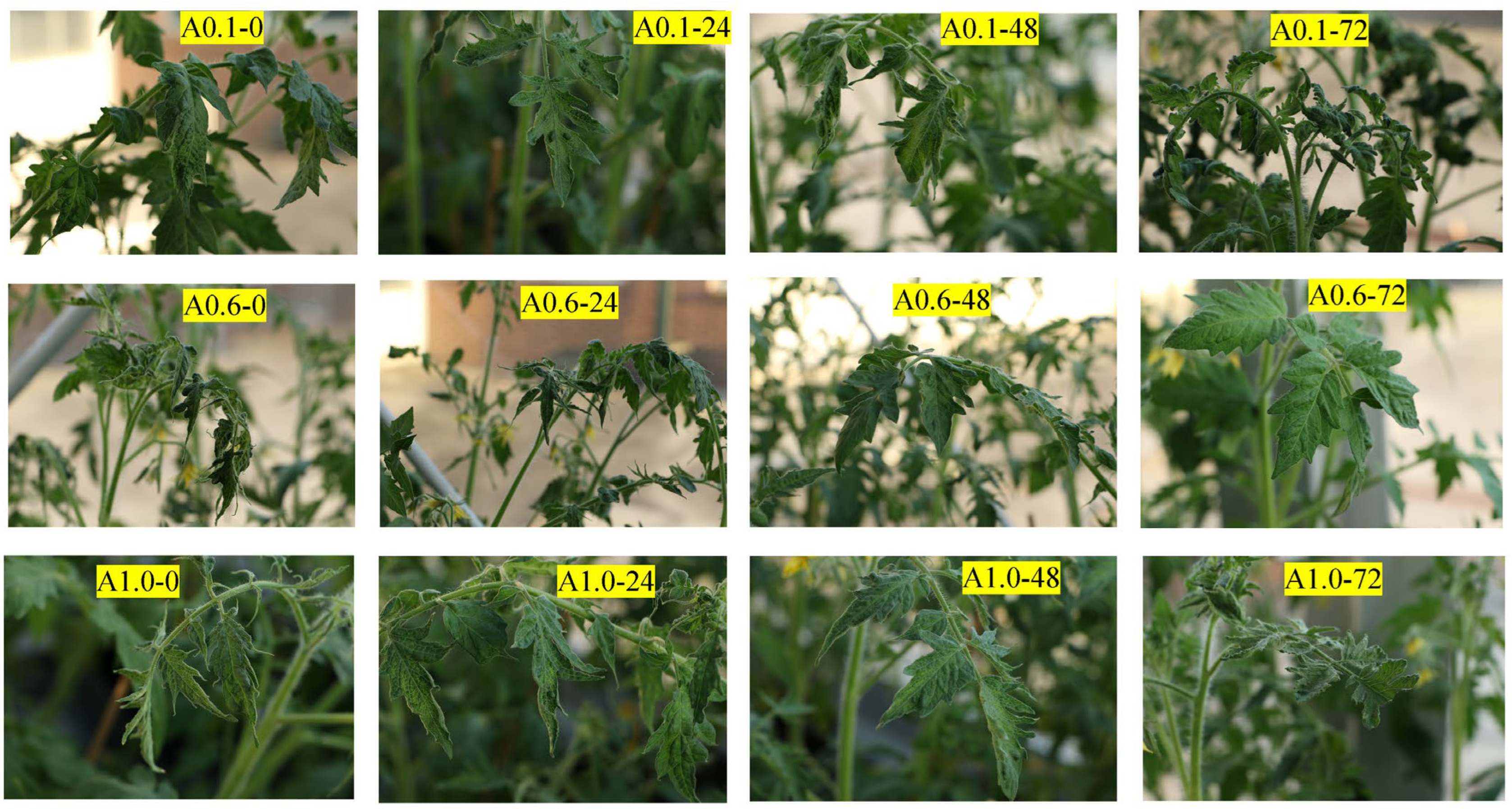
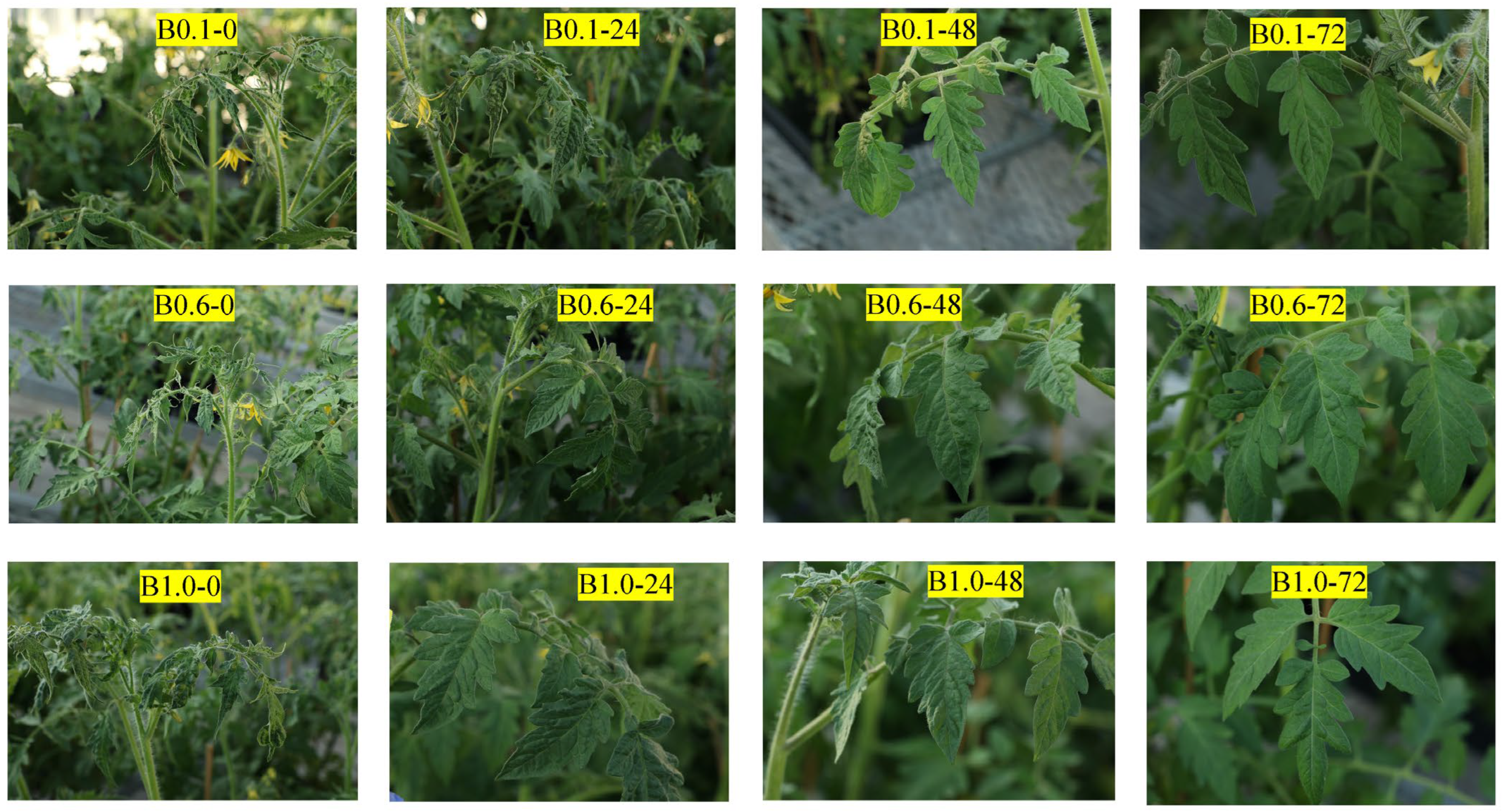
3.4. Efficacy of Cold Plasma-Generated Ozone Treatment against the Infectivity of ToBRFV

Upon proving that ToBRFV existing in the runoff water solutions from commercial greenhouses could trigger virus infection in the bioassay by directly inoculating the water solution onto tomato plants, which indicated that ToBRFV could potentially be transmitted through a hydroponic irrigation line to other parts of a greenhouse, we were interested in developing a water treatment using cold plasma ozone against ToBRFV infection. To evaluate the efficacy of the treatment, we conducted bioassays to assess the infectivity of ToBRFV inocula prepared in different dilutions after being exposed to different ozone concentrations for pre-selected timeframes. With a combination of three ozone concentrations and four treatment timeframes, fifteen different treatments including a mock inoculation were evaluated for three dilutions of virus inocula (1:100, 1:1000, and 1:10,000 dilutions) (Figure 2, Supplementary Table S2). The results showed that, under the highest concentration of ToBRFV inoculum (1:100 dilution), a prolonged exposure (72 min) to the two higher ozone concentration inputs (0.6 mg/L and 1.0 mg/L) resulted in a lower percentage of the test plants (33.3% and 16.7%) developing virus infection (Figure 3), although the infectivity of ToBRFV was not completely aborted. Under a medium concentration of the virus inoculum (1:1000 dilution), although an exposure for 24 min at two higher ozone concentrations (0.6 mg/L and 1.0 mg/L) only reduced the virus infection rate to 16.7%, a longer exposure for 48 min completely suppressed virus infectivity, as demonstrated by the 0% infection of the inoculated plants (Figure 4). The same effect was achieved using even the lowest concentration of ozone input (0.1 mg/L). Furthermore, under the lowest concentration of virus inoculum (1:10,000 dilution), the virus was completely inactivated even at a shorter exposure time (24 min) in any of the three ozone concentrations (0.1 mg/L–1.0 mg/L) (Figure 5). Given that the level of infectious ToBRFV in the greenhouse-collected runoff water samples, as determined above, was comparable to the lowest concentration of the virus inoculum (1:10,000 dilution) used here, which could be fully inactivated using a minimal ozone input within the shortest treatment timeframe selected, the results from this study show great promise in inactivating the ToBRFV residing in runoff water solutions from greenhouses.

4. Discussion

The existence of plant viruses in water environments has been described for many years with a primary concern that the contaminated water could serve as an inoculum to initiate a secondary virus infection, exacerbating disease problems [57,71,72]. However, the virus concentration of water samples collected from rivers, creeks, stream, drainage, and sea water was often too low to directly work on, and therefore, they were often concentrated by ultracentrifugation or polyethylene glycol (PEG) precipitation before any in-depth test such as RT-PCR could be performed [73,74]. Agricultural manufacturing facilities such as large-scale hydroponic farms equipped with closed-recycling fertigation water systems, on the other hand, aid in accumulating water-borne pathogens. Previous studies did reveal that some highly transmissible viruses such as the tomato mosaic virus (ToMV) and the cucumber green mottle mosaic virus (CGMMV) could spread easily from one single infected plant to the entire hydroponic system in greenhouses [75,76].
A bioassay is the only approach to infer the infectivity of a virus existing in a given sample even though DAS-ELISA and qRT-PCR may have a higher sensitivity for detecting the presence of the virus (Table 1 and Table 3). It is noteworthy that neither of the two methods could differentiate between infectious and non-infectious virions in a particular sample, considering that molecular detection assays only target specific regions of a virus’ genetic material, and in the case of ToBRFV, it is a 98-nucleotide segment of the movement protein gene, whereas a serological assay detects the presence of structural components of the virion, for instance, the viral capsid protein. These components could be present in samples containing either intact virus particles or remnants of degraded virions [77,78], and the latter could still yield positive results in molecular and/or serological detection but would fail to trigger virus infection in the bioassay. For this reason, a bioassay was utilized in the current study to evaluate the infectivity of the collected runoff water solutions and the efficacy of the cold plasma-generated ozone treatment against ToBRFV. In conjunction with the results of the bioassay, none of the further-diluted runoff water solutions led to ToBRFV infection, indicating that the approximate active virus titer in the samples collected from Farms #1 and #2 approached the 1:105 dilution of the ToBRFV-infected tissue (Table 2 and Table 3). This number was at an alarmingly high level considering that these runoff water solutions were uncondensed environmental samples. On the other hand, none of the 97 runoff water sample collected from Farm #3 could trigger ToBRFV infection, suggesting that the active virus titer was below this threshold, despite a number of samples testing positive in the qRT-PCR. In addition, Mehle and colleagues proved that the ToBRFV residing in a nutrient solution or in irrigation water could infect healthy plants through their roots [57], further substantiating the water-borne nature of ToBRFV and the fact that a virus-contaminated hydroponic irrigation system is a potential high-risk factor for disseminating the virus and hence should be targeted as a vulnerable stage in the ToBRFV disease cycle. To minimize potential disease dispersal, the water solution circulating in a hydroponic system should be treated properly to inactivate ToBRFV infectivity prior to being recirculated within the same hydroponic system.
Although chemical disinfectants have been shown to be effective against ToBRFV through various treatments [31,32,34,45,46,47,48,49,50,51], offering practical solutions to manage both the vertical and horizontal transmission of the virus in the greenhouse, applying chemotherapy such as using these chemicals in hydroponic water systems can be challenging due to the concern of phytotoxicity and chemical residues. It is therefore imperative to explore other environmentally friendly methods that can be safely applied in water treatments. Different non-chemical methods, such as heat, sonication, media and membrane filtrations, etc., have also been used to control viruses [79]. Ultraviolet (UV) radiation, another commonly used sanitation measure in hydroponic production systems, has been shown to greatly reduce tobacco mosaic virus (TMV) and confine the spread of lettuce ring necrosis virus (an ophiovirus transmitted by Olpidium brassicae), respectively [80,81]. However, the disinfection effect of UV radiation on many other targeted viruses in water environments is generally considered to be minimal [66]. To cope with this challenge, we treated the virus-contaminated water reservoir with ozone generated by cold plasma and subsequently evaluated the efficacy of this approach in inactivating ToBRFV infectivity. The results showed that the effectiveness of the ozone treatment was affected by a combination of virus titer in the contaminated water, the input ozone concentration, and the timeframe of the exposure to the ozone treatment. When the virus inoculum was at 1:100 dilution of ToBRFV, virus infectivity was not affected until the water was exposed to higher ozone concentrations for 72 min (Figure 2). As the relative virus titer reduced to the 1:1000 dilution, a 48 min exposure at the lowest ozone level (0.1 mg/L) was sufficient to inactivate the virus. Furthermore, a minimal exposure time (24 min) at the lowest level of ozone input was adequate to inactivate the virus when the virus inoculum dropped to the 1:10,000 dilution, which was equivalent to the active virus titer detected from the greenhouse-collected runoff water samples. Taken together, our results suggest that the cold plasma-generated ozone treatment is effective in inactivating ToBRFV infectivity in water if appropriate ozone concentrations and exposure timeframes are applied. Given that the lowest active virus titer tested in this experiment (1:104 dilution of the ToBRFV inoculum), which could be completely inactivated by the lowest ozone input with 24 min of exposure, was 10 times higher than those in the runoff water solutions collected from commercial greenhouse farms (approximately 1:105 dilution of the ToBRFV inoculum), the cold plasma ozone treatment could offer a promising solution for preventing potential ToBRFV outbreaks in a hydroponic greenhouse. Future work will focus on evaluating the effects of this ozone treatment on plant growth and development, including but not limited to whether the treatment could lead to phytotoxicity, how the stability of nutrient fertilizers is impacted, and how crop yield and quality are affected. The ultimate goal is to further assess the feasibility of using cold plasma ozone in treating ToBRFV in a real-world situation through specifying the optimized parameters for practical applications.

5. Conclusions

ToBRFV has become a most troubling plant pathogen threatening global tomato production in recent years, with its high mechanical transmissibility and seed-borne characteristic serving as the driving forces behind its quick expansion worldwide. Apart from known transmission pathways, here we proved the water-borne nature of the virus and added a new element to its epidemiology. Although additional studies are needed to thoroughly assess water-mediated ToBRFV transmission to answer questions on whether virus-infected water is the disease trigger directly causing outbreaks, the fact that ToBRFV-contaminated nutrient solutions and irrigation water could infect healthy plants through their roots presents an alarming new sign for vegetable growers [57] while raising special concerns for hydroponic farms as virus outbreaks continue to occur even when strict sanitation measures are implemented. Although the number of water samples that tested positive in this study is relatively low (4/134), in the closed-loop fertigation system where a nutrient solution is captured and recirculated, virus infection occurring in only one spot could be readily spread to the entire system [37]. With the proven efficacy of the ozone treatment in disinfecting ToBRFV in the nutrient solution reported here, we are one step closer to conquering this devastating virus through minimizing its spread in greenhouse tomato production.

Supplementary Materials

The following supporting information can be downloaded at https://www.mdpi.com/article/10.3390/horticulturae10040416/s1, Table S1. Preliminary screening results of ToBRFV in runoff water samples collected from three commercial greenhouses; Table S2. Assay to assess the effectiveness of cold plasma ozone treatment against ToBRFV infectivity in experimental virus-spiked water reservoir.

Author Contributions

Conceptualization, K.-S.L.; methodology, K.-S.L., J.Z. and A.G.; validation, K.-S.L., J.Z. and A.G.; formal analysis, K.-S.L. and J.Z.; investigation, J.Z., K.-S.L. and A.G.; resources, K.-S.L.; data curation, K.-S.L., J.Z. and A.G.; writing—original draft preparation, J.Z.; writing—review and editing, K.-S.L. and A.G.; visualization, K.-S.L. and J.Z; supervision, K.-S.L.; project administration, K.-S.L.; funding acquisition, K.-S.L. All authors have read and agreed to the published version of the manuscript.

Funding

This research was funded in part by the USDA-ARS National Plant Disease Recovery System (grant number: 6080-22000-032-000D) and the USDA-NIFA Crop Protection and Pest Management program, with grant number 2023-70006-40603 to KSL.

Data Availability Statement

The data are presented in the Supplementary Materials.

Acknowledgments

We thank the anonymous greenhouse tomato growers who helped to collect and ship the runoff water samples for this study. The technical support provided by the Clear Path Holdings Corp in setting up the PMOSafe system is greatly appreciated. This research was supported in part by an appointment (JZ) to the Agricultural Research Service (ARS) Research Participation Program administered by the Oak Ridge Institute for Science and Education (ORISE) through an interagency agreement between the U.S. Department of Energy (DOE) and the U.S. Department of Agriculture (USDA). ORISE is managed by ORAU under DOE contract number DE-SC0014664. All the opinions expressed in this paper are the authors’ and do not necessarily reflect the policies and views of the USDA, the DOE, or ORAU/ORISE. The mention of trade names or commercial products in this article is solely for the purpose of providing specific information and does not imply recommendation or endorsement by the USDA. The USDA is an equal opportunity provider and employer.

Conflicts of Interest

The authors declare no conflicts of interest. The funders had no role in the design of this study; in the collection, analyses, or interpretation of the data; in the writing of the manuscript; or in the decision to publish the results.

References

  1. Ofori, P.A.; Owusu-Nketia, S.; Opoku-Agyemang, F.; Agbleke, D.; Naalamle Amissah, J. Greenhouse tomato production for sustainable food and nutrition security in the tropics. In Tomato—From Cultivation to Processing Technology; Viskelis, P., Urbonavičienė, D., Viskelis, J., Eds.; IntechOpen: London, UK, 2022. [Google Scholar] [CrossRef]
  2. Minor, T.; Baskins, S.; Bond, J.K. Imported Greenhouse Tomatoes from Mexico Illustrate the Growing Diversity in Fresh-Market Tomatoes. 2019. Available online: https://www.ers.usda.gov/amber-waves/2019/may/imported-greenhouse-tomatoes-from-mexico-illustrate-the-growing-diversity-in-fresh-market-tomatoes/ (accessed on 18 March 2024).
  3. Salem, N.; Mansour, A.; Ciuffo, M.; Falk, B.W.; Turina, M. A new tobamovirus infecting tomato crops in Jordan. Arch. Virol. 2016, 161, 503–506. [Google Scholar] [CrossRef]
  4. Luria, N.; Smith, E.; Reingold, V.; Bekelman, I.; Lapidot, M.; Levin, I.; Elad, N.; Tam, Y.; Sela, N.; Abu-Ras, A.; et al. A new Israeli tobamovirus isolate infects tomato plants harboring Tm-22 resistance genes. PLoS ONE 2017, 12, e0170429. [Google Scholar] [CrossRef]
  5. EPPO Global Database. 2024. Available online: https://gd.eppo.int/taxon/TOBRFV/distribution (accessed on 18 March 2024).
  6. Salem, N.M.; Jewehan, A.; Aranda, M.A.; Fox, A. Tomato brown rugose fruit virus pandemic. Annu. Rev. Phytopathol. 2023, 61, 137–164. [Google Scholar] [CrossRef]
  7. Fidan, H.; Sarikaya, P.; Calis, O. First report of Tomato brown rugose fruit virus on tomato in Turkey. New Dis. Rep. 2019, 39, 18. [Google Scholar] [CrossRef]
  8. Ghorbani, A.; Rostami, A.; Seifi, S.; Izadpanah, K. First report of Tomato brown rugose fruit virus in greenhouse tomato in Iran. New Dis. Rep. 2021, 44, e12040. [Google Scholar] [CrossRef]
  9. Sabra, A.; Al-Saleh, M.A.; Al-Shahwan, I.M.; Amer, M.A. First report of Tomato brown rugose fruit virus infecting the tomato crop in Saudi Arabia. Plant Dis. 2022, 106, 1310. [Google Scholar] [CrossRef] [PubMed]
  10. Yan, Z.-Y.; Ma, H.-Y.; Han, S.-L.; Geng, C.; Tian, Y.-P.; Li, X.-D. First report of Tomato brown rugose fruit virus infection tomato in China. Plant Dis. 2019, 103, 2973. [Google Scholar] [CrossRef]
  11. Alfaro-Fernández, A.; Castillo, P.; Sanahuja, E.; Rodríguez-Salido, M.C.; Font, M.I. First report of Tomato brown rugose fruit virus in tomato in Spain. Plant Dis. 2021, 105, 515. [Google Scholar] [CrossRef] [PubMed]
  12. Beris, D.; Malandraki, I.; Kektsidou, O.; Theologidis, I.; Vassilakos, N.; Varveri, C. First report of Tomato brown rugose fruit virus infecting tomato in Greece. Plant Dis. 2020, 104, 2035. [Google Scholar] [CrossRef]
  13. Eichmeier, A.; Hejlova, M.; Orsagova, H.; Frejilchova, L.; Hakalova, E.; Tomankova, K.; Linhartova, S.; Kulich, P.; Cermak, V.; Cechova, J. Characterization of tomato brown rugose fruit virus (ToBRFV) detected in Czech Republic. Diversity 2023, 15, 301. [Google Scholar] [CrossRef]
  14. Hamborg, Z.; Blystad, D.-R. First report of Tomato brown rugose fruit virus in tomato in Norway. Plant Dis. 2022, 106, 2004. [Google Scholar] [CrossRef] [PubMed]
  15. Hasan, Z.M.; Salem, N.M.; Ismail, I.D.; Akel, E.H.; Ahmad, A.Y. First report of Tomato brown rugose fruit virus on greenhouse tomato in Syria. Plant Dis. 2022, 106, 772. [Google Scholar] [CrossRef]
  16. Mahillon, M.; Kellenberger, I.; Dubuis, N.; Brodard, J.; Bunter, M.; Weibel, J.; Sandrini, F.; Schumpp, O. First report of Tomato brown rugose fruit virus in tomato in Switzerland. New Dis. Rep. 2022, 45, 12065. [Google Scholar] [CrossRef]
  17. Menzel, W.; Knierim, D.; Winter, S.; Hamacher, J.; Heupel, M. First report of Tomato brown rugose fruit virus infecting tomato in Germany. New Dis. Rep. 2019, 39, 1. [Google Scholar] [CrossRef]
  18. Orfanidou, C.G.; Cara, M.; Merkuri, J.; Papadimitriou, K.; Katis, N.I.; Maliogka, V.I. First report of Tomato brown rugose fruit virus in tomato in Albania. J. Plant Pathol. 2022, 104, 855. [Google Scholar] [CrossRef]
  19. Panno, S.; Caruso, A.G.; Davino, S. First report of Tomato brown rugose fruit virus on tomato crops in Italy. Plant Dis. 2019, 103, 1443. [Google Scholar] [CrossRef]
  20. Skelton, A.; Buxton-Kirk, A.; Ward, R.; Harju, V.; Frew, L.; Fowkes, A.; Long, M.; Negus, A.; Forde, S.; Adams, I.; et al. First report of Tomato brown rugose fruit virus in tomato in the United Kingdom. New Dis. Rep. 2019, 40, 12. [Google Scholar] [CrossRef]
  21. Skelton, A.; Gentit, P.; Porcher, L.; Visage, M.; Fowkes, A.; Adams, I.P.; Harju, V.; Webster, G.; Pufal, H.; McGreig, S.; et al. First report of Tomato brown rugose fruit virus in tomato in France. New Dis. Rep. 2022, 45, e12061. [Google Scholar] [CrossRef]
  22. Van de Vossenberg, B.T.K.H.; Visser, M.; Bruinsma, M.; Koenraadt, H.M.S.; Westenberg, M. Real-time tracking of Tomato brown rugose fruit virus outbreaks in the Netherlands using Nextstrain. PLoS ONE 2020, 15, e0234671. [Google Scholar] [CrossRef]
  23. Camacho-Beltrán, E.; Pérez-Villarreal, A.; Leyva-López, N.E.; Rodríguez-Negrete, E.A.; Ceniceros-Ojeda, E.A.; Méndez-Lozano, J. Occurrence of Tomato brown rugose fruit virus infecting tomato crops in Mexico. Plant Dis. 2019, 103, 1440. [Google Scholar] [CrossRef]
  24. Cambrón-Crisantos, J.M.; Rodríguez-Mendoza, J.; Valencia-Luna, J.B.; Alcasio-Rangel, S.; García-Ávila, C.J.; López-Buenfil, J.A.; Ochoa-Martínez, D.L. First report of tomato brown rugose fruit virus (ToBRFV) in Michoacan, Mexico. Rev. Mex. Fitopatol. 2018, 37, 185–192. [Google Scholar]
  25. Ling, K.-S.; Tian, T.; Gurung, S.; Salati, R.; Gilliard, A. First report of Tomato brown rugose fruit virus infecting tomato in the United States. Plant Dis. 2019, 103, 1439. [Google Scholar] [CrossRef]
  26. Sarkes, A.; Fu, H.; Geindel, D.; Harding, M.; Feng, J. Development and evaluation of a loop-mediated isothermal amplification (LAMP) assay for the detection of Tomato brown rugose fruit virus (ToBRFV). PLoS ONE 2020, 15, e0230403. [Google Scholar] [CrossRef]
  27. Obregón, V.G.; Ibañez, J.M.; Lattar, T.E.; Juszczak, S.; Growth-Helms, D. First report Tomato brown rugose fruit virus in tomato in Argentina. New Dis. Rep. 2023, 48, e12203. [Google Scholar] [CrossRef]
  28. Amer, M.A.; Mahmoud, S.Y. First report of tomato brown rugose fruit virus on tomato in Egypt. New. Dis. Rep. 2020, 41, 24. [Google Scholar] [CrossRef]
  29. New Zealand: ToBRFV Detected in Small Seed Lot. 2021. Available online: www.hortidaily.com/article/9281236/new-zealand-tobrfv-detected-in-small-seed-lot/ (accessed on 18 March 2024).
  30. Zhang, S.; Griffiths, J.S.; Marchand, G.; Bernards, M.A.; Wang, A. Tomato brown rugose fruit virus: An emerging and rapidly spreading plant RNA virus that threatens tomato production worldwide. Mol. Plant. Pathol. 2022, 23, 1262–1277. [Google Scholar] [CrossRef] [PubMed]
  31. Davino, S.; Caruso, A.G.; Bertacca, S.; Barone, S.; Panno, S. Tomato brown rugose fruit virus: Seed transmission rate and efficiency of different seed disinfection treatments. Plants 2020, 9, 1615. [Google Scholar] [CrossRef] [PubMed]
  32. Salem, N.M.; Sulaiman, A.; Samarah, N.; Turina, M.; Vallino, M. Location of mechanical transmission of tomato brown rugose fruit virus in tomato seeds. Plant Dis. 2022, 106, 275–281. [Google Scholar] [CrossRef] [PubMed]
  33. Chanda, B.; Gilliard, A.; Jaiswal, N.; Ling, K.-S. Comparative analysis of host range, ability to infect tomato cultivars with Tm-22 gene and real-time RT-PCR detection of tomato brown rugose fruit virus. Plant Dis. 2021, 105, 3643–3652. [Google Scholar] [CrossRef] [PubMed]
  34. Chanda, B.; Shamimuzzaman, M.; Gilliard, A.; Ling, K.-S. Effectiveness of disinfectants against the spread of tobamoviruses: Tomato brown rugose fruit virus and Cucumber green mottle mosaic virus. Virol. J. 2021, 18, 7. [Google Scholar] [CrossRef]
  35. Klap, C.; Luria, N.; Smith, E.; Bakelman, E.; Belausov, E.; Laskar, O.; Lachman, O.; Gal-On, A.; Dombrovsky, A. The potential risk of plant-virus disease initiation by infected tomatoes. Plants 2020, 9, 623. [Google Scholar] [CrossRef] [PubMed]
  36. Levitzky, N.; Smith, E.; Lachman, O.; Luria, N.; Mizrahi, Y.; Bakelman, H.; Sela, N.; Laskar, O.; Milrot, E.; Dombrovsky, A. The bumblebee Bombus terrestris carries a primary inoculum of Tomato brown rugose fruit virus contributing to disease spread in tomatoes. PLoS ONE 2019, 14, e0210871. [Google Scholar] [CrossRef] [PubMed]
  37. Panno, S.; Caruso, A.G.; Barone, S.; Lo Bosco, G.; Rangel, E.A.; Davino, S. Spread of tomato brown rugose fruit virus in Sicily and evaluation of the spatiotemporal dispersion in experimental conditions. Agronomy 2020, 10, 834. [Google Scholar] [CrossRef]
  38. Dey, K.K.; Velez-Climent, M.; Soria, P.; Batuman, O.; Mavrodieva, V.; Wei, G.; Zhou, J.; Adkins, S.; McVay, J. First report of Tomato brown rugose fruit virus infecting tomato in Florida, USA. New Dis. Rep. 2021, 44, e12028. [Google Scholar] [CrossRef]
  39. Avni, B.; Gelbart, D.; Sufrin-Ringwald, T.; Zinger, A.; Chen, L.; Machbash, Z.; Bekelman, I.; Segoli, M.; Dombrovsky, A.; Kamenetsky, R.; et al. Tomato genetic resistance to tobamoviruses is compromised. Acta Hortic. 2021, 1316, 89–98. [Google Scholar] [CrossRef]
  40. Jewehan, A.; Salem, N.; Tóth, Z.; Salamon, P.; Szabó, Z. Screening of Solanum (sections Lycopersicon and Juglandifolia) germplasm for reactions to the tomato brown rugose fruit virus. J. Plant Dis. Prot. 2022, 129, 117–123. [Google Scholar] [CrossRef]
  41. Jewehan, A.; Salem, N.; Tóth, Z.; Salamon, P.; Szabó, Z. Evaluation of responses to tomato brown rugose fruit virus (ToBRFV) and selection of resistant lines in Solanum habrochaites and Solanum peruvianum germplasm. J. Gen. Plant Pathol. 2022, 88, 187–196. [Google Scholar] [CrossRef]
  42. Kabas, A.; Fidan, H.; Kucukaydin, H.; Atan, H.N. Screening of wild tomato species and interspecific hybrids for resistance/tolerance to Tomato brown rugose fruit virus (ToBRFV). Chil. J. Agric. Res. 2022, 82, 189–196. [Google Scholar] [CrossRef]
  43. Jaiswal, N.; Chanda, B.; Gilliard, A.; Shi, A.; Ling, K.-S. Evaluation of tomato germplasm against tomato brown rugose fruit virus and identification of resistance in Solanum pimpinellifolium. Plants 2024, 13, 581. [Google Scholar] [CrossRef]
  44. Zinger, A.; Lapidot, M.; Harel, A.; Doron-Faigenboim, A.; Gelbart, D.; Levin, I. Identification and mapping of tomato genome loci controlling tolerance and resistance to tomato brown rugose fruit virus. Plants 2021, 10, 179. [Google Scholar] [CrossRef]
  45. Samarah, N.; Sulaiman, A.; Salem, N.M.; Turina, M. Disinfection treatments eliminated tomato brown rugose fruit virus in tomato seeds. Eur. J. Plant Pathol. 2021, 159, 153–162. [Google Scholar] [CrossRef]
  46. Dombrovsky, A.; Mor, N.; Gantz, S.; Lachman, O.; Smith, E. Disinfection efficacy of tobamovirus-contaminated soil in greenhouse-grown crops. Horticulturae 2022, 8, 563. [Google Scholar] [CrossRef]
  47. Ehlers, J.; Nourinejhad Zarghani, S.; Kroschewski, B.; Büttner, C.; Bandte, M. Cleaning of tomato brown rugose fruit virus (ToBRFV) from contaminated clothing of greenhouse employees. Horticulturae 2022, 8, 751. [Google Scholar] [CrossRef]
  48. Ehlers, J.; Nourinejhad Zarghani, S.; Kroschewski, B.; Büttner, C.; Bandte, M. Decontamination of tomato brown rugose fruit virus-contaminated shoe soles under practical conditions. Horticulturae 2022, 8, 1210. [Google Scholar] [CrossRef]
  49. Ling, K.-S.; Gilliard, A.C.; Zia, B. Disinfectants useful to manage the emerging tomato brown rugose fruit virus in greenhouse tomato production. Horticulturae 2022, 8, 1193. [Google Scholar] [CrossRef]
  50. Rodriguez-Diaz, C.I.; Zamora-Macorra, E.J.; Ochoa-Martinez, D.L.; Gonzalez-Garza, R. Disinfectants effectiveness in tomato brown rugose fruit virus (ToBRFV) transmission in tobacco plants. Rev. Mex. Fitopatol. 2022, 40, 240–253. [Google Scholar] [CrossRef]
  51. Skelton, A.; Frew, L.; Ward, R.; Hodgson, R.; Forde, S.; McDonough, S.; Webster, G.; Chisnall, K.; Mynett, M.; Buxton-Kirk, A.; et al. Tomato brown rugose fruit virus: Survival and disinfection efficacy on common glasshouse surfaces. Viruses 2023, 15, 2076. [Google Scholar] [CrossRef] [PubMed]
  52. Bačnik, K.; Kutnjak, D.; Pecman, A.; Mehle, N.; Žnidarič, M.T.; Aguirre, I.G.; Ravnikar, M. Viromics and infectivity analysis reveal the release of infective plant viruses from wastewater into the environment. Water Res. 2020, 177, 115628. [Google Scholar] [CrossRef] [PubMed]
  53. Natarajan, A.; Fremin, B.J.; Schmidtke, D.T.; Wolfe, M.K.; Zlitni, S.; Graham, K.E.; Brook, E.F.; Severyn, C.J.; Sakamato, K.M.; Lacayo, N.J.; et al. The tomato brown rugose fruit virus movement protein gene is a novel microbial source tracking marker. Appl. Environ. Microbiol. 2023, 89, e0058323. [Google Scholar] [CrossRef]
  54. Rothman, J.; Whiteson, K. Sequencing and variant detection of eight abundant plant-infecting tobamoviruses across southern California wastewater. Microbiol. Spectr. 2022, 10, e0305022. [Google Scholar] [CrossRef]
  55. Sherchan, S.P.; Malla, B.; Haramoto, E. First quantitative detection of tomato brown rugose fruit virus in wastewater in Louisiana. Sci. Total Environ. 2023, 888, 164001. [Google Scholar] [CrossRef] [PubMed]
  56. Nash, D.; Ellmen, I.; Knapp, J.J.; Menon, R.; Overton, A.K.; Cheng, J.; Lynch, M.D.J.; Nissimov, J.I.; Charles, T.C. A novel tiled amplicon sequencing assay targeting the tomato brown rugose fruit virus (ToBRFV) genome reveals widespread distribution in municipal wastewater treatment systems in the province of Ontario, Canada. Viruses 2024, 16, 460. [Google Scholar] [CrossRef]
  57. Mehle, N.; Bačnik, K.; Bajde, I.; Brodarič, J.; Fox, A.; Gutiérrez-Aguirre, I.; Kitek, M.; Kutnjak, D.; Loh, Y.L.; Carvalho Ferreira, O.M.; et al. Tomato brown rugose fruit virus in aqueous environments—Survival and significance of water-mediated transmission. Front. Plant Sci. 2023, 14, 1187920. [Google Scholar] [CrossRef]
  58. Baysan, A.; Lynch, E. The use of ozone in dentistry and medicine. Prim. Dent. J. 2005, 12, 47–52. [Google Scholar] [CrossRef]
  59. Khadre, M.A.; Yousef, A.E.; Kim, J.-G. Microbiological aspects of ozone applications in food: A review. J. Food Sci. 2001, 66, 1242–1252. [Google Scholar] [CrossRef]
  60. Seridou, P.; Kalogerakis, N. Disinfection application of ozone micro- and nanobubbles. Environ. Sci. Nano 2021, 8, 3493–3510. [Google Scholar] [CrossRef]
  61. Sato, H.; Wananabe, Y.; Miyata, H. Virucidal effect of ozone treatment of laboratory animal viruses. Jikken Dobutsu. 1990, 39, 223–229. [Google Scholar]
  62. Boast, N.; Heselton, D.; Hudson, J. Apparatus and Method for Using Ozone as a Disinfectant. International Publication Number WO2005087278A1, 22 September 2005. [Google Scholar]
  63. Zhang, J.; Zheng, C.; Xiao, G.; Zhou, Y.; Gao, R. Examination of the efficacy of ozone solution disinfectant in in-activating SARS virus. Chin. J. Disinfect. 2004, 21, 27–28. [Google Scholar]
  64. Hu, X.; Chen, Z.; Su, Z.; Deng, F.; Chen, X.; Yang, Q.; Li, P.; Chen, Q.; Ma, J.; Guan, W.; et al. Ozone water is an effective disinfectant for SARS-CoV-2. Virol. Sin. 2021, 36, 1066–1068. [Google Scholar] [CrossRef]
  65. Filipić, A.; Primc, G.; Zaplotnik, R.; Mehle, N.; Gutierrez Aguirre, I.; Ravnikar, M.; Mozetič, M.; Žel, J.; Dobnik, D. Cold atmospheric plasma as a novel method for inactivation of potato virus Y in water samples. Food Environ. Virol. 2019, 11, 220–228. [Google Scholar] [CrossRef]
  66. Filipić, A.; Dobnik, D.; Tušek Žnidarič, M.; Žegura, B.; Štern, A.; Primc, G.; Mozetič, M.; Ravnikar, M.; Žel, J.; Aguirre, I.G. Inactivation of pepper mild mottle virus in water by cold atmospheric plasma. Front. Microbiol. 2021, 12, 618209. [Google Scholar] [CrossRef] [PubMed]
  67. Mahnot, N.K.; Mahanta, C.L.; Keener, K.M.; Misra, N.N. Strategy to achieve a 5-log Salmonella inactivation in tender coconut water using high voltage atmospheric cold plasma (HVACP). Food Chem. 2019, 284, 303–311. [Google Scholar] [CrossRef] [PubMed]
  68. Nguyen, D.V.; Ho, P.Q.; Pham, T.V.; Nguyen, T.V.; Kim, L. Treatment of surface water using cold plasma for domestic water supply. Environ. Eng. Res. 2019, 24, 412–417. [Google Scholar] [CrossRef]
  69. Patange, A.; Lu, P.; Boehm, D.; Cullen, P.J.; Bourke, P. Efficacy of cold plasma functionalized water for improving microbiological safety for fresh produce and wash water recycling. Food Microbiol. 2019, 84, 103226. [Google Scholar] [CrossRef] [PubMed]
  70. Qin, H.; Qiu, H.; He, S.; Hong, B.; Liu, K.; Lou, F.; Li, M.; Hu, P.; Kong, H.; Song, Y.; et al. Efficient disinfection of SARS-CoV-2-like coronavirus, pseudotyped SARS-CoV-2 and other coronaviruses using cold plasms induces spike protein damage. J. Hazard. Mater. 2022, 430, 128414. [Google Scholar] [CrossRef] [PubMed]
  71. Büttner, C.; Nienhaus, F. Virus contamination of soils in forest ecosystems of the Federal Republic of Germany. Eur. J. For. Path. 1989, 19, 47–53. [Google Scholar] [CrossRef]
  72. Koenig, R. Plant viruses in rivers and lakes. Adv. Virus Res. 1986, 31, 321–333. [Google Scholar] [PubMed]
  73. Gosalvez, B.; Navarro, J.A.; Norca, A.; Botella, R.; Sánchez-Pina, M.A.; Pallas, V. Detection of melon necrotic spot virus in water samples and melon plants molecular methods. J. Virol. Methods 2003, 113, 87–93. [Google Scholar] [CrossRef] [PubMed]
  74. Mehle, N.; Ravnikar, M. Plant viruses in aqueous environment—Survival, water mediated transmission and detection. Water Res. 2012, 46, 4902–4917. [Google Scholar] [CrossRef] [PubMed]
  75. Paludan, N. Spread of viruses by recirculated nutrient solutions in soilless cultures. Tidsskr. Planteavl 1985, 89, 467–474. [Google Scholar]
  76. Pategas, K.G.; Schuerger, A.C.; Wetter, C. Management of tomato mosaic virus in hydroponically grown pepper (Capsicum annuum). Plant Dis. 1989, 73, 570–573. [Google Scholar] [CrossRef]
  77. Girt, G.C.; Lakshminarayanan, A.; Huo, J.; Dormon, J.; Norman, C.; Afrough, B.; James, W.; Owens, R.J.; Naismith, J.H. The use of nanobodies in a sensitive ELISA test for SARS-CoV-2 spike 1 protein. R. Soc. Open Sci. 2021, 8, 211016. [Google Scholar] [CrossRef] [PubMed]
  78. Motley, M.P.; Bennett-Guerrero, E.; Fries, B.C.; Spitzer, E.D. Review of viral testing (Polymerase chain reaction) and antibody/serology testing for severe acute respiratory syndrome-coronavirus-2 for the intensivist. Crit. Care Explor. 2020, 2, e0154. [Google Scholar] [CrossRef] [PubMed]
  79. Mori, J.; Smith, R. Transmission of waterborne fish and plant pathogens in aquaponics, and their control with physical disinfection and filtration: A systematized review. Aquaculture 2019, 504, 380–395. [Google Scholar] [CrossRef]
  80. Gharbi, S.; Verhoyen, M. Sterilization UV irradiation of nutrients solution with view to avoiding viral infections transmitted by O. brassicae in hydroponic culture of lettuce. HortScience 1993, 58, 873–874. [Google Scholar]
  81. Runia, W. Elimination of root-infecting pathogens in recirculation water from closed cultivation systems by ultra-violet radiation. Acta Hortic. 1994, 361, 361–371. [Google Scholar] [CrossRef]
Figure 1. Phenotypes of tomato plants of the “Moneymaker” variety in a bioassay to evaluate ToBRFV infectivity four weeks post mechanical inoculation using runoff water samples collected from commercial greenhouses. (A) Typical mottling, mosaic, and chlorosis symptoms on tomato plants inoculated with water sample V22-43, which tested positive for ToBRFV during qRT-PCR. (B) A typical asymptomatic tomato leaf on a tomato plant inoculated with the other water samples that did not cause virus infection and tested negative for ToBRFV.
Horticulturae 10 00416 g001
Figure 2. Assessing the effectiveness of a treatment against ToBRFV infectivity in bioassays using three concentrations of cold plasma ozone on three dilutions of ToBRFV-infected plant tissue extracted in experimental water reservoirs. Panel (A): Percentage of the test tomato plants (6) infected in the bioassays in a virus inoculum at a 1:100 dilution upon exposure for 0 min, 24 min, 48 min, and 72 min using three concentrations of cold plasma ozone (0.1, 0.6, and 1 mg/L). Panel (B): Percentage of the test tomato plants (6) infected in the bioassays in a virus inoculum at a 1:1000 dilution upon exposure for 0 min, 24 min, 48 min, and 72 min using three concentrations of cold plasma ozone (0.1, 0.6, and 1 mg/L). Panel (C): Percentage of the test tomato plants (6) infected in the bioassays in a virus inoculum at a 1:10,000 dilution upon exposure for 0 min, 24 min, 48 min, and 72 min using three concentrations of cold plasma ozone (0.1, 0.6, and 1 mg/L).
Horticulturae 10 00416 g002
Figure 3. The phenotypes of the tomato plants in the bioassays to assess the efficacy of a cold plasma ozone treatment using a virus inoculum at a 1:100 dilution upon exposure for 0 min, 24 min, 48 min, and 72 min using three concentrations of cold plasma ozone (0.1, 0.6, and 1 mg/L). The label in each panel represented by A is the virus inoculum in a 1:100 dilution of ToBRFV-infected tomato tissue sap, followed by the ozone concentration (0.1, 0.6, or 1.0 mg/L), and, finally, the exposure time (0, 24, 48, or 72 min).
Horticulturae 10 00416 g003
Figure 4. The phenotypes of the tomato plants in the bioassays to assess the efficacy of a cold plasma ozone treatment using a virus inoculum at a 1:1000 dilution (B) upon exposure for 0 min, 24 min, 48 min, and 72 min using three concentrations of cold plasma ozone (0.1, 0.6, and 1 mg/L). The label in each panel represented by B is the virus inoculum in a 1:1000 dilution of ToBRFV-infected tomato tissue sap, followed by the ozone concentration (0.1, 0.6, or 1.0 mg/L), and, finally, the exposure time (0, 24, 48, or 72 min).
Horticulturae 10 00416 g004
Figure 5. The phenotypes of the tomato plants in the bioassays to assess the efficacy of a cold plasma ozone treatment using a virus inoculum at a 1:10,000 dilution (C) upon exposure for 0 min, 24 min, 48 min, and 72 min using three concentrations of cold plasma ozone (0.1, 0.6, and 1 mg/L). The label in each panel represented by C is the virus inoculum in a 1:10,000 dilution of ToBRFV-infected tomato tissue sap, followed by the ozone concentration (0.1, 0.6, or 1.0 mg/L), and, finally, the exposure time (0, 24, 48, or 72 min).
Horticulturae 10 00416 g005
Table 1. Summary of initial screening of ToBRFV in runoff water samples collected from different commercial greenhouses.
Sample SourceWater SampleBioassay on Tomato Plant
Sample NameqRT-PCR aSymptomsDAS-ELISA b
Farm #1V22-1427.83 (+)Yes0.55 (+)
V22-1526.18 (+)Yes2.65 (+)
Farm #2V22-2919.90 (+)Yes2.56 (+)
V22-4322.97 (+)Yes2.35 (+)
33 other water samples21.25 (+) to No Ct (−)No0.01 (−) to 0.03 (−)
Farm #397 water samples22.62 (+) to 32.13 (−)No0.01 (−) to 0.04 (+)
Positive controlToBRFV-infected tomato13.43 (+)Yes2.67 (+)
Negative controlHealthy tomato31.22 (−)No0.01 (−)
a Number represents the Ct value of the corresponding sample. The Ct cut-off value for qRT-PCR was 30 in this experiment. Water samples with a Ct (∆Rn) reading <30 were considered positive (+) for ToBRFV, while those >30 were negative (−). b Number represents ELISA absorbance values OD405. OD405 > 0.3 was considered positive (+), and those ≤0.3 were negative −).
Table 2. Secondary test to evaluate ToBRFV infectivity in selected water samples.
Sample Name
Plant Sap Dilutions
V22-14V22-15V22-29V22-43
ELISA aIC-qRT-PCR bELISAIC-qRT-PCRELISAIC-qRT-PCRELISAIC-qRT-PCR
Undiluted−(0/6)−(0/6)+(1/6) c+(1/6) c−(0/6)−(0/6)+(3/6) d+(3/6) d
1:10−(0/6)−(0/6)−(0/6)−(0/6)−(0/6)−(0/6)−(0/6)−(0/6)
1:100−(0/6)−(0/6)−(0/6)−(0/6)−(0/6)−(0/6)−(0/6)−(0/6)
1:1000−(0/6)−(0/6)−(0/6)−(0/6)−(0/6)−(0/6)−(0/6)−(0/6)
a,b Fraction in parenthesis represents the number of plants that tested positive for ToBRFV/the number of plants tested in total using individual methods. (+) and (−) indicate positive and negative testing results for ToBRFV, respectively. c Tested positive at 8 WPI. d Two plants tested positive at 4 WPI, and another one tested positive at 8 WPI.
Table 3. Comparative analysis to assess relative virus titer of ToBRFV inocula prepared in plant extract.
Plant Sap DilutionsVirus DetectionBioassay on Tomato Plants
DAS-ELISA aIC-qRT-PCR bSymptoms cDAS-ELISA dIC-qRT-PCR d
1:1003.3327.35Yes (3/3)2.02 (3/3)26.75 (3/3)
1:1033.3227.01Yes (3/3)2.12 (3/3)28.52 (3/3)
1:1043.0430.15Yes (2/3)2.25 (2/3)27.73 (2/3)
1:1051.2431.32Yes (1/3)2.02 (1/3)26.49 (1/3)
1:1060.2134.17No (0/3)0.11 (0/3)No Ct
1:1070.03No CtNo (0/3)0.11 (0/3)No Ct
1:1080.01No CtNo (0/3)0.10 (0/3)No Ct
1:1090.01No CtNo (0/3)0.10 (0/3)No Ct
1:10100.01No CtNo (0/3)0.10 (0/3)No Ct
1:10110.09No CtNo (0/3)0.10 (0/3)No Ct
1:10120.01No CtNo (0/3)0.10 (0/3)No Ct
Positive
Control e
3.1826.37Yes (3/3)2.66 (3/3)20.02 (3/3)
Mock
Control
0.02No CtNo (0/3)0.11 (0/3)No Ct
a Number represents the average OD405 values of DAS-ELISA. OD405nm > 0.2 is considered positive. b Number represents the Ct values of IC-qRT-PCR. The cut-off Ct value for IC-qRT-PCR is 35. No Ct represents the fact that a Ct value is not detected. c Number in parenthesis represents the ratio of the number of plants exhibiting ToBRFV symptoms to the total number of plants tested. d Number in parenthesis represents the ratio of the number of plants that tested positive using individual methods to the total number of plants tested. e Positive control: a virus inoculum prepared through the processing of ToBRFV-infected leaf tissue in a tissue extraction buffer for ELISA (1:10 w/v) was used as the positive control.
Disclaimer/Publisher’s Note: The statements, opinions and data contained in all publications are solely those of the individual author(s) and contributor(s) and not of MDPI and/or the editor(s). MDPI and/or the editor(s) disclaim responsibility for any injury to people or property resulting from any ideas, methods, instructions or products referred to in the content.

