Article

LED White Light Treatment Delays Postharvest Senescence of ‘Zaosu’ Pear Fruit with Inhibited Chlorophyll Degradation

1
College of Food Science and Engineering, Bohai University, Jinzhou 121000, China
2
Hami Melon Research Center, Xinjiang Academy of Agricultural Sciences, Urumqi 830091, China
*
Authors to whom correspondence should be addressed.
Horticulturae 2024, 10(1), 32; https://doi.org/10.3390/horticulturae10010032
Submission received: 1 December 2023 / Revised: 16 December 2023 / Accepted: 21 December 2023 / Published: 28 December 2023

Abstract

:
To investigate the effects of LED white light treatment (LWT) on the storage quality of postharvest ‘Zaosu’ pears, as well as its role in maintaining fruit greenness and delaying senescence, pear fruits were treated with intermittent irradiation using LED white light for 12 h per day, with a light source distance of 30 ± 1 cm and a photon flux density of 151 μmoL/(m2·s) at 25 °C. The results showed that LWT preserved the postharvest quality of ‘Zaosu’ pear fruit by reducing weight loss and ascorbic acid degradation and promoting the ratio of sugar and organic acid. Meanwhile, LWT also substantially inhibited the respiration intensity and ethylene production during the storage process. Accordingly, the expressions of genes related to ethylene biosynthesis and signaling were reduced in LWT fruit. Notably, LWT retarded the decrease in chlorophyll content of fruit by increasing the activities of enzymes associated with chlorophyll synthase. Additionally, LWT also suppressed the chlorophyll degradation-related enzymes and their gene expressions in pear peel. These findings suggest that a moderate light irradiation can delay the de-green progress and benefit post-harvest storage of ‘Zaosu’ pear.

1. Introduction

The ‘Zaosu’ pear (Pyrus ussuriensis Var. Zaosu) is a high-quality, high-yielding, strong resistance, and early maturing pear variety bred by the Research Institute of Pomology of the Chinese Academy of Agricultural Sciences (Xingcheng). It is also one of the most widely cultivated early maturing pear varieties in China [1]. The ‘Zaosu’ pear is favored by Chinese consumers due to its vibrant green color, minimal stone cells, abundant juice, and exquisite taste [2]. However, the high water content in the pear results in postharvest wrinkling and shrinkage, leading to physiological disorders and a decline in overall quality [3]. The pear fruit exhibits high respiration intensity and rapid nutrient consumption, which accelerate senescence and diminish its nutritional value [4]. Notably, the peel of the ‘Zaosu’ pear undergoes a rapid transition from green to yellow shortly after harvest, typically within ten days, which greatly compromises both the edible quality and commercial value of the fruit [5,6].
Currently, numerous approaches have been explored to preserve the green color of the fruit peel during storage and transportation. These include a controlled atmosphere, 1-methylcyclopropene (1-MCP), heat treatment, ozone, ethanol, plant growth regulators, plant extracts, and film preservatives [7,8,9,10,11,12,13,14,15,16]. Interestingly, researchers in the horticultural post-harvest field have shown great interest in light-emitting diodes (LEDs) due to their remarkable attributes. LEDs are recognized for being environmentally friendly, energy efficient, capable of close-range irradiation, and for generating minimal heat [17]. Research studies have demonstrated that LED light irradiation is highly effective in suppressing the growth of surface microorganisms on fruits and vegetables [18]. Furthermore, it has been observed that it slows down ethylene release and respiration rate, thereby achieving successful post-harvest preservation [19]. In addition, fruits and vegetables treated with LED exhibit well-preserved nutritional value, flavor quality, and color, as evidenced by studies [19].
Based on the above, in this study, the ‘Zaosu’ pear was used as the experimental material to investigate the impact of LED white light treatment on the post-harvest quality and chlorophyll metabolism of the fruit during storage at 25 °C, providing an experimental basis and theoretical reference for the green preservation effect and storage preservation technology of ‘Zaosu’ pear fruits after harvest.

2. Materials and Methods

2.1. Plant Materials and Treatments

The ‘Zaosu’ pear fruit was harvested from a local orchard in Jinzhou City on September 10th, 2021 and transported to the Bohai University laboratory within 2 h after harvesting. A total of 112 fruits with a weight of 200–230 g, no diseases or pests, no mechanical damage, uniform size, similar color, and excellent texture were selected and stored in conditions of 25 ± 1 °C and RH 85–90%. The pear fruits were randomly divided into two groups: (i) control group: dark treatment group (control); (ii) LED white light treatment group (LWT): the fruit was placed in conditions of 25 ± 1 °C at a distance of 30 ± 1 cm from the light source, with a light density of 151 μmol/m2·s, and subjected to intermittent light for 12 h/d (from 08:00 to 20:00 every day, remaining in darkness for the rest of the time). The specific experimental setup is shown in Figure 1. The pear fruit samples were turned over every 24 h and sampled on days 0, 5, 10, 15, 20, and 25. On the sampling day, color difference, weight loss rate, respiration intensity, and firmness were measured. The remaining parts (peel, flesh) were frozen with liquid nitrogen and stored in a −80 °C refrigerator for subsequent experiments.

2.2. Color, Weight Loss, and Firmness

The fruit color was measured using a color spectrum CS-520 spectrophotometer (Suzhou Liuxinyan Precision Machinery Inc., Suzhou, China). Nine pear fruits were randomly selected from each group. Three equidistant positions were chosen on the equatorial part of the fruit, and the corresponding a* and b* values were measured and recorded. Subsequently, the h value was calculated using the formula [20]:
h ° = a r c t a n ( b * / a * )                                         ( a * > 0 , b * > 0 )
h ° = 180 + a r c t a n ( b * / a * )                       a * < 0 , b * > 0
where the a* value represents the red–green chromaticity and the b* value represents the yellow–blue chromaticity. The h value has four color boundary points: h = 0° (red), h = 90° (yellow), h = 180° (green), and h = 270° (blue) [21].
For each group, five pear fruits were selected. The weight loss rate of the fruit was determined using a weighing method, and the following formula was employed to calculate the mass difference:
Weight loss rate (%) = (Weight before storage − Weight after storage)/Quality before storage × 100
The fruit firmness was measured using the method described by Villarreal [6], using a digital fruit hardness meter (GY-4, Beijing Sunshine Yishida Technology Inc., Beijing, China) with a probe diameter of 11 mm and a testing distance of 10 mm. Three equidistant positions were selected on the equatorial part of the fruit, and the fruit peel, approximately 1.0 mm in thickness, was removed. For each group, nine fruits were randomly selected from each treatment group. Firmness was expressed in Newton (N).

2.3. Ascorbic Acid Content

The ascorbic acid (AsA) content in the fruit was determined using the 2,6-dichlorophenol indophenol titration method, as described by Skrovankova et al. [22] with some modifications. Five grams of frozen fruit powder was put into 100 mL of oxalic acid solution (20 g/L) and mixed thoroughly. The mixture was kept in dark conditions for 10 min and was filtered using a Whatman® qualitative filter paper. Ten milliliters of filtrate were transferred into a conical flask and titrated with a standard 2,6-dichlorophenol indophenol solution. Ten milliliters of oxalic acid solution (20 g/L) was used as a blank control. This was repeated three times for each sample. The content of ascorbic acid was expressed in mg/100 g.
A s c o r b i c   a c i d   c o n t e n t mg / 100   g = [ V × ( V 1 V 0 ) × C / V s × W ] × 100
where V1 represents the volume of dye consumed in the sample titration (mL); V0 denotes the volume of dye consumed in blank titration (mL); C indicates the 1 mL of dye solution equivalent to the amount of ascorbic acid (mg); Vs expresses the volume of sample solution taken during titration (mL); V signifies the total volume of sample extraction solution (mL); and W refers to the sample weight (g).

2.4. Sugar-to-Acid Ratio

The content of total soluble solids (TSS) was measured using a PAL-1 refractometer (Tokyo, Japan). Nine ‘Zaosu’ pear fruits were randomly selected, and their flesh was cut and mixed thoroughly. The juice was extracted and centrifuged, and the supernatant was used to measure the value of TSS, which was expressed as a percentage.
The titratable acid (TA) content was determined using the acid-base titration method as described by Skrovankova et al. [22]. Approximately 1.5 g of pear fruit samples was weighed into 3.0 mL of CO2-free distilled water and mixed thoroughly. The mixture was then centrifuged at 4 °C and 10,000 rpm for 20 min. Subsequently, 1.0 mL of the supernatant was taken, and 2 drops of 1% (w/v) phenolphthalein indicator were added. The volume of 0.1 M NaOH solution consumed during titration was recorded, and the titratable acid was expressed as a mass fraction (%). The conversion factor was calculated based on the malic acid (0.067).
The sugar-to-acid ratio was calculated as the ratio of soluble solid content to titratable acid content in the pear fruit.

2.5. Respiratory Intensity and Ethylene Production

The method for respiratory intensity and ethylene production measurement was in line with Sun’s [23], with some modification. Nine pear fruits were randomly selected and weighed, then placed in a sealed desiccator at 20 °C. After sealing for one hour, one milliliter of the gas at the top of the container was collected for further analysis.
For gas chromatography analysis, the carrier gases used were 0.05 MPa of nitrogen, 0.1 MPa of hydrogen, and 0.1 MPa of air. The gas flow rate was set at 40.0 mL/min (7890A, Agilent Technologies Inc., Santa Clara, CA, USA). The respiration intensity and ethylene release were expressed as mg CO2/(kg·h) and μL/(kg·h), respectively.
E t h y l e n e   e v o l u t i o n ( µ L · k g 1 · h 1 ) = C × V / W × t × 1000
where C represents the ethylene content in the sample gas determined by gas chromatography (µL/L); V denotes the volume of the glass container’s confined space (mL); T indicates the measurement time (h); and W signifies the fruit weight (kg).

2.6. Chlorophyll Content and Related Enzyme Activities

As described by Esteban et al. [24], with some modifications, the chlorophyll content was measured via spectrophotometry. Approximately 1.0 g of pear peel sample was weighed and placed into 3.0 mL of pre-cooled extraction solution (80% (v/v) acetone: 95% (v/v) ethanol = 2:1). The mixture was stirred and kept at 4 °C in darkness for 30 min. The obtained suspension was centrifuged at 4 °C and 10,000 rpm for 15 min. The supernatant was collected and the absorbance values measured at 645 nm and 663 nm using a spectrophotometer (GS-520, Suzhou Liuxinyan Precision Machinery Inc., Suzhou, China). The chlorophyll content was expressed as mg/g FW (milligrams per gram of fresh weight). The contents of chlorophyll a and b, and total chlorophyll were calculated using the following formulas.
C h l o r o p h y l l - a = 12.72 A 663 2.59 A 645
C h l o r o p h y l l -b = 22.88 A 645 4.67 A 663
T o t a l   c h l o r o p h y l l   c o n t e n t = 20.29 A 645 + 8.05 A 663
To determine the activities of chlorophyll metabolism-related enzymes (chlorophyll synthase, chlorophyll a oxygenase, chlorophyllase, Mg-dechelatase, pheophytinase, and pheophorbide a oxygenase) in the fruit peel sample, 1 g of fruit peel sample was taken and added into 9 mL of phosphate buffer (pH 7.2–7.4, 0.01 M elemental acid, hydrophosphate and sodium chloride). The mixture was homogenized in an ice bath, centrifuged at 4 °C and 4500× g for 20 min, and the supernatant was collected for further analysis. The plant enzyme-linked immunosorbent assay (ELISA) kits (Plant CAO ELISA Kit, Plant PPH ELISA Kit, Plant CS ELISA Kit, Plant Chkase ELISA Kit, Plant MDCase ELISA Kit, Plant PAO ELISA Kit, Shanghai Enzyme-linked Biotechnology Inc., Shanghai, China) were used. The ELISA assays were performed according to the kit instructions, with triplicate measurements for each group. The activities of enzymes were expressed as U/g FW (units per gram of fresh weight). A change of 0.01 per minute was considered as one unit of enzyme activity.

2.7. Fluorescence Quantitative PCR

The RNA extraction was performed using the CTAB method, referring to the method described by Chen et al. [25]. The reverse transcription was carried out using the FastKing RT Kit cDNA first strand synthesis kit (Cat # KR116-02, Tiangen Biochemical Technology Co., Ltd., Beijing, China). In line with previous research [26,27,28], PbNYC, PbNOL, PbCLH, PbSGR, PbPPH, PbPAO, PbRCCR, PbACS4, PbACO1, PbACO4, PbETR1, PbETR2, PbERS1, PbERS2, PbCTR1, PbEIN2, PbEIL1, and PbERF1 were selected, and their primers are listed in Table 1. The relative expression of the genes was calculated using the 2−ΔΔCT algorithm [29].

2.8. Statistical Analysis

All the data were expressed as mean ± standard deviation and analyzed using Excel 2016 software (Microsoft, Washington, DC, USA). SPSS 21.0 software (International Business Machines Corporation, Armonk, NY, USA) was used for one-way analysis of variance (ANOVA) with significant differences between groups at p < 0.05 (Duncan).

3. Results

3.1. Effect of LED White Light on the Postharvest Quality of Pear Fruit

During the entire storage period, the weight loss rate of pear fruit in the dark (control) and LED white light treatment (LWT) groups showed an increasing trend. The weight loss of the LWT group was significantly lower than the control group (p < 0.05), indicating that LED white light treatment, to some extent, inhibited the evaporation of water and respiration consumption of ‘Zaosu’ pear fruit during storage (Figure 2A).
Figure 2B shows that the levels of ascorbic acid (AsA) in both groups of pear fruit exhibited a decreasing trend during storage. However, by day 15, the AsA content in the LWT group was significantly higher than in the control group (p < 0.05). Therefore, it can be observed that LED white light treatment effectively inhibits the decline in AsA content in ‘Zaosu’ pear fruit during storage, particularly in the early stages of storage.
During the storage period, there was no significant difference in the firmness of ‘Zaosu’ pear fruit between the two treatment groups (p > 0.05, Figure 2C), indicating that LED white light treatment did not have an impact on the changes in fruit firmness during storage.
In both treatment groups, the sugar-to-acid ratio showed a similar trend of initially increasing and then decreasing, reaching its peak on day 10 (Figure 2D). The LWT group was significantly higher than the dark treatment group (p < 0.05), indicating that LED white light is beneficial in maintaining the favorable taste of ‘Zaosu’ pear fruit (Figure 2D).

3.2. Effect of LED White Light on Respiratory Intensity and Ethylene Production of Pear Fruit

As shown in Figure 3, ‘Zaosu’ pears exhibited typical peaks for respiration rate and ethylene production during storage. The respiration rate and ethylene production of the pear fruit in both treatment groups showed a trend of initially increasing and then decreasing, with peak values occurring on days 15 and 20, respectively. However, the respiration rate and ethylene production of pear fruit in the LWT group were significantly lower than in the control group (p < 0.05). This indicates that LWT can inhibit the respiration process and ethylene synthesis in pear fruit.

3.3. Effect of LED White Light on the Expression of Genes Related to the Ethylene Biosynthesis Pathway in Pear Fruit

Throughout the entire storage period, the relative expression levels of PbACS4 and PbACO4 in ‘Zaosu’ pear fruit treated with LED white light showed a continuous increase, whilst their relative expression levels in the control group exhibited an initial increase followed by a decrease, with peak values occurring on day 20 (Figure 4A,C). Within the first 20 days of storage, the expression levels of PbACS4 and PbACO4 in the LWT group were significantly lower than in the control group (p < 0.05). The expression levels of PbACO1 in both treatment groups showed a trend of initially increasing and then decreasing, with peak values occurring on days 15 (control group) and 20 (LWT), and the LWT group was significantly lower than the control group (p < 0.05, Figure 4B).

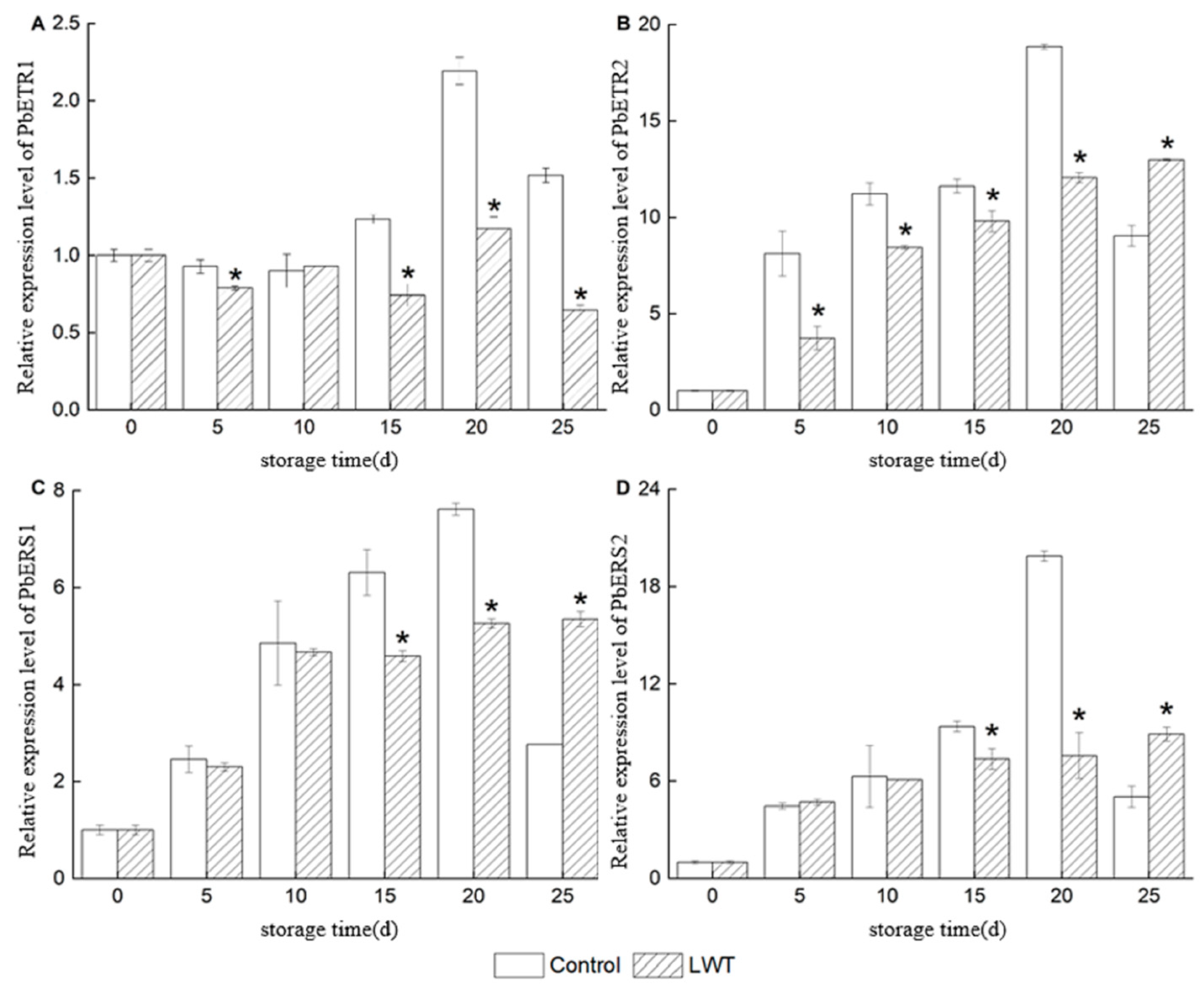
3.4. Effect of LED White Light on the Expression of Ethylene Signal Transduction Pathway-Related Genes in Pear Fruit

Figure 5 and Figure 6 show the expression levels of ethylene receptor genes (ETR1, ETR2, ERS1, and ERS2) and signal transduction factors (CTR1, EIN2, EIL1, and ERF1) during storage. Figure 5 shows that during storage, the expression levels of ethylene receptor genes in the control group pear fruit exhibited an initial increase followed by a decrease trend, reaching a peak on day 20, which was consistent with the change in ethylene release. Prior to the ethylene peak, the expression levels of ethylene receptor genes in LWT pear fruit were significantly lower compared to the control group (p < 0.05). Then, on day 25, the expression level of PbETR1 in the LWT group fruit was significantly lower than the control group (p < 0.05), while the expression levels of PbETR2, PbERS1, and PbERS2 in the LWT group fruit were significantly higher than the control group (p < 0.05).
Figure 6 shows that during storage, the ethylene signal transduction genes (PbCTR1, PbEIN2, PbEIL1, and PbERF1) in the pear fruit exhibited an initial increase followed by a decrease trend. Prior to the ethylene peak, the expression levels of PbCTR1 and PbERF1 in the LWT group fruit were significantly lower than the control group (p < 0.05), while there was no significant difference in the expression levels of PbEIN2 and PbEIL1 between the two groups (p > 0.05). After the ethylene peak, the expression level of PbERF1 in the LWT group fruit was significantly higher than the control group (p < 0.05), while the expression level of PbEIL1 in the LWT group fruit was significantly lower than the control group (p < 0.05).

3.5. Effect of LED White Light on the Color Change and Chlorophyll Content of Pear Fruit

Color change is an important indicator of quality decay of the fruit. As shown in Figure 7, the pear fruit in the control group exhibited significant yellowing of the skin after 15 days of storage, while the pear fruit in the LWT group displayed this phenomenon after 20 days of storage. This indicated that LED white light treatment delayed the occurrence of skin fading and yellowing in ‘Zaosu’ pear fruit.
The skin greasiness can make fruit appear brighter and show higher L* values. During storage, the L* values of pear fruit in both groups showed an increasing trend, but the LWT group was significantly lower than the control group (p < 0.05, Figure 7B), indicating that LED white light treatment delayed the greasiness process of the pear skin.
The h values of pear fruit in both groups showed a decreasing trend (Figure 7C), indicating a gradual color transformation from green to yellow. Furthermore, the h value of the LWT group fruit was significantly higher than the control group (p < 0.05), which further confirmed that LED white light treatment effectively delayed the yellowing of ‘Zaosu’ pear fruit during storage.
During storage, the chlorophyll a, chlorophyll b, and total chlorophyll content of the pear fruit in both groups showed a decreasing trend (Figure 7D–F). However, the chlorophyll content of the pear fruit in the LWT group was significantly higher than the control group (p < 0.05), consistent with the trend of color change during storage. This indicated that LWT maintained the vibrant green color of the pear skin by inhibiting the degradation of chlorophyll in the fruit during storage.

3.6. Effect of LED White Light on the Activities of Enzymes Related to Chlorophyll Synthesis and Metabolism in Pear Fruit

During storage, the pear fruit in both groups showed a trend of initial increase followed by a decrease in CAO enzyme activity. The CAO enzyme activity in the LWT group reached its maximum value on day 20 of storage, while in the control group it reached its maximum value on day 15. The CAO enzyme activity in the LWT group was significantly higher than the control group (Figure 8A, p < 0.05). Similarly, the CS enzyme activity in the ‘Zaosu’ pear fruit subjected to dark treatment also showed a trend of initial increase followed by a decrease and reached its maximum value on day 20 of storage. The CS enzyme activity in the pear fruit subjected to LED white light treatment showed an increasing trend and was significantly higher than the dark treatment group (Figure 8B, p < 0.05). This suggested that LED white light treatment enhanced the activity of chlorophyll synthesis-related enzymes.

3.7. Effect of LED White Light on the Activities and Gene Expression of Enzymes Related to Chlorophyll Metabolism in Pear Fruit

Chlorophyll degradation is the main reason for the yellowing of ‘Zaosu’ pears. As shown in Figure 9A, during the entire storage period, the Chlase activity in both groups showed a decreasing trend. The LWT group exhibited a particularly significant decrease on day 5, while a pronounced decrease in the control group occurred on day 20 of storage. The Chlase activity in the LWT group was significantly lower than the control group (p < 0.05). Additionally, the PAO activity in the LWT group of pear fruit was significantly lower than the control group (Figure 9B, p < 0.05). Furthermore, the expression levels of PbPAO, PbNYC, PbNOL, PbPPH, and PbRCCR genes in the LWT group of fruit were also significantly lower than the control group (Figure 10, p < 0.05). So, it can be observed that LED white light treatment to some extent inhibited the activity of chlorophyll degradation-related enzymes or the expression of enzyme genes.

4. Discussion

‘Zaosu’ pear fruit have a bright green skin and a crisp texture when harvested. However, this kind of pear fruit are prone to yellowing and have a short storage period [1]. In this study, LED white light treatment (LWT) inhibited weight loss in ‘Zaosu’ pear fruit during storage (Figure 2A). At the same time, LWT also substantially curbed the decline in ascorbic acid content in the pear fruit during the early storage period (Figure 2B,C), which is consistent with the findings of Joanna [30], who demonstrates that light exposure can prevent a decrease in ascorbic acid content in citrus fruit peel caused by bagging. Ascorbic acid (AsA) has been known to play a crucial role in scavenging free radicals, as well as in the protection and maintenance of the reduced state of compounds such as flavonoids and polyphenols [26].
The sugar-to-acid ratio is one of the important indicators that affect fruit texture and consumer preference [27]. LWT also effectively increased the sugar-to-acid ratio content of ‘Zaosu’ pear fruit (Figure 2D), thus maintaining their good taste. LED light treatment has been proved to be able to promote the accumulation of sucrose and fructose in fruit, affecting carbohydrate synthesis and transport, thereby influencing the soluble sugar content and the sugar-to-acid ratio [31].
‘Zaosu’ pear fruit treated with LED white light maintained a lower respiratory intensity and ethylene release (Figure 3A,B), contributing to the delayed ripening and aging processes of the fruit. This finding was consistent with the process of green-to-yellow color fading in the peel (Figure 3A,B). Similar results have been reported by Hasperué et al. [32], who show that the respiratory intensity and ethylene release of Brussels sprouts could also be reduced by LED light treatment, resulting in delayed aging and yellowing processes. In addition, the LWT suppressed the expression levels of ethylene synthesis and signal transduction-related genes and delayed the occurrence of the peak of ethylene production in pear fruit after harvest (Figure 4A–C and Figure 5A–D). This result confirmed the findings reported by Lv et al. [33] in apple fruit.
The occurrence of peel yellowing is closely related to the degradation metabolism of chlorophyll in the fruit [34]. As the pear fruit ripened, their peel color changed from green to yellow (Figure 7A–C). During storage, LWT effectively inhibited chlorophyll degradation and peel yellowing of ‘Zaosu’ pear fruit (Figure 7D–F). Mehmet et al. [21] revealed that LED white light treatment could prolong the storage time of lettuce and showed it had strong effect on maintaining green color and reducing chlorophyll degradation. Additionally, Vergara et al. [35] found that in olives, LED white light treatment not only effectively maintained the content of chlorophyll but also reduced the activity of enzymes related to chlorophyll degradation metabolism, which may be the main reason for its inhibition of yellowing. In this study, LED white light treatment also effectively increased the activity of chlorophyll synthesis-related enzymes in ‘Zaosu’ pear fruit (Figure 8), such as chlorophyll synthase (CS) and chlorophyll a oxygenase (CAO), and reduced the activity of chlorophyll degradation-related enzymes (Figure 9) such as chlorophyllase (Chlase) and pheophorbide a oxygenase (PAO). The expression pattern of chlorophyll degradation metabolism-related genes in ‘Zaosu’ pear fruit further confirmed the above findings (Figure 10).

5. Conclusions

In summary, LED white light treatment (LWT) reduced the respiration rate and ethylene production of ‘Zaosu’ pear fruit and inhibited the expression of genes related to ethylene synthesis and signal transduction, thereby delaying fruit ripening and senescence. Meanwhile, LWT delayed the yellowing of the pear fruit by increasing the chlorophyll content and the activities of its synthesis-related enzymes and inhibiting the activities of chlorophyll degradation-related enzymes and their gene expression. Additionally, LWT effectively curbed the decline in ascorbic acid content in ‘Zaosu’ pear fruit and improved the sugar–acid ratio. Thus, LED white light treatment can effectively retain the green color and delay the yellowing of ‘Zaosu’ pear fruit, prolonging their post-harvest storage and shelf life.

Author Contributions

H.M.: design of the work; analysis of data; validation; writing—review and editing; funding acquisition. X.Z. and J.Y.: acquisition of data; software; visualization; validation; data curation; writing—original draft preparation. J.C. and B.L.: design of the work; data interpretation; writing—review and editing; funding acquisition. All authors have read and agreed to the published version of the manuscript.

Funding

This research was funded by the National Natural Science Foundation of China, grant numbers 31901745 and 31902035; the Xinjiang Natural Science Foundation for Distinguished Young Scholars, grant number 2022D01E60; the Department of Science and Technology of Liaoning Province, grant number 2020-BS-238; and the Educational Department of Liaoning Province, grant numbers LJKZ1025 and LJKZ1018.

Data Availability Statement

Data are contained within the article.

Conflicts of Interest

The authors declare no conflicts of interest.

References

  1. Wei, D.M.; Xu, J.; Dong, F.S.; Liu, X.G.; Wu, X.H.; Zheng, Y.Q. Penicillium and patulin distribution in pears contaminated with Penicillium expansum. Determination of patulin in pears by UHPLC-MS/MS. J. Integr. Agric. 2017, 16, 1645–1651. [Google Scholar] [CrossRef]
  2. Saquet, A.A. Storage of pears. Sci. Hortic. 2019, 246, 1009–1016. [Google Scholar] [CrossRef]
  3. Zhang, H.; Wang, R.; Wang, T.; Fang, C.; Wang, J. Methyl salicylate delays peel yellowing of ‘Zaosu’ pear (Pyrus bretschneideri) during storage by regulating chlorophyll metabolism and maintaining chloroplast ultrastructure. J. Sci. Food Agric. 2019, 99, 4816–4824. [Google Scholar] [CrossRef] [PubMed]
  4. Armitage, A.D.; Cockerton, H.M.; Sreenivasaprasad, S.; Woodhall, J.; Lane, C.R.; Harrison, R.J.; Clarkson, J.P. Genomics evolutionary history and diagnostics of the Alternaria alternata species group including apple and Asian pear pathotypes. Front. Microbiol. 2020, 10, 3124. [Google Scholar] [CrossRef] [PubMed]
  5. Kaewsuksaeng, S.; Tatmala, N.; Srilaong, V.; Pongprasert, N. Postharvest heat treatment delays chlorophyll degradation and maintains quality in Thai lime (Citrus aurantifolia Swingle cv. Paan) fruit. Postharvest Biol. Technol. 2015, 100, 1–7. [Google Scholar] [CrossRef]
  6. Villarreal, N.M.; Rosli, H.G.; Martínez, G.A.; Civello, P.M. Polygalacturonase activity and expression of related genes during ripening of strawberry cultivars with contrasting fruit firmness. Postharvest Biol. Technol. 2008, 47, 141–150. [Google Scholar] [CrossRef]
  7. Zhan, L.; Li, J.; Huang, W.; Song, C.; Li, J.; Pang, L.; Li, Y. Light irradiation affects the total antioxidant capacity, total phenolic compounds, phenolic acids, and related enzyme activities of minimally processed spinach (Spinacia oleracea L.). J. Food Process. Preserv. 2020, 44, e14825. [Google Scholar] [CrossRef]
  8. Zhang, X.-J.; Zhang, M.; Chitrakar, B.; Devahastin, S.; Guo, Z. Novel combined use of red-white LED illumination and modified atmosphere packaging for maintaining storage quality of postharvest pakchoi. Food Bioprocess Technol. 2022, 15, 590–605. [Google Scholar] [CrossRef]
  9. Bae, J.M.; Lee, D.U.; Jeong, M.C.; Choi, J.H. Change of quality characteristics in fresh-cut ‘Romaine’ lettuce by heat treatment. Korean J. Food Preserv. 2016, 23, 27–33. [Google Scholar] [CrossRef]
  10. Youn, K.-S.; Park, E.-H.; Yoon, K.Y. Quality characteristics and antioxidant activity of bitter melon (Momordica charantia L.) dried by different methods. Korean J. Food Preserv. 2019, 26, 185–193. [Google Scholar] [CrossRef]
  11. Tkaczewska, J. Peptides and protein hydrolysates as food preservatives and bioactive components of edible films and coatings—A review. Trends Food Sci. Technol. 2020, 106, 298–311. [Google Scholar] [CrossRef]
  12. Lum, G.B.; DeEll, J.R.; Hoover, G.J.; Subedi, S.; Shelp, B.J.; Bozzo, G.G. 1-Methylcylopropene and controlled atmosphere modulate oxidative stress metabolism and reduce senescence-related disorders in stored pear fruit. Postharvest Biol. Technol. 2017, 129, 52–63. [Google Scholar] [CrossRef]
  13. García-Martín, J.F.; Olmo, M.; García, J.M. Effect of ozone treatment on postharvest disease and quality of different citrus varieties at laboratory and at industrial facility. Postharvest Biol. Technol. 2018, 137, 77–85. [Google Scholar] [CrossRef]
  14. Han, Q.; Gao, H.; Chen, H.; Fang, X.; Wu, W. Precooling and ozone treatments affects postharvest quality of black mulberry (Morus nigra) fruits. Food Chem. 2017, 221, 1947–1953. [Google Scholar] [CrossRef] [PubMed]
  15. Lin, Y.; Lin, Y.; Lin, H.; Lin, M.; Li, H.; Yuan, F.; Chen, Y.; Xiao, J. Effects of paper containing 1-MCP postharvest treatment on the disassembly of cell wall polysaccharides and softening in Younai plum fruit during storage. Food Chem. 2018, 264, 1–8. [Google Scholar] [CrossRef] [PubMed]
  16. Aparicio-García, P.F.; Ventura-Aguilar, R.I.; del Río-García, J.C.; Hernández-López, M.; Guillén-Sánchez, D.; Salazar-Piña, D.A.; Ramos-García, M.d.L.; Bautista-Baños, S. Edible chitosan/propolis coatings and their effect on ripening, development of Aspergillus flavus, and sensory quality in fig fruit, during controlled storage. Plants 2021, 10, 112. [Google Scholar] [CrossRef] [PubMed]
  17. Cackett, L.; Luginbuehl, L.H.; Schreier, T.B.; Lopez-Juez, E.; Hibberd, J.M. Chloroplast development in green plant tissues: The interplay between light, hormone, and transcriptional regulation. New Phytol. 2022, 233, 2000–2016. [Google Scholar] [CrossRef]
  18. Kim, B.-S.; Youm, S.; Kim, Y.-K. Sterilization of harmful microorganisms in hydroponic cultivation using an ultraviolet LED light source. Sens. Mater. 2020, 32, 3773–3785. [Google Scholar] [CrossRef]
  19. Shi, J.; Gao, L.; Zuo, J.; Wang, Q.; Wang, Q.; Fan, L. Exogenous sodium nitroprusside treatment of broccoli florets extends shelf life, enhances antioxidant enzyme activity, and inhibits chlorophyll-degradation. Postharvest Biol. Technol. 2016, 116, 98–104. [Google Scholar] [CrossRef]
  20. Xu, F.; Tang, Y.; Dong, S.; Shao, X.; Wang, H.; Zheng, Y.; Yang, Z. Reducing yellowing and enhancing antioxidant capacity of broccoli in storage by sucrose treatment. Postharvest Biol. Technol. 2016, 112, 39–45. [Google Scholar] [CrossRef]
  21. Kasim, M.U.; Kasim, R. While continuous white LED lighting increases chlorophyll content (SPAD), green LED light reduces the infection rate of lettuce during storage and shelf-life conditions. J. Food Process. Preserv. 2017, 41, e13266. [Google Scholar] [CrossRef]
  22. Skrovankova, S.; Mlcek, J.; Sochor, J.; Baron, M.; Kynicky, J.; Jurikova, T. Determination of ascorbic acid by electrochemical techniques and other methods. Int. J. Electrochem. Sci. 2015, 3, 2421–2431. [Google Scholar] [CrossRef]
  23. Sun, Q.; Liu, L.; Zhang, L.; Lv, H.; He, Q.; Guo, L.; Zhang, X.; He, H.; Ren, S.; Zhang, N.; et al. Melatonin promotes carotenoid biosynthesis in an ethylene-dependent manner in tomato fruits. Plant Sci. 2020, 298, 110580. [Google Scholar] [CrossRef] [PubMed]
  24. Esteban, R.; García-Plazaola, J.I.; Hernández, A.; Fernández-Marín, B. On the recalcitrant use of Arnon’s method for chlorophyll determination. New Phytol. 2018, 217, 474–476. [Google Scholar] [CrossRef] [PubMed]
  25. Chen, J.; Zhang, D.; Zhang, X.; Feng, X.; Mi, H.; Ge, Y.; Lv, J. Comprehensive responses of aroma production in ‘Benihoppe’ strawberry to low oxygen associated with the changes of key gene expressions and energy levels. J. Sci. Food Agric. 2023, 103, 1856–1863. [Google Scholar] [CrossRef] [PubMed]
  26. Charoenchongsuk, N.; Ikeda, K.; Itai, A.; Oikawa, A.; Murayama, H. Comparison of the expression of chlorophyll-degradation-related genes during ripening between stay-green and yellow-pear cultivars. Sci. Hortic. 2015, 181, 89–94. [Google Scholar] [CrossRef]
  27. Cheng, Y.; Dong, Y.; Yan, H.; Ge, W.; Shen, C.; Guan, J.; Liu, L.; Zhang, Y. Effects of 1-MCP on chlorophyll degradation pathway-associated genes expression and chloroplast ultrastructure during the peel yellowing of Chinese pear fruits in storage. Food Chem. 2012, 135, 415–422. [Google Scholar] [CrossRef] [PubMed]
  28. Zhang, H.; Zhao, L.; Fan, C.; Wang, P.; Cui, M.; Liu, L.; Yang, H.; Wang, J. Impact of methyl salicylate on storage quality, ethylene action, and protein profiling of ‘Zaosu’ pear (Pyrus bretschneideri). Sci. Hortic. 2020, 264, 109196. [Google Scholar] [CrossRef]
  29. Livak, K.J.; Schmittgen, T.D. Schmittgen. Analysis of relative gene expression data using real-time quantitative PCR and the 2−ΔΔCT method. Methods 2001, 25, 402–408. [Google Scholar] [CrossRef]
  30. Lado, J.; Alós, E.; Rodrigo, M.J.; Zacarías, L. Light avoidance reduces ascorbic acid accumulation in the peel of Citrus fruit. Plant Sci. 2015, 231, 138–147. [Google Scholar] [CrossRef]
  31. Chen, X.-L.; Wang, L.-C.; Li, T.; Yang, Q.-C.; Guo, W.-Z. Sugar accumulation and growth of lettuce exposed to different lighting modes of red and blue LED light. Sci. Rep. 2019, 9, 6926. [Google Scholar] [CrossRef] [PubMed]
  32. Hasperué, J.H.; Rodoni, L.M.; Guardianelli, L.M.; Chaves, A.R.; Martínez, G.A. Use of LED light for Brussels sprouts postharvest conservation. Sci. Hortic. 2016, 213, 281–286. [Google Scholar] [CrossRef]
  33. Lv, J.; Zhang, M.; Zhang, J.; Ge, Y.; Li, C.; Meng, K.; Li, J. Effects of methyl jasmonate on expression of genes involved in ethylene biosynthesis and signaling pathway during postharvest ripening of apple fruit. Sci. Hortic. 2018, 229, 157–166. [Google Scholar] [CrossRef]
  34. Chen, A.; Liu, L.; Liu, X.; Fu, Y.; Li, J.; Zhao, J.; Hou, X. Exploring the differential stages of the pigment metabolism by pre-harvest bagging and post-harvest ethylene de-greening of Eureka lemon peel. PeerJ 2021, 9, e11504. [Google Scholar] [CrossRef]
  35. Vergara-Domínguez, H.; Ríos, J.J.; Gandul-Rojas, B.; Roca, M. Chlorophyll catabolism in olive fruits (var. Arbequina and Hojiblanca) during maturation. Food Chem. 2016, 212, 604–611. [Google Scholar] [CrossRef]
Figure 1. Schematic diagram of experimental apparatus (A), spectra (B), and physical parameters (C) of LED white light.
Horticulturae 10 00032 g001
Figure 2. Effects of LED white light treatment (LWT) on weight loss rate (A), ascorbic acid content (B), firmness (C), and sugar–acid ratio (D) of pear fruit. The asterisk on the column represents the significant difference between groups (p < 0.05).
Horticulturae 10 00032 g002
Figure 3. Effects of LED white light treatment (LWT) on the respiration rate (A) and ethylene production (B) of pear fruit. The asterisk on the column represents the significant difference between groups (p < 0.05).
Horticulturae 10 00032 g003
Figure 4. Effects of LED white light on the expression of PbACS4 (A), PbACO1 (B) and PbACO4 (C) genes, which are related to the ethylene biosynthesis pathway of pear fruit. The asterisk on the column represents the significant difference between groups (p < 0.05).
Horticulturae 10 00032 g004
Figure 5. Effect of LED white light on the expression of PbETR1 (A), PbETR2 (B), PbERS1 (C) and PbERS2 (D) genes, which are ethylene receptors in pear fruit. The asterisk on the column represents significant differences between groups (p < 0.05).
Horticulturae 10 00032 g005
Figure 6. Effect of LED white light on the expression of PbCTR1 (A), PbEIN2 (B), PbEIL1 (C) and PbERF1 (D) genes, which are ethylene signal transduction factors in pear fruit. The asterisk in the figure represents significant differences between groups (p < 0.05).
Horticulturae 10 00032 g006
Figure 7. Effect of LED white light on color change ((A), pear fruit; (B), L* valuses; (C), h values) and chlorophyll content ((D), chlorophyll a content; (E), chlorophyll b content; (F), total chlorophyll content) of pear fruit. The asterisk in the figure represents significant differences between groups (p < 0.05).
Horticulturae 10 00032 g007
Figure 8. Effect of LED white light on the activities of CAO (A) and CS (B) enzymes related to chlorophyll synthesis in pear fruit. The asterisk in the figure represents significant differences between groups (p < 0.05).
Horticulturae 10 00032 g008
Figure 9. Effect of LED white light on the activities of Chase (A) and PAO (B) enzymes related to chlorophyll degradation in pear fruit. The asterisk in the figure represents significant differences between groups (p < 0.05).
Horticulturae 10 00032 g009
Figure 10. Effect of LED white light on the expression of PbNYC(A), PbNOL (B), PbSGR1 (C), PbPPH (D), PbPAO (E) and PbRCCR (F) genes related to chlorophyll degradation in pear fruit. The asterisk in the figure represents significant differences between groups (p < 0.05).
Horticulturae 10 00032 g010
Table 1. Primers for genes selected for fluorescence quantitative PCR.
Target Gene Gene IDPrimer Sequence
PbNYCJN167997 F: GGCATAGCATGTGATGTTTG
R: GCACCAGCATTGTTTATCC
PbNOLJN167996 F: CCGTGAGGCAATAAAAATGATG
R: CGCTTTGTTGCCCCGTAT
PbCLH1JN168001 F: CAACGTTCATAGACTTCCCCTACG
R: GGAGAGTACAAGGCAAGAGCTGC
PbSGR1JN168000 F: ATTCAACAAGGTGGAGTGCTGG
R: GCCGCTGTTGTTTTCCTGG
PbPPHJN168999 F: CTGCTGCATCATTGGCCTC
R: CTCGTAATATGGAGCTTCAGGC
PbPAOJN168998 F: GCGACATCGACGAGCAAGTT
R: TCGAAAGCGACCCACGATTC
PbRCCRJN168002 F: TTCATAGACTTCCCCTACGTGTCG
R: GGAGAGTACAAGGCAAGAGCTGC
EF1-αAH009876 F: CAAGTATGCCTGGGTGCTTG
R: TCAGCCTGTGAAGTTCCAGT
Disclaimer/Publisher’s Note: The statements, opinions and data contained in all publications are solely those of the individual author(s) and contributor(s) and not of MDPI and/or the editor(s). MDPI and/or the editor(s) disclaim responsibility for any injury to people or property resulting from any ideas, methods, instructions or products referred to in the content.

