Article

Effect of Lactobacillus curvatus HY7602-Fermented Antler on Sarcopenia in Mice

R&BD Center, hy Co., Ltd., 22, Giheungdanji-ro 24beon-gil, Giheung-gu, Yongin-si 17086, Gyeonggi-do, Republic of Korea
*
Authors to whom correspondence should be addressed.
Fermentation 2023, 9(5), 429; https://doi.org/10.3390/fermentation9050429
Submission received: 5 April 2023 / Revised: 27 April 2023 / Accepted: 27 April 2023 / Published: 28 April 2023
(This article belongs to the Special Issue High Quality Functional Food: Potential of Probiotics 2.0)

Abstract

:
Sarcopenia, commonly found in the elderly, causes a decrease in muscle mass and function. Lactobacillus curvatus HY7602-fermented antler (FA) is a functional food that increases muscle strength and ameliorates dexamethasone-induced muscle atrophy. In the present study, we evaluated the effects of FA on age-related muscle atrophy and its mechanism of action using 100-week-old BALB/c mice. Physiological changes during fermentation were investigated. The results showed that the oral administration of FA substantially recovered muscle loss up to 23.6% and improved physical performance, such as treadmill running distance and limb grip strength, in aged mice. A gene expression analysis of muscle fibers showed that the effect of FA on age-related muscle atrophy was significantly associated with the inhibition of protein degradation and apoptosis, muscle fiber regeneration, and increased mitochondrial biogenesis. In addition, the acetate and butyrate contents increased by more than 50% during the fermentation of antler. In conclusion, FA can be considered as a functional food ingredient capable of effectively controlling muscle atrophy caused by aging and can be a novel alternative treatment for sarcopenia.

1. Introduction

Sarcopenia is a condition characterized by the loss of muscle mass and function due to aging. Muscle mass decreases by 1% per year from middle age, and in severe cases, it can decrease by up to 50% until old age [1]. This is accompanied by quantitative changes in muscle size, grip strength, isokinetic strength, power, and fatigability, as well as qualitative changes in muscle architecture, fiber morphology and biochemistry, myonuclei, and satellite cells [2]. As these changes also affect independence and mobility, which have a critical impact on the quality of life of the elderly, various therapeutic approaches, such as exercise, dietary intake, and hormones, are being studied [3,4,5].
Age-related loss of muscle mass and function is associated with several factors. The two E3 ubiquitin ligases, Atrogin-1 and MuRF1, are important regulators of ubiquitin-mediated protein degradation in skeletal muscles, and the ubiquitin–proteasome system is one of the main pathways that regulates muscle protein degradation. These two factors are upregulated in age-related skeletal muscle atrophy [6]. Apoptosis is a highly regulated form of cell death, and the apoptosis of posterior tissues such as skeletal muscle can degrade tissue function by activating receptors through increased levels of physiological oxidants and tumor necrosis factor alpha, as well as increased mitochondrial oxidant production with normal aging [7,8]. In aged skeletal muscles, pro-apoptotic factors are upregulated, while anti-apoptotic factors are downregulated, both of which are known to exacerbate muscle atrophy [9]. Conversely, mitochondrial function and muscle fiber regeneration in the skeletal muscles are significantly reduced during age-related muscle atrophy [5]. Mitochondria play an important role in maintaining normal cellular function and homeostasis by contributing to metabolism and apoptosis as well as generating the energy required for cell function through oxidative phosphorylation. Mitochondrial enzyme activity is also known to increase during muscle regeneration. Mitochondrial proliferation is active in the muscles in response to exercise training and electrical stimulation and can be lost due to inactivity and aging [10,11].
Deer antler is the only mammalian organ that can regenerate annually and is widely known for its traditional medicinal value in East Asia [12]. Its efficacy in immunity [13] as well as its anti-fatigue [14] and anti-arthritis [15] properties have been studied. In Korea, deer antler is widely used as a functional food, as it is listed as a food ingredient in the Food Code of the Ministry of Food and Drug Safety, as well as in traditional medicine. The known effects of deer antlers are presumed to be caused by amino acids, peptides, and saccharides present in the antlers [16]. The functionality of deer antlers has been studied for a long time and the number of studies on the functionality of fermented deer antlers is steadily increasing.
Fermentation is an important food production process used to extend the shelf life of food and improve its organoleptic properties. Recently, it has been consumed in fermented foods and beverages as a rich source of probiotics, along with probiotic foods [17,18]. Fermentation is also known to increase the bioactive components of foods; therefore, studies are being actively conducted to elucidate the useful functionalities of fermenting various foods [19]. Lactic acid bacteria are the most commonly used starters in fermentation; they produce useful metabolites, such as short-chain fatty acids (SCFAs) and lactic acid, during fermentation [20,21,22]. SCFAs are used involved in intestinal microbial metabolism. The number of SCFA-producing bacteria decreases in patients with inflammatory bowel disease, irritable bowel syndrome, and obesity [23]. In addition, the amount and type of SCFAs produced are associated with human diseases and intestinal microorganisms [24,25].
In previous studies, we confirmed that supplementation with Lactobacillus curvatus (named Latilactobacillus curvatus in the new taxonomy) HY7602-fermented antler (FA) improved young and middle-aged mice’s endurance during exercise and muscle strength as well as dexamethasone-induced muscle atrophy [26,27,28]. These effects were associated with the sialic acid content of FA [28]. However, the effects of FA on muscle atrophy in an aged model and changes in metabolites due to fermentation have not yet been confirmed. Thus, the aim of this study was to investigate the effects of FA on sarcopenia and the changes in SCFA content produced during fermentation.

2. Materials and Methods

2.1. Preparation of Fermented Deer Antler

The middle and lower parts of antlers (Cervus elaphus Linné) were sliced, mixed with distilled water (1:30, w/w), and extracted at 95 ± 5 °C to 1.0 ± 0.5 °Brix. The samples met herbal medicine standards (Korean Pharmacopoeia, Ministry of Food and Drug Safety). L. curvatus strain HY7602 freeze-dried powder (hy Pyeongtaek Factory Co., Ltd., Pyeongtaek-si, Republic of Korea) was added to the antler extract (1% w/w) and fermented at 37 ± 2 °C for 24 ± 1 h. Meanwhile, L. curvatus strain HY7602 was inactivated via pasteurization after fermentation, concentrated, mixed with maltodextrin powder (Samyang Corp., Seoul, Republic of Korea), and lyophilized. The strains used in this study were selected through previous studies [26].

2.2. Animals, Diet, and Experimental Design

Seven-week-old (n = 5) and 100-week-old (n = 15) male BALB/c mice were purchased from DooYeol Biotech (Seoul, Republic of Korea) and acclimatized for 1 week with 18% protein rodent diet (crude protein 18.6%, fat 6.2%, carbohydrates 44.2%, crude fiber 3.5%, neutral detergent fiber 14.7%, and ash 5.3%; Envigo, Indianapolis, IN, USA). The mice were allowed free access to autoclaved distilled water and housed in a controlled environment (humidity 40–60%, temperature 20–22 °C) with a 12 h light/dark cycle. After the acclimation period, mice were divided into four groups (n = 5): young mice (Young), old mice (Old), old mice administered non-fermented antler (Old + NFA), and old mice administered L. curvatus HY7602-fermented antler (Old + FA). Non-fermented antler (NFA) and FA were orally administered once a day for 4 weeks (120 mg/kg body weight), and the Young and Old groups were orally administered saline during the same period. The calf muscles and whole blood were collected at the end of the study period. The calf muscle was separated into the gastrocnemius (GA) and soleus (SOL) and then weighed. The SOL was then stored in a freezer at −80 °C for further analysis. The animal study was conducted according to the guidelines of hy Co., Ltd. (Yongin-si, Republic of Korea) and approved by the Institutional Animal Care and Use Committee of hy Co., Ltd. (IACUC approval number, AEC-2023-0001-Y).

2.3. Treadmill Exercise Performance and Limb Grip Strength Tests

Mice were subjected to treadmill exercise weekly using a variable-speed belt treadmill (JD-A-09, Jeong Do B&P, Seoul, Republic of Korea). During the acclimatization period, each mouse was trained to run on a treadmill. Exercise performance was started at 5 m/min without an incline, and the speed was increased by 5 m/min to a maximum of 30 m/min at 5 min intervals. Each mouse exercised for up to 45 min or until exhaustion.
Grip strength of the mouse limbs was measured weekly using a grip strength testing machine (BIO-GS3, Bioseb, Vitrolles CEDEX, France). Each mouse was placed on the grid, with its body kept at its height; all four paws were attached and then pulled back gently by holding the tail. Data are reported as the mean of three trials.

2.4. Histological Analysis

A histological analysis of muscle cross-sectional area (CSA) was performed by fixing GA in 10% formalin, embedding it in paraffin, and staining it with hematoxylin and eosin (H&E). The muscle fiber size of each mouse was measured, and three representative images were captured at 10× magnification. Muscle fiber area and diameter were determined by calculating the mean of 100 fibers per group using a Motic digital microscope image analysis system (Motic Optical Instruments Co., Ltd., Xiamen, China).

2.5. Serum Biochemistry

Blood samples were collected via cardiac puncture, and sera were separated via centrifugation at 2000× g for 15 min at 4 °C. The sera were then stored at −80 °C until further use. Serum levels of alanine aminotransferase (ALT), aspartate aminotransferase (AST), alkaline phosphatase (ALP), creatine kinase (CK), and creatinine (Crea) were measured using an automated analyzer (Hitachi 7020, Hitachi, Japan).

2.6. Real-Time Polymerase Chain Reaction (RT-PCR)

Total RNA from SOL (20 mg) was isolated using the easy-spin Total RNA Extraction Kit (iNtRON Biotechnology, Gyeonggi, Republic of Korea), according to the manufacturer’s instructions. The cDNA template was synthesized using 2 μg of total RNA and the Omniscript RT Kit (QIAGEN, Hilden, Germany). The resulting cDNA template was used for real-time polymerase chain reaction (RT-PCR).
Gene expression analysis was performed using the QuantStudio 6 RT-PCR program (Thermo Fisher Scientific, Waltham, MA, USA), as previously described [28]. Quantifications of muscle atrophy F-box (Atrogin-1, Mm00499523_m1), muscle RING-finger protein-1 (MuRF1, Mm01185221_m1), B-cell lymphoma 2 (Bcl-2, Mm00477631_m1), Bcl-2associated X protein (Bax, Mm_00432051_m1), myoblast determination protein 1 (MyoD, Mm_00440387_m1), myogenic factor 5 (Myf5, Mm00435125_m1), myogenin (Myog, Mm00446194_m1), type 2X myosin heavy chain (MYH1, Mm01332489_m1), protein kinase AMP-activated alpha 1 catalytic subunit (Prkaa1(AMPK), Mm01296700_m1), peroxisome proliferator-activated receptor gamma coactivator 1 alpha (Ppargc1a(PGC1-α), Mm01208835_m1), uncoupling protein 3 (Ucp3, Mm01163394_m1), transcription factor A mitochondrial (TFAM, Mm00447485_m1), and glyceraldehyde-3-phosphate dehydrogenase (GAPDH, Mm_99999915_g1) were performed using gene specific primers purchased from Applied Biosystems (Middlesex Country, MA, USA). Expression data were determined after normalization to the expression of GAPDH using the comparative CT method.

2.7. Measurement of SCFA Content

SCFA content was determined using high-performance liquid chromatography (HPLC, Agilent Technologies, Santa Clara, CA, USA). For the pretreatment, each sample (NFA, FA) was boiled for 15 min and then centrifuged with 13,000 rpm at 4 °C for 15 min. The supernatant was filtered in a 0.22 μm filter and used for the analysis. Lactic acid, acetic acid, propionic acid (Sigma-Aldrich, St. Louis, MO, USA), and butyric acid (Kanto Chemical Co., Inc., Tokyo, Japan) were used as the standard. The following operating conditions were used: column, Aminex HPX-87H (Bio-Rad, 1300 × 7.8 mm, 9 μm); flow rate, 0.6 mL/min; column temperature, 50 °C; injection volume, 20 μL; detector, UV 210 nm (Agilent 1260 G4212B); and mobile phase, 4 mM sulfuric acid.

2.8. Statistical Analysis

Statistical analysis was performed using SPSS version 26.0 (IBM, Somers, NY, USA). All data are expressed as mean ± SD. Analysis was performed using an unpaired Student’s t-test. Significant differences between the Young and Old groups are presented by p-values < 0.05 (*), <0.01 (**), and <0.005 (***), and significant differences between the Old and experimental groups are indicated by p-values < 0.05 (#), <0.01 (##), and <0.005 (###). Significant differences in muscle fiber diameter and area between the Old + NFA and Old + FA groups are indicated by p-values < 0.005 ().

3. Results

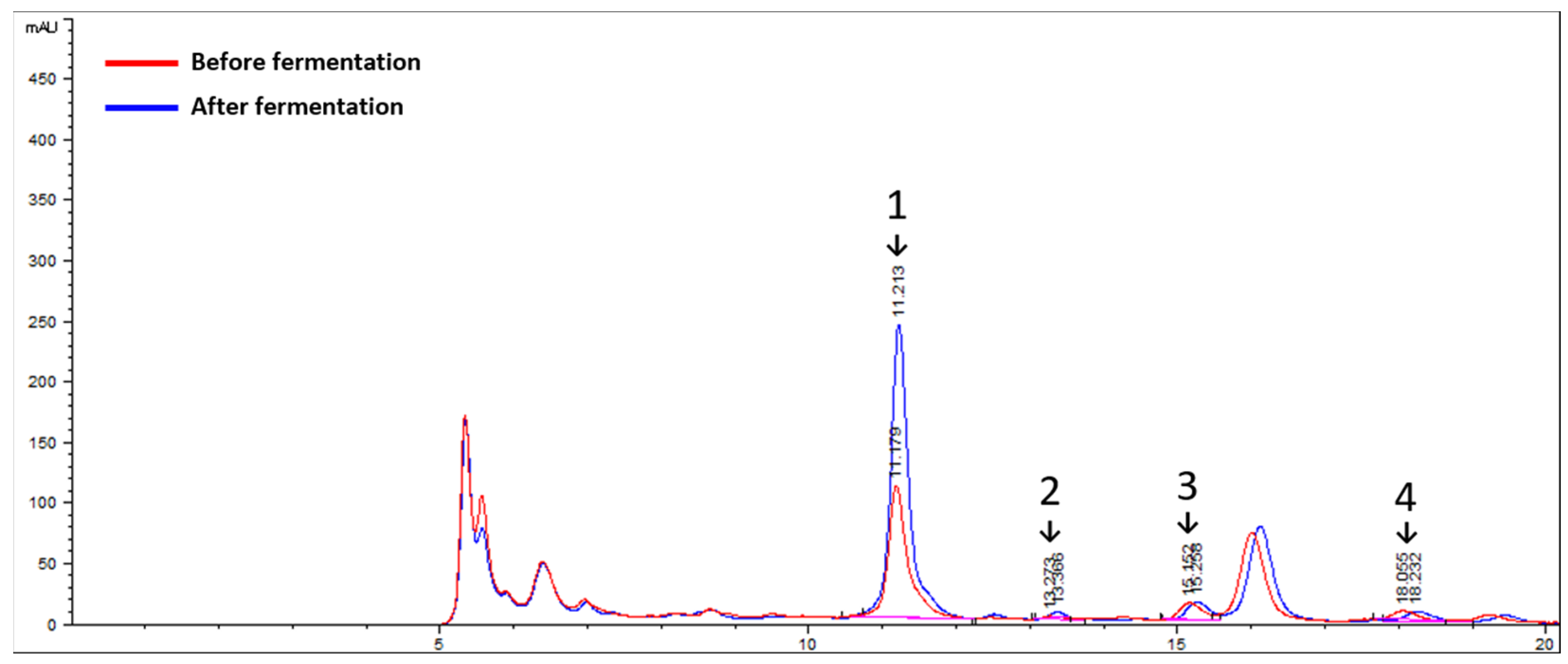
3.1. Characterization of L. curvatus HY7602 Fermentation in Antlers

3.1.1. Changes in pH Values

pH is an important physicochemical property of lactic acid fermentation, and changes in pH are known to be in good agreement with changes in lactic acid bacteria population and organic acid content [29]. During the L. curvatus HY7602 fermentation in antlers, the pH decreased over time. The pH at 0 to 2 h, the early stage of fermentation, rapidly decreased by 20% (pH 6.20 to pH 4.96), and a pH of 4 was maintained for 24 h (Table 1).

3.1.2. Changes in SCFA Content

SCFAs, such as acetate, propionate, and butyrate, are metabolites produced by lactic acid bacteria during fermentation and have various beneficial effects in humans. The SCFA profiles of the antlers before and after fermentation were quantified using HPLC (Figure 1). After fermentation, the acetate and butyrate levels increased by 71.42% and 53.44%, respectively. On the other hand, the propionate levels decreased by 11.13% (Table 2). Lactate, a representative metabolite of lactic acid bacterial fermentation, increased by 95.83% after fermentation from 121.17 ± 25.51 to 237.29 ± 22.23.

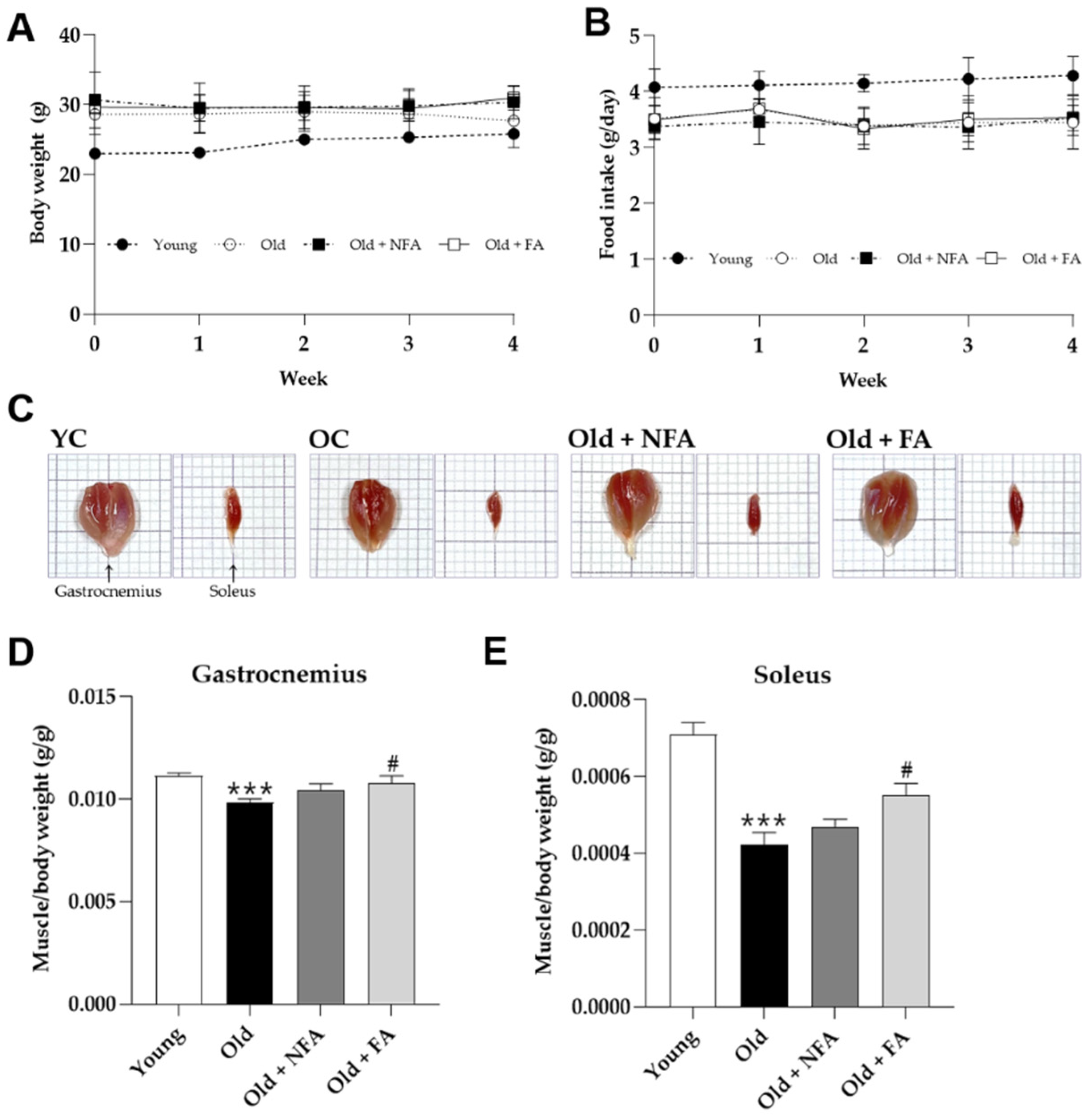
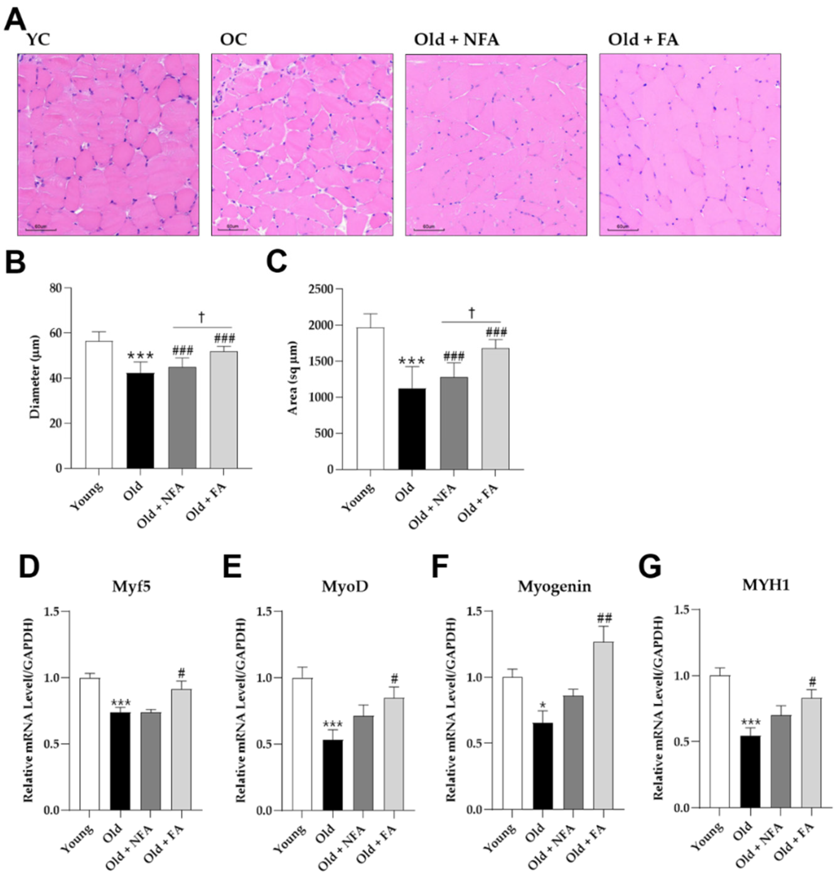
3.2. FA Recovers Muscle Loss in Aged Mice

Age-related muscle loss, known as sarcopenia, can occur during middle age and result in >50% muscle loss by old age [1]. The effects of FA on age-induced muscle loss were investigated using 100-week-old mice. The body weight of the Old group decreased during the first 4 weeks, whereas those of the Old + NFA and Old + FA groups increased from week 3 (Figure 2A). Food intake did not differ significantly between the Old mice groups during the experimental period (Figure 2B). To determine the change in calf muscle mass (GA and SOL) following FA treatment, the muscle tissue was extracted and weighed after the mice were sacrificed (Figure 2C). The Old group had a lower GA muscle mass (0.0098 ± 0.0004 g/kg) compared with the Young group (0.0111 ± 0.0003 g/kg) (p < 0.005). In contrast, the Old + NFA and Old + FA groups had GA muscle masses of 0.0104 ± 0.0007 and 0.0108 ± 0.0008 g/kg, respectively, and the Old + FA showed a significant increase compared to Old group (p < 0.05) (Figure 2D). Likewise, Sol muscle mass was significantly lower in the Old group (0.00042 ± 0.00007 g/kg) compared to that of the Young group (0.00071 ± 0.00007 g/kg) (p < 0.005). The Old + NFA and Old + FA groups had Sol muscle masses of 0.00047 ± 0.00004 and 0.00055 ± 0.00007 g/kg, respectively, and that of the Old + FA group was significantly increased compared to that of the Old group (p < 0.05) (Figure 2E).

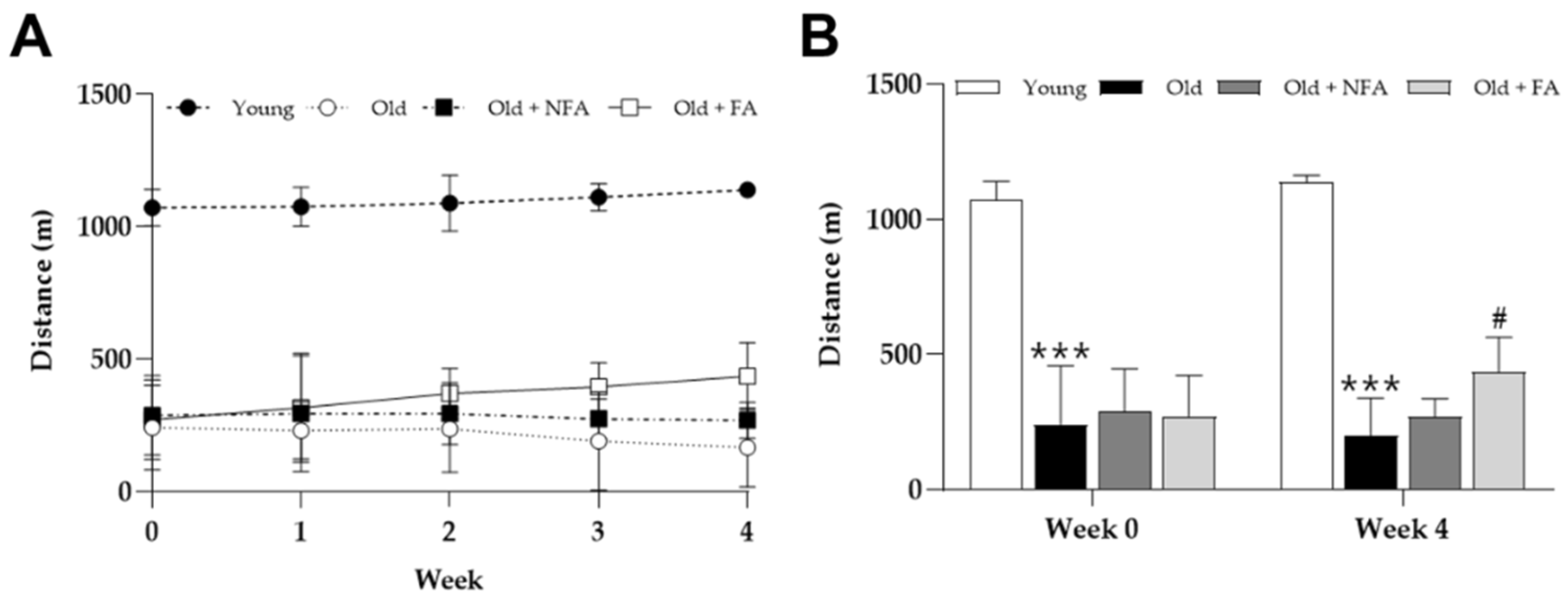
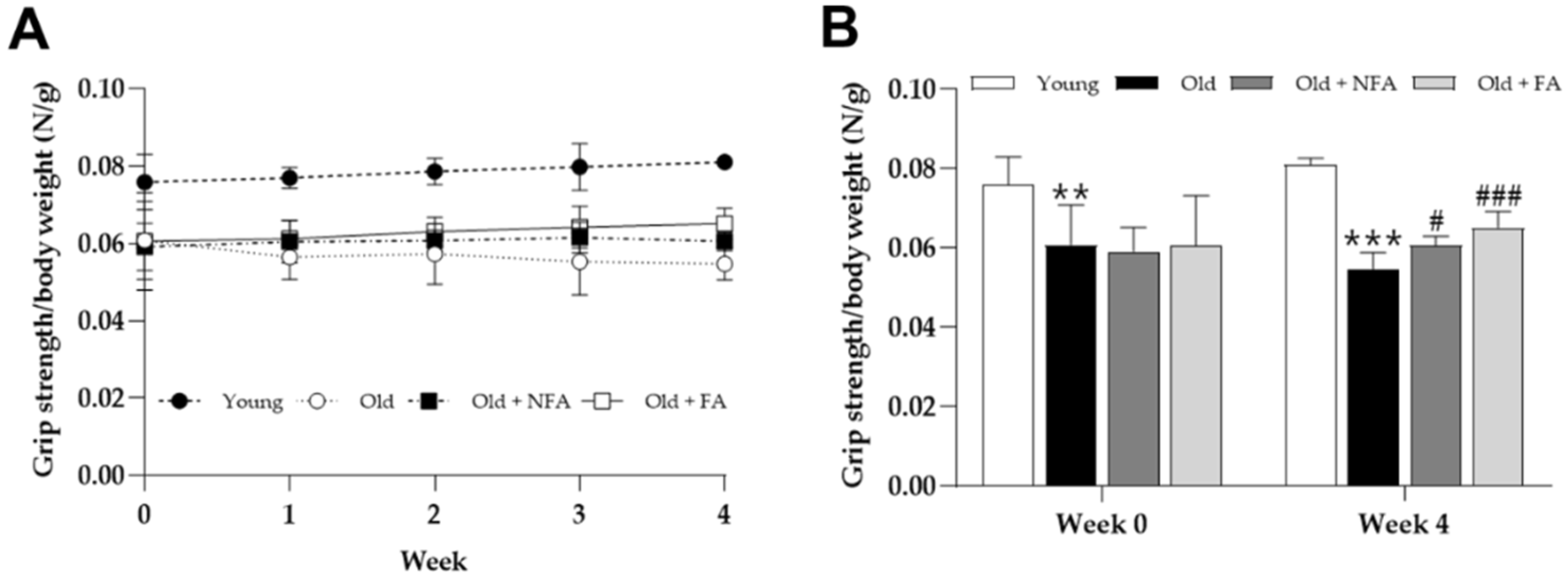
3.3. FA Ameliorates Limb Grip Strength and Exercise Performance Reduction in Aged Mice

Sarcopenia results in the loss of muscle weight and function in the elderly. Loss of muscle function can lead to a decrease in muscle strength and exercise performance, resulting in various disabilities [30]. Weekly grip strength and treadmill running tests were performed to evaluate the effects of FA on grip strength and exercise capacity in aged mice. During the 4-week experimental period, the total running distance of the Old group decreased, that of the Old + NFA group also slightly decreased, and those of the Young and Old + FA groups increased (Figure 3A). At week 0, the total running distance of the Old group was significantly shorter compared to that of the Young group (242.02 ± 159.20 m vs. 1071.40 ± 69.01 m; p < 0.005). Meanwhile, the total running distance of the Old + NFA and Old + FA groups were 287.82 ± 159.20 m and 272.12 ± 150.52 m, respectively, and no significant difference was observed with that of the Old group. After 4 weeks of treatment, the Old and Young groups showed a significant difference in the total running distance (166.74 ± 148.98 m vs. 1198.90 ± 24.82 m; p < 0.005). That of the Old + NFA group (269.13 ± 67.34 m) was slightly longer than that of the Old group, but no significant difference was observed. The total running distance of the Old + FA group showed a significant increase (436.18 ± 127.88 m) compared to that of the Old group (p < 0.05) (Figure 3B).
Meanwhile, the limb grip strength of the Old group decreased during the 4-week experimental period, whereas those of the other groups showed a slight increase (Figure 4A). At week 0, the limb grip strength of the Old group was significantly weaker compared with that in the Young group (0.0608 ± 0.0101 N/g vs. 0.0759 ± 0.0071 N/g; p < 0.01). Those of the Old + NFA and Old + FA groups were 0.0591 ± 0.00061 N/g and 0.0606 ± 0.0126 N/g, respectively, and no significant difference was observed compared with that of the Old group. After 4 weeks of treatment, the limb grip strength of the Old group decreased (0.0547 ± 0.0042 N/g) compared with that in the Young group (0.0810 ± 0.0016 N/g) (p < 0.005). That of the Old + NFA group (0.0605 ± 0.0024 N/g) was significantly increased than that of the Old group (p < 0.05), and that of the Old + FA group showed a significant increase (0.0651 ± 0.0040 N/g; p < 0.001) (Figure 4B).

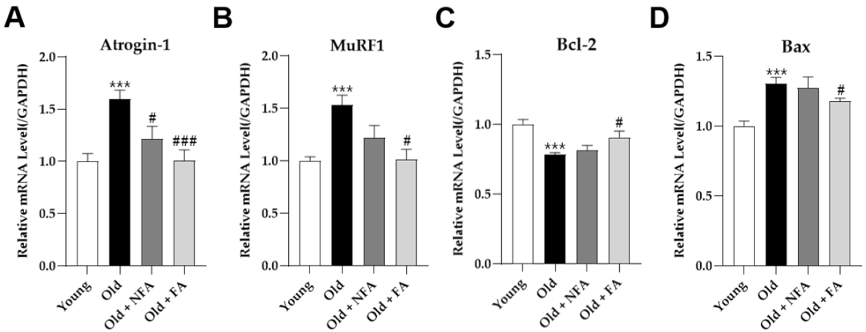
3.4. FA Inhibits Muscle Protein Degradation and Apoptosis and Promotes Muscle Fiber Regeneration

Atrogin1 and MuRF1 are induced in response to myostatin/TGFβ signaling and promote muscle protein degradation. Both genes are key regulators of muscle atrophy and are upregulated in skeletal muscle tissues during sarcopenia [6]. The mRNA levels of Atrogin-1 and MuRF1 were higher in the Old group than those in the Young group (p < 0.005). FA downregulated both the high levels of Atrogin-1 and MuRF1 (p < 0.005, p < 0.05), while NFA only slightly decreased Atrogin-1 (p < 0.05) (Figure 5A,B).
In sarcopenia, apoptosis promotes muscle cell death, leading to muscle atrophy. In atrophied skeletal muscle tissues, increased levels of pro-apoptotic Bax and decreased anti-apoptotic Bcl-2 levels have been observed [31]. As shown in Figure 5C, Bcl-2 was significantly decreased in the Old group compared to that in the Young group (p < 0.005) and increased in the Old + FA group (p < 0.05). In contrast, Bax expression was higher in the Old group than in the Young group (p < 0.005) and significantly decreased in the Old + FA group (p < 0.05). The Old + NFA group did not show any significant differences in either gene.
A CSA analysis is commonly used to evaluate muscle quality and strength; with aging, the thickness of muscle fibers decreases, which can lead to a decrease in muscle strength [32]. Therefore, the diameter and area of muscle fibers were evaluated using a histological analysis of the GA muscle. The CSA of the GA was measured using a digital microscope (Figure 6A). The diameters of muscle fibers were the smallest in the Old group (42.36 ± 4.84 µm), which was significantly different compared to that in the Young group (56.43 ± 4.19 µm) (p < 0.005). On the other hand, that of the Old + NFA group was significantly different from that of the Old + FA group (44.88 ± 4.08 µm vs. 51.93 ± 2.19 µm) (Figure 6B), and both values were larger compared to that of the Old group (p < 0.005). The area of the Old group was 1123.60 ± 296.72 sq µm, which was lower than that of the Young group (1966.79 ± 191.45 sq µm) (p < 0.005). Meanwhile, those of the Old + NFA and Old + FA groups were 1277.26 ± 197.81 sq µm and 1680.48 ± 118.34 sq µm, respectively, and showed a significant increase compared to that of the Old group (p < 0.005). Furthermore, a significant difference was observed between the Old + FA and Old + NFA groups (p < 0.005) (Figure 6C).
Furthermore, we confirmed the changes in gene expression related to muscle regeneration. Skeletal muscle fiber regeneration is a complex process of recovery from injury that is blunted by aging [33,34]. The expression of Myf5 and MyoD, which are related to early differentiation, was the lowest in the Old group (p < 0.005). The Old + FA group showed an increase compared to the Old group (p < 0.05) (Figure 6D,E). Myogenin, associated with late differentiation, also decreased significantly in the Old group compared to the Young group (p < 0.05), and increased significantly in the Old + FA group (p < 0.01) (Figure 6F). Furthermore, MYH1 expression decreased significantly in the Old group (p < 0.005) and increased in the Old + FA group (p < 0.05) (Figure 6G). The Old + NFA group showed a slight increase in the expression of all genes except Myf5, but no significant difference was observed.

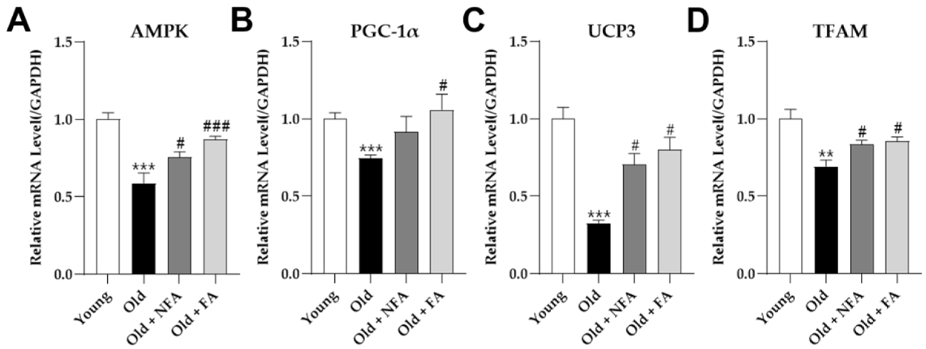
3.5. FA Promotes Mitochondrial Biogenesis in Aged Mice

The responsiveness of AMPK signaling, maintenance of cellular energy homeostasis, and mitochondrial function decrease with age [35]. As shown in Figure 7A, the expression level of AMPK was lower in the Old group than that in the Young group (p < 0.005). In addition, the levels of PGC-1α, UCP3, and TFAM, which are related to mitochondrial biogenesis, were also reduced in the Old group (Figure 7B–D). The Old + FA group showed increased levels of AMPK (p < 0.005), PGC-1α, UCP3, and TFAM compared to that in the Old group (p < 0.05). The expression of AMPK, UCP3, and TFAM in the Old + NFA group was significantly different from that in the Old group (p < 0.05).

3.6. Effects of the FA on Serum Biochemistry

The serum levels of ALT, AST, ALP, CK, and Crea were analyzed to determine the in vivo toxicity of NFA and FA. The Old group showed a significant increase in serum ALT and AST levels compared to those of the Young group, while the ALP levels were significantly decreased (p < 0.05). The CK and Crea levels were not significantly different between the Young and Old groups. The Old + FA group showed significantly decreased levels of ALT, CK, and Crea (p < 0.05), whereas the Old + NFA group showed significantly decreased levels of Crea (p < 0.05) (Table 3).

4. Discussion

Deer antler is known to be an effective treatment in recovering from fatigue and improving liver function, immunity, and arthritis. It is widely consumed as a traditional herbal medicine in East Asia and is also used in various health functional foods [13,14,15].
Our previous study has shown that L. curvatus HY7602-fermented antler can increase endurance during exercise in young mice and improves muscle strength in middle-aged mice and dexamethasone-induced muscle atrophy models [26,27,28]. However, its effects on age-related muscle atrophy, sarcopenia, and changes in metabolites after fermentation have not been confirmed. Therefore, we aimed to establish the effect of FA on sarcopenia and SCFA content.
Age-related loss of muscle mass and function can be easily observed in skeletal muscles. Recently, in addition to measuring muscle mass, measurements of exercise performance, such as grip strength and walking speed, have been included in various studies to improve the diagnostic accuracy of sarcopenia [1]. In the present study, the Old group showed a trend of weight loss over time, and the muscle weight after 4 weeks was much lower than that of the Young group. Furthermore, the exercise performance of the older group decreased, with the total running distance and limb grip strength decreasing over time. This was consistent with previous studies on the symptoms of sarcopenia due to aging [2,30]. Interestingly, FA improved the changes in body weight, muscle mass, and exercise performance in old mice. Our data indicate that FA reduces age-related muscle loss and affects exercise performance, for example, improving treadmill running distance and muscle strength.
In the muscle tissue, the expression of Atrogin-1 and MuRF1, which are known to be upregulated in sarcopenia, was significantly increased in the Old group compared to that in the Young group. Both genes are muscle-proteolysis-related factors that are mainly expressed in skeletal muscle tissues and are significantly increased by stressors that induce muscle atrophy. Inhibiting their expression is known to conserve muscles [36,37]. FA significantly suppressed the expression of Atrogin-1 and MuRF1, which was increased in old mice. These results indicated that FA contributes to muscle conservation by preventing the degradation of muscle protein owing to age-related muscle atrophy.
Apoptosis, another mechanism associated with muscular atrophy, is a form of programmed cell death that contributes to the normal development of organisms, cell replacement, and the remodeling of diseased tissues. Inappropriate apoptosis can cause physical defects, such as the exacerbation of muscle atrophy [38]. In the Old group, the expression of Bcl-2 was significantly reduced compared to that in the Young group, whereas Bax was significantly increased. These results may be explained by previously published findings in which aging is one of the causes of disproportionate apoptosis, which increases pro-apoptotic Bax and decreases anti-apoptotic Bcl-2 [39]. FA significantly inhibited gene expression in old mice. From these results, we assume that the promotion of apoptosis in the muscles contributed to the decrease in muscle mass in the Old group. In addition, FA can prevent the loss of muscle mass by significantly inhibiting the disproportionate apoptosis promoted by aging.
Muscle fibers are the main components of muscles, and their number and size determine muscle growth and ultimate mass. Myofibers are maintained and regenerated through a highly regulated process called muscle regeneration and gradually decrease with age, resulting in muscle fiber atrophy [40,41]. Consistent with previous studies, the Old group had a significantly lower diameter and area of muscle fibers than the Young group, and also the expression of myoblast differentiation-related genes (Myf5, MyoD, and Myogenin) and type 2X-MYH was significantly decreased. In contrast, FA restored muscle fiber atrophy in old mice and upregulated the expression of related genes. Myf5, MyoD, and Myogenin are myogenic regulatory factors that control myofibril differentiation. Myosin heavy chain (MYH), a component protein of myosin that is most abundant in skeletal muscle and is involved in contraction, is mainly expressed in the early stages of muscle differentiation and continues until the late stages [42,43]. Our findings show that the change in muscle fiber size in the Old group may be due to a decrease in the related genes, and that FA reversed myofibril atrophy by promoting muscle regeneration in old mice.
Mitochondria are the major energy sources for cellular activity, and their mass and functional homeostasis are maintained through mitochondrial biogenesis [44]. In the present study, the expression of mitochondrial-biogenesis-related factors was significantly lower in the Old group than that in the Young group. These results are consistent with previous findings showing that aging reduces mitochondrial biosynthesis [45]. Conversely, FA improved the expression of decreased AMPK, PGC-1α, UCP3, and TFAM in the Old group. In NFA, the expression of the remaining factors except PGC-1α was improved. Earlier, we found that exercise performance (treadmill running distance and grip strength) in the Old group was reduced and improved by FA. Based on these data, we confirmed that the efficacy of FA observed in previous exercise performance studies may be associated with improved mitochondrial biogenesis. In addition, the fact that NFA showed some efficacy in improving exercise performance and limb grip strength appears to be related to this result.
FA administration did not adversely affect blood concentrations of ALT and AST, which are indicators of hepatotoxicity, or ALP, CK, and Crea, which are indicators of renal function, but rather significantly reduced the blood ALT concentration, which increases due to aging [46]. However, it is not possible to infer all the effects of FA on liver function improvement, such as ALT reduction, from this study alone, and additional independent studies are needed to confirm this.
Our results showed the superior efficacy of FA compared to NFA, and a further analysis was conducted under the assumption that changes in metabolites due to fermentation result in this difference. The pH level can indirectly confirm changes in bacterial populations and organic acid production during fermentation, and the pH level of FA decreases over time [29]. The final pH level after fermentation was 4.09 ± 0.01, which is a common value in lactic acid bacteria fermentation. Subsequently, the SCFA content before and after fermentation was quantified. After fermentation, acetate and butyrate increased by more than 50%, and propionate decreased by approximately 11.13%. Acetate, propionate, and butyrate are the major metabolites produced by bacterial fermentation and are known to have various functions in the human body, along with the intestinal microbiota [47]. Previous studies have shown that SCFAs affect carbohydrate, lipid, and protein metabolism within skeletal muscle tissues [48]. In particular, acetate contributes to glycogen synthesis in muscle, and butyrate regulates lipid absorption, storage, and oxidation in muscle. In addition, the key mechanism of these SCFAs is likely to be the activation of AMPK and PGC-1α [49]. Therefore, the expression of AMPK and PGC-α in the old + FA group may be due to increased acetated and butyrate after fermentation. However, the effect of decreased propionate levels on muscles is unknown and further research is needed.
Overall, FA led to the quantitative and qualitative recovery of muscles by inhibiting muscle protein degradation and apoptosis and increasing muscle regeneration and mitochondrial biogenesis in age-related muscle atrophy. This effect was significantly increased after fermentation, which may be due to the increased acetate and butyrate contents. However, the effect of decreased propionate levels in sarcopenia requires further investigation.

5. Conclusions

This study showed that FA is a potential functional food for improving age-related muscle atrophy and sarcopenia. FA attenuated the loss of muscle mass and function by inhibiting muscle protein degradation and apoptosis and promoting muscle fiber regeneration and mitochondrial biogenesis. Increased acetate and butyrate levels after fermentation are expected to contribute to this effect, but their actual efficacy is not yet known. Therefore, further studies on the effects of short-chain fatty acids on sarcopenia are needed.

Author Contributions

Conceptualization, H.J. and J.-J.S.; Methodology, H.J.; Software, H.J.; Validation, H.J.; Formal Analysis, H.J.; Investigation, H.J. and K.L.; Data Curation, H.J.; Writing—Original Draft Preparation, H.J.; Writing—Review and Editing, J.-Y.K. and J.-L.L.; Visualization, H.J.; Supervision, J.-Y.K. and J.-L.L.; Project Administration, J.-Y.K. and J.-L.L. All authors have read and agreed to the published version of the manuscript.

Funding

This study received no external funding.

Institutional Review Board Statement

This animal study was approved by the Institutional Animal Care and Use Committee (IACUC) of hy Co., Ltd. (approval number: AEC-2023-0001-Y).

Informed Consent Statement

Not applicable.

Data Availability Statement

The data presented in this study are available.

Conflicts of Interest

The authors declare that the research was conducted in the absence of any commercial or financial relationships that could be construed as a potential conflict of interest.

References

  1. Wilkinson, D.J.; Piasecki, M.; Atherton, P.J. The age-related loss of skeletal muscle mass and function: Measurement and physiology of muscle fibre atrophy and muscle fibre loss in humans. Ageing Res. Rev. 2018, 47, 123–132. [Google Scholar] [CrossRef]
  2. Mitchell, W.K.; Williams, J.; Atherton, P.; Larvin, M.; Lund, J.; Narici, M. Sarcopenia, dynapenia, and the impact of advancing age on human skeletal muscle size and strength; a quantitative review. Front. Physiol. 2012, 3, 260. [Google Scholar] [CrossRef] [PubMed]
  3. Phu, S.; Boersma, D.; Duque, G. Exercise and sarcopenia. J. Clin. Densitom. 2015, 18, 488–492. [Google Scholar] [CrossRef] [PubMed]
  4. Liguori, I.; Russo, G.; Aran, L.; Bulli, G.; Curcio, F.; Della-Morte, D.; Gargiulo, G.; Testa, G.; Cacciatore, F.; Bonaduce, D.; et al. Sarcopenia: Assessment of disease burden and strategies to improve outcomes. Clin. Interv. Aging 2018, 13, 913–927. [Google Scholar] [CrossRef]
  5. Larsson, L.; Degens, H.; Li, M.; Salviati, L.; Lee, Y.I.; Thompson, W.; Kirkland, J.L.; Sandri, M. Sarcopenia: Aging-related loss of muscle mass and function. Physiol. Rev. 2019, 99, 427–511. [Google Scholar] [CrossRef] [PubMed]
  6. Clavel, S.; Coldefy, A.S.; Kurkdjian, E.; Salles, J.; Margaritis, I.; Derijard, B. Atrophy-related ubiquitin ligases, atrogin-1 and MuRF1 are up-regulated in aged rat tibialis anterior muscle. Mech. Ageing Dev. 2006, 127, 794–801. [Google Scholar] [CrossRef]
  7. Marzetti, E.; Leeuwenburgh, C. Skeletal muscle apoptosis, sarcopenia and frailty at old age. Exp. Gerontol. 2006, 41, 1234–1238. [Google Scholar] [CrossRef]
  8. Leeuwenburgh, C. Role of apoptosis in sarcopenia. J. Gerontol. Ser. A 2003, 58, M999–M1001. [Google Scholar] [CrossRef]
  9. Alway, S.E.; Siu, P.M. Nuclear apoptosis contributes to sarcopenia. Exerc. Sport Sci. Rev. 2008, 36, 51–57. [Google Scholar] [CrossRef]
  10. Wagatsuma, A.; Sakuma, K. Mitochondria as a potential regulator of myogenesis. Sci. World J. 2013, 2013, 593267. [Google Scholar] [CrossRef]
  11. Hood, D.A.; Memme, J.M.; Oliveira, A.N.; Triolo, M. Maintenance of skeletal muscle mitochondria in health, exercise, and aging. Annu. Rev. Physiol. 2019, 81, 19–41. [Google Scholar] [CrossRef] [PubMed]
  12. Li, C.; Zhao, H.; Liu, Z.; McMahon, C. Deer antler—A novel model for studying organ regeneration in mammals. Int. J. Biochem. Cell Biol. 2014, 56, 111–122. [Google Scholar] [CrossRef] [PubMed]
  13. Gilbey, A.; Perezgonzalez, J.D. Health benefits of deer and elk velvet antler supplements: A systematic review of randomised controlled studies. N. Z. Med. J. 2012, 125, 80–86. [Google Scholar] [PubMed]
  14. Chen, J.C.; Hsiang, C.Y.; Lin, Y.C.; Ho, T.Y. Deer antler extract improves fatigue effect through altering the expression of genes related to muscle strength in skeletal muscle of mice. Evid. Based Complement. Altern. Med. 2014, 2014, 540580. [Google Scholar] [CrossRef] [PubMed]
  15. Kim, K.S.; Choi, Y.H.; Kim, K.H.; Lee, Y.C.; Kim, C.H.; Moon, S.H.; Kang, S.G.; Park, Y.G. Protective and anti-arthritic effects of deer antler aqua-acupuncture (DAA), inhibiting dihydroorotate dehydrogenase, on phosphate ions-mediated chondrocyte apoptosis and rat collagen-induced arthritis. Int. Immunopharmacol. 2004, 4, 963–973. [Google Scholar] [CrossRef]
  16. Sui, Z.; Zhang, L.; Huo, Y.; Zhang, Y. Bioactive components of velvet antlers and their pharmacological properties. J. Pharm. Biomed. Anal. 2014, 87, 229–240. [Google Scholar] [CrossRef]
  17. Parvez, S.; Malik, K.A.; Ah Kang, S.; Kim, H.Y. Probiotics and their fermented food products are beneficial for health. J. Appl. Microbiol. 2006, 100, 1171–1185. [Google Scholar] [CrossRef]
  18. Ranadheera, C.S.; Vidanarachchi, J.; Rocha, R.; Cruz, A.; Ajlouni, S. Probiotic Delivery through Fermentation: Dairy vs. Non-Dairy Beverages. Fermentation 2017, 3, 67. [Google Scholar] [CrossRef]
  19. Verardo, V.; Gómez-Caravaca, A.M.; Tabanelli, G. Bioactive components in fermented foods and food by-products. Foods 2020, 9, 153. [Google Scholar] [CrossRef]
  20. Rhee, S.J.; Lee, J.E.; Lee, C.H. Importance of lactic acid bacteria in Asian fermented foods. Microb. Cell Fact. 2011, 10 (Suppl. S1), S5. [Google Scholar] [CrossRef]
  21. Denisa, L.; Zuzana, M.; Ľubomír, V. 12. Lactic acid bacteria and fermentation of cereals and pseudocereals. In Fermentation Processes; Faustino, J.A., Ed.; IntechOpen: Rijeka, Croatia, 2017. [Google Scholar]
  22. Wang, Y.; Wu, J.; Lv, M.; Shao, Z.; Hungwe, M.; Wang, J.; Bai, X.; Xie, J.; Wang, Y.; Geng, W. Metabolism characteristics of lactic acid bacteria and the expanding applications in food industry. Front. Bioeng. Biotechnol. 2021, 9, 612285. [Google Scholar] [CrossRef]
  23. Markowiak-Kopeć, P.; Śliżewska, K. The effect of probiotics on the production of short-chain fatty acids by human intestinal microbiome. Nutrients 2020, 12, 1107. [Google Scholar] [CrossRef] [PubMed]
  24. LeBlanc, J.G.; Chain, F.; Martín, R.; Bermúdez-Humarán, L.G.; Courau, S.; Langella, P. Beneficial effects on host energy metabolism of short-chain fatty acids and vitamins produced by commensal and probiotic bacteria. Microb. Cell Fact. 2017, 16, 79. [Google Scholar] [CrossRef] [PubMed]
  25. Blaak, E.E.; Canfora, E.E.; Theis, S.; Frost, G.; Groen, A.K.; Mithieux, G.; Nauta, A.; Scott, K.; Stahl, B.; van Harsselaar, J.; et al. Short chain fatty acids in human gut and metabolic health. Benef. Microbes. 2020, 11, 411–455. [Google Scholar] [CrossRef] [PubMed]
  26. Jung, S.; Kim, S.; Jeung, W.; Ra, J.; Heo, K.; Shim, J.; Lee, J. Fermented antler improves endurance during exercise performance by increasing mitochondrial biogenesis and muscle strength in mice. Appl. Sci. 2021, 11, 5386. [Google Scholar] [CrossRef]
  27. Kim, Y.-T.; Jeon, H.; Kim, S.; Heo, K.; Shim, J.; Lee, J.; Yang, D.; Kang, S.C. Fermented antler recovers stamina, muscle strength and muscle mass in middle-aged mice. Appl. Sci. 2022, 12, 106. [Google Scholar] [CrossRef]
  28. Jeon, H.; Kim, Y.; Jang, W.Y.; Kim, J.; Heo, K.; Shim, J.; Lee, J.; Yang, D.; Kang, S.C. Effects of Lactobacillus curvatus HY7602-fermented antlers in dexamethasone-induced muscle atrophy. Fermentation 2022, 8, 454. [Google Scholar] [CrossRef]
  29. Bintsis, T. Lactic acid bacteria as starter cultures: An update in their metabolism and genetics. AIMS Microbiol. 2018, 4, 665–684. [Google Scholar] [CrossRef]
  30. Keller, K.; Engelhardt, M. Strength and muscle mass loss with aging process. Age and strength loss. Muscle Ligaments Tendons J. 2013, 3, 346–350. [Google Scholar] [CrossRef]
  31. Song, W.; Kwak, H.B.; Lawler, J.M. Exercise training attenuates age-induced changes in apoptotic signaling in rat skeletal muscle. Antioxid. Redox Signal. 2006, 8, 517–528. [Google Scholar] [CrossRef]
  32. Frontera, W.R.; Reid, K.F.; Phillips, E.M.; Krivickas, L.S.; Hughes, V.A.; Roubenoff, R.; Fielding, R.A. Muscle fiber size and function in elderly humans: A longitudinal study. J. Appl. Physiol. 2008, 105, 637–642. [Google Scholar] [CrossRef] [PubMed]
  33. Tedesco, F.S.; Dellavalle, A.; Diaz-Manera, J.; Messina, G.; Cossu, G. Repairing skeletal muscle: Regenerative potential of skeletal muscle stem cells. J. Clin. Investig. 2010, 120, 11–19. [Google Scholar] [CrossRef]
  34. Domingues-Faria, C.; Vasson, M.P.; Goncalves-Mendes, N.; Boirie, Y.; Walrand, S. Skeletal muscle regeneration and impact of aging and nutrition. Ageing Res. Rev. 2016, 26, 22–36. [Google Scholar] [CrossRef]
  35. Peterson, C.M.; Johannsen, D.L.; Ravussin, E. Skeletal muscle mitochondria and aging: A review. J. Aging Res. 2012, 2012, 194821. [Google Scholar] [CrossRef] [PubMed]
  36. Gumucio, J.P.; Mendias, C.L. Atrogin-1, MuRF-1, and sarcopenia. Endocrine 2013, 43, 12–21. [Google Scholar] [CrossRef] [PubMed]
  37. Bodine, S.C.; Baehr, L.M. Skeletal muscle atrophy and the E3 ubiquitin ligases MuRF1 and MAFbx/atrogin-1. Am. J. Physiol. Endocrinol. Metab. 2014, 307, E469–E484. [Google Scholar] [CrossRef]
  38. Elmore, S. Apoptosis: A review of programmed cell death. Toxicol. Pathol. 2007, 35, 495–516. [Google Scholar] [CrossRef]
  39. Marzetti, E.; Calvani, R.; Bernabei, R.; Leeuwenburgh, C. Apoptosis in skeletal myocytes: A potential target for interventions against sarcopenia and physical frailty—A mini-review. Gerontology 2012, 58, 99–106. [Google Scholar] [CrossRef]
  40. Bentzinger, C.F.; Wang, Y.X.; Rudnicki, M.A. Building muscle: Molecular regulation of myogenesis. Cold Spring Harb. Perspect. Biol. 2012, 4, a008342. [Google Scholar] [CrossRef]
  41. Muñoz-Cánoves, P.; Neves, J.; Sousa-Victor, P. Understanding muscle regenerative decline with aging: New approaches to bring back youthfulness to aged stem cells. FEBS J. 2020, 287, 406–416. [Google Scholar] [CrossRef]
  42. LaFramboise, W.A.; Guthrie, R.D.; Scalise, D.; Elborne, V.; Bombach, K.L.; Armanious, C.S.; Magovern, J.A. Effect of muscle origin and phenotype on satellite cell muscle-specific gene expression. J. Mol. Cell. Cardiol. 2003, 35, 1307–1318. [Google Scholar] [CrossRef]
  43. Schiaffino, S.; Rossi, A.C.; Smerdu, V.; Leinwand, L.A.; Reggiani, C. Developmental myosins: Expression patterns and functional significance. Skelet. Muscle 2015, 5, 22. [Google Scholar] [CrossRef]
  44. Gan, Z.; Fu, T.; Kelly, D.P.; Vega, R.B. Skeletal muscle mitochondrial remodeling in exercise and diseases. Cell Res. 2018, 28, 969–980. [Google Scholar] [CrossRef] [PubMed]
  45. Jornayvaz, F.R.; Shulman, G.I. Regulation of mitochondrial biogenesis. Essays Biochem. 2010, 47, 69–84. [Google Scholar] [CrossRef]
  46. Le Couteur, D.G.; Blyth, F.M.; Creasey, H.M.; Handelsman, D.J.; Naganathan, V.; Sambrook, P.N.; Seibel, M.J.; Waite, L.M.; Cumming, R.G. The association of alanine transaminase with aging, frailty, and mortality. J. Gerontol. A. Biol. Sci. Med. Sci. 2010, 65, 712–717. [Google Scholar] [CrossRef] [PubMed]
  47. Silva, Y.P.; Bernardi, A.; Frozza, R.L. The role of short-chain fatty acids from gut microbiota in gut-brain communication. Front. Endocrinol. 2020, 11, 25. [Google Scholar] [CrossRef]
  48. Frampton, J.; Murphy, K.G.; Frost, G.; Chambers, E.S. Short-chain fatty acids as potential regulators of skeletal muscle metabolism and function. Nat. Metab. 2020, 2, 840–848. [Google Scholar] [CrossRef]
  49. Han, Q.; Huang, X.; Yan, F.; Yin, J.; Xiao, Y. The role of gut microbiota in the skeletal muscle development and fat deposition in pigs. Antibiotics 2022, 11, 793. [Google Scholar] [CrossRef]
Figure 1. Comparison of the high-performance liquid chromatography chromatograms before and after fermentation at 210 nm. 1. Lactate; 2. Acetate; 3. Propionate; 4. Butyrate. The quantification of short-chain fatty acids is shown in Table 2.
Fermentation 09 00429 g001
Figure 2. Effect of L. curvatus HY7602-fermented antler (FA) on age-induced muscle loss. (A) Body weight and (B) food intake were measured weekly. (C) Representative images of the gastrocnemius and soleus muscles in mice after 4 weeks of treatment. The weights of (D) gastrocnemius and (E) soleus muscles were measured. Data are expressed as the mean ± SD (n = 5 mice per group). Statistical analysis was performed using unpaired Student’s t-tests. Differences between the Young and Old groups are presented by p-values < 0.005 (***), while that between the Old and experimental groups are indicated by p-values < 0.05 (#). Young, young mice; Old, old mice; Old + NFA, old mice treated with non-fermented antler; Old + FA, old mice treated with FA.
Fermentation 09 00429 g002
Figure 3. Effect of FA on exercise endurance in aged mice. The exercise endurance of the mice was evaluated using a treadmill test. Changes in (A) total running distance on a treadmill during the 4 weeks of treatment. (B) Comparison of total running distance before (week 0) and after (week 4) treatment. Data are expressed as the mean ± SD (n = 5 mice per group). Statistical analysis was performed using unpaired Student’s t-tests. Differences between the Young and Old groups are presented by p-values < 0.005 (***), and those between the Old and experimental groups are indicated by p-values < 0.05 (#). Young, young mice; Old, old mice; Old + NFA, old mice treated with non-fermented antler; Old + FA, old mice treated with FA.
Fermentation 09 00429 g003
Figure 4. Effect of FA on the limb grip strength in aged mice. Changes in (A) hindlimb grip strength during the 4 weeks of treatment. (B) Comparison of limb grip strength before (week 0) and after (week 4) treatment. Data are expressed as the mean ± SD (n = 5 mice per group). Statistical analysis was performed using unpaired Student’s t-tests. Differences between the Young and Old groups are presented by p-values < 0.01 (**) and < 0.005 (***), and those between the Old and experimental groups are indicated by p-values < 0.05 (#) and <0.005 (###). Young, young mice; Old, old mice; Old + NFA, old mice treated with non-fermented antler; Old + FA, old mice treated with FA.
Fermentation 09 00429 g004
Figure 5. Effect of FA on gene expression in soleus muscle tissues related to muscle protein degradation and apoptosis in aged mice. The expression of (A) Atrogin-1 and (B) MuRF1 related to muscle atrophy, and the expression of (C) Bcl-2 and (D) Bax related to apoptosis were detected using real-time polymerase chain reaction (RT-PCR). Data are expressed as the mean ± SD (n = 5 mice per group). Statistical analysis was performed using unpaired Student’s t-tests. Differences between the Young and Old groups are presented by p-values < 0.005 (***), while those between the Old and experimental groups are indicated by p-values < 0.05 (#) and <0.005 (###). Young, young mice; Old, old mice; Old + NFA, old mice treated with non-fermented antler; Old + FA, old mice treated with FA.
Fermentation 09 00429 g005
Figure 6. Effect of FA on cross-sectional area of muscle fiber in aged mice. (A) Representative images of stained gastrocnemius muscle tissue. (B) The diameter and (C) area of calf muscle fiber were measured using a digital microscope image. The expression of (D) Myf5, (E) MyoD, (F) Myogenin, and (G) MYH1 related to muscle regeneration was detected using RT-PCR. Data are expressed as the mean ± SD (n = 5 mice per group). Statistical analysis was performed using unpaired Student’s t-tests. Differences between the Young and Old groups are presented by p-values <0.05 (*) and <0.005 (***), while those between the Old and experimental groups are indicated by p-values < 0.05 (#), <0.01 (##), and <0.005 (###). In addition, significant differences in muscle fiber diameter and area between the Old + NFA and Old + FA groups are indicated by p-values < 0.005 (). Young, young mice; Old, old mice; Old + NFA, old mice treated with non-fermented antler; Old + FA, old mice treated with FA.
Fermentation 09 00429 g006
Figure 7. Effect of FA on gene expression in soleus muscle tissues related to mitochondrial biogenesis in aged mice. The expression of (A) AMPK, (B) PGC-1α, (C) UCP3, and (D) TFAM was detected using RT-PCR. Data are expressed as the mean ± SD (n = 5 mice per group). Statistical analysis was performed using unpaired Student’s t-tests. Differences between the Young and Old groups are presented by p-values < 0.01(**) and <0.005 (***), while those between the Old and experimental groups are indicated by p-values < 0.05 (#) and <0.005 (###). Young, young mice; Old, old mice; Old + NFA, old mice treated with non-fermented antler; Old + FA, old mice treated with FA.
Fermentation 09 00429 g007
Table 1. pH changes during 24 h fermentation.
0 h2 h4 h6 h8 h24 h
pH6.20 ± 0.104.96 ± 0.054.51± 0.034.27 ± 0.124.11 ± 0.084.09 ± 0.01
The data are expressed as the mean ± SD (n = 3).
Table 2. Short-chain fatty acid contents before and after fermentation.
Contents (µg/mL)
AcetatePropionateButyrate
Before Fermentation14.17 ± 2.417.73 ± 1.051.31 ± 0.56
After Fermentation24.29 ± 5.70 *6.87 ± 0.692.01 ± 0.81
The data are expressed as the mean ± SD (n = 3). Statistical analysis was performed using unpaired Student’s t-tests. Differences between the values before fermentation and after fermentation are presented by p-values < 0.05 (*).
Table 3. Serum biochemical analysis in different groups of mice after 4 weeks of treatment.
ALT (U/L)AST (U/L)ALP (U/L)CK (U/L)Crea (mg/dL)
Young31.2 ± 3.354.3 ± 5.5409 ± 24132 ± 180.38 ± 0.01
Old36.7 ± 3.9 *70.9 ± 11.0 *316 ± 60 *158 ± 360.35 ± 0.04
Old + NFA32.6 ± 5.462.5 ± 8.8272 ± 22139 ± 260.31 ± 0.02 #
Old + FA31.3 ± 2.6 #60.2 ± 4.9298 ± 3599 ± 26 #0.31 ± 0.01 #
The data are expressed as the mean ± SD (n = 5 mice per group). Statistical analysis was performed using unpaired Student’s t-tests. Differences between the Young and Old groups are presented by p-values < 0.05 (*), while those between the Old and experimental groups are indicated by p-values < 0.05 (#). ALT, alanine aminotransferase; AST, aspartate aminotransferase; ALP, alkaline phosphatase; CK, creatine kinase; Crea, creatinine. Young, young mice; Old, old mice; Old + NFA, old mice treated with unfermented antler; Old + FA, old mice treated with FA.
Disclaimer/Publisher’s Note: The statements, opinions and data contained in all publications are solely those of the individual author(s) and contributor(s) and not of MDPI and/or the editor(s). MDPI and/or the editor(s) disclaim responsibility for any injury to people or property resulting from any ideas, methods, instructions or products referred to in the content.

