Article

The Profile of Exopolysaccharides Produced by Various Lactobacillus Species from Silage during Not-Fat Milk Fermentation

1
Department of Meat and Milk Technology, Kazan National Research Technological University, 420015 Kazan, Russia
2
Institute of Fundamental Medicine and Biology, Kazan (Volga Region) Federal University, 420008 Kazan, Russia
3
Kazan Institute of Biochemistry and Biophysics, FRC Kazan Scientific Center of RAS, 420111 Kazan, Russia
*
Authors to whom correspondence should be addressed.
Fermentation 2023, 9(2), 197; https://doi.org/10.3390/fermentation9020197
Submission received: 31 December 2022 / Revised: 6 February 2023 / Accepted: 18 February 2023 / Published: 20 February 2023
(This article belongs to the Special Issue Application of Microbial Fermentation in Organic Matter Production)

Abstract

:
The exopolysaccharides (EPS) produced by lactic acid bacteria (LAB) and released into fermented milk play a protective role from stress factors as well as improve emulsifying and thickening properties of the product, reduce syneresis, and increase elasticity. Here we report the relationship between the properties, composition, and microstructure of EPS produced by six different strains of lactobacilli (L. bulgaricus and five strains isolated from silage). The presence of fructose together with negative-charged uronic acid was found to play a significant role in changing the EPS properties. Thus, the increased fraction of rhamnose and arabinose and a decrease in xylose leads to compaction of the EPS, decreased porosity and increased both OH- and superoxide scavenging and Fe-chelating activities. By contrast, increased xylose and low rhamnose and arabinose apparently leads to loss of large aggregates and high DPPH activity and FRAP. The high content of glucose, however, provides the formation of large pores. The increased fructan fraction (69.9 mol%) with a high fraction of galacturonic (18.2 mol%) and glucuronic acids (6.7 mol%) apparently determines the highly porous spongy-folded EPS microstructure. Taken together, our results indicate that both the quantitative characteristics of the individual components of the fraction and the structural features of EPS are important for the antioxidant potential of fermented milk and depend on the strain used for milk fermentation, suggesting the advantage of a multicomponent starter to achieve the optimal beneficial properties of fermented milk.

1. Introduction

Exopolysaccharides (EPS) produced by microorganisms as a capsule, biofilm matrix, or loosely attached slime layer play an important role in bacterial protection from environmental and stress factors, cell recognition, as well as provide the attachment of cells to tissues and inorganic surfaces [1,2]. In the last two decades, exopolysaccharides produced by lactic acid bacteria (LAB) during milk fermentation have attracted increased attention due to their safety, excellent biocompatibility, high viscosity, emulsion stabilizing capacity, beneficial health effects, and therapeutic potential [3,4,5].
The EPS properties depend on the features and diversity of their structure. Generally, all EPS produced by LAB are divided into homo- and heteropolysaccharides. Homopolysaccharides (HoPS) are composed of only one structural residue, are divided to α-glucans, β-glucans, β-fructans, and α-galactans, and are produced by Lactobacillus, Streptococcus, and Leuconostoc from sucrose [6]. The most EPS produced by LAB belong to heteropolysaccharides (HePS) and include several structural types of monomers at once, mostly represented by glucose, galactose, and rhamnose, as well as other monomers in various ratios [7]. In addition to hydroxyl, carbonyl, and carboxyl groups, carbohydrate residues in EPS produced by Lactobacillus can also carry a number of modifying functional substituents (for example, phosphate and sulfonic groups), which also provide specific physicochemical and biological properties of EPS.
The properties of EPS produced by many LAB species, such as Streptococcus thermophilus, Lactobacillus reuteri, Lactobacillus rhamnosus, Lactobacillus plantarum, Lactobacillus casei, Lactobacillus helveticus, Lactococcus lactis, etc., have been extensively studied. Due to their specific rheological properties and biocompatibility, EPS produced by LAB are widely demanded in the food industry. In general, EPS have a major function as a natural biothickening agents, which stabilize and reduce the syneresis of dairy ingredients and increase the viscosity and elasticity of products [8,9]. These properties of EPS are mainly governed by their ability to interact with other biopolymers (for example, proteins), ions and metabolites produced by LAB depending on a charge, the molecular weight, and the stiff conformation of EPS. For example, a neutral EPS interacts more slowly with caseins and contributes to viscosity but not to elasticity. Negatively charged EPS, and vice versa, can strongly interact with positive-charged casein [10], and even a small quantity EPS could be sufficient for the formation of a good texture of fermented milk products. Additionally, EPS reduces the syneresis and affects the texture due to its water-binding capacity via, in particular, the modification of the yogurt gel during the coagulation process [11] or when EPS-producing starter cultures were employed in the manufacturing of low-fat cheese [12]. EPS can be introduced into the product as additives, but the stimulation of EPS synthesis with starter cultures is more beneficial [11]; therefore, EPS-synthesizing strains are specially introduced as a minor component to the starter culture.
Together with changing the rheological and texture properties of the product, EPS of LAB perform the role of a postbiotic when used as a functional food component. Postbiotics, which include vitamins, peptides, acids, and proteins in addition to EPS, are characterized as metabolic products released into the fermentation matrix or during cell lysis that can have a positive impact on health and beneficial biological properties [13,14]. These polysaccharides reduce the level of cholesterol and exhibited prebiotic [15,16], antitumor, immunomodulatory [17,18,19], antiviral, and anticoagulant effects [9,20]. Most of the commercial probiotics produce EPS. These polymers positively influence the LAB viability in the gastrointestinal tract, aid in colonization of the human gut, and provide the prebiotic activity of EPS producers [21,22].
Moreover, the EPS produced by LAB may also exhibit antioxidant activities, which further increase their application potential in the food industry [13,23,24,25]. Thus, the antioxidant activity was demonstrated for a number of EPS synthesized by strains of LAB [8,26]. For example, EPS from L. plantarum C88 effectively inactivated reactive oxygen species (ROS), while EPS from other strains of L. plantarum had the ability to bind DPPH and superoxide radicals [27,28]. It has been proven that EPS from L. gasseri FR4 has good activity against free hydroxyl and superoxide radicals, and their scavenging activity depends on the concentration of EPS [29]. The EPS from L. helveticus MB2-1 also showed the ability to scavenge hydroxyl, superoxide, and DPPH radicals, and a chelating ability on ferrous ions. The antioxidant activity depends on the concentration and the structure of the EPS. The EPS with a greater proportion of uronic acid with negatively charged carboxyl groups are characterized by a higher metal-chelating activity [30]. Metal-chelating activity may be one of the mechanisms by which EPS scavenges hydroxyl radicals. Among the monosaccharides in the EPS’ composition, rhamnose was the most significant factor associated with antioxidant and immune-stimulating properties [31]. The presence of rhamnose-containing HeEPS significantly increased antioxidant enzyme (superoxide dismutase and catalase) activity and contributed to the inhibition of lipid peroxidation [26]. The antioxidant activity of heteropolysaccharides was found to be generally higher than that of homopolysaccharides. For example, the scavenging ability of HeEPS from Enterococcus faecium based on the mannose, glucose, and galactose on the DPPH, hydroxyl, and superoxide anion radicals was significantly higher than that of HoEPS containing only mannose [32]. However, with increasing concentration, homopolysaccharides also show high antioxidant activity, which was found for glucan from Lactobacillus sp. Ca6 [33]. Additionally, the antioxidant activity of EPS extracted from Dendrobium denneanum corelated with its glucose components. The inclusion of a phosphoryl group in the EPS of Lactococcus lactis and L. plantarum increased its superoxide anion, hydroxyl radical, and DPPH scavenging activity and antioxidant enzyme activities (catalase, superoxide dismutase, and glutathione peroxidase) and decreased malondialdehyde level in the serum and livers of mice, as well as prevented the progression of d-galactose-induced oxidative stress of hepatocytes [34,35]. Additionally, it has been proved that the antioxidant property is much associated with chain length: with a decreasing molecular weight, the antioxidant activity increased [36,37,38].
However, despite the many studies of EPS produced by LAB, there are still many gaps regarding the relationship between the structural features and properties of various structural and functional types of EPS produced by various strains of Lactobacilli due to the wide variety of EPS produced by these microorganisms.
In our previous studies, a number of novel EPS-producing LAB strains (L. plantarum AG1, L. fermentum AG8, L. plantarum AG9, L. plantarum AG10, and L. rhamnosus AG16) with probiotic properties were isolated from clover silage [39]. The strains demonstrated a high acidification rate and pronounced antibacterial activity against biofilm-embedded pathogens (the growth of Escherichia coli and Staphylococcus aureus was completely repressed; Pseudomonas aeruginosa to a lesser extent) as well as exhibited appropriate milk-fermenting properties. Among them, L. plantarum AG10 has been characterized as promising for the food industry [40]. Here, we report a comparative analysis of properties, composition, and microstructure of EPS produced by six different strains of lactobacilli (L. bulgaricus, L. plantarum AG1, L. fermentum AG8, L. plantarum AG9, L. plantarum AG10, and L. rhamnosus AG16) during skimmed milk fermentation. We show the relationship between these characteristics and discuss the possibility of using these strains for functional food production.

2. Materials and Methods

2.1. Strains and Cultivation

The commercial strain L. delbrueckii subs. bulgaricus (“Lactosynthesis”, Moscow, Russia) was used as a reference; Lactiplantibacillus plantarum AG1, Limosilactobacillus fermentum AG8, Lactiplantibacillus plantarum AG9, Lactiplantibacillus plantarum AG10, and Lacticaseibacillus rhamnosus AG16 (earlier identified as L. fermentum) strains were earlier isolated from silage, provide a high milk acidification rate, and exhibit potential probiotic properties [39]. LAB were stored in de Man, Rogosa, and Sharpe (MRS) broth (Himedia, Maharashtra, India) with 50% glycerol at −80 °C. To obtain a working culture, a 100 μL aliquot of each culture was individually transferred into MRS broth and grown for 18 h under static conditions at 37 °C. The pre-cultures of lactic acid bacteria (LAB) were prepared by incubation at 40 °C for 16 h in skimmed UHT milk (Valio®, Moscow, Russia). The pre-cultures were inoculated into the skimmed milk and incubated at 40 °C for 8 h with the following stabilization at 6 °C. The EPS were extracted after 7 days of storage at 6 °C.

2.2. EPS Isolation and Preparation of Extract

Isolation and quantification of exopolysaccharides were carried out as described in [40] with modifications. Briefly, 150 g of the fermented milk was incubated in a flask at 100 °C for 30 min. After cooling until 4 °C, the samples were centrifuged at 4000 rpm for 30 min and 2.8 mL of 85% trichloroacetic acid was added to 80 mL of the supernatant, and after 15 min incubation the centrifugation at 8000 rpm for 15 min was carried out. Part of the resulting supernatant was used as a protein-free extract (PFE) for analysis, and the pH was adjusted to 6.5 with 1N NaOH. An amount of 40 mL of another part was mixed with 120 mL of chilled (−20 °C) ethanol and incubated for 72 h at 4 °C to precipitate the EPS. The mixture was centrifuged for 15 min at 8000 rpm and EPS was lyophilized.

2.3. The Characterization of EPS Composition

The molecular weight of EPS was analyzed by size exclusion chromatography on an OHpak SB-806M HQ (8.0 × 300 mm) column with an OHpak SB-G (6.0 × 50 mm) guard column (Shodex, New York, NY, USA). Elution was carried out by deionized water at the flow rate of 0.3 mL/min and column temperature of 60 °C with refractive index detection on an Agilent 1260 Infinity detector (Agilent, Santa Clara, CA, USA) at an optical unit temperature of 35 °C. Pullulan samples of 800, 380, 186, 100, 48, 23.7, 12.2, and 5.8 kDa (Showa Denko, Tokyo, Japan) with low polydispersity index (1.09–1.19) and d-(+)-glucose (Merck, Darmstadt, Germany) were used for column calibration. The data analysis was made by using Agilent GPC/SEC software v. a.02.01.
To determine the monosaccharide composition of EPS, the samples were hydrolyzed with 2 M TFA (Sigma, Livonia, MI, USA) at 120 °C for 1 h and air-dried at 60 °C. Monosaccharide analysis was carried out by high performance anion-exchange chromatography (HPAEC, ICS-6000, Thermo, Waltham, MA, USA) on a CarboPac PA-1 column (4 × 250 mm, Thermo, Waltham, MA, USA) with a guard column, using integrated amperometric detection. Quadruple pulse waveform for the analysis of carbohydrates by ion chromatography using a gold working electrode was applied. The analysis was performed at the flow rate of 1 mL/min and column temperature of 30 °C. Separation of neutral monosaccharides was carried out in 18 mM NaOH over 15 min. The linear gradient from 90% of 15 mM NaOH and 10% 100 mM NaOH in 1 M NaOAc to 70% of 15 mM NaOH and 30% 100 mM NaOH in 1 M NaOAc over 10 min was used for uronic acid determination. Monosaccharide standards (Merck, Darmstadt, Germany, Sigma-Aldrich, St Louis, MO, USA) were treated with 2 M TFA at 120 °C, 1 h before the calibration. Chromeleon 7.0 software was used for data analysis.
The glucose content in PFE was determined by Accu-Chek active GC (Roche, Munich, Germany).
Total phenolic compounds (TPCs) in PFE were determined using Folin–Ciocalteu reagent. For that, 250 μL of the sample solution was added to 250 μL of Folin–Ciocalteu reagent; 1.25 mL of sodium carbonate solution (5%) and 0.75 mL of pure water were added after 10 min and mixed. Then, the mixture was incubated for 1 h in the dark and the absorbance was measured at 750 nm on a Spectrophotometer SF-2000 (St Petersburg, Russia). The results were expressed in tyrosine equivalents, μg/mL.
The peptide concentration analysis using an O-phthaldialdehyde (OPA) assay was performed as follows. PFE (100 μL) was mixed with 1.0 mL of OPA-reagent solution (Thermo Scientific, Waltham, MA, USA) and incubated for 2 min. The absorbance was measured at 340 nm.

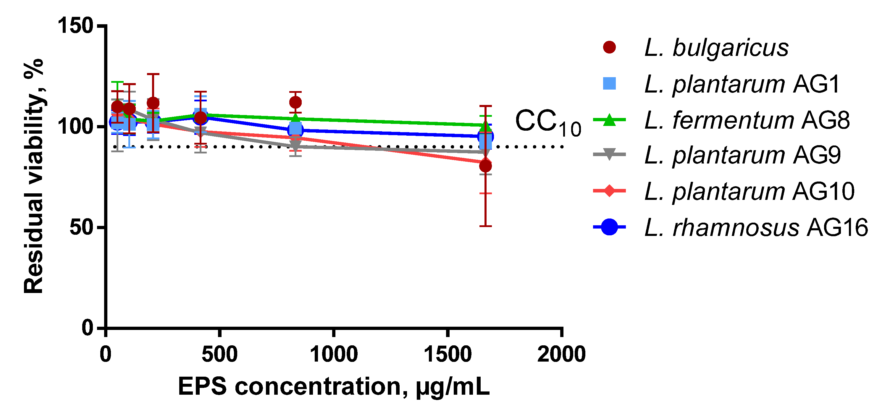
2.4. Cytotoxicity Assay

The cytotoxicity of EPS was assessed using a proliferative MTT assay (3-(4, 5-dimethylthiazol-2-yl)-2, 5-diphenyltetrazolium bromide) (PanEko, Moscow, Russia) [41] on primary human skin fibroblast cells (HSF). Cells were cultured in α-MEM (PanEko, Moscow, Russia) supplemented with 10% fetal bovine serum (PAA, Jannali, Australia), 2 mM L-glutamine, and 1% penicillin-streptomycin and maintained at 37 °C with 5% CO2 in a humidified atmosphere. Cells were treated with trypsin-EDTA (2.5%/0.02%) with subsequent inactivation of trypsin by adding α–MEM with FBS and seeded 24 h prior to treatment in a 96-well plate (2000 cells per well). Then, aliquots of filter-sterilized EPS solutions were added until final concentrations of 1667, 833, 417, 208, 104, and 52 μg/mL, and the plates were incubated for the next 72 h. Then, the medium was replaced with a fresh one (90 μL) and 10 μL of MTT reagent (5 mg/mL in DMSO) was added. After 1 h of incubation, the medium was removed and 100 μL of DMSO was added. After 10 min, the absorbance was measured at 555 nm with the reference wavelength 750 nm using the Tecan Infinite M200 Pro plate reader (Tecan, Männedorf, Switzerland). The results were presented as the percent of residual metabolic activity compared with untreated samples and CC10 has been calculated.

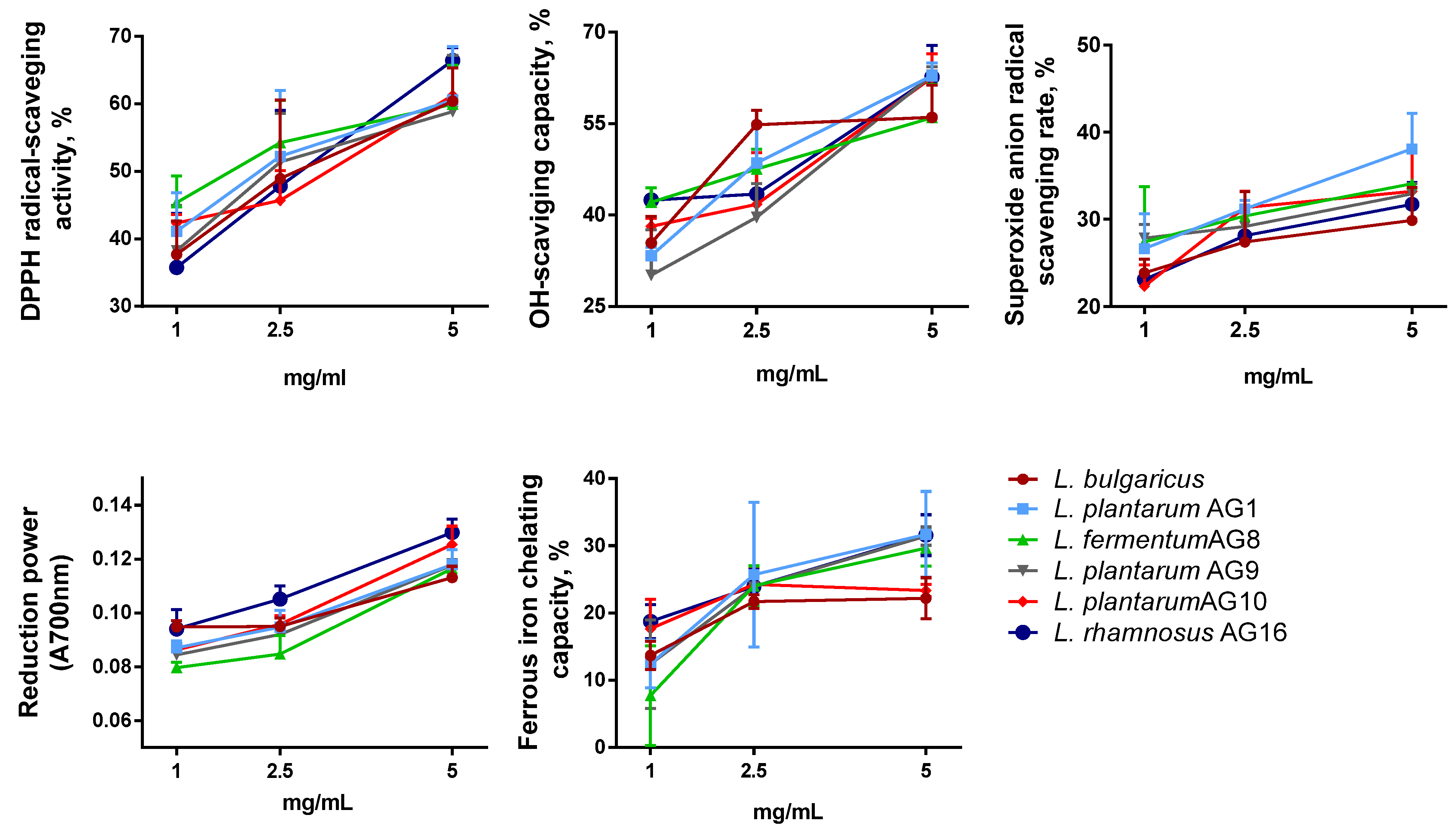
2.5. Antioxidant Activity

2.5.1. Evaluation of Radical Scavenging Ability (RSA) by 2, 2-Di-phenyl-1-picrylhydrazyl (DPPH) Assay

The radical scavenging capacity was analyzed as in [40] with modifications. EPS were tested at concentrations of 1, 2.5, and 5 mg/mL diluted in pure water, and PFE was diluted 5 times with pure water. Briefly, 1 mL of sample was mixed with 1 mL of freshly prepared DPPH solution (0.12 mM ethanol) and incubated at 25 °C in the dark for 30 min. The reaction mixture was centrifuged for 2 min at 10,000 rpm and the absorbance was measured at 517 nm on a Spectrophotometer SF-2000 (St Petersburg, Russia). As a reference, ethanol was used as control for the absorbance DPPH solution (0.12 mM ethanol). The radical scavenging activity was calculated as:
DPPH scavenging activity% = [(control absorbance − extract absorbance)/(control absorbance)] × 100.

2.5.2. Ferric Reducing Antioxidant Power Assay (FRAP)

The ferric reducing antioxidant power (FRAP) assay was carried out following the procedure described in [42] with modifications. EPS were tested at concentrations of 1, 2.5, and 5 mg/mL diluted in pure water, and PFE was diluted 5 times with pure water. Briefly, 1 mL of the sample was mixed with 1 mL of 0.2 M potassium sodium phosphate buffer (pH 6.5) and 1 mL of 1% potassium ferricyanide. The reaction mixture was incubated for 20 min at 50 °C, cooled, and 1 mL of 10% trichloroacetic acid was added. The mixture was centrifuged at 2000 rpm for 10 min at 25 °C; the supernatant was diluted twice with water (2 mL + 2 mL) and 400 μL of 0.1% FeCl3. For reference, a buffer was added instead of the potassium ferricyanide. The absorbance was measured at 700 nm (Spectrophotometer SF-2000, St Petersburg, Russia) and expressed as reducing force, which was expressed as absorbance at 700 nm relative to the control.

2.5.3. HO Free Radical Scavenging Ability

Analysis of HO free radical scavenging ability was carried out following the procedure described by Qin et al. [43] with modifications. EPS were tested at concentrations of 1, 2.5, and 5 mg/mL diluted in pure water, and PFE was diluted 10 times with pure water. Then, 0.5 mL 5 mM/L ferrous sulfate (FeSO4) solution, 0.5 mL 5 mM/L salicylic acid ethanol solution, and 0.5 mL 3 mM/L hydrogen peroxide solution were added into 0.5 mL of sample, followed by mixing and incubation at 37 °C for 30 min, 9000 r/min, and centrifugation for 5 min. The supernatant OD510 nm was taken as B1, ddH2O was used to replace the cell suspension of the cell-free extract as the control group, and OD510 nm was measured as B0.
OH scavenging activity (inhibition)% = (B0 − B1)/B0 × 100.

2.5.4. Determination of Superoxide Anion Radical Scavenging Activity

Analysis of the superoxide anion radical scavenging activity of the polysaccharides was conducted as described in [40,44]. EPS were tested at concentrations of 1, 2.5, and 5 mg/mL diluted in pure water, and PFE was diluted 5 times with pure water. The sample solution (0.2 mL) with different concentrations was added to Tris-HCl buffer (1 mL) and incubated at 25 °C for 20 min, and then pyrogallol solution (1 mL, 7 mM) was added. After 5 min, hydrochloric acid (0.2 mL, 10 mol/L) was added to stop the reaction. The absorbance of the resulting mixture (A1) was measured at 420 nm. The absorbance of the solution where pyrogallol was replaced with pure water was denoted as A2, and the absorbance of the solution with water instead of the sample solution was denoted as A0. The spectrophotometer was zeroed using pure water before measuring A1, A2, and A0. The superoxide anion radical scavenging activity was calculated with the following equation:
O2 − scavenging rate (%) = [1 − (A1 − A2)/A0] × 100.

2.5.5. Fe-Chelating Activity

The FIC assay was carried out as follows [45]. EPS were tested at concentrations of 1, 2.5, and 5 mg/mL diluted in pure water, and PFE was diluted 10 times with pure water. The iron(II) chloride (FeCl2) solution (2 mM) and ferrozine solution (5 mM) were diluted 20 times prior to assay. Diluted FeSO4·H2O solution (1 mL) was added to the sample and mixed. Diluted ferrozine solution (1 mL) was then added to the mixture followed by incubation for 10 min at room temperature. Absorbance at 562 nm (B1) was measured against pure water as a blank on a spectrophotometer (SF-2000, St Petersburg, Russia). A mixture of FeCl2·H2O solution (1 mL), ferrozine solution (1 mL), and pure water (1 mL) served as a control (B0). The FIC ability of samples was calculated using the mentioned equation.
FIC (inhibition)% = (B0 − B1)/B0 × 100.

2.6. Scanning Electron Microscopy

The microstructure of fermented milk samples was evaluated by SEM. Briefly, samples were fixed with 2.5% glutaraldehyde for 4–5 h, subsequently washed three times with 0.2 M Na-K phosphate buffer (pH 7.0), then dehydrated using 30%, 40%, 50%, 60%, 70%, and 80% ethanol (twice for each concentration) for 15 min, and 95% ethanol dehydrated three times for 30 min. The samples were mounted on metal stubs and coated with gold-palladium alloy (~10 nm thickness) using the Quorum Q150T ES coating machine. Samples were then observed using the self-emission scanning electron microscope Merlin (Carl Zeiss, Oberkochen, Germany) at an acceleration voltage of 5 kV, a secondary electrons detector. Magnifications are as indicated at the bottom of each figure.

2.7. Statistical Analysis

All experiments were carried out in triplicate. Five replicates were performed for antioxidants’ analysis. Significance was established at p < 0.05. The results were analyzed for statistical significance with Mann–Whitney or Kruskal–Wallis tests by GraphPad Prism software at a significance level of p < 0.05. The graphical representation of the principal component analysis (PCA) allows the data to be analyzed on a two-dimensional P1/P2 map by Statistica12 (Statsoft, Tulsa, OK, USA) and to identify the trends between variables at a significance level of p < 0.05. When two variables are far from the center and close together, they correlate significantly. If they are on the opposite side of the center, then they are significantly negatively correlated.

3. Results

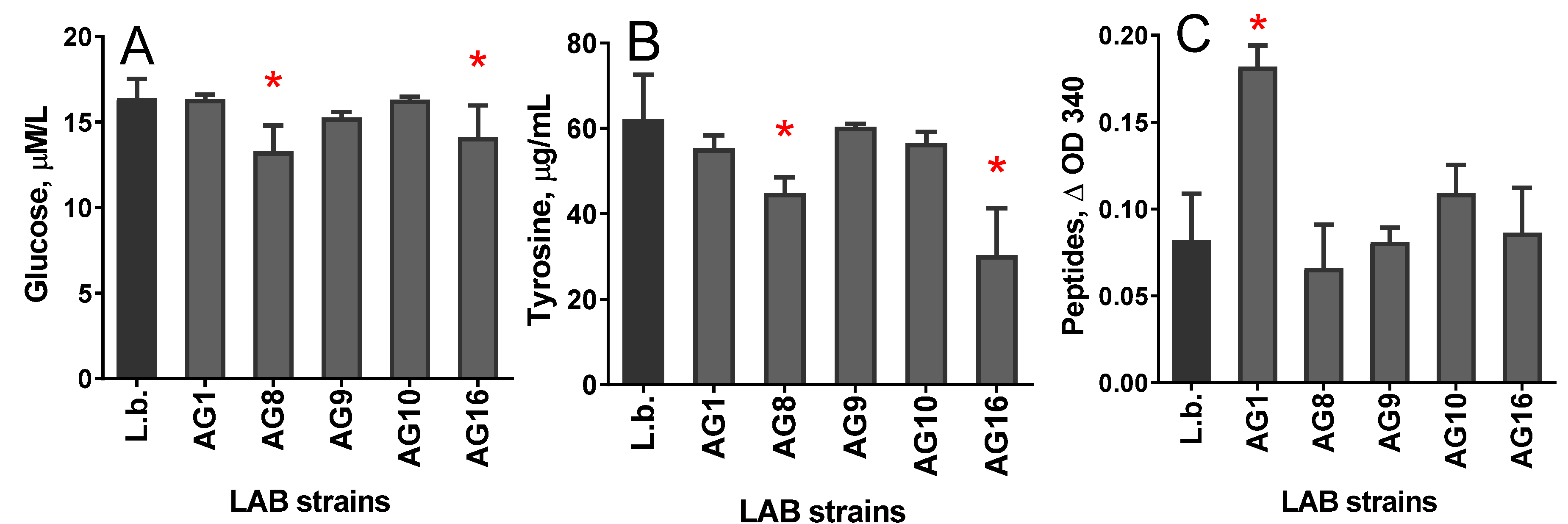
3.1. Chemical Composition of the Protein-Free Extract (PFE)

During milk fermentation, lactic acid bacteria of the starter culture produce various biologically active substances such as vitamins, amino acids, enzymes, exopolysaccharides (EPS), short-chain fatty acids, organic acids, phenolic compounds, bioactive peptides, etc., which have a positive impact on human health [46]. Therefore, the extracts with pre-precipitated high-molecular proteins (protein-free extract, PFE) were prepared by protein precipitation with TCA. The resulting PFE contained a pool of carbohydrates, including EPS, and low-molecular-weight peptide components, as well as amino acids, vitamins, and other low-molecular-weight compounds. The level of glucose, the product of the lactose metabolism, was significantly lower in the PFE of milk fermented with L. fermentum AG8 and L. rhamnosus AG16 compared with L. bulgaricus (Figure 1). The same strains led to the lowest total amount of phenolic compounds, the indicators of the presence of compounds with an aromatic group with antioxidant properties. Surprisingly, L. plantarum AG1 led to a two-fold higher amount of peptides in PFE compared with all other strains tested, suggesting increased proteolytic activity of this strain.

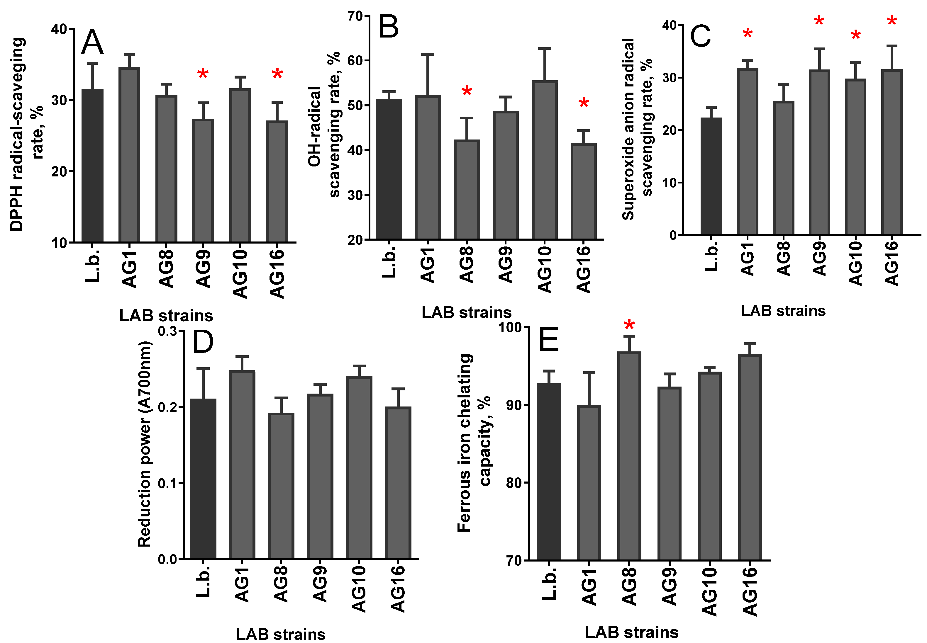
3.2. Antioxidant Properties of PFE

The ability to bind free radicals was high in the PFE of milk fermented by L. plantarum AG1, L. plantarum AG10, and L. bulgaricus (Figure 2A), and low in L. fermentum AG8, L. plantarum AG9, and L. rhamnosus AG16, which correlates with the level of free peptides. The PFE of milk fermented by the L. rhamnosus AG16 strain showed the lowest level of OH-binding activity, and the PFE of L. plantarum AG10 exhibited the highest one (Figure 2B). The PFE of milk fermented by the L. fermentum AG8 strain bound the superoxide radicals similarly to L. bulgaricus, while other strains led to higher activity (Figure 3C). The chelating activity of PFE was highest in the case of L. fermentum AG8 and L. rhamnosus AG16 strains, which showed the least ability to bind reactive oxygen species (Figure 2E). These data allow assuming that different molecular mechanisms are responsible for antioxidant and chelating abilities.

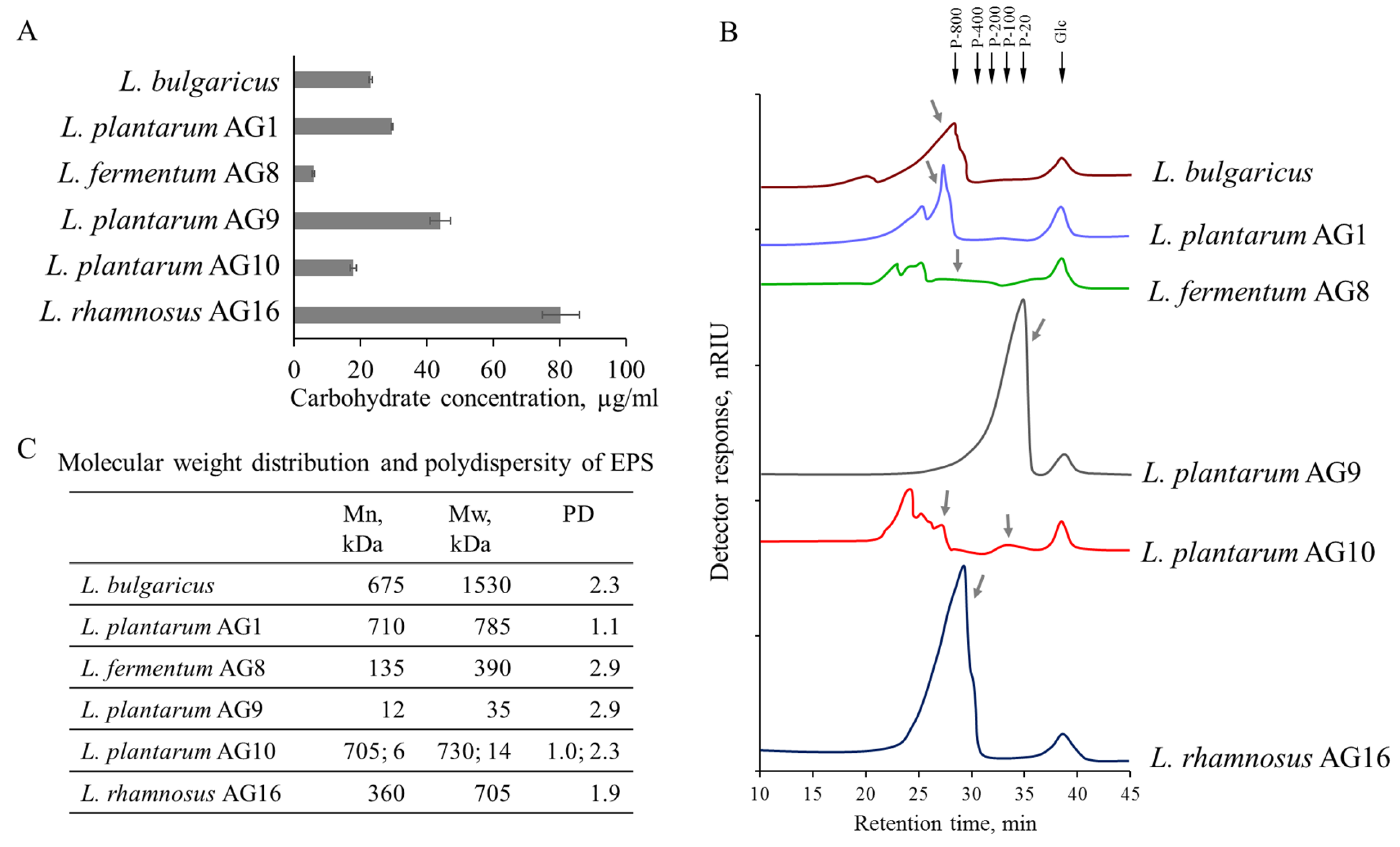
3.3. Yield and Composition of Exopolysaccharides (EPS)

The observed differences between antioxidant properties in PFE of milk fermented by different strains can be a result of quantitative and qualitative variations of exopolysaccharides. The maximum EPS yield (calculated from the sum of monosaccharides in fractions) was observed for L. rhamnosus AG16, and the minimum EPS yield for L. fermentum AG8 (Figure 3A). A relatively high EPS yield was also observed for L. plantarum AG9. L. bulgaricus, L. plantarum AG10, and L. plantarum AG1 produced comparable amounts of EPS, which were slightly smaller compared with L. plantarum AG9 and more than twice decreased compared with L. rhamnosus AG16 (Figure 3A).
Next, the lyophilized EPS produced with various strains were analyzed. During the EPS separation by size-exclusion chromatography, carbohydrates were detected only in peaks with molecular weights more than 10 kDa, which confirms the polymeric form of all EPS. The molecular weight distribution of polysaccharides produced by different strains differed (Figure 3B). The EPS produced by L. plantarum AG9 and L. rhamnosus AG16 were represented by one clearly defined peak with molecular weights of about 35 and 700 kDa, respectively. For the other samples, in addition to the peaks identified in the region of the good resolution of the calibration curve, a series of peaks with a high molecular weight of more than 1500 kDa were observed. The monosaccharide composition of these high-molecular-weight peaks was very close to the composition of lower-molecular-weight ones (data not shown), which shows that the high-molecular-weight peaks contain the aggregates of EPS molecules.
The composition of EPS produced by different Lactobacilli strains during the fermentation of skimmed milk also varied significantly (Table 1). Galactose and glucose predominated in five out of six obtained EPS, but all five EPS differed by the ratio of the glucose and galactose monomers as well as by the presence of other monosaccharides in the composition. EPS of L. plantarum AG9 mainly consisted of glucose (69 mol%). In addition to glucose, this EPS contained galactose (13 mol%). The EPS of L. plantarum AG10 and L. plantarum AG1, which are also characterized by a high proportion of glucose (46 and 26 mol%, respectively) and galactose (22 and 43 mol%), contained high proportions of arabinose and rhamnose. The EPS of L. bulgaricus differ from the EPS of L. plantarum AG1, which is similar in composition and the ratio of glucose and galactose, by a reduced fraction of arabinose and rhamnose and an increased proportion of xylose. These changes in the EPS composition correlate with an increased tendency of the EPS of L. bulgaricus to aggregate, expressed by the appearance of a higher-molecular-weight peak on the elution profile (Figure 1B), whose composition is similar to the EPS of the main peak. Compared to EPS of other strains also enriched with glucose and galactose, the EPS of L. rhamnosus AG16 contains a significantly increased proportion of xylose (29 mol%). At the same time, such a xylose-enriched polysaccharide did not demonstrate a pronounced tendency to form large aggregates (Figure 3B). Except for the high proportion of xylose, one of the additional reasons for this may be the increased proportion of the acidic monosaccharide –galacturonic acid (6.9 mol%) in its composition (Table 1). Unlike all the others, EPS produced by L. fermentum AG8 were represented by fructans (70 mol% of fructose) enriched in charged uronic acids (18 mol% of galacturonic acid and 7 mol% of glucuronic acid). Molecules of this EPS also showed a tendency to self-aggregate, even despite the high proportion of uronic acids in the composition (galacturonic acid and glucuronic acid) detected in high-molecular peaks, although their proportion was slightly reduced.
It has been shown that the monosaccharide composition of EPS strongly depends on the cultivation conditions. Despite the identical cultivation conditions, even one species group of Lactobacilli produces EPS of different composition, and even within EPS with a similar proportion of the main components, significant differences in galactose and glucose have also been revealed. So, with a similar tendency in the key components’ ratio, galactose and glucose (L. plantarum AG9 and L. plantarum AG10), EPS produced by these strains differ in the proportion of other neutral monomers, rhamnose and ara, as well as NAcGlcN. Another strain of this group of Lactobacilli (L. plantarum AG1) produces EPS with a similar proportion of rhamnose and arabinose to L. plantarum AG10, but with a significantly different ratio of the main monomers. At the same time, according to the galactose/glucose ratio, this polysaccharide is close to the EPS produced by a strain of another group of LactobacilliL. bulgaricus. EPS based on glucans and galactans are produced by analyzed Lactobacilli in a higher yield compared with fructans. The presence of arabinose and rhamnose in the composition increases the tendency of galactose- and glucose-enriched EPS to self-aggregate. The ability to form aggregates is also possessed by fructans produced by L. fermentum, despite the presence of uronic acids in their composition.

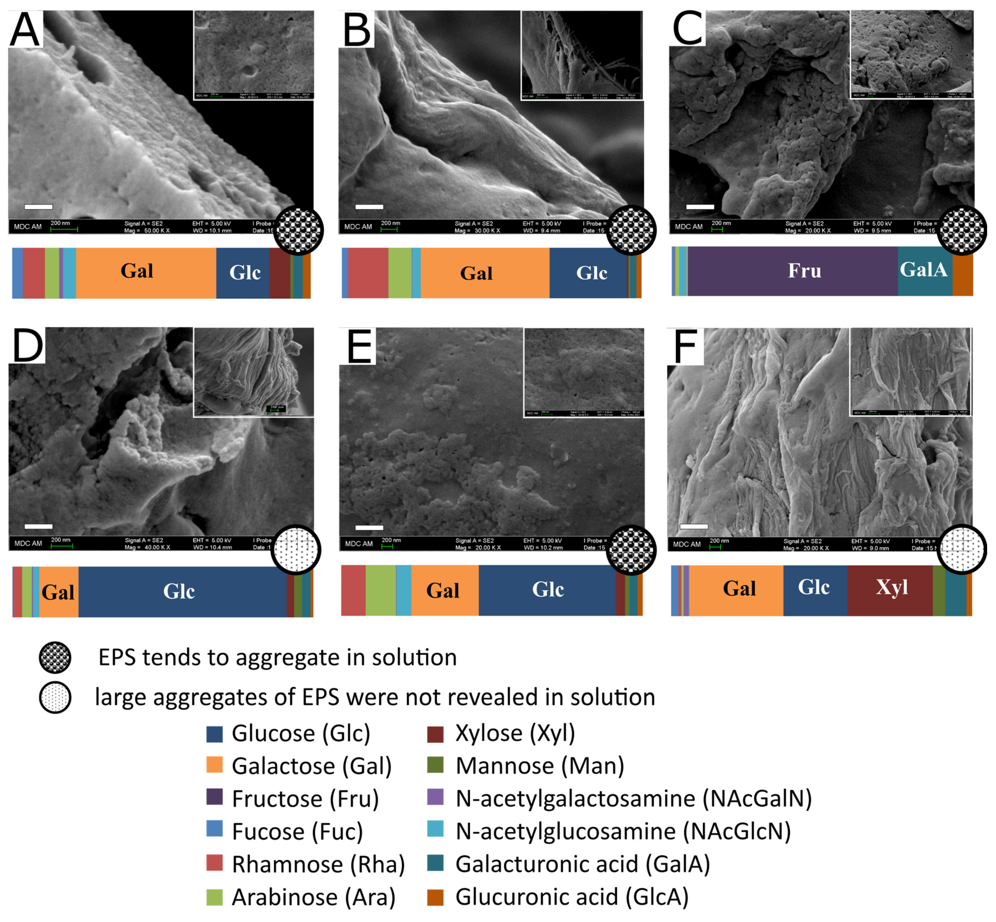
3.4. EPS Microstructureand Surface Morphology

Differences in the composition and properties of lyophilized EPS reflected in the organization and morphology of the surfaces of the microstructures they form at dehydration have been analyzed with SEM. EPS produced by L. bulgaricus (Figure 4) has an uneven, fine spongy-like structure interspersed with voids with some layering in the spatial section, and the surface is relatively smooth. EPS of L. plantarum AG1 by hydrodynamic properties and composition is similar to EPS of L. bulgaricus, while it differs by the absence of xylose and an increased proportion of rhamnose and arabinose, and has a relatively smooth structure of densely packed threads tightly glued to each other. Additionally, there are areas with EPS fibrils of irregular shape not arranged into compartments. EPS produced by L. fermentum AG8, represented mainly by fructans containing galacturonic acid, are characterized by a tendency to aggregate in solution and form spongy-like structures appearing as a loose substance with relatively large pores. The EPS of L. plantarum AG9, the most enriched in glucose, form areas of both spongy-like and filamentous structures. The spongy-like areas are fine-grained but with large voids, and no layering could be detected. However, the presence of areas in the form of granularly packed strands is periodically observed, which then turn into fine-grained areas. Apparently, the stage of EPS formation when polysaccharide chains begin to form a polysaccharide network has been fixed during samples preparation. Moreover, this happens precisely during dehydration, since in solution this type of EPS does not show a tendency to form large aggregates (Figure 3B). The EPS of L. plantarum AG10, which, along with a high proportion of glucose and galactose, also contains rhamnose and arabinose, forms a microstructure like that of a fine-grained sponge; however, filamentous areas and layering could not be observed. The surface layer seems rough and inhomogeneous, partly similar to the structure formed by the EPS of L. bulgaricus; however, large voids are not visualized. EPS of L. rhamnosus AG16 are characterized by a high proportion of xylose in the composition and the absence of a tendency for the self-aggregation of molecules in solution. These EPS have a filamentous type of structure; the filaments are assembled into compartments of different thicknesses, which are eventually covered with a layer of an amorphous smooth substance. The pores in the EPS structures are very small, without significant voids.

3.5. EPS Cytotoxicity

To exclude possible toxic effects, we checked the cytotoxicity of EPS (Figure 5). Only at a concentration of 800 µg/mL and more was a slight decrease in cell viability revealed in the MMT test with human fibroblasts. Only for L. plantarum AG1, L. plantarum AG9, and L. rhamnosus AG16 could the CC10 be calculated (934, 844, and 893 µg/mL, respectively). Thus, EPS of the studied strains, which are synthesized during the fermentation of skimmed milk, do not show toxicity to eukaryotic cells and are safe for food products.

3.6. EPS Antioxidant Activity

The analysis of the EPS’s ability to bind radicals of different chemical nature (free radical, hydroxyl radical, superoxide radical) revealed specificity, while a dose-dependent effect was not always manifested (Figure 6). At the highest concentration (5 mg/mL), the highest activity to bind free radicals was in EPS strain AG16. Hydroxyl radicals at a concentration of 5 mg/mL bound the EPS of strains AG1, 9, 10, and 16 (activities above 60%), while AG8 and L. bulgaricus had activities below 60%. The superoxide radical bound EPS of strain AG1 most actively. A decrease in concentration by 5 times (to 2.5 and 1 mg/mL) did not lead to a directly proportional decrease in activity. Restorative activity in the FRAP test was the highest in EPS strains AG10 and AG16. The chelating activity of EPS was studied in the test for the ability to bind iron ions, where the leaders were EPS strains AG1, AG9, and AG16. Thus, the difference in the manifestation of different aspects of antioxidant properties should mean a difference in the structural organization of different EPS.

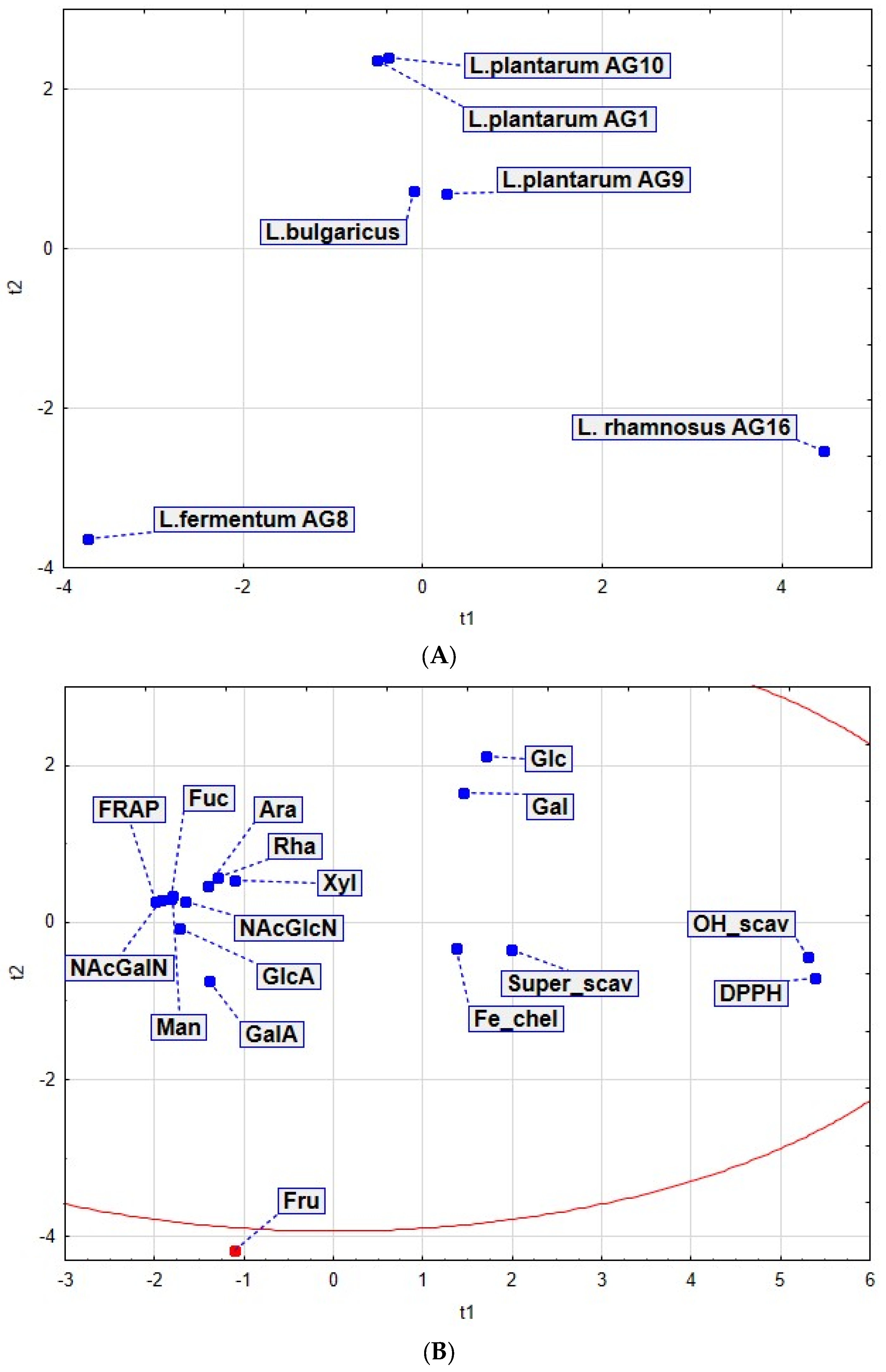
3.7. Principal Component Analysis (PCA)

The PCA revealed a high degree of similarity in chemical composition and antioxidant properties between EPS produced by L. bulgaricus and L. plantarum AG9 strains. At some distance from them lie the EPS of L. plantarum AG1 and L. plantarum AG10. According to the complex of chemical and antioxidant properties, strains of L. fermentum AG8 and L. rhamnosus AG16 differ greatly both from each other and from L. plantarum strains (Figure 7).
An analysis of the antioxidant properties of EPS and their composition revealed a high degree of correlation between the FRAP index and the presence of most of the detected indicators, except for the glucose, galactose, and fructose. The presence of fructose, especially as the main monomer, plays a significant role in changing the properties of EPS. Radical-binding (DPPH) and hydroxyl-binding (OH-scavenging) activities are highly correlated with each other. Additionally, there is a correlation between the Fe-chelating activity and superoxide-binding activity.
Thus, the presence of fructose as the main monomer and negative-charged galacturonic acid in L. fermentum AG8 EPS determines its antioxidant properties, which are different from others. The uniqueness of EPS L. rhamnosus AG16 is determined by the unique combination of glucose, galactose, and xylitolose. The similarity of the indicators’ structure properties of L. plantarum show their closely related relationship.

4. Discussion

The milk fermentation by LAB has been widely used for dairy food production since medieval times. Together with prolongation of the storage time, the fermentation of milk by LAB provides its enrichment by various beneficial properties. Among the factors responsible for the structural properties and the antioxidant activity of the product, the extracellular polysaccharides play an important role.
In this work, EPS produced by various LAB strains were prepared as a complex extract after the precipitation of high-molecular proteins. The yield of EPS produced by all analyzed strains was not large. However, for example, for L. bulgaricus, this indicator was close to the one shown for L. bulgaricus NCFB 2772 grown in a chemically defined medium with glucose (36.8 mg/L, [47,48]). The EPS yield depends on multiple factors such as dissolved oxygen, pH, nutritional media composition, and incubation temperature and duration [49]. For example, the EPS yield depending on the incubation conditions varies from 36.8 to 830 mg/L for L. bulgaricus [48,50,51]; 58.7 to 956 mg/L for L. plantarum [52,53]; 111 to 2767 mg/L for L. rhamnosus [54]; and 100 to 2000 mg/L for L. fermentum [55]. Thus, to determine optimal growing conditions for maximum EPS yield from novel isolated strains, further experiments will be needed.
The molecular weight distribution of EPS produced by analyzed Lactobacillus species from silage and isolated from fermented skim milk was comparable to the earlier reports of EPS produced by lactic acid bacteria. The EPS from L. plantarum MTCC 9510 had molecular weights of 268 kDa, 255 kDa and 283 kDa [56]; and Lactobacillus sp. CFR-2182 produced heteropolysaccharides fractions with molecular weights in the range 33–1320 kDa [57]. However, for the EPS of some strains analyzed in this work, the pattern of molecular weight distribution was similar, but for others it was significantly different. A similar picture was characteristic of EPS capable of self-aggregation (L. bulgaricus, L. plantarum AG1, AG10, L. fermentum AG8); the molecular weight of such polysaccharides exceeded 390 kDa. EPS from L. rhamnosus AG16, despite its high molecular weight, did not show a tendency to form large aggregates, as well as EPS from L. plantarum AG9, which had the lowest molecular weight (35 kDa). The influence of EPS molecular weight on yoghurt texture was illustrated by the work of [58]. It was shown that polysaccharides produced by S. thermophilus Rs and Sts in skimmed milk had the same composition, structure, yield, and had a similar effect on the protein–polysaccharide network. Milk fermented with two strains of S. thermophilus (Rs and Sts) was different in Posthumus viscosity (39 and 126 s, respectively). Authors noted that the only clear difference between both strains, which may cause the difference in ropiness of the milk cultures, is the difference in molecular mass of the EPS (3700 and 2600 kDa for Sts and Rs strains, respectively).
During the fermentation of milk, EPS is heated up and its role in providing product properties can be significant. The selected Lactobacillus isolates from silage show a different ability to synthesize exopolysaccharides. Carbohydrates make up from 90 to 96% of the composition of EPS produced by L. plantarum [53]; nevertheless, their chemical composition is different, which in turn affects the microstructure and properties of EPS. Differentiation of the chemical composition for different types of lactobacilli has been shown in several works. Thus, for L. plantarum GD2 and L. rhamnosus E9, the main monosaccharides were mannose and glucose, while mannose, glucose, galactosamine, and glucosamine were for the EPS of L. plantarum NCU116, and galactose, glucose, rhamnose, and mannose were for the EPS of L. rhamnosus ATCC9595 [59,60]. The EPS of L. plantarum consists of such monomers as mannose, fructose, galactose, and glucose; and EPS produced by L. plantarum NTU 102 contain fructose, arabinose, galactose, glucose, mannose, and maltose in various proportions. In our case, five out of six polysaccharides contained glucose and galactose as the main monomers, and in the composition of one of them (EPS of L. plantarum AG9), glucose significantly prevailed. In addition to these monomers, five polysaccharides also contained rhamnose, arabinose, N-acetylglucosamine, xylose, mannose, and uronic acids in various proportions. For some EPS, the presence of fucose and N-acetylgalactosamine in small amounts was also noted. The EPS of one strain (L. fermentum AG8) was represented by fructose and a high proportion of uronic acids, among which galacturonic acid predominated. A similar type of polysaccharide among those previously described was not revealed.
In this work, we have identified several variants of EPS comparisons that allow us to identify the potential role of the components of the EPS composition in providing their microstructure and antioxidant properties. The identification of such EPS pairs can serve as a basis for the subsequent deciphering of the mechanisms that provide the properties of these polysaccharides. For example, when comparing L. bulgaricus and L. plantarum AG1, the EPS of the second strain shows an increase in the proportion of rhamnose and arabinose, as well as a decrease in the proportion of xylose, even though the proportions of the main components (galactose and glucose) and the molecular weight of the EPS of these strains are comparable. The noted changes in the composition of EPS from L. plantarum AG1 lead to a decrease in the degree of porosity and compaction of the stacking of microstructures formed by it and are also accompanied by an increase in the ability to chelate Fe-ions and remove OH- and superoxide radicals. The comparison of EPS from L. plantarum AG1 and L. rhamnosus AG16 shows an opposite trend in composition changes: in AG16, the proportion of xylose increases, but rhamnose and arabinose decrease, and the proportions of the main components (galactose and glucose) are still comparable. At the same time, changes in morphology are no longer observed, and superoxide scavenging decreases. The result of this comparison demonstrates the significant role of rhamnose and arabinose in the binding of the superoxide radical and the reduction in the porosity of the microstructure formed by EPS. The participation of rhamnose in providing the antioxidant properties of EPS as well as an indirect relationship between the characteristics of those properties and various types of links between arabinose residues were demonstrated earlier [26,31]. Nothing is known about the effect of xylose, both on its properties and on the formation of supramolecular structures by EPS. The comparison of EPS from L. plantarum AG9 and AG10 demonstrates that the EPS, even within the same species but different strains, show differences in composition and properties. Thus, for EPS from L. plantarum AG10, an increase in the proportions of rhamnose, arabinose, NAcGlcN, and galactose is observed with a decrease in the proportion of glucose. This leads to an increase in the molecular weight of the EPS and a significant change in the morphology of the microstructures formed by it (the appearance of large pores and fibrils packed into strands with small pores). Apparently, such sharp changes in the microstructure are associated primarily with a change in the ratio of the main components (galactose and glucose); an additional role in this can be played by NAcGlcN through the formation of hydrophobic interactions with other molecules involving the methyl group and hydrogen bonds through the amino groups. Moreover, even though the proportions of rhamnose and arabinose in the EPS from L. plantarum AG10 were increased, the effect observed for the EPS from L. plantarum AG1 on antioxidant activity against superoxide was not revealed, and Fe-chelating also decreased. This suggests that such properties require not just the presence of rhamnose and arabinose but their certain combination with other components and features of the EPS structure (accessibility of groups, types of bonds).
In the case of EPS of the studied strains, there was no strong law of reducing DPPH scavenging activity with an increase in molecular weight. The high HO-scavenging activity and Fe-chelating capacity in the L. fermentum AG8 strain may be due to an increased content of uronic acids. Previously, the role of uronic acids in the formation of the antioxidant potential has been reported [44]. In addition, the metal ion chelating ability of polysaccharides could be due to the formation of a linkage between the carboxyl group in galacturonic acid and the divalent ion. The results for low Fe-reducing power EPS (0.1 D 700, 5 mg/mL) for the studied strains are opposite to results for L. plantarum LS5 and LU5 strains, in which FRAP was at the level of 0.4 D700 at a concentration of 4 mg/mL; however, the EPS of these strains is determined by DPPH scavenging activity. Apparently, this may be due to the high glucose level in EPS of L. plantarum LS5 and LU5. Homopolysaccharide EPS (monomer–glucose) from L. pentosus 14FE, L. plantarum 47FE, and L. pentosus 68FE showed DPPH scavenging activity at the level of 37–52% at a concentration of 5 mg/mL, which is 10–25% lower than in the heteropolysaccharide EPS of strains studied in this work. On the other hand, EPS produced by L. plantarum CNPC003 have a heteropolysaccharide structure based on mannose and glucose and showed DPPH scavenging activity at the level of 40% at a concentration of 4 mg/ml [23]. The EPS of L. plantarum BR2 at a concentration of 2 mg/mL showed 30% DPPH radical-binding activity [61]; the same activity in our case was shown by EPS at a concentration of 12 mg/mL. The activity may be due to the presence of a hydroxyl group and other functional groups in EPS, which convert the free radical into a more stable form.
In addition to EPS because of the fermentation by LAB, a number of different metabolites are formed, including milk proteins derivatives, which are cleaved to oligopeptides by proteinases. Intracellular or cell-wall-associated peptidases digest large peptides into smaller oligopeptides with antioxidant properties [62,63,64]. Studies of various protein components of milk and fermented milk products revealed their role in protection against oxidative stress [65]. Thus, free radicals are believed to be deactivated by peptides containing hydrophobic and aromatic amino acids. Low-molecular-weight phosphoserine residues as a hydrolysis product of milk casein have ABTS radical scavenging, hydroxyl, peroxyl, and metal chelating activity. It was previously found that low-molecular-weight casein hydrolysates were more effective peroxyl radical scavengers, while hydrolysis products rich in casein phosphopeptides had greater metal chelating properties. When characterizing the antioxidant activity of the PFE of fermented milk, it demonstrated relatively high potential to bind free, hydroxyl, and superoxide radicals, as well as metal ions that indicate the presence of both low and high molecular components of milk proteins in the extract [66].

5. Conclusions

In this work, a comparison of the composition, microstructure, and antioxidant properties of EPS produced by various Lactobacillus species from silage during not-fat milk fermentation, as well as PFE obtained after the removal of high-molecular-weight proteins from samples enriched with EPS, allowed us to identify some structural criteria and composition indicators that affect the features of their antioxidant properties and the specificity of supramolecular structures formed by them. Thus, the increased fraction of rhamnose and arabinose and low xylose leads to compaction of the EPS, a decrease in porosity, and increased both their OH- and superoxide scavenging and Fe-chelating activities. By contrast, increased xylose and low rhamnose and arabinose apparently leads to loss of large aggregates and high DPPH activity and FRAP. The high content of glucose, however, provides the formation of large pores. The increased fructan fraction (69.9 mol%) with a high fraction of galacturonic (18.2 mol%) and glucuronic acids (6.7 mol%) apparently determines the highly porous spongy-folded EPS microstructure. Both the quantitative characteristics of the individual components of the fraction and structural features are important for the antioxidant potential of fermented milk and depend on the strain used for milk fermentation. Therefore, the differences in the antioxidant properties provided with different strains should be considered, and the use of a multicomponent starter to achieve the optimal beneficial properties of a fermented milk product is preferred.

Author Contributions

Conceptualization, E.N. and A.K.; methodology, E.N., P.M. and T.P.; software, A.K; validation, E.N., A.K.; formal analysis, E.N., P.M. and T.P.; investigation, E.N., P.M., A.S., M.K., O.B. and T.P.; resources, A.K. and E.N.; data curation, A.K. and E.N.; writing—original draft preparation, E.N., P.M., E.G. and A.K.; writing—review and editing, E.N., P.M., E.G. and A.K.; visualization, E.N., E.G., P.M. and O.B.; supervision, A.K. and E.N.; project administration, E.N. and A.K.; funding acquisition, E.N. All authors have read and agreed to the published version of the manuscript.

Funding

The study was funded by RSF project number 22-26-20022 (to E.N.), https://rscf.ru/project/22-26-20022/ (accessed on 20 February 2023).

Institutional Review Board Statement

Not applicable.

Informed Consent Statement

Not applicable.

Data Availability Statement

All data are available in the manuscript file.

Acknowledgments

This research was performed in frames of the Kazan Federal University Strategic Academic Leadership Program PRIORITY-2030 (toxicity assays) and the government assignment for the FRC Kazan Scientific Center of RAS (monosaccharide analysis).

Conflicts of Interest

The authors declare no conflict of interest.

References

  1. Harapanahalli, A.K.; Younes, J.A.; Allan, E.; van der Mei, H.C.; Busscher, H.J. Chemical Signals and Mechanosensing in Bacterial Responses to Their Environment. PLoS Pathog. 2015, 11, e1005057. [Google Scholar] [CrossRef] [PubMed]
  2. Oleksy, M.; Klewicka, E. Exopolysaccharides produced by Lactobacillus sp.: Biosynthesis and applications. Crit. Rev. Food Sci. Nutr. 2018, 58, 450–462. [Google Scholar] [CrossRef] [PubMed]
  3. Daba, G.M.; Elnahas, M.O.; Elkhateeb, W.A. Contributions of exopolysaccharides from lactic acid bacteria as biotechnological tools in food, pharmaceutical, and medical applications. Int. J. Biol. Macromol. 2021, 173, 79–89. [Google Scholar] [CrossRef] [PubMed]
  4. Jurášková, D.; Ribeiro, S.C.; Silva, C.C.G. Exopolysaccharides Produced by Lactic Acid Bacteria: From Biosynthesis to Health-Promoting Properties. Foods 2022, 11, 156. [Google Scholar] [CrossRef]
  5. Angelin, J.; Kavitha, M. Exopolysaccharides from probiotic bacteria and their health potential. Int. J. Biol. Macromol. 2020, 162, 853–865. [Google Scholar] [CrossRef]
  6. Nabot, M.; Guérin, M.; Sivakumar, D.; Remize, F.; Garcia, C. Variability of Bacterial Homopolysaccharide Production and Properties during Food Processing. Biology 2022, 11, 171. [Google Scholar] [CrossRef]
  7. Kavitake, D.; Devi, P.B.; Singh, S.P.; Shetty, P.H. Characterization of a novel galactan produced by Weissella confusa KR780676 from an acidic fermented food. Int. J. Biol. Macromol. 2016, 86, 681–689. [Google Scholar] [CrossRef]
  8. Ale, E.C.; Perezlindo, M.J.; Pavón, Y.; Peralta, G.H.; Costa, S.; Sabb, A.G.N.; Bergamini, C.; Reinheimer, J.A.; Binetti, A.G. Technological, rheological and sensory characterizations of a yogurt containing an exopolysaccharide extract from Lactobacillus fermentum Lf2, a new food additive. Food Res. Int. 2016, 90, 259–267. [Google Scholar] [CrossRef]
  9. Badel, S.; Bernardi, T.; Michaud, P. New perspectives for Lactobacilli exopolysaccharides. Biotechnol. Adv. 2011, 29, 54–66. [Google Scholar] [CrossRef]
  10. Duboc, P.; Mollet, B. Applications of exopolysaccharides in the dairy industry. Int. Dairy J. 2001, 11, 759–768. [Google Scholar] [CrossRef]
  11. Doleyres, Y.; Schaub, L.; Lacroix, C. Comparison of the functionality of exopolysaccharides produced in situ or added as bioingredients on yogurt properties. J. Dairy Sci. 2005, 88, 4146–4156. [Google Scholar] [CrossRef] [Green Version]
  12. Perry, D.B.; McMahon, D.J.; Oberg, C.J. Effect of Exopolysaccharide-Producing Cultures on Moisture Retention in Low Fat Mozzarella Cheese1. J. Dairy Sci. 1997, 80, 799–805. [Google Scholar] [CrossRef]
  13. Korcz, E.; Kerényi, Z.; Varga, L. Dietary fibers, prebiotics, and exopolysaccharides produced by lactic acid bacteria: Potential health benefits with special regard to cholesterol-lowering effects. Food Funct. 2018, 9, 3057–3068. [Google Scholar] [CrossRef]
  14. Wegh, C.A.M.; Geerlings, S.Y.; Knol, J.; Roeselers, G.; Belzer, C. Postbiotics and Their Potential Applications in Early Life Nutrition and Beyond. Int. J. Mol. Sci. 2019, 20, 4673. [Google Scholar] [CrossRef] [Green Version]
  15. İspirli, H.; Demirbaş, F.; Dertli, E. Glucan type exopolysaccharide (EPS) shows prebiotic effect and reduces syneresis in chocolate pudding. J. Food Sci. Technol. 2018, 55, 3821–3826. [Google Scholar] [CrossRef]
  16. Bello, F.D.; Walter, J.; Hertel, C.; Hammes, W.P. In vitro study of prebiotic properties of levan-type exopolysaccharides from Lactobacilli and non-digestible carbohydrates using denaturing gradient gel electrophoresis. Syst. Appl. Microbiol. 2001, 24, 232–237. [Google Scholar] [CrossRef]
  17. Ismail, B.; Nampoothiri, K.M. Exposition of antitumour activity of a chemically characterized exopolysaccharide from a probiotic Lactobacillus plantarum MTCC 9510. Biologia 2013, 68, 1041–1047. [Google Scholar] [CrossRef] [Green Version]
  18. Sasikumar, K.; Kozhummal Vaikkath, D.; Devendra, L.; Nampoothiri, K.M. An exopolysaccharide (EPS) from a Lactobacillus plantarum BR2 with potential benefits for making functional foods. Bioresour. Technol. 2017, 241, 1152–1156. [Google Scholar] [CrossRef]
  19. Liu, Z.; Zhang, Z.; Qiu, L.; Zhang, F.; Xu, X.; Wei, H.; Tao, X. Characterization and bioactivities of the exopolysaccharide from a probiotic strain of Lactobacillus plantarum WLPL04. J. Dairy Sci. 2017, 100, 6895–6905. [Google Scholar] [CrossRef]
  20. Zhou, T.; Huo, R.; Kwok, L.-Y.; Li, C.; Ma, Y.; Mi, Z.; Chen, Y. Effects of applying Lactobacillus helveticus H9 as adjunct starter culture in yogurt fermentation and stor AGe. J. Dairy Sci. 2019, 102, 223–235. [Google Scholar] [CrossRef] [Green Version]
  21. Das, D.; Baruah, R.; Goyal, A. A food additive with prebiotic properties of an α-d-glucan from Lactobacillus plantarum DM5. Int. J. Biol. Macromol. 2014, 69, 20–26. [Google Scholar] [CrossRef] [PubMed]
  22. Hongpattarakere, T.; Cherntong, N.; Wichienchot, S.; Kolida, S.; Rastall, R.A. In vitro prebiotic evaluation of exopolysaccharides produced by marine isolated lactic acid bacteria. Carbohydr. Polym. 2012, 87, 846–852. [Google Scholar] [CrossRef] [PubMed]
  23. Bomfim, V.B.; Neto, J.H.P.L.; Leite, K.S.; de Andrade Vieira, É.; Iacomini, M.; Silva, C.M.; dos Santos, K.M.O.; Cardarelli, H.R. Partial characterization and antioxidant activity of exopolysaccharides produced by Lactobacillus plantarum CNPC003. LWT 2020, 127, 109349. [Google Scholar] [CrossRef]
  24. Nehal, F.; Sahnoun, M.; Smaoui, S.; Jaouadi, B.; Bejar, S.; Mohammed, S. Characterization, high production and antimicrobial activity of exopolysaccharides from Lactococcus lactis F-mou. Microb. Pathog. 2019, 132, 10–19. [Google Scholar] [CrossRef] [PubMed]
  25. Du, R.; Qiao, X.; Zhao, F.; Song, Q.; Zhou, Q.; Wang, Y.; Pan, L.; Han, Y.; Zhou, Z. Purification, characterization and antioxidant activity of dextran produced by Leuconostoc pseudomesenteroides from homemade wine. Carbohydr. Polym. 2018, 198, 529–536. [Google Scholar] [CrossRef]
  26. Pan, D.; Mei, X. Antioxidant activity of an exopolysaccharide purified from Lactococcus lactis subsp. lactis 12. Carbohydr. Polym. 2010, 80, 908–914. [Google Scholar] [CrossRef]
  27. Xu, Y.; Cui, Y.; Wang, X.; Yue, F.; Shan, Y.; Liu, B.; Zhou, Y.; Yi, Y.; Lü, X. Purification, characterization and bioactivity of exopolysaccharides produced by Lactobacillus plantarum KX041. Int. J. Biol. Macromol. 2019, 128, 480–492. [Google Scholar] [CrossRef]
  28. Wang, J.; Zhao, X.; Yang, Y.; Zhao, A.; Yang, Z. Characterization and bioactivities of an exopolysaccharide produced by Lactobacillus plantarum YW32. Int. J. Biol. Macromol. 2015, 74, 119–126. [Google Scholar] [CrossRef]
  29. Li, S.; Huang, R.; Shah, N.P.; Tao, X.; Xiong, Y.; Wei, H. Antioxidant and antibacterial activities of exopolysaccharides from Bifidobacterium bifidum WBIN03 and Lactobacillus plantarum R315. J. Dairy Sci. 2014, 97, 7334–7343. [Google Scholar] [CrossRef] [Green Version]
  30. Li, W.; Ji, J.; Rui, X.; Yu, J.; Tang, W.; Chen, X.; Jiang, M.; Dong, M. Production of exopolysaccharides by Lactobacillus helveticus MB2-1 and its functional characteristics in vitro. LWT Food Sci. Technol. 2014, 59, 732–739. [Google Scholar] [CrossRef]
  31. Lo, T.C.-T.; Chang, C.A.; Chiu, K.-H.; Tsay, P.-K.; Jen, J.-F. Correlation evaluation of antioxidant properties on the monosaccharide components and glycosyl link AGes of polysaccharide with different measuring methods. Carbohydr. Polym. 2011, 86, 320–327. [Google Scholar] [CrossRef]
  32. Jia, K.; Tao, X.; Liu, Z.; Zhan, H.; He, W.; Zhang, Z.; Zeng, Z.; Wei, H. Characterization of novel exopolysaccharide of Enterococcus faecium WEFA23 from infant and demonstration of its in vitro biological properties. Int. J. Biol. Macromol. 2019, 128, 710–717. [Google Scholar] [CrossRef]
  33. Trabelsi, I.; Slima, S.B.; Chaabane, H.; Riadh, B.S. Purification and characterization of a novel exopolysaccharides produced by Lactobacillus sp. Ca6. Int. J. Biol. Macromol. 2015, 74, 541–546. [Google Scholar] [CrossRef]
  34. Guo, Y.; Pan, D.; Sun, Y.; Xin, L.; Li, H.; Zeng, X. Antioxidant activity of phosphorylated exopolysaccharide produced by Lactococcus lactis subsp. lactis. Carbohydr. Polym. 2013, 97, 849–854. [Google Scholar] [CrossRef]
  35. Wang, K.; Li, W.; Rui, X.; Li, T.; Chen, X.; Jiang, M.; Dong, M. Chemical modification, characterization and bioactivity of a released exopolysaccharide (r-EPS1) from Lactobacillus plantarum 70810. Glycoconj. J. 2015, 32, 17–27. [Google Scholar] [CrossRef]
  36. Błaszczyk, K.; Wilczak, J.; Harasym, J.; Gudej, S.; Suchecka, D.; Królikowski, T.; Lange, E.; Gromadzka-Ostrowska, J. Impact of low and high molecular weight oat beta-glucan on oxidative stress and antioxidant defense in spleen of rats with LPS induced enteritis. Food Hydrocoll. 2015, 51, 272–280. [Google Scholar] [CrossRef]
  37. Lei, N.; Wang, M.; Zhang, L.; Xiao, S.; Fei, C.; Wang, X.; Zhang, K.; Zheng, W.; Wang, C.; Yang, R. Effects of low molecular weight yeast β-glucan on antioxidant and immunological activities in mice. Int. J. Mol. Sci. 2015, 16, 21575–21590. [Google Scholar] [CrossRef] [Green Version]
  38. Sun, L.; Wang, C.; Shi, Q.; Ma, C. Preparation of different molecular weight polysaccharides from Porphyridium cruentum and their antioxidant activities. Int. J. Biol. Macromol. 2009, 45, 42–47. [Google Scholar] [CrossRef]
  39. Gavrilova, E.; Anisimova, E.; Gabdelkhadieva, A.; Nikitina, E.; Vafina, A.; Yarullina, D.; Bogachev, M.; Kayumov, A. Newly isolated lactic acid bacteria from silage targeting biofilms of foodborne pathogens during milk fermentation. BMC Microbiol. 2019, 19, 248. [Google Scholar] [CrossRef]
  40. Nikitina, E.; Petrova, T.; Adel, V.; Asya, E.; Monyr, N.Y.; Airat, K. Textural and Functional Properties of Skimmed and Whole Milk Fermented by Novel Lactiplantibacillus plantarum AG10 Strain Isolated from Silage. Fermentation 2022, 8, 290. [Google Scholar] [CrossRef]
  41. Cory, A.H.; Owen, T.C.; Barltrop, J.A.; Cory, J.G. Use of an aqueous soluble tetrazolium/formazan assay for cell growth assays in culture. Cancer Commun. 1991, 3, 207–212. [Google Scholar] [CrossRef] [PubMed]
  42. Nikitina, E.V.; Yurtaeva, T.A.; Tsyganov, M.S.; Ezhkova, G.O. Physico-Chemical and Antioxidant Properties of Skimmed Varenets (Slavic Baked Milk Yogurt) Mixed with Enzyme-Modified Potato Starches. Curr. Res. Nutr. Food Sci. 2021, 9, 88–99. [Google Scholar] [CrossRef]
  43. Qin, S.; Huang, Z.; Wang, Y.; Pei, L.; Shen, Y. Probiotic potential of Lactobacillus isolated from horses and its therapeutic effect on DSS-induced colitis in mice. Microb. Pathog. 2022, 165, 105216. [Google Scholar] [CrossRef] [PubMed]
  44. Hu, S.-M.; Zhou, J.-M.; Zhou, Q.-Q.; Li, P.; Xie, Y.-Y.; Zhou, T.; Gu, Q. Purification, characterization and biological activities of exopolysaccharides from Lactobacillus rhamnosus ZFM231 isolated from milk. LWT 2021, 147, 111561. [Google Scholar] [CrossRef]
  45. Muniandy, P.; Shori, A.B.; Baba, A.S. Influence of green, white and black tea addition on the antioxidant activity of probiotic yogurt during refrigerated stor AGe. Food Pack. Aging Shelf Life 2016, 8, 1–8. [Google Scholar] [CrossRef]
  46. Diez-Ozaeta, I.; Astiazaran, O.J. Fermented foods: An update on evidence-based health benefits and future perspectives. Food Res. Int. 2022, 156, 111133. [Google Scholar] [CrossRef]
  47. Grobben, G.J.; Sikkema, J.; Smith, M.R.; de Bont, J.A. Production of extracellular polysaccharides by Lactobacillus delbrueckii ssp. bulgaricus NCFB 2772 grown in a chemically defined medium. J. Appl. Bacteriol. 1995, 79, 103–107. [Google Scholar] [CrossRef]
  48. Grobben, G.J.; Smith, M.R.; Sikkema, J.; de Bont, J.A. Influence of fructose and glucose on the production of exopolysaccharides and the activities of enzymes involved in the sugar metabolism and the synthesis of sugar nucleotides in Lactobacillus delbrueckii subsp. bulgaricus NCFB 2772. Appl. Microbiol. Biotechnol. 1996, 46, 279–284. [Google Scholar] [CrossRef]
  49. Gamar, L.; Blondeau, K.; Simonet, J.M. Physiological approach to extracellular polysaccharide production by Lactobacillus rhamnosus strain C83. J. Appl. Microbiol. 1997, 83, 281–287. [Google Scholar] [CrossRef]
  50. Goh, K.K.; Haisman, D.R.; Singh, H. Development of an improved procedure for isolation and purification of exopolysaccharides produced by Lactobacillus delbrueckii subsp. bulgaricus NCFB 2483. Appl. Microbiol. Biotechnol. 2005, 67, 202–208. [Google Scholar] [CrossRef]
  51. Kimmel, S.A.; Roberts, R.F. Development of a growth medium suitable for exopolysaccharide production by Lactobacillus delbrueckii ssp. bulgaricus RR. Int. J. Food Microbiol. 1998, 40, 87–92. [Google Scholar] [CrossRef]
  52. Wang, J.; Zhao, X.; Tian, Z.; He, C.; Yang, Y.; Yang, Z. Isolation and characterization of exopolysaccharide-producing Lactobacillus plantarum SKT109 from Tibet Kefir. Pol. J. Food Nutr. Sci. 2015, 65, 269–279. [Google Scholar] [CrossRef] [Green Version]
  53. Imran, M.Y.; Reehana, N.; Jayaraj, K.A.; Ahamed, A.A.; Dhanasekaran, D.; Thajuddin, N.; Alharbi, N.S.; Muralitharan, G. Statistical optimization of exopolysaccharide production by Lactobacillus plantarum NTMI05 and NTMI20. Int. J. Biol. Macromol. 2016, 93, 731–745. [Google Scholar] [CrossRef]
  54. Berecka, M.P.; Waśko, A.; Szwajgier, D.; Choma, A. Bifidogenic and antioxidant activity of exopolysaccharides produced by Lactobacillus rhamnosus E/N cultivated on different carbon sources. Pol. J. Microbiol. 2013, 62, 181–189. [Google Scholar] [CrossRef]
  55. Fukuda, K.; Shi, T.; Nagami, K.; Leo, F.; Nakamura, T.; Yasuda, K.; Senda, A.; Motoshima, H.; Urashima, T. Effects of carbohydrate source on physicochemical properties of the exopolysaccharide produced by Lactobacillus fermentum TDS030603 in a chemically defined medium. Carbohydr. Polym. 2010, 79, 1040–1045. [Google Scholar] [CrossRef]
  56. Ismail, B.; Nampoothiri, K.M. Molecular characterization of an exopolysaccharide from a probiotic Lactobacillus plantarum MTCC 9510 and its efficacy to improve the texture of starchy food. J. Food Sci. Technol. 2014, 51, 4012–4018. [Google Scholar] [CrossRef] [Green Version]
  57. Vijayendra, S.; Palanivel, G.; Mahadevamma, S.; Tharanathan, R. Physico-chemical characterization of a new heteropolysaccharide produced by a native isolate of heterofermentative Lactobacillus sp. CFR-2182. Arch. Microbiol. 2009, 191, 303–310. [Google Scholar] [CrossRef]
  58. Faber, E.J.; Zoon, P.; Kamerling, J.P.; Vliegenthart, J.F. The exopolysaccharides produced by Streptococcus thermophilus Rs and Sts have the same repeating unit but differ in viscosity of their milk cultures. Carbohydr. Res. 1998, 310, 269–276. [Google Scholar] [CrossRef]
  59. Wu, J.; Zhang, Y.; Ye, L.; Wang, C. The anti-cancer effects and mechanisms of lactic acid bacteria exopolysaccharides in vitro: A review. Carbohydr. Polym. 2021, 253, 117308. [Google Scholar] [CrossRef]
  60. Tallon, R.; Bressollier, P.; Urdaci, M.C. Isolation and characterization of two exopolysaccharides produced by Lactobacillus plantarum EP56. Res. Microbiol. 2003, 154, 705–712. [Google Scholar] [CrossRef]
  61. Ramsey, I.; Corsini, N.; Peters, M.D.J.; Eckert, M. A rapid review of consumer health information needs and preferences. Patient Educ. Couns. 2017, 100, 1634–1642. [Google Scholar] [CrossRef] [PubMed]
  62. Arakawa, K.; Matsunaga, K.; Takihiro, S.; Moritoki, A.; Ryuto, S.; Kawai, Y.; Masuda, T.; Miyamoto, T. Lactobacillus gasseri requires peptides, not proteins or free amino acids, for growth in milk. J. Dairy Sci. 2015, 98, 1593–1603. [Google Scholar] [CrossRef] [Green Version]
  63. Griffiths, M.W.; Tellez, A.M. Lactobacillus helveticus: The proteolytic system. Front. Microbiol. 2013, 4, 30. [Google Scholar] [CrossRef] [PubMed] [Green Version]
  64. Trimigno, A.; Bøge Lyndgaard, C.; Atladóttir, G.A.; Aru, V.; Balling Engelsen, S.; Harder Clemmensen, L.K. An NMR Metabolomics Approach to Investigate Factors Affecting the Yoghurt Fermentation Process and Quality. Metabolites 2020, 10, 293. [Google Scholar] [CrossRef]
  65. Power, O.; Jakeman, P.; FitzGerald, R.J. Antioxidative peptides: Enzymatic production, in vitro and in vivo antioxidant activity and potential applications of milk-derived antioxidative peptides. Amino Acids 2013, 44, 797–820. [Google Scholar] [CrossRef] [PubMed]
  66. Díaz, M.; Decker, E.A. Antioxidant mechanisms of caseinophosphopeptides and casein hydrolysates and their application in ground beef. J. Agric. Food Chem. 2004, 52, 8208–8213. [Google Scholar] [CrossRef] [PubMed]
Figure 1. Chemical composition of protein-free extract of skim milk fermented by different strains of LAB (A) concentration of dextrose, (B) phenolic compounds (by tyrosine), (C) free peptides, reaction with OPA-reagent). L. b.—L. bulgaricus, AG1—L. plantarum AG1, AG8—L. fermentum AG8, AG9—L. plantarum AG9, AG10—L. plantarum AG10, AG16—L. rhamnosus AG16. Asterisks indicate statistically significant differences with L. bulgaricus according to non-parametric one-way analysis of variance (Kruskal–Wallis) test, p < 0.05.
Fermentation 09 00197 g001
Figure 2. Antioxidant and chelating properties of a protein-free extract (pre-neutralized to pH = 7) isolated from skim milk fermented with different strains of LAB (L. b.—L. bulgaricus, AG1—L. plantarum AG1, AG8—L. fermentum AG8, AG9—L. plantarum AG9, AG10—L. plantarum AG10, AG16—L. rhamnosus AG16). (A) DPPH scavenging activity; (B) OH scavenging activity; (C) O2−scavenging rate; (D) Ferric Reducing Antioxidant Power Assay (FRAP); (E) Fe-Chelating Activity. Asterisks indicate statistically significant differences with L. bulgaricus according to non-parametric one-way analysis of variance (Kruskal–Wallis) test, p < 0.05.
Fermentation 09 00197 g002
Figure 3. Yield (A) and molecular weight distribution (B,C) of EPS produced by various Lactobacillus species of silage and isolated from fermented skim milk. The number average (Mn) and weight average (Mw) molecular weights (C) are given for peaks indicated by gray arrows (B). Some of the peaks in the chromatograms (B) are outside the calibration curve (molecular weight parameters are not given for them). Pullulans with a low polydispersity index and glucose were used as molecular weight markers at gel filtration.
Fermentation 09 00197 g003
Figure 4. Scanning electron microscopy of EPS isolated from skim milk fermented by different strains of LAB ((A) L. bulgaricus, (B) L. plantarum AG1, (C) L. fermentum AG8, (D) L. plantarum AG9, (E) L. plantarum AG10, (F) L. rhamnosus AG16).
Fermentation 09 00197 g004
Figure 5. Cytotoxicity of EPS isolated from skim milk fermented by different strains of LAB (MTT-test, human fibroblasts).
Fermentation 09 00197 g005
Figure 6. Antioxidant and chelating properties of EPS isolated from skim milk fermented by different strains of LAB.
Fermentation 09 00197 g006
Figure 7. Principal component analysis of antioxidant properties of LAB strains (A) and antioxidant properties of EPS (B).
Fermentation 09 00197 g007
Table 1. Monosaccharide composition of EPS produced by various Lactobacillus species.
Sample of EPSMonosaccharide Composition, mol%
FucRhaAraNAcGalNNAcGlcNGalGlcXylManFruGalAGlcA
L. bulgaricus3.6 ± 0.17.3 ± 0.24.9 ± 0.11.3 ± 0.04.2 ± 0.146.9 ± 1.617.9 ± 0.77.1 ± 0.21.0 ± 0.00.0 ± 0.03.2 ± 0.12.6 ± 0.1
L. plantarum AG12.2 ± 0.113.4 ± 0.47.8 ± 0.20.1 ± 0.02.9 ± 0.142.9 ± 1.325.7 ± 0.80.6 ± 0.00.6 ± 0.00.0 ± 0.02.1 ± 0.11.7 ± 0.1
L. fermentum AG80.7 ± 0.00.3 ± 0.01.4 ± 0.00.0 ± 0.02.4 ± 0.10.3 ± 0.00.1 ± 0.00.0 ± 0.00.0 ± 0.069.9 ± 2.118.2 ± 0.56.7 ± 0.2
L. plantarum AG90.3 ± 0.02.9 ± 0.13.3 ± 0.10.2 ± 0.02.1 ± 0.113.1 ± 0.669.1 ± 2.82.6 ± 0.12.6 ± 0.10.0 ± 0.02.9 ± 0.10.9 ± 0.0
L. plantarum AG100.0 ± 0.08.1 ± 0.210.2 ± 0.30.1 ± 0.05.0 ± 0.222.3 ± 0.945.5 ± 1.63.0 ± 0.11.4 ± 0.00.0 ± 0.02.7 ± 0.11.7 ± 0.1
L. rhamnosus AG162.5 ± 0.10.8 ± 0.00.8 ± 0.01.7 ± 0.10.2 ± 0.031.3 ± 1.121.1 ± 0.828.5 ± 0.94.2 ± 0.10.0 ± 0.06.9 ± 0.22.0 ± 0.1
Disclaimer/Publisher’s Note: The statements, opinions and data contained in all publications are solely those of the individual author(s) and contributor(s) and not of MDPI and/or the editor(s). MDPI and/or the editor(s) disclaim responsibility for any injury to people or property resulting from any ideas, methods, instructions or products referred to in the content.

