Article

ε-Polylysine Derived from Marine Bacteria-A Possible Natural Preservative for Raw Milk Storage

by
Sourish Bhattacharya
1,*,
Sandhya Mishra
2,*,
Antonio Zuorro
3,
Heba Hassan Salama
4,
Alexandru Vasile Rusu
5 and
Monica Trif
6,*
1
Process Design and Engineering Cell, CSIR-Central Salt and Marine Chemicals Research Institute, Bhavnagar 346002, India
2
Applied Phycology and Biotechnology Division, CSIR–Central Salt and Marine Chemicals Research Institute, Bhavnagar 346002, India
3
Department of Chemical Engineering, Materials & Environment, Sapienza-University of Rome, V. Eudossiana 18, 00184 Rome, Italy
4
National Research Centre, Dairy Department, Food Industries and Nutrition Research Institute, 33 El-Buhouth St. (Former El-Tahrir St.), Dokki, Giza 12622, Egypt
5
Animal Science and Biotechnology Faculty, University of Agricultural Sciences and Veterinary Medicine Cluj-Napoca, 400372 Cluj-Napoca, Romania
6
Food Research Department, Centre for Innovative Process Engineering (CENTIV) GmbH, 28857 Syke, Germany
*
Authors to whom correspondence should be addressed.
Fermentation 2023, 9(2), 156; https://doi.org/10.3390/fermentation9020156
Submission received: 30 November 2022 / Revised: 1 February 2023 / Accepted: 2 February 2023 / Published: 6 February 2023
(This article belongs to the Special Issue Advances in Beverages, Food, Yeast and Brewing Research 2.0)

Abstract

:
Despite the fact that researchers have been working on the preservation of raw milk at room temperature for several decades, most of the processes are limited to the use of chemical preservatives. One of the major problems of raw milk self-life is its spoilage at ambient temperature during the summer season. Therefore, in the present study, research has been conducted to control raw milk spoilage at 4 °C and 35 °C (considered in different regions’ ambient temperatures). ε-Polylysine, a natural preservative approved for food use, was isolated from the fermentation broth of Bacillus licheniformis PL26 grown in an M3G medium, and its antimicrobial preservation properties for milk applications were tested. The raw milk samples containing 0.02% w/v ε-polylysine could be stored at 4 °C for up to 16 days without spoilage, however, raw milk samples without ε-polylysine as preservative spoiled on the 8th day even at 4 °C refrigeration conditions. Raw milk containing 0.02% ε-polylysine in combination with 0.2% sodium bicarbonate (added to avoid acidification) could be stored at ambient temperature (35 °C) for up to 48 h. The changes in milk composition, especially of the casein, lactose, and fat stability, during storage under different conditions with/without ε-polylysine, were studied as well. The present study proves that ε-polylysine can be successfully used as a new biopreservative. Therefore, for the dairy industry, a natural preservative to store milk at room temperature during the summer season, replacing synthetic preservatives derived from renewable sources, can be proposed. Once again, marine bacteria seem to be one of the promising sustainable and renewable sources of biologically active compounds such as new food biopreservatives

1. Introduction

Milk and dairy products are considered a major category of food products due to their taste and high nutritive value [1,2]. However, as a rich source of proteins and vitamins, these products represent a good growth medium for several pathogenic organisms such as Klebsiella, Bacillus, Pseudomonas, and Staphylococcus spp. which are the most common contaminants in milk [3,4]. The major challenge is the preservation of milk, especially in the regions where refrigeration facilities are a limitation or in places where ambient temperature exceeds 30–35 °C [5,6,7]. Therefore, for the last few decades, researchers were focussing on the preservation of milk at ambient temperature and improving its shelf life under refrigeration conditions. Natural and biopreservatives commonly used in milk and its products are lactic acid bacteria (LAB), bacteriocin, hydrogen peroxide, honey and lecithin, or different plant-based [3,8,9,10,11]. Nisin, a group of proteins with antibiotic effect, technically called bacteriocins [12,13,14] is permitted as a food additive by the FAO/WHO Codex Committee on Milk and Milk Products. They are formed by bacteria (Streptococcus lactis) that are often found in raw milk [15]. Although the pH of milk is close to neutral, which encourages the growth of harmful and spoilage bacteria, the reduced solubility and stability of nisin in milk are anticipated to decrease nisin effectiveness [16,17,18]. Honey is well known for its antibacterial activity against S. aureus, E. coli Clostridium perfringens, and Salmonella spp., and as a natural preservative wherein, microbial contamination can be reduced up to a level of 50% for up to 4 days [19,20,21]. Further, Sešķēna & Jankevica [22] have used 0.02% sodium azide, 0.06% hydrogen peroxide, 0.04% bronopol, 0.4% azidiol, 1.0% boric acid and 0.5% potassium sorbate and found that bronopol, sodium azide, and azidiol are safe preservatives at the specific concentration for storage of milk under 4 °C up to 96 h. Researchers have also succeeded in preserving milk for up to 24 h using 0.05% hydrogen peroxide [23]. The ideal circumstances for preserving milk samples were given by the tripled quantities of sodium azide and chloramphenicol. Since this concentration allowed for reliable bacterial counts for up to 12 days when stored at 3 °C and for up to 8 days when maintained at 6 °C, it was the best option for extending the analytical shelf life of the samples [24].
ε-Polylysine (ε-PL) is a homopolymer of L-lysine linked by the peptide bond between the carboxyl and ε- amino groups [25,26,27]. ε-PL is non-toxic to humans and animals, environmentally friendly, water-soluble, and biodegradable. Over the past few years, its derivatives have been utilized in a wide range of industrial applications, including those related to electronics, food, medicine, and the environment [28]. Gram-positive and Gram-negative bacteria, yeasts, and molds are just a few of the pathogens that ε-PL demonstrates effective antibacterial properties [25,29,30]. Furthermore, due to its antimicrobial and biodegradable nature, ε-PL has been introduced as a safe food biopreservative [31]. When ε-PL used in food, the influence on the taste of the food should be considered. The addition of a large amount of lysine produces a bitter taste and can influence the taste of the food [32,33]. In food processing, it is combined with protein and acidic polysaccharides and will also change the texture of food to some extent [27]. However, the bacteriostatic activity of ε-PL is relatively high and its concentration used in food is relatively low. Therefore, the risk of side effects of ε-PL on food quality and sensorial is very low [34].
In Japan, it has already entered the commercial market and is produced at an industrial scale by fermentation method using a mutant derived from Streptomyces albulus [29,35,36]. A wide variety of other novel uses for ε-PL and its derivatives, including emulsifying agents, dietary agents, biodegradable fibers, highly water-absorbable hydrogels, anticancer agent enhancers, drug and gene delivery carriers, coatings, nano- and micro-capsules for drug delivery, and the encapsulation of cell lines for in vivo delivery of bioactive molecules. [37,38,39,40].
Purification of ε-PL can be done directly from the fermentation broth in the form of ε-PL hydrochloride via the precipitation method with tetraphenylborate (TPB-) anion [28]. At pH 3.5, the (TPB-) anion and the ε-PL-Hn+ cation associate to precipitate the 1:n stoichiometric: ε-PL-H (TPB)n. Other monovalent cations are also able to precipitate with TPB-. The more soluble NH4TPB and KTPB are removed by washing the precipitate with acetone, and finally, ε-PLH can be further precipitated [41].
In the present study, research has been focused on aiming for raw milk preservation at a higher temperature i.e., (35 °C), considered in different regions’ ambient temperatures. One of the biggest challenges for milk-based products is the preservation of raw milk at these considered ambient temperatures in these regions (above 35 °C). Therefore, ε-PL, a natural biopreservative with antimicrobial properties was isolated from the fermentation broth of Bacillus licheniformis grown in an M3G medium. Bacillus licheniformis PL26, which has been isolated from the west coast of India, was found to be able to produce ε-PL [42].

2. Materials and Methods

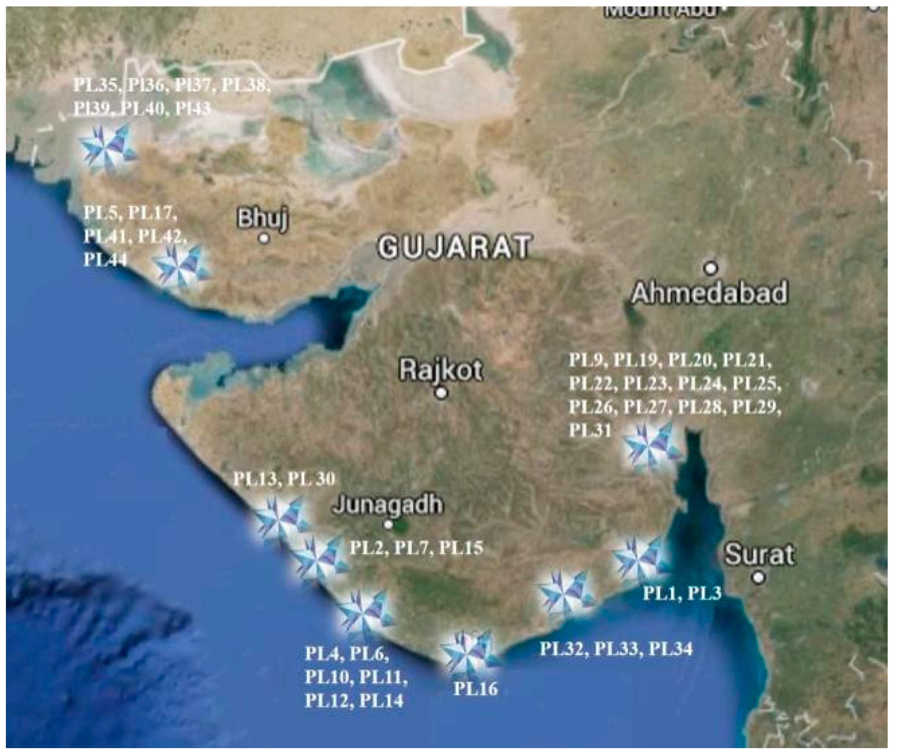
2.1. Study Sites and Collection Strategy

The locations of the present study are shown in Figure 1. Mainly were based on the west coast of India, and samples of seawater were taken at low tide from different areas: Adri, Chakratirth, CSIR-CSMCRI’s experimental salt farm, Jafarabad, Okha coast, Nagoa Beach (Diu), Madhavpur, Veraval, Jakhau port, Koteshalay Jetty, Mandavi temple, Mandavi beach, Mundra port, Vector port, along the west coast of India [42,43].

2.2. Isolation of Bacteria

The protocol used to isolate the bacteria has been optimized and described in a previous study by Bhattacharya et al. [43]. Ten-fold dilutions of the water and sediment samples were prepared and 0.1 mL from each sample solution was poured on Zobell marine agar plates containing (g/L) peptone 5.0; yeast extract 1.0; ferric citrate 0.1; sodium chloride 19.45; magnesium chloride 8.8; sodium sulfate 3.24; calcium chloride 1.8; potassium chloride 0.55; sodium bicarbonate 0.16; potassium bromide 0.08; strontium chloride, 0.034; boric acid, 0.022; sodium silicate, 0.004; sodium fluoride, 0.0024; ammonium nitrate, 0.0016; disodium phosphate, 0.008; agar, 1.5, at pH 7.6 ± 0.2. The plates were incubated at 37 °C temperature for 48 h.

2.3. Screening of Bacteria

Based on bacterial growth and ε-PL content, a novel and easier preliminary screening has been used, previously described by Bhattacharya et al. [43]. Bacterial isolates were screened by streaking them on agar plates containing methylene blue [44]. A novel and faster preliminary screening were implemented on the basis of bacterial growth and ε-PL content. The screening was carried out by streaking bacterial isolates on agar plates containing the basic dye methylene blue [44,45]. The growth medium containing (g/L) glycerol 10, ammonium sulfate 1.0, disodium hydrogen phosphate 0.5, magnesium sulfate 0.25, yeast extract 0.5, potassium dihydrogen phosphate 0.5, agar 2%, and methylene blue 0.02% were maintained at pH 7.0. The isolated bacterial strains were streaked on the plates containing methylene blue as the basic dye and kept at an incubation period of 168 h at 32 °C. The growth of microorganism-producing extracellular basic polymers such as ε-PL on agar plates containing methylene blue dye shows an outer zone which was later confirmed by adding the biomass of ε-PL-producing strain as well as standard ε-PL (100 mg) in the wells of the plates containing the 0.02% basic dye (methylene blue). 44 isolates (PL 1-44) were obtained which showed non-uniform zone distribution on the methylene blue plates.

2.4. Fermentation

2.4.1. Culture Media

The strain was cultivated in Zobell marine medium as described in a previous study [43]. The media was adjusted to pH 7.6 ± 0.2. For 48 h, the seed media was incubated at a temperature of 37 °C.

2.4.2. Inoculum

Bacillus licheniformis PL26 was first grown on a slant. Additionally, 100 mL of Zobell marine broth and a loopful of the bacteria Bacillus licheniformis PL26 were added to a seed culture before being incubated at 30°C for an entire night at 120 rpm.

2.4.3. Production of ε-PL

ε-PL was produced by cultivation of Bacillus licheniformis PL26 in a two-stage process through submerged fermentation in a shake flask scale (100 mL). In the first stage, a loopful of Bacillus licheniformis inoculated to seed culture medium containing (g/L) peptone 5.0, yeast extract 1.0, ferric citrate 0.1, sodium chloride 19.45, magnesium chloride 8.8, sodium sulfate 3.24, calcium chloride 1.8, potassium chloride 0.55, sodium bicarbonate 0.16, potassium bromide 0.08, strontium chloride 0.034, boric acid 0.022, sodium silicate 0.004, sodium fluoride 0.0024, ammonium nitrate 0.0016 and disodium phosphate 0.008 and then cultivated for 48 h at 30°for getting the dense seed inoculum appropriate for inoculating into production medium. In the second stage, 5% of the seed culture was inoculated to a production medium containing (g/L) ammonium sulphate-10.0, potassium dihydrogen ortho phosphate-1.36, dipotassium hydrogen phosphate-0.8, magnesium sulphate-0.5, ferrous sulphate-0.04, zinc sulphate-0.03, and Glucose-50 and pH 6.8 ± 0.2. The production batch was kept at 30 °C in an incubator shaker at 220 rpm for 120 h.
Downstream processing for extraction and purification was carried out according to [42].

2.4.4. ε-Polylysine Assay

After the fermentation was complete, the culture was centrifuged (9000 rpm, 10 min), and the concentration of ε-PL in the supernatant was determined. This method is based on ε--PL’s preferential binding of trypan blue [43], and optimized and described by Bhattacharya et al. [44].

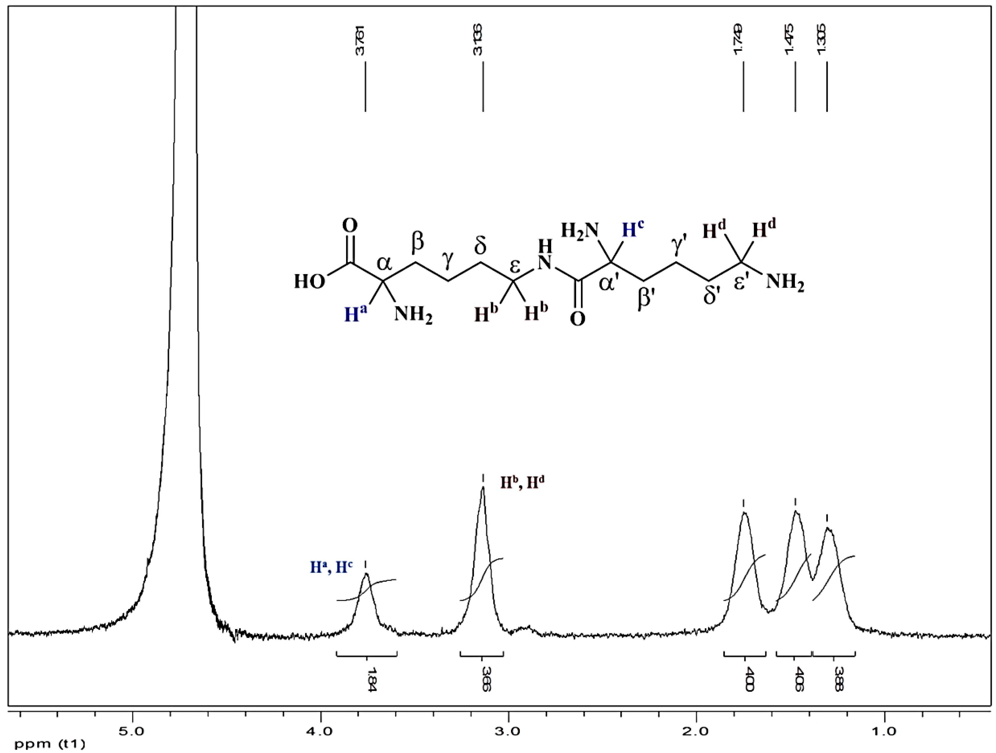
2.4.5. Characterization of ε-PL

Isolated ε-PL from Bacillus licheniformis was characterized using NMR in D2O water. 1H -NMR spectra were recorded on a Bruker 400 MHz spectrometer. For 1H NMR, tetramethylsilane (TMS) served as internal standard (δ = 0) and data are reported as follows: chemical shift, integration, multiplicity (s = singlet, d = doublet, t = triplet, q = quartet, m = multiplet) and coupling constant(s) in Hz.
1H NMR (D2O, 400 MHz, δ ppm): 3.87 (1H, t, J 4.0), 3.20–3.11 (2H, m), 1.80–1.77 (2H, m), 1.49 (2H, t j 4.0), 1.31 (2H, t, J 4.0).

2.5. Application of ε-PL for Milk Preservation

2.5.1. Preliminary Study to Select the Best Concentration of ε-PL as a Natural Preservative at 4 °C (Refrigeration)

Raw cow milk samples were distributed to five Erlenmeyer flasks (100 mL each), boiled at 80 °C, and cooled at ambient temperature (pasteurization). ε-PL (natural preservative) was measured and added to each flask at a concentration of 0.005%, 0.02%, 0.08%, and 0.32% (w/v) respectively, while one milk sample without any addition of ε-PL, was kept as control. All the samples were stored at 4 °C temperature in the refrigerator.

2.5.2. Preservation Studies Using 0.02% ε-PL along with Various Concentrations of Sodium Bicarbonate under Ambient Conditions

Raw cow milk samples were distributed to 5 Erlenmeyer flasks (100 mL each), boiled at 80 °C, and cooled at ambient temperature (Pasteurization). Three flasks A, B, and C were supplemented with various concentrations of sodium bicarbonate along with 0.02% ε-PL (Table 1). One milk sample not containing any concentration of ε-PL was kept as a positive control, while another milk sample containing 0.02% ε-PL without any concentration of sodium bicarbonate was considered a negative control. All the samples were stored at ambient temperature (35 °C).

2.5.3. Microbiological Analysis

Milk samples were tested for coliforms, yeast, and mold counts using standard microbiological methods.

Minimum Inhibitory Concentration Assay

To obtain a minimum inhibitory concentration of ε-PL necessary to prevent bacterial growth, an assay was performed to obtain MIC values. Briefly, bacterial cultures including Salmonella, Staphylococcus aureus, Listeria monocytogenes, and Clostridium perfringes were cultured in LB broth at 28 °C, 200 rpm, for 12 h. Bacteria were cultured overnight in LB broth and OD600nm adjusted with 0.1 with LB medium (≈106 CFU/mL). ε-PL was added to each bacterial suspension at the previously determined MICs (unpublished data) and incubated for 2 h at 28 °C and 200 rpm. Further, aliquots at different time intervals were serially diluted and plated on LB agar plates. Thereafter, plates were kept for 2 days at 28 °C and the CFU count was measured.
Similarly, 12 h grown bacterial cultures of Salmonella, Staphylococcus aureus, Listeria monocytogenes, and Clostridium perfringes having 106 CFU/mL in LB medium were added to UHT milk. Thereafter, ε-PL was added to the UHT milk in previously determined concentration and incubated at 28 °C and 200 rpm for 2 h. Further, aliquots at different time intervals were serially diluted and plated on LB agar plates. Finally, plates were kept for 2 days at 28 °C and the CFU count was measured.

Determination of Casein in Milk at Ambient Condition after Addition of Preservatives

40 mL of cow’s milk was taken in a clean dry beaker, and 40 mL of saturated (NH4)2 SO4 solution was added slowly by stirring. The fat along with casein will be precipitated. The solution was filtered, and the precipitate was transferred into another beaker. Then, about 60 mL of distilled water was added to the precipitate. Only casein dissolves in water forming a milky solution and leaving fat undissolved. The solution was further heated to about 40 °C and 1% acetic acid solution was added dropwise until casein gets precipitated. The precipitate was filtered and washed with water, and let to dry; its weight before and after drying was measured [46].

Determination of Lactose in Milk at Ambient Condition after Addition of Preservatives

50 mL milk in a beaker was diluted to 250 mL using distilled water. About 35 mL of “colloidal iron” was added to this solution in drops with constant stirring. The purpose of “colloidal iron” is to clarify the milk (i.e., to remove the protein contents and fat contents in the milk). After the complete addition of “colloidal iron”, 165 mL of distilled water was added and this final mixture was filtered through a filter paper. The filtrate was a 10% milk serum solution.
In a conical flask on 25 mL of the milk serum, 25 mL of N/10 solution of iodine, and 28 mL of N/10 NaOH solution were added to it. The solution was thoroughly mixed, treated with 38 mL of N/10 sodium thiosulfate, and after 5 min titrated against N/20 sodium thiosulfate using starch as an indicator. The percentage of lactose in the milk serum was calculated by using the equation:
X × 0.036,
where X is the difference between the titer values of the sample and the blank volumes. The factor 0.036 is based on the fact that an average of 1 mL of milk contains 0.036 g of lactose [47].

Estimation of Fat Content in Milk under Ambient Conditions after Addition of Preservatives

About 17 mL of milk was taken in a graduated measuring jar. 17 mL of concentrated sulphuric acid was added and then centrifuged. The separated fat was measured by the gravimetric method through oven drying the centrifuged material [47].

Estimation of Acidity in Milk at Ambient Condition after Addition of Preservatives

The determination of acidity in milk was estimated according to the Alizarol test. 2 mL of milk sample was added with 2 mL of Alizarol solution (68% (v/v) ethanol + 0.4 gm. Alizarin; pH 6.7) and observed for desired coloration, texture, and pH. The acid milk presents a mixture where the coloration of the alizarol was pink-rosa (little acid) or yellow (very acid), besides presenting clots. The normal milk presented a mixture with light purple coloration and was clotless. The acidic milk presented brownish pink (sour) or yellow (very sour). The alkaline milk (usually by an increment of water or neutralizing substance) presented violet coloration [47].

3. Results and Discussions

3.1. Distribution of ε-PL Producing Bacteria in Sea Water along the West Coast of India

The west coast of India is a significant location for gathering a variety of marine microorganisms with the ability to produce -PL in a salty environment. In this attempt, 200 marine isolates from the west coast of India [42,43]. Isolates were mainly from the Adri coast, Bhavnagar coast, Diu (Nagoa beach), Gopnath coast, Rajula (Victor Port), Jafrabad coast, Madhavpur (Junagarh), Mandvi Beach (Kutch) and Veraval coast were isolated.

3.2. Isolation and Screening for ε-PL Producing Bacteria

On the basis of the exclusion of methylene blue from the outer zone of the separated colonies (possessing ε-PL generating potential) on the methylene blue agar plates, 44 bacterial strains were found to be positive out of a total of 200 marine bacteria which were isolated from various seawater samples from the west coast of India. However, when the pure powdered ε-PL (positive charge material) was put to the agar plates containing 0.02% methylene blue, the strong electrostatic contact between ε-PL (a material with a positive charge) and the basic dye (negative charge) resulting in the formation of a zone and complete disintegration of the components present in the region (Nishikawa and Ogawa, 2002). Further, the interaction between the charged groups of the released polypeptides by the isolated bacterium during the screening and the basic dye methylene blue enabled the above-discussed method to investigate many microbes in a single run of screening multiple bacterial strains. The potential bacterial isolates were then added to the M3G production medium as the reported production medium to check the yield of the ε-PL. After screening 44 potential marine bacterial isolates in the production medium (M3G medium), Bacillus licheniformis PL26 was found to be the most potential and suitable for further studies as it can grow on normal M3G medium using submerged fermentation to produce ε-PL at a concentration of 3.6 g/L in 36 h (production age). However, it was also observed that for the first time, it was reported that any such strain of Bacillus licheniformis can produce ε-PL. Also, the production date of ε-PL is reported only from Streptomyces albulus with a production age of 120 h and on the other hand, our isolated strain of Bacillus licheniformis can produce a maximum concentration of 3.6 g/L ε-PL in 36 h which makes the upstream process more sustainable and cost-effective.

3.3. Characterization of ε-PL

Protons (Ha, Hc) attached to α-amino groups arrived together as a broad singlet at δ 3.76 ppm, and protons (Hb, Hd) attached to ε-amino groups arrived together as a broad singlet at δ 3.14 ppm. Protons attached to β and β’ carbons come at δ 1.75 ppm as a broad singlet. While the other protons attached to carbons come at δ 1.47 ppm (4H, ε and εʹ) and δ 1.30 ppm (4H, γ and γʹ) respectively (Figure 2).

3.4. Application of ε-PL for Milk Preservation

3.4.1. Preservation of Milk at 4 °C

Based on pH, the control milk sample (without ε-PL) was spoiled on day 8 at 4 °C, whereas the milk sample containing 0.02% w/v ε-PL could be stored at 4 °C up to 16 days (Table 2). Less than 4 °C, after 16 days, milk samples were spoiled due to acidification.
However, no microbial growth was found in the milk which may be due to the inhibitory action of ε-PL towards most of the Gram-positive and Gram-negative microbe. The optimum ε-PL concentration for the storage of milk samples was 0.02% w/v ε-PL. It was found that at ambient temperature 0.02% w/v ε-PL spoiled the milk samples due to the acidification process, although there were no traces of microbial growth. Therefore, to avoid acidification of milk, 0.1% and 0.2% w/v sodium bicarbonate (NaHCO3) were added along with 0.02% w/v ε-PL (Table 3). It was observed that the milk samples could be stored for 48 h at ambient conditions (35 °C) without any spoilage due to neither microbial growth nor acidification and the storing of milk effectively for 48 h during the summer season was possible.

3.4.2. Preservation of Milk at Ambient Temperature

One of the major concerns with raw milk is that it spoils when left at room temperature in summertime. Further, to avoid spoilage and extend the self-life, efforts have been made to control the spoilage of milk at 35 °C, but it was observed that the acidification occurred after the addition of ε-PL due to a slightly acidic pH of ε-PL. Further, ε-PL was added with various concentrations of sodium bicarbonate for neutralizing the pH of the milk and it was observed that when 0.02% ε-PL was added with 0.2% ε-PL having neutral pH = 7.092, the milk can be stored under ambient temperature up to 48 h to 2 days without any spoilage. Further, after 48 h, the milk with 0.02% ε-PL was in good condition and the growth of bacteria as well as coliforms were in negligible concentrations (<10 cfu/g) obtained in the samples (Table 4). However, both the controls, the milk samples without any preservative and samples containing 0.02% ε-PL were spoiled within 12–24 h after pasteurization.
Although for the last few decades, researchers have been working on the preservation of raw milk at ambient temperature, most of the processes are limited to preservation using chemical preservatives [48]. According to one of the reports from the Food and Agriculture Organisation (FAO) of the United Nations, milk is being preserved by using the lactoperoxidase (LPO) system, but during the process, harmful chemicals such as sodium thiocyanate and sodium percarbonate are used which are toxic for human health if consumed in larger concentration [49]. Simultaneously, researchers have attempted to use hydrogen peroxide for preserving raw sheep milk under ambient conditions (35 °C) for up to 15 h and storage at 4 °C, but, using only hydrogen peroxide, it is difficult to store raw milk for a longer time period. Therefore, the LPO system was considered an effective system for raw milk preservation; however, a major drawback is the addition of sodium thiocyanate and hydrogen peroxide, which are toxic to human health if consumed above the permissible limit [50]. Further, nisin and reuterin were used as antimicrobial agents for raw milk storage, but, their addition along with the LPO system was found to be more efficient for antimicrobial activity effectiveness. Besides, ε-polylysine as a new food biopreservative has shown higher antimicrobial properties compared with natamycin and nisin for the preservation of different foods [16,17,18]. There are major concerns about the addition of chemical preservatives (toxic to human health if consumed more), and there is a need to replace them with only natural/food-grade preservatives for milk and milk-based food products [51].

Minimum Inhibitory Concentration Assay

The antimicrobial effect of ε-PL was evaluated through in-vitro assays. The minimum inhibitory concentration (MIC) generally are determined based on the lowest concentration of the peptide solution that was able to inhibit the bacterial growth. In the present context, ε-PL inhibited the growth of all four bacterial species namely Salmonella, Staphylococcus aureus, Listeria monocytogenes, and Clostridium perfringes. The MIC of ε-PL varied between 200 µg/mL to inhibit Salmonella growth, 400 µg/mL to inhibit the growth of Staphylococcus aureus, 260 µg/mL to inhibit the growth of Listeria monocytogenes and 500 µg/mL to inhibit the growth of Clostridium perfringes. Similar concentrations were obtained for the bacteria (Salmonella, Staphylococcus aureus, Listeria monocytogenes, and Clostridium perfringes) inoculated to milk. ε-PL efficiently inhibited the growth of all four bacteria in the UHT milk as well. However, it was also found that the peptide ε-PL efficiently inhibited the growth of colonies after 40 min of ε-PL treatment with a reduction in bacterial growth close to 100%, which means no growth was observed after that. After two hours, the reduction remained, confirming the antibacterial activity of ε-PL. Further, a possible limitation of the methods used is that the initial 0 of Listeria monocytogenes was not carried out and this may be beneficial for future studies.

Effect of ε-PL and Sodium Bicarbonate of Casein, Lactose, and Fat Stability

It was found that after 24h under ambient temperature (35 °C), 55% of casein (Table 5), 40% of lactose (Table 6), and 11% fat (Table 7) were degraded in the milk containing 0.02% ε-PL and 0.1% sodium bicarbonate. However, to crosscheck the acceptability of milk, the ALIZAROL test has been performed, and it was found that after the addition of the prepared solution, the color of the milk changed to dark purple which clearly confirms it as fresh milk having pH 6.6 and acceptable for human consumption. However, on the other hand, the color of the milk containing only ε-PL and the milk without preservatives changed to yellow with a pH less than 6.0 (acidic) after 16 h which confirmed its unacceptability due to acidification as well as the sour texture.

4. Conclusions

In the present study has been proven that ε-Polylysine isolated from the fermentation broth of Bacillus licheniformis PL26 grown in M3G medium, a new natural biopreservative with antimicrobial properties in combination with sodium bicarbonate (added to avoid acidification) could be a good candidate to store at 35°C up to 48h the raw milk. Therefore, for the dairy industry, such a natural biopreservative replacing synthetic preservatives derived from renewable sources can be proposed for practical applications, which can be further researched and extended to different dairy products. Once again, marine microorganisms represent valuable sources of bioactive substances that need to be explored, and the present study demonstrates another beneficial application of them.

Author Contributions

Conceptualization, S.B. and S.M.; methodology, S.B.; formal analysis, H.H.S. and S.B.; writing—review and editing, A.V.R.; M.T. and A.Z.; APC funding acquisition, M.T. All authors have read and agreed to the published version of the manuscript.

Funding

This research received no external funding.

Data Availability Statement

Not applicable.

Acknowledgments

Financial assistance from CSIR as a part of the CSIR-FTT project (MLP 0049) is gratefully acknowledged. We acknowledge the analytical science division of CSIR-CSMCRI for their timely support and cooperation. We extend our gratitude to BDIM for providing PRIS no. CSIR-CSMCRI–212/2014.

Conflicts of Interest

The authors declare no conflict of interest.

References

  1. Șanta, A.; Mierlita, D.; Dărăban, S.; Socol, C.T.; Vicas, S.I.; Șuteu, M.; Maerescu, C.M.; Stanciu, A.S.; Pop, I.M. The Effect of Sustainable Feeding Systems, Combining Total Mixed Rations and Pasture, on Milk Fatty Acid Composition and Antioxidant Capacity in Jersey Dairy Cows. Animals 2022, 12, 908. [Google Scholar] [CrossRef]
  2. Shakeel, K.; Rabail, R.; Iahtisham-Ul-Haq; Sehar, S.; Nawaz, A.; Manzoor, M.F.; Walayat, N.; Socol, C.T.; Maerescu, C.M.; Aadil, R.M. Camel milk protectiveness toward multiple liver disorders: A review. Front. Nutr. 2022, 9, 944842. [Google Scholar] [CrossRef] [PubMed]
  3. Ameer, S.; Aslam, S.; Saeed, M. Preservation of Milk and Dairy Products by Using Biopreservatives. Middle East J. Appl. Sci. Technol. (MEJAST) 2019, 2, 72–79. [Google Scholar]
  4. Chaudhary, V.; Kajla, P.; Kumari, P.; Bangar, S.P.; Rusu, A.; Trif, M.; Lorenzo, J.M. Milk protein-based active edible packaging for food applications: An eco-friendly approach. Front. Nutr. 2022, 9, 942524. [Google Scholar] [CrossRef] [PubMed]
  5. Nurliyani, P.; Aryuanti, F.; Indratiningsih. Change of Mare Milk Quality During Storage at Room Temperature. Pak. J. Nutr. 2015, 14, 642–646. [Google Scholar] [CrossRef]
  6. Munsch-Alatossava, P.; Alatossava, T. Quality and Safety of Bovine Raw Milk: Present Challenges and Technological Solutions. In Milk Production, Processing and Marketing; IntechOpen: London, UK, 2019. [Google Scholar] [CrossRef]
  7. Fei, Y.; Yang, Z.; Niazi, S.; Chen, G.; Nasir, M.A.; Khan, I.M.; Rehman, A.; Aadil, R.M.; Trif, M.; Coşier, V. Proteolysis of β-Lactoglobulin Assisted by High Hydrostatic Pressure Treatment for Development of Polysaccharides-Peptides Based Coatings and Films. Coatings 2022, 12, 1577. [Google Scholar] [CrossRef]
  8. Gálvez, A.; López, R.L.; Pulido, R.P.; Burgos, M.J.G. Biopreservation of Milk and Dairy Products. In Food Biopreservation; SpringerBriefs in Food, Health, and Nutrition; Springer: New York, NY, USA, 2014. [Google Scholar] [CrossRef]
  9. Shi, C.; Maktabdar, M. Lactic Acid Bacteria as Biopreservation Against Spoilage Molds in Dairy Products—A Review. Front. Microbiol. 2022, 12, 819684. [Google Scholar] [CrossRef]
  10. Islam, S.; Pramanik, M.J.; Biswas, S.; Moniruzzaman, M.; Biswas, J.; Akhtar-E-Ekram, M.; Zaman, S.; Uddin, M.S.; Saleh, M.A.; Hassan, S. Biological Efficacy of Compounds from Stingless Honey and Sting Honey against Two Pathogenic Bacteria: An In Vitro and In Silico Study. Molecules 2022, 27, 6536. [Google Scholar] [CrossRef]
  11. Vithushana, T.; Jayaweera, B.P.A. Aloe Vera Gel as a Natural Preservative on the Shelf life of Flavored Pasteurized Milk. Int. Res. J. Eng. Technol. 2020, 7, 3502–3504. [Google Scholar]
  12. Mokoena, M.P.; Omatola, C.A.; Olaniran, A.O. Applications of Lactic Acid Bacteria and Their Bacteriocins against Food Spoilage Microorganisms and Foodborne Pathogens. Molecules 2021, 26, 7055. [Google Scholar] [CrossRef]
  13. Bangar, S.P.; Sharma, N.; Kumar, M.; Ozogul, F.; Purewal, S.S.; Trif, M. Recent developments in applications of lactic acid bacteria against mycotoxin production and fungal contamination. Food Biosci. 2021, 44, 101444. [Google Scholar] [CrossRef]
  14. Fidan, H.; Esatbeyoglu, T.; Šimat, V.; Trif, M.; Tabanelli, G.; Sensoy, I.; Ibrahim, S.A.; Özogul, F. Recent Developments of Lactic Acid Bacteria and Their Metabolites on Foodborne Pathogens and Spoilage Bacteria: Facts and Gaps. Food Biosci. 2022, 47, 101741. [Google Scholar] [CrossRef]
  15. Zapaśnik, A.; Sokołowska, B.; Bryła, M. Role of Lactic Acid Bacteria in Food Preservation and Safety. Foods 2022, 11, 1283. [Google Scholar] [CrossRef]
  16. Gharsallaoui, A.; Oulahal, N.; Joly, C.; Degraeve, P. Nisin as a Food Preservative: Part 1: Physicochemical Properties, Antimicrobial Activity, and Main Uses. Crit. Rev. Food Sci. Nutr. 2016, 56, 1262–1274. [Google Scholar] [CrossRef]
  17. Ibarra-Sanchez, L.A.; El-Haddad, N.; Mahmoud, D.; Miller, M.J.; Karam, L. Invited review: Advances in nisin use for preservation of dairy products. J. Dairy Sci. 2020, 103, 2041–2052. [Google Scholar] [CrossRef] [PubMed]
  18. Bangar, S.P.; Suri, S.; Trif, M.; Ozogul, F. Organic acids production from lactic acid bacteria: A preservation approach. Food Biosci. 2022, 46, 101615. [Google Scholar]
  19. Oinaala, D.; Lehesvaara, M.; Lyhs, U.; Tikkanen-Kaukanen, C. Antimicrobial activity of organic honeys against food pathogenic bacterium Clostridium perfringens. Org. Agr. 2015, 5, 153–159. [Google Scholar] [CrossRef]
  20. Hossain, M.L.; Lim, L.Y.; Hammer, K.; Hettiarachchi, D.; Locher, C. A Review of Commonly Used Methodologies for Assessing the Antibacterial Activity of Honey and Honey Products. Antibiotics 2022, 11, 975. [Google Scholar] [CrossRef] [PubMed]
  21. Krushna, N.S.A.; Kowsalya, A.; Radha, S.; Narayanan, R.B. Honey as a natural preservative of milk. Indian J. Exp. Biol. 2007, 45, 459–464. [Google Scholar]
  22. Sešķēna, R.; Jankevica, L. Influence of chemical preservatives on the quality and composition indices of raw milk. Acta Univ. Lativiensis 2007, 723, 171–180. [Google Scholar]
  23. Saha, B.K.; Ali, M.Y.; Chakraborty, M.; Islam, Z.; Hira, A.K. Study on the Preservation of Raw Milk with Hydrogen Peroxide (H2O2) for Rural Dairy Farmers. Pak. J. Nutr. 2003, 2, 36–42. [Google Scholar]
  24. Vancin, F.R.; Webber, B.; Bondan, C.; Daroit, L.; dos Santos, L.R.; Rodrigues, L.B. Effects of different concentrations of sodium azide and chloramphenicol on the preservation of raw milk samples. Cienc. Rural. 2020, 50, e20190425. [Google Scholar] [CrossRef]
  25. Shih, I.L.; Shen, M.H.; Van, Y.T. Microbial synthesis of poly (ε-lysine) and its various applications. Bioresour. Technol. 2006, 97, 1148–1159. [Google Scholar] [CrossRef]
  26. Meng, Y.; Lou, L.; Shao, Z.; Chen, J.; Li, Y.; Zhang, T. Antibacterial Activity and Mechanism of Action of Whey Protein-ε-Polylysine Complexes against Staphylococcus aureus and Bacillus subtilis. Foods 2022, 11, 2311. [Google Scholar] [CrossRef] [PubMed]
  27. Meng, Y.; Xue, Q.; Chen, J.; Li, Y.; Shao, Z. Structure, stability, rheology and texture properties of ε-polylysine-whey protein complexes. J. Dairy Sci. 2022, 105, 3746–3757. [Google Scholar] [CrossRef] [PubMed]
  28. Li, S.; Mao, Y.; Zhang, L.; Wang, M.; Meng, J.; Liu, X.; Bai, Y.; Guo, Y. Recent advances in microbial ε-poly-L-lysine fermentation and its diverse applications. Biotechnol Biofuels 2022, 15, 65. [Google Scholar] [CrossRef]
  29. Gu, Y.; Yang, C.; Wang, X.; Geng, W.; Sun, Y.; Feng, J.; Wang, Y.; Quan, Y.; Che, Y.; Zhang, C.; et al. Genome Sequence of the ε-Poly-l-Lysine-Producing Strain Streptomyces albulus NK660, Isolated from Soil in Gutian, Fujian Province, China. Genome Announc. 2014, 2, e00532-14. [Google Scholar] [CrossRef]
  30. Ushimaru, K.; Hamano, Y.; Katano, H. Antimicrobial activity of ε-Poly-l-lysine after forming a water-insoluble complex with an anionic surfactant. Biomacromolecules 2017, 18, 1387–1392. [Google Scholar] [CrossRef]
  31. Shu, C.; Cui, K.; Li, Q.; Cao, J.; Jiang, W. Epsilon-poly-l-lysine (ε-PL) exhibits multifaceted antifungal mechanisms of action that control postharvest Alternaria rot. Int. J. Food Microbiol. 2021, 348, 109224. [Google Scholar] [CrossRef] [PubMed]
  32. Hyldgaard, M.; Mygind, T.; Vad, B.S.; Stenvang, M.; Otzen, D.E.; Meyer, R.L. The antimicrobial mechanism of action of epsilon-poly-l-lysine. Appl. Environ. Microbiol. 2014, 80, 7758–7770. [Google Scholar] [CrossRef] [PubMed]
  33. Nath, A.; Eren, B.A.; Zinia Zaukuu, J.-L.; Koris, A.; Pásztorné-Huszár, K.; Szerdahelyi, E.; Kovacs, Z. Detecting the Bitterness of Milk-Protein-Derived Peptides Using an Electronic Tongue. Chemosensors 2022, 10, 215. [Google Scholar] [CrossRef]
  34. Wang, L.; Zhang, C.; Zhang, J.; Rao, Z.; Xu, X.; Mao, Z.; Chen, X. Epsilon-poly-L-lysine: Recent Advances in Biomanufacturing and Applications. Front. Bioeng. Biotechnol. 2021, 9, 748976. [Google Scholar] [CrossRef]
  35. Wang, C.; Ren, X.; Yu, C.; Wang, J.; Wang, L.; Zhuge, X.; Liu, X. Physiological and transcriptional responses of Streptomyces albulus to acid stress in the biosynthesis of ε-poly-L-lysine. Front. Microbiol. 2020, 11, 1379. [Google Scholar] [CrossRef] [PubMed]
  36. Huang, R.; Liu, H.; Zhao, W.; Wang, S.; Wang, S.; Cai, J.; Yang, C. AdpA, a developmental regulator, promotes ε-poly-l-lysine biosynthesis in Streptomyces albulus. Microb. Cell Fact. 2022, 21, 60. [Google Scholar] [CrossRef]
  37. Silva, C.C.G.; Silva, S.P.M.; Ribeiro, S.C. Application of Bacteriocins and Protective Cultures in Dairy Food Preservation. Front. Microbiol. 2018, 9, 594. [Google Scholar] [CrossRef]
  38. Punia Bangar, S.; Chaudhary, V.; Thakur, N.; Kajla, P.; Kumar, M.; Trif, M. Natural Antimicrobials as Additives for Edible Food Packaging Applications: A Review. Foods 2021, 10, 2282. [Google Scholar] [CrossRef]
  39. Chen, S.; Huang, S.; Li, Y.; Zhou, C. Recent advances in epsilon-poly-L-lysine and L-lysine-based dendrimer synthesis, modification, and biomedical applications. Front. Chem. 2021, 9, 1–14. [Google Scholar] [CrossRef]
  40. Chaudhary, V.; Punia Bangar, S.; Thakur, N.; Trif, M. Recent Advancements in Smart Biogenic Packaging: Reshaping the Future of the Food Packaging Industry. Polymers 2022, 14, 829. [Google Scholar] [CrossRef]
  41. Katano, H.; Yoneoka, T.; Kito, N.; Maruyama, C.; Hamano, Y. Separation and purification of ε-poly-l-lysine from the culture broth based on precipitation with the tetraphenylborate anion. Anal. Sci. 2012, 28, 1153–1157. [Google Scholar] [CrossRef]
  42. Bhattacharya, S.; Dubey, S.; Singh, P.; Shrivastava, A.; Mishra, S. Biodegradable Polymeric Substances Produced by a Marine Bacterium from a Surplus Stream of the Biodiesel Industry. Bioengineering 2016, 3, 34. [Google Scholar] [CrossRef] [PubMed]
  43. N ishikawa, M.; Ogawa, K. Distribution of microbes producing antimicrobial ε-poly-L-lysine polymers in soil microflora determined by a novel method. Appl. Environ. Microbiol. 2002, 68, 3575–3581. [Google Scholar] [CrossRef]
  44. . Bhattacharya, S.; Singh, P.; Maity, N.C.; Mishra, S. Distribution of Antimicrobial ε-polylysine Producing Marine Microbe in Sea Water along West Coast of India. Biomater. Med. Appl. 2018, 2, 1. [Google Scholar] [CrossRef]
  45. Shen, W.C.; Yang, D.; Ryser, H.G. Colorimetric determination of microgram quantities of polylysine by trypan blue precipitation. Anal. Biochem. 1984, 142, 521–524. [Google Scholar] [CrossRef]
  46. Coroian, A.; Coroion, C.O.; Vodnar, D.C.; Trif, M. Study on the main microbiological traits in Romanian buffalo milk. Bioflux 2010, 2, 92–98. [Google Scholar]
  47. Garau, V.; Manis, C.; Scano, P.; Caboni, P. Compositional Characteristics of Mediterranean Buffalo Milk and Whey. Dairy 2021, 2, 469–488. [Google Scholar] [CrossRef]
  48. Desouky, M.M.; Salama, H.H.; El-Sayed, S.M. The effects of camel milk powder on the stability and quality properties of processed cheese sauce. Acta Sci. Pol. Technol. Aliment. 2019, 18, 349–359. [Google Scholar] [CrossRef]
  49. Guidelines for the Preservation of Raw Milk by the Use of Lactoperoxidase System. In Milk and Milk Products, 2nd ed.; World Health Organization, Food and Agriculture Organization of United Nations: Geneva, Switzerland, 2011; Available online: www.fao.org/input/download/standards/29/CXG_013e.pdf (accessed on 7 November 2022).
  50. Arqués, J.L.; Rodríguez, E.; Nuñez, M.; Medina, M. Antimicrobial activity of nisin, reuterin, and the lactoperoxidase system on Listeria monocytogenes and Staphylococcus aureus in cuajada, a semisolid dairy product manufactured in Spain. J. Dairy Sci. 2008, 91, 70–75. [Google Scholar] [CrossRef]
  51. Rusu, A.; Randriambelonoro, M.; Perrin, C.; Valk, C.; Álvarez, B.; Schwarze, A.-K. Aspects influencing food intake and approaches towards personalising nutrition in the elderly. J. Popul. Ageing 2020, 13, 239–256. [Google Scholar] [CrossRef] [Green Version]
Figure 1. Distribution of ε-PL producing bacteria in seawater along the west coast of India.
Fermentation 09 00156 g001
Figure 2. 1H of ε-PL (left lane) in D2O water.
Fermentation 09 00156 g002
Table 1. Preservation studies of milk using 0.02% ε-PL along with various concentrations of sodium bicarbonate.
Sample CodeConcentration
Positive ControlRaw milk with 0.02% ε-PL
Negative controlRaw milk without any preservatives
Preservative ARaw milk + 0.02% ε-PL + 0.1% sodium bicarbonate
Preservative BRaw milk + 0.02% ε-PL + 0.2% sodium bicarbonate
Table 2. pH monitoring of control milk sample and other milk samples added with various concentrations of ε-PL.
ε-PL (%)Initial dayDay 2Day 4Day 6Day 8Day 10Day 12Day 14Day 16Day 18
Control6.606.606.606.546.426.416.406.366.306.337
0.0056.606.606.606.566.506.476.426.406.386.32
0.026.606.606.606.626.606.506.526.506.506.44
0.086.606.606.606.586.566.466.446.406.326.23
0.326.606.606.606.506.516.386.306.286.186.04
Table 3. pH monitoring of control milk sample and other milk samples added with bicarbonate and ε-PL.
ε-PL Concentration (% w/v)Initial Day24 h48 h
Positive control (without ε-PL)6.65.44.9
Negative Control (with 0.02% w/v ε-PL)6.65.44.3
0.02% w/v ε-polylysine + 0.1% w/v NaHCO36.85.94.3
0.02% w/v ε-polylysine + 0.2% w/v NaHCO37.17.14.2
Table 4. Microbiological analysis of milk samples without ε-PL, with 0.02% w/v ε-PL, with 0.02% w/v ε-polylysine + 0.1% w/v NaHCO3 and with 0.02% w/v ε-polylysine + 0.2% w/v NaHCO3.
Test ParameterMeasurement UnitMethod UsedResults (without ε-PL)Results
(with 0.02% w/v ε-PL)
Results
(with 0.02% w/v ε-polylysine + 0.1% w/v NaHCO3)
Results
(with 0.02% w/v ε-polylysine + 0.2% w/v NaHCO3)
Total plate countCfu/mLIS 5402: 20127.5 × 1042.1 × 1031.8 × 1031.1 × 103
Escherichia coli (E. coli)Per mLIS5887 (Part 1)>10absentabsentabsent
SalmonellaPer 25 mLIS5887 (Part 3)absentabsentabsentabsent
Staphylococcus aureusPer mLIS5887 (Part 2)absentabsentabsentabsent
Listeria monocytogenesPer mLIS14988 (Part 1)absentabsentabsentabsent
Bacillus cereusPer mLFSSAI Manual 2022>10absentabsentabsent
Clostridium perfringesPer mLFSSAI Manual 2022absentabsentabsentabsent
Coliform countCfu/mLIS 5401 (Part 1)>10≤8≤8≤5
Table 5. Effect of ε-PL and sodium bicarbonate as the preservative on casein degradation.
Hours% Casein Present
without Preservativewith 0.02% ε-PLwith 0.02% ε-PL +0.1% NaHCO3with 0.02% ε-PL + 0.2% NaHCO3
0100.00100.00100.00100.00
1230.6779.3380.0066.67
248.0080.0044.6724.67
366.6721.3332.6724.67
485.3316.6728.6713.33
Table 6. Effect of ε-PL and sodium bicarbonate as the preservative on lactose degradation.
Hours% Lactose
without Preservativewith 0.02% ε-PLwith 0.02% ε-PL +0.1% NaHCO3with 0.02% ε-PL + 0.2% NaHCO3
0100.00100.00100.00100.00
1270.0050.0060.0060.00
2462.7840.0060.0060.00
3650.0040.0060.0060.00
4840.0032.2230.0040.00
Table 7. Effect of ε-PL and sodium bicarbonate as the preservative on fat degradation.
Hours% Fat Degradation
without Preservativewith 0.02% ε-PLwith 0.02% ε-PL +0.1% NaHCO3with 0.02% ε-PL + 0.2% NaHCO3
0100.00100.00100.00100.00
1266.7413.45100.00100
2465.8011.3789.1681.96
3652.8710.4377.6975.18
4852.146.2674.4561.21
Disclaimer/Publisher’s Note: The statements, opinions and data contained in all publications are solely those of the individual author(s) and contributor(s) and not of MDPI and/or the editor(s). MDPI and/or the editor(s) disclaim responsibility for any injury to people or property resulting from any ideas, methods, instructions or products referred to in the content.

