Article

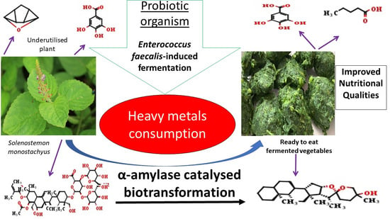
Enterococcus faecalis-Induced Biochemical Transformation during Fermentation of Underutilized Solenostemon monostachyus Leaves

by
Israel Sunmola Afolabi
1,*,
Eze Frank Ahuekwe
2,
Precious Amaneshi Garuba
1,
Aderinsola Jumai Adigun
1,
Oluwatofunmi E. Odutayo
1 and
Alaba Oladipupo Adeyemi
1
1
Biochemistry Department, College of Science and Technology, Covenant University, Canaanland, Ota 112233, Ogun State, Nigeria
2
Biological Sciences Department, College of Science and Technology, Covenant University, Canaanland, Ota 112233, Ogun State, Nigeria
*
Author to whom correspondence should be addressed.
Fermentation 2023, 9(1), 33; https://doi.org/10.3390/fermentation9010033
Submission received: 30 October 2022 / Revised: 16 November 2022 / Accepted: 16 November 2022 / Published: 30 December 2022

Abstract

:
Solenostemon monostachyus is an underutilized plant that is yet to be explored for consumption. This study assessed the biochemical impacts of a probiotic Enterococcus faecalis as an agent of fermentation, promoting the edible properties of the leaves from S. monostachyus using either an unfermented water leaf or S. monostachyus. The results with p < 0.05 were considered statistically significant. The α-amylase activity, proteins, carbohydrates, and ash, iron, and copper contents significantly increased (p < 0.05), while fats, crude fiber, cadmium, and manganese contents of the fermented S. monostachyus leaves were significantly reduced (p < 0.05), compared to the control (unfermented water leaves). The total phenol and saponin contents of the leaves were 1.98 ± 0.03 and 2.77 ± 0.04 mg GAE/mL for the S. monostachyus, 2.20 ± 0.01 and 2.39 ± 0.51 mgGAE/mL for water leaf, respectively on Day 5. Spirostanol (20.7343 mg/10 g) and two possible yet-to-be-identified saponin compounds P-S1 (33.5773 mg/10 g) and P-S2 (23.5718 mg/10 g) were newly synthesized along with one possible novel volatile compound by the fermentation process. Furostanol (19.873–29.420), gallic acid (88.111–98.949 mg/10 g), luteolin (0.954–11.712 mg/10 g) were retained, and aescin (69.510 mg/10 g) was completely consumed by the fermentation process. E. faecalis derived some micronutrients to drive the α-amylase catalyzed biotransformation of phytochemicals to improve the health benefits in the leafy vegetable.

Graphical Abstract

1. Introduction

Food preservation through fermentation is a method that has been around for a very long time. Bacteria and yeast use fermentation to break down carbs [1]. It is used to make yogurt, cheese, and sauerkraut among other things, and has a particularly tasty flavor. Fermentation can be employed in food processing to preserve food, liberate predigested nutrients, or make ethanolic beverages. Almost every country and ethnic group have produced its unique fermented dishes [2]. Improved digestive health, greater immunity, and higher availability of vital nutrients have all been linked to fermented meals. Some fermented foods include: cheese, yogurt, beer, wines, and olives.
Food insecurity affects people all around the world, where poverty limits their purchasing power and prevents them from having reliable access to food [3]. Fermentation generates products using a large culture of microorganisms, including aerobic and anaerobic processes. It is an old way of preserving food and has been for a long time. There are 800 million people who do not have enough to eat. If we consider individuals who do not have access to food, the number increases to 1.2 billion people. This number of people affected by food insecurity represents one-fifth of the global population [4]. Further, two billion people are micronutrient deficient in one or more nutrients [3]. Fermentation technologies are crucial in ensuring the food security of millions of people worldwide, particularly marginalized and vulnerable groups [5].
The nutritional quality is the primary reason for consuming leafy vegetables, which are usually bitter and sour in their natural state [6]. The taste and sensory properties of vegetables are of secondary importance since they are not often consumed directly. These properties during the consumption of leafy vegetables are most usually influenced by those of the spices and oil added to improve their edible qualities [7,8]. Fermented vegetables are beneficial to one’s health, and kimchi and sauerkraut are probably the most popular fermented vegetables [9]. Solenostemon monostachyus, popularly known as monkey’s potato belongs to the family of Lamiaceae, it is a valuable herb found in West and Central Africa that has many phytochemical properties, but it is presently rarely consumed. The plant is locally called Ntorikwot among the Ibibio and Olojogbodu among the Yoruba tribe in Nigeria [10]. S. monostachyus provides the body with various minerals, phytochemicals, anti-inflammatory effects, and antisickling activities [11]. The plant has the potential to benefit humans globally since it is also geographically located in Costa Rica and some other parts of the world [11,12].
Food insecurity affects people worldwide, where poverty limits their purchasing power and prevents reliable access to food [4]. Food prices worldwide have grown considerably in recent years, and they are expected to climb even more due to inflation in the pricing of other commodities, and the increasing global population [4]. It was projected that 795 million people out of 7.3 billion people worldwide suffered from hunger and malnutrition between 2014 and 2016, with developing nations accounting for 780 million of the hungry [13]. Hunger and malnutrition, such as micronutrient deficiencies, are problems that most developing countries in the world experience.
While hunger, diminishing food resources, and food insecurity persist, many underutilized plant leaves could serve as alternate sources of food and nutrients if properly harnessed. Numerous parts from these underutilized plants are often discarded due to their likely high antinutrient contents limiting the utilization of the nutrients they possess. Fermentation can increase the nutritional value of plant leaves [14,15]. The leaves of the S. monostachyus plant were selected for this study due to the numerous medicinal benefits, such as antisickling potential, helping to facilitate childbirth, and others associated with it. The leaves present low toxicity and may be a useful source of health-beneficial phytochemicals and nutrients when administered and consumed nutritionally after improvements with necessary processing techniques [16]. This study is aimed to provide a scientific foundation for the use of fermentation in the processing of underutilized plant leaves for safety and edibility [9,14]. This study aims to evaluate the effect of specific lactic acid bacteria as a fermenting agent of enhancing the edible properties of the leaves from Solenostemon monostachyus and identify the specific phytochemicals serving as the prebiotic carbon source for the lactic acid bacteria, improve consumption and the nutritional value of the yet-to-be-consumed plant leaves, and to assess the in vitro antioxidant capacities of fermented leaves.

2. Materials and Methods

2.1. Chemicals

Absolute ethanol (Sigma Aldrich, St. Louis, MO, USA), methanol (Sigma Aldrich, St. Louis, MO, USA), potato starch (Sigma Aldrich, St. Louis, MO, USA), 2,2-diphenyl-1-picrylhydrazyl (Sigma Aldrich, St. Louis, MO, USA), De Man Rogosa and Sharpe (MRS) agar (Oxoid medium, Basingstoke, UK), Mueller-Hinton agar (Oxoid medium, Basingstoke, UK), Analytical Profile Index (API) 50 CHL kit (Biomereux ltd, Grenoble, France), gum acacia (Mallinckrodt Baker, Phillipsburg, NJ, USA), glycerol tributyrate (Merck Schuchardt, Hohenbrunn, Germany), glacial acetic acid (analytical grade), methanol (Lichrosolv, Darmstadt, Germany), acetonitrile (Lichrosolv, Darmstadt, Germany), and formic acid used for this study were of analytical grade. The other HPLC grades reference standards chemicals for the bioflavonoids used were methanol (Lichrosolv, Darmstadt, Germany), acetonitrile (Lichrosolv, Darmstadt, Germany), rutin (95%), quercetin (95%), quercitrin (85%), kaempferol (90%) and isorhamnetin (99%). Methanol (Lichrosolv, Darmstadt, Germany), acetonitrile (Lichrosolv, Darmstadt, Germany), ascorbic acid, gallic acid, catechin, methyl gallate, caffeic acid, syringic acid, ellagic acid, chlorogenic acid (HPLC grades) served as the reference standards for the phenolic compounds. Diosgenin, gitogenin, and hecogenin (HPLC grades) among others served as reference standards for saponins.

2.2. Collection of Plant Leaves

Solenostemon monostachyus were collected within the lawns of Covenant University, Ota, Ogun State, Nigeria. These plants were identified at the Applied Biology Unit of the Biological Sciences Department at Covenant University. For each plant that was deposited at FRIN, voucher numbers were assigned and received. For the experiment, the unfermented leaves of S. monostachyus plant serves as the control for monitoring the effects of fermentation, while the southwest of Nigeria’s Talinum fruticosum edible leaves—locally known as water leaf—were employed as the control for monitoring the edible status of the experimental leaves.

2.3. Procedure for Preparation of Organism for the Experimentation

Collection and Sub-Culturing of Lactic Acid Bacteria

Enterococcus faecalis (MW481698) used for this study was earlier identified, confirmed as Gram-positive, catalase-negative, and non-hemolytic, and deposited in the NCBI database [17]. E. faecalis was collected from the Molecular Biology laboratory at Covenant University and suspended in MRS broth at 37 °C under static conditions for this experimentation [18]. They were grown in Brain Heart Infusion (BHI) broth at 37 °C under shaking conditions (200 rpm) for 4–6 h. The above-mentioned cultures were maintained at 40 °C in MRS and BHI media with 40% glycerol (v/v). Before use, the cultures were propagated twice in their respective broths [18].

2.4. Procedure for the Fermentation of Plant Leaves

2.4.1. Preparation of the Plant Leaves for Fermentation

The harvested plant leaves were submerged in clean water and rinsed thoroughly before removing the washed leaves from any suspended dirt. This washing process was additionally repeated twice. The final cleaned leaves were then carefully chopped into tiny pieces, soaked in water, and blended. The prepared fermenting medium consisting of a combination of an equal volume of salt solution (3.0%) and sugar solution (3.0%), was also prepared, sterilized, and allowed to stand for 48 h.

2.4.2. Sterilization of Materials for Use during Fermentation

The inoculum was prepared by suspending a loopful of the fresh cultures of Enterococcus faecalis in a 2 mL sterile saline solution and washing twice at 10,000× g for 10 min. An aliquot (2.0 mL) of the inoculum (2 × 10³ CFU/mL) was aseptically dispensed into the sterile 1000 mL fermentation jars containing 500 mL of the sterile 2.5% of NaCl (brine) and 1 kg of the already chopped vegetables. This setup was incubated with a Thermo Scientific Ltd. incubator (model: 371, serial no: 153729-9146, Waltham, MA, USA) at room temperature of about 37 °C for 3 and 5 days, alongside an uninoculated jar serving as control [19].

2.5. Post-Fermentation Analyses

After each day of fermentation of leafy vegetables, the solution containing both the vegetables and fermentation inoculum was blended and sieved out for further analysis. The oven-dried residues from the blending process were analyzed for proximate and mineral contents. The collected filtrates from the fermented and unfermented leaves portioned into a filtrate A (FA) were directly analyzed for pH, proximate contents, antioxidant qualities (total antioxidant capacities, DPPH and FRAP), biochemical enzymes, quantification of phytochemical contents (phenols, flavonoids, and saponins), and GC-MS profile of the chemical constituents. Another portion of the filtrate B (FB) was further condensed using a rotary evaporator at 20 °C for a period of 30 and 20 min for water leaf, and S. monostachyus, respectively. It was this condensed filtrate (CF) that was reconstituted for the identification and quantification of the antioxidant vitamins (A, C, and E), saponins, phenolic, and bioflavonoids compounds using high-performance liquid chromatography (HPLC). The pH of the extracts was determined using a Hanna Instruments pH meter (model: pHep, serial no: HI96107, Villafranca Padovana, Italy) as described by Afolabi and Oloyede [20].

2.5.1. Quantitative Proximate Composition and Total Phytochemical Analysis

The moisture, total fats, crude fiber, and ash contents were determined by the AOAC method [21]. The total protein content of the extracts was assayed spectrometrically (Thermo Scientific Ltd., model: GEN10S UV-Vis, serial no: 2L5V095205, Waltham, MA, USA) at 540 nm wavelength using the biuret method [22] and total carbohydrate content was assayed spectrometrically (Thermo Scientific Ltd., model: GEN10S UV-Vis, serial no: 2L5V095205, Waltham, MA, USA) at 620 nm wavelength using the Anthrone reagent method described by Ibrahim and Jaafar [23]. The total saponin, total flavonoids, and total phenol contents were determined according to the method described by Olawole et al. [24].

2.5.2. Assay for Total Antioxidant Capacity (TAC) and Ferric-Reducing Antioxidant Power (FRAP)

Total antioxidant capacity (TAC) and the ferric-reducing antioxidant power (FRAP) of extracts and residues from the control and the fermented leaves were assayed spectrometrically (Thermo Scientific Ltd., model: GEN10S UV-Vis, serial no: 2L5V095205, Waltham, MA, USA), respectively, 695 nm and at 700 nm wavelengths following the methods described by Sharma et al. [25]. The absorbance of all solutions was read and expressed as mg of ascorbic acid equivalent per gram of extract (mg AAE/g).

2.5.3. Diphenyl-1-Picrylhydrazyl Radical Scavenging Activity

The abilities of the extracts to scavenge DPPH radicals were determined spectrometrically (Thermo Scientific Ltd., Waltham, MA, USA, (model: GEN10S UV-Vis, serial no: 2L5V095205) 517 nm wavelength as described by Sakat and Juvekar [26]. The percentage inhibition (DPPH scavenging activity) was calculated using the following formula:
%   Inhibition = Absorbance   of   control   Absorbance   of   sample Absorbance   of   control × 100

2.5.4. Assay for Micronutrients in the Sample Leaves

Procedure for Preparing the Samples for Mineral Analysis

The previous ashed samples (1.0 g) were digested using the AOAC wet-ash method described by Boyer [27].
Procedure for Micronutrients Determination Using Atomic Absorption Spectrometric Technique.
Atomic Absorption Spectrometry (Buck Scientific 210VGP, East Norwalk, CT, USA) pre-set at absorbance/emission of −0.0820, −3.200, and 50 psi air/acetylene flame and integration periods of 0.5–10 s were engaged in the determination of the cadmium, chromium, copper, iron, lead, and nickel constituents. The method previously described for the determination of calcium, selenium, potassium, magnesium, sodium, and zinc using an Atomic Absorption Spectrophotometer (Shimadzu, model AA-7000, Nishi-ku, Japan) was adopted for this study [28].

Assay for Vitamin Contents of the Sample Leaves with HPLC

The extracted samples (10 µL), including the standard for vitamin A (1.08 mg/mL) and vitamin E (1.0 mg/mL), were injected into the HPLC (model: Agilent 1200) operating with 100% methanol (absolute) mobile phase, and 2.0 mL/min flow rate. HPLC equipped with a diode array (DA) detector, Zorbax Eclipse XDB-C18 (4.6 nm × 150 nm, 5 µL) column set at 280 nm wavelength, 40 °C was engaged to analyze the vitamins contents of the sample extracts.

Water Soluble Vitamin C Analysis

The extracted samples (10 µL), including the standard for vitamin C (1.0 mg/mL, BDH, 99.89%), were injected into the HPLC (model: Agilent 1200) operating with acidified water (pH 2.16): acetonitrile (99:1) mobile phase, and 2.0 mL/min flow rate. HPLC equipped with a diode array (DA) detector, Zorbax Eclipse XDB-C18 (4.6 nm × 150 nm) column set at 220 nm wavelength, 40 °C was engaged to analyze the vitamins contents of the sample extracts previously mixed with the mobile phase (25% v/v).

2.5.5. Assay for Phytochemical Constituents of the Sample Leaves

Preparation of Sample for HPLC Analysis

An aliquot of sample extract (0.1 g) was mixed with 10 mL of 70% methanol and was allowed to stand for 1–2 h in a closed test tube. The extracted sample was thereafter decanted, centrifuged with a refrigerated centrifuge (model: CR21G, serial no: S2025709), and filtered using a micron filter into a 5 mL sample bottle. The sample filtrate obtained was employed for the HPLC assessment of the saponins, phenolic, and bioflavonoid compounds in the extracts of the leaves.

Procedure for the HPLC Analysis of the Saponins Fractions

The extracted saponin samples (40 µL) were injected into the HPLC (model: Agilent LC-8518) operating with acetonitrile/water (70:30) mobile phase and at 205 nm wavelength and a run time of 14 min. HPLC equipped with a high-sensitivity LC-8518 ultraviolet (UV) detector, column (150 mm × 4.6 mm) set at 40 °C, and a low-pressure gradient and solvent delivery LC-8518 pump with a high-pressure switching valve was engaged to analyze the flavonoids content of the extracts, and amount in the extracted sample using N2000 chromatography software (4.0 version).

Procedure for the HPLC Analysis of the Phenolic Fractions

The extracted phenolic samples (40 µL) were injected into the HPLC (model: Agilent LC-8518) operating with acetonitrile/water/acetic acid (19:80:1) mobile phase and at 272 nm wavelength and a run time of 25 min. HPLC equipped with a high-sensitivity LC-8518 diode array (DA) detector, column (150 mm × 4.6 mm) set at 35 °C, and a low-pressure gradient and solvent delivery LC-8518 pump with a high-pressure switching valve was engaged to analyze the flavonoids content of the extracts, and amount in the extracted sample using N2000 chromatography software.

Procedure for the HPLC Analysis of the Flavonoids Fractions

The extracted flavonoids samples (40 µL) were injected into the HPLC (model: Agilent LC-8518) operating with acetonitrile/water/formic acid (25:74:1) mobile phase and at 210 nm wavelength and a run time of 25 min. HPLC equipped with a high-sensitivity LC-8518 diode array (DA) detector, column (150 mm × 4.6 mm) set at 40 °C, and a low-pressure gradient and solvent delivery LC-8518 pump with a high-pressure switching valve was engaged to analyze the flavonoids content of the extracts, and amount in the extracted sample using N2000 chromatography software.

Procedure for the Phytochemical Analysis Using GC-MS

The procedure described by Odutayo, Adegboye, Omonigbehin, Olawole, Ogunlana, and Afolabi [17] was followed in the estimation of the phytochemical constituents of the extracts. The phytochemical compounds obtained in this study were drawn structurally and sequenced into a proposed scheme of bio-transformational mechanisms using ChemAxon MarvinSketch software (version 15.9.14.0, ChemAxon limited, Budapest, Hungary).

2.5.6. Procedure for Enzyme Analysis

α-Amylase Activity Assay

The α-amylase activity of the enzymes, the method described by Ho and Yin Sze [29], was adopted. The absorbance was read at 550 nm. Distilled water served as blank. A unit of amylase activity was expressed as
milligram   of   maltose   released time   of   incubation × mL   enzyme   in   the   reaction   mixture

Lactate Dehydrogenase Activity Assay

The activity of lactate dehydrogenase (LDH) in extracts was assessed using the semi-micro method described in the Randox kit, and the equivalent activity (U/l) was calculated using the following formula.
U / l = 8095 ×   Δ Absorbance   340   nm / min

2.6. Method of Statistical Analysis

All analysis was analyzed with the aid of MegaStats software (version 10.3 Release 3.2.1) using the analysis of variance (ANOVA). Procedures were all performed in triplicates to obtain data reported as mean ± standard deviation and probability of less than 0.05 were considered to be statistically significant.

3. Results

3.1. Effect of Fermentation on the Acidity of the Leaves of S. monostachyus

The acidity of the fermenting medium expectedly changed, although from 4.623 to 8.610 as the fermentation progressed (Table 1). This indicates an increase in alkalinity, instead of an increased acidity during the fermentation, suggesting the process follows an alkaline fermentation process.

3.2. Effect of Fermentation on the Nutritional Qualities of the Leaves of S. monostachyus

The need to extensively appreciate the bioremediation role of the E. faecalis encouraged the extension of the relevant control to day 3 and day 5. The ash content obtained was generally less the 50% of the corresponding values for the S. monostachyus leaves (Table 1). Hence, the ash samples generated from water leaves were extremely small after fermentation due to the nature of the control leaves. The mineral nutrient levels in the S. monostachyus are similar to those of the commonly consumed water leaves vegetables. The S. monostachyus is richer in calcium, selenium, potassium, magnesium, and sodium. The underutilized leaves were rich in all the basic mineral nutrients required for the survival of the consumers, and the minerals were relatively stable, suggesting they were not depleted by the fermentation-induced E. faecalis process (Table 2).
The cadmium concentration reduced significantly (p < 0.05) for the three-days and five-days fermentation of the leaves of S. monostachyus and T. fruticosum compared to the control (unfermented T. fruticosum). An initial significant increase (p < 0.05) in the cadmium concentration in the one-day-fermented leaves of T. fruticosum (Table 2). The manganese concentration reduced significantly (p < 0.05) during the five-days fermentation of the leaves of S. monostachyus and T. fruticosum compared to the control (unfermented T. fruticosum). An initial significant increase (p < 0.05) in the manganese concentration in the one-day fermented leaves of both the S. monostachyus and T. fruticosum (Table 2). An initial significant increase (p < 0.05) in the lead concentration in the one-day fermented leaves of T. fruticosum was mainly observed (Table 2).
The resultant chromatograms for the GC/MS analysis were as indicated (Figures S1–S7). S. monostachyus has lower levels of vitamins A and E and is richer in vitamin C relative to their corresponding contents of vitamins in the commonly consumed water leaf vegetables. The fermentation depleted the levels of vitamin E in S. monostachyus, possibly to serve as a substrate for the E. faecalis-fermenting organism (Table 2).

3.3. Effect of Fermentation on the Antioxidant Qualities of the Leaves of S. monostachyus

The level of DPPH inhibition reduced significantly (p < 0.05) for three–five days and the first three days of fermentation in the leaves of S. monostachyus and T. fruticosum, respectively, compared to the control (unfermented T. fruticosum). The final five-days fermentation significantly increased (p < 0.05) the DPPH inhibition in the T. fruticosum (Table 3). The level of free radical scavenging activity decreased significantly (p < 0.05) for the three–five days of fermentation in the leaves of S. monostachyus compared to the control (unfermented T. fruticosum). On the contrary, the level of free radical scavenging activity reduced (p < 0.05) significantly (p < 0.05) for only the first day and throughout the five-days fermentation of the leaves S. monostachyus and T. fruticosum, respectively (Table 3). The total antioxidant capacity increased significantly (p < 0.05) throughout the five days and significantly reduced (p < 0.05) throughout the five-days fermentation in the leaves of S. monostachyus and T. fruticosum, respectively, compared to the control, unfermented T. fruticosum (Table 3).

3.4. Effect of Fermentation on the Phytochemical Constituents of the Leaves of S. monostachyus

The phenols and saponin concentrations increased significantly (p < 0.05) throughout the five-days fermentation of the leaves of S. monostachyus and T. fruticosum compared to the control, unfermented T. fruticosum (Table 3). The five-day fermentation of the leaves of S. monostachyus and T. fruticosum produced no significant effect (p > 0.05) in the level of flavonoids compared to the control, unfermented T. fruticosum (Table 3).

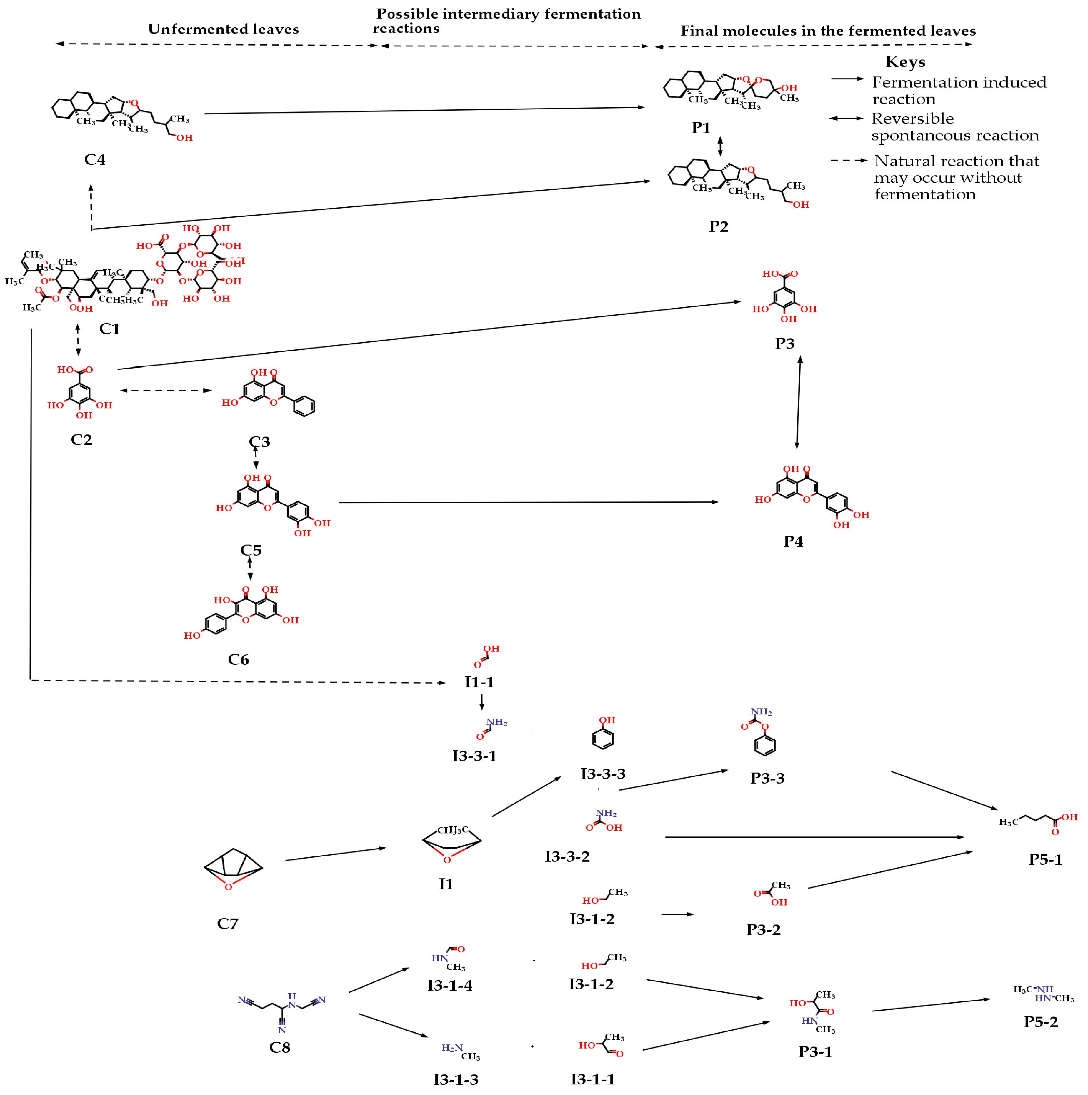
3.5. Metabolism of Phytochemicals during the Fermentation of the Leaves of S. monostachyus

The levels of saponins in S. monostachyus leaves metabolized by E. faecalis-induced fermentation are indicated (Table 4). The major saponin in the leaves, aescin, was completely catabolized by the E. faecalis while also partially catabolizing furastanol. This finding suggests the organism possesses a higher preference for aescin than furastanol. The E. faecalis under this fermentation conditions synthesized spirostanol as two possible yet-to-be-identified saponin compounds (P-S1 and P-S2), in addition to a new yet-to-be-identified saponin compound (S-S1). Considering the metabolism of phenolic compounds during this fermentation, gallic acid was detected as a main phenolic compound in the leaves, which remained relatively stable during the 5-days fermentation. The E. faecalis largely consumed luteolin from 11.71 to 0.95 mg/10 g extract and also completely catabolized kaempferol and chrysin during the 5-days fermentation of the leaves of S. monostachyus (Table 4).
The resultant chromatograms for the GC/MS analysis were as indicated (Figures S8–S10). One possible novel volatile compound was additionally identified with GC/MS technique (Table 5). A 1,2-dimethylhydrazine detected as a major volatile compound on the 5-days-fermented leaves of S. monostachyus is a carcinogen with a strong ability to alter DNA. Although, this study reveals zinc as a major micronutrient in the extract of the leaves. More careful heat-degrading processing techniques such as boiling, frying, cooking, and microwaving should be employed in treating the plant for consumption.

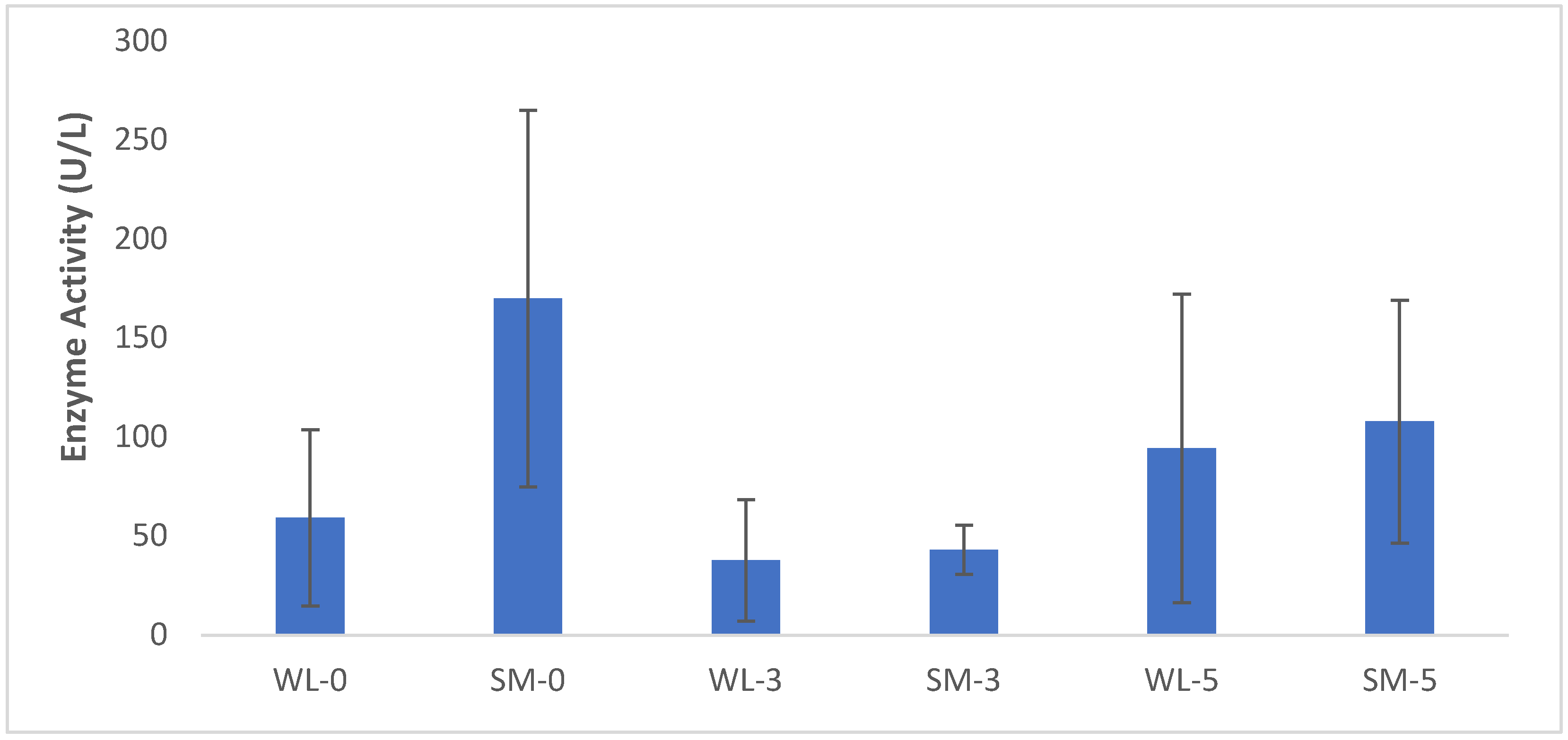
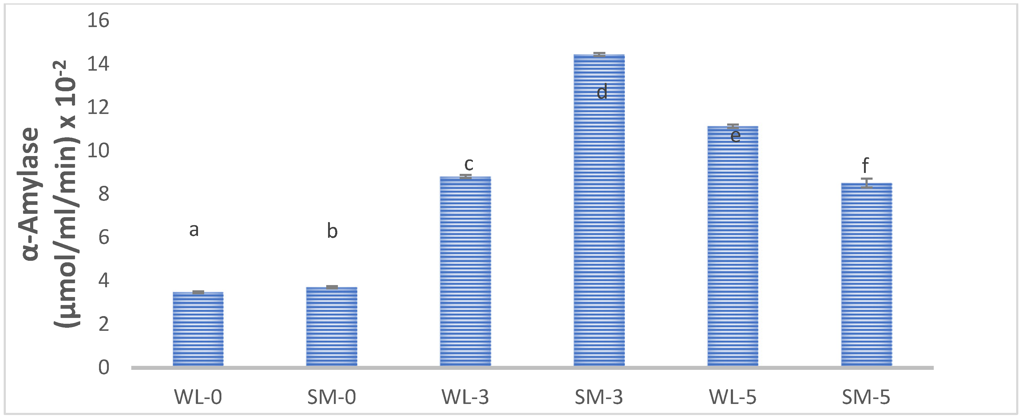
3.6. Effect of Fermentation on the Biochemical Status of the Leaves of S. monostachyus

The five-day fermentation of the leaves of S. monostachyus and T. fruticosum produced no significant effect (p > 0.05) on the LDH activity compared to the control, unfermented.
T. fruticosum (Figure 1). The activity of α-amylase activity increased significantly (p < 0.05) throughout the five-days fermentation of the leaves of S. monostachyus and T. fruticosum compared to the control, unfermented T. fruticosum (Figure 2). This suggests the use of the amylase enzyme as a possible mechanism of action for Enterococcus faecalis, which is the fermenting organism.

4. Discussion

The pH values recorded during this fermentation indicate that the E. faecalis initiated alkaline fermentation to facilitate the process (Table 1). The release of ammonia from the proteolysis of protein was implicated in alkaline fermentation. Ammonia and hydroxyl compound production reduces the pH of the fermenting medium to the alkaline region. Alkaline fermentation is used in food processing to enhance nutrients and bioactive qualities [31,32,33,34,35].
A strain of E. faecalis was implicated in the citrate fermentation pathway during food processing and glycerol metabolism [36,37]. Numerous strains of E. faecalis that were so far identified are harmful and implicated in drug resistance and biofilm formation in the human intestine [38,39,40]. Interestingly, the strain of E. faecalis used in this study was non-virulent due to its Gram-positive, catalase-negative, and non-hemolytic nature previously established for probiotic use [17,41,42].

4.1. Effect of Fermentation on the Nutritional Qualities of the Leaves of S. monostachyus

The fermentation improved nutrients (proteins, carbohydrates, and ash contents) and antioxidant qualities, while it reduced the fats and crude fiber contents of the S. monostachyus (Table 1). In numerous physiological and metabolic processes in plants, iron plays a crucial role. It is needed for a variety of biological processes as it is a component of numerous essential enzymes, including cytochromes of the electron transport chain. Iron is a component of chlorophyll production in plants and is necessary for the preservation of chloroplast structure and function. Fermentation technologies are crucial in ensuring food security. Fermentation of the S. monostachyus plant leaves moderately reduced the iron and copper levels of the plants, which could be used as a suitable product in the treatment of anemia and it is also a known nutrient for E. faecalis [43]. The body needs copper, which is an essential nutrient. It helps the body produce red blood cells along with iron. It contributes to iron absorption and maintains strong bones, blood vessels, nerves, and the immune system. Enough copper in the diet may also help prevent osteoporosis and cardiovascular disease.
The fermentation of the S. monostachyus plant leaves decreased the cadmium level, which causes plant tissue death by causing the production of reactive oxygen species, impeding the use, absorption, and transportation of vital nutrients and water, and altering the photosynthetic apparatus. High cadmium levels also cause health issues, such as kidney disease and cardiovascular disease [44]. The fermentation of the S. monostachyus plant leaves reduced the manganese level. The mineral is important at a moderate level since more than 300 metabolic processes in the body require manganese. It supports healthy immunological function, regulates heartbeat and glucose metabolism, keeps bones strong, and maintains appropriate nerve and muscle function. It assists in the synthesis of protein and energy [44]. Lead is an element that should be avoided in diets due to the numerous serious health hazards it causes to consumers and plants. The effects of this fermentation on the leaves of S. monostachyus could not be ascertained since it was not detected in both the water leaves or the unfermented leaves of S. monostachyus that served as the control for this study. Interestingly, the reduction or elimination of lead in the fermented leaves of the plant can be postulated to follow the pattern presented by other heavy metals such as cadmium during this fermentation process [45].

4.2. Total Antioxidants Qualities

Humans can benefit greatly from plant nutrients’ numerous health advantages. Antioxidant, hormonal, anti-microbial, anti-tumor, and anti-inflammatory properties are just a few of these advantages. As antioxidants, these bioactive molecules combat free radicals. As a result, they boost metabolites that detoxify carcinogens and prevent DNA from being damaged [46]. The improved DPPH levels of the fermented S. monostachyus plant leaves can also be linked to the action of E. faecalis [47,48]. The fermentation of S. monostachyus plant leaves reduced the free reducing scavenging activity (FRAP) level, which shows that S. monostachyus has shown to have a suitable potent reducing power and total antioxidant activity after fermentation, which may be due to its high phenolic, saponin content revealed in this study. Its strong antioxidant ability was previously reported [11,49]. This strong antioxidant ability of the plant also indicates it may arrest the possible side effects of heavy metals such as cadmium and other very few identified phytochemical compounds in this study.

4.3. Effect of Fermentation on the Phytochemical Constituents of the Leaves of S. monostachyus

The phytochemical makeup of a plant affects how it functions physiologically in the body [50]. The fermentation improved the antioxidant-phenolic compounds in the fermented leaves (Table 3). Gallic acid (88.111–98.949 mg/10 g) and luteolin (0.954–11.712 mg/10 g) were the phenolic compounds detected and retained by the fermented leaves of S. monostachyus, while kaempferol (0.066 mg/10 g), chrysin (0.071 mg/10 g), and aescin (69.510 mg/10 g) were completely consumed by the fermentation process (Table 4). These compounds frequently provide a variety of health advantages, such as metabolites with crucial benefits to the cosmetic, pharmaceutical, and food industries. The fermentation conditions used in this study affect the phytochemical components found in these leaves. With each additional day of fermentation, more phytochemical substances (phenols and saponins) are discovered (Table 3). Polyphenol compounds have many health benefits, including the regulation of blood pressure and glucose levels and actions against inflammatory, viral, and cytotoxic properties. They also have anti-malarial, hepaticidal, and diuretic activities in humans [46].
The fermentation improved the antioxidant-phenolic compounds in the fermented leaves (Table 3). Furostanol (19.873–29.420 mg/10 g) is among the saponins detected in the fermented leaves of S. monostachyus. Spirostanol (20.7343 mg/10 g) and two possible yet-to-be-identified saponin compounds (P-S1 (33.5773 mg/10 g) and P-S2 (23.5718 mg/10 g)) were newly synthesized by the fermentation process (Table 4). Some saponins boost the immune system in the body and may have certain cytotoxic effects [51]. Saponins possess several medicinal qualities, such as anti-inflammatory, anti-fungal, anti-bacterial, anti-parasitic, anti-cancer, and antiviral actions [52]. Overall, the fermentation condition used in the study improved the secondary metabolites (saponin and phenols) during the fermentation of the S. monostachyus plant leaves. This strong influence of these groups of compounds may be responsible for the enhanced ability to scavenge DPPH radicals in this study [24,49]. Most of the phytochemicals identified in this study and contributing to the strong antioxidant properties of the leaves of S. monostachyus were previously reported [53]. However, the fermentation of S. monostachyus plant leaves had no remarkable influence on the flavonoid content of the S. monostachyus plant leaves.
Interestingly, one possible novel volatile compound, along with other molecules, was additionally identified with the GC/MS technique. A carcinogenic compound, 1,2-dimethylhydrazine, was among the volatile compounds generated solely by the extended 5-days fermentation of the leaves. Consequently, we proposed the use of high-temperature food processing techniques and other established catalytic-degradation treatments to degrade the harmful compounds provided the extended 5-days fermentation is adopted [54,55,56]. More careful heat-degrading processing techniques such as boiling, frying, cooking, and microwaving should be employed in treating the plant for consumption. Zinc is a necessary nutrient to prevent or regulate the possible carcinogenic effects of the 1,2-dimethylhydrazine in the extracts of fermented leaves of S. monostachyus [57,58]. Fortunately, the extract from the seeds of pumpkin may hopefully contain the bioactive(s) that could eliminate the harmful effects of the compound [59]. The compound (1,2-dimethylhydrazine) was previously identified in grilled foods and heat-treated foods containing preservatives [60]. Consumption of gallic and other extracts from plants has proven to alleviate or eliminate the toxic effects since they provide relevant therapeutic phytochemicals [61,62]. We suggest processing the leaves of the S. monostachyus with palm oil since it is usually used to reduce poisons and harmful compounds similar to 1,2-dimethylhydrazine. Palm oil is incorporated into the processing of cassava and the products of cassava (garri) to counter the effects of cyanide toxicity [63,64,65].
The biosynthesis of the numerous bioactive phytochemicals in plants through the phenylpropanoid pathway was previously verified [66]. The pathway elucidated in this study was distinct from the established phenylpropanoid pathway indicating the role of the fermenting organisms in facilitating the identified metabolic process (Figure 3).
Aescin seems to be the parent phytochemical compound from where most of the other simpler phytochemical compounds are derived. The fermentation produced alcohols and acids, similar to most of the fermentation process [17,67]. Ethanoic acid and valeric acid are the major acids produced by the 3-days and 5-days E. faecalis-induced fermentation process, respectively. This finding indicates that the fermenting organisms are capable of utilizing either or both of the normal alcoholic fermentation pathway and another unique anaerobic fermentation pathway that leads to the production of valeric acid. The choice of a particular fermentation pathway depends on the length of the fermentation or the nature of the phytochemical available as substrate [68,69]. The formation of ethanoic acid from ethanol is a common phenomenon associated with fermentation [70,71]. Valeric acid synthesis was also previously noted during fermentation [72]. This study shows the possible synthesis of valeric acid from ethanoic acid, carbamic acid, or phenyl carbamate. Valeric acid has the potential to impede cancer in the liver and was found to biologically protect against the development of radiation injuries, eczema, and food allergy [73,74].
Formic acid and carbamic acid were revealed to as intermediary metabolites that spontaneously participated during the 5-days fermentation (Figure 3). Spirostanol and furostanol emerged as the major alcoholic compounds from the 5-day E. faecalis-induced fermentation process, while ethanol seems to play a role as a precursor-intermediary metabolite formed preceding the two alcoholic compounds. Similar to the findings in this study, both phenol and 2,5-dimethyloxolane were previously identified with wine, a product of fermentation [75]. The formation of the phenol from 2,5-dimethyloxolane is importantly reported for the first time in this study. The formations of formic acid and gallic acid were previously implicated with fermentation [76,77]. Interestingly, this study elucidates for the first time the possible synthesis of formic acid and gallic acid from aescin-containing plants. The formation of formamide during fermentation was previously established [78]. Electrochemical procedures were recently inaugurated to facilitate the synthesis of formamide due to the importance it presents to the industry [79,80]. Carbamic acid derivatives derived their importance in possessing the required common core structure for the synthesis of most approved drugs. Hence, it attracts interest from drug manufacturing scientists and industries [81]. This study implicates these carbamate derivatives with fermentation for the first time. 2-Hydroxy-N-methylpropanamide and 2-hydroxypropanal were implicated with fermentation for the first time in this study. The formation of 2-hydroxy-N-methylpropanamide from 2-hydroxypropanal was also indicated in this study (Figure 3).

4.4. Effect of Fermentation on the Enzyme Activities of the Leaves of S. monostachyus

4.4.1. Involvement of LDH Activity in the Fermentation

Lactate dehydrogenase essentially contributes to the anaerobic metabolism of glucose when oxygen is absent or scarce. When cells are subjected to anaerobic or hypoxic circumstances, oxidative phosphorylation, which produces ATP, is disturbed. Cells must use an alternative metabolism to provide energy for this procedure. LDH is thus upregulated in these circumstances to meet the demand for energy generation [82]. Enterococcus faecalis is a lactic acid bacterium, but after the fermentation of the S. monostachyus plant leaves, there was no significant difference in the LDH activity. It showed that the LDH levels were normal. The indifference in the LDH activity may account for the inability to detect lactic acid in the GC/MS profiling of molecules in the extract from fermented S. monostachyus leaves. Hence, it is ascertained in this study that the mobilization of LDH enzyme is not a mode of biochemical action in the E. faecalis probiotics organisms concerning the fermentation of the leaves of S. monostachyus. Lactate dehydrogenase is a broad-spectrum enzyme that is influenced by numerous factors such as pH and types of phytochemicals (such as 2-hydroxybutanoic acid, 2-hydroxypentanoic acid, 3-phenyllactic acid, and cyclopropaneglycodic acid) that are constantly changing during fermentation [83,84,85]. The erratic-low preference of the organisms to engage the LDH enzymes compared to the alpha-amylase enzyme under the fermenting conditions used for this experimentation could also generate the indicated high deviations in Figure 1 [86]. Such a wide spectrum in behavior influences LDH activity, leading to slightly higher erratic behavior leading to error deviation [86,87,88,89,90].

4.4.2. Involvement of α-Amylase Activity in the Fermentation

α-Amylase is widespread among microorganisms in the digestive systems of humans and other mammals. α-amylase is a digestive enzyme found in humans, and improvement in the fermented S. monostachyus plant leaves may indicate the increased digestible ability of the leaves aided by the E. faecalis probiotic bacteria [2]. The transformation of starches into oligosaccharides depends on the synthesis of α-amylase. Numerous microbial α-amylases are used in industries, including the food industry [67,91]. The fermentation of the S. monostachyus plant leaves improved the α-amylase activity, thus establishing the enzymic action as a possible mechanism of action of the E. faecalis-induced fermentation of the leaves [92].

5. Conclusions

S. monostachyus possess specific phytochemical components, including phenolic, saponins, and flavonoids, among others, that may enhance human health. Relative to the regularly eaten vegetable leaves of T. fruticosum, the nutritional, mineral, and antioxidant composition of the S. monostachyus was improved by the fermentation process. E. faecalis uses lactic acid dehydrogenase to drive the fermentation that turns the S. monostachyus into potential vegetable food. In addition to its nutritional benefits, this potential fermented vegetable might influence consumer dietary habits in favor of a healthy populace. This study highlights the enormous advantages of enhancing plant leaves by using starter cultures that could increase the bioavailability of nutrients such as minerals, antioxidants, enzyme activity, and vitamins.
In conclusion, fermentation can enhance the phytochemical advantages of vegetable products as well as increase or decrease some antioxidant and mineral concentrations. By using the S. monostachyus leaves as an additional supply of vegetables for people, the hunger crisis could be reduced. The E. faecalis is a possible agent for the bioremediation of heavy metals in soil or water polluted environment. The E. faecalis and fermentation were implicated as possible bioremediation agents of heavy metals (cadmium, manganese, and lead) for the first time. It was noticed that the probiotic organism exhibits a preference for metabolizing iron, copper, cadmium, saponins, and phenolic compounds. In addition, this probiotic organism shows no preference for flavonoids. The E. faecalis possibly drives the process of fermenting the leaves of S. monostachyus using an alkaline fermentation system since the biochemical activities detected in this study occurs in the alkaline pH range.
This study established the possibility of using probiotics-aided fermentation to improve the nutritional value, consumption properties, and health benefits of the rarely consumed leaves of S. monostachyus. α-amylase enzyme was also established as the biochemical tools engaged by E. faecalis to facilitate the fermentation of the potential leafy vegetable derived from S. monostachyus. The fermented leaves should further be complemented with other heat-processing techniques to eliminate a harmful-volatile compound generated by the five-days fermentation. For the best nutritional value of these fermented S. monostachyus plant leaves, the fermentation duration should not be longer than five days. Although fermentation was proven as a useful tool to generate safe products for consumption to a certain extent, more toxicological tests are recommended for the fermented leaves in this study.

Supplementary Materials

The following supporting information can be downloaded at: https://www.mdpi.com/article/10.3390/fermentation9010033/s1, Figure S1: Chromatogram for the HPLC vitamin C standard analysis; Figure S2: Chromatogram for the HPLC vitamin C analysis of the aqueous extract of unfermented S. monostachyus leaves; Figure S3: Chromatogram for the HPLC vitamin C analysis of the aqueous extract of 5-days fermented S. monostachyus leaves; Figure S4: Chromatogram for the HPLC vitamin A standard analysis; Figure S5: Chromatogram for the HPLC vitamin E standard analysis; Figure S6: Chromatogram for the HPLC vitamin A and E analysis of the aqueous extract of unfermented S. monostachyus leaves; Figure S7: Chromatogram for the HPLC vitamin A and E analysis of the aqueous extract of 5-days fermented S. monostachyus leaves; Figure S8: Chromatogram for the GC/MS phytochemical analysis of the aqueous extract of unfermented S. monostachyus leaves; Figure S9: Chromatogram for the GC/MS phytochemical analysis of the aqueous extract of 3-days fermented S. monostachyus leaves; Figure S10: Chromatogram for the GC/MS phytochemical analysis of the aqueous extract of 5-days fermented S. monostachyus leaves.

Author Contributions

Conceptualization, I.S.A. and O.E.O.; methodology, I.S.A., E.F.A., P.A.G., A.J.A. and A.O.A.; formal analysis, I.S.A. and A.J.A.; investigation, I.S.A., E.F.A., P.A.G., A.J.A. and A.O.A.; resources, O.E.O.; data curation, I.S.A.; writing—original draft preparation, I.S.A. and P.A.G.; writing—review and editing, I.S.A. and E.F.A.; visualization, I.S.A.; supervision, I.S.A.; project administration, I.S.A. All authors have read and agreed to the published version of the manuscript.

Funding

This research received no external funding. The authors appreciate the management of Covenant University for anticipated publication support received to fund the article processing fees of this article.

Data Availability Statement

The deposited sequenced detail including the accession number (MW481698) of the E. faecalis that was used to promote the biotransformation reported in this report are available in the National Centre for Biotechnology Information (NCBI) database—https://www.ncbi.nlm.nih.gov/nuccore/MW481698 (accessed on 18 November 2022). Also, the probiotic organism can be made available on request.

Acknowledgments

The privilege granted to the authors to utilize software tools provided by ChemAxon Ltd., Budapest, Hungary, for the chemical drawings and computational prediction of the reaction scheme is highly appreciated. The technical assistance from Bose Adegboye of the Biochemistry Department, Covenant University, Ota, Nigeria, is highly appreciated.

Conflicts of Interest

The authors declare no conflict of interest.

References

  1. Solomons, N.W. Fermentation, fermented foods and lactose intolerance. Eur. J. Clin. Nutr. 2002, 56, S50–S55. [Google Scholar] [CrossRef] [PubMed] [Green Version]
  2. Taveira, I.C.; Nogueira, K.M.V.; Oliveira, D.L.G.d.; Silva, R.d.N. Fermentation: Humanity’s oldest biotechnological tool. Front. Young Minds 2021, 9, 1–7. [Google Scholar] [CrossRef]
  3. Codjoe, S.N.; Okutu, D.; Abu, M. Urban household characteristics and dietary diversity: An analysis of food security in Accra, Ghana. Food Nutr. Bull. 2016, 37, 202–218. [Google Scholar] [CrossRef] [PubMed] [Green Version]
  4. Matemilola, S.; Elegbede, I. The challenges of food security in Nigeria. OALib 2017, 4, 1–22. [Google Scholar] [CrossRef]
  5. Scarborough, M.J.; Hamilton, J.J.; Erb, E.A.; Donohue, T.J.; Noguera, D.R. Diagnosing and predicting mixed culture fermentations with unicellular and guild-based metabolic models. mSystems 2020, 5, e00755-20. [Google Scholar] [CrossRef]
  6. Feng, Y.; Albiol Tapia, M.; Okada, K.; Castaneda Lazo, N.B.; Chapman-Novakofski, K.; Phillips, C.; Lee, S.Y. Consumer acceptance comparison between seasoned and unseasoned vegetables. J. Food Sci. 2018, 83, 446–453. [Google Scholar] [CrossRef] [Green Version]
  7. Fritts, J.R.; Fort, C.; Quinn Corr, A.; Liang, Q.; Alla, L.; Cravener, T.; Hayes, J.E.; Rolls, B.J.; D’Adamo, C.; Keller, K.L. Herbs and spices increase liking and preference for vegetables among rural high school students. Food Qual. Preference 2018, 68, 125–134. [Google Scholar] [CrossRef]
  8. Thomas, A.; Boobyer, C.; Borgonha, Z.; van den Heuvel, E.; Appleton, K.M. Adding flavours: Use of and attitudes towards sauces and seasonings in a sample of community-dwelling UK older adults. Foods 2021, 10, 2828. [Google Scholar] [CrossRef]
  9. Ayed, L.; M’hir, S.; Hamdi, M. Microbiological, biochemical, and functional aspects of fermented vegetable and fruit beverages. J. Chem. 2020, 2020, 5790432. [Google Scholar] [CrossRef]
  10. Ehigiator, B.E.; Adesida, A.S. Chicoric acid, a phytochemical compound of Solenostemon monostachyus: Possible drug candidate for the relief of erectile dysfunction. Int. J. Eng. Appl. Sci. Technol. 2020, 4, 509–518. [Google Scholar] [CrossRef]
  11. Afolabi, I.S.; Osikoya, I.O.; Fajimi, O.D.; Usoro, P.I.; Ogunleye, D.O.; Bisi-Adeniyi, T.; Adeyemi, O.A.; Adekeye, B.T. Solenostemon monostachyus, Ipomoea involucrata and Carica papaya seed oil versus Glutathione, or Vernonia amygdalina: Methanolic extracts of novel plants for the management of sickle cell anemia disease. BMC Complement. Altern. Med. 2012, 12, 262. [Google Scholar] [CrossRef] [Green Version]
  12. Brenes-Prendas, S.; Agüero-Alvarado, R.; Sánchez-Vindas, P.; Poveda-Álvarez, L. Primer reporte de la arvense Solenostemon monostachyus (P. Beauv.) Briq. (Fam. Lamiaceae) en agroecosistemas de Costa Rica. Agron. Mesoam. 2013, 24, 427–431. [Google Scholar] [CrossRef] [Green Version]
  13. Webb, P.; Stordalen, G.A.; Singh, S.; Wijesinha-Bettoni, R.; Shetty, P.; Lartey, A. Hunger and malnutrition in the 21st century. BMJ 2018, 361, k2238. [Google Scholar] [CrossRef] [Green Version]
  14. Takahata, M.; Frémont, M.; Desreumaux, P.; Rousseaux, C.; Dubuquoy, C.; Shimomiya, Y.; Nakamura, Y.; Miyake, Y. Evaluation of therapeutic properties of fermented vegetables extract (OM-X®) in the model of colitis induced by Citrobacter rodentium in mice. J. Funct. Foods 2014, 10, 117–127. [Google Scholar] [CrossRef] [Green Version]
  15. Park, K.Y.; Jeong, J.K.; Lee, Y.E.; Daily, J.W., 3rd. Health benefits of kimchi (korean fermented vegetables) as a probiotic food. J. Med. Food 2014, 17, 6–20. [Google Scholar] [CrossRef]
  16. Olusa, A.S.; Oyemitan, I.A. Acute toxicity test and behavioural activity of aqueous and ethanol dried leaf extracts of Solenostemon monostachyus in mice. Niger. J. Pharm. Res. 2021, 17, 1–14. [Google Scholar] [CrossRef]
  17. Odutayo, O.E.; Adegboye, B.E.; Omonigbehin, E.A.; Olawole, T.D.; Ogunlana, O.O.; Afolabi, I.S. Structural transformation and creativity induced by biological agents during fermentation of edible nuts from Terminalia catappa. Molecules 2021, 26, 5874. [Google Scholar] [CrossRef]
  18. Devi, S.M.; Archer, A.C.; Halami, P.M. Screening, characterization and In vitro evaluation of probiotic properties among lactic acid bacteria through comparative analysis. Probiotics Antimicrob. Proteins 2015, 7, 181–192. [Google Scholar] [CrossRef]
  19. Stoll, D.A.; Wafula, E.N.; Mathara, J.M.; Trierweiler, B.; Kulling, S.E.; Huch, M. Fermentation of African nightshade leaves with lactic acid bacterial starter cultures. Int. J. Food Microbiol. 2021, 342, 109056. [Google Scholar] [CrossRef]
  20. Afolabi, I.S.; Oloyede, O.B. Biochemical response of sweet potato to bemul-wax coating combined with calcium chloride treatment during ambient storage. Afr. J. Biotechnol. 2011, 10, 2724–2732. [Google Scholar] [CrossRef]
  21. AOAC. AOAC Official Methods of Analysis; Association of Official Analytical Chemists, Inc.: Boulevard Arlington, VA, USA, 1990. [Google Scholar]
  22. Kalaydzhiev, H.; Ivanova, P.; Uzunova, G.; Manolov, I.; Chalova, V. Biuret and Bradford methods suitability for protein quantification in rapeseed meal. Contemp. Agric. 2018, 67, 87–92. [Google Scholar] [CrossRef] [Green Version]
  23. Ibrahim, M.H.; Jaafar, H.Z.E. Involvement of carbohydrate, protein and phenylanine ammonia lyase in up-regulation of secondary metabolites in Labisia pumila under various CO2 and N2 level. Molecules 2011, 16, 4172–4190. [Google Scholar] [CrossRef] [Green Version]
  24. Olawole, T.D.; Olalere, A.T.; Adeyemi, O.A.; Okwumabua, O.; Afolabi, I.S. Tannin and antioxidant status of fermented and dried Sorghum bicolor. Rasāyan J. Chem. 2019, 12, 523–530. [Google Scholar] [CrossRef]
  25. Sharma, D.C.; Shukla, R.; Ali, J.; Sharma, S.; Bajpai, P.; Pathak, N. Phytochemical evaluation, antioxidant assay, antibacterial activity and determination of cell viability (J774 and THP1 alpha cell lines) of P. sylvestris leaf crude and methanol purified fractions. EXCLI J. 2016, 15, 85–94. [Google Scholar] [CrossRef] [PubMed]
  26. Sakat, S.; Juvekar, A. Comparative study of Erythrina indica Lam. (Febaceae) leaves extracts for antioxidant activity. J. Young Pharm. 2010, 2, 63–67. [Google Scholar] [CrossRef] [Green Version]
  27. Boyer, K.W. Metals and other Elements at Trace Levels in Foods; Association of Official Analytical Chemists, Inc.: Boulevard Arlington, VA, USA, 1984; pp. 444–476. [Google Scholar]
  28. Afolabi, I.S.; Nwachukwu, I.C.; Ezeoke, C.S.; Woke, R.C.; Adegbite, O.A.; Olawole, T.D.; Martins, O.C. Production of a new plant-based milk from Adenanthera pavonina seed and evaluation of its nutritional and health benefits. Front. Nutr. 2018, 5, 9. [Google Scholar] [CrossRef] [Green Version]
  29. Ho, J.C.K.; Yin Sze, L. Isolation, identification and characterization of enzyme-producing lactic acid bacteria from traditional fermented foods. Biosci. Horiz. Int. J. Stud. Res. 2018, 11, 1–11. [Google Scholar] [CrossRef]
  30. FAO. Human Vitamin and Mineral Requirements; FAO of the United Nations, Ed.; Food and Agricultural Organisation of the United Nations: Rome, Italy, 2001; p. 303. [Google Scholar]
  31. Shi, M.; Gu, J.; Wu, H.; Rauf, A.; Emran, T.B.; Khan, Z.; Mitra, S.; Aljohani, A.S.M.; Alhumaydhi, F.A.; Al-Awthan, Y.S.; et al. Phytochemicals, nutrition, metabolism, bioavailability, and health benefits in lettuce-A comprehensive review. Antioxidants 2022, 11, 1158. [Google Scholar] [CrossRef]
  32. Yakubu, C.M.; Sharma, R.; Sharma, S.; Singh, B. Influence of alkaline fermentation time on in vitro nutrient digestibility, bio- & techno-functionality, secondary protein structure and macromolecular morphology of locust bean (Parkia biglobosa) flour. LWT 2022, 161, 113295. [Google Scholar] [CrossRef]
  33. Anal, A. Quality ingredients and safety concerns for traditional fermented foods and beverages from Asia: A review. Fermentation 2019, 5, 8. [Google Scholar] [CrossRef]
  34. Nkhata, S.G.; Ayua, E.; Kamau, E.H.; Shingiro, J.B. Fermentation and germination improve nutritional value of cereals and legumes through activation of endogenous enzymes. Food Sci. Nutr. 2018, 6, 2446–2458. [Google Scholar] [CrossRef] [Green Version]
  35. Bautista-Exposito, S.; Penas, E.; Silvan, J.M.; Frias, J.; Martinez-Villaluenga, C. pH-controlled fermentation in mild alkaline conditions enhances bioactive compounds and functional features of lentil to ameliorate metabolic disturbances. Food Chem. 2018, 248, 262–271. [Google Scholar] [CrossRef]
  36. Ouoba, L.I.I.; Vouidibio Mbozo, A.B.; Anyogu, A.; Obioha, P.I.; Lingani-Sawadogo, H.; Sutherland, J.P.; Jespersen, L.; Ghoddusi, H.B. Environmental heterogeneity of Staphylococcus species from alkaline fermented foods and associated toxins and antimicrobial resistance genetic elements. Int. J. Food Microbiol. 2019, 311, 108356. [Google Scholar] [CrossRef] [Green Version]
  37. De Vuyst, L.; Vaningelgem, F.; Ghijsels, V.; Tsakalidou, E.; Leroy, F. New insights into the citrate metabolism of Enterococcus faecium FAIR-E 198 and its possible impact on the production of fermented dairy products. Int. Dairy J. 2011, 21, 580–585. [Google Scholar] [CrossRef]
  38. Martino, G.P.; Perez, C.E.; Magni, C.; Blancato, V.S. Implications of the expression of Enterococcus faecalis citrate fermentation genes during infection. PLoS ONE 2018, 13, e0205787. [Google Scholar] [CrossRef]
  39. Anderson, A.C.; Jonas, D.; Huber, I.; Karygianni, L.; Wolber, J.; Hellwig, E.; Arweiler, N.; Vach, K.; Wittmer, A.; Al-Ahmad, A. Enterococcus faecalis from food, clinical specimens, and oral sites: Prevalence of virulence factors in association with biofilm formation. Front. Microbiol. 2015, 6, 1534. [Google Scholar] [CrossRef] [Green Version]
  40. Wajda, Ł.; Ostrowski, A.; Błasiak, E.; Godowska, P. Enterococcus faecium isolates present in human breast milk might be carriers of multi-antibiotic resistance genes. Bacteria 2022, 1, 66–87. [Google Scholar] [CrossRef]
  41. Baccouri, O.; Boukerb, A.M.; Farhat, L.B.; Zebre, A.; Zimmermann, K.; Domann, E.; Cambronel, M.; Barreau, M.; Maillot, O.; Rince, I.; et al. Probiotic potential and safety evaluation of Enterococcus faecalis OB14 and OB15, isolated from traditional Tunisian testouri cheese and rigouta, using physiological and genomic analysis. Front. Microbiol. 2019, 10, 881. [Google Scholar] [CrossRef]
  42. Jiao, Y.; Yang, H.; Shigwedha, N.; Zhang, S.; Liu, F.; Zhang, L. Probiotic effects and metabolic products of Enterococcus faecalis LD33 with respiration capacity. Foods 2022, 11, 606. [Google Scholar] [CrossRef]
  43. Rout, G.R.; Sahoo, S. Role of iron in plant growth and metabolism. Rev. Agric. Sci. 2015, 3, 1–24. [Google Scholar] [CrossRef]
  44. Fatima, G.; Raza, A.M.; Hadi, N.; Nigam, N.; Mahdi, A.A. Cadmium in human diseases: It’s more than just a mere metal. Indian J. Clin. Biochem. 2019, 34, 371–378. [Google Scholar] [CrossRef] [PubMed]
  45. Tchounwou, P.B.; Yedjou, C.G.; Patlolla, A.K.; Sutton, D.J. Heavy metal toxicity and the environment. EXS 2012, 101, 133–164. [Google Scholar] [CrossRef] [PubMed] [Green Version]
  46. Saxena, M.; Saxena, J.; Nema, R.; Singh, D.; Gupta, A. Phytochemistry of medicinal plants. J. Pharmacogn. Phytochem. 2013, 1, 168–182. [Google Scholar]
  47. Hussein, W.E.; Abdelhamid, A.G.; Rocha-Mendoza, D.; Garcia-Cano, I.; Yousef, A.E. Assessment of safety and probiotic traits of Enterococcus durans OSY-EGY, isolated from Egyptian artisanal cheese, using comparative genomics and phenotypic analyses. Front. Microbiol. 2020, 11, 608314. [Google Scholar] [CrossRef] [PubMed]
  48. Dapkevicius, M.L.E.; Sgardioli, B.; Camara, S.P.A.; Poeta, P.; Malcata, F.X. Current trends of Enterococci in dairy products: A comprehensive review of their multiple roles. Foods 2021, 10, 821. [Google Scholar] [CrossRef]
  49. Afolabi, I.S.; Uchendu, J.O.; Mustapha, O.S. Antioxidant and phytochemical qualities of Solenostermon monostachyus (SoleMon). Rasāyan J. Chem. 2021, 14, 2154–2160. [Google Scholar] [CrossRef]
  50. Ayoola, G.A.; Coker, H.A.; Adesegun, S.A.; Adepoju-Bello, A.A.; Obaweya, K.; Ezennia, E.C.; Atangbayila, T.O. Phytochemical screening and antioxidant activities of some selected medicinal plants used for malaria therapy in Southwestern Nigeria. Trop. J. Pharm. Res. 2008, 7, 1019–1024. [Google Scholar] [CrossRef] [Green Version]
  51. Marrelli, M.; Conforti, F.; Araniti, F.; Statti, G.A. Effects of saponins on lipid metabolism: A review of potential health benefits in the treatment of obesity. Molecules 2016, 21, 1404. [Google Scholar] [CrossRef] [Green Version]
  52. Podolak, I.; Galanty, A.; Sobolewska, D. Saponins as cytotoxic agents: A review. Phytochem. Rev. Proc. Phytochem. Soc. Eur. 2010, 9, 425–474. [Google Scholar] [CrossRef] [Green Version]
  53. Afolabi, I.S.; Jolaoluwa, A.F.; Awogbindin, V.O.; Amosun, P.T. Phytonutrients and bioactive compounds in the leaves of Solenostemon monostachyus. Planta Med. 2016, 81, S1–S381. [Google Scholar] [CrossRef]
  54. Roberto Thewes, F.; Both, V.; Brackmann, A.; Rodrigo Thewes, F.; Junior Soldateli, F.; Roberto Pasquetti Berghetti, M.; Ludwig, V.; Mallmann Wendt, L.; Ribas Schiefelbein, H. Dynamic and static drying temperatures for ‘Barton’ pecans: Impacts on the volatile compounds profile and kernel color. LWT 2022, 161, 113393. [Google Scholar] [CrossRef]
  55. Liang, M.; Li, W.; Qi, Q.; Zeng, P.; Zhou, Y.; Zheng, Y.; Wu, M.; Ni, H. Catalyst for the degradation of 1,1-dimethylhydrazine and its by-product N-nitrosodimethylamine in propellant wastewater. RSC Adv. 2016, 6, 5677–5687. [Google Scholar] [CrossRef]
  56. Mu, X.; Wang, X.; Zhang, Y.; Liu, B.; Yang, J. Major products and their formation and transformation mechanism through degrading UDMH wastewater via DBD low temperature plasma. Environ. Technol. 2021, 42, 2709–2720. [Google Scholar] [CrossRef]
  57. Dani, V.; Goel, A.; Vaiphei, K.; Dhawan, D.K. Chemopreventive potential of zinc in experimentally induced colon carcinogenesis. Toxicol. Lett. 2007, 171, 10–18. [Google Scholar] [CrossRef]
  58. Jain, K.; Dhawan, D.K. Regulation of biokinetics of (65)Zn by curcumin and zinc in experimentally induced colon carcinogenesis in rats. Cancer Biother. Radiopharm. 2014, 29, 310–316. [Google Scholar] [CrossRef]
  59. Chari, K.Y.; Polu, P.R.; Shenoy, R.R. An appraisal of pumpkin seed extract in 1, 2-dimethylhydrazine induced colon cancer in wistar rats. J. Toxicol. 2018, 2018, 6086490. [Google Scholar] [CrossRef] [Green Version]
  60. Tudosie, M.; Pauna, A.; Stefani, C.; Staicu, I. Diet and food chemicals increasing the risk of colorectal cancer—Literature review. J. Mind Med. Sci. 2022, 9, 118–124. [Google Scholar] [CrossRef]
  61. Katsuki, T.; Hirata, K.; Ishikawa, H.; Matsuura, N.; Sumi, S.; Itoh, H. Aged garlic extract has chemopreventative effects on 1,2-dimethylhydrazine-induced colon tumors in rats. J. Nutr. 2006, 136, 847S–851S. [Google Scholar] [CrossRef] [Green Version]
  62. Torabi, F.; Dadkhah, A.; Fatemi, F.; Dini, S.; Taghizadeh, M.; Malayeri, M.R.M. Prevention and therapy of 1,2-dimethyl hydrazine induced colon carcinogenesis by Ferula assafoetida hydroalcoholic extract / 1,2-dimetilhidrazin ile indüklenmiş kalın bağırsak kanserlerinin Ferula assa-foetida ekstraktı kullanılarak önlenmesi ve tedavisi. Turk. J. Biochem. 2015, 40, 390–400. [Google Scholar] [CrossRef]
  63. Chikezie, P.C.; Ojiako, O.A. Cyanide and aflatoxin loads of processed cassava (Manihot esculenta) tubers (garri) in Njaba, Imo State, Nigeria. Toxicol. Int. 2013, 20, 261–267. [Google Scholar] [CrossRef] [Green Version]
  64. Ihedioha, J.I. The clinicopathologic significance of enriching grated cassava mash with red palm oil in the production of gari. Plant Foods Hum. Nutr. 2002, 57, 295–305. [Google Scholar] [CrossRef] [PubMed]
  65. Uvere, P.O. Reactivity of red palm oil and cyanide ion: Implications for the cyanogen content of palm oil-treated gari. Plant Foods Hum. Nutr. 1999, 53, 249–253. [Google Scholar] [CrossRef] [PubMed]
  66. Petersen, M.; Abdullah, Y.; Benner, J.; Eberle, D.; Gehlen, K.; Hucherig, S.; Janiak, V.; Kim, K.H.; Sander, M.; Weitzel, C.; et al. Evolution of rosmarinic acid biosynthesis. Phytochemistry 2009, 70, 1663–1679. [Google Scholar] [CrossRef] [PubMed]
  67. Odutayo, O.E.; Omonigbehin, E.A.; Olawole, T.D.; Ogunlana, O.O.; Afolabi, I.S. Fermentation enhanced biotransformation of compounds in the kernel of Chrysophyllum albidum. Molecules 2020, 25, 6021. [Google Scholar] [CrossRef] [PubMed]
  68. Gonzalez-Garcia, R.; McCubbin, T.; Navone, L.; Stowers, C.; Nielsen, L.; Marcellin, E. Microbial propionic acid production. Fermentation 2017, 3, 21. [Google Scholar] [CrossRef]
  69. Kim, H.; Jeon, B.S.; Sang, B.I. An efficient new process for the selective production of odd-chain carboxylic acids by simple carbon elongation using Megasphaera hexanoica. Sci. Rep. 2019, 9, 11999. [Google Scholar] [CrossRef] [Green Version]
  70. Deshmukh, G.; Manyar, H. Production pathways of acetic acid and its versatile applications in the food industry. In Biotechnological Applications of Biomass; Basso, T.P., Basso, T.O., Basso, L.C., Eds.; IntechOpen Limited: London, UK, 2021; p. 11. [Google Scholar]
  71. Song, J.; Wang, J.; Wang, X.; Zhao, H.; Hu, T.; Feng, Z.; Lei, Z.; Li, W.; Zheng, Y.; Wang, M. Improving the acetic acid fermentation of Acetobacter pasteurianus by enhancing the energy metabolism. Front. Bioeng. Biotechnol. 2022, 10, 815614. [Google Scholar] [CrossRef]
  72. Zhang, J.; Barajas, J.F.; Burdu, M.; Wang, G.; Baidoo, E.E.; Keasling, J.D. Application of an acyl-coA ligase from Streptomyces aizunensis for lactam biosynthesis. ACS Synth. Biol. 2017, 6, 884–890. [Google Scholar] [CrossRef] [Green Version]
  73. Han, R.; Nusbaum, O.; Chen, X.; Zhu, Y. Valeric acid suppresses liver cancer development by acting as a novel HDAC inhibitor. Mol. Ther. Oncolytics 2020, 19, 8–18. [Google Scholar] [CrossRef]
  74. Gio-Batta, M.; Spetz, K.; Barman, M.; Braback, L.; Norin, E.; Bjorksten, B.; Wold, A.E.; Sandin, A. Low concentration of fecal valeric acid at 1 year of age is linked with eczema and food allergy at 13 years of age: Findings from a Swedish birth cohort. Int. Arch. Allergy Immunol. 2022, 183, 398–408. [Google Scholar] [CrossRef]
  75. Duan, G.; Liu, Y.; Lv, H.; Wu, F.; Wang, R. Optimization of “Zaoheibao” wine fermentation process and analysis of aroma substances. Biotechnol. Biotechnol. Equip. 2020, 34, 1056–1064. [Google Scholar] [CrossRef]
  76. Wang, C.; Zheng, M.; Wu, S.; Zou, X.; Chen, X.; Ge, L.; Zhang, Q. Effects of gallic acid on fermentation parameters, protein fraction, and bacterial community of whole plant soybean silage. Front. Microbiol. 2021, 12, 662966. [Google Scholar] [CrossRef]
  77. Sharma, R.; Garg, P.; Kumar, P.; Bhatia, S.K.; Kulshrestha, S. Microbial fermentation and its role in quality improvement of fermented foods. Fermentation 2020, 6, 106. [Google Scholar] [CrossRef]
  78. Chen, C.; Xiang, J.Y.; Hu, W.; Xie, Y.B.; Wang, T.J.; Cui, J.W.; Xu, Y.; Liu, Z.; Xiang, H.; Xie, Q. Identification of key micro-organisms involved in Douchi fermentation by statistical analysis and their use in an experimental fermentation. J. Appl. Microbiol. 2015, 119, 1324–1334. [Google Scholar] [CrossRef] [Green Version]
  79. Meng, N.; Shao, J.; Li, H.; Wang, Y.; Fu, X.; Liu, C.; Yu, Y.; Zhang, B. Electrosynthesis of formamide from methanol and ammonia under ambient conditions. Nat. Commun. 2022, 13, 5452. [Google Scholar] [CrossRef]
  80. Guo, C.; Zhou, W.; Lan, X.; Wang, Y.; Li, T.; Han, S.; Yu, Y.; Zhang, B. Electrochemical upgrading of formic acid to formamide via coupling nitrite co-reduction. J. Am. Chem. Soc. 2022, 144, 16006–16011. [Google Scholar] [CrossRef]
  81. Ghosh, A.K.; Brindisi, M. Organic carbamates in drug design and medicinal chemistry. J. Med. Chem. 2015, 58, 2895–2940. [Google Scholar] [CrossRef] [Green Version]
  82. Passarella, S.; Schurr, A. L-lactate transport and metabolism in mitochondria of hep G2 cells-The Cori cycle revisited. Front. Oncol. 2018, 8, 120. [Google Scholar] [CrossRef] [Green Version]
  83. McDonald, A.G.; Tipton, K.F. Parameter reliability and understanding enzyme function. Molecules 2022, 27, 263. [Google Scholar] [CrossRef]
  84. Kim, M.J.; Whitesides, G.M. L-Lactate dehydrogenase: Substrate specificity and use as a catalyst in the synthesis of homochiral 2-hydroxy acids. J. Am. Chem. Soc. 2002, 110, 2959–2964. [Google Scholar] [CrossRef]
  85. Sharma, R.; Diwan, B.; Singh, B.P.; Kulshrestha, S. Probiotic fermentation of polyphenols: Potential sources of novel functional foods. Food Prod. Process. Nutr. 2022, 4, 21. [Google Scholar] [CrossRef]
  86. Robinson, P.K. Enzymes: Principles and biotechnological applications. Essays Biochem. 2015, 59, 1–41. [Google Scholar] [CrossRef] [PubMed]
  87. Bisswanger, H. Enzyme assays. Perspect. Sci. 2014, 1, 41–55. [Google Scholar] [CrossRef] [Green Version]
  88. Chakraborty, S.; Asgeirsson, B.; Rao, B.J. A measure of the broad substrate specificity of enzymes based on ’duplicate’ catalytic residues. PLoS ONE 2012, 7, e49313. [Google Scholar] [CrossRef] [Green Version]
  89. Huang, J.; Xu, Q.; Liu, Z.; Jain, N.; Tyagi, M.; Wei, D.Q.; Hong, L. Controlling the substrate specificity of an enzyme through structural flexibility by varying the salt-bridge density. Molecules 2021, 26, 5693. [Google Scholar] [CrossRef]
  90. Scopes, R.K. Enzyme activity and assays. Encycl. Life Sci. 2002, 1–6. [Google Scholar] [CrossRef]
  91. de Souza, P.M.; de Oliveira Magalhaes, P. Application of microbial alpha-amylase in industry—A review. Braz. J. Microbiol. 2010, 41, 850–861. [Google Scholar] [CrossRef]
  92. Unban, K.; Kanpiengjai, A.; Takata, G.; Uechi, K.; Lee, W.C.; Khanongnuch, C. Amylolytic enzymes acquired from L-lactic acid producing Enterococcus faecium K-1 and improvement of direct lactic acid production from cassava starch. Appl. Biochem. Biotechnol. 2017, 183, 155–170. [Google Scholar] [CrossRef]
Figure 1. Lactate dehydrogenase activity of fermented leaves of S. monostachyus. Results = mean ± sd (n = 3).
Fermentation 09 00033 g001
Figure 2. α-Amylase activities in fermented leaves of S. monostachyus. Results = mean ± sd (n = 3). a–f The results with different superscript alphabets are significantly different (p < 0.05). Keys: SM = S. monostachyus; WL = water leaf (T. fruticosum).
Fermentation 09 00033 g002
Figure 3. The proposed mechanisms for the E. faecalis-induced biotransformation during fermentation of the leaves of S. monostachyus. Keys: P1: spirostanol (20.7%); P2: furostanol (19.9 mg/10 g); P3: gallic acid (99.0 mg/10 g); P4: luteolin (1.0 mg/10 g); P3-1: 2-Hydroxy-N-methylpropanamide (24.7%); P3-2: ethanoic acid (28.1%); P3-3: phenyl carbamate (47.1%); P5-1: valeric acid (13.0%); P5-2: 1,2-Dimethylhydrazine (87.0%); I1: 2,5-dimethyloxolane; I1-1: formic acid; I3-1-1: 2-hydroxypropanal; I3-1-2: ethanol; I3-1-3: N-methylformamide; I3-1-4: methanamine; I3-3-1: formamide; I3-3-2: carbamic acid; I3-3-3: phenol; C1: aescin (69.5 mg/10 g); C2: gallic acid (88.1 mg/10 g); C3: chrysin (0.1 mg/10 g); C4: furostanol (29.4 mg/10 g); C5: luteolin (11.7 mg/10 g); C6: kaempferol (0.1 mg/10 g); C7: 1-4,7-Epoxytricyclo [4.1.0.0(3,5)]heptane (48.9%); C8: 2-(Cyanomethylamino)pentanedinitrile (51.1%).
Fermentation 09 00033 g003
Table 1. pH and proximate contents of fermented leave extracts from S. monostachyus and T. fruticosum.
ParametersLeaf TypesDuration of Fermentation (Days)
035
pHWL4.310 ± 0.017 a7.500 ± 0.000 b8.000 ± 0.000 c
SM4.623 ± 0.040 a8.047 ± 0.081 b8.610 ±0.017 c
Moisture (%)WL11.62 ± 0.03 a20.77 ± 0.06 b23.49 ± 0.00 c
SM13.30 ± 0.11 a11.00 ± 0.36 b10.46 ± 0.04 c
Protein (%)WL12.20 ± 0.10 a21.40 ± 0.10 b12.13 ± 0.06 c
SM13.07 ± 0.06 a20.47 ± 0.45 b23.07 ± 0.06 c
Fats (%)WL14.34 ± 0.11 a19.10 ± 0.17 b29.90 ± 0.09 c
SM16.94 ± 0.10 a12.82 ± 0.28 b7.20 ± 0.08 c
Carbohydrates (%)WL17.54 ± 0.04 a18.36 ± 0.13 b16.98 ± 0.04 c
SM15.83 ± 0.07 a19.98 ± 0.01 c16.24 ± 0.01 c
Crude fiber (%)WL32.56 ± 0.01 a10.01 ± 0.01 b11.22 ± 0.01 c
SM25.10 ± 0.05 a14.43 ± 0.02 b20.01 ± 0.01 c
Ash (%)WL11.73 ± 0.01 a10.35 ± 0.01 b6.27 ± 0.01 c
SM15.76 ± 0.02 a21.30 ± 0.01 b23.02 ± 0.01 c
Value = mean ± standard deviation (n = 3); a–c data within each row with different superscript alphabets are statistically different (p < 0.05). Keys: SM = S. monostachyus; WL = water leaf (T. fruticosum).
Table 2. Some micronutrients in fermented leave extracts from S. monostachyus and T. fruticosum.
MineralsLeaf TypesDuration of Fermentation (Days)
035Recommended Daily Intake
Ca (mg/L)WL0.507 ± 0.000NANA1000 mg
SM0.967 ± 0.0020.969 ± 0.001NA
Se (mg/L)WL0.226 ± 0.000NANA25–34 μg
SM0.293 ± 0.002 a0.289 ± 0.000 bNA
Mg (mg/L)WL0.692 ± 0.002NANA400 mg
SM1.003 ± 0.0020.998 ± 0.002NA
K (mg/L)WL1.932 ± 0.000 aNANA3500 mg
SM2.530 ± 0.0102.540 ± 0.000NA
Na (mg/L)WL0.485 ± 0.000NANA2400 mg
SM0.720 ± 0.0000.719 ± 0.000NA
Zn (mg/L)WL0.801 ± 0.000NANA15 mg
SM0.916 ± 0.0000.916 ± 0.0000NA
Cu (mg/L)WL0.190 ± 0.000 a0.275 ± 0.1610.000 ± 0.000 b2 mg
SM0.420 ± 0.015 a0.264 ± 0.000 b0.207 ± 0.0118 c
Fe (mg/L)WL8.728 ± 0.066 a16.524 ± 0.112 b0.000 ± 0.000 c18 mg
SM15.379 ± 0.130 a11.576 ± 0.000 b10.351 ± 0.000 c
Cd (mg/L)WL0.045 ± 0.000 a0.050 ± 0.0000.000 ± 0.000 b3.6 μg/kg bw
SM0.068 ± 0.030 a0.027 ± 0.023 b0.046 ± 0.000 c
Mn (mg/L)WL1.241 ± 0.000 a0.911 ± 0.0100.000 ± 0.000 b2 mg
SM0.707 ± 0.000 a1.016 ± 0.0100.509 ± 0.000 a
Pb (mg/L)WL0.000 ± 0.0000.000 ± 0.0000.000 ± 0.0001.0 μg/kg bw
SM0.000 ± 0.0000.000 ± 0.0000.000 ± 0.000
Vit. A (mg/mL)WL0.132 ± 0.000NANA5000 (I.U.)
SM0.016 ± 0.0010.010 ± 0.000NA
Vit. C (mg/mL)WL0.610 ± 0.000NANA60 mg
SM0.820 ± 0.006 a1.059 ± 0.023 bNA
Vit. E (mg/mL)WL0.821 ± 0.000NANA30 (I.U.)
SM1.547 ± 0.064 a0.321 ± 0.009 bNA
Value= mean ± standard deviation (n = 3); a–c data within each row with different superscript alphabets are statistically different (p < 0.05). Keys: NA = not applicable; I.U. = international unit; SM = S. monostachyus; WL = water leaf (T. fruticosum). Sources: Afolabi, Nwachukwu, Ezeoke, Woke, Adegbite, Olawole, and Martins [28], FAO [30].
Table 3. Phytochemical components and antioxidant qualities in the fermented leaves of S. monostachyus and T. fruticosum.
DaysPhenol (mgGAE/g) × 10−2Saponin (mgGAE/g)Flavonoids (mgGAE/g)TAC (µg/mL)FRAP (µg/mL) × 10−5DPPH Inhibition (%)
WLSMWLSMWLSMWLSMWLSMWLSM
02.008 ± 0.007 a1.858 ± 0.024 a1.61 ± 0.17 a1.90 ± 0.08 a0.05 ± 0.000.04 ± 0.0026.38 ± 0.001 a25.76 ± 0.02 a24.10 ± 0.00 a6.71 ± 0.06 a75.92 ± 0.25 a83.53 ± 0.74 a
32.085 ± 0.007 b1.951 ± 0.019 b2.47 ± 0.32 b2.67 ± 0.05 b0.05 ± 0.000.05 ± 0.0025.89 ± 0.02 b26.01 ± 0.02 b8.49 ± 0.00 b7.05 ± 0.01 b70.52 ± 0.00 b72.48 ± 0.25 b
52.202 ± 0.007 c1.983 ± 0.031 c2.39 ± 0.51 b2.77 ± 0.04 b0.06 ± 0.000.05 ± 0.0026.51 ± 0.02 c27.10 ± 0.02 c6.72 ± 0.01 c8.42 ± 0.01 c80.02 ± 0.14 c71.75 ± 0.25 c
Results = mean ± SD (n = 3). a–c The results within the same column with different superscript alphabets are significantly different (p < 0.05). Keys: SM = S. monostachyus; WL = water leaf (T. fruticosum).
Table 4. Phytochemical compounds in E. faecium fermented S. monostachyus leaves.
S/NIdentified Compounds
(Tr (min)Peak nos.)
Concentration (mg/10 g Extract)
SaponinsControl (Day 0)5-Days Fermented
1S-S1 (0.9903–0.9486)0.0111.313
2Spirostanol (1.6828)-20.734
3P-S1 (1.8489)-33.577
4Furostanol (2.5238–2.53210)29.42019.873
5Aescin (2.5739)69.510-
6P-S2 (3.90711)-23.572
PhenolicControl (Day 0)5-Days Fermented
7Gallic acid (1.2152–1.3732)88.11198.949
8Luteolin (3.4733–3.3653)11.7120.954
9Kaempferol (4.6654)0.066-
10Chrysin (6.1325)0.071-
Keys: S-S = unidentified natural saponins; P-S = unidentified newly synthesized saponins products.
Table 5. Compounds present in the gas chromatography-mass spectrometry (GC-MS) analysis of the unfermented leaves extract of S. monostachyus.
S/NPeaksTrArea (%)Similarity Index (%)Class of CompoundIUPAC NameCommon Name
Unfermented—Day 0
114.33348.8365Cycloalkanes4,7-Epoxytricyclo [4.1.0.0(3,5)]heptaneNot applicable
229.41651.1777Nitrile2-(Cyanomethylamino)pentanedinitrilePossible novel compound
Fermented—Day 3
114.38324.6873Hydroxylamines2-Hydroxy-N-methylpropanamideNot applicable
225.19128.1984Fatty acidEthanoic acidAcetic acid
339.10847.1375Fatty acid
ester
Carbamic acid, phenyl esterPhenyl carbamate
Fermented—Day 5
115.09013.0067Fatty acid1-Butanecarboxylic acidValeric acid
225.67987.0089Hydrazines1,2-DimethylhydrazineHydrazomethane
Disclaimer/Publisher’s Note: The statements, opinions and data contained in all publications are solely those of the individual author(s) and contributor(s) and not of MDPI and/or the editor(s). MDPI and/or the editor(s) disclaim responsibility for any injury to people or property resulting from any ideas, methods, instructions or products referred to in the content.

