Article

The Purification and Biochemical Characterization of a Weissella cibaria F1 Derived β-Mannanase for Its Use in the Preparation of Konjac Oligo-Glucomannan with Immunomodulatory Properties

1
Engineering Research Center of Agricultural Microbiology Technology, Ministry of Education, Heilongjiang University, Harbin 150500, China
2
Key Laboratory of Microbiology, School of Life Sciences, College of Heilongjiang Province, Heilongjiang University, Harbin 150080, China
3
Guangxi Key Laboratory for Polysaccharide Materials and Modifications, School of Marine Sciences and Biotechnology, Guangxi University for Nationalities, Nanning 530008, China
*
Authors to whom correspondence should be addressed.
Fermentation 2022, 8(9), 468; https://doi.org/10.3390/fermentation8090468
Submission received: 14 August 2022 / Revised: 8 September 2022 / Accepted: 15 September 2022 / Published: 18 September 2022
(This article belongs to the Section Microbial Metabolism, Physiology & Genetics)

Abstract

:
Mannanase with a molecular weight of 33.1 kDa was purified from Weissella cibaria F1. The F1 mannanase contained 289 amino acid residues and shared 70.0% similarity with mannanase from Bacillus subtilis (P55278 (MANB_BACIU)). The optimum reaction conditions of F1 mannanase were 50 °C and pH 6.5. After incubation at pH 4.5–8.0 and 30–60 °C for 2 h, the enzyme activity remained above 60%. The effects of metal ions on mannanase enzyme activity were measured, and Mn2+, Mg2+, and Cu2+ increased enzyme activity. The Km (16.96 ± 0.01 μmol·mL−1) and Vmax (1119.05 ± 0.14 μmol·min−1) values showed that the enzyme exhibited high affinity for locust bean gum. Mannanase was used to hydrolyze konjac glucomannan to produce konjac oligo-glucomannan (KOGM). KOGM increased the proliferation and phagocytosis of RAW264.7 macrophages and enhanced nitric oxide, and cytokine production in macrophages, which showed potent immunostimulatory activity. In this study, the advantages of mannanase derived from lactic acid bacteria were utilized to expand the application of KOGM in the medical field, which is helpful to explore the broad prospects of KOGM in functional food or medicine.

1. Introduction

Endo-beta 1,4-mannanase (EC. 3.2.1.78, also known as β-1,4-mannan hydrolase, hereinafter referred to as mannanase) randomly hydrolyzes the β-1,4-mannan bonds on the backbone [1]. Many bacterial mannanases, such as those derived from Bacillus [2], Streptomyces [3], and Klebsiella [4], have received attention due to their properties, economical production costs, and abundance of the mannan-rich substrates upon which they act. In recent years, mannanase has been increasingly focused on due to its high safety applications, in food processing to clarify fruit juices [5], in feed to improve their digestibility, as a powerful natural immune system stimulant, and in prebiotics to promote gut microbiota health [6,7].
Lactic acid bacteria (LAB), which are generally regarded as safe (GRAS), are ideal mannanase sources, providing a guarantee for direct application of mannanase in food and medical industries [8]. So far in the literature, there are only four LAB species; six strains produce mannanase directly, including Lactobacillus casei HDS-01 [9], Pediococcus acidilactici M17 [10], Lactobacillus plantarum M24 [11], Lb. plantarum ATCC 14917TM [12], Lb. plantarum RI 11 [13], and Weissella viridescens LB37 [14]. These reports focused on purification, biochemical characterization, and application in clarifying fruit juices. There have been no further results reported about the protein identification of LAB mannanases and immunomodulatory aspects of Konjac oligo-glucomannan (KOGM) [15]. In particular, mannanases have been used to hydrolyze konjac glucomannan (KGM) to produce KOGM, which shows good functional characteristics of improving immune functions and scavenging free radicals in vitro and in vivo. Higher requirements for biosafety of mannanase application have been proposed [16,17,18]. KOGM is a functional oligosaccharide composed of 2–10 monosaccharide units [17]. Antioxidant activities of KOGM and KGM have been confirmed in vitro and in vivo; meanwhile, KOGM with low molecular weight has stronger antioxidant capacity than KGM with high molecular weight [19]. In recent years, it has been understood that KOGMs differ in biological effect. KOGM has good hypoglycemic effects in mice [20], and also has widespread pharmacological action, including anticoagulant, antitumor, and immunomodulation [21].
In a previous study, the mannanase-producing strain Weissella cibaria F1 was obtained. The yield of F1 mannanase was optimized, and its potential application of crude mannanase in juice clarification was studied [22]. In this study, F1 mannanase was purified, and its biochemical characteristics were preliminarily studied. KGM was hydrolyzed by mannanase to produce KOGM, which acted on immune cell RAW264.7, evaluated the proliferation and phagocytosis of RAW264.7, and allowed exploring its potential effects in the secretion of NO and other related cytokines. The results of this study explore the potential role of KOGM in human immunity, which can lay the foundation for KOGM to develop functional foods or alternative drugs.

2. Materials and Methods

2.1. Bacterial Strains, Cultures and Cell Line

W. cibaria F1 (GenBank Accession Number: MW011786.1), isolated from the liquid of fermented tomato, has been preserved in the Microbiological Lab of Heilongjiang University [22]. The strain stored at −80 °C in 30% C3H8O3 was added to deMan, Rogosa, Sharpe (MRS) medium for transfer and storage. The strain was cultured in konjac power-MRS medium for the production of mannanase. The RAW264.7 murine macrophage cell line was provided by the Harbin Veterinary Research Institute, Heilongjiang Province.

2.2. Material and Reagents

Konjac powder was purchased from Jinxing Konjac powder factory (Chengdu, China). Pectin, locust bean gum, and guar gum were purchased from Sigma Aldrich (Shanghai, China; CAS No. 9000-69-5, 9000-40-2, and 9000-30-0, respectively). The protein marker was purchased from Beyotime (Shanghai, China, CAS No. P0060M). Dulbecco’s modified Eagle’s medium (DMEM) was purchased from cytiva (Shanghai, China, CAS No. SH30022.01). Fetal bovine serum (FBS) was purchased from Tianhang (Zhejiang, China, CAS No. 11011-8611). Lipopolysaccharide (LPS) was purchased from Biotopped (Beijing China CAS No. 93572-42-0). Penicillin–streptomycin solution was purchased from HyClone (Shanghai, China). Cell Counting Kit-8 (CCK-8) was purchased from Beyotime (Shanghai, China). Enzyme-linked immunosorbent assay (ELISA) kits for nitric oxide (NO), mouse tumor necrosis factor-α (TNF-α), mouse monocyte chemoattractant protein-1 (MCP-1), mouse interleukin-6 (IL-6), mouse interleukin-8 (IL-8), interleukin-10 (IL-10), and interleukin-1β (IL-1β) were purchased from Mlbio (Shanghai, China).

2.3. Mannanase Activity and Protein Detection

Mannanase activity was measured using locust bean gum 0.5% (w·v−1) predissolved in disodium phosphate–citric acid buffer (pH 6.0) as the substrate [23]. The reaction was terminated by adding an equal volume of dinitrosalicylic acid (DNS) reagent and boiling for 10 min after incubation at 55 °C for 30 min. With mannose standard solution as a standard, the OD was verified at 575 nm by using a spectrophotometer. One unit of enzyme was defined as the amount of enzyme that releases 1 mol of reducing sugar per minute. The protein concentration was determined by the Bradford method [24] mentioned previously, using bovine serum albumin (BSA) as a reference.

2.4. Purification and Determination of Molecular Mass

The cell-free supernatant was obtained by centrifuging the F1 fermentation to prepare extracellular mannanase. Total mannanase was extracted by 200% acetone at 4 °C. The centrifuged precipitate was dissolved in 50 mL 20 mM sodium citrate (pH 6.0) and dialyzed overnight with the same buffer at 4 °C. After concentration with polyethylene glycol (PEG), the proteins loaded onto a DEAE-cellulose column were eluted with a 0–1.0 M gradient of chloride ion buffer at a flow rate of 1 mL·min−1. Sodium dodecyl sulfate–polyacrylamide gel electrophoresis (SDS-PAGE) was used to verify the purity and molecular mass of the purified enzyme.

2.5. Enzyme Identification by Liquid Chromatography–Tandem Mass Spectrometry (LC-MS/MS)

The enzyme protein bands on SDS-PAGE gel were cut after coomassie brilliant blue R-250 staining, and the samples were tested for protein spectrum analysis and identification. After reduced alkylation and enzymolysis, the polypeptides were identified by the Ekspert NanoLC TripleTOF 5600-plus instrument (AB Sciex, Framingham, MA, USA).

2.6. Biochemical Characterization

To have access to the activity of the purified enzyme under different conditions, locust bean gum (0.5%, w·v−1) was used as substrate in 20 mM sodium citrate buffer (pH 6.0). The optimal temperature was tested between 10 and 90 °C at intervals of 10 °C, and thermal stability was determined after the mixture was incubated for 0 to 72 h. The optimal pH was determined in the pH range from 3.0 to 11.0 at intervals of 0.5 pH unit. At the same time, pH stability was tested after incubating the mixture for 0 to 3.5 h. The purified enzyme was incubated in the presence of various metal ions (K+, Na+, NH4+, Mg2+, Mn2+, Zn2+, Fe2+, Cu2+, and Ni2+) at three concentrations (5.0, 15.0, and 50.0 mM) to determine the enzyme activity, which was conducted at 55 °C for 1 h. Similarly, the effects of urea were tested at 1 to 6 M at intervals of 1.0 units. The effects of surfactants, including DTT sodium dodecyl sulfate (SDS), ethylenediaminetetraacetic acid (EDTA), hexadecyltrimethylammonium bromide (CTAB), glutathione (GSH), and TritonX-100, on mannanase activity were tested at 1.0, 5.0, and 25.0 mM. Organic solvents, including dimethyl sulfoxide (DMSO), ethanol, methanol, isopropanol, n-hexane, ethyl acetate, acetonitrile (ACN), and benzene, were examined by measuring enzyme activity at 5% (v·v−1), 10% (v·v−1), and 20% (v·v−1).

2.7. Enzymatic Preparation KOGM

The purified mannanase was added to PBS buffer containing 0.6% konjac powder to obtain KOGM at 50 °C for 6 h. After that, KOGM hydrolysate was centrifuged to obtain the supernatant. KOGM sample with a final concentration of 10 mg·mL−1 was prepared for subsequent tests.

2.8. Immunomodulatory Activity Detection

2.8.1. Cell Culture

The RAW264.7 macrophages were maintained in DMEM supplemented with 10% FBS and 100 U·mL−1 penicillin and streptomycin as complete medium at the condition of 37 °C in a humidified atmosphere containing 5% CO2 (v·v−1).

2.8.2. Cell Viability Assay

The cell viability of macrophages treated with KOGM was determined by the CCK-8 assay. Cells were cultured in 96-well plates for 24 h and then treated with complete medium as negative control, lipopolysaccharide (LPS, 1 μg·mL−1) as positive control, and KOGM at different concentrations (10, 50, 100 200, and 400 μg·mL−1) for 24 h. Ten percent (v·v−1) of CCK-8 reagent was added to each well and further incubated for 2 h [25]. The absorbance was measured at 450 nm using a microplate reader, and all of these were expressed as a percentage versus the negative control group.

2.8.3. Phagocytosis Measurement

The phagocytic uptake activity of RAW264.7 cells was investigated by using the neutral red uptake method described previously with some modifications [26]. The RAW264.7 cells were preincubated in a 96-well plate for 24 h and then treated with different reagents as 2.7.2. After adding 100 μL of 0.075% (m·v−1) neutral red to each well, the cells were incubated at 37 °C for 1 h. Then, 100 μL of the cell lysate was incubated at 25 °C for 30 min, and the optical density was measured at 540 nm.

2.8.4. NO Assay

Griess reagent, as described by the manufacturer, was used to measure NO levels in the cultured supernatants. The cells were stimulated with various reagents for 24 h. The collected supernatants were treated by the manufacturer’s instrument.

2.8.5. Cytokine Detection

According to the manufacturer’s instructions, the concentrations of TNF-α, MCP-1, IL-10, IL-8, IL-6, and IL-1β in the culture supernatant of the cell from different treatments were measured by using commercial ELISA kits.

2.9. Statistical Analysis

Three independent tests were carried out for all tests in this study, and the data are expressed as the mean ± standard deviation. JMP software (9.0.2, SAS Institute Inc.) was used for the statistical analysis of the data. Analysis of variance and Tukey’s test identified significant differences at p < 0.05 and extremely significant differences at p < 0.01.

3. Results

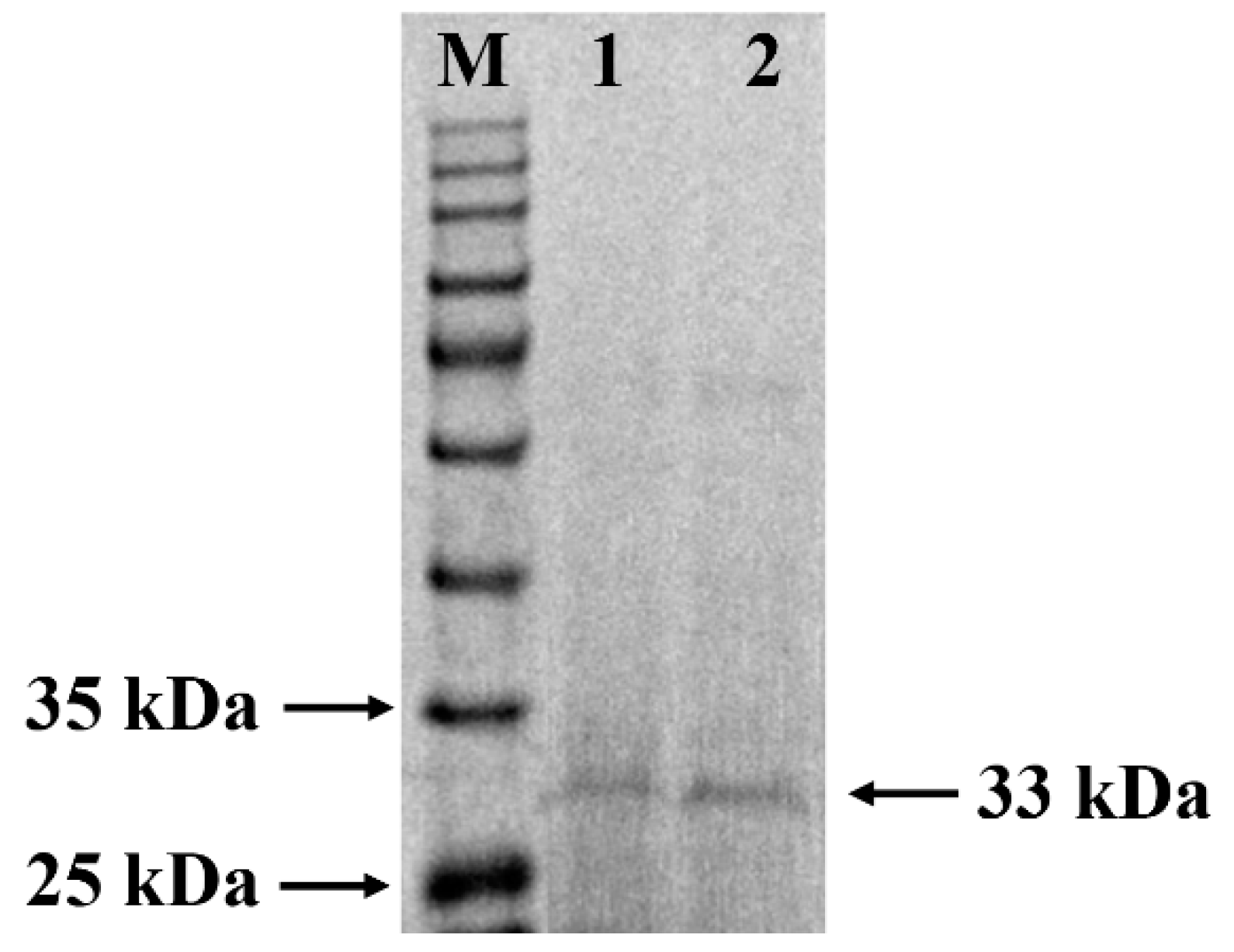
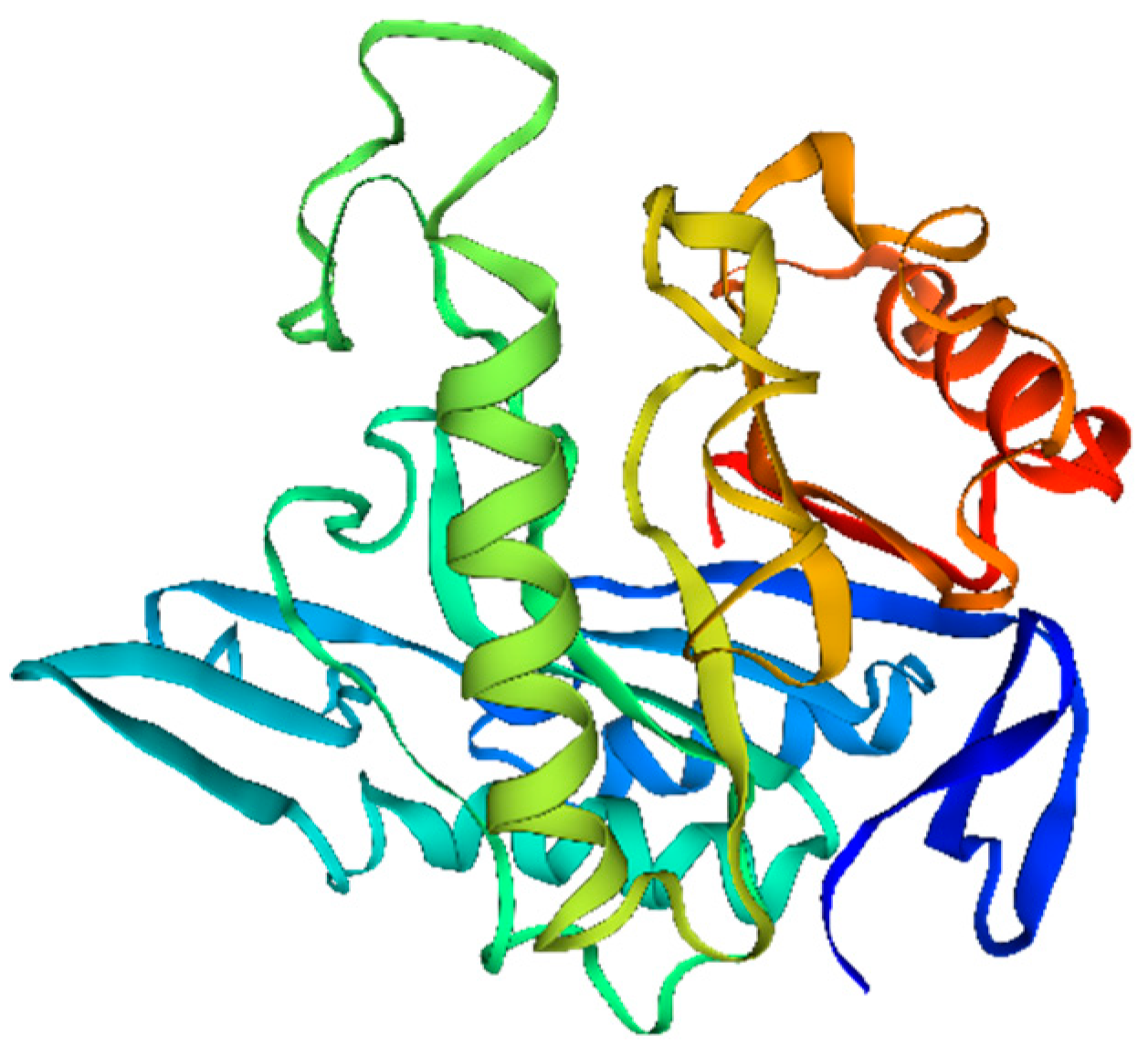
3.1. Purification and Identification of W. cibaria F1 Mannanase

The specific activity of the purified mannanase from W. cibaria F1 was 902.69 U·mg−1, and the protein content was 1.41 mg·mL−1. The final enzyme preparation was purified 2.46 folds with a yield of 19.35% (Table 1). The SDS-PAGE (Figure 1) showed a clearly single protein band with the molecular mass of the protein between 25 and 35, which is close to the molecular weight of other mannanases from Lb. casei HDS-01 37 kDa [9], P. acidilactici 38 kDa [10], and Weissella viridescens 36.5 kDa [14], while the exception is Lb. plantrum [12], which consisted of two subunits of 36.4 kDa and 55.3 kDa.
LC-MS/MS identification of the sequences of W. cibaria F1 mannanase protein showed a molecular mass of 33.1 kDa mannanase, consistent with the results of SDS-PAGE, and it was composed of 289 amino acids. The 3D structure model of the protein was constructed as shown in Figure 2. The protein belongs to the glycosidic hydrolase (GH) family 26 by amino acid sequence alignment. It had the highest coverage with B. subtilis mannanase protein (P55278 (MANB_BACIU)), and its similarity reached 70.7%.

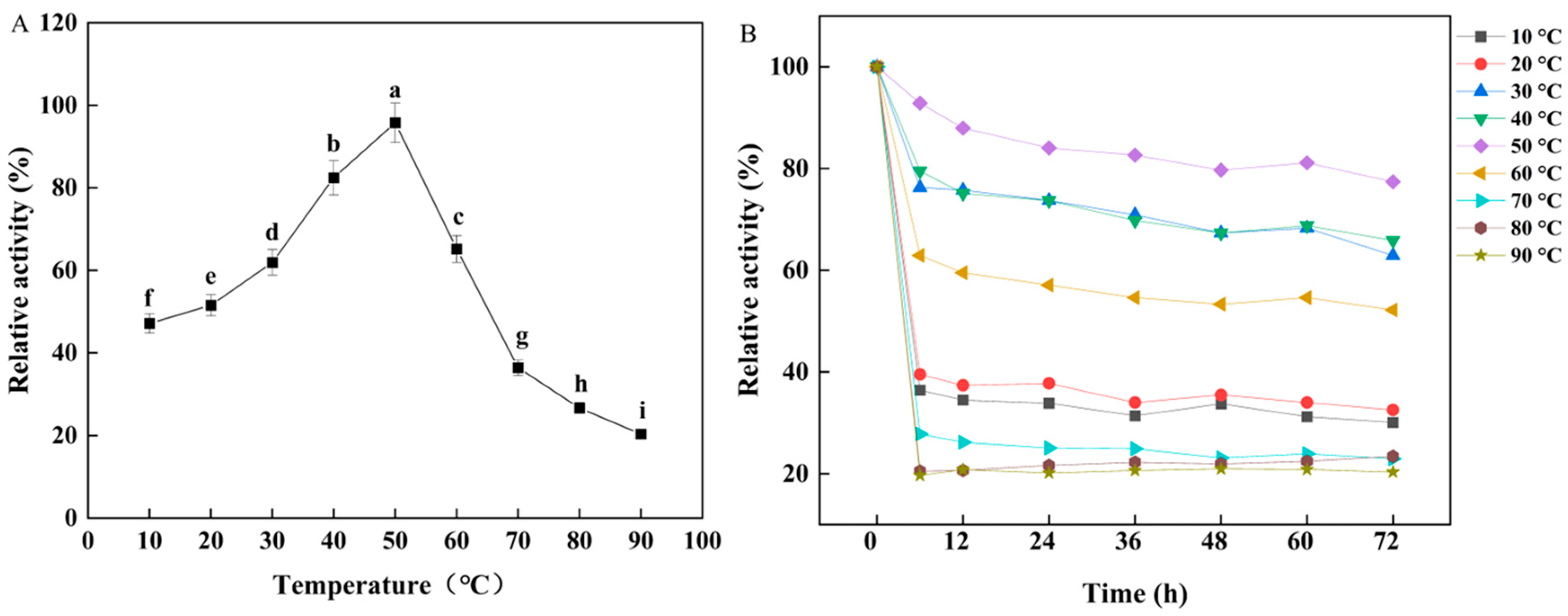
3.2. Effects of Temperature and pH on W. cibaria F1 Mannanase

The effects of pH on mannanase activity and stability were evaluated by assaying the enzyme activity in the pH range of 3.5–11.0 (Figure 3). The pH profile revealed that mannanase had over 60% relative activity between pH 4.5 and 8.0, which indicated that mannanase possesses the ability to work under a wide pH range. Figure 3A also illustrates that the optimum pH of mannanase was 6.5. This result was in accordance with the mannanase of Streptomyces galbus [27] (pH 6.5), while was higher than the mannanase of P. acidilactici [10] and W. viridescens [14]. Additionally, mannanase remained excellent pH stability, retaining 80% of activity after incubation for 3.5 h at pH 6.5. The pH stability was better than Lb. casei [9] mannanase, retaining 50% activity after 3 h incubation.
As shown in Figure 4, the optimal reaction temperature of the purified mannanase was 50 °C, which brings it into correspondence with mannanase from Lb. plantarum M24 [11]. Although that was different from the other two LAB strains Lb. casei HDS-01 (40 °C) [9] and P. acidilactici M17 [10] (60 °C), the F1 mannanase retained favorable activity of about 60% in the range from 30 °C to 60 °C. After 6 h incubation at 30 °C to 50 °C, the enzyme activity still remained at 80%, which showed better stability of B. subtilis MAFIC-S11 [28]. So, the optimal conditions and stability results related to temperature and pH of F1 mannanase would be conducive to its easy application in fields requiring high biosafety and provide a reference for technological parameters.

3.3. Effects of Metal Ions and Chemical Agents on W. cibaria F1 Mannanase

LAB mannanase activities can be activated or inhibited by many metal ions. For mannanase from Lb. plantarum ATCC 14917 [12] and Lb. plantarum M24 [11], Co2+ metal ion had an activation effect on the enzyme with a ratio of 375.3% and 254.1%. Co2+ also had the effect of enhancing mannanase activity manifested by increasing it to 235.4% in other species P. acidilactici M17 [10]. Besides that, both Mn2+ and Cu2+ could enhance the LAB mannanase activities [10,11,12], which was in accordance with W. cibaria F1, for which mannanase yield was increased at the rate of 124.5% and 112.4% at 5 mM concentration, respectively. With increasing the concentration of Mn2+ having the most obvious enhancing effect from 15 mM to 50 mM, W. cibaria F1 mannanase activity increased to 165.6% from 156.9%. Furthermore, Fe2+ was an inhibitor for W. cibaria F1 mannanase, and the same suppression of mannanase activity was also found in Lb. casei HDS-01 [9]. However, Fe2+ had a positive effect on mannanase from Lb. plantarum M24 [11]. All the study results showed that the same metal ion can exert differential effects. The enzyme activity of W. cibaria F1 mannanase gradually decreased with the increasing concentration of urea (Figure 5B). It could be that urea affected the active center of the mannanase, which in turn affected the structure of the enzyme and led to the unfolding of and ultimately a change in the properties of the enzyme. The inhibitory effect of urea on the enzyme was observed in different species such as Aspergillus nidulans [29] and B. subtilis [30].
The negative influence of several substances, including organic solvents, surfactants, and inhibitors, on mannanase activity is illustrated in Figure 5C,D. The surfactant SDS invaded the hydrophobic part of the enzyme and modified its three-dimensional structure to decrease the mannanase activity of Bacillus sp., such as B. subtilis G1 [31], B. subtilis CAe24, and B. subtilis HM7 [32]. In this study, SDS showed its obviously inhibitory effect on W. cibaria F1 mannanase in 5 mmol·L−1, similar to Lb. casei HDS-01 [9], Streptomyces sp. NRRL B-24484 [33], A. niger USM F4 [34], Paenibacillus thiaminolyticus [35], and Psychrobacter pulmonis [36]. The other surfactants and inhibitors, such as EDTA, TritionX-100, DTT, and CTAB, inhibited W. cibaria F1 mannanase activity in the present study, similar to previous reports [4,34,35]. Inhibition of enzyme activities also occurred in organic solvents, as shown in Figure 5D. Organic solvents generally bound the outer surface of the enzyme at low concentrations. As the concentration of the organic solvents increased, perhaps a larger conformational change was induced that led to the unfolding and inactivation of the enzyme [37]. Thus, the inhibitory effect of organic solvents depended upon their concentration for the denaturation of mannanase.

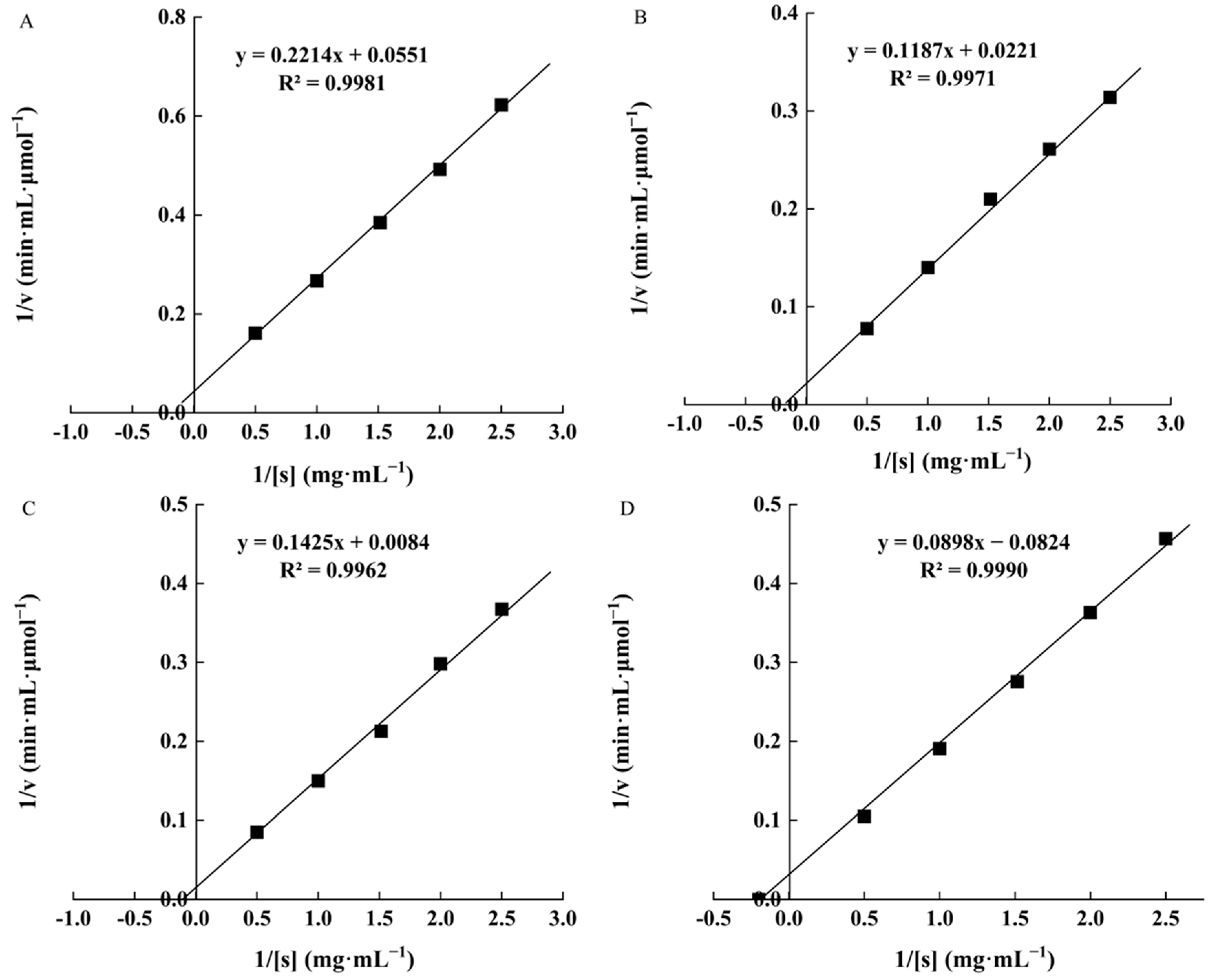
3.4. Kinetic Parameters of W. cibaria F1 Mannanase under Different Substrates

As Figure 6 shows, the kinetic parameters of F1 mannanase from W. cibaria were determined with four natural polysaccharides using double reciprocal (Lineweaver–Bark) plots. When locust bean gum was used as substrate, the Km value was 16.96 ± 0.01 μmol·mL−1, and Vmax was 1119.05 ± 0.14 μmol·min−1, indicating that W. cibaria F1 mannanase had the highest affinity catalytic efficiency. This property was similar to HDS-01 mannanase, exhibiting the highest affinity and reactivity for locust bean gum (Km 2.68 ± 0.02 mg·mL−1, Vmax 400.03 ± 1.22 μmol·mL−1·min−1) [9]. Other microbial mannanases have reported the affinity and reactivity for locust bean gum, including Bacillus nealsonii PN11 (Km 7.22 mg·mL−1, Vmax 400.03 ± 1.22 μmol·mL−1·min−1) [38], B. subtilis BE-91(Km 7.14 mg·mL−1, Vmax 107.5 μmol·mL−1·min−1) [39], Aspergillus fumigatus (Km 10.48 mg·mL−1, Vmax 948.4 U·mL−1) [40], and Aspergillus oryza (Km 2.77 mg·mL−1, Vmax 1388.8 μmol·mL−1·min−1) [41]. The Vmax values of mannanase in W. cibaria F1 for guar gum and konjac powder were 24.45 ± 0.33 and 45.24 ± 0.43 μmol·min−1, whereas in B. subtilis, the WY 34 Vmax values of mannanase for guar gum and konjac power were 556.4 ± 15.2 and 435.3 ± 29.1 μmol·mL−1·min−1, respectively [42]. Comparing the kinetic constants of mannanase from different microorganisms was limited by the lack of standardization in the substrates utilized, a suitable method, and variations in the pH values and temperature during the assay [35].

3.5. Immunomodulatory Activity of W. cibaria F1 Mannanase

3.5.1. Effect of W. cibaria F1 Mannanase on Cell Viability

RAW264.7 cells play a role in host inflammation and other immune processes by increasing proliferation, phagocytic ability, NO production, and cytokine secretion. In this study, RAW264.7 cells were directly treated with KOGM obtained by enzymatic hydrolysis of konjac powder by mannanase. As shown in Figure 7A, compared with the control group, there was no decrease in RAW264.7 cell viability. At the concentration of 200 μg·mL−1, the cell viability was higher than other concentrations and LPS (p < 0.05). In general, KOGM was nontoxic to the cells at the tested concentrations. Based on research by Jian [43], LO2 cell proliferation was not inhibited by KOGM at the concentrations of 0.005, 0.05, 0.5, and even 5 mg·mL−1.

3.5.2. Effect of W. cibaria F1 Mannanase on Macrophages Phagocytosis

Phagocytosis is an important index to evaluate the function of macrophages. As shown in Figure 7B, the positive control LPS (1 μg·mL−1) obviously increased the phagocytosis index of macrophages. The phagocytosis rate was similarly enhanced by KOGM. The phagocytic rate of all the different groups treated by KOGM was significantly higher than the negative control group (p < 0.05). The results indicated that KOGM activated RAW264.7 macrophages by promoting phagocytic ability, which was comparable to or better than the positive control group.

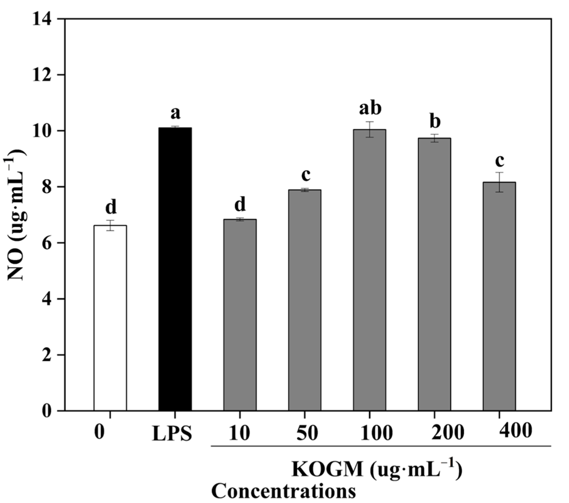
3.5.3. Effect of W. cibaria F1 Mannanase on Production of NO

NO is a mediator of immune regulation and is well known to jumpstart the immune system, where it plays a vital role in the host defense system [44]. In the current study, all various concentrations of KOGM could promote high levels of NO production (Figure 8). NO production reached the maximum value at a concentration of 100 μg·mL−1 KOGM similar to positive controls. Yan et al. [45] showed that NO was induced by konjac oligosaccharide (KOS) in a dose-dependent manner, and NO production reached 22.5 μmol·L−1 under 200 μg·mL−1 KOS, which was similar to that induced by the positive control of 1 μg·mL−1 LPS. Several previous studies have also indicated that oligosaccharides regulate NO synthesis in vitro [46]. The increase in NO secretion revealed that KOGM could activate the immunomodulatory activity of RAW264.7.

3.5.4. Effect of W. cibaria F1 Mannanase on Production of Cytokines

Cytokines, including TNF-α, MCP-1, IL-10, IL-8, IL-6, and IL-1β, produced by activated macrophages play a prominent role in immune responses [47]. Different concentrations of KOGM for 24 h were used to induce macrophages to release cytokines. As shown in Figure 9, KOGM markedly stimulated RAW264.7 macrophages to release TNF-α, MCP-1, IL-10, IL-8, IL-6, and IL-1β. Cytokine production induced by KOGM was larger than those untreated. When the concentration of KOGM was 200 μg·mL−1, the release of the cytokines of TNF-α, IL-8, and IL-1β was the largest at different concentrations, while MCP-1, IL-10, and IL-6 production of RAW264.7 cells were the highest at 100 μg·mL−1 KOGM. Many other reports have shown similar phenomena about the inhibitory effects of polysaccharides at high concentrations, but the underlying mechanism is unclear. [48]. To summarize, KOGM could stimulate the production of cytokines TNF-α, MCP-1, IL-10, IL-8, IL-6, and IL-1β in RAW264.7 cells. Among the cytokines secreted by activated macrophages, IL-6, IL-1β, and IL-10 were the most important proinflammatory cytokines, while IL-10 was the main anti-inflammatory cytokine, counteracting the negative effects of the proinflammatory cytokines [49]. Cytokines are involved in the pathogenesis of a variety of inflammatory and immunologic diseases and activate macrophages to enhance various functional responses [50].
As a prebiotic, KOGM had similar functional properties to some other common oligosaccharide prebiotics. Relevant studies have shown that inulin [51], galactooligosaccharide [52], fructooligosaccharide [53], and xylooligosaccharides [54] promote the secretion of cytokines and participate in immune regulation. The biological activity of KOGM was strongly correlated with their chemical composition, physical properties, Mw, chain conformation, and structure configurations [55]. Therefore, the composition of KOGMs remains very complex, and further studies are needed to elucidate specific immune mechanisms.

4. Conclusions

The extracellular mannanase (36.7 kDa) was purified from W. cibaria F1. This protein contains 289 amino acid residues, belonging to the GH 26 Family. The enzyme had the optimum reaction conditions at acidic to neutral pH (4.5–8.0) and moderate temperature and maintained relatively strong activity after long incubation periods. It was strongly activated by Mn2+ and exhibited high affinity for locust bean gum. KOGM obtained by mannanase hydrolysis of konjac powder activated RAW264.7 macrophages and stimulated the secretion of nitric oxide and cytokines in macrophages, which showed potent immunostimulatory activity. So far, the relationship between KOGM structure and immunity has not been fully understood. The relationship between the two will be further studied in our laboratory and will be reported in future work.

Author Contributions

S.W.: Investigation, Formal analysis, Writing—Original Draft Preparation. H.J.: Visualization, Investigation. R.D.: Conceptualization, Methodology, Software. W.P.: Resources. J.G.: Project administration. D.Z.: Supervision, Funding acquisition. All authors have read and agreed to the published version of the manuscript.

Funding

This work was financially supported by the National Natural Science Foundation of China (32072189 and 32071519) and the Project of Guangxi Key Laboratory for Polysaccharide Materials and Modifications, China (GXPSMM20-2).

Institutional Review Board Statement

Not applicable.

Informed Consent Statement

Not applicable.

Data Availability Statement

The data presented in this study are available on request from the corresponding author.

Conflicts of Interest

The authors declare no conflict of interest.

References

  1. Srivastava, P.K.; Kapoor, M. Production, properties, and applications of endo-β-mannanases. Biotechnol. Adv. 2016, 35, 1–19. [Google Scholar] [CrossRef] [PubMed]
  2. Nopvichai, C.; Charoenwongpaiboon, T.; Luengluepunya, N.; Ito, K.; Pichyangkura, R. Production and purification of mannan oligosaccharide with epithelial tight junction enhancing activity. PeerJ 2019, 7, e7206. [Google Scholar] [CrossRef] [PubMed]
  3. Mohapatra, B.R. Characterization of β-mannanase extracted from a novel Streptomyces species Alg-S25 immobilized on chitosan nanoparticles. Biotechnol. Biotechnol. Equip. 2021, 35, 150–161. [Google Scholar] [CrossRef]
  4. Singh, S.; Singh, G.; Khatri, M.; Kaur, A.; Arya, S.K. Thermo and alkali stable β-mannanase: Characterization and application for removal of food (mannans based) stain. Int. J. Biol. Macromol. 2019, 134, 536–546. [Google Scholar] [CrossRef] [PubMed]
  5. Gurler, H.N.; Yilmazer, C.; Erkan, S.B.; Ozcan, A.; Yatmaz, E.; Öziyci, H.R.; Karhan, M.; Turhan, I. Applicability of recombinant Aspergillus sojae crude mannanase enzyme in carrot juice production. J. Food Process. Preserv. 2020, 45, 1–9. [Google Scholar] [CrossRef]
  6. Dhawan, S.; Singh, R.; Kaur, R.; Kaur, J. A β-mannanase from Paenibacillus sp.: Optimization of production and its possible prebiotic potential. Biotechnol. Appl. Biochem. 2016, 63, 669–678. [Google Scholar] [CrossRef] [PubMed]
  7. Ahirwar, S.; Soni, H.; Rawat, H.K.; Prajapati, B.P.; Kango, N. Experimental design of response surface methodology used for utilisation of palm kernel cake as solid substrate for optimised production of fungal mannanase. Mycology 2016, 7, 143–153. [Google Scholar] [CrossRef] [PubMed]
  8. Peterbauer, C.; Maischberger, T.; Haltrich, D. Food-grade gene expression in lactic acid bacteria. Biotechnol. J. 2011, 6, 1147–1161. [Google Scholar] [CrossRef]
  9. Zhao, D.; Zhang, X.; Wang, Y.; Na, J.; Ge, J.P. Purification, biochemical and secondary structural characterisation of β-mannanase from Lactobacillus casei HDS-01 and juice clarification potential. Int. J. Biol. Macromol. 2020, 154, 826–834. [Google Scholar] [CrossRef] [PubMed]
  10. Nadaroglu, H.; Adiguzel, G.; Adiguzel, A.; Sonmez, Z. A thermostable-endo-β-(1,4)-mannanase from Pediococcus acidilactici (M17): Purification, characterization and its application in fruit juice clarification. Eur. Food Res. Technol. 2017, 243, 193–201. [Google Scholar] [CrossRef]
  11. Nadaroglu, H.; Adiguzel, A.; Adiguzel, G. Purification and characterisation of β-mannanase from Lactobacillus plantarum (M24) and its applications in some fruit juices. Int. J. Food Sci. Technol. 2015, 50, 1158–1165. [Google Scholar] [CrossRef]
  12. Nadaroglu, H.; Dikbas, N. Purification and characterization of a β-mannanase from Lactobacillus plantarum (ATCC 14917TM). Int. J. Innov. Res. Rev. 2018, 2, 1–6. [Google Scholar]
  13. Zabidi, N.; Ling, F.H.; Chwen, L.T.; Rosfarizan, M.; Raha, A.R. Enhancement of versatile extracellular cellulolytic and hemicellulolytic enzyme productions by Lactobacillus plantarum RI 11 isolated from Malaysian food using renewable natural polymers. Molecules 2021, 25, 2607. [Google Scholar] [CrossRef]
  14. Adiguzel, G.; Sonmez, Z.; Adiguzel, A.; Nadaroglu, H. Purification and characterization of a thermostable endo-beta-1,4 mannanase from Weissella viridescens LB37 and its application in fruit juice clarification. Eur. Food Res. Technol. 2016, 242, 769–776. [Google Scholar] [CrossRef]
  15. Guo, L.; Yokoyama, W.; Chen, L.; Liu, F.; Zhong, F. Characterization and physicochemical properties analysis of konjac glucomannan: Implications for structure-properties relationships. Food Hydrocoll. 2021, 120, 106818. [Google Scholar] [CrossRef]
  16. Liu, J.H.; Xu, Q.H.; Zhang, J.J.; Zhou, X.X.; Lyu, F.; Zhao, P.C.; Ding, Y.T. Preparation, composition analysis and antioxidant activities of konjac oligo-glucomannan. Carbohydr. Polym. 2015, 130, 398–404. [Google Scholar] [CrossRef] [PubMed]
  17. Yang, W. Preparation of konjac oligoglucomannans with different molecular weights and their in vitro and in vivo antioxidant activities. Open Life Sci. 2020, 15, 799–807. [Google Scholar] [CrossRef]
  18. Chen, Z.J.; Wang, S.S.; Shang, L.C.; Zhou, P.Y.; Li, J.; Li, B. An efficient and simple approach for the controlled preparation of partially degraded konjac glucomannan. Food Hydrocoll. 2020, 108, 106017. [Google Scholar] [CrossRef]
  19. Andrew, M.; Jayaraman, G. Structural features of microbial exopolysaccharides in relation to their antioxidant activity. Carbohydr. Res. 2020, 487, 107881. [Google Scholar] [CrossRef] [PubMed]
  20. Mao, Y.H.; Xu, Y.X.; Song, F.L.; Wang, Z.M.; Li, Y.L.; Zhao, M.Z.; He, F.; Tian, Z.Z.; Yang, Y. Protective effects of konjac glucomannan on gut microbiome with antibiotic perturbation in mice. Carbohydr. Polym. 2022, 290, 119476. [Google Scholar] [CrossRef] [PubMed]
  21. Zhou, Z.; Zhang, B.; Yang, X.; Shang, W.; Ma, Q.; Strappe, P. Regulation of hyperglycemia in diabetic mice by autolysates from. J. Sci. Food Agric. 2019, 99, 6981–6988. [Google Scholar] [CrossRef] [PubMed]
  22. Ji, H.R.; Cao, H.Y.; Zhao, L.; Na, R.Y.; Ping, W.X.; Ge, J.P.; Zhao, D. The response surface optimization of β-mannanase produced by Weissella cibaria F1 and its potential in juice clarification. Prep. Biochem. Biotechnol. 2022, 1–9. [Google Scholar] [CrossRef] [PubMed]
  23. Toshiro, A.; Nobuyuki, N.; Koki, H. Characterization of three BETA-mannanases of an alkalophilic Bacillus sp. Agric. Biol. Chem. 1988, 52, 773–779. [Google Scholar] [CrossRef]
  24. Bradford, M.M. A rapid and sensitive method for the quantitation of microgram quantities of protein utilizing the principle of protein-dye binding. Anal. Chem. 1976, 2, 248–253. [Google Scholar] [CrossRef]
  25. Liu, Y.; Li, Q.Z.; Li, L.; Zhou, X.W. Immunostimulatory effects of the intracellular polysaccharides isolated from liquid culture of Ophiocordyceps sinensis (Ascomycetes) on RAW264.7 cells via the MAPK and PI3K/Akt signaling pathways. J. Ethnopharmacol. 2021, 275, 114130. [Google Scholar] [CrossRef] [PubMed]
  26. Liu, X.; Xie, J.H.; Jia, S.; Huang, L.X.; Wang, Z.J.; Li, C.; Xie, M.Y. Immunomodulatory effects of an acetylated Cyclocarya paliurus polysaccharide on murine macrophages RAW264.7. Int. J. Biol. Macromol. 2017, 98, 576–581. [Google Scholar] [CrossRef] [PubMed]
  27. Kansoh, A.L.; Nagieb, Z.A. Xylanase and mannanase enzymes from Streptomyces galbus NR and their use in biobleaching of softwood kraft pulp. Antonie Van Leeuwenhoek 2004, 85, 103–114. [Google Scholar] [CrossRef]
  28. Lv, J.N.; Chen, Y.Q.; Pei, H.L.; Yang, W.H.; Li, Z.M.; Dong, B.; Cao, Y.H. Cloning, expression, and characterization of β-mannanase from Bacillus subtilis MAFIC-S11 in Pichia pastoris. Appl. Biochem. Biotechnol. 2013, 169, 2326–2340. [Google Scholar] [CrossRef]
  29. Lu, H.Q.; Luo, H.Y.; Shi, P.J.; Huang, H.Q.; Meng, K.; Yang, P.L.; Yao, B. A novel thermophilic endo-β-1,4-mannanase from Aspergillus nidulans XZ3: Functional roles of carbohydrate-binding module and Thr/Ser-rich linker region. Appl. Microbiol. Biotechnol. 2014, 98, 2155–2163. [Google Scholar] [CrossRef]
  30. Sudip, R.; Pradeep, G.C.; Yun, H.C.; Yoon, S.C.; Ji, E.; Seung, S.C.; Jin, C.Y. A multi-tolerant low molecular weight mannanase from Bacillus sp CSB39 and its compatibility as an industrial biocatalyst. Enzym. Microb. Technol. 2016, 92, 76–85. [Google Scholar] [CrossRef]
  31. Vu, T.T.; Quyen, D.T.; Dao, T.T.; Nguyen, S.T. Cloning, high-level expression, purification, and properties of a novel endo-beta-1,4-mannanase from Bacillus subtilis G1 in Pichia pastoris. J. Microbiol. Biotechnol. 2012, 22, 331–338. [Google Scholar] [CrossRef] [PubMed]
  32. Leerawatthanakun, S.; Charoenwongpaiboon, T.; Klaewkla, M.; Chunsrivirot, S.; Sirirak, J.; Sriwitool, T.-e.; Wangpaiboon, K.; Pichyangkura, R. High surfactant-tolerant β-mannanase isolated from Dynastes hercules larvae excrement, and identification of its hotspot using site-directed mutagenesis and molecular dynamics simulation. Enzym. Microb. Technol. 2021, 154, 109956. [Google Scholar] [CrossRef] [PubMed]
  33. Sakai, K.; Kimoto, S.; Shinzawa, Y.; Minezawa, M.; Suzuki, K.; Jindou, S.; Kato, M.; Shimizu, M. Characterization of pH-tolerant and thermostable GH 134 β-1,4-mannanase SsGH134 possessing carbohydrate binding module 10 from Streptomyces sp. NRRL B-24484. J. Biosci. Bioeng. 2018, 125, 287–294. [Google Scholar] [CrossRef] [PubMed]
  34. Syarifah, A.R.; Darah, I.; Ibrahim, C.O.; Ramli, H.; Tong, W.Y. Purification and physicochemical characterisation of Aspergillus niger USM F4 β-mannanase. Malays. J. Microbiol. 2020, 16, 396–406. [Google Scholar] [CrossRef]
  35. Dhawan, S. Purification of a thermostable β-mannanase from Paenibacillus Thiaminolyticus-characterization and its potential use as a detergent additive. J. Pure Appl. Microbiol. 2021, 15, 368–381. [Google Scholar] [CrossRef]
  36. Badejo, O.A.; Olaniyi, O.O.; Oluwadare, A.A.; Lawal, O.T. Biochemical properties of partially purified surfactant-tolerant alkalophilic endo beta-1,4 xylanase and acidophilic beta-mannanase from bacteria resident in ruminants’ guts. Biocatal. Agric. Biotechnol. 2021, 34, 101982. [Google Scholar] [CrossRef]
  37. Daniel, E.O. Protein unfolding in detergents: Effect of micelle structure, ionic strength, pH, and temperature. Biophys. J. 2002, 83, 2219–2230. [Google Scholar]
  38. Chauhan, P.S.; Sharma, P.; Puri, N.; Gupta, N. Purification and characterization of an alkali-thermostable β-mannanase from Bacillus nealsonii PN-11 and its application in mannooligosaccharides preparation having prebiotic potential. Eur. Food Res. Technol. 2014, 238, 927–936. [Google Scholar] [CrossRef]
  39. Cheng, L.F.; Duan, S.W.; Feng, X.Y.; Zheng, K.; Yang, Q.; Liu, Z.C. Purification and characterization of a thermostable -mannanase from Bacillus subtilis be-91: Potential application in inflammatory diseases. BioMed Res. Int. 2019, 2016, 6380147. [Google Scholar] [CrossRef]
  40. Karahalil, E.; Germe, M.; Karaoglan, M.; Yatmaz, E.; Turhan, I. Partial purification and characterization of a recombinant β-mannanase from Aspergillus fumigatus expressed in Aspergillus sojae grown on carob extract. Biomass Convers. Biorefinery 2020, 10, 1189–1205. [Google Scholar] [CrossRef]
  41. Uttam, K.J.; Rahul, K.S.; Bhanu, P.P.; Hemant, S.; Naveen, K. Production optimization and characterization of mannooligosaccharide generating β-mannanase from Aspergillus oryzae. Bioresour. Technol. 2018, 268, 308–314. [Google Scholar] [CrossRef]
  42. Jiang, Z.Q.; Yun, W.; Li, D.Y.; Li, L.; Chai, P.P.; Kusakabe, I. High-level production, purification and characterization of a thermostable β-mannanase from the newly isolated Bacillus subtilis WY34. Carbohydr. Polym. 2006, 66, 88–96. [Google Scholar] [CrossRef]
  43. Jian, W.; Chen, Y.H.; Wang, L.; Tu, L.; Xiong, H.; Sun, Y.M. Preparation and cellular protection against oxidation of Konjac oligosaccharides obtained by combination of γ -irradiation and enzymatic hydrolysis. Food Res. Int. 2018, 107, 93–101. [Google Scholar] [CrossRef] [PubMed]
  44. Wang, J.; Fang, X.B.; Wu, T.; Fang, L.; Liu, C.L.; Min, W.H. In vitro immunomodulatory effects of acidic exopolysaccharide produced by Lactobacillus planetarium JLAU103 on RAW264.7 macrophages. Int. J. Biol. Macromol. 2020, 156, 1308–1315. [Google Scholar] [CrossRef] [PubMed]
  45. Yan, Z.; Zhang, J.G.; Ying, Z.; Yan, M.; Bo, Z.; Sun, Y.X. Prebiotic, immunomodulating, and antifatigue effects of Konjac Oligosaccharide. J. Food Sci. 2018, 12, 3110–3117. [Google Scholar] [CrossRef]
  46. Meng, X.X.; Yang, X.Y.; Lin, G.; Fang, Y.; Ruan, Z.L.; Liu, M.F.; Liu, G.X.; Li, M.Z.; Yang, D. Mannan oligosaccharide increases the growth performance, immunity and resistance capability against Vibro Parahemolyticus in juvenile abalone Haliotis discus hannai Ino. Fish Shellfish Immunol. 2019, 94, 654–660. [Google Scholar] [CrossRef] [PubMed]
  47. Zhang, P.; Sun, F.F.; Cheng, X.; Li, X.J.; Mu, H.B.; Wang, S.C.; Geng, H.L.; Duan, J.Y. Preparation and biological activities of an extracellular polysaccharide from Rhodopseudomonas palustris. Int. J. Biol. Macromol. 2019, 131, 933–940. [Google Scholar] [CrossRef] [PubMed]
  48. You, X.; Li, Z.; Ma, K.; Zhang, C.L.; Chen, X.H.; Wang, G.X.; Yang, L.; Dong, M.S.; Rui, X.; Zhang, Q.Q.; et al. Structural characterization and immunomodulatory activity of an exopolysaccharide produced by Lactobacillus helveticus LZ-R-5. Carbohydr. Polym. 2020, 235, 115977. [Google Scholar] [CrossRef]
  49. Busse, M.; Campe, K.N.J.; Nowak, D.; Schumacher, A.; Zenclussen, A.C. IL-10 producing B cells rescue mouse fetuses from inflammation-driven fetal death and are able to modulate T cell immune responses. Sci. Rep. 2019, 9, 9335. [Google Scholar] [CrossRef]
  50. Shi, Y.; Ye, Y.F.; Zhang, B.W.; Liu, Y.; Wang, J.H. Purification, structural characterization and immunostimulatory activity of polysaccharides from Umbilicaria esculenta. Int. J. Biol. Macromol. 2021, 181, 743–751. [Google Scholar] [CrossRef]
  51. Akram, W.; Garud, N.; Joshi, R. Role of inulin as prebiotics on inflammatory bowel disease. Drug Discov. Ther. 2019, 13, 1–8. [Google Scholar] [CrossRef] [PubMed]
  52. Vulevic, J.; Juric, A.; Walton, G.E.; Claus, S.P.; Tzortzis, G.; Toward, R.E.; Gibson, G.R. Influence of galacto-oligosaccharide mixture (B-GOS) on gut microbiota, immune parameters and metabonomics in elderly persons. Br. J. Nutr. 2015, 114, 586–595. [Google Scholar] [CrossRef] [PubMed] [Green Version]
  53. Zhang, C.N.; Wang, J.H.; Qi, Q.; Yang, L.; Sun, P.; Yuan, X.Y. Modulatory effect of fructooligosaccharide against triphenyltin-induced oxidative stress and immune suppression in goldfish (Carassius auratus). Ecotoxicol. Environ. Saf. 2021, 212, 111966. [Google Scholar] [CrossRef]
  54. Pang, J.M.; Zhou, X.J.; Ye, H.; Wu, Y.J.; Wang, Z.Y.; Lu, D.D.; Wang, J.J.; Han, D.D. The high level of xylooligosaccharides improves growth performance in weaned piglets by increasing antioxidant activity, enhancing immune function, and modulating gut microbiota. Front. Nutr. 2021, 8, 764556. [Google Scholar] [CrossRef] [PubMed]
  55. Ma, L.P.; Jiao, K.P.; Luo, L.; Xiang, J.L.; Fan, J.L.; Zhang, X.Y.; Yi, J.P.; Zhu, W.X. Characterization and macrophage immunomodulatory activity of two polysaccharides from the flowers of Paeonia suffruticosa Andr. Int. J. Biol. Macromol. 2019, 124, 955–962. [Google Scholar] [CrossRef] [PubMed]
Figure 1. SDS-PAGE of purified W. cibaria F1 mannanase. Lane M: protein marker; Lane 1 and 2: purified W. cibaria F1 mannanase.
Fermentation 08 00468 g001
Figure 2. Three-dimensional structural model of F1 mannanase.
Fermentation 08 00468 g002
Figure 3. Effects of pH value (A) and pH value stability (B) on the enzyme activity of W. cibaria F1. Different letters in superscript donate significant difference (p < 0.05).
Fermentation 08 00468 g003
Figure 4. Effects of temperature (A) and temperature stability (B) on the enzyme activity of W. cibaria F1. Different letters in superscript donate significant difference (p < 0.05).
Fermentation 08 00468 g004
Figure 5. Effects of metal ions (A), urea concentration (B), surfactant and inhibitions (C) and organic solvents (D) on the enzyme activity of W. cibaria F1 mannanase. Different letters in superscript donate significant difference (p < 0.05).
Fermentation 08 00468 g005
Figure 6. Specificity of W. cibaria F1 mannanase for different substrates guar gum (A), konjac power (B), locust bean gum (C), and pectin (D).
Fermentation 08 00468 g006
Figure 7. Effects of W. cibaria F1 mannanase on cell viability (A) and macrophages phagocytosis (B) of RAW264.7 macrophages in vitro. Different letters in superscript donate significant difference (p < 0.05).
Fermentation 08 00468 g007
Figure 8. Effects of W. cibaria F1 mannanase on production of NO of RAW264.7 macrophages in vitro. Different letters in superscript donate significant difference (p < 0.05).
Fermentation 08 00468 g008
Figure 9. Effects of W. cibaria F1 mannanase on production of cytokine of RAW264.7 macrophages in vitro (AF). Different letters in superscript donate significant difference (p < 0.05).
Fermentation 08 00468 g009
Table 1. Purification process of mannanase from W. cibaria F1.
Enzyme FractionVolume (mL)Activity (U·mL−1)Total Activity (U·mL−1)Protein (mg·mL−1)Recovery (%)Specific Activity (U·mg−1)Purification Fold
Crude extract100020.6820680.0107.00100.00193.271.00
Acetone extraction10065.776577.017.9024.47367.431.90
DEAE-Sepharose FF10127.281272.81.4119.35902.692.46
Publisher’s Note: MDPI stays neutral with regard to jurisdictional claims in published maps and institutional affiliations.

