Article

Isolation and Identification of Lactose-Degrading Yeasts and Characterisation of Their Fermentation-Related Ability to Produce Ethanol

by
Murni Kurniawati
1,
Nurliyani Nurliyani
1,
Wiratni Budhijanto
2,3 and
Widodo Widodo
1,3,*
1
Department of Technology of Animal Products, Faculty of Animal Science, Universitas Gadjah Mada, Yogyakarta 55281, Indonesia
2
Chemical Engineering Department, Faculty of Engineering, Universitas Gadjah Mada, Yogyakarta 55281, Indonesia
3
The Graduate School of Biotechnology, Universitas Gadjah Mada, Yogyakarta 55281, Indonesia
*
Author to whom correspondence should be addressed.
Fermentation 2022, 8(4), 183; https://doi.org/10.3390/fermentation8040183
Submission received: 12 March 2022 / Revised: 1 April 2022 / Accepted: 5 April 2022 / Published: 12 April 2022

Abstract

:
Kefir is traditionally produced by fermenting cow’s milk using kefir grains as a starter culture. As the viability of microbes within kefir grains is limited and preparing the grains for kefir fermentation is laborious, here, a single starter that ferments lactose and produces ethanol is developed. For this purpose, it is important to isolate yeasts that can ferment lactose and subsequently produce alcohol. This study aimed to isolate and identify yeasts from kefir and characterise their ability as single starters to produce kefir. Based on morphological and physiological evaluations, 15 presumptive yeast isolates were obtained, 10 of which grew well on lactose-containing media. Those that were able to grow on lactose using only carbon sources were subjected to molecular identification based on the internal transcribed spacer (ITS) of the 5.8 rDNA using PCR technology. Molecular identification confirmed four isolates—namely, KFA 3, KFA 7, KFA 9 and KFB 1—as belonging to Kluyveromyces marxianus. The batch fermentation data of these strains were fitted on a logistic model to obtain the carrying capacity coefficients and strain performances were compared. The kinetic modelling revealed that KFA 9 had the highest values for the carrying capacity coefficient, biomass yield and product yield, indicating that, among the four K. marxianus strains, this was superior due to its relatively fast growth and good ethanol productivity.

1. Introduction

Kefir is a traditional fermented milk product with high nutritional content [1], and is considered a health food [2,3,4]. Some of the reported health benefits of kefir are associated with the bioactivity of several metabolites [5] which can act as anti-inflammatory [6], antioxidant [7] and anti-tumour [8] compounds, modulators of the immune system [9] and pathogen inhibitors [10]. Kefir is traditionally produced by fermenting cow’s milk using kefir grains as a starter culture for fermentation [11]. These grains mainly contain lactic acid bacteria, particularly species belonging to the genera Lactobacillus and Leuconostoc (Lactobacillaceae), Lactococcus and Streptococcus (Streptococcaceae), Acetobacter (acetic acid bacteria) and the genera Kluyveromyces and Saccharomyces (Saccharomycetaceae) [12,13,14,15]. The microbial composition of kefir grains depends on their origin, and their viability can be maintained by ensuring a balance in the number of bacteria/yeasts using a continuous fermentation method that can increase the microbial cell biomass in the grains [12]. This increase highly depends on the fermentation temperature, pH and the presence of nutrients for microbial growth. Due to the stable composition of the microbiota, kefir grains retain their activity if they are stored under proper conditions [16,17].
Yeasts are unicellular eukaryotic organisms belonging to the Ascomycetes, Basidiomycetes and Deuteromycetes classes [18,19]. Their activity during fermentation processes can significantly modify the taste, aroma and texture of food. Yeasts grow at optimal temperatures between 25 and 30 °C, within a maximum temperature range between 35 and 47°C, and pH between 4.0 and 4.5 [15]. Yeasts can be isolated from several different sources. Qvirist et al. [20] have successfully isolated strains of Kluyveromyces marxianus, Pichia fermentans, Saccharomyces cerevisiae and Kazachstania unispora from fermented goat’s milk. Pedersen et al. [21] isolated Candida krusei, Kluyveromyces marxianus, Candida tropicalis, Candida rugosa, Candida fabianii, Candida norvegensis and Trichosporon asahii from Fura, a fermented product consumed in West African countries. Yeasts are the most effective microorganisms in producing ethanol. However, not all species have a good lactase activity and some are unable to degrade lactose and directly ferment fresh milk to produce ethanol. As kefir grains have a limited viability and their preparation on an industrial scale is laborious and costly, it is important to develop a single starter culture that ferments fresh milk directly into kefir products. This study aimed to isolate and identify yeast isolates and to evaluate their ability to ferment lactose as the sole carbon source in milk, and to measure their effectiveness in fermenting lactose into ethanol.

2. Materials and Methods

2.1. Yeast Isolation from Kefir

Yeast was isolated from 1 mL of local kefir products (Kefira, Yogyakarta, Indonesia) and was grown on an enrichment medium of glucose yeast peptone broth (GYP; Thermo Scientific™ Oxoid™, Jakarta, Indonesia) (20 g/L glucose, 10 g/L bacto-peptone, 5 g/L bacto yeast extract); then, it was incubated at 37 °C for 24 h with aeration [11,22]. The culture (100 µL) was then poured on malt extract agar (MEA; Thermo Scientific™ Oxoid™, Jakarta, Indonesia) (48 g/L) supplemented with 50 ppm chloramphenicol [22]. Plates were incubated at 37 °C for 24–48 h in aerobic growth conditions. The colonies that appeared were purified by successive streaking on MEA agar supplemented with chloramphenicol and were also incubated at 37 °C for 24–48 h in aerobic growth conditions [11]. They were then subjected to morphological and physiological identification. The former was carried out based on colony size and colour and cell type, while the latter was based the isolates’ ability to grow on media with lactose as the sole carbon source [22].

2.2. Screening for Lactose-Degrading Yeast

The presumptive yeast isolates were assessed in terms of their ability to grow on lactose media (20 g/L lactose, 5 g/L beef extract, 5 g/L C2H3NaO2, 4 g/L yeast extract, 2 g/L K2HPO4, 2 g/L ammonium citrate, 1 g/L tween 80, 0.2 g/L MgSO4 and 0.05 g/L MnSO4) supplemented with 50 ppm chloramphenicol. A loop of each isolate was transferred into test tubes containing 10 mL of sterile lactose media and these were incubated at 37 °C for 24 h in aerobic conditions [22]. The isolates that were able to grow on the media were inoculated on GYP broth media. An aliquot of 100 µL of this inoculum was separately transferred on lactose media; then, glucose-, sucrose- and fructose-containing media on Durham tubes added with 0.4% bromothymol blue as indicator, and these tubes were incubated at 37 °C for 72 h under aerobic conditions. The ability of cultures to ferment these sugars was indicated by the presence of gas and change in the colour of media from green to yellow. The presumptive yeast species with the ability to ferment lactose were further identified based on molecular approaches.

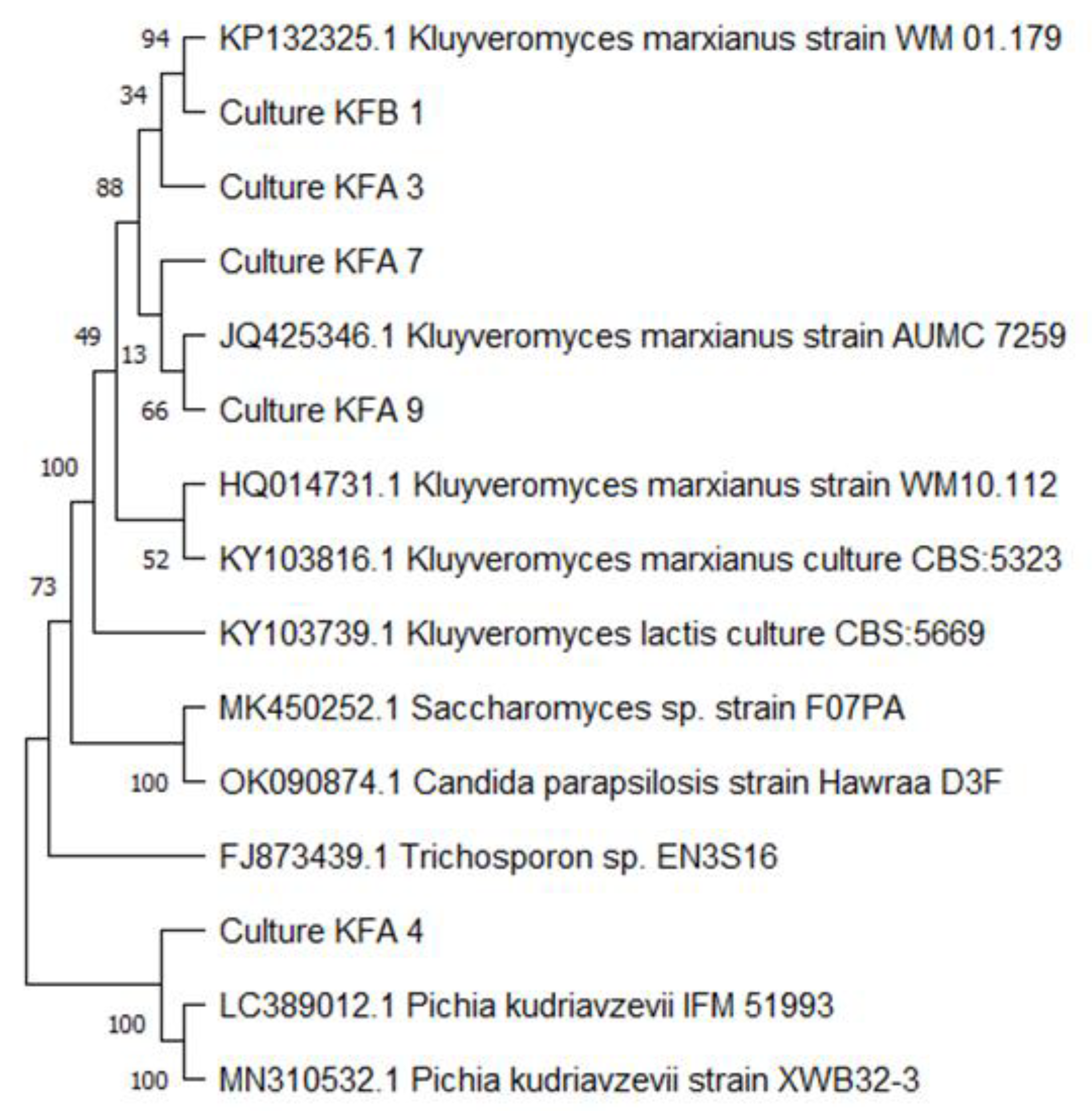
2.3. Molecular Identification of Isolates

Genomic DNA was extracted from 1.5 mL of yeast cells using Favorgen DNA isolation kit (Favorgen, Ping-Tung, Taiwan) according to manufacturer’s instructions. DNA amplification of the ITS1-5.8S-ITS4 region was carried via PCR (BioProducts, New Taipei City, Taiwan) using specific primers. A forward ITS1 primer (5’-TCCGTAGGTGAACCTGCGG) and a reverse ITS4 primer (5’-TCCTCCGCTTATTGATATGC) (Genetika Science Indonesia, Tangerang, Indonesia) were used for the target amplification of a 500–700 bp section of ribosomal DNA. Amplification was carried out in a 50 µL volume and the reaction mixture contained 1 µL (200 ng) of DNA template, 2 µL (1 µM) of reverse and 2 µL (1 µM) forward primers, 10 µL of Smobio PCR master mix (Smobio, Jakarta, Indonesia), which were finally added with 35 µL of nuclease free water. The following PCR cycling conditions were employed: pre-denaturation at 94 °C for 2 min, followed by 30 amplification cycles at 94 °C for 30 s, annealing at 60 °C for 1 min, extension at 72 °C for 30 s and final extension at 72 °C for 1 min. PCR products were then analysed using 1.5% TBE agarose gel electrophoresis (GeneDirex, New Taipei City, Taiwan). The gel was stained with ethidium bromide, was visualised under UV light and was then photographed. The amplified bands were sequenced at the 1st BASE Sequencing laboratories (1st BASE, Singapore). The nucleotide sequence data were compared with sequences stored in the GeneBank database using the BLAST algorithm. Phylogenetic analysis was performed by comparing several sequences of close and distant reference genera (outgroups) obtained from the National Centre for Biotechnology Information database using the neighbour-joining tree method in the ClustalX2 programme. The clustering stability was calculated using 1000 bootstrap replications. The phylogenetic tree view was opened and edited in Mega v. 5.05 and it was constructed based on the ITS gene sequences of isolates using the same programme (www.megasoftware.net) (accessed on 26 November 2021).

2.4. Inoculum Preparation and Ethanolic Fermentation

Four yeast isolates identified as Kluyveromyces marxianus (KFA3, KFA7, KFA9 and KFB1) and one isolate identified as Pichia kudriavzevii (KFA4), which were all able to ferment lactose, were reactivated in GYP broth at 37 °C for 24 h under aerobic conditions. The cultures were centrifuged at 8000× g at 5 min (Corning® LSE, Phoenix, AZ, USA) and were removed from the supernatant; they were then resuspended in 0.1% (w/v) peptone water and adjusted to an optical density of 0.5 at 600 nm (SP-V1100 spectrophotometer; DLAB, Beijing, China). Aliquots of 10 mL were inoculated on 100 mL of sterile lactose media and were incubated at 37 °C under micro-aerobic conditions. A volume of 500 mL containing sterile lactose media (20 g/L lactose, 5 g beef extract, 5 g/L C2H3NaO2, 4 g/L yeast extract, 2 g/L K2HPO4, 2 g/L ammonium citrate, 1 g/L tween 80, 0.2 g/L MgSO4 and 0.05 g/L MnSO4) was placed in a 1000 mL erlenmeyer flask with the pH set at 5.0. To obtain micro-aerobic conditions, 200 mg/L sodium thioglycolate was added [22]. The media was inoculated with 5% (v/v) of the inoculum and was incubated at 37 °C. During 24 h of batch fermentation, biomass and ethanol concentration and lactose content (as reducing sugar concentration) were measured every 3 h. Biomass concentration was measured based on cell dry weight. Yeast cells were centrifuged at 8000× g for 5 min and the pellets were then frozen at −80 °C for 24 h. The dry frozen cells were crushed, weighed and dissolved into sterile aquadest to obtain concentrations of 1, 2, 4, 6, 8 and 10 mg/mL. The solution was measured by OD600 and applied as standard cell dry weight. The reducing sugar content was obtained using the Nelson–Somogyi method [23] by measuring OD540 and by entering the measured absorbance value into the regression equation based on the standard curve of lactose. The standard curve was prepared using 1 mL of lactose solution with concentrations of 0, 4, 8, 12, 16 and 20 mg/100 mL. The ethanol concentration was calculated using the Conway microdiffusion method [24] by measuring OD480 and it was compared to the standard curve regression equation. The ethanol standard curve was prepared using ethanol solution with concentrations of 0.025, 0.05, 0.075 and 0.1% (v/v).

2.5. Batch Kinetics of Biomass and Ethanolic Production

In a batch fermentation, the accumulating cell mass will reach a maximum value under a particular set of conditions, which include the substrate concentration, the presence of inhibitors and pH or temperature stability. The maximum cell mass, usually denoted as X, is identical to the ecological concept of carrying capacity. When the specific growth rate is related to the amount of unused carrying capacity, a logistic equation can well represent is condition [25].
The general form of the growth rate equation in a batch fermentation is presented in Equation (1), where X (g/L) is the cell dry weight, t (hour) is time and µg (h−1) is the specific growth rate. Furthermore, the specific growth rate (µg) is correlated to the carrying capacity in Equation (2), where k is the carrying capacity coefficient. Hence, the logistic equation to be applied in this study is obtained and is presented in Equation (3).
dX dt = μ g X
μ g = k [ 1 X X ]
dX dt = kX [ 1 X X ]
The differential Equation (3) is approximated by using the numerical finite difference method. The experimental data must be fitted to Equation (3) in order to determine the k value that provides the best fit of the calculated X to the measured X values. The formula to calculate X is obtained by integrating Equation (3), which results in Equation (4).
X = X o e kt 1 X o X [ 1 e kt ] .
The yield coefficients YP/S (product yield factor) and YX/S (cell yield factor) are estimated from the experimental data of ethanol concentration (P, g/L), cell dry weight (X, g/L) and substrate (S, g/L) using Equations (5) and (6), respectively, as follows:
Y P / S = P S
Y X / S = X S

3. Results and Discussion

3.1. Isolation and Identification of Yeast Isolates

Kefir grains have been reported to contain several species of yeast, including lactose-fermenting Kluyveromyces marxianus and non-lactose-fermenting Saccharomyces cerevisiae and Saccharomyces exiguous [14,15]. In this study, yeast isolation from kefir grains was conducted on MEA agar supplemented with 50 ppm chloramphenicol to inhibit the growth of bacteria. Joseph et al. [26] previously reported the effectiveness of this compound as a bacteriostatic for the inhibition of bacterial growth. Using this selection media, a total of 15 isolates were obtained, which were further tested on lactose-containing media to determine the ones capable of degrading this sugar. Of the 15 isolates tested, 10 were able to grow on lactose media and they were then identified based on their morphological characteristics, including colony colour and shape, colony elevation and cell shape [19,27].
Macroscopic observations showed that the yeast-specific colour of the colonies was cream, and their shape was ellipsoidal. Isolates KFA 4 and KFB 2 had a pellicle on the surface of the media. According to Fleischman and Sripuntanagoon [28], a ring-shaped and yellowish pellicle on the surface indicates the presence of a surface yeast (or top yeast), namely, a yeast that grows in clusters and releases CO2 rapidly, causing cells to float upwards. In contrast, the formation of a precipitate indicates the presence of a bottom yeast, which does not grow in clusters and produces CO2 slowly so that the cells accumulate at the bottom of the tube. Based on morphological identification, these 10 isolates were considered as presumptive yeast species potentially belonging to the Saccharomycetaceae family. Further physiological identification was carried out to determine the ability of these isolates to degrade various carbohydrate sources, as these degradation processes are associated with enzymatic and metabolic activities inside the cells. The yeasts’ physiological activity was identified through biochemical tests, specifically, by testing for the ability to ferment glucose, lactose, sucrose and fructose. The data on the ability to ferment this range of sugars are presented in Table 1.
Yeasts have different sugar fermentation abilities, as measured by the carbon dioxide (CO2) produced. Members of the Kluyveromyces and Saccharomyces genera are able to ferment glucose, while those of the Trichosporon genus are not [21]. In particular, the genus Kluyveromyces is capable of fermenting lactose, glucose and galactose [18]. Table 1 shows that all isolates were able to change the medium colour, except for KFA4 on sucrose medium and KFB2 on lactose medium. The change in medium colour is an indicator of the ability to ferment sugars and produce organic acids [29]. Sugar fermentation is characterised by the formation of CO2 by-products and it finally produces ethanol and organic acids. The presence of CO2 is identified by visible bubbles produced in the Durham tubes. Based on the fermentation pattern, seven isolates (KFA 3, KFA 4, KFA 7, KFA 9, KFB 1, KFB 2 and KFB 4) were selected for further molecular identification tests. Isolates KFA 3, KFA 4, KFA 7, KFA 9, KFB 1 and KFB 4 were chosen because they were able to ferment lactose and produce CO2, while isolate KFB 2 was selected as a negative control because unable to do so (Table 1).
Molecular methods were applied due to their high level of accuracy, which allows the identification of isolates to the strain level. Using this approach, the internal transcribed spacer (ITS) of the 5.8 ribosomal DNA (rDNA) was amplified by PCR using specific primers previously reported by Naumova et al. [30] and Hesham et al. [31]. The amplified DNA was then sequenced and the resulting nucleotide sequence was used to search sequence homology and to construct a phylogenetic tree. Table 2 shows the ITS rDNA nucleotide similarity of the five tested yeast isolates with sequences from the Gene Bank database.
Table 2 shows a high similarity (>99%) of the KFA 3, KFA 7, KFA 9 and KFB 1 nucleotide sequences with the corresponding sequence of K. marxianus, suggesting that these isolates belong to this species. In contrast, the nucleotide sequence of KFA 4 showed a high similarity (>99%) with the corresponding sequence of P. kudriavzevii, suggesting that isolate KFA 4 is a strain of this species. According to Claverie and Notredame [32], a nucleotide similarity of above 70% is categorised as homologous. The relationship between the yeast isolates reported in this study and several strains of K. marxianus and P. kudriavzevii was described using a dendrogram and is presented in Figure 1.
The phylogeny tree was constructed based on the neighbour-joining method and the nucleotide sequences used as references were those of the complete 5.8S rDNA gene of the following strains: Kluyveromyces marxianus WM 01.179, Kluyveromyces marxianus AUMC 7259, Kluyveromyces lactis CBS:5669, Candida parapsilosis Hawraa D3F, Saccharomyces sp. F07PA and Trichosporon sp. EN3S16. The phylogenetic tree is described according to scale, with the branch length in units being equal to the distance of the relationship. Figure 1 shows that KFA 3 and KFB 1 were grouped in the same cluster of K. marxianus, strain WM 01.179, whereas KFA 7 and KFA 9 were grouped in the same cluster of K. marxianus strain AUMC 7259 and KFA 4 was grouped with P. kudriavzevii strains XWB32-3 and IFM 51993.
The origin of yeast habitat is likely to contribute to the diversity of the rDNA nucleotide sequences of yeast species. K. marxianus WM 01.179 was identified from human and animal pathogenic fungi; P. kudriavzevii XWB32-3 was isolated from fermented Capsicum frutescens; K. marxianus WM10.112 and AUMC 7259 were isolated from clinical samples in Oman and from citrus and grapevine plantations, respectively. Saccharomyces sp. F07PA was isolated from Indian Ocean waters, and Trichosporon sp. EN3S16 was obtained from soil and plants in Taiwan. Although each strain is closely related, its ability to grow in a particular environment depends on its natural habitat. The identified yeast strains were then assessed in terms of their growth pattern on lactose-containing media.

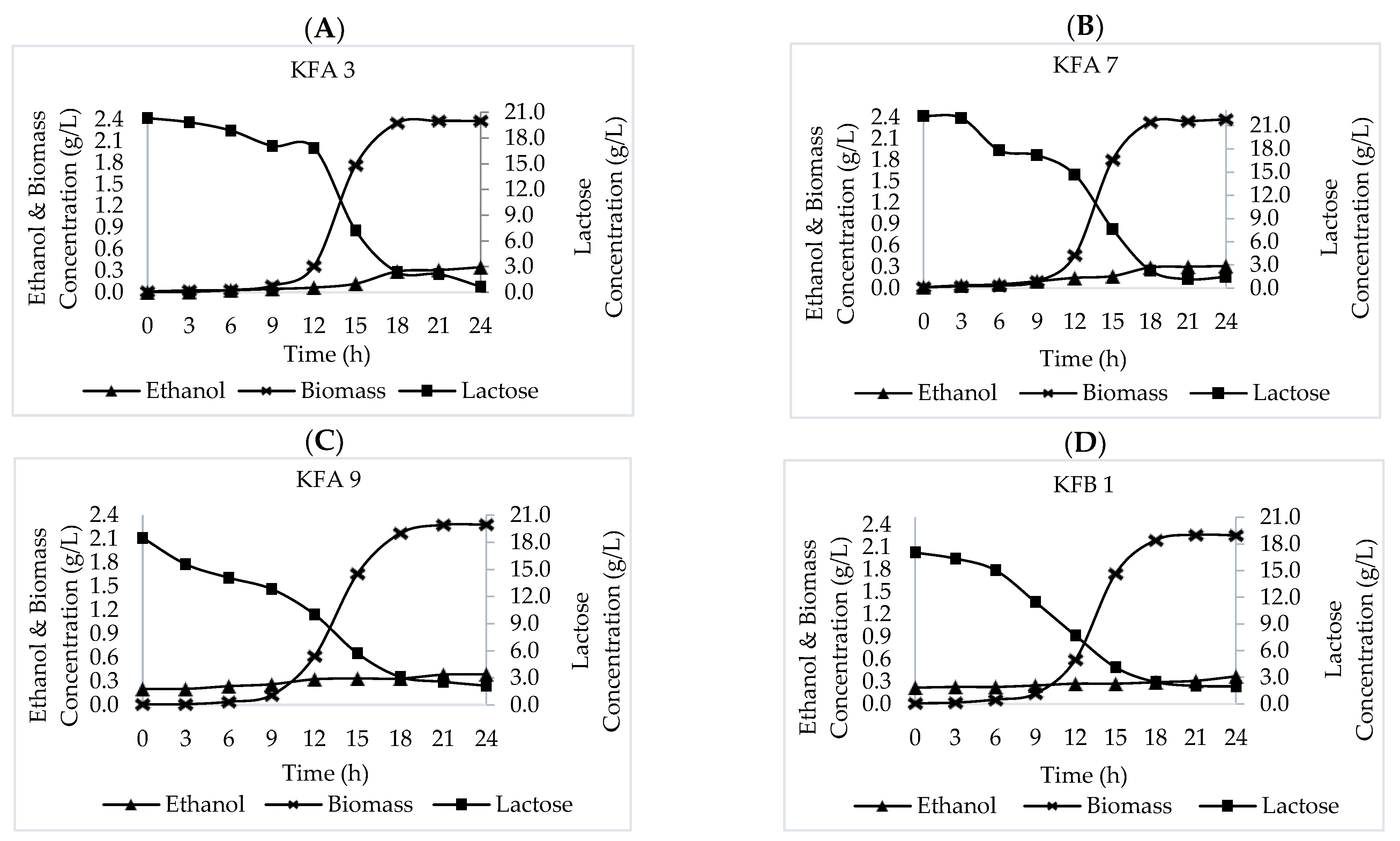
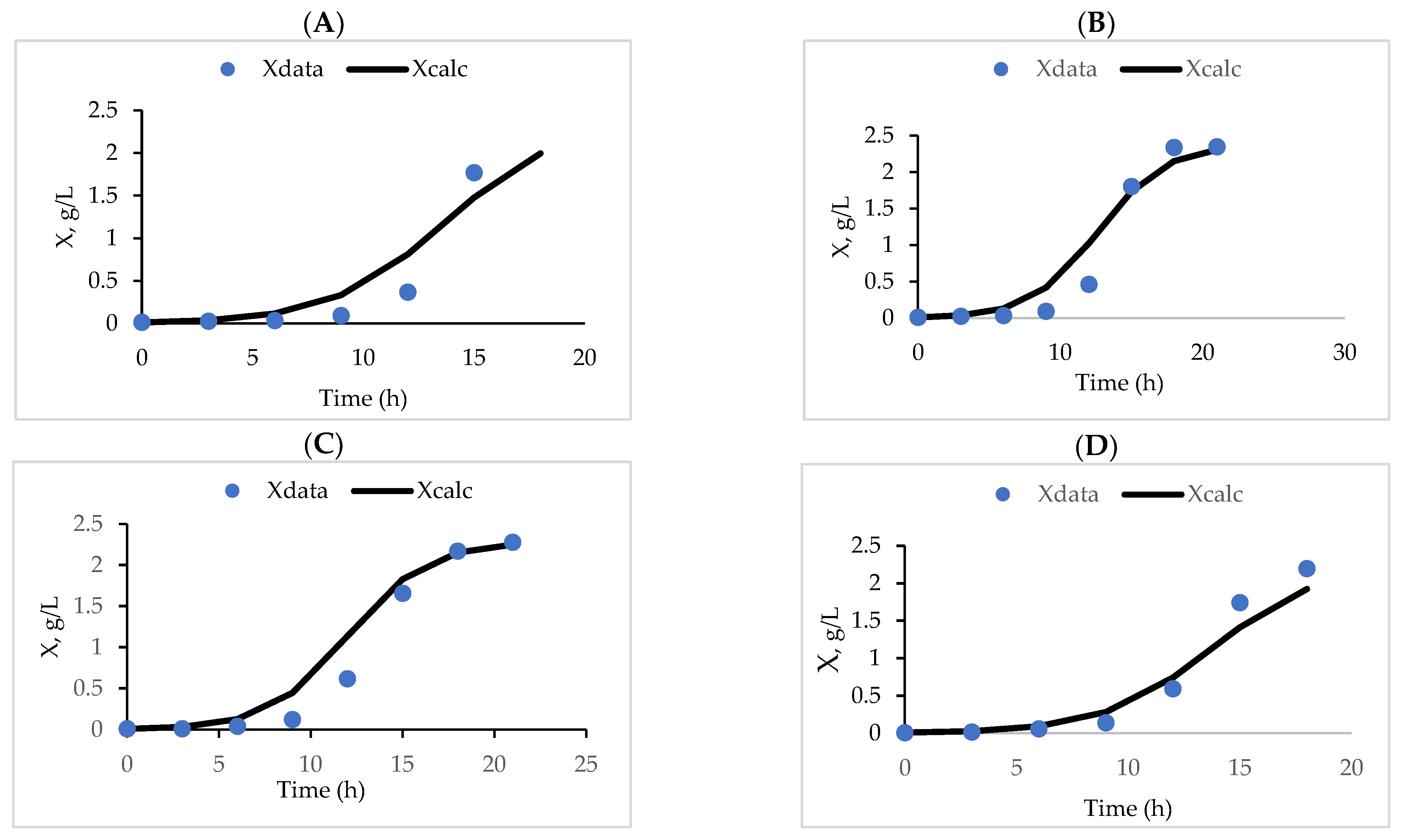
3.2. Growth Pattern on Lactose-Containing Media

The growth pattern of the K. marxianus KFA 3, KFA 7, KFA 9 and KFB 1 strains, and P. kudriavzevii KFA 4 strain was obtained by observing biomass concentration (g/L) for 24 h. Figure 2 shows the growth pattern of the above-mentioned strains on lactose media.
The K. marxianus KFA 3, KFA 7, KFA 9 and KFB 1 strains grew well on lactose-containing media, suggesting that they were all able to consume lactose as main energy source and use it to support their growth. Their growth pattern showed four stages of microbial growth: a lag phase, a logarithmic (exponential) phase, a stationary phase and a death phase. The first occurred after inoculation and it lasted three hours, until the yeast cells adapted to the new environment. The exponential phase was initiated between 9 and 18 h after inoculation, while the stationary phase started 18 h after inoculation (Figure 2). In contrast to K. marxianus, the growth of the P. kudriavzevii KFA 4 strain in lactose-containing media was very slow, as indicated by the very small growth observed. These data suggest that this strain is not a good candidate as starter culture for lactose fermentation and, therefore, it was not included in the subsequent experimental stages.

3.3. Fermentation Kinetics

Kinetic parameters can serve as an objective tool to compare the outcomes of various treatments in a fermentation process. The use of these parameters as a comparison tool is especially essential when the data are limited and the measured variables look so similar that it is difficult to make inferences based on the evaluation of the plots. Therefore, in this study, the kinetic parameters of each K. marxianus strain based on the cell growth, substrate consumption and ethanol production plots were compared.
During ethanolic fermentation, chemical composition changes due to the bioconversion of lactose into lactic acid (by homofermentative LAB), so the pH decreases and the acidity increases [12,33]. This is followed by a fermentation process by yeast and heterofermentative LAB to produce lactic acid, ethanol and CO2 [2]. Microbial growth kinetics describe how microbes grow during fermentation and this information is important to determine optimal batch times. Figure 3A–D show the growth of four K. marxianus strains (KFA 3, KFA 7, KFA 9 and KFB 1) on lactose-containing media and the pattern of lactose depletion, as well as the biomass and ethanol production. The exponential growth phase of these strains started between 9 and 18 h after the initial growth. During this phase, the cell biomass doubled as the substrate (lactose) decreased. The increase in biomass also coincided with a slight increase in ethanol production (Figure 3A–D).
The batch fermentation kinetics were modelled using the logistic equation described in Equation (4). Figure 4A–D show the fit of the calculated X compared to the measured X in the experiments. The reasonably good fit of the calculated and measured X values indicates that the logistic equation adequately represents all the K. marxianus strains (KFA 3, KFA 7, KFA 9 and KFB 1). The carrying capacity coefficients and the maximum cell mass obtained in each batch are summarised in Table 3.
Table 3 shows the growth kinetics of fermentation by K. marxianus strains and the bioconversion of lactose into ethanol at 37 °C. Most of the initial lactose (20 g/L) was metabolised by strains KFA 3, KFA 7, KFA 9 and KFB 1 within 15 h. These activities produced ethanol at concentrations of 0.120, 0.168, 0.334 and 0.270 g/L, respectively; and increased biomass concentrations to 1.768, 1.802, 1.658 and 1.745 g/L, respectively. Zafar and Owais [34] previously reported that most of the lactose was metabolised by yeast for 22 h, while Longhi et al. [35] argued that this occurred for 17.5 h. Another study by Ariyanti and Hadiyanto [36] stated that 46 g/L of initial lactose was metabolised by yeast for 16 h of fermentation.
In terms of the carrying capacity (X), no significant differences were detected among the four K. marxianus strains. Nevertheless, the carrying capacity coefficients (k) indicate that the KFA3 strain was the most slow-growing of all. This result narrows down the prospective strains to KFA7, KFA9 and KFB1, which all have a similar growth rate, as shown by their carrying capacity coefficients.
The yield coefficients for biomass on substrate (Yx/s) of the K. marxianus KFA 3, KFA 7, KFA 9 and KFB 1 strains were 0.170, 0.142, 0.179, 0.216 and 0.218 gX/gS, respectively. The Yx/s value of K. marxianus strains reported in a recent study was similar that reported by Zafar et al. [37], but higher than the value reported by Ariyanti and Hadiyanto [36]. The yield coefficients for products on substrate (Yp/s) of strains KFA 3, KFA 7, KFA 9 and KFB 1 were 0.007, 0.008, 0.010 and 0.003 gP/gS, respectively. Based on both the Yx/s and Yp/s values, the highest value was recorded for strain KFA 9.
Although the kinetic data suggest that KFA9 is superior to the other three K. marxianus strains, the yield coefficient for products on substrate (Yp/s) was lower than the maximum theoretical yield of 0.53 gP/gS. The Yp/s value, which was far from the maximum value, was likely due to differences in yeast strains, fermentation conditions and the substrate used. In this study, K. marxianus strains were used as a single starter to degrade lactose and ferment into ethanol. Compared to kefir grains, which consist of a complex of microbial population with lactic acid bacteria to degrade lactose and yeast to ferment ethanol, the low yield coefficients for products on substrate (Yp/s) suggest the ineffectiveness of a single starter consisting of a K. marxianus strain for ethanol production. By using kefir grains, the chemical composition of kefir changes rapidly due to the bioconversion of lactose into lactic acid (by homofermentative LAB) so that pH decreases and acidity increases [12]. This is followed by a fermentation process by heterofermentative LAB and yeast to produce lactic acid, ethanol and CO2 [38]. In contrast, this study showed that a single starter of a K. marxianus strain for ethanol fermentation has a limited microbial activity compared to kefir grains. To increase the yield coefficients for products on substrate (Yp/s), it would be important to test a mutualistic combination of the K. marxianus strains reported in this study with lactic acid bacteria. In addition, temperature and pH during fermentation, as well as inoculum, should be optimised to increase the yield coefficient for products on substrate (Yp/s). The inoculation of high amounts of starter cultures increases the production of lactic acid and results in a sharp decrease in pH. A low amount of kefir seed inoculum also has an impact on the high levels of lactose in the resulting kefir products [38,39].

4. Conclusions

This study successfully isolated and identified KFA 3, KFA 7, KFA 9 and KFB 1 as strains of Kluyveromyces marxianus (99.86%) and KFA 4 as a strain of Pichia kudriavzevii (99.80%). The four K. marxianus strains were able to ferment lactose, while the P. kudriavzevii one was not, so it was considered inadequate as dairy starter culture. Further studies on the batch fermentation kinetics of strains KFA 3, KFA 7, KFA 9 and KFB 1 should be conducted to confirm that KFA 9 is the best-performing strain. In this study, KFA 9 reported the highest values for carrying capacity coefficient, biomass yield factor and product yield factor, which indicate a relatively faster growth and better productivity, compared to other strains.

Author Contributions

Conceptualisation, W.W. and N.N.; methodology, W.W., M.K., N.N. and W.B.; formal analysis, W.W., M.K., N.N. and W.B.; data curation, W.W., M.K., N.N. and W.B.; writing—original draft preparation, W.W., M.K. and W.B.; writing—review and editing, W.W., M.K., N.N. and W.B.; funding acquisition, W.W. All authors have read and agreed to the published version of the manuscript.

Funding

This study was supported and funded by Universitas Gadjah Mada.

Institutional Review Board Statement

Not applicable.

Informed Consent Statement

Not applicable.

Data Availability Statement

Not applicable.

Conflicts of Interest

The authors declare no conflict of interest.

References

  1. Bourrie, B.C.T.; Willing, B.P.; Cotter, P.D. The microbiota and health promoting characteristics of fermented baverage Kefir. Front. Microbiol. 2016, 7, 1–17. [Google Scholar] [CrossRef] [Green Version]
  2. Agarbati, A.; Ciani, M.; Canonico, L.; Galli, E.; Comitini, F. Exploitation of yeast with probiotic traits for kefir production: Effectiveness of the microbial consortium. Fermentation 2022, 8, 9. [Google Scholar] [CrossRef]
  3. Iraporda, C.; Romanin, D.E.; Rumbo, M.; Garrote, G.L.; Abraham, A.G. The role of lactate on the immunomodulatory properties of the nonbacterial fraction of kefir. Food Res. Int. 2014, 62, 247–253. [Google Scholar] [CrossRef]
  4. Vinderola, C.G.; Duarte, J.; Thangavel, D.; Perdigon, G.; Farnworth, E.; Matar, C. Immunomodulating capacity of Kefir. J. Dairy Res. 2005, 72, 195–202. [Google Scholar] [CrossRef] [Green Version]
  5. Ahmed, Z.; Wang, Y.; Ahmad, A.; Khan, S.T.; Nisa, M.; Ahmad, H.; Afreen, A. Kefir and Health: A contemporary perspective. Crit. Rev. Food Sci. 2013, 53, 422–434. [Google Scholar] [CrossRef]
  6. Rodrigues, K.L.; Carvalho, J.C.T.; Schneedorf, J.M. Anti-inflammatory properties of kefir and its polysaccharide extract. Inflammopharmacology 2005, 13, 485–492. [Google Scholar] [CrossRef]
  7. Liu, J.-R.; Chen, M.-J.; Lin, C.-W. Antimutagenic and antioxidant properties of milk-Kefir and soymilk-Kefir. J. Agric. Food Chem. 2005, 53, 2467–2474. [Google Scholar] [CrossRef]
  8. Gao, J.; Gu, F.; Ruan, H.; Chen, Q.; He, J.; He, G. Induction of apoptosis of gastric cancer cells SGC7901 in vitro by a cell-free fraction of Tibetan kefir. Int. Dairy J. 2013, 30, 14–18. [Google Scholar] [CrossRef]
  9. Carasi, P.; Racedo, S.M.; Jacquot, C.; Romanin, D.E.; Serradell, M.A.; Urdaci, M.C. Impact of kefir derived Lactobacillus kefiri on the mucosal immune responseand gut microbiota. J. Immunol. Res. 2015, 2015, 361604. [Google Scholar] [CrossRef] [Green Version]
  10. Iraporda, C.; Junior, M.A.; Neumann, E.; Nunes, A.C.; Nicoli, J.R.; Abraham, A.G.; Garrote, G.L. Biological activity of the non-microbial fraction of kefir: Antagonism against intestinal pathogens. J. Dairy Res. 2017, 84, 339–345. [Google Scholar] [CrossRef] [Green Version]
  11. Simova, E.; Beshkova, D.; Angelov, A.; Hristozova, T.; Frengova, G.; Spasov, Z. Lactic acid bacteria and yeasts in kefir grains and kefir made from them. J. Ind. Microbiol. Biotechnol. 2002, 28, 1–6. [Google Scholar] [CrossRef] [PubMed]
  12. Garrote, G.L.; Abraham, A.G.; De Antoni, G.L. Chemical and microbial characterisation of kefir grains. J. Dairy Sci. 2001, 96, 4149–4159. [Google Scholar]
  13. Leite, A.M.O.; Leite, D.C.A.; Del Aguila, E.M.; Alvares, T.S.; Peixoto, R.S.; Miguel, M.A.L.; Silva, J.T.; Paschoalin, V.M.F. Microbiological and chemical characteristics of Brazilian kefir during fermentation and storage processes. J. Dairy Sci. 2013, 96, 4149–4159. [Google Scholar] [CrossRef]
  14. Pogacic, T.; Sinko, S.; Zamberlin, S.; Samarzija, D. Microbiota of kefir grains. Mljekarstvo 2013, 63, 3–14. [Google Scholar]
  15. Bengoa, A.A.; Iraporda, C.; Garrote, G.L.; Abraham, A.G. Kefir microorganisms: Their role in grain assembly and health properties of fermented milk. J. Appl. Microbiol. 2019, 126, 686–700. [Google Scholar] [CrossRef] [PubMed] [Green Version]
  16. Vardjan, T.; Lorbeg, P.M.; Rogelj, I.; Majhenic, A.C. Characterization and stability of lactobacilli and yeast microbiota in kefir grains. J. Dairy Sci. 2013, 96, 2729–2736. [Google Scholar] [CrossRef] [PubMed] [Green Version]
  17. Witthuhn, R.C.; Cilliers, A.; Britz, T.J. Evaluation of different preservation techniques on the storage potential of kefir grains. J. Dairy Res. 2005, 72, 125–128. [Google Scholar] [CrossRef]
  18. Barnett, J.A.; Payne, R.W.; Yarrow, D. Yeasts: Characteristics and Identification, 3rd ed.; Cambridge University Press: Cambridge, UK, 2000. [Google Scholar]
  19. Belloch, C.; Querol, A.; Barrio, E. Yeasts and Molds-Kluyveromyces spp.; Elsevier: Edinburgh, UK, 2011; Volume 3, pp. 1417–1428. [Google Scholar]
  20. Qvirist, L.A.; Filippo, C.D.; Strati, F.; Stefanini, I.; Sordo, M.; Andlid, T.; Felis, G.E.; Mattarelli, P.; Cavalieri, D. Isolation, identification and characterization of yeast from fermented goat milk of the Yaghnob Valley in Tajikistan. Front. Microbiol. 2016, 7, 1–17. [Google Scholar] [CrossRef] [Green Version]
  21. Pedersen, L.L.; Owusu-Kwarteng, J.; Thorsen, L.; Jespersen, L. Biodiversity and probiotic potential of yeasts isolated from Fura, a West African spontaneously fermented cereal. Int. J. Food Microbiol. 2012, 159, 144–151. [Google Scholar] [CrossRef]
  22. Ianeva, O.D.; Voronina, G.O.; Pidgorskyi, V.S. Isolation and characteristics of the lactose-fermenting yeast Candida kefyr. Cytol. Gen. 2013, 47, 359–365. [Google Scholar] [CrossRef] [Green Version]
  23. Somogyi, M. Notes on sugar determination. J. Biol. Chem. 1952, 195, 19–23. [Google Scholar] [CrossRef]
  24. Conway, E.J. Microdiffusion Analysis & Volumetric Error; Crosby Lockwood & Son Ltd.: London, UK, 1962. [Google Scholar]
  25. Shuler, M.L.; Kargi, F. Bioprocess Engineering Basic Concepts; Prentice Hall International: Englewood Cliffs, NJ, USA, 2002. [Google Scholar]
  26. Joseph, M.R.P.; Al-Hakami, A.M.; Assiry, M.M.; Jamil, A.S.; Assiry, A.M.; Shaker, M.A.; Hamid, M.E. In vitro anti-yeast activity of chloramphenicol: A preliminary report. Med. Mycol. 2015, 25, 17–22. [Google Scholar] [CrossRef]
  27. Kurtzman, C.P.; Fell, J.W. Definition, Classification and Nomenclature of the Yeast. In The Yeasts, 4th ed.; Elsevier: New York, NY, USA, 1998; pp. 3–5. [Google Scholar]
  28. Fleischman, J.; Sripuntanagoon, E.M. Pellicle associated adherence film above incubation broth surface-an inexpensive adjunct to recognizing Candida krusei in the laboratory: California. BMC Res. Notes 2011, 4, 74. [Google Scholar] [CrossRef] [Green Version]
  29. Harley, J.P.; Prescott, L.M. Laboratory Exercises in Microbiology, 5th ed.; Mc-Graw Hill Companies: New York, NY, USA, 2002. [Google Scholar]
  30. Naumova, E.S.; Sukhotina, N.N.; Naumov, G.I. Molecular-genetic differentiation of the dairy yeast Kluyveromyces lactis and its closet wild relatives. FEMS Yeast Res. 2004, 5, 263–269. [Google Scholar] [CrossRef] [Green Version]
  31. Hesham, A.E.; Wambui, V.; Ogola, J.; Maina, J.M. Phylogenetic analysis of isolated biofuel yeasts based on 5.8S-ITS rDNA and D1/D2 26S rDNA sequences. J. Genetic Eng. Biotechnol. 2014, 12, 37–43. [Google Scholar] [CrossRef]
  32. Claverie, J.-M.; Notredame, C. Bioinformatics for Dummies, 2nd ed.; John Wiley & Sons Inc.: Hoboken, NJ, USA, 2006. [Google Scholar]
  33. Viera, C.P.; Alvares, T.S.; Gomes, L.S.; Torres, A.G.; Paschoalin, V.M.F.; Conte-Junior, C.A. Kefir grains change fatty acid profile of milk during fermentation and storage. PLoS ONE 2015, 10, e0139910. [Google Scholar]
  34. Zafar, S.; Owais, M. Ethanol production from crude whey by Kluyveromyces marxianus. Biochem. Eng. J. 2006, 27, 295–298. [Google Scholar] [CrossRef]
  35. Longhi, L.G.S.; Luvizetto, D.J.; Ferreira, L.S.; Rech, R.; Ayub, M.A.Z.; Secchi, A.R. A growth kinetic model of Kluyveromyces marxianus cultures on cheese whey as substrate. J. Ind. Microbiol. Biotechnol. 2004, 31, 35–40. [Google Scholar]
  36. Ariyanti, D.; Hadiyanto, H. Ethanol production from whey by Kluyveromyces marxianus in batch fermentation system: Kinetics parameters estimation. Bull. Chem. React. Eng. Catal. 2013, 7, 179–184. [Google Scholar] [CrossRef]
  37. Zafar, S.; Owais, M.; Saleemuddin, M.; Husain, S. Batch kinetics and modelling of ethanolic fermentation of whey. Int. J. Food Sci.Technol. 2005, 40, 597–604. [Google Scholar] [CrossRef]
  38. Irigoyen, A.; Arana, I.; Castiella, M.; Torre, P.; Ibanez, F. Microbiological, physicochemical and sensory chatracteristics of kefir during storage. Food Chem. 2005, 90, 613–620. [Google Scholar] [CrossRef]
  39. Alves, E.; Ntungwe, E.N.; Gregorio, J.; Rodrigues, L.M.; Pereira-Leite, C.; Caleja, C.; Pereira, E.; Barros, L.; Aguilar-Vilas, M.V.; Rosado, C.; et al. Characterization of kefir produced in household conditions: Physicochemical and nutritional profile and storage stability. Foods 2021, 10, 1057. [Google Scholar] [CrossRef]
Figure 1. Neighbour-joining dendrogram based on the 5.8 ITS rDNA nucleotide sequences of five yeast isolates with relevant corresponding sequences in the GeneBank database.
Fermentation 08 00183 g001
Figure 2. Growth pattern of K. marxianus (KFA 3, KFA 7, KFA 9 and KFB 1) and P. kudriavzevii (KFA 4) on lactose-containing media.
Fermentation 08 00183 g002
Figure 3. Lactose depletion and biomass and ethanol production of K. marxianus isolates: (A) KFA 3, (B) KFA 7, (C) KFA 9 and (D) KFB 1.
Fermentation 08 00183 g003
Figure 4. Logistic equation fitting on the four K. marxianus isolates: (A) KFA 3, (B) KFA 7, (C) KFA 9 and (D) KFB 1.
Fermentation 08 00183 g004
Table 1. Ability of yeast isolates to ferment different carbon sources. The ability of isolates to ferment different sugars was indicated by the presence of gas and change in the colour of media from green to yellow.
IsolatesColourGas
GlucoseLactoseSucroseFructoseGlucoseLactoseSucroseFructose
KFA 3++++++
KFA 4+++++
KFA 7++++++++
KFA 9+++++++
KFA 10++++++++
KFA 11+++++++
KFB 1++++++
KFB 2+++++
KFB 4++++++++
KFB 6++++++
+: A colour change from green to yellow is present and gas is produced. −: Neither colour change nor gas are detected.
Table 2. Nucleotide sequence similarity of the ITS rDNA of five yeast isolates with sequences of Kluyveromyces and Pichia species.
NoIsolatesSequence Access NumberSpeciesSimilarity (%)
1.KFA 3KP132325.1Kluyveromyces marxianus99.86
2.KFA 4MN310532.1Pichia kudriavzevii99.80
3.KFA 7HQ014731.1Kluyveromyces marxianus99.86
4.KFA 9KY103816.1Kluyveromyces marxianus99.86
5.KFB 1JQ425346.1Kluyveromyces marxianus99.86
Table 3. Fermentation kinetics of K. marxianus strains.
K. marxianus StrainsX (g/L)S (g/L)P (g/L)K
(h−1)
Yx/sYp/s
X0XS0StP0Pt
KFA 30.0922.38017.0807.2010.0470.1200.3850.1700.007
KFA 70.0942.37017.1777.6370.0950.1680.4250.1790.008
KFA 90.1242.28112.8185.7000.2610.3340.4700.2160.010
KFB 10.1372.25811.4624.1020.2460.2700.4100.2180.003
Publisher’s Note: MDPI stays neutral with regard to jurisdictional claims in published maps and institutional affiliations.

