Article

Health Benefits of Postbiotics Produced by E. coli Nissle 1917 in Functional Yogurt Enriched with Cape Gooseberry (Physalis peruviana L.)

1
Dairy Microbiology Laboratory, Dairy Department, Faculty of Agriculture, Mansoura University, Mansoura 35516, Egypt
2
Key Laboratory of Preventive Veterinary Medicine and Biotechnology, Longyan University, Longyan 364012, China
3
Fujian Provincial Key Laboratory for the Prevention and Control of Animal Infectious Diseases and Biotechnology, Longyan 364012, China
4
Agricultural Chemistry Department, Faculty of Agriculture, Mansoura University, Mansoura 35516, Egypt
5
Department of Pharmacognosy, Faculty of Pharmacy, Mansoura University, Mansoura 35516, Egypt
6
Department of Pharmacognosy, Faculty of Pharmacy, Horus University-Egypt, New Damietta 34518, Egypt
7
Veterinary Medicine Directorate, Mansoura 35516, Egypt
8
Department of Food Science and Nutrition, College of Science, Taif University, P.O. Box 11099, Taif 21944, Saudi Arabia
*
Authors to whom correspondence should be addressed.
Fermentation 2022, 8(3), 128; https://doi.org/10.3390/fermentation8030128
Submission received: 31 January 2022 / Revised: 6 March 2022 / Accepted: 7 March 2022 / Published: 16 March 2022

Abstract

:
Changes in the activities of antimicrobial, antitumor, and antioxidant properties of postbiotics (YCG) are related to changes in the composition of phenolic compounds. Antimicrobial activity was found to be highest in postbiotic (YCG-7) against P. aeruginosa, S. aureus, and E. faecalis with an MIC of 3.1 µg/mL. YCG-7 revealed the most cytotoxicity against LS-174T and PC-3 cell lines with an IC50 of 5.78 and 6.56 µg/mL, respectively. YCG-7 was far more effective for scavenging free radicals in the NO and DPPH assays with a scavenging activity of 70.73% and 85.6%, respectively. YCG-7’s total phenolic acid content is up to eightfold higher compared with control. Escherichia coli Nissle 1917 retained high viable counts during refrigerated storage, particularly in YCG (>108 cells g−1) revealing a potential prebiotic activity of Cape gooseberry juice. EcN affected the phenolic profile of the YCG. Pyrogallol, p-coumaric acid, ellagic acid, 4-hydroxybenzoic acid, salicylic acid, gallic acid, vanillic acid, o-coumaric acid, caffeic acid, catechol, syringic acid, and rutin were the predominant phenolic compounds in YCG-7 or YCG-15. Chlorogenic, rosmarinic, cinnamic acid, naringin, and kaempferol were degraded by EcN in YCG-7 and YCG-15. The YCG had significantly higher sensory scores for appearance, smoothness, sourness, mouthfeel, and overall acceptance. These results provide the basis to target the functional benefits of YCG for further human health applications.

1. Introduction

Foods may be considered ‘functional’ that are whole, enriched, fortified, and enhanced and provide additional benefits for health beyond providing the body with essential nutrients [1]. The functional characteristics of many plants or fruits, particularly, utilized as new nutraceuticals in functional foods are being detected. Remarkably, dairy products are one of the most preferred types of functional foods by consumers [2]. Those are classified as the typical host of functional ingredients, subsequently, their useful characteristics have been widely studied [3], they have been considered as perfect agents for the delivery of probiotic bacteria in humans and they have been considered as a part of meals for many years [4]. Recent technological advancements in exploring the diversity of microorganisms have revealed that a large proportion of microorganisms are still undetected, and their beneficial effects are largely unknown. Careful selection of beneficial microbes is a critical point for enhancing novel technologies for efficient use of beneficial microbes for environmental protection, sustainable agriculture, functional food technology, and human health [5,6,7]. Regarding the limited biodiversity of commercial starter cultures, there has been a new method of selection and improvement of new probiotic strains isolated from traditional fermented food products (or any sources) and their use in the industrial production of fermented foods production [8].
Escherichia coli Nissle 1917 (EcN) belongs to the Enterobacteriaceae family and is not a pathogenic bacterium [9]. It is considered a probiotic strain with many health benefits on balancing intestinal microbiota. Initially, EcN is contradictory to other strains of E.coli that do not produce any virulence agents, so which it is not able to stimulate damage to the intestinal epithelium surface [10].
Consumers continue to seek ‘natural’ food products, without the addition of synthetic ingredients, such as artificial sweeteners, flavorings, and preservatives [11]. Following this trend, there is increasing attention for the improvement of fortified fermented dairy products with phenolic compounds derived from edible sources, generally due to their potential health benefits and safety [12]. These compounds are mainly distributed in vegetables, fruits, cereals, herbs, and seeds [13]. Phenol-Explorer, a polyphenol contents database in foods, classified 501 phenolic compounds into six groups and 31 sub-classes according to their chemical structures [14]. The health benefits of phenolic compounds are typically related to their anti-inflammatory, anti-carcinogenic, anti-allergic, anti-arthritic, anti-arthritic, antimicrobial, antioxidant, and antihypertensive activities [15]. Besides, phenolic compounds may act as a prebiotic function and increase the viability of probiotic strains, suggesting a mutual relationship between probiotic and phenolic compounds [16]. The dairy products enriched with phenolic-rich products such as Cape gooseberry (Physalis peruviana L.) juices seem a typical manner to achieve the benefits of functional dairy products with high concentration phenolic ingredients [17].
Cape gooseberry is also defined as goldenberry in many countries (English-speaking countries), there are several names for the Cape gooseberry around the world [18]. It is a climacteric fruit grown in countries such as Egypt, South Africa, Australia, Peru, and Venezuela [19]. It is widely applied as a medicinal herb for treating many illnesses such as malaria, cancer, hepatitis, asthma, rheumatism, and dermatitis. It has been reported to offer some health benefits due to its therapeutic activities such as antioxidant, antimicrobial, antipyretic, anti-inflammatory, anti-allergic, and anti-ulcer [20] which might be explained by its high content of flavonoids, phenolic acids, proanthocyanidins, flavonols, tannins, coumarins, anthocyanins, and carotenoids [21].
The biotransformation of phenolic compounds was achieved by probiotic strains, which might enhance their absorption and bioavailability [22]. Recent findings reported that the health benefits of phenolic compounds were attributed to their microbial-derived metabolites (postbiotics) rather than their parent compounds [23,24]. Postbiotics are the metabolic products resulting from probiotics in cell-free supernatants such as short-chain fatty acids, enzymes, vitamins, secreted proteins, amino acids, organic acids, and secreted biosurfactants [25].
Although increasing numbers of studies have shown the intestinal microflora contribute to the metabolism of phenolic compounds isolated from different sources [26,27], there is a lack of information about the biotransformation of polyphenols in postbiotics produced by EcN in functional yogurt enriched with Cape gooseberry. Their antimicrobial, antioxidant, and antitumor activities are still unknown. The present study aimed to assess the biotransformation of phenolic profiles in postbiotics produced by EcN and their antimicrobial, antitumor, and antioxidant activities. In addition to evaluating the sensorial characteristics of yogurt and EcN during storage periods.

2. Materials and Methods

2.1. Material

2.1.1. Chemicals and Reagents

1,1-Diphenyl-2picrylhydrzyl (DPPH), Gallic acid, sodium hydroxide, and polyphenols standards for HPLC were purchased from Sigma Aldrich (St. Louis, MO, USA). Doxorubicin, Amikacin, and Fluconazole were purchased from Merck (Darmstadt, Germany). MacConkey agar, tryptone soya broth (TSB), tryptone soya agar (TSA), and Sabouraud dextrose agar were purchased from Thermo Fisher scientific (Cairo, Egypt).

2.1.2. Microbial Strains and Cells Line

All microbial strains (Escherichia coli Nissle 1917, Klebsiella pneumonia FML5, Escherichia coli BAA 2472, Enterobacter cloacae ATCC 13,047, Pseudomonas aeruginosa FML102, Proteus vulgaris ATCC 13,315, Bacillus cereus ATCC 14,579, Bacillus subtilis ATCC 6633, Staphylococcus epidermidis ATCC 12,228, Enterococcus faecalis ATCC 29,212, Staphylococcus aureus ATCC 25,923, C. parapsilosis ATCC 22,019, Candida albicans ATCC 90,028, Candida tropicalis ATCC 20,336, Candida glabrata ATCC 32,554 and Candida krusei ATCC 6258) were used in this study. They were taken from stock strains collection of food microbiology laboratory (Dairy department, Faculty of Agriculture, Mansoura University, Egypt).
Breast (MDA-MB231), colon (LS-174T), and prostate (PC-3) were obtained from American Type Tissue Culture Collection (ATCC). Tissue culture media and cell culture reagents were purchased from Thermo Fisher Scientific (Cairo, Egypt).

2.1.3. Plant Materials

The fruits of Cape gooseberry (Physalis peruviana L.) were purchased from the local market in Mansoura City, Egypt.

2.2. Preparation of Cape Gooseberry Juice (CGJ)

The fully ripened CG fruits were collected from a local market (Mansoura City, Egypt) and the calyx was removed. The fruits were crushed in a blender (Braun JB3060) for preparing fresh CG juice. The CG juice was filtered by cheesecloth to remove the skin residues and seeds [28]. CGJ was pasteurized using a water bath at atmospheric pressure. The CGJ was filled into a glass container and placed in a water bath at 80 °C for 2 min. The temperature for pasteurization was determined according to previously reported findings [29].

2.3. A Dual Acidification Process for the Preparation of Functional Yogurt

At the first, the sterilized skim milk was inoculating with overnight of E. coli Nissle 1917 (2%) and incubation at 37 °C for 24 h. After incubation, this strain seemed to be a suitable for yogurt production. The standardized bovine milk (milk fat 3%, TS 12%, protein 3.5% and pH 6.68) was heated at 94 °C for 15 min. The milk was divided into two equal parts, followed by cooling to 40 °C. The 2% of E. coli Nissle 1917 culture was added to all of the treatments. The CGJ was added to the first part in a large container with strong stirring for the even distribution of CGJ until the milk’s pH was 5.5 (YCG) while the second part is considered as control (without the addition of CG juice). The mixture was distributed in plastic cups and incubated at 37 °C until complete coagulation (until the pH value reached 4.6–4.5). The obtained yogurt was refrigerated immediately after the acid coagulation of milk and stored at 5 °C for 15 days. The yogurt samples were taken for different analyses at zero time, 7 and 15 days.

2.4. Preparation of Yogurt Water Extracts (Postbiotic)

Yogurt water extracts were prepared following a standard procedure [30]. A 10-g of yogurt samples were mixed with 2.5 mL of sterile deionized water and the pH of the mixture was adjusted to 4.0 by HCl (0.1 N) and then heating for 10 min at 50 °C, followed by centrifugation at 6000× g for 10 min. the pH of supernatants was readjusted to 7.0 using NaOH (0.1 N). The supernatant was re-centrifuged at 6000× g for 10 min for further salts and protein precipitation. The final supernatants were filtered with sterilized 0.22 µm Millipore filters. Finally, the supernatant (postbiotic) was lyophilized at −50 °C for 48 h.

2.5. Determination of Phenolic Profiles of YCG by HPLC

The phenolic compounds of YCG were extracted by liquid-liquid extraction (LLE), following the methodology described by Taher, et al. [31]. An aliquot of 1 g of dried yogurt was extracted twice with 10 mL of ethyl acetate under agitation for 5 min (vortex mixer). The organic phases of the two extractions were combined and evaporated in a rotatory evaporator with a controlled temperature (35 ± 1 °C). The remaining residue was re-dissolved in 2 mL of methanol 50% v/v. Identification and quantification of phenolic profiles of yogurt samples were carried out using Agilent 1260 infinity high-performance liquid chromatography (HPLC, Santa Clara, CA, USA) equipped with a quaternary pump. The phenolic compounds in methanol were passed through 0.45 μm filters and then injected in a volume of 20 μL. The separation route was performed on a kintex-R 5 µM EVO C18 (4.6 mm × 100 mm, Phenomenex, Los Angeles, CA, USA), at a temperature of 30 °C. A binary solvent mixture involves water acidified with 0.2% H3PO4 (solvent A) and acetonitrile/methanol 1:1 (solvent B) was used as a gradient elution system in a constant flow rate (1 mL min−1). The linear model of the mobile phase was carried out according to Ghomari, et al. [32]. The UV absorbance at 280 (for phenolic acids) and 360 nm (for flavonoids) was recorded in the eluate. All phenolic compounds were recognized by matching their retention times with those of reference phenolic ingredients.

2.6. Antimicrobial Activity of YCG Supernatant

The antimicrobial activity was evaluated using disc diffusion assay as previously described by Jovanovic, et al. [33]. Briefly, the following strains were used as indicator bacteria: Gram (−) bacteria (K. pneumonia, E.coli, E. cloacae, P. aeruginosa and P. vulgaris), Gram (+) bacteria (B. cereus, B. subtilis S. aureus, E. faecalis, and S. epidermidis), and yeasts (C. parapsilosis, C. albicans C. tropicalis, C. glabrata, and C. krusei). The overnight bacterial cultures (100 µL) were streaked onto tryptone soya agar synthetic media (TSA), while Sabouraud dextrose agar was for fungal cultures. The YCG supernatant filtrates were added to the well, followed by incubation at 37 °C for 24 h, the inhibition zone diameter is used as an indication for antimicrobial activity and measured by Vernier caliper.

2.6.1. Estimation of Minimum Inhibitory Concentrations (MICs) of YCG Supernatants

The minimum inhibitory concentrations of YCG supernatant were determined by the broth microdilution method following protocols by the clinical laboratory standards institute [34]. The MIC of supernatants was assayed in tryptone soya broth (TSB) at pH 6.8 and in Sabouraud dextrose broth at pH 7.4 for bacteria and fungi, respectively.

2.6.2. Determination of Minimum Bactericidal Concentrations (MBC) or Minimum Fungicidal Concentration (MFC) of YCG Supernatant

After the MIC determination of supernatants, aliquots of 50 µL from the three lowest concentrations of the YCG supernatant presenting invisible microbial growth and streaks into TSA plates for bacteria and Sabouraud dextrose agar for fungi, followed by incubation at 37 °C for 24 h. then estimated for microbial growth corresponding to YCG supernatant concentrations. The MBC was measured as the supernatant concentration that did not exhibit any microbial growth on plates [35].

2.7. Cytotoxicity Assay of YCG Supernatant

The human cancer cell lines: breast (MDA-MB231), colon (LS-174T), and prostate (PC-3) were maintained as a monolayer in Dulbecco’s modified Eagle’s medium (DMEM) enriched with 2 mM of L-glutamine, 1% streptomycin/penicillin mixtures, and 10% fetal bovine serum. The cytotoxic effect of YCG supernatant on MDA-MB231, LS-174T, and PC-3 was tested using doxorubicin as a reference drug following the previously reported standard procedure [33].

2.8. Total Phenolic Content Assay

The content of total polyphenols was assessed according to the protocol reported by Taher, et al. [36]. A volume of 0.5 mL of each yogurt supernatant was mixed with 0.1 mL Folin-Ciocalteau reagent and 0.5 mL of sodium carbonate 7.5% (w/v). The mixture was incubated for 60 min in the dark at room temperature and the absorbance was recorded at 740 nm, against distilled water as blank. The standard curve was plotted using gallic acid as a reference for the phenolic compound. Total phenolic content in methanol extract was expressed as mg gallic acid equivalent per mL.

2.9. Antioxidant Capacity

2.9.1. DPPH Inhibition Assay

The free radical scavenging activity was determined by adopting the DPPH assay following the previously reported procedure by Taher, et al. [37]. The free scavenging activity of yogurt supernatants was tested in three replicates. A volume of 2.5 mL of 0.06 mM DPPH solution in methanol was added into a test tube containing 1 mL of yogurt supernatant. Then, the mixture was maintained in dark for 20 min at 25 °C. The absorbance of the incubated mixture was spectrophotometrically recorded at 517 nm. In control, yogurt supernatant was substituted by an equal volume of distilled water. The percentage of inhibition of DPPH scavenging activity was calculated using the following equation:
% inhibition = A° − A/A° × 100
where A° = Absorbance of the control; A = Absorbance of the sample under investigation.

2.9.2. Determination of Nitric Oxide (NO) Radical Scavenging

This scheme is based on the ability of scavengers to inhibit NO produced in sodium nitroprusside/phosphate buffer solution [36]. In the presence of Griess reagent, NO inhibitors compete with O2, leading to the formation of a chromophore ranging in color from light pink to deep purple. Griess reagent was freshly prepared by adding the mixture of naphthyl ethylenediamine dihydrochloride (0.1-g) and sulphanilamide (1.0-g) in 100 mL of 2% ortho-phosphoric acid. Each yogurt supernatant was tested as nitric oxide scavengers in triplicate. A volume of 0.5 mL of yogurt supernatant was moved into a test tube containing 2.0 mL of 10 mM sodium nitroprusside/PBS (pH 7.4). The reaction mixture was then incubated for two hours at room temperature, followed by the addition of Griess reagent (0.5 mL). The resultant mixture was re-incubated at room temperature for 5 min, the absorbance of the resultant chromophore was measured at 546 nm. Control was prepared by substitution of yogurt supernatant with 0.5 mL of distilled water. The percentage of inhibition of NO scavenging activity was calculated using the above formula for the DPPH assay.

2.10. E.coli Nissle 1917 (EcN) Counts

A 1-mL yogurt sample was placed into a culture tube including 9 mL buffered peptone water (BPW) (Thermo Scientific TM, CM0509, Waltham, MA, USA). Each sample was serially diluted with BPW using a suitable dilution to obtain around 50–250 CFU/plate to determine the viable count of E.coli Nissle 1917 in yogurt, followed by spreading onto MacConkey agar (Thermo Scientific TM, CM0007B) and incubated for 24 h at 37 °C. Plates including a suitable number of colonies (50–250 colonies) were counted. The results were indicated as colony-forming units per mL (CFU/mL) [38].

2.11. Sensory Evaluation

The sensory evaluation of yogurt samples was measured with slight modification according to a previously reported method by Mohan, et al. [39]. Briefly, all yogurt samples, after cold storage overnight, 7 and 15 days were subjected to a consumer acceptance test (hedonic sensory assessment) by a total of 72 participants. The untrained panelists included mostly staff members and university students at Mansoura University, who were consumers of yogurts with various frequencies. A seven-point hedonic scale with ratings from “extreme like” on the right (a score of 7), “neither like nor dislike” in the middle (a score of 4), and “extreme dislike” on the left (a score of 1) was used. The sensory characteristics assessed by the consumer panelists were appearance, color, smoothness, sourness, mouthfeel, and overall acceptability. No details about the types of sample types were provided to the panelists to prevent any biases.

2.12. Statistical Analysis

All tests were performed in triplicate. ANOVA test with the significance level at p < 0.05 was used to evaluate the changes in polyphenols profile, sensory properties, EcN survival, antimicrobial, antioxidant, antitumor activities. The data were presented as average ± standard deviation. Significant divergences between values were determined using Duncan’s multiple range tests. SPSS Statistics software was applied to assess all statistical tests in the present study. A heat map analysis and principal components analysis (PCA) was carried out using the XLSTAT program.

3. Results

3.1. Phenolic Profiles of Yogurt

The heat map was performed for the simultaneous analysis of samples clustering and variables and samples in a synthetic way (Figure 1). Cluster analysis quantified the similarity level between phenolic compounds as well as the treatments, by appreciating the distance among the possible pairs of treatments and molecules. The hierarchical clustering classified treatments into clusters T1 and T2, corresponding to the group without and with CGJ, respectively. Cluster T2 included two subgroups: (T2A) indicates to YCG at zero time and (T2B) indicates to YCG after 7 or 15 days of refrigerated storage period. However, the phenolic compounds were grouped into two essential groups: P1 and P2. Cluster P1 contained two subgroups: P1A contained chlorogenic acid. This compound was detected in YCG at zero time, while the extension of the storage period to 7 or 15 days caused complete degradation of chlorogenic acid. P1B included kaempferol, naringin, cinnamic acid, and rosmarinic acid. These compounds significantly decreased along with the storage period (Figure 1). P2 involved two subgroups: P2A contains catechol, caffeic acid, syringic acid, and rutin. The concentrations of catechol, caffeic acid, syringic acid, and rutin increased by 1.74, 1.67, 1.27 and 1.52-fold, respectively after 7 or 15 days compared with the concentration of these compounds in YCG at zero time (Figure 1). However, P2B contained gallic acid, vanillic acid, o-coumaric acid, quercetin, salicylic acid, myricetin, pyrogallol, p-coumaric acid, 4-hydroxybenzoic acid, and ellagic acid (Figure 1). Quercetin, salicylic acid, and myricetin were not detected in YCG at zero time and were synthesized through 7 and 15 days of refrigerated storage period (Figure 1). The concentration of gallic acid, vanillic acid o-coumaric acid, pyrogallol, p-coumaric acid, 4-hydroxybenzoic acid, and ellagic acid notable increased by 3.95, 2.87, 2.29, 8.96, 9.34, 7.79, and 8.41, respectively, after 7 or 15 days compared with YCG at zero time (Figure 1). All tested phenolic compounds were not detected in control at different times of storage period (Figure 1). No significant (p ˂ 0.05) difference was detected in polyphenols profile between YCG after 7 and 15 days of cold storage period (Figure 1).

3.2. Antimicrobial Activity of YCG Supernatant

The results revealed that all yogurt supernatants at different storage periods have potent antimicrobial activity against indicator microorganisms with a variable degree (Table 1). The supernatant of YCG after seven days of storage period (YCG-7) showed a significant inhibition zone against Gram-positive bacteria, especially B. cereus, B. subtilis, S. aureus, E. faecalis and S. epidermidis with inhibition zones ranging from 26.7 to 29.2 mm at 100 µg/mL. The inhibition zones against S. aureus and E. faecalis were larger than those of amikacin. The same supernatant inhibited the growth of G- bacteria, especially K. pneumonia, E.coli, E. cloacae, P. aeruginosa and P. vulgaris with inhibition zones ranging from 21.3 to 26.7 mm at 100 µg/mL. The inhibition zones against P. aeruginosa and P. vulgaris were larger than those of amikacin (Table 1). The YCG supernatant showed a moderate influence on the tested fungi, inhibition zones from 18.1 to 19.7 mm, according to the sensitivity of the fungi strains. All of the inhibition zones against fungi strains were smaller than those of fluconazole. The supernatant separated from plain yogurt (negative control) was found to not be active against all tested strains (Table 1). No significant difference was detected in antimicrobial activity between YCG-7 and YCG-15 (Table 1).

3.2.1. Minimum Inhibitory Concentrations (MIC) of YCG Supernatant

The bacteriostatic and fungistatic activities of YCG were investigated against tested indicator microorganisms and represented as the MIC (Table 1). MIC of YCG was ranged from 3.1–25 µg/mL for Gram-negative bacteria and 3.1 to 6.15 µg/mL for Gram-positive bacteria. However, YCG’s for fungal strains was ranged 12.5 and 50 µg/mL. P. aeruginosa, E. faecalis and S. aureus were more sensitive, while K. pneumonia, E. cloacae, P. vulgaris, C. tropicalis, C. parapsilosis , C. krusei, C. albicans and C. glabrata were more resistant to the YCG. Moreover, the antimicrobial activity of YCG significantly (p < 0.05) increased after seven days of storage period compared with zero time. However, there were no significant differences in the antimicrobial activity of YCG between the storage times of 7 or 15 days against all indicator microorganisms (Table 1).

3.2.2. Minimum Bactericidal Concentrations (MBC) and Minimum Fungicidal Concentration (MFC) of YCG Supernatant

The MBC of YCG supernatant against all tested bacterial strains ranged from 6.15 to 50 depending on the susceptibility of the strains, while the minimal fungicidal concentration of YCG reached 25 µg/mL (Table 1). The ratio MBC/MIC or MFC/MIC values of YCG range between 1–2 (Table 1).

3.3. Cytotoxic Characteristics of YCG Supernatants

The antitumor potential of lyophilized supernatants of YCG and plain yogurt (as negative control) at different concentrations ranged between 3.125–100 µg/mL were tested in vitro against three different human tumor cell lines, namely breast (MDA-MB231), colon (LS-174T), and prostate (PC-3). Doxorubicin was used as a positive control. The anti-tumor activity of the supernatant is directly proportional to the supernatant concentration (Figure 2). In consequence, YCG-7 exhibited potent cytotoxicity with inhibition percentages between 78.18–83.42% at a concentration of 100 µg/mL, while the use of 3.125 µg/mL of the same supernatant resulted in inhibition percentages ranging from 33.21% to 44.35% (Figure 2). In general, the results demonstrated that the YCG supernatant had potent cytotoxicity activity mostly equivalent to doxorubicin (positive control) against the different human cancer cell lines (Figure 2). The cytotoxicity degree of plain yogurt and YCG supernatants significantly (p < 0.05) increased until seven days of the refrigerated storage period, and then no significant (p < 0.05) difference in the cytotoxicity activity until the last day of storage (day 15) (Figure 2). Moreover, the YCG supernatant after 7 days of storage period revealed the most cytotoxicity against LS-174T, PC-3, and MDA-MB231 cell lines with an IC50 of 5.78, 6.56, and 8.14 µg/mL, respectively, indicating high effectiveness of the YCG supernatant in the first order (Figure 3).

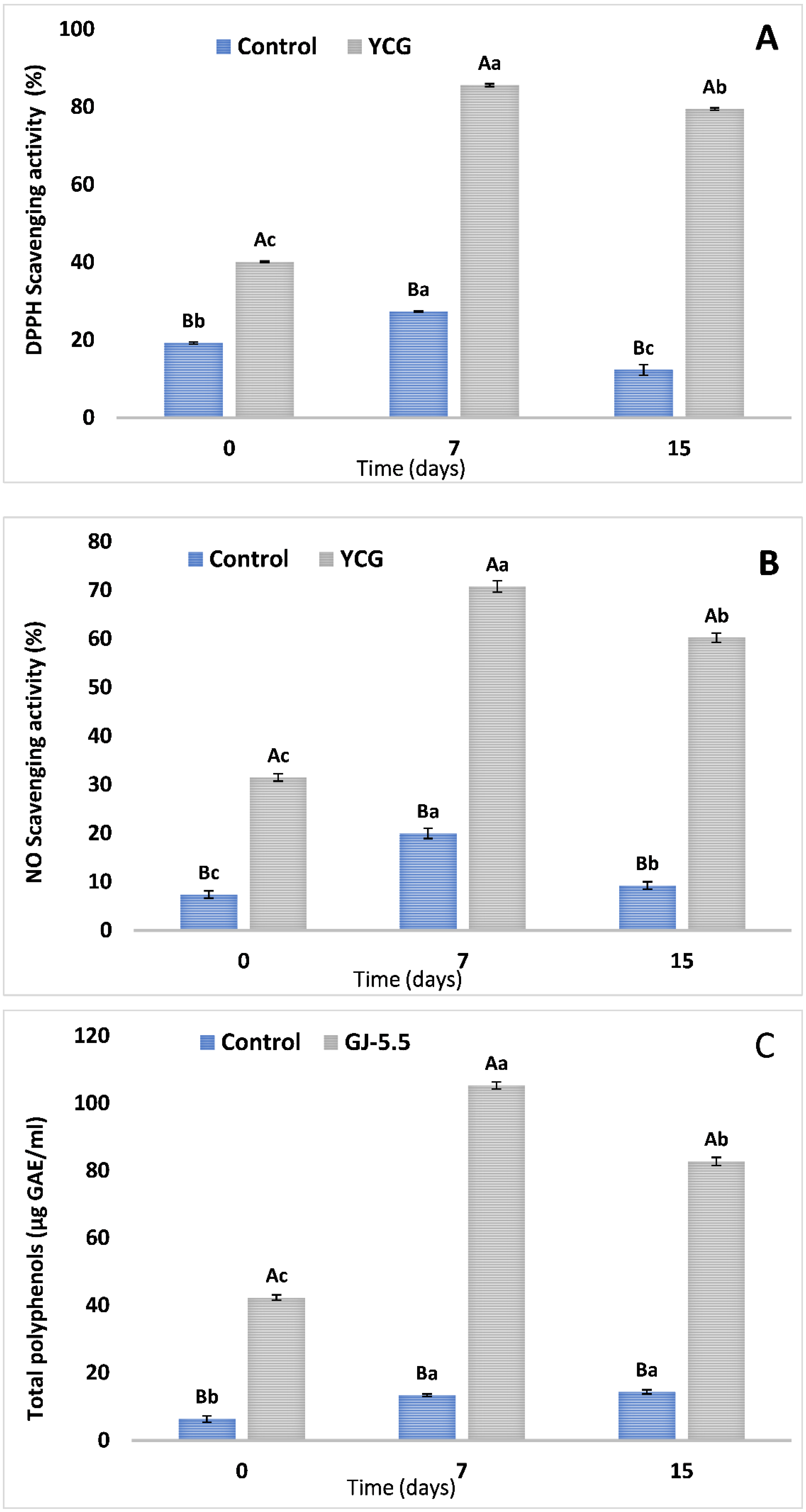
3.4. Assessment of Antioxidant Activities and Total Phenolic Components in Yogurt

In the DPPH assay, The YCG supernatant recorded a higher significant value than the control sample at zero time (40.16 vs. 19.23%, respectively) (Figure 4A). The maximum inhibition value of DPPH radical (85.6%) was obtained by YCG-7 supernatant. Overall, refrigerated storage significantly improved DPPH radical scavenging activity in YCG till the end of storge period (Figure 4A). In NO assay, the maximum inhibition of 70.73% was observed after seven days of cold storage of the YCG sample (Figure 4B). The total phenolic components (TPC) in the YCG supernatant were higher (p < 0.05) than control (Figure 4C). YCG supernatant displayed a notable increase in TPC and reached the maximum values at day seven (105.3 ± 1.04 μg GAE/mL), followed by a notable decrease (82.73 ± 1.23 μg GAE/mL; p < 0.05) by day 15. However, the plain-yogurt supernatant displayed a slight increase in TPC till the end of the storage period.

3.5. Total Viable Counts of E.coli Nissle 1917

Counts of EcN at zero time were above 7 log CFU mL−1 in all tested groups of yogurts, with the maximum values existing in YCG (7.38 log CFU mL−1). As can be noticed in Figure 5, the total viable counts of EcN gradually increased till the last day of cold storage, except in the control (Figure 5). The viable counts of EcN in all instances were upheld above the proposed minimum level of 6 logs CFU mL−1 during storage periods of yogurt (Figure 5). YCG maintained an extensive level of EcN survival (8.80 log CFU mL−1) after two-week cold storage.

3.6. Sensory Evaluation of Yogurt Samples

The sensory characteristic of the yogurt samples was rated in terms of appearance, color, smoothness, sourness, mouthfeel, and overall acceptance as shown in Figure 6. Compared with the control, YCG had higher sensory scores for appearance, smoothness, sourness, mouthfeel, and overall acceptance. Overall acceptance was, thus, based on the consumer predilection of the sourness and color degrees in the yogurts. The addition of CGJ to yogurt (YCG) significantly (p < 0.05) decreased the color scores compared with control (Figure 6). Generally, there was a significant (p < 0.05) decline in sensory properties of all yogurt samples with progressing the storage period.

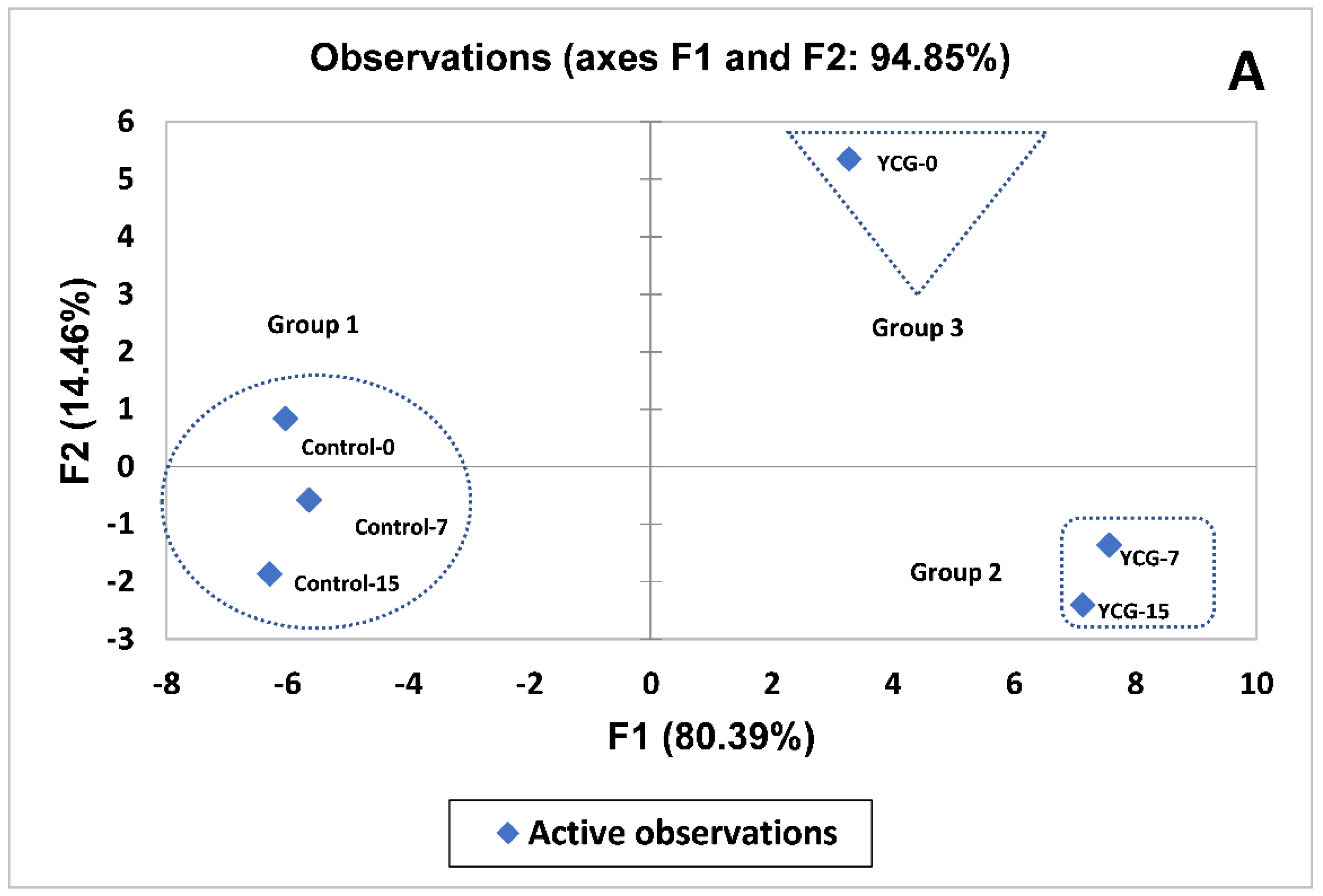
3.7. Multivariate Analysis of YCG Parameters

Principal component analysis (PCA) of polyphenols profile, EcN survival, sensory properties, antimicrobial, antitumor, and antioxidant activities of YCG explained 94.85%of the variability on 2PC (Figure 7A). PC1(80.39%) included sensory properties (appearance, color, and smoothness), antitumor activity against MDA, LS and PC-3, E.coli Nissle 1917 survival, antioxidant activity (DPPH and NO), total phenolic contents, antimicrobial activity against all tested strains, and polyphenols profile (pyrogallol, gallic acid, catechol,4-hydroxybenzoic acid, vanillic acid, caffeic acid, syringic acid, kaempferol, rutin, ellagic acid, o-coumaric acid, salicylic acid, cinnamic acid, myricetin, quercetin, rosmarinic acid, and naringin). However, the second dimension (14.46%) was primarily associated with sourness, mouthfeel, overall acceptance, chlorogenic acid (Figure 7A). The treatments were classified into three groups. Group 1 was found on the left side of PC1. However, the second and the third groups were positioned on the positive values side of PC1. The first group was characterized by the high level of IC50 against Ls, PC-3, and MDA cell lines, as well as a high score of color degree for resulting yogurt samples, whereas group 2 was characterized by the highest values of antioxidant activity (DPPH and NO), total phenolic contents, and antimicrobial activity against all tested strains. In addition, it has a high concentration of pyrogallol, gallic acid, catechol, 4-hydroxybenzoic acid, vanillic acid, caffeic acid, syringic acid, p-coumaric acid, kaempferol, rutin, ellagic acid, o-coumaric acid, salicylic acid, myricetin, and quercetin (Figure 7A). The high concentration of chlorogenic acid, cinnamic acid, rosmarinic acid, and naringin was detected in group 3 compared with other groups. Besides, its scores the highest on the sensory properties except for the color of resulted yogurt (Figure 7A).
The variable correlation analysis of PCA was especially needed when there are several explanatory variables, probably correlated. Here, the analysis of variable correlation was used to assess the relationship between changes in polyphenols profile and sensory properties, EcN survival, antioxidant, antimicrobial, and antitumor activities (Figure 7B). The high concentration of pyrogallol, gallic acid, catechol, 4-hydroxybenzoic acid, vanillic acid, caffeic acid, syringic acid, p-coumaric acid, kaempferol, rutin, ellagic acid, o-coumaric acid, salicylic acid, myricetin, and quercetin are strong positive correlated with antimicrobial, antioxidant, antitumor activities and survive of EcN. However, the high concentration of chlorogenic acid, cinnamic acid, rosmarinic acid, and naringin are strongly positively correlated with appearance, sourness, mouthfeel, and overall acceptance. Further, all o these phenolic compounds are negative with color and IC50 of MDA, LS, and PC-3 (Figure 7B).

4. Discussion

The proposed changes in the phenolic contents after incubation with EcN are summarized in Figure 8. The concentrations of pyrogallol, gallic and ellagic acids were largely increased in YCG after 7 and 15 days of cooled storage when compared with the unfermented sample. This might be due to the ability of EcN to degrade the amount of hydrolyzable tannins found in the used juice, similar results have been reported by other microorganisms [40]. Overall, the quantified amount of tannins in Cape gooseberry fruit aqueous extract (as 0.71 mg CE/g DW) as noted by Namiesnik, et al. [41], supported the previous declaration. The identification of bound polyphenols such as chlorogenic acid, 1-p-coumaroyl-β-D-glucoside, 1-O-caffeoyl-β-D-glucopyranoside, naringin, and myricetin-3-O-neohesperidoside in Cape gooseberry fruit has been reported [42,43,44]. In this regard, the ability of microorganisms and their enzymatic systems, including glycosidases and esterases, to hydrolyze such these bound polyphenols into free ones or other lower molecular weight bioactive metabolites via further biotransformation mechanisms has been reported [45,46,47,48]. So, the high release of some free polyphenols in this study such as myricetin, coumaric, 4-hydroxy benzoic and salicylic acids as well as the sharp reduction of naringin, chlorogenic and rosmarinic acids might be partially due to the hydrolytic role of EcN enzymes on polyphenol derivatives of Cape gooseberry.
Interestingly, the deglycosylation of flavonoid glycosides releases their biological activities [49]. Likewise, the physiological properties of one of the naringin metabolites named 3-(4ʹ-Hydroxyphenyl) propanoic acid have been reported [46]. Noticeably, the low level of quercetin increase in this study may reflect the weaker ability of starter strains to deglycosylate the flavonoid of rutin into quercetin and other metabolites. However, other microbial strains can release higher significant quantities of quercetin and quercetin-3-glucoside as the major metabolites of rutin rich extracts [50]. The notable increase in rutin might be explained by the microbial demethylation of related compounds such as isorhamnetin 3-rutinoside. In this respect, demethylation of poly methoxy flavones by human gut bacterium has been reported [51].
Overall, this study clarified an improvement in the bioavailability of different polyphenols of CGJ when used in the acidification of cow milk due to the microbial action. In polyphenol-rich foods, biotransformation can be carried out by incubating them with probiotics which initiate different types of chemical modifications such as decarboxylation, deglycosylation, ring fission, demethylation, dehydroxylation, lactonization, aromatic hydroxylation, or reduction of carbon-carbon double bonds [52] shifting some polyphenols into more bioavailable and/or bioactive forms than their original forms [53].
The postbiotics produced by EcN used in this study presented board antibacterial activity, where they could inhibit Gram-negative and Gram-positive pathogens. Besides, they had moderate antifungal activity. These results are consistent with a previous report by Liasi, et al. [54], who studied that the antibacterial agent resulted from Lactobacillus plantarum which inhibited the growth of E. coli, Listeria monocytogenes and Staphylococcus aureus. Similarly, Kareem, et al. [55] observed the positive effect of inulin towards the antimicrobial activity of postbiotics produced by L. plantarum. The antimicrobial activity of postbiotics in our study might be attributed to various antimicrobial compounds such as organic acid, bioactive peptides, short-chain fatty acids, and phenolic compounds [55,56].
The polyphenols in the obtained postbiotics in our finding were classified into 3 groups according to molecular weight, 11 phenolic compounds had a molecular weight < 200 g·mol−1, 7 phenolic compounds had a molecular weight between 200 and 400 g·mol−1, and lastly, one phenolic compound had molecular weight > 400 g·mol1. The high antimicrobial activity of postbiotic after 7 or 15 days of storage period compared with postbiotic at zero time may be due to the high concentration of phenolic compounds having a molecular weight < 200 g.mol−1. Many previous studies have reported that the strong antimicrobial activity phenolic compounds that had low molecular weight based on quantitative structure-activity relationship (QSAR) [56]. However, the prediction by QSAR for antimicrobial activity of phenolic compounds which had high molecular weight remains limited [56]. Besides, a high concentration of salicylic acid was detected in postbiotics after 7 and 15 days of storage periods compared with other compounds. However, it was not detected in postbiotic at zero time. Several previous studies exhibited the antimicrobial activity of salicylic acid [57]. Many previous studies have proven the antimicrobial of pyrogallol and caffeic acid [58,59]. Their mechanism of the action takes place by enzymatic inhibition by oxidized compounds [58,59].
The MIC values of YCG (3.1–25 µg/mL) were lower than those reported in the literature for Cape gooseberry [34].
The estimation of ratio MBC/MIC or MFC/MIC values is considered to be a perfect evaluation of the antibacterial or antifungal influence of bioactive compounds, where Pankey and Sabath [60] reported that an antimicrobial compound considers bacteriostatic or fungistatic only when the ratio MBC/MIC or MFC/MIC higher than 4. Otherwise such substance is fungicidal or bactericidal. The values of the ratio of MBC/MIC or MFC/MIC for YCG Exhibited bactericidal and fungicidal activities.
Our finding presented that the antitumor activity of YCG, significantly higher than the previous study suggested, according to Ramadan, et al. [61], who reported that the inhibition level of Cape gooseberry extract against colon cell lines (IC50:142µg/mL) was higher than breast cell line (IC50:371 µg/mL) [61]. Also, some studies confirmed that goldenberry extract has presented significant antiproliferative activity toward colon cancer cells (colo-205) lung cancer cell lines (H1299) and others including renal, prostate, mammary, and colorectal cell lines [62].
The concentration of pyrogallol in YCG-7 and YCG-15 increased about ninefold compared with YCG-0 (that is, one of the natural phenolic compounds) was known to have antitumor and anti-inflammatory against colon and breast cancers. The underlying anticancer mechanisms of pyrogallol, remain unclear so far [63]. While many previous findings examined the antitumor influences of combinations of two or three phenolic compounds, we used more comprehensive mixtures (postbiotics) of phenolic compounds with bioactive peptides, vitamins, and other micronutrients. Salicylic acid, myricetin, and quercetin in YCG-7 or YCG-15 appear to work in synergy with a high concentration of low molecular weight phenolic compounds for increasing antitumor against tested human tumor cell lines (MDA, LS and PC) compared with YCG-0. This results are in consistent with those by Niedzwiecki, et al. [64], who reported that a combination of phenolic compounds (green tea extract and quercetin) with amino acids, vitamin C and other micronutrients increased the anticancer activity against ovarian cancer ES-2 xenograft tumor growth.
Our finding presented that the biotransformation of phenolic compounds in postbiotics (YCG-7 and YCG-15) play a critical role in antitumor activities compared with other compounds and micronutrients. This could be a reason for the reduction of antitumor activity by postbiotic produced from plain yogurt.
The findings of the present study regarding the strong antioxidant activity of YCG largely agreed with those obtained by Shori and Baba [30]. They found that yogurt supplemented by Azadirachta indica had significant values of TPC and antioxidant ability throughout the entire period of cold storage when compared with the negative control. The notable decrease in TPC in supernatant of YCG-15 may be explained by the microbial degradation of higher molecular weight polyphenols or breakdown of aromatic ring structure of phenolic compounds [30]. Overall, raised TPC in food extracts was normally related to increased antioxidant potentials [36] despite some discrepancies [65]. Likewise, CGJ has been formerly stated to show antioxidant capacity [66] which might be explained by its high content of total polyphenols and vitamin C [43]. The action of EcN on phenolic acids derivatives can lead to their conversion into other phenolic acids owning antioxidant potentials such as p-hydroxybenzoic and vanillic acids in postbiotic (YCG-7) compared with postbiotic at zero time (YCG-0) [67]. Other likely sources of radical scavengers ascribed to yogurt may be obtained from milk protein proteolysis [68] and organic acids [69] as a result of post-acidification and fermentation during storage.
The presence of glycosyl or amine conjugated groups to phenolic compounds can improve the growth of EcN by providing essential nutrients, respectively fermentable sugars or nitrogen [56]. This could explain the EcN growth improving in YCG during the storage period compared with plain yogurt.
As expected, YCG expressed the maximum preference for appearance, smoothness, sourness, mouthfeel, and overall acceptance. The results are in full accordance with the findings of Mohan et al. [39].

5. Conclusions

The current finding presented that the addition of Cape gooseberry juice had no notable effect on the sensory properties of the resulting yogurt. On the other hand, the postbiotics produced from YCG after 7 or 15 days of storage periods significantly increased antimicrobial, antitumor, antioxidant activities, total phenolic content, and EcN survival. These results are opening new prospects for the utilization of polyphenol-rich products by the dairy sector by design and development of modern fermented dairy products associated with increased functional properties. However, more studies are needed to explain the mechanisms of the effect of biotransformation of phenolic compounds by EcN on antimicrobial, antitumor, and antioxidant. Besides, in vivo studies could also be auxiliary to assess the possible health benefits of postbiotic extracted from YCG.

Author Contributions

Conceptualization M.S.D.; methodology M.S.D., M.A.T., A.A.E., L.Q., A.A.Z., E.K., N.A.A.-Z., O.M.A.K.S. and D.H.D.; software M.S.D., M.A.T., A.A.E., L.Q., A.A.Z., E.K., N.A.A.-Z., O.M.A.K.S. and D.H.D.; validation, M.S.D., M.A.T., A.A.E., L.Q., A.A.Z., E.K., N.A.A.-Z., O.M.A.K.S. and D.H.D.; formal analysis, M.S.D., M.A.T., A.A.E., L.Q., A.A.Z., E.K., N.A.A.-Z., O.M.A.K.S. and D.H.D.; investigation, M.S.D., M.A.T., A.A.E., L.Q., A.A.Z., E.K., N.A.A.-Z., O.M.A.K.S. and D.H.D.; resources L.Q., E.K., M.S.D., A.A.E. and M.A.T.; data curation M.S.D., M.A.T., A.A.E., L.Q., A.A.Z., E.K., N.A.A.-Z., O.M.A.K.S. and D.H.D.; writing—original draft preparation, M.S.D. and M.A.T.; review and editing, M.S.D., M.A.T., A.A.E., L.Q., A.A.Z., E.K., N.A.A.-Z., O.M.A.K.S. and D.H.D.; visualization, M.S.D., M.A.T., A.A.E., L.Q., A.A.Z., E.K., N.A.A.-Z., O.M.A.K.S. and D.H.D.; supervision, M.S.D., L.Q., M.A.T. and E.K. All authors have read and agreed to the published version of the manuscript.

Funding

This research was funded by the special projects for local science and technology development guided by the China central government (grant No. 2019L3011). And Taif University Researchers supporting project number (TURSP-2020/307), Taif University, Taif, Saudi Arabia.

Institutional Review Board Statement

Not applicable.

Informed Consent Statement

Not applicable.

Data Availability Statement

All data generated or analyzed during this study are available from the corresponding author on reasonable request.

Acknowledgments

In Taif University Researchers supporting project number (TURSP-2020/307), Taif University, Taif, Saudi Arabia, and the special projects for local science and technology development guided by the China central government (grant No. 2019L3011).

Conflicts of Interest

All data generated or analyzed during this study are available from the corresponding author on reasonable request.

References

  1. Nazhand, A.; Souto, E.B.; Lucarini, M.; Souto, S.B.; Durazzo, A.; Santini, A. Ready to Use Therapeutical Beverages: Focus on Functional Beverages Containing Probiotics, Prebiotics and Synbiotics. Beverages 2020, 6, 26. [Google Scholar] [CrossRef] [Green Version]
  2. Bimbo, F.; Bonanno, A.; Nocella, G.; Viscecchia, R.; Nardone, G.; De Devitiis, B.; Carlucci, D. Consumers’ acceptance and preferences for nutrition-modified and functional dairy products: A systematic review. Appetite 2017, 113, 141–154. [Google Scholar] [CrossRef] [PubMed] [Green Version]
  3. Beltrán-Barrientos, L.; Hernández-Mendoza, A.; Torres-Llanez, M.; González-Córdova, A.; Vallejo-Córdoba, B. Invited review: Fermented milk as antihypertensive functional food. J. Dairy Sci. 2016, 99, 4099–4110. [Google Scholar] [CrossRef] [PubMed] [Green Version]
  4. Kandylis, P.; Pissaridi, K.; Bekatorou, A.; Kanellaki, M.; Koutinas, A.A. Dairy and non-dairy probiotic beverages. Curr. Opin. Food Sci. 2016, 7, 58–63. [Google Scholar] [CrossRef]
  5. Ahmad, I.; Khan, M.S.A.; Aqil, F.; Singh, M. Microbial Applications in Agriculture and the Environment: A Broad Perspective. In Microbes and Microbial Technology: Agricultural and Environmental Applications; Ahmad, I., Ahmad, F., Pichtel, J., Eds.; Springer: New York, NY, USA, 2011; pp. 1–27. [Google Scholar]
  6. Raza, M.A.; Kanwal, Z.; Rauf, A.; Sabri, A.N.; Riaz, S.; Naseem, S. Size-and shape-dependent antibacterial studies of silver nanoparticles synthesized by wet chemical routes. Nanomaterials 2016, 6, 74. [Google Scholar] [CrossRef] [Green Version]
  7. Riaz, S.; Raza, Z.A.; Majeed, M.I.; Jan, T. Synthesis of zinc sulfide nanoparticles and their incorporation into poly (hydroxybutyrate) matrix in the formation of a novel nanocomposite. Mater. Res. Express 2018, 5, 055027. [Google Scholar] [CrossRef]
  8. Terzić-Vidojević, A.; Veljović, K.; Tolinački, M.; Živković, M.; Lukić, J.; Lozo, J.; Fira, Đ.; Jovčić, B.; Strahinić, I.; Begović, J.; et al. Diversity of non-starter lactic acid bacteria in autochthonous dairy products from Western Balkan Countries—Technological and probiotic properties. Food Res. Int. 2020, 136, 109494. [Google Scholar] [CrossRef]
  9. Schultz, M. Clinical use of E. coli Nissle 1917 in inflammatory bowel disease. Inflamm. Bowel Dis. 2008, 14, 1012–1018. [Google Scholar] [CrossRef]
  10. Grozdanov, L.; Raasch, C.; Schulze, J.; Sonnenborn, U.; Gottschalk, G.; Hacker, J.; Dobrindt, U. Analysis of the genome structure of the nonpathogenic probiotic Escherichia coli strain Nissle 1917. J. Bacteriol. 2004, 186, 5432–5441. [Google Scholar] [CrossRef] [Green Version]
  11. Caleja, C.; Barros, L.; Antonio, A.L.; Carocho, M.; Oliveira, M.B.; Ferreira, I.C. Fortification of yogurts with different antioxidant preservatives: A comparative study between natural and synthetic additives. Food Chem. 2016, 210, 262–268. [Google Scholar] [CrossRef] [Green Version]
  12. Ramos, L.R.; Santos, J.S.; Daguer, H.; Valese, A.C.; Cruz, A.G.; Granato, D. Analytical optimization of a phenolic-rich herbal extract and supplementation in fermented milk containing sweet potato pulp. Food Chem. 2017, 221, 950–958. [Google Scholar] [CrossRef]
  13. Hossen, M.S.; Ali, M.Y.; Jahurul, M.H.A.; Abdel-Daim, M.M.; Gan, S.H.; Khalil, M.I. Beneficial roles of honey polyphenols against some human degenerative diseases: A review. Pharmacol. Rep. 2017, 69, 1194–1205. [Google Scholar] [CrossRef]
  14. Neveu, V.; Perez-Jiménez, J.; Vos, F.; Crespy, V.; du Chaffaut, L.; Mennen, L.; Knox, C.; Eisner, R.; Cruz, J.; Wishart, D.; et al. Phenol-Explorer: An online comprehensive database on polyphenol contents in foods. Database 2010, 2010, bap024. [Google Scholar] [CrossRef]
  15. Bhuyan, D.J.; Basu, A. Phenolic compounds potential health benefits and toxicity. In Utilisation of Bioactive Compounds from Agricultural and Food Production Waste; Routledge: New York, NY, USA, 2017; pp. 27–59. [Google Scholar]
  16. de Llano, D.G.; Gil-Sánchez, I.; Esteban-Fernández, A.; Ramos, A.M.; Fernández-Díaz, M.; Cueva, C.; Moreno-Arribas, M.V.; Bartolomé, B. Reciprocal beneficial effects between wine polyphenols and probiotics: An exploratory study. Eur. Food Res. Technol. 2017, 243, 531–538. [Google Scholar] [CrossRef]
  17. Erkaya, T.; Dağdemir, E.; Şengül, M. Influence of Cape gooseberry (Physalis peruviana L.) addition on the chemical and sensory characteristics and mineral concentrations of ice cream. Food Res. Int. 2012, 45, 331–335. [Google Scholar] [CrossRef]
  18. Puente, L.A.; Pinto-Muñoz, C.A.; Castro, E.S.; Cortés, M. Physalis peruviana Linnaeus, the multiple properties of a highly functional fruit: A review. Food Res. Int. 2011, 44, 1733–1740. [Google Scholar] [CrossRef]
  19. Narváez-Cuenca, C.E.; Mateus-Gómez, Á.; Restrepo-Sánchez, L.P. Antioxidant capacity and total phenolic content of air-dried cape gooseberry (Physalis peruviana L.) at different ripeness stages. Agron. Colomb. 2014, 32, 232–237. [Google Scholar] [CrossRef]
  20. Tammu, J.; Ramana, K.V. Pharmacological review on Physalis species: A potential herbal cure-all. World J. Pharm. Res. 2015, 4, 247–256. [Google Scholar]
  21. Ramadan, M.F. Bioactive phytochemicals, nutritional value, and functional properties of cape gooseberry (Physalis peruviana): An overview. Food Res. Int. 2011, 44, 1830–1836. [Google Scholar] [CrossRef]
  22. De Souza, E.L.; de Albuquerque, T.M.R.; dos Santos, A.S.; Massa, N.M.L.; de Brito Alves, J.L. Potential interactions among phenolic compounds and probiotics for mutual boosting of their health-promoting properties and food functionalities—A review. Crit. Rev. Food Sci. Nutr. 2019, 59, 1645–1659. [Google Scholar] [CrossRef]
  23. Hervert-Hernández, D.; Goñi, I. Dietary polyphenols and human gut microbiota: A review. Food Rev. Int. 2011, 27, 154–169. [Google Scholar] [CrossRef]
  24. Possemiers, S.; Bolca, S.; Verstraete, W.; Heyerick, A. The intestinal microbiome: A separate organ inside the body with the metabolic potential to influence the bioactivity of botanicals. Fitoterapia 2011, 82, 53–66. [Google Scholar] [CrossRef]
  25. Wegh, C.A.; Geerlings, S.Y.; Knol, J.; Roeselers, G.; Belzer, C. Postbiotics and their potential applications in early life nutrition and beyond. Int. J. Mol. Sci. 2019, 20, 4673. [Google Scholar] [CrossRef] [Green Version]
  26. Kay, C.D.; Pereira-Caro, G.; Ludwig, I.A.; Clifford, M.N.; Crozier, A. Anthocyanins and flavanones are more bioavailable than previously perceived: A review of recent evidence. Annu. Rev. Food Sci. Technol. 2017, 8, 155–180. [Google Scholar] [CrossRef]
  27. Stevens, Y.; Rymenant, E.V.; Grootaert, C.; Camp, J.V.; Possemiers, S.; Masclee, A.; Jonkers, D. The intestinal fate of citrus flavanones and their effects on gastrointestinal health. Nutrients 2019, 11, 1464. [Google Scholar] [CrossRef] [Green Version]
  28. Hassan, H.A.; Serag, H.M.; Qadir, M.S.; Ramadan, M.F. Cape gooseberry (Physalis peruviana) juice as a modulator agent for hepatocellular carcinoma-linked apoptosis and cell cycle arrest. Biomed. Pharmacother. 2017, 94, 1129–1137. [Google Scholar] [CrossRef]
  29. Margean, A.; Lupu, M.I.; Alexa, E.; Padureanu, V.; Canja, C.M.; Cocan, I.; Negrea, M.; Calefariu, G.; Poiana, M.-A. An Overview of Effects Induced by Pasteurization and High-Power Ultrasound Treatment on the Quality of Red Grape Juice. Molecules 2020, 25, 1669. [Google Scholar] [CrossRef] [Green Version]
  30. Shori, A.; Baba, A. Antioxidant activity and inhibition of key enzymes linked to type-2 diabetes and hypertension by Azadirachta indica-yogurt. J. Saudi Chem. Soc. 2013, 17, 295–301. [Google Scholar] [CrossRef]
  31. Taher, M.; El-Daly, N.M.; El-Khateeb, A.Y.; Hassan, S.M.; Elsherbiny, E.A. Chemical Composition, Antioxidant, Antitumor and Antifungal Activities of Methanolic Extracts of Coleus blumei, Plectranthus amboinicus and Salvia splendens (Lamiaceae). J. Agric. Chem. Biotechnol. 2021, 12, 177–187. [Google Scholar] [CrossRef]
  32. Ghomari, O.; Sounni, F.; Massaoudi, Y.; Ghanam, J.; Drissi Kaitouni, L.B.; Merzouki, M.; Benlemlih, M. Phenolic profile (HPLC-UV) of olive leaves according to extraction procedure and assessment of antibacterial activity. Biotechnol. Rep. 2019, 23, e00347. [Google Scholar] [CrossRef]
  33. Jovanovic, M.; Petrovic, M.; Miocinovic, J.; Zlatanovic, S.; Lalicic Petronijevic, J.; Mitic-Culafic, D.; Gorjanovic, S. Bioactivity and Sensory Properties of Probiotic Yogurt Fortified with Apple Pomace Flour. Foods 2020, 9, 763. [Google Scholar] [CrossRef] [PubMed]
  34. Helvaci, S.; Kokdil, G.; Kawai, M.; Duran, N.; Duran, G.; Guvenc, A. Antimicrobial activity of the extracts and physalin D from Physalis alkekengi and evaluation of antioxidant potential of physalin D. Pharm. Biol. 2010, 48, 142–150. [Google Scholar] [CrossRef] [PubMed] [Green Version]
  35. Parvekar, P.; Palaskar, J.; Metgud, S.; Maria, R.; Dutta, S. The minimum inhibitory concentration (MIC) and minimum bactericidal concentration (MBC) of silver nanoparticles against Staphylococcus aureus. Biomater. Investig. Dent. 2020, 7, 105–109. [Google Scholar] [CrossRef] [PubMed]
  36. Taher, M.A.; Tadros, L.K.; Dawood, D.H. Phytochemical constituents, antioxidant activity and safety evaluation of Kei-apple fruit (Dovyalis caffra). Food Chem. 2018, 265, 144–151. [Google Scholar] [CrossRef] [PubMed]
  37. Taher, M.A.; MennatAllah, E.; Tadros, L.K.; Sanad, M.I. The effects of new formulations based on Gum Arabic on antioxidant capacity of tomato (Solanum lycopersicum L.) fruit during storage. J. Food Meas. Charact. 2020, 14, 2489–2502. [Google Scholar] [CrossRef]
  38. Lee, S.-B.; Cosmas, B.; Park, H.-D. The Antimutagenic and Antioxidant Activity of Fermented Milk Supplemented with Cudrania tricuspidata Powder. Foods 2020, 9, 1762. [Google Scholar] [CrossRef] [PubMed]
  39. Mohan, A.; Hadi, J.; Gutierrez-Maddox, N.; Li, Y.; Leung, I.K.; Gao, Y.; Shu, Q.; Quek, S.-Y. Sensory, Microbiological and Physicochemical Characterisation of Functional Manuka Honey Yogurts Containing Probiotic Lactobacillus reuteri DPC16. Foods 2020, 9, 106. [Google Scholar] [CrossRef] [Green Version]
  40. Mingshu, L.; Kai, Y.; Qiang, H.; Dongying, J. Biodegradation of gallotannins and ellagitannins. J. Basic Microbiol. 2006, 46, 68–84. [Google Scholar] [CrossRef]
  41. Namiesnik, J.; Vearasilp, K.; Kupska, M.; Ham, K.-S.; Kang, S.-G.; Park, Y.-K.; Barasch, D.; Nemirovski, A.; Gorinstein, S. Antioxidant activities and bioactive components in some berries. Eur. Food Res. Technol. 2013, 237, 819–829. [Google Scholar] [CrossRef] [Green Version]
  42. Chang, J.; Lin, C.; Wu, S.; Lin, D.; Wang, S.; Miaw, C.; Ng, L. Antioxidative and hepatoprotective effects of Physalis peruviana extract against acetaminophen-induced liver injury in rats. Pharm. Biol. 2008, 46, 724–731. [Google Scholar] [CrossRef] [Green Version]
  43. Deng, K.J.; Zang, L.L.; Lan, X.H.; Zhong, Z.H.; Xiong, B.Q.; Zhang, Y.; Zheng, X.L. Antioxidant Components from Cape Gooseberry. J. Food Processing Preserv. 2016, 40, 893–898. [Google Scholar] [CrossRef]
  44. Muñoz, P.; Parra, F.; Simirgiotis, M.J.; Sepúlveda Chavera, G.F.; Parra, C. Chemical Characterization, Nutritional and Bioactive Properties of Physalis peruviana Fruit from High Areas of the Atacama Desert. Foods 2021, 10, 2699. [Google Scholar] [CrossRef]
  45. Bel-Rhlid, R.; Thapa, D.; Kraehenbuehl, K.; Hansen, C.E.; Fischer, L. Biotransformation of caffeoyl quinic acids from green coffee extracts by Lactobacillus johnsonii NCC 533. Amb Express 2013, 3, 28. [Google Scholar] [CrossRef] [Green Version]
  46. Chen, T.; Wu, H.; He, Y.; Pan, W.; Yan, Z.; Liao, Y.; Peng, W.; Gan, L.; Zhang, Y.; Su, W. Simultaneously quantitative analysis of naringin and its major human gut microbial metabolites naringenin and 3-(4′-hydroxyphenyl) propanoic acid via stable isotope deuterium-labeling coupled with RRLC-MS/MS method. Molecules 2019, 24, 4287. [Google Scholar] [CrossRef] [Green Version]
  47. Raimondi, S.; Anighoro, A.; Quartieri, A.; Amaretti, A.; Tomás-Barberán, F.A.; Rastelli, G.; Rossi, M. Role of bifidobacteria in the hydrolysis of chlorogenic acid. Microbiologyopen 2015, 4, 41–52. [Google Scholar] [CrossRef] [Green Version]
  48. Zheng, Y.; Guo, Z.; Cai, Z. Combination of β-elimination and liquid chromatography/quadrupole time-of-flight mass spectrometry for the determination of O-glycosylation sites. Talanta 2009, 78, 358–363. [Google Scholar] [CrossRef]
  49. Cavia-Saiz, M.; Busto, M.D.; Pilar-Izquierdo, M.C.; Ortega, N.; Perez-Mateos, M.; Muñiz, P. Antioxidant properties, radical scavenging activity and biomolecule protection capacity of flavonoid naringenin and its glycoside naringin: A comparative study. J. Sci. Food Agric. 2010, 90, 1238–1244. [Google Scholar] [CrossRef]
  50. Lin, S.; Zhu, Q.; Wen, L.; Yang, B.; Jiang, G.; Gao, H.; Chen, F.; Jiang, Y. Production of quercetin, kaempferol and their glycosidic derivatives from the aqueous-organic extracted residue of litchi pericarp with Aspergillus awamori. Food Chem. 2014, 145, 220–227. [Google Scholar] [CrossRef]
  51. Burapan, S.; Kim, M.; Han, J. Curcuminoid demethylation as an alternative metabolism by human intestinal microbiota. J. Agric. Food Chem. 2017, 65, 3305–3310. [Google Scholar] [CrossRef]
  52. Aura, A.-M. Microbial metabolism of dietary phenolic compounds in the colon. Phytochem. Rev. 2008, 7, 407–429. [Google Scholar] [CrossRef]
  53. Thilakarathna, W.W.; Langille, M.G.; Rupasinghe, H.V. Polyphenol-based prebiotics and synbiotics: Potential for cancer chemoprevention. Curr. Opin. Food Sci. 2018, 20, 51–57. [Google Scholar] [CrossRef]
  54. Liasi, S.A.; Azmi, T.I.; Hassan, M.D.; Shuhaimi, M.; Rosfarizan, M.; Ariff, A.B. Antimicrobial activity and antibiotic sensitivity of three isolates of lactic acid bacteria from fermented fish product, Budu. Malays. J. Microbiol. 2009, 5, 33–37. [Google Scholar]
  55. Kareem, K.Y.; Hooi Ling, F.; Teck Chwen, L.; May Foong, O.; Anjas Asmara, S. Inhibitory activity of postbiotic produced by strains of Lactobacillus plantarum using reconstituted media supplemented with inulin. Gut Pathog. 2014, 6, 23. [Google Scholar] [CrossRef] [Green Version]
  56. Bouarab-Chibane, L.; Forquet, V.; Lantéri, P.; Clément, Y.; Léonard-Akkari, L.; Oulahal, N.; Degraeve, P.; Bordes, C. Antibacterial Properties of Polyphenols: Characterization and QSAR (Quantitative Structure–Activity Relationship) Models. Front. Microbiol. 2019, 10, 829. [Google Scholar] [CrossRef]
  57. Kantouch, A.; El-Sayed, A.A.; Salama, M.; El-Kheir, A.A.; Mowafi, S. Salicylic acid and some of its derivatives as antibacterial agents for viscose fabric. Int. J. Biol. Macromol. 2013, 62, 603–607. [Google Scholar] [CrossRef]
  58. Mason, T.L. Inactivation of red beet β-glucan synthase by native and oxidized phenolic compounds. Phytochemistry 1987, 26, 2197–2202. [Google Scholar] [CrossRef]
  59. Simões, C.C.; Araújo, D.B.d.; Araújo, R.P.C.d. Study, in vitro and ex vivo, of the action of different concentrations of propolis extracts against microorganisms present in human saliva. Rev. Bras. Farmacogn. 2008, 18, 84–89. [Google Scholar] [CrossRef] [Green Version]
  60. Pankey, G.A.; Sabath, L.D. Clinical Relevance of Bacteriostatic versus Bactericidal Mechanisms of Action in the Treatment of Gram-Positive Bacterial Infections. Clin. Infect. Dis. 2004, 38, 864–870. [Google Scholar] [CrossRef] [Green Version]
  61. Ramadan, M.; El-Ghorab, A.; Ghanem, K. Volatile compounds, antioxidants, and anticancer activities of Cape gooseberry fruit (Physalis peruviana L.): An in vitro study. J. Arab Soc. Med. Res. 2015, 10, 56–64. [Google Scholar] [CrossRef]
  62. Xu, Y.-M.; Wijeratne, E.K.; Babyak, A.L.; Marks, H.R.; Brooks, A.D.; Tewary, P.; Xuan, L.-J.; Wang, W.-Q.; Sayers, T.J.; Gunatilaka, A.L. Withanolides from aeroponically grown Physalis peruviana and their selective cytotoxicity to prostate cancer and renal carcinoma cells. J. Nat. Prod. 2017, 80, 1981–1991. [Google Scholar] [CrossRef]
  63. Ahn, H.; Im, E.; Lee, D.Y.; Lee, H.-J.; Jung, J.H.; Kim, S.-H. Antitumor effect of Pyrogallol via miR-134 mediated S phase arrest and inhibition of PI3K/AKT/Skp2/cMyc signaling in hepatocellular carcinoma. Int. J. Mol. Sci. 2019, 20, 3985. [Google Scholar] [CrossRef] [PubMed] [Green Version]
  64. Niedzwiecki, A.; Roomi, M.W.; Kalinovsky, T.; Rath, M. Anticancer Efficacy of Polyphenols and Their Combinations. Nutrients 2016, 8, 552. [Google Scholar] [CrossRef] [PubMed] [Green Version]
  65. Kähkönen, M.P.; Hopia, A.I.; Vuorela, H.J.; Rauha, J.-P.; Pihlaja, K.; Kujala, T.S.; Heinonen, M. Antioxidant activity of plant extracts containing phenolic compounds. J. Agric. Food Chem. 1999, 47, 3954–3962. [Google Scholar] [CrossRef] [PubMed]
  66. Al-Olayan, E.M.; El-Khadragy, M.F.; Omer, S.A.; Shata, M.T.M.; Kassab, R.B.; Abdel Moneim, A.E. The beneficial effect of Cape gooseberry juice on carbon tetrachloride-induced neuronal damage. CNS Neurol. Disord.-Drug Targets 2016, 15, 344–350. [Google Scholar] [CrossRef]
  67. Blum, U. Effects of microbial utilization of phenolic acids and their phenolic acid breakdown products on allelopathic interactions. J. Chem. Ecol. 1998, 24, 685–708. [Google Scholar] [CrossRef]
  68. Lourens-Hattingh, A.; Viljoen, B.C. Yogurt as probiotic carrier food. Int. Dairy J. 2001, 11, 1–17. [Google Scholar] [CrossRef]
  69. Correia, I.; Nunes, A.; Duarte, I.F.; Barros, A.; Delgadillo, I. Sorghum fermentation followed by spectroscopic techniques. Food Chem. 2005, 90, 853–859. [Google Scholar] [CrossRef]
Figure 1. Heat map estimated from quantitative data of phenolic compounds concentration in different treatments. The scale of color moves from red to green with increased concentration.
Fermentation 08 00128 g001
Figure 2. Comparison of the inhibition percentage between different types of postbiotics with different concentrations against tumor cells. Where (A,D), (B,E) and (C,F) for breast (MDA-MB231), colon (LS-174T), and prostate (PC-3) cell lines, respectively. Besides, (G) for positive control (Doxorubicin). Different uppercase superscripts point out significant (p < 0.05) differences between the values for different concentrations. Different lowercase superscripts point out significant (p < 0.05) differences between the values during the storage period. Vertical bars present the standard division of the treatment means.
Fermentation 08 00128 g002
Figure 3. Comparison of the IC50 values of the lyophilized supernatants against human cancer cells. (AC) for breast (MDA-MB231), colon (LS-174T), and prostate (PC-3) cell lines, respectively. Different uppercase superscripts point out significant (p < 0.05) differences between the values for different treatments. Different lowercase superscripts point out significant (p < 0.05) differences between the values during the storage period. Vertical bars present the standard division of the treatment means.
Fermentation 08 00128 g003
Figure 4. Antioxidant activity and total phenolic content of tested yogurts samples. (A) DPPH scavenging activity (%), (B) NO scavenging activity (%), and (C) total polyphenols. Different uppercase superscripts point out significant (p < 0.05) differences between the values for different treatments. Different lowercase superscripts point out significant (p < 0.05) differences between the values during the storage period. Vertical bars present the standard division of the treatment means.
Fermentation 08 00128 g004
Figure 5. E.coli Nissle 1917 (EcN) counts (log CFU/mL) in YCG during the cold storage period. Different uppercase superscripts indicate significant (p < 0.05) differences between the values for different treatments. Different lowercase superscripts indicate significant (p < 0.05) differences between the values during the storage period. Error bars represent the standard division of means.
Fermentation 08 00128 g005
Figure 6. The average score of sensory evaluation of different yogurt types over storage periods. (A) zero-time, (B) 7 days of the storage period, and (C) 15 days of storage period. Different lowercase superscripts indicate significant (p < 0.05) differences between the values for different treatments. Different uppercase superscripts indicate significant (p < 0.05) differences between the values during the storage period. Error bars represent the standard division of means.
Fermentation 08 00128 g006
Figure 7. (A) Principal component analysis (PCA) biplot of YCG parameters. (B) The analysis of correlation variables.
Fermentation 08 00128 g007aFermentation 08 00128 g007b
Figure 8. Proposed scheme for changes of the phenolic contents.
Fermentation 08 00128 g008
Table 1. Antimicrobial activity, Minimum inhibitory concentrations (MIC), and Minimum bactericidal/fungicidal concentrations (MBC/MFC) of YCG supernatant.
MicroorganismInhibition Zone (mm) Supernatants.
Plain YogurtYCG (µg/mL)Amikacin (µg/mL) Fluconazole (µg/mL)
100 µg/mLMICMBC30 µg/mLMICMBC100 µg/mLMICMBC
Zero Time
Gram (−) Bacteria
K.pneumoniae-18.2 Bb*12.52524.9 A6.1512.5---
E.coli-19.2 Bb255029.1 A3.16.15---
E.cloacae-17.1 Bb255023.2 A12.525---
P. aeruginosa-23.8 Bb12.52525.3 A6.1512.5---
P. vulgaris-17.4 Bb12.52521.5 A12.525---
Gram (+) bacteria
B. cereus-23.2 Bb6.156.1526.8 A6.1512.5---
B.subtilis-26.6 Bb6.1512.529.4 A6.1512.5---
S. aureus-27.5 Ab6.1512.524.5 B3.16.15---
E. faecalis-23.7 Ab6.1512.522.1 B6.1512.5---
S. epidermidis-22.3 Bb6.1512.532.7 A3.16.15---
Fungi
C. parapsilosis-11.6 Bb50100---23.2 A12.525
C. krusei-12.1 Bb2550---27.4 A6.1512.5
C. albicans-12.5 Bb50100---22.9 A12.525
C. tropicalis-10.1 Bb50100---22.3 A2550
C. glabrata-13.7 Bb2550---30.2 A6.1512.5
7 days
Gram (−) bacteria
K.pneumoniae-22.7 Ba1 2.52524.9 A6.1512.5---
E.coli-24.4 Ba6.1512.529.1 A3.16.15---
E.cloacae-21.3 Ba12.512.523.2 A12.525---
P. aeruginosa-26.7 Aa3.16.1525.3 B6.1512.5---
P. vulgaris-22.5 Aa12.52521.5 B12.525---
Gram (+) bacteria
B. cereus-27.5 Aa6.156.1526.8 A6.1512.5---
B.subtilis-28.8 Aa6.1512.529.4 A6.1512.5---
S. aureus-29.5 Aa3.16.1524.5 B3.16.15---
E. faecalis-26.7 Aa3.16.1522.1 B6.1512.5---
S. epidermidis-29.2 Ba6.1512.532.7 A3.16.15---
Fungi
C. parapsilosis-18.2 Ba12.525---23.2 A12.525
C. krusei-19.1 Ba12.525---27.4 A6.1512.5
C. albicans-18.5 Ba12.525---22.9 A12.525
C. tropicalis-18.1 Ba2550---22.3 A2550
C. glabrata-19.7 Ba12.525---30.2 A6.1512.5
15 days
Gram (−) bacteria
K.pneumoniae-22.3 Ba12.52524.9 A6.1512.5---
E.coli-24.0 Ba6.1512.529.1 A3.16.15---
E.cloacae-20.8 Ba12.512.523.2 B12.525---
P. aeruginosa-26.5 Aa3.16.1525.3 B6.1512.5---
P. vulgaris-22.1 Aa12.52521.5 A12.525---
Gram (+) bacteria
B. cereus-27.1 Aa6.156.1526.8 A6.1512.5---
B.subtilis-28.2 Aa6.1512.529.4 A6.1512.5---
S. aureus-29.0 Aa3.16.1524.5 B3.16.15---
E. faecalis-26.1 Aa3.16.1522.1 B6.1512.5---
S. epidermidis-29.6 Ba6.1512.532.7 A3.16.15---
Fungi
C. parapsilosis-17.8 Ba12.525---23.2 A12.525
C. krusei-18.8 Ba12.525---27.4 A6.1512.5
C. albicans-18.1 Ba12.525---22.9 A12.525
C. tropicalis-17.7 Ba2550---22.3 A2550
C. glabrata-19.4 Ba12.525---30.2 A6.1512.5
* Different uppercase superscripts in columns point out significant (p < 0.05) differences between the values for different treatments (YCG and positive control). Different lowercase superscripts in the column point out significant (p < 0.05) differences between the values during the storage period.
Publisher’s Note: MDPI stays neutral with regard to jurisdictional claims in published maps and institutional affiliations.

