Review

Mechanical Cell Disruption Technologies for the Extraction of Dyes and Pigments from Microorganisms: A Review

1
Centre de Recherche Royallieu, ESCOM, TIMR (Integrated Transformations of Renewable Matter), Université de Technologie de Compiègne, CS 60 319, 60 203 Compiègne CEDEX, France
2
Laboratoire CTA, UR TVA, Centre d’Analyses et de Recherche, Faculté des Sciences, Université Saint-Joseph, Beyrouth 1104 2020, Lebanon
3
Laboratoire E2D, UR-EGP, Centre d’Analyses et de Recherche, Faculté des Sciences, Université Saint-Joseph, Beyrouth 1104 2020, Lebanon
4
Micalis Institute, AgroParisTech, INRAE, Université Paris-Saclay, 78352 Jouy-en-Josas, France
*
Authors to whom correspondence should be addressed.
Fermentation 2021, 7(1), 36; https://doi.org/10.3390/fermentation7010036
Submission received: 31 January 2021 / Revised: 28 February 2021 / Accepted: 3 March 2021 / Published: 9 March 2021

Abstract

:
The production of pigments using single cell microorganisms is gaining traction as a sustainable alternative to conventional syntheses, which rely, in no negligible proportions, on petrochemicals. In addition to depending on petroleum, these syntheses involved the use of toxic organic solvents, which may be inadequately disposed of across a range of industries, thus compounding the deleterious effects of fossil fuel exploitation. Literature suggests that notable research efforts in the area of sustainable pigment production using single cell microorganisms are focused on the production of pigments coveted for their interesting qualities, which transcend their mere capacity to dye various fabrics both natural and synthetic. As interest in sustainable pigment biosynthesis grows, the need to devise effective and efficient cell disruption processes becomes more pressing given that the viability of pigment biosynthesis is not only dependent on microorganisms’ yield in terms of production, but also on researchers’ ability to recover them. This review chiefly reports findings as to mechanical cell disruption methods, used individually or in various combinations, and their aptitude to recover biosynthetic pigments.

1. Introduction

Microorganisms, whose functions and output can be extensively modified through genetic engineering, can be used to produce a wide variety of compounds of interest in a growing number of fields with textiles, food, and pharmaceuticals leading the charge. Their use to produce pigments, such as carotenoids for example, presents numerous advantages over alternate biomass sources such as monocrop-type agriculture. When contrasted against compound production from microbial sources, purposeful plant cultivation appears to show numerous drawbacks such as it being time-consuming, resource-intensive, vulnerable to inclement weather, highly dependent on soil composition, and marked by use of considerable surfaces of land [1,2]. Additionally, in the context of producing high value products from microbial sources, industrial byproducts such as sugarcane and sugar beet molasses can be valorized and used as alternative nutrient sources [3,4]. This contributes to a reduction of the overall cost of production, which could be driven further down through the optimization of various process parameters pertaining to both the fermentation and extraction processes.
While arguably less stable than their synthetic counterparts, microbial pigments often possess numerous bioactive properties which make them desirable in the pharmaceutical, cosmetic, and even textile sectors. These properties, coupled to their high yields and capacity to proliferate in low-cost substrates, make microorganisms prime candidates for the industrial-scale production of various pigments [5]. Indeed, the industrial-scale production of pigments such as natural carotenoids from microbial sources is already a common industry practice [6]. The extensive research which has culminated in the widespread adoption of microbial synthesis of carotenoids in particular was driven by considerable demand due to their interesting pharmaceutical and cosmetic properties in addition to increased consumer wariness of synthetic food dyes [7,8]. Efforts are currently underway to similarly produce diverse pigments such as violacein, a purple bisindole pigment with antioxidant, antimicrobial, antipyretic, analgesic, and antitumoral properties [9]. The impetus to produce biosynthetic natural pigments is animated by shifting consumer trends towards health-conscious and sustainable consumption which, in this context, entails eschewing synthetic dyes and alleviating dependance on petrol derivatives. The economic implications of contributing to this shift are also considerable, with the size of the agricultural and food colorant markets expected to reach 2.03 billion USD and 3.75 billion USD respectively [10]. Generally, microbial pigments are produced and accumulated intracellularly and thus the cell wall must be ruptured for pigment recovery to be performed. Cell disruption techniques such as mechanical processes or chemical methods may be used individually or in various combinations to ensure consummate recovery of the pigments. The mechanical cell disruption technologies of bead milling, high-pressure homogenization (HPH), and ultrasonication (USN) will be described in the following sections.

2. Mechanical Cell Disruption
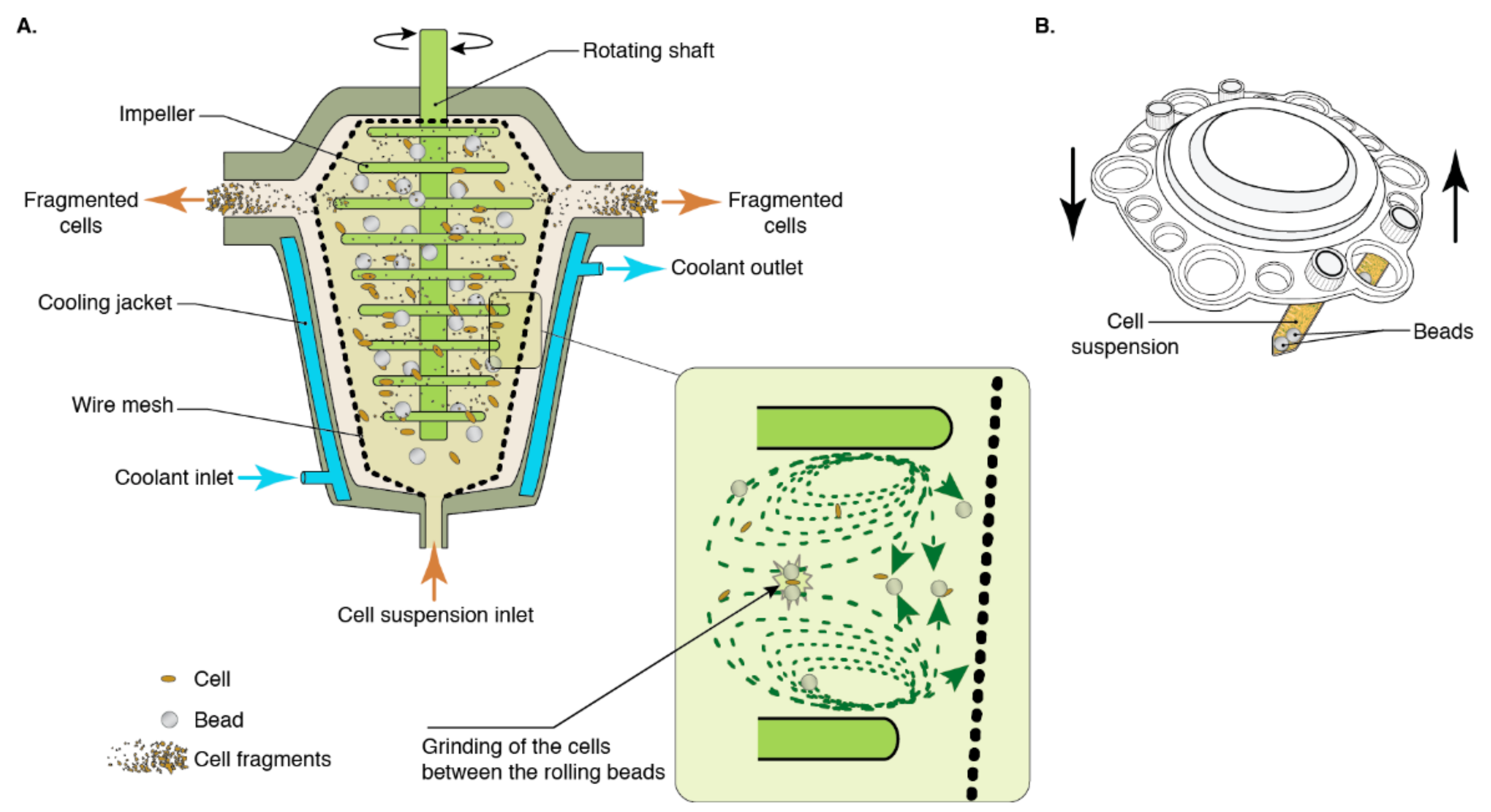
A considerable advantage of mechanical cell disruption techniques is that, in addition to being quite effective, they can generally be scaled up with relative ease. However, a salient caveat is that they have poor selectivity, which may negatively influence the recovery of the pigments diffused into the medium. Indeed, considerable amounts of cell debris can cause a number of downstream complications [11]. Although this shortcoming could be remedied with downstream separation processes (membrane filtration, centrifugation, etc.), this recourse may contribute to some loss of pigment and result in increased energy consumption, which compounds the relatively costly mechanical cell disruption processes. Additionally, as with all cell disruption processes relying on frictional effects, temperature increases in the medium are to be expected and this constitutes a significant drawback for the extraction of thermolabile compounds. Depending on their chemical nature, these compounds, should they be damaged, may undergo detrimental reactions or denaturation which result in either color attenuation or undesirable color changes.

2.1. Bead Milling

Bead milling or bead homogenization is a mechanical cell disruption process with some degree of complexity. Through this technique, cell disruption is induced through shear forces produced during the rotary movement of the cells and the beads and cell grinding between beads [12] (Figure 1). Bead-cell collisions are also purportedly implicated in the mechanism whereby bead milling disrupts cells [13]. Its parameters are bead diameter, bead density, bead filling, agitator speed, and feed rate [14]. Given that a number of components may vary depending on the make and model of the apparatus, such as the design of the grinding chamber or the agitator to name a few, some incongruencies are likely to arise should protocols be replicated with different apparatuses. Parameters like bead size, bead density, and bead filling must be optimized depending on the rigidity of the microorganism’s cell wall, the viscosity of the medium, as well as the flow rate. Optimal biomass concentration for maximum cell disruption must also be determined to enhance the yield of the overall process. There is a complex interaction between the parameters of the process, which require fine-tuning to ensure efficacy and efficiency. Indeed, inadequate parameters may lead to excessive energy consumption, which can be significantly reduced by optimization [15].
The results of bead milling can prove difficult to predict due to the complexity of parameter interactions, and experimental protocols are devised to fit the system being investigated [16].
Figure 1. Cell disruption by bead milling. (A) Continuous large-scale equipment. (B) Batch laboratory scale equipment. Insert represents the grinding mechanism between two rolling beads. Reprinted with permission from [17].
Fermentation 07 00036 g001

2.1.1. Microalgae

Microalgae are deemed a good source for the biosynthesis of carotenoid pigments in addition to a variety of fatty acids and proteins [18]. They have been extensively investigated for the production of high value compounds. However, pigments among other intracellular products resulting from commercial microalgae production are few in number and this is owed to the difficulty associated to compound recovery. Some microalgae species have cell wall structures which are particularly resilient which hinders their widespread exploitation in industrial settings [19]. Additionally, microalgal cell wall structures and compositions exhibit considerable diversity across species, emphasizing the need to determine the various chemical and mechanical attributes of specific cell walls and consequently understand how various types of treatment affect the biomass and its pigment load [20].
Bead milling offers a potentially attractive approach for large scale cell disruption for microalgae [12]. For example, different methods were investigated for the extraction of carotenoids from Tetraselmis sp. CTP4, a strain of microalgae with a mechanically resistant cell wall [21]. In the context of this study, the researchers compared the effectiveness of solubilization via mechanical dispersion to that of bead milling for wet biomass and freeze-dried biomass in a variety of solvents: ethanol, acetone, methanol, and tetrahydrofuran (THF). Mechanical dispersion was performed by way of an IKA Ultra-Turrax T18D Basic running at 25,000 rpm and two processing cycles of 45 s each were performed. Bead milling was performed using 0.7 g of glass beads with diameters ranging from 425 to 600 µm in a tube vortexed for 2 min at the highest setting available on an IKA Vortex Genius 3 shaker, which corresponds to 2500 rpm. Overall, THF appeared to be the solvent for choice for carotenoids and glass beads resulted in better overall solubilization. The configuration resulting in the highest solubilization of carotenoids was THF and glass beads, particularly for wet biomass.
Bead milling exhibits adequate potential at extracting chlorophylls and carotenoids from Chlorella vulgaris [22]. Bead milling was performed using a Netzsch LabStar bead miller and 0.3 to 0.5 mm Y2O3-stabilized ZrO2 grinding beads with grinding times in the range of 1 to 60 min while stirring speed was set at 2500 rpm and the solid:water ratio was 1:13 w v−1. In the context of this study, the researchers sought to evaluate the extraction effectiveness of the following configurations: (a) supercritical CO2 on raw C. vulgaris, (b) supercritical CO2 on C. vulgaris which was previously subject to bead milling, and (c) supercritical CO2 + 5% w v−1 ethanol co-solvent on raw C. vulgaris biomass. Although the co-solvent configuration (c) extracted the most chlorophylls and carotenoids, it is seconded by the bead-milling configuration (b), which is relatively more cost effective.
In the context of investigating the effect of cell disruption methods on protein and pigment diffusion when processing C. vulgaris, bead milling was demonstrated as highly effective at inflicting damage to the cell wall and enabled the release of chlorophylls and carotenoids into the aqueous medium [22]. Bead milling was performed using a Netzsch Labstar and 1 to 1.6 mm zirconium silicate grinding beads, with an agitation speed of 2500 rpm and a 1/13 solid to water ratio. The pigments were quantified using spectrophotometry. Observations via confocal microscopy at 30 min of grinding indicate considerable damage was inflicted onto the cell wall, and it is suspected that the considerable contact surface between the cells and the beads in this instance made bead milling particularly effective. With the parameters previously described, carotenoid concentration in the aqueous medium peaked at 40 min of milling time and dwindled henceforth indicating that pigment degradation had occurred beyond this point.

2.1.2. Fungi

In a laboratory setting, bead milling was proven effective for extracting carotenoids from Sporobolomyces ruberrimus H110 [23]. The pigments were quantified using spectrophotometry. The researchers sought to evaluate numerous experimental configurations wherein mechanical methods, planetary ball mill (Fritsch Pulverisette 5) or a tube shaker (vortex extraction), were used in combination with a variety of cell lysis agents: irregular quartz stones, regular glass beads, and dimethyl sulfoxide (DMSO). Findings suggest that the use of irregular quartz stones in the mechanical techniques resulted in lower extraction yields compared to their counterparts implicating the use of regular glass beads. Although the irregular stones should theoretically have a greater stone-cell-stone contact surface compared to the bead-cell-bead contact surface (this is owed to the fact that for equivalent volume/mass, a spherical configuration has a minimal contact surface), thus enabling more cell crushing between colliding stones, results indicate that this was not the case. A number of parameters can be examined to determine why uniform beads resulted in greater pigment yield. Moreover, the researchers determined the optimal extraction parameters, lysis agent concentration and agitation speed, for both mechanical methods and concluded that bead milling yielded better overall carotenoid extraction and was the most energy-efficient given its capacity to process greater amounts of cells per individual actuation.
In a laboratory setting, the implications of using agricultural waste products as carbon sources for Rhodotorula mucilaginosa on the synthesis of carotenoids was investigated [3]. Bead milling was used to extract the synthesized carotenoids, which were subsequently quantified using spectrophotometry. However, bead milling processing parameters were not disclosed.
Bead milling was also investigated as a cell disruption technique to recover violacein from Yarrowia lipolytica, an oleaginous yeast [24]. The Yarrowia yeast species is characterized by a rigid cell wall mainly comprised of mannose and galactose, granting it considerable resistance to mechanical and osmotic pressure [25]. Bead milling was thus a good candidate for this application, and was used in multiple configurations whereby the impact borne by processing time and pretreatment process (solvent-based via ethyl acetate or methanol/enzymatic via Zymolase 20-T) was evaluated. Identical configurations without bead milling were used as a benchmark. The researchers quantified the pigment using HPLC and microplate photometry. Findings indicate that the highest yield and overall violacein purity was obtained with bead milling superseding ethyl acetate pretreatment, although the purity was slightly lower than that of the benchmark in which cells were incubated in 30 °C ethyl acetate for 24 h. Additionally, it must be noted that bead milling for 1 min yielded 7% less violacein than bead milling for 5 min. Assessing the economic implications of processing the yeasts for an additional 4 min from a pigment cost vs. energy consumption standpoint would provide insight into the most suitable processing time.
Bead milling was the cell disruption method used to extract violacein and deoxyviolacein, among five aromatic-derived natural compounds, from the YL33 strain of Y. lipolytica [26]. The chief objective of this endeavor was to achieve de novo synthesis of organic compounds through the circumvention of the rate-limiting steps in the shikimate pathway. The fermentation culture was first mixed with a five-fold volume of ethyl acetate and vortexed at 30 °C for 24 h with glass beads of undisclosed proportions, then subsequently centrifuged at 12,000 rpm for 10 min.
Y. lipolytica is a highly versatile species of yeast as is evidenced by the considerable number of articles wherein it is used to synthesize an impressive roster of products in both academic and industrial contexts [27]. In the extraction of carotenoids from the β-carotene-producing strain ob-CHCTEF CTEF, bead milling was used to extract the pigments and subsequently determine total β-carotene output via HPLC [28]. The fermentation samples were freeze-dried before a solvent mixture (50:50 v/v heptane, ethyl acetate and 0.01 butylated hydroxytoluene) was added along with glass beads of diameters ranging from 0.710 to 1.18 mm. They were subsequently vortexed and ultrasonicated for 20 min before being centrifuged at 13,000× g for 5 min.
Bead milling was also used to recover astaxanthins from two Pantoea stewartii strains, pTOPOcrtW and pCR-Blunt II-TOPO, in a patent filed by Desouza et al. [29]. Five-day old cultures were washed and suspended in acetone before the addition of glass beads and subsequent incubation in a dark setting. The cells were occasionally vortexed before being centrifuged for 10 min at 15,000× g. The patent offers few details with regards to vortexing rpm, the type of apparatus used, and the proportions of the glass beads.
Carotenoids were recovered from Knufia petricola A95 using bead milling in the context of evaluating changes in the organism’s pigment composition when it is subjected to oxidative stress and cold temperatures [30]. The mill in question, a Precellys 24 from Peqlab/Bertin instruments, had an integrated CO2 cooling system to prevent pigment degradation and the researchers specify having used a 1:3 (w/w) mixing ratio wherein 1 part was freeze-dried K. petricola cells and 3 parts were glass beads with an average diameter of 0.25 mm. The solvent was a 40:20:40 v/v/v acetonitrile, ethyl acetate, and n-propanol mixture. The researchers determined that carotenoid concentration, except for β-carotene, increased under oxidative stress whereas the overall pigment composition did not change in a statistically significant manner when the organism was exposed to cold temperatures.

2.1.3. Bacteria

As spirulina gains popularity among health-conscious consumers, producing and optimally extracting phycocyanin, a blue photosynthetic pigment-protein complex with antioxidative and anti-inflammatory activities, is becoming a highly active area of research namely in the fields of food, cosmetics, and pharmaceuticals. Bead milling exhibited promising results extracting phycocyanin and phenolic compounds from Arthrospira spirulina [31]. The researchers investigated two drying methods, spouted bed and tray drying, and three cell disruption methods being bead milling, autoclaving, and microwaving to extract the pigments [31]. The bead mill of unspecified make was used with porcelain beads of unspecified diameter that occupied 20% of the overall jar volume. The 150 g samples were processed for 2 h at 60 rpm. 0.5 g samples were microwaved using a Sanyo EM804TGR for 120 s, at a frequency of 1450 MHz and 1400 W of power. Five grams of sample were autoclaved using a Stermax 16052 for 30 min at 121 °C and 200 kPa absolute pressure. The researchers evaluated all six possible combinations of drying and cell disruption methods, with results indicating that the spouted bed was conducive to better post-cell disruption yield with respect to total phenolic content. Microwaving appeared to be the best cell disruption technique from a total phenolic content standpoint and its results were comparable to those of bead milling. However, phycocyanin content was higher for microwave-based cell disruption rather than bead milling. Fourier Transform Infrared Spectroscopy (FTIR) analysis ascribes this differential to phycocyanin degradation as a result of bead milling.
C-phycocyanin is typically recovered from Spirulina sp. through a lengthy and time-consuming process. First, the cultivated biomass is harvested through filtration and exposed to a low-temperature process, which disrupts the cell wall. The dried biomass is then submerged in a buffer solution wherein the phycocyanin is solubilized. After a second filtration and the discarding of the waste biomass, impurities are removed and the phycocyanin is subject to a final drying step [32]. Ca(II) ions among others in a buffer solution were investigated as a possible extraction technique. The total amounts of carotenoids in the cell sample was determined using bead milling, and the amounts extracted via the method implicating Ca(II) ions were contrasted against this benchmark. Bead milling using an overhead stirrer equipped with a Rushton turbine blade operating at 330 rpm and 150 g of zirconia beads ranging from 0.5 to 1.4 mm in diameter was performed to determine the total number of C-phycocyanin in the processed sample. Its effectiveness is evidenced in the context of this study wherein it was performed to create a benchmark for optimal extraction against which the novel technique of using Ca(II) ions, among others, in a buffer solution was contrasted [33].

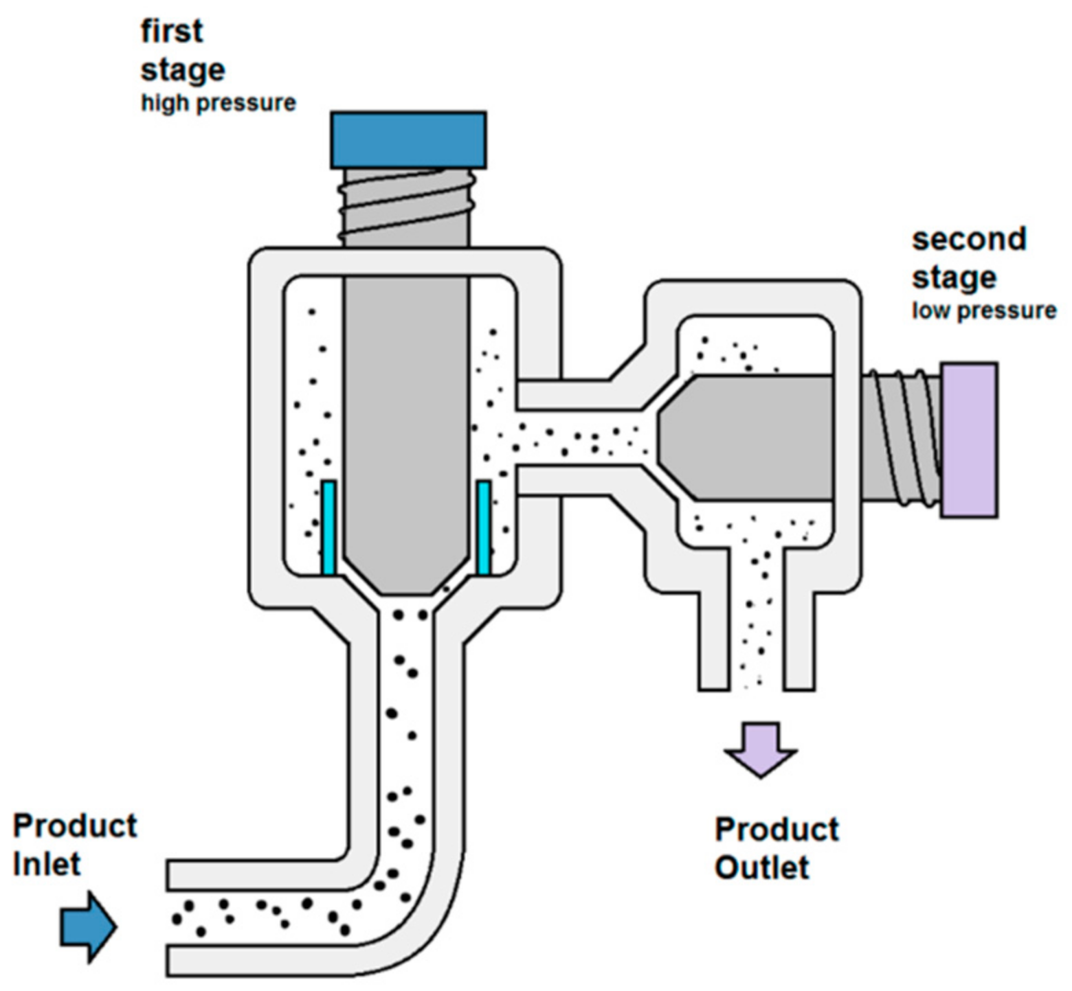
2.2. High Pressure Homogenization

High pressure homogenization (HPH) is a highly effective and scalable method to perform cell disruption. HPH entails forcing a suspension through a strait nozzle or a valve actuated by high pressure. After passing through the valve, the suspension is released into a low-pressure chamber (Figure 2). It is chiefly suspected that cells within the suspension are subject to disruption induced by the collision between the high-speed suspension jet onto the valve surface, and shear stress induced by the pressure drop [34,35]. However, the mechanism through which cell disruption occurs via HPH is still subject for debate, and its effects are even ascribed in various sources to cavitation, turbulence, or extensional stress [36,37]. Nevertheless, empirical evidence suggests that HPH is a highly capable cell disruption method whose mettle was tested thoroughly in a variety of contexts. Homogenization effectiveness and efficiency can be enhanced by weakening the cell wall through pre-treatment via chemical or enzymatic attacks on the compounds that contribute to its overall strength [38]. Samples are usually subjected to a number of passes through a high-pressure homogenizer to yield the desired results. The method is nevertheless not without its drawbacks, with cooling being required in virtually all instances very high pressures are used, naturally resulting in temperature increases detrimental to the chemical integrity of thermolabile compounds. HPH results in the formation of cell debris, which can incur additional downstream costs to filter and eliminate these waste products. Additionally, the solutions must be pumpable. The risk of agglomeration within the low-pressure chamber or within the narrow nozzle is non-negligible, particularly when the sample is subjected to temperature increases due to high shear stress, and the apparatus is usually costly to maintain and operate [39]. These drawbacks still present challenges for the adoption of this particular technique in industrial settings.

2.2.1. Microalgae

HPH of microalgae is a tried and tested method of cell disruption, which can easily overcome the considerable rigidity of the algal cell walls. In some instances, the method exhibits impressive ability to extract astaxanthin from algae, particularly when coupled to chemical (acid/alkali) or enzymatic treatment. In the latter case, their yield exceeded that of HCl, NaOH, enzymatic and spray-drying methods threefold [41].
HPH is highly effective at retrieving pigments from microalgae, as for example the extraction of compounds from Nannochloropsis sp. [42]. Compared to other cell disruption techniques such as pulsed electric fields (PEF), high voltage electrical discharges (HVED), and ultrasonication (USN), HPH exhibited better pigment extraction than its counterparts. While the electricity-based extraction techniques showed considerable promise in the extraction of water-soluble ionic compounds and water-soluble proteins, their effect on pigment extraction was negligible. USN and HPH proved to be the most effective techniques for the extraction of carotenoids and chlorophylls, although the admittedly more power-consuming HPH was the most effective one.
In the context of investigating biomolecule recovery from Parachlorella kessleri, HPH was used to recover pigments among other compounds [43]. The method superseded ultrasonication and the effects of using the former and the latter consecutively is evaluated. When contrasted against ultrasonication, HPH is incapable of handling suspensions with elevated dry matter concentrations. Consequently, the 10% w/w dry matter samples used to evaluate ultrasonication required dilution by a factor of 10. The conducted study chiefly highlights the effectiveness of such a combination on the extraction of various molecules although pigments are the focal point of this review. The homogenizer used was an NS 100L-PANDA 2K and the ultrasound processor was a Hielscher UP-400S. The experimental parameters are consummately detailed in the publication. The researchers noted that cell wall disruption, evaluated via scanning electron microscope (SEM), was significantly more pronounced for samples processed using the USN + HPH combination compared to the samples solely processed via USN. The researchers posit that the integrity of the cell wall was “completely lost”. Their data reveals that USN + HPH was conducive to a more efficient extraction of carotenes and chlorophylls, as evidenced by spectrophotometric analyses, although it must be noted that the effects were not similar for the remaining molecule types.
HPH was investigated as a cell disruption technique to extract carotenoids from Desmodesmus sp. F51 [44]. The authors’ findings suggest that, in this instance, homogenization pressure contributed more than the number of cycles to the achievement of efficient cell disruption [44]. Six cell disruption techniques were used: autoclaving, microwaving, osmotic shock using a 10% NaCl solution, sonication, bead milling, and HPH. During the preliminary optimization stage for HPH, findings indicated that cell density, ranging from 2 g/L up to 90 g/L did not have an impact on cell disruption effectiveness in this instance, leading up to the conclusion that lower cell densities would consequently possess higher specific energy consumption. HPH was found to have the second highest extraction yield, determined via HPLC, second only to bead milling.
HPH also possesses the quality of engendering rapid pigment diffusion in the medium when used to disrupt C. vulgaris cells [45]. When conducting a study to assess the influence of cell disruption methods on the diffusion of proteins and pigments in an aqueous medium after processing C. vulgaris, Safi et al. noted that HPH resulted in a rapid destruction of the cells, leading up to a high diffusion of proteins and chlorophylls compared to the other methods considered in this assessment which included USN, HPH, bead milling, and chemical hydrolysis.

2.2.2. Fungi

In the context of investigating the methods to rupture the cell wall of Sporidiobolus pararoseus to recover carotenoids, HPH was compared to four other cell disruption methods: acid heating, dimethyl sulfoxide (DMSO), enzymatic disruption, and cell autolysis [46]. The acid heating was performed by subjecting the centrifugation precipitate to an acid bath in 3 mol/L HCl for 60 min and subsequently bathing it in boiling water for 3 min. The DMSO method implied the addition of 30 mL of DMSO preheated to 55 °C to the precipitate and stirring the mixture for 5 h. This step was followed by isolation and cleaning of the cell pellet before extraction in 20 mL of acetone. For the enzymatic method, 4 mg of alkaline protease per gram of yeast were added to the broth, and the temperature of the mixture was maintained at 37 °C for 9 h. The enzyme was deactivated by bringing the broth to a boil for 10 min, after which the cells were retrieved and cleaned before being placed in acetone for pigment extraction. The cell autolysis method was performed by adding 5 g of NaCl to 50 mL of broth and subjecting the mixture to shaking using a bath shaking bed at 100 rpm. The sample was subsequently cleaned and the pigments were extracted by placing the cells in acetone. HPH was conducted at a pressure of 80 MPa applied to a sample with 8% biomass concentration for three passes. Carotenoid concentration was determined via spectrophotometry and cell wall breaking was evaluated using optical microscopy and a blood cell count board. The acid heating method resulted in higher overall cell wall disruption, exceeding that of HPH, the second most effective method, by 30%. However, this did not necessarily translate into a higher yield of carotenoids for the acid heating method, as HPH resulted in the recovery of the highest carotenoid amounts.

2.2.3. Bacteria

C-phycocyanin pigments were extracted from Arthrospira maxima using microwaves and HPH [47]. The authors note that conventional methods for the extraction of C-phycocyanin present drawbacks hence the need to develop greener approaches. The maceration method incurs considerable solvent consumption whereas the freezing and thawing approach can be quite lengthy. The researchers compared the extraction yield of HPH and microwave cell disruption to those of conventional maceration and freeze-thawing, concluding that HPH at 1400 bar of pressure had the highest yield, exceeding the comparable yields of microwave (MW) and freeze-thawing by 30%. The researchers also noted that the maceration method performed the most poorly from an extraction yield standpoint.
C-phycocyanin was also recovered from Spirulina platensis using HPH as a cell disruption technique [48]. The homogenizer used was a PhD Technology International model D-15M. The cells were subjected to wall-weakening enzymatic pretreatment via lysozyme prior to being processed at pressure values in the range of 689.48 to 827.37 bar, with the mixture being maintained at temperatures in the range of 4 to 8 °C. The researchers subsequently attempted to purify C-phycocyanin from the resulting slurry using an assortment of techniques.
HPH can also be used as a cell disruption method for Serratia marcescens in order to recover prodigiosin according to a patent filed by Jiang et al. [49]. The description of the experimental protocol provided by the inventors of the referenced patent is, however, not comprehensive and pertinent experimental parameters are undisclosed.
While the evidence suggests that HPH is a highly capable cell disruption method in the context of recovering intracellular carotenoids, a scarcity of publications assessing its potential in the extraction of bacterial and fungal pigments is noted. While this dearth is observable in the context of pigment extraction, it surely does not bespeak general inflexibility of HPH as a cell disruption technique. HPH has a conspicuous advantage over USN with regards to disrupting microalgal cell walls and a scalability advantage over both USN and bead milling [50]. However, unlike HPH, ultrasonication and bead milling possess the added benefit of being equipped with active cooling apparatuses, which can be engaged in real time as the processing is ongoing, contributing to a minimization of thermolabile compound degradation.

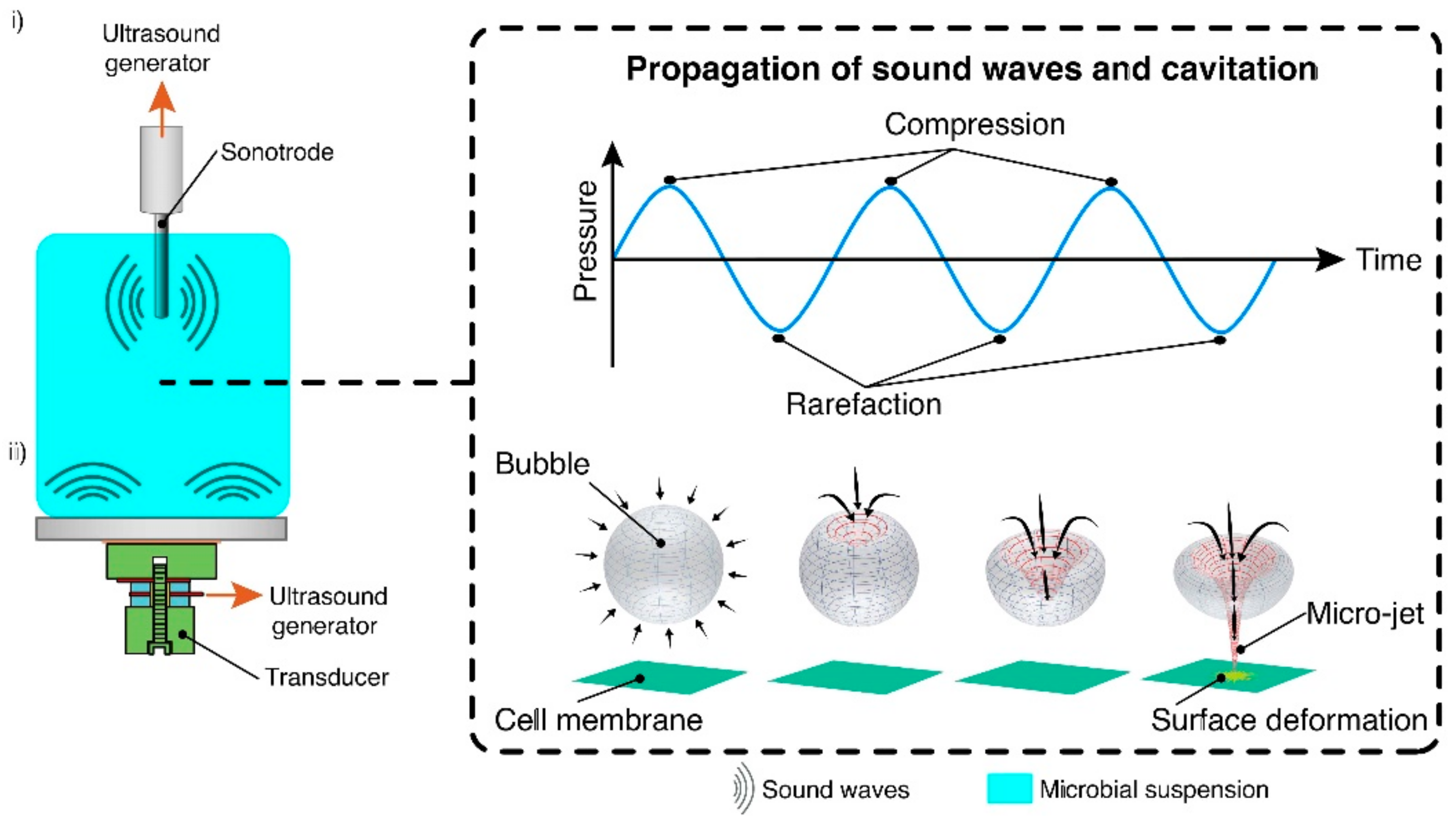
2.3. Ultrasonication

Ultrasonication is a technique whereby cavitation is created by introducing ultrasonic waves into a liquid medium via a resonance rod. It occurs when the vapor bubbles of a liquid form in a locus where the liquid’s pressure is lower than that of its vapor pressure. The bubbles distend under negative pressure and compress under positive pressure, causing a brusque and powerful collapse of these bubbles (Figure 3) [22]. Its capacity to act as a cell disruption technique is predicated on the aptness of cavitation forces at inflicting damage to the cell wall, and is this highly dependent on said cell wall’s composition. The characteristics of the cell wall considerably impact the effectiveness of this technique and microbial species with high cell wall rigidity may be inadequately processed through USN.

2.3.1. Microalgae

USN was evaluated as a candidate for the extraction of carotenoids and other compounds from C. vulgaris in an aqueous medium [45]. The authors noted that hydrophilic entities diffused rapidly into the aqueous medium whereas hydrophobic ones like carotenoids and chlorophylls did not. Additionally, they noted that the technique did not contribute to the rupture of the majority of the cells’ walls given their high mechanical strength, which is a notable characteristic of C. vulgaris.
The applicability of USN in the extraction of phycocyanin from Spirulina platensis was investigated in the context of developing ultrasonic equipment capable of extracting phycocyanin on an industrial scale [52]. The apparatus used in these experiments was a modified “Bioruptor” system. The researchers noted, after comparing the concentration of USN-extracted phycocyanin to the concentration obtained using conventional aqueous extraction, that no product was destroyed by cavitation and that the maximum extraction concentrations were comparable. They also determined that a specific frequency band conducive to optimal extraction was between 28 and 40 kHz and that there was no lag between the cell wall disruption and the release of phycocyanin from the cells into the medium.
The effect of temperature and time of exposure in the context of recovering phycocyanin using USN as a cell disruption method for S. platensis was also investigated [53]. The temperatures chosen were 30, 45, and 60 °C whereas exposure times were 20, 35, and 50 min. Ultrasound frequencies of 28 and 42 kHz were considered. Phycocyanin content was determined using spectrophotometry, and antioxidant activity was determined using DPPH assay in accordance with a protocol detailed by Herrero et al. [54]. Results lower frequency of 28 kHz was unable to inflict considerable damage to the cell wall, resulting in the lower phycocyanin yield compared to 42 kHz. Optimal extraction temperature was 45 °C, with the authors noting that temperatures exceeding 55 °C would result in protein denaturation and coagulation detrimental to the antioxidant activity of phycocyanin. Antioxidant activity and pigment yield peaked at 35 min of exposure to ultrasonic frequencies.
USN was also successfully used to recover C-phycocyanin, allophycocyanin, and phycoerythrin from marine Spirulina maxima [55]. The apparatus used to perform the cell disruption was an Asia Ultrasonic AUG-R3-900. Ten grams of cells were suspended in 500 mL of distilled water before being subjected to USN at frequencies ranging from 20 to 100 kHz, 120 W of power, and extraction times ranging from 1 to 5 h. The experiments were conducted at 25 °C, and the pigments were quantified in accordance to a method described by Moraes et al. [56]. C-phycocyanin was quantified via spectrophotometry. Findings indicate that optimal frequency was 60 kHz while optimal extraction time was 3 h, a considerable decrease compared to the 24 h required for conventional water extraction. The chemical integrity of C-phycocyanin was evaluated via HPLC and the researchers determined that the USN method outperformed conventional water extraction from a chemical integrity standpoint. Moreover, products yielded by the USN method exhibited better antioxidant and anti-inflammatory properties evaluated via DPPH assay and modified Green assay, respectively [57,58].
A USN-solvent approach was also explored as a means to simultaneously extract lipids and carotenoids from Heterochlorella luteoviridis [59]. The USN apparatus used in this study was a Sonics Vibracell with a frequency of 20 kHz. Initially, the researchers created a mixture wherein 4 g of freeze-dried yeast cells were added to 80 mL of an ethanol-water solution (25% v/v). Secondly, they subjected the mixture to USN pretreatment at different intensities, with 100% corresponding to 50 W cm−2, to investigate the impact of ultrasound intensity on lipid and carotenoid yields. The mixture was maintained at temperatures lower than 30 °C throughout the pretreatment phase using a cooling jacket. After this pretreatment phase, the samples were transferred to vessels with varying ethanol-water ratios to determine the combination most conducive to diffusion. Carotenoids were quantified using a spectrophotometric approach detailed in Rodriguez-Amaya [60], whereas lipids were quantified using a spectrophotometric protocol delineated in Jaeschke et al. [61]. Findings indicate that optimal extraction of both carotenoids and lipids occurs in the range of 40–80% USN intensity and 60–75% ethanol concentration, although the researchers noted that the extraction yield of lipids was unaffected by USN.
Pigments, chlorophylls, and phenolic compounds were successfully extracted from Nannochloropsis sp. using USN and environmentally-friendly solvents [62]. The solvents used were water, DMSO, and ethanol as well as binary water-solvent mixtures to determine the one best fit for purpose. USN cell disruption was conducted using a Hielscher UP400S with total power output of 400 W and a frequency of 24 kHz. The researchers determined that a microalgae concentration was conducive to optimal results. Additionally, the USN treatment time of 5 min at 400 W resulted in the highest yield for both chlorophylls and phenolic compounds. The solvents resulting in optimal recovery were water-DMSO and water-ethanol with water rations ranging between 25 and 30%. The researchers’ findings also indicate that increases in microalgal concentration in the ultrasonicated sample resulted in a more efficient cell disruption process, with concentrations of 10% wt requiring one tenth of the power required to process the same amount in 1% wt batches.

2.3.2. Fungi

Among others, USN was investigated as a cell disruption technique for the recovery of astaxanthin from a mutant Phaffia rhodozyma [63]. Multiple configurations were chosen by the researchers: (a) 3 combinations of organic solvents including dimethyl sulfoxide (DMSO), acetone, and hexane followed by ultrasonication, (b) acid bath with 2 M HCl while subjecting the cultures to bead milling followed by USN, subsequent division of the batch into 2 equal moieties and the addition of acetone to one of them, (c) adding differing concentrations (1 mg/mL, 5 mg/mL, 10 mg/mL) of β-glucanase to two sets of three tubes each, then subjecting the contents of one set to USN, (d) adding a 10 mg/mL solution of β-glucanase, lyophilizing the cells, and subjecting the dried biomass to supercritical CO2 extraction at 60 °C for 1 h with a CO2 flow of 15 mL/min with one sample being subjected to 300 bar of pressure whereas the other was subject to 500 bar of pressure in the presence of 10% ethanol, (e) adding a 10 mg/mL solution of β-glucanase before subjecting to HPH. For HPH, the extraction parameters were 1000 bars of pressure applied for 10 passes via a Stansted Fluid Power apparatus. USN was performed using a Sonics VCX 130 at 100% amplitude for 5 min and bead milling was performed using 15 g of glass beads and a rotatory incubator shaker at 250 rpm. Quantification was performed with Ultra Performance Liquid Chromatography coupled to mass spectrometry. Solvents were shown to have poor cell disruption potential compared to their mechanical cell disruption counterparts. Additionally, the use of enzymes greatly enhanced the extraction yield of these mechanical techniques, with greater enzyme concentrations resulting in higher pigment extraction. HPH had greater yield than supercritical CO2 extraction, but USN coupled to enzymatic cell disruption had the highest yield.
Carotenoids were also extracted from Xanthophyllomyces dendrorhous using USN [64]. The researchers evaluated the potential of this technique by contrasting it to extraction via DMSO. The researchers evaluated the DMSO technique detailed in their publication using a central composite rotatable design wherein reaction time and agitation were the independent variables. The USN technique was also evaluated using a central composite rotatable design wherein the independent variables were temperature and time of exposure. Cell morphology was assessed using scanning electron microscopy and carotenoids were quantified using spectrophotometry. The DMSO-based extraction method produced optimal results with 99% extraction at 150 rpm and 102 min of reaction time. The USN method produced optimal results with 90% extraction at a temperature of 30 °C and 56 min of exposure time to ultrasounds. While the extraction yield of USN did not match that of DMSO in this study, it provides further evidence of the technique’s aptness to extract pigments from yeasts.
USN was used to extract astaxanthin from Phaffia rhodozyma [65]. The USN method performed in the presence of lactic acid was compared to a benchmark DMSO-based technique. USN was performed using a 120 W Dakshin Ultrasonics ultrasonic horn with a diameter of 1.1 cm and a maximum operating frequency of 20 kHz. The researchers exposed the used various power values ranging from 40 W and 120 W, applied to the sample for 30 min, to evaluate the effect of power dissipation and determined the value most conducive to astaxanthin extraction which was 80 W. Subsequently, they determined the lactic acid concentration resulting in the highest extraction yield which was 3 M. Thirdly, they determined the optimal disruption time which was 15 min. Finally, the researchers determined the optimal duty cycle, which had been set to 80% in the previous experiments, to be 60% for this analog unit. The DMSO-based technique implicated the addition of 2 mL of 60 °C DMSO to 0.05 g of biomass in accordance to the protocol detailed by Michelon et al. [66]. The USN method resulted in the extraction of 90% of the total carotenoids extracted by the DMSO method. Despite the marginally lower yield, the authors claimed that the astaxanthin recovered using USN is suitable for use in the food and pharmaceutical industries unlike the astaxanthin recovered using DMSO, which may contain minute amounts of solvent.
Manosonication, a method that combines high pressure and ultrasound, was investigated as a sustainable solvent-free technique to extract carotenoids from Rhodotorula glutinis in an aqueous medium [67]. The apparatus in question consisted of a 100 mL treatment chamber pressurized with nitrogen in which protruded a sonication horn connected to a 2000 W Digital Sonifier from Branson Ultrasonics Corporation. The frequency was set at 20 kHz and the chamber was actively cooled with a coil to maintain temperatures below 30 °C. The researchers explored, through a surface response model, the effect of hydrostatic pressure, time of exposure, and sonication amplitude on the overall extraction yield of carotenoids which was evaluated via HPLC, as well as cell inactivation evaluated via CFU determination. The best extraction yields were obtained using the highest pressure and longer processing time. Optimal extraction conditions for carotenoids were 200 kPa of pressure, 120 µm of amplitude, and a time of exposure of 30 s resulting in the recovery of 82% of total carotenoid. The researchers noted that cell inactivation and carotenoid extraction were not subject to a linear relationship, as increases in extracted carotenoid quantities were detected after 99% of the yeast population was inactivated. This observation lends reasonable credence to the supposition that ultrasounds play a role, in addition to cell wall disruption, in the disassembling of carotenoids from the yeast structure. The high yield was also attributed to the formation of a nanoemulsion wherein the dispersed phase, droplets of carotenoids, was stabilized by cytoplasmic phospholipids.

2.3.3. Bacteria

Prodigiosin, a deep red pigment chiefly produced by Serratia marescens and Streptomyces, possesses potent antimicrobial, antifungal, cytotoxic, and immunosuppressive properties [68]. Apart to prodigiosin intriguing visual qualities, its therapeutic properties have spurred research to develop cell disruption protocols to isolate the intracellular pigment, a matter particularly difficult in light of the rigidity of the cell wall of endophytes. USN was considered, among other techniques as a cell disruption method in the context of extracting a red pigment from S. marcescens, which was confirmed to be prodigiosin through spectroscopy, thin layer chromatography, HPLC, and FTIR [69]. Homogenization, USN, freezing and thawing, heat treatment, organic solvents and inorganic acids were the methods considered to disrupt the cell wall. For USN, the researchers used a Sonics VCX750 at 30 kHz, 35% amplitude for 6 min on a 5 mL aliquot. Homogenization was performed using a mortar and pestle in presence of liquid nitrogen. Freezing and thawing was performed by quickly exposing the biomass to −20 °C for 30 min and thawing at 40 °C for 30 min. Heat treatment was performed by dissolving the biomass in 5 mL of ethanol and exposing one mixture to 30 °C and the other to 60 °C for 6 h. For the organic solvent approach, the researchers dissolved biomass in 5 mL of a variety of solvents for 24 h. The inorganic solvent method consisted of placing dry biomass in 5 mL of either HCl (0.1 N) or NaOH (0.1 N) for 24 h at room temperature. The researchers concluded that USN resulted in a higher prodigiosin yield. The method was conducive to the extraction of nearly twice the amount yielded by the second-best performing heat treatment method.
Evidence suggests that buffer pH plays a role in the extraction of C-phycocyanin from Spirulina platensis via USN, but also via MW [70]. The researchers optimized extraction parameters for USN and MW before comparing the optimal result for each technique. For MW, the researchers used a Panasonic NN-S674 with 1.1 kW of microwave energy and a frequency of 2.45 GHz. Identical amounts of S. platensis were added to buffers which stabilized pH levels at 5, 6, 7, and 8 to evaluate the impact of pH on MW and USN cell disruption. The impact of time was investigated by subjecting samples to 90 W of power for time intervals ranging from 0 to 50 s followed by a freeze-thaw cycle. For USN, the researchers used a Sonics VWR B2500A-MT batch bath ultrasound followed by a freeze-thaw cycle. Frequency was set at 20 kHz and power at 100 W, with samples subjected to USN for 1 to 10 min. C-phycocyanin was quantified using spectrophotometry. Findings indicate that the MW technique yielded the best results at an extraction time of 25 min and pH of 7, whereas USN yielded better results at an extraction time of 7 min and a pH of 6. Despite being faster, however, USN exhibited lower purity.
USN was also successfully used to extract phycocyanin from A. platensis [71]. Its ability to rupture A. platensis cells was contrasted against that of high-pressure processing (HPP), PEF, a pH-adjusted phosphate buffer, and NaCl solution. For the NaCl-based method, 2.4 g of dried biomass were hydrated in 37.6 mL of NaCl (10%, wt%) poured into a 50 mL conical tube, which was then centrifuged at 80 rpm for 12 h. For the phosphate buffer method, 2.4 g of dried biomass were hydrated into 37.6 mL of phosphate buffer in a 50 mL conical tube. For PEF, 21 g of biomass were hydrated in different buffer solutions and subjected to 50 to 200 pulses at 20 kV using an ELEA PEF pilot unit. For high-pressure processing, the biomass was hydrated into different extraction buffers at a ratio of 6% (wt%) before being transferred into high-density plastic polyethylene bottles and vacuum-sealed in plastic bags. They were then subjected to HPP using a Doral Hiperbaric-55 for 3.5 min with pressures between 50 and 600 MPa. For USN, 24 g of biomass were dispersed in 37.6 mL of different extraction buffers and processed with a Sonics VC 505 at an amplitude of 40% and time periods of 2, 4, and 6 min. Samples were analyzed using spectrophotometry. For the NaCl method, results show that no significant extraction enhancements were observed after 7 h. They also suggested that higher NaCl concentrations (25–100 g/L) resulted in increased phycocyanin extraction and lower chlorophyll contamination within the sample. PEF did not contribute considerably to the extraction process, and it is speculated that some phycocyanin recovery can be ascribed to cell wall damage during the drying process. HPP was found to denature the pigment-protein complex, engendering a decrease in pigment color intensity. USN was able to extract considerable amounts of phycocyanin, but the researchers suggested that the rehydrated A. platensis was destroyed and that fine cell debris was consequently generated, resulting in some difficulty in the separation phase. While USN is apt at extracting phycocyanin in this scenario, the researchers found that a 3 h soak in a 100 mM phosphate buffer at a pH of 7.5 produced better results.
USN was also used in the extraction of prodigiosin from S. marcescens [72]. The researchers undertaking the study sought to determine optimal extraction parameters through a central composite design. The apparatus used was a Shanghai Zhixin Instruments JYD 650 cell crusher with a 6 mm diameter probe. Multiple variables were considered in the context of this study: time of exposure to 30 kHz ultrasonication, temperature, solute to solvent ratio (g/mL). The predicted values were 17.5 min of USN, a temperature of 23.4 °C, and a solute to solvent ration of 1:27.2 (g/mL) culminating in the extraction of 4.31 g of prodigiosin from 100 g S. marcescens cells. The experimental value was remarkably similar to the predicted one at 4.3 g of prodigiosin with the statistically optimized parameters.
Janthinobacterium lividum XT1, a relatively recent strain of violet-pigment-producing bacteria, was processed with USN to extract the pigment and subsequently identify it as violacein [73]. Cell pellets were placed in 2 mL of 99.7% ethanol and subject to sonication until the cells were completely bleached. Although the researchers did not detail the parameters of this cell disruption protocol, they claimed that the extraction was virtually complete due to the cells being completely stripped of color.

3. Mechanical Cell Disruption Limitations

This review evinces that mechanical cell disruption techniques possess considerable potential in the field of biosynthetic pigment production. Their effectiveness is somewhat undermined by the relative complexity that lies in the optimization of their parameters in order to engender maximum extraction yield and economic viability, bearing in mind that optimal extraction and economic viability may sometimes entail compromises. Such shortcomings may be addressed, for example, by replacing a single power-intensive process with two moderately energy-consuming steps with a similar pigment yield and whose cumulative power needs undercut that of the process being substituted. Table 1 sums up the adavantages and drawbacks of the discussed techniques. Furthermore, the methods used to assess the effects of mechanical cell disruption techniques on cell populations are numerous, and insight as to the state of the cell wall post-processing may help determine reasons for subpar and on-par yields to refine/alter the process and complement knowledge available in extant literature. The methods expounded in this review can be completed with appropriate stain assays, supporting the viability of the full process from production to utilization. Although the ostensibly smooth appearance of certain cells may compel researchers to assume they are intact, viability stain assays can contribute to drawing more dependable conclusions which, in certain scenarios, could prevent bootless or even detrimental further processing [74].

4. Conclusions

The roster of pigments sourced from microorganisms only genuinely grows whenever successful and plentiful pigment biosynthesis is complemented by an effective and economically viable extraction method. Mechanical cell disruption techniques have exhibited considerable potential in both laboratory and pilot applications as well as industrial use at extracting various pigments from various microorganisms. Their parameters can be optimized to ensure maximum extraction yield, minimal pigment loss due to heat generation, and minimized energy consumption. One technique can possess attributes which render it a more adequate option than its counterparts at extracting pigments from defined microorganisms, and their effectiveness is highly contingent on the shape of the microorganism, the structure of its cell wall, its size, and the thermolability of its pigments among others. These attributes are further complemented by economical constraints which factor in the overall time to complete the process, the energy it consumes, its overall pigment yield, and the ease with which pigments are isolated and purified as well as industry-specific constraints such as severely discouraged solvent use in the food industry.

Funding

This research was funded by the Embassy of France in Beirut, the “Conseil de Recherche de l’Université Saint-Joseph de Beyrouth” (SAFAR program), and the “Université de technologie de Compiègne (France)”.

Conflicts of Interest

The authors declare no conflict of interest.

References

  1. Martínez, J.M.; Delso, C.; Álvarez, I.; Raso, J. Pulsed Electric Field-Assisted Extraction of Valuable Compounds from Microorganisms. Compr. Rev. Food Sci. Food Saf. 2020, 19, 530–552. [Google Scholar] [CrossRef] [PubMed]
  2. Valduga, E.; Tatsch, P.O.; Tiggemann, L.; Treichel, H.; Toniazzo, G.; Zeni, J.; Di Luccio, M.; Fúrigo, A. Carotenoids Production: Microorganisms as Source of Natural Dyes. Quim. Nova 2009, 32, 2429–2436. [Google Scholar] [CrossRef]
  3. Aksu, Z.; Tuǧba Eren, A. Carotenoids Production by the Yeast Rhodotorula Mucilaginosa: Use of Agricultural Wastes as a Carbon Source. Process Biochem. 2005, 40, 2985–2991. [Google Scholar] [CrossRef]
  4. Whallans, R.C.M.; de Janaina, F.M.B. Optimization of Agroindustrial Medium for the Production of Carotenoids by Wild Yeast Sporidiobolus Pararoseus. Afr. J. Microbiol. Res. 2015, 9, 209–219. [Google Scholar] [CrossRef] [Green Version]
  5. Buzzini, P.; Martini, A. Production of Carotenoids by Strains of Rhodotorula Glutinis Cultured in Raw Materials of Agro-Industrial Origin. Bioresour. Technol. 2000, 71, 41–44. [Google Scholar] [CrossRef]
  6. Park, P.K.; Kim, E.Y.; Chu, K.H. Chemical Disruption of Yeast Cells for the Isolation of Carotenoid Pigments. Sep. Purif. Technol. 2007, 53, 148–152. [Google Scholar] [CrossRef]
  7. Vílchez, C.; Forján, E.; Cuaresma, M.; Bédmar, F.; Garbayo, I.; Vega, J.M. Marine Carotenoids: Biological Functions and Commercial Applications. Mar. Drugs 2011, 9, 319–333. [Google Scholar] [CrossRef] [PubMed] [Green Version]
  8. Guedes, A.C.; Amaro, H.M.; Malcata, F.X. Microalgae as Sources of Carotenoids. Mar. Drugs 2011, 9, 625–644. [Google Scholar] [CrossRef] [PubMed]
  9. Im, H.; Choi, S.Y.; Son, S.; Mitchell, R.J. Combined Application of Bacterial Predation and Violacein to Kill Polymicrobial Pathogenic Communities. Sci. Rep. 2017, 7, 1–10. [Google Scholar] [CrossRef] [Green Version]
  10. Venil, C.K.; Dufossé, L.; Devi, P.R. Bacterial Pigments: Sustainable Compounds With Market Potential for Pharma and Food Industry. Front. Sustain. Food Syst. 2020, 4, 1–17. [Google Scholar] [CrossRef]
  11. Kholany, M.; Trébulle, P.; Martins, M.; Ventura, S.P.M.; Nicaud, J.M.; Coutinho, J.A.P. Extraction and Purification of Violacein from Yarrowia Lipolytica Cells Using Aqueous Solutions of Surfactants. J. Chem. Technol. Biotechnol. 2020, 95, 1126–1134. [Google Scholar] [CrossRef]
  12. Gong, M.; Bassi, A. Carotenoids from Microalgae: A Review of Recent Developments. Biotechnol. Adv. 2016, 34, 1396–1412. [Google Scholar] [CrossRef]
  13. Nooralabettu, K.P. Cell Disruption Techniques. In Downstream Process Technology: A New Horizon In Biotechnology; PHI Leaning Private Limited: New Delhi, India, 2010; pp. 101–126. [Google Scholar]
  14. Montalescot, V.; Rinaldi, T.; Touchard, R.; Jubeau, S.; Frappart, M.; Jaouen, P.; Bourseau, P.; Marchal, L. Optimization of Bead Milling Parameters for the Cell Disruption of Microalgae: Process Modeling and Application to Porphyridium Cruentum and Nannochloropsis Oculata. Bioresour. Technol. 2015, 196, 339–346. [Google Scholar] [CrossRef]
  15. Postma, P.R.; Miron, T.L.; Olivieri, G.; Barbosa, M.J.; Wijffels, R.H.; Eppink, M.H.M. Mild Disintegration of the Green Microalgae Chlorella Vulgaris Using Bead Milling. Bioresour. Technol. 2015, 184, 297–304. [Google Scholar] [CrossRef]
  16. Burmeister, C.F.; Kwade, A. Process Engineering with Planetary Ball Millss. Chem. Soc. Rev. 2013, 42, 7660–7667. [Google Scholar] [CrossRef]
  17. Koubaa, M.; Imatoukene, N.; Drévillon, L.; Vorobiev, E. Current Insights in Yeast Cell Disruption Technologies for Oil Recovery: A Review. Chem. Eng. Process. Process Intensif. 2020, 150, 107868. [Google Scholar] [CrossRef]
  18. Kermanshahi-Pour, A.; Zimmerman, J.B.; Anastas, P.T. Microalgae-Derived Chemicals: Opportunity for an Integrated Chemical Plant. In Natural and Artificial Photosynthesis; John Wiley & Sons Inc.: Hoboken, NJ, USA, 2013; pp. 387–433. [Google Scholar]
  19. Burczyk, J.; Zych, M.; Ioannidis, N.E.; Kotzabasis, K. Polyamines in Cell Walls of Chlorococcalean Microalgae. Z. Nat. C 2014, 69, 75–80. [Google Scholar] [CrossRef] [Green Version]
  20. Alhattab, M.; Kermanshahi-Pour, A.; Brooks, M.S.-L. Microalgae Disruption Techniques for Product Recovery: Influence of Cell Wall Composition. J. Appl. Phycol. 2019, 31, 61–88. [Google Scholar] [CrossRef]
  21. Schüler, L.M.; Gangadhar, K.N.; Duarte, P.; Placines, C.; Molina-Márquez, A.M.; Léon-Bañares, R.; Sousa, V.S.; Varela, J.; Barreira, L. Improvement of Carotenoid Extraction from a Recently Isolated, Robust Microalga, Tetraselmis Sp. CTP4 (Chlorophyta). Bioprocess Biosyst. Eng. 2020, 43, 785–796. [Google Scholar] [CrossRef]
  22. Safi, C.; Camy, S.; Frances, C.; Varela, M.M.; Badia, E.C.; Pontalier, P.Y.; Vaca-Garcia, C. Extraction of lipids and pigments of Chlorella vulgaris by supercritical carbon dioxide: Influence of bead milling on extraction performance. J. Appl. Phycol. 2014, 26, 1711–1718. [Google Scholar] [CrossRef] [Green Version]
  23. Gomes, M.; Pennacchi, C.; Rodríguez-fernández, D.E.; Maranho, L.T.; Marc, I.; Alves, L.; Master, P.; Cnrs, U.P.R.; Svs, P. A Comparison of Cell Disruption Procedures for the Recovery of Intracellular Carotenoids from Sporobolomyces Ruberrimus H110. Int. J. Appl. Biol. Pharm. Technol. 2014, 6, 136–144. [Google Scholar]
  24. Tong, Y.; Zhou, J.; Zhang, L.; Xu, P. Engineering Oleaginous Yeast Yarrowia Lipolytica for Violacein Production: Extraction, Quantitative Measurement and Culture Optimization. bioRxiv 2019, 1, 1–19. [Google Scholar] [CrossRef]
  25. Liu, D.; Ding, L.; Sun, J.; Boussetta, N.; Vorobiev, E. Yeast Cell Disruption Strategies for Recovery of Intracellular Bio-Active Compounds—A Review. Innov. Food Sci. Emerg. Technol. 2016, 36, 181–192. [Google Scholar] [CrossRef]
  26. Gu, Y.; Ma, J.; Zhu, Y.; Ding, X.; Xu, P. Engineering Yarrowia Lipolytica as a Chassis for de Novo Synthesis of Five Aromatic-Derived Natural Products and Chemicals. ACS Synth. Biol. 2020, 9, 2096–2106. [Google Scholar] [CrossRef]
  27. Zhu, Q.; Jackson, E.N. Metabolic Engineering of Yarrowia Lipolytica for Industrial Applications. Curr. Opin. Biotechnol. 2015, 36, 65–72. [Google Scholar] [CrossRef] [Green Version]
  28. Jacobsen, I.H.; Ledesma-Amaro, R.; Martinez, J.L. Recombinant β-Carotene Production by Yarrowia Lipolytica—Assessing the Potential of Micro-Scale Fermentation Analysis in Cell Factory Design and Bioreaction Optimization. Front. Bioeng. Biotechnol. 2020, 8, 1–9. [Google Scholar] [CrossRef]
  29. Desouza, M.A.; Schroeder, W.A.; Kollman, S.R.; May, C.A. Carotenoid Biosynthesis. Patent Number WO 02/079395 A2, 10 October 2002. [Google Scholar]
  30. Flieger, K.; Knabe, N.; Toepel, J. Development of an Improved Carotenoid Extraction Method to Characterize the Carotenoid Composition under Oxidative Stress and Cold Temperature in the Rock Inhabiting Fungus Knufia Petricola A95. J. Fungi 2018, 4, 124. [Google Scholar] [CrossRef] [Green Version]
  31. Larrosa, A.P.Q.; Camara, Á.S.; Moura, J.M.; Pinto, L.A.A. Spirulina Sp. Biomass Dried/Disrupted by Different Methods and Their Application in Biofilms Production. Food Sci. Biotechnol. 2018, 27, 1659–1665. [Google Scholar] [CrossRef]
  32. Bux, F.; Ramesh, D. Lipid Identification and Extraction Techniques. Biotechnol. Appl. Microalgae 2013, 89–98. [Google Scholar] [CrossRef]
  33. Pott, R.W.M. The Release of the Blue Biological Pigment C-Phycocyanin through Calcium-Aided Cytolysis of Live Spirulina sp. Coloration Technol. 2019, 135, 17–21. [Google Scholar] [CrossRef] [Green Version]
  34. D’Hondt, E.; Martín-Juárez, J.; Bolado, S.; Kasperoviciene, J.; Koreiviene, J.; Sulcius, S.; Elst, K.; Bastiaens, L. Cell Disruption Technologies. Microalgae-Based Biofuels Bioprod. Feedstock Cultiv. End-Prod. 2017, 133–154. [Google Scholar] [CrossRef]
  35. Donsì, F.; Ferrari, G.; Lenza, E.; Maresca, P. Main Factors Regulating Microbial Inactivation by High-Pressure Homogenization: Operating Parameters and Scale of Operation. Chem. Eng. Sci. 2009, 64, 520–532. [Google Scholar] [CrossRef]
  36. Kleinig, A.R.; Middelberg, A.P.J. On the Mechanism of Microbial Cell Disruption in High-Pressure Homogenisation. Chem. Eng. Sci. 1998, 53, 891–898. [Google Scholar] [CrossRef]
  37. Diels, A.M.J.; Michiels, C.W. High-Pressure Homogenization as a Non-Thermal Technique for the Inactivation of Microorganisms. Crit. Rev. Microbiol. 2006, 32, 201–216. [Google Scholar] [CrossRef] [PubMed]
  38. Middelberg, A.P.J. Microbial Cell Disruption by High-Pressure Homogenization. In Downstream Processing of Proteins; Desai, M.A., Ed.; Humana Press: Totowa, NJ, USA, 2000; Volume 9, pp. 11–21. [Google Scholar]
  39. Hu, Y.; Bassi, A. Extraction of Biomolecules from Microalgae; Elsevier Inc.: Amsterdam, The Netherlands, 2020; ISBN 9780128185360. [Google Scholar]
  40. Comuzzo, P.; Calligaris, S. Potential Applications of High Pressure Homogenization in Winemaking: A Review. Beverages 2019, 5, 56. [Google Scholar] [CrossRef] [Green Version]
  41. Mendes-Pinto, M.M.; Raposo, M.F.J.; Bowen, J.; Young, A.J.; Morais, R. Evaluation of Different Cell Disruption Process on Encysted Cells of Haematococcus Pluvialis. J. Appl. Phycol. 2001, 13, 19–24. [Google Scholar] [CrossRef]
  42. Grimi, N.; Dubois, A.; Marchal, L.; Jubeau, S.; Lebovka, N.I.; Vorobiev, E. Selective Extraction from Microalgae Nannochloropsis Sp. Using Different Methods of Cell Disruption. Bioresour. Technol. 2014, 153, 254–259. [Google Scholar] [CrossRef]
  43. Zhang, R.; Grimi, N.; Marchal, L.; Lebovka, N.; Vorobiev, E. Effect of Ultrasonication, High Pressure Homogenization and Their Combination on Efficiency of Extraction of Bio-Molecules from Microalgae Parachlorella Kessleri. Algal Res. 2019, 40. [Google Scholar] [CrossRef]
  44. Xie, Y.; Ho, S.H.; Chen, C.N.N.; Chen, C.Y.; Jing, K.; Ng, I.S.; Chen, J.; Chang, J.S.; Lu, Y. Disruption of Thermo-Tolerant Desmodesmus Sp. F51 in High Pressure Homogenization as a Prelude to Carotenoids Extraction. Biochem. Eng. J. 2016, 109, 243–251. [Google Scholar] [CrossRef]
  45. Safi, C.; Frances, C.; Ursu, A.V.; Laroche, C.; Pouzet, C.; Vaca-Garcia, C.; Pontalier, P.Y. Understanding the effect of cell disruption methods on the diffusion of chlorella vulgaris proteins and pigments in the aqueous phase. Algal Res. 2015, 8, 61–68. [Google Scholar] [CrossRef] [Green Version]
  46. Liu, C.; Cheng, Y.; Du, C.; Lv, T.; Guo, Y.; Han, M.; Pi, F.; Zhang, W.; Qian, H. Study on the Wall-Breaking Method of Carotenoids Producing Yeast Sporidiobolus Pararoseus and the Antioxidant Effect of Four Carotenoids on SK-HEP-1 Cells. Prep. Biochem. Biotechnol. 2019, 49, 767–774. [Google Scholar] [CrossRef]
  47. Ruiz-Domínguez, M.C.; Jáuregui, M.; Medina, E.; Jaime, C.; Cerezal, P. Rapid Green Extractions of C-Phycocyanin from Arthrospira Maxima for Functional Applications. Appl. Sci. 2019, 9, 1987. [Google Scholar] [CrossRef] [Green Version]
  48. Song, W.; Zhao, C.; Wang, S. A Large-Scale Preparation Method of High Purity c-Phycocyanin. Int. J. Biosci. Biochem. Bioinform. 2013, 3, 293–297. [Google Scholar] [CrossRef]
  49. Jiang, D.; Wei, F.; Caim, Q.; Lan, L.; Ji, H.; Gao, A. High-Prodigiosin-Yield Serratia Marcescens Strain Sm-128 and Use Thereof. Patent Number CN102277323B, 6 February 2013. [Google Scholar]
  50. Paton, K.R.; Varrla, E.; Backes, C.; Smith, R.J.; Khan, U.; O’Neill, A.; Boland, C.; Lotya, M.; Istrate, O.M.; King, P.; et al. Scalable Production of Large Quantities of Defect-Free Few-Layer Graphene by Shear Exfoliation in Liquids. Nat. Mater. 2014, 13, 624–630. [Google Scholar] [CrossRef] [PubMed]
  51. Peng, K.; Koubaa, M.; Bals, O.; Vorobiev, E. Recent Insights in the Impact of Emerging Technologies on Lactic Acid Bacteria: A Review. Food Res. Int. 2020, 137, 109544. [Google Scholar] [CrossRef] [PubMed]
  52. Furuki, T.; Maeda, S.; Imajo, S.; Hiroi, T.; Amaya, T.; Hirokawa, T.; Ito, K.; Nozawa, H. Rapid and Selective Extraction of Phycocyanin From. J. Appl. Phycol. 2003, 15, 319–324. [Google Scholar] [CrossRef]
  53. Hadiyanto; Suttrisnorhadi; Sutanto, H.; Suzery, M. Phyocyanin Extraction from Microalgae Spirulina Platensis Assisted by Ultrasound Irradiation: Effect of Time and Temperature. Songklanakarin J. Sci. Technol. 2016, 38, 391–398. [Google Scholar]
  54. Herrero, M.; Martín-Álvarez, P.J.; Señoráns, F.J.; Cifuentes, A.; Ibáñez, E. Optimization of Accelerated Solvent Extraction of Antioxidants from Spirulina Platensis Microalga. Food Chem. 2005, 93, 417–423. [Google Scholar] [CrossRef] [Green Version]
  55. Choi, W.Y.; Lee, H.Y. Effect of Ultrasonic Extraction on Production and Structural Changes of C-Phycocyanin from Marine Spirulina Maxima. Int. J. Mol. Sci. 2018, 19, 220. [Google Scholar] [CrossRef] [Green Version]
  56. Moraes, C.C.; Sala, L.; Cerveira, G.P.; Kalil, S.J. C-Phycocyanin Extraction from Spirulina Platensis Wet Biomass. Braz. J. Chem. Eng. 2011, 28, 45–49. [Google Scholar] [CrossRef] [Green Version]
  57. Green, L.C.; Wagner, D.A.; Glogowski, J.; Skipper, P.L.; Wishnok, J.S.; Tannenbaum, S.R. Analysis of Nitrate, Nitrite, and [15N]Nitrate in Biological Fluids. Anal. Biochem. 1982, 126, 131–138. [Google Scholar] [CrossRef]
  58. Dietz, B.M.; Kang, Y.-H.; Liu, G.; Eggler, A.L.; Yao, P.; Chadwick, L.R.; Pauli, G.F.; Farnsworth, N.R.; Mesecar, A.D.; van Breemen, R.B.; et al. Xanthohumol Isolated from Humulus Lupulus Inhibits Menadione-Induced DNA Damage through Induction of Quinone Reductase. Chem. Res. Toxicol. 2005, 18, 1296–1305. [Google Scholar] [CrossRef]
  59. Jaeschke, D.P.; Rech, R.; Marczak, L.D.F.; Mercali, G.D. Ultrasound as an Alternative Technology to Extract Carotenoids and Lipids from Heterochlorella Luteoviridis. Bioresour. Technol. 2017, 224, 753–757. [Google Scholar] [CrossRef] [PubMed]
  60. Rodriguez-Amaya, D.B. A Guide to Carotenoid Analysis in Foods; ILSI Press: Washington, DC, USA, 2001; ISBN 1578810728. [Google Scholar]
  61. Jaeschke, D.P.; Menegol, T.; Rech, R.; Mercali, G.D.; Marczak, L.D.F. Carotenoid and Lipid Extraction from Heterochlorella Luteoviridis Using Moderate Electric Field and Ethanol. Process Biochem. 2016, 51, 1636–1643. [Google Scholar] [CrossRef]
  62. Parniakov, O.; Apicella, E.; Koubaa, M.; Barba, F.J.; Grimi, N.; Lebovka, N.; Pataro, G.; Ferrari, G.; Vorobiev, E. Ultrasound-Assisted Green Solvent Extraction of High-Added Value Compounds from Microalgae Nannochloropsis spp. Bioresour. Technol. 2015, 198, 262–267. [Google Scholar] [CrossRef]
  63. Hasan, M.; Azhar, M.; Nangia, H.; Bhatt, P.C.; Panda, B.P. Influence of High-Pressure Homogenization, Ultrasonication, and Supercritical Fluid on Free Astaxanthin Extraction from β-Glucanase-Treated Phaffia Rhodozyma Cells. Prep. Biochem. Biotechnol. 2016, 46, 116–122. [Google Scholar] [CrossRef]
  64. Urnau, L.; Colet, R.; Soares, V.F.; Franceschi, E.; Valduga, E.; Steffens, C. Extraction of Carotenoids from Xanthophyllomyces Dendrorhous Using Ultrasound-Assisted and Chemical Cell Disruption Methods. Can. J. Chem. Eng. 2018, 96, 1377–1381. [Google Scholar] [CrossRef]
  65. Gogate, P.R.; Nadar, S.G. Ultrasound-Assisted Intensification of Extraction of Astaxanthin from Phaffia Rhodozyma. Indian Chem. Eng. 2015, 57, 240–255. [Google Scholar] [CrossRef]
  66. Michelon, M.; de Matos de Borba, T.; da Silva Rafael, R.; Burkert, C.A.V.; de Medeiros Burkert, J.F. Extraction of Carotenoids from Phaffia Rhodozyma: A Comparison between Different Techniques of Cell Disruption. Food Sci. Biotechnol. 2012, 21, 1–8. [Google Scholar] [CrossRef]
  67. Martínez, J.M.; Delso, C.; Aguilar, D.E.; Álvarez, I.; Raso, J. Organic-Solvent-Free Extraction of Carotenoids from Yeast Rhodotorula Glutinis by Application of Ultrasound under Pressure. Ultrason. Sonochem. 2020, 61, 104833. [Google Scholar] [CrossRef]
  68. Huryn, D.M.; Wipf, P. Natural Product Chemistry and Cancer Drug Discovery, 2nd ed.; Elsevier: Amsterdam, The Netherlands, 2013; ISBN 9780123965219. [Google Scholar]
  69. Khanam, B.; Chandra, R. Comparative Analysis of Prodigiosin Isolated from Endophyte Serratia Marcescens. Lett. Appl. Microbiol. 2018, 66, 194–201. [Google Scholar] [CrossRef] [PubMed]
  70. Vali Aftari, R.; Rezaei, K.; Mortazavi, A.; Bandani, A.R. The Optimized Concentration and Purity of Spirulina PlatensisC-Phycocyanin: A Comparative Study on Microwave-Assisted and Ultrasound-Assisted Extraction Methods. J. Food Process. Preserv. 2015, 39, 3080–3091. [Google Scholar] [CrossRef]
  71. Li, Y.; Zhang, Z.; Paciulli, M.; Abbaspourrad, A. Extraction of Phycocyanin—A Natural Blue Colorant from Dried Spirulina Biomass: Influence of Processing Parameters and Extraction Techniques. J. Food Sci. 2020, 85, 727–735. [Google Scholar] [CrossRef]
  72. Sun, S.-Q.; Wang, Y.-J.; Xu, W.; Zhu, C.-J.; Liu, X.-X. Optimizing Ultrasound-Assisted Extraction of Prodigiosin by Response Surface Methodology. Prep. Biochem. Biotechnol. 2015, 45, 101–108. [Google Scholar] [CrossRef] [PubMed]
  73. Lu, Y.; Wang, L.; Xue, Y.; Zhang, C.; Xing, X.H.; Lou, K.; Zhang, Z.; Li, Y.; Zhang, G.; Bi, J.; et al. Production of Violet Pigment by a Newly Isolated Psychrotrophic Bacterium from a Glacier in Xinjiang, China. Biochem. Eng. J. 2009, 43, 135–141. [Google Scholar] [CrossRef]
  74. Bernaerts, T.M.M.; Gheysen, L.; Foubert, I.; Hendrickx, M.E.; Van Loey, A.M. Evaluating Microalgal Cell Disruption upon Ultra High Pressure Homogenization. Algal Res. 2019, 42, 101616. [Google Scholar] [CrossRef]
Figure 2. Schematic representation of a two-stage high-pressure homogenizer. Reprinted from [40].
Fermentation 07 00036 g002
Figure 3. Schematic presentation of the formation of micro-jets through the collapse of acoustic cavitation bubbles. (i) Ultrasonic probe-based system, (ii) Ultrasonic transducer-based system (ultrasonic bath). Reprinted from [51].
Fermentation 07 00036 g003
Table 1. Summary of the advantages and drawbacks of the reviewed mechanical extraction techniques, bearing in mind that they share the common shortcoming of producing significant heat.
Cell Disruption TechniqueAdvantagesDrawbacks
Bead milling
  • Considerably effective against cell walls with considerable rigidity.
  • The apparatus can be actively cooled to prevent heat-dependent damage to the pigment meant to be extracted
  • Results can be unpredictable and fine-tuning is required.
  • Can consume considerable amounts of energy if parameters are not optimized.
High-pressure homogenization
  • High scalability
  • Can overcome high cell wall rigidity.
  • Immediate intracellular compound dispersion into the medium
  • Fine cell debris can engender downstream complications and costs.
  • The processed product is cooled Ex-Post.
  • The apparatus can be difficult and expensive to repair should it be damaged.
Ultrasonication
  • Active cooling can minimize thermolabile compound degradation.
  • High reproducibility.
  • Fairly straightforward process optimization.
  • Cell disruption is most effective in close vicinity of the resonance rod.
  • High cell wall rigidity can be a significant impediment.
Publisher’s Note: MDPI stays neutral with regard to jurisdictional claims in published maps and institutional affiliations.

