Review

Fusarium Photoreceptors

by
Javier Pardo-Medina
1,2,
M. Carmen Limón
1,* and
Javier Avalos
1,*
1
Departamento de Genética, Facultad de Biología, Universidad de Sevilla, 41012 Sevilla, Spain
2
Centro de Biotecnología y Genómica de Plantas, Instituto Nacional de Investigación y Tecnología Agraria y Alimentaria (INIA), Universidad Politécnica de Madrid, 28223 Madrid, Spain
*
Authors to whom correspondence should be addressed.
J. Fungi 2023, 9(3), 319; https://doi.org/10.3390/jof9030319
Submission received: 30 December 2022 / Revised: 20 February 2023 / Accepted: 22 February 2023 / Published: 4 March 2023
(This article belongs to the Special Issue Signal Transductions in Fungi 2.0)

Abstract

:
Light is an important modulating signal in fungi. Fusarium species stand out as research models for their phytopathogenic activity and their complex secondary metabolism. This includes the synthesis of carotenoids, whose induction by light is their best known photoregulated process. In these fungi, light also affects other metabolic pathways and developmental stages, such as the formation of conidia. Photoreceptor proteins are essential elements in signal transduction from light. Fusarium genomes contain genes for at least ten photoreceptors: four flavoproteins, one photolyase, two cryptochromes, two rhodopsins, and one phytochrome. Mutations in five of these genes provide information about their functions in light regulation, in which the flavoprotein WcoA, belonging to the White Collar (WC) family, plays a predominant role. Global transcriptomic techniques have opened new perspectives for the study of photoreceptor functions and have recently been used in Fusarium fujikuroi on a WC protein and a cryptochrome from the DASH family. The data showed that the WC protein participates in the transcriptional control of most of the photoregulated genes, as well as of many genes not regulated by light, while the DASH cryptochrome potentially plays a supporting role in the photoinduction of many genes.

1. Introduction

Light controls important physiological and morphological responses in fungi, including circadian rhythms, morphogenesis, tropism, and the synthesis of pigments, among others [1]. The integration of external signals helps organisms to improve their competitiveness in their environment and constitutes an important driving force of evolution and adaptation [2]. Most organisms, including fungi, have evolved systems to detect light and adapt their activity accordingly. After decades of studying light responses in fungi, the application of new “omics” techniques, supported by the availability of many fungal genome sequences, has provided novel insights into light sensing and signaling pathways in these organisms [3]. From a transcriptomic point of view, light regulates the expression of hundreds of genes in different fungi [4,5,6,7].
Responses to light frequently include the modulation of characteristic fungal processes, such as sporulation and the production of secondary metabolites and hydrolytic enzymes. In addition to their ecological importance, many fungi are interesting due to their outstanding metabolic diversity [8]. They are the source of valuable compounds for the chemical or food industry [9,10,11], but they also produce many toxic metabolites, known as mycotoxins [12]. Furthermore, their adaptability and resilience make them efficient pathogens that affect animal and plant biodiversity, causing epidemics in staple crops, and even affecting human health [13]. The fungi belonging to the genus Fusarium stand out as models in important fields of research, such as the mechanisms that control their pathogenicity, or the production of potentially beneficial or harmful compounds. Light affects the secondary metabolism and pathogenic activity of fungi, and the regulatory mechanisms involved have received much attention [14]. Therefore, light is also expected to play an important role in many aspects of the Fusarium life cycle.

2. Fusarium Photobiology

The genus Fusarium comprises a large and heterogeneous group of ascomycetes widely distributed in nature. Many of them have received attention as phytopathogens, with great impacts on crops and as mycotoxin producers, but others are non-pathogenic, endophytic, saprophytic, or parasitic species of other organisms [15,16]. The global distribution of the genus is attributed both to its metabolic diversity, which broadens its accessibility to very diverse potential substrates, and to its efficient dispersal mechanism, which is based on the production of different types of conidia [16]. The complex taxonomy of Fusarium species has been clarified by DNA-based phylogenetic analyses, which revealed a monophyletic lineage consisting of 20 species complexes including almost 300 phylogenetically distinct species [17]. At present, different species of the Fusarium genus are widely used in research, e.g., Fusarium graminearum, Fusarium oxysporum, and Fusarium fujikuroi, which are normally associated with pathogenesis or secondary metabolism. Some features of the biology of these species, especially those related to development and metabolite production, are influenced by light. Over 80 years, considerable progress has been made in describing Fusarium photoresponses, the first reports dating back to 1962 [18].

2.1. Effects of Light on Development

The developmental processes related to asexual reproduction in Fusarium are influenced by different factors and environmental cues, including light [19]. Fungi of this species spread asexually through the formation of three types of spores, macroconidia, microconidia, and chlamydospores [16]. The different kinds of spores share common characteristics in different Fusarium species, but there is considerable morphological diversity [20,21]. Macroconidia are long, typically sickle-shaped, with transverse septa, usually containing several cells [19]. Microconidia are usually unicellular, although they are not produced by all Fusarium species, and a few species can arrange microconidia in chains [16]. Chlamydospores are thick-walled cells, usually formed inside the hyphae, capable of surviving in adverse conditions and for long periods of time [22]. Conidia of either type are usually produced in abundance to promote rapid dispersal and the colonization of new habitats, including other pathogenic hosts. Due to their multinucleated nature, it can be expected that the macroconidia are more resistant than the microconidia to adverse conditions, and it has been reported that they can develop chlamydospores [23].
Visible and near-UV lights have been reported to enhance conidia production in different Fusarium species [24]. Thus, in Fusarium verticillioides, short-wavelength blue light is particularly effective in stimulating conidia production [25]. Conidiation levels and the presence of macroconidia are very variable among different F. fujikuroi strains [26]. Macroconidia are rarely observed in IMI58289, a widely used wild-type strain, but they are frequently found in FKMC1995, which has been used in different works described in this review. Regulatory differences are also observed between both strains. For example, light induces conidiation in IMI58289 [24,26] although it has a negative effect in FKMC1995 [27]. The differential fitness of conidia produced at different wavelengths towards light has also been described [27].
In F. graminearum, conidiation under near-UV light requires the abaA gene product [28] conserved in other fungi. The induction of conidiation by light in F. fujikuroi IMI58289 does not occur in mutants of the GATA factor Csm1 [29]. The latter, and other proteins, such as histone methyltransferase Set1 and demethylase Kdm5, control the expression of the key conidiation regulator gene abaA [30]. In this research, conidiation was tested under illumination regimes, but the effect of light was not investigated. The elimination of Fvve1 in F. verticillioides alters the aerial development of the hyphae and reduces their hydrophobicity, and in submerged cultures it activates conidiation. Interestingly, this mutation increases the ratio of macroconidia to microconidia [31]. The relationship of this protein with light regulation (see Section 2.2) provides a possible explanation for the influence of light on conidiation. The possible involvement of photoreceptors in conidiation, investigated through the effect of their gene mutations, is mentioned in Section 4, Section 5, Section 6 and Section 7.
Regulation by light also involves sexual reproduction. In sexually competent species, the formation of perithecia during mating is favored under specific light conditions. This has been investigated in F. graminearum [32,33], in which the perithecia are not formed in the dark but under light, with 4 h of daily light being enough for their optimal production. Moreover, reducing UV exposure lowers the number of perithecia. Ascospore release is also stimulated by light in this fungus [34].

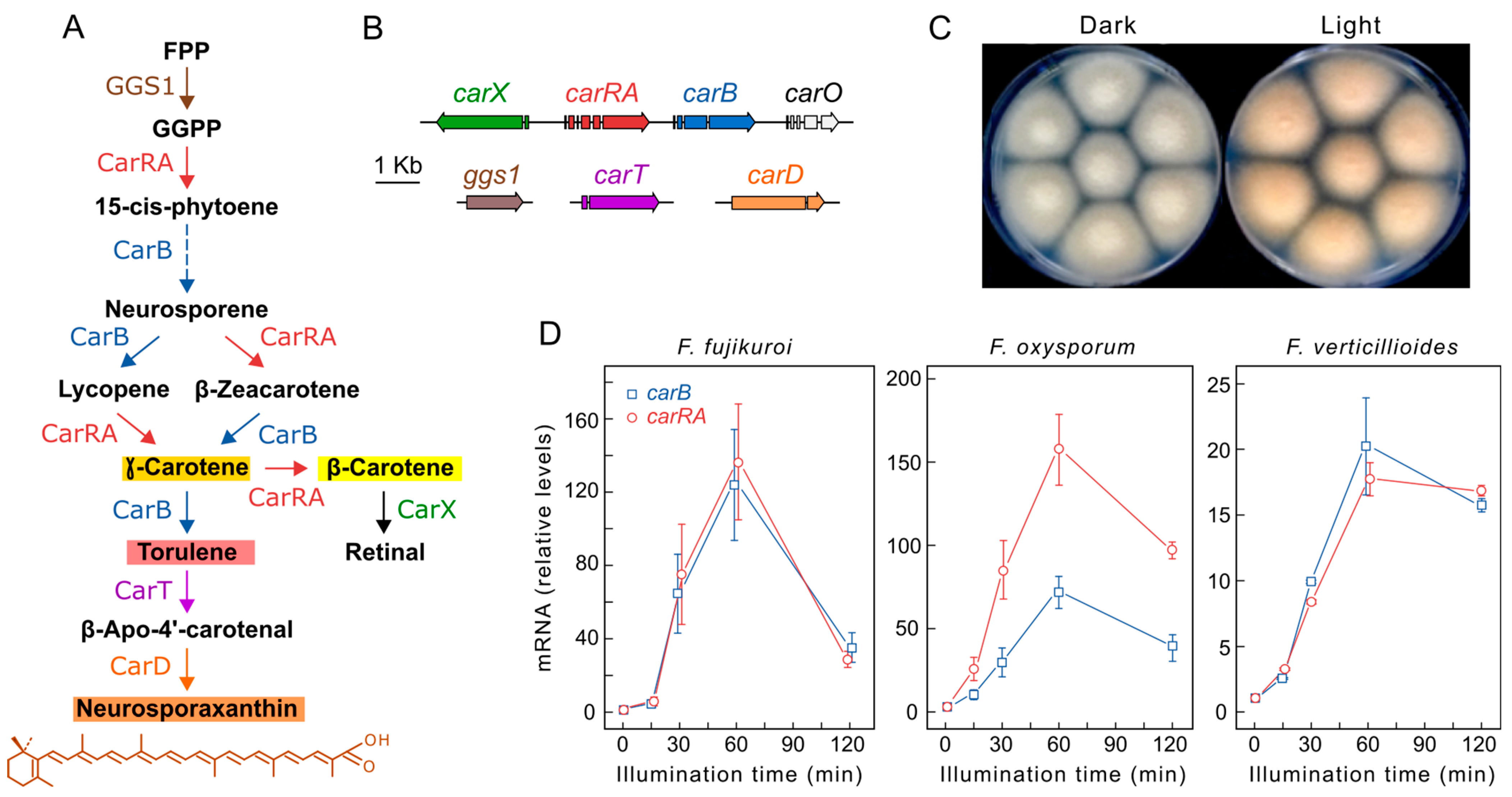
2.2. Effect of Light on Carotenogenesis

Photocarotenogenesis is the most well-characterized light-regulated process in Fusarium [35]. The first studies on the effect of light on carotenogenesis were carried out on Fusarium aquaeductuum, which showed a gradual accumulation of carotenoids after illumination, reaching a maximum at 12 h [36]. The carotenogenetic reaction to light in this species is independent of temperature in the range of 5 to 25 °C but requires oxygenation and active protein synthesis [37]. The light-inducing effect can be partially replaced by the addition of oxidizing reagents [36,38], suggesting that the oxidation of the -SH groups plays a role in the light sensing system, which disappears when reducing agents are added [39]. However, while brief exposure to light is sufficient for photoinduction, oxidizing agents must be continuously present to maintain their stimulatory effect in addition to that of light [40], indicating different mechanisms of action. Nevertheless, the oxidizing agent p-hydroxymercuribenzoate had no effect on other Fusarium species [41].
All Fusarium carotenoid synthesis genes have been identified [35], and the pathway is well established (Figure 1A). Three structural genes, carRA, carB, and carX, required for torulene, β-carotene, and retinal production, are organized in a gene cluster coregulated with a rhodopsin gene, carO (Figure 1B). The photoinduction of carotenogenesis in Fusarium mycelium grown in the dark involves a rapid increase in the transcript levels of most structural genes during the first hour of illumination, followed by an accumulation of carotenoids in the following hours, providing an orange pigmentation to the mycelium (Figure 1C). Northern blot and RT-PCR analyses of the four clustered car genes of F. fujikuroi showed a similar induction kinetics, also found for the carT gene, which is required for neurosporaxanthin synthesis. Similar results were obtained in F. oxysporum [42] and F. verticillioides [43,44] (Figure 1D). Recent RNA-seq data have also revealed the significant photoinduction of ggs1 coding for a prenyl transferase [6], which has been previously underestimated [45]. The carD gene exhibited a lower photoresponse in F. fujikuroi [46], as corroborated by RNA-seq data [35]. Therefore, in F. fujikuroi, all carotenoid metabolism genes are regulated by light. The regulatory proteins responsible for this photoresponse are mentioned in later sections, but other proteins involved in the light signal transduction pathway may also be responsible. Fusarium has the predicted components of a Velvet complex, FfLae1, FfVel, and FfVel2 [47], which is connected to light regulation in other fungi [48]. The carRA gene is upregulated in Fflae1 mutants, indicating a repressor function for lae1 gene [49].

2.3. Effect of Light on the Production of Other Secondary Metabolites

Light modulates the production of other metabolites in addition to carotenoids. Gibberellin biosynthesis is stimulated by light in some strains of F. fujikuroi [24,51], although its effect is minor compared to that caused by nitrogen shortage. The influence of light on the synthesis of enniatins, cyclohexadepsipeptide antibiotics produced by different Fusarium species, has also been investigated. Enniatin production is enhanced by light in Fusarium sambucinum [52]. In other fungi, such as those of the genera Aspergillus or Neurospora, light influences the production of secondary metabolites through the Velvet VelB/VeA/LaeA complex [47,53]. In Aspergillus, this occurs through light controlling the VeA passage into the nucleus in response to a signal from photoreceptor proteins [54]. In Neurospora crassa, light promotes the degradation of the Velvet Ve-1 protein [55]. Disruption of the Velvet complex genes in F. fujikuroi almost completely halts the biosynthesis of gibberellins, fumonisin, fusarins, and fusaric acid [48,49], as well as conidiation. F. graminearum deletion mutants of the FgVeA and FgVe1 genes show reduced aerial hyphal formation, as well as reduced biosynthesis of deoxynivalenol, aurofusarin, and trichotecene [56,57]. No attention has been paid, for either of the two Fusarium species, to the effect of light on these phenotypic changes. However, deleting the Velvet complex genes veA, velB, and laeA drastically reduces beauvericin production in F. oxysporum under light and dark conditions, in addition to affecting conidia production and morphology [58]. These mutants exhibited fewer differences in pigmentation and morphology between light and dark growth colonies than those exhibited by the wild type, confirming their connection to light regulation.

3. Fungal Photoreceptors

The proteins responsible for light detection and signal transmission are known as photoreceptors. They bind to small molecules called chromophores, which can absorb light and cause a conformational or chemical change in the cognate protein. This triggers a direct response or initiates a signal transduction pathway [59]. Depending on the nature of their chromophore, photoreceptors can detect light or radiation within a specific wavelength range. Thus, UV-, blue/UV-, green-, or red-light photoreceptors can be distinguished. Flavin, retinal, and tetrapyrrole chromophores are the typical fungal photoreceptor chromophores [3]. Most light responses studied in fungi are caused by the detection of blue light, although responses at other wavelengths are also known [3,60,61,62]. The main families of photoreceptors in fungi and their presence in Fusarium are described below. Fusarium genomes contain genes for ten photoreceptors (Table 1). Most of them have been studied by targeted deletion in several Fusarium species, while others await investigation.

4. Flavoproteins

Flavoprotein photoreceptors are a heterogenous group of proteins that detect blue light through the absorption of a flavin chromophore [73], bound to a LOV domain (from Light, Oxygen and Voltage). Best known classes in fungi are the WC-1 proteins of the White Collar system and VIVID proteins, but there are other LOV-containing proteins that have received poor attention in fungal photobiology.

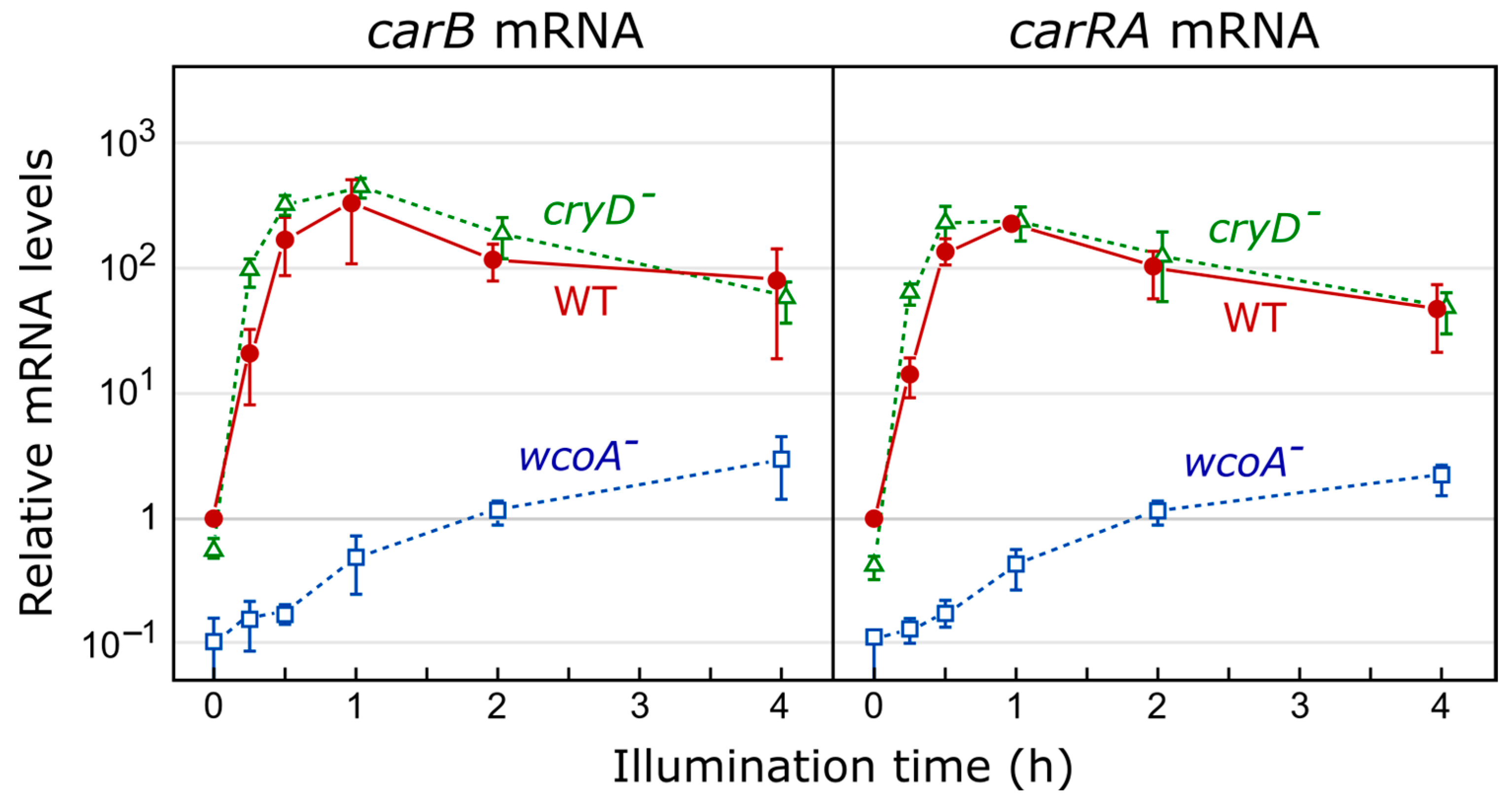
4.1. White Collar Complex

The White Collar complex (WCC), the first photoreceptor system investigated in fungi, was discovered in N. crassa as a photoreceptor needed to induce carotenogenesis by light [74]. The WCC is composed of two proteins, WC-1 and WC-2 [75,76], which centralize all known photoresponses in this fungus, including the light control of carotenogenesis, conidiation, circadian rhythm, and the orientation of their perithecial beaks [75,77]. The photosensitivity of WC-1 relies on its LOV domain (light, oxygen, and voltage), which is a variant of the PAS domain of interaction between proteins. LOV-containing photoreceptors regulate most responses to light in fungi [3,62] using flavin-type chromophores [78].
A flavin is non-covalently attached to the LOV domain through hydrogen bonds, Van der Waals forces, and electrostatic interactions. Light excitation of the bound flavin, either flavin mononucleotide (FMN) or flavin dinucleotide (FAD), produces a transient covalent bond with a conserved cysteine of the LOV domain [79,80]. The formation of this link modifies the structure of WC-1 by initiating a cascade of signals in response to light. When light excitation stops, this link is slowly broken, restoring the protein to its initial fully oxidized state. WC-1 also contains two additional PAS domains [81,82] and a zinc-finger DNA-binding domain. The partner of WC-1, WC-2, is a smaller protein that lacks a LOV domain, meaning it cannot act as a photoreceptor on its own, but does have PAS and a zinc-finger domains.
In N. crassa, the WCC in the dark binds to specific DNA elements mostly located in the promoters of light-regulated genes [83,84]. Light promotes the interaction of two WC1 proteins through their LOV domains, to form a WCC dimer [85]. The mechanism by which the WCC controls the transcription of target genes in response to light in this fungus involves the participation of other regulatory proteins (Figure 2) [3,62]. WCC binding mediates chromatin remodeling, facilitating RNA polymerase accessibility and transcription initiation. WC-1 mediates H3 histone acetylation (H3K14ac) by histone acetyltransferase NGF-1 [86,87], and nucleosomes are displaced after the binding of the transcription factor SUB-1 [84]. Activation of the WCC by light [88] leads to the transcriptional induction of early photoinducible genes, including VVD (VIVID, see next section). VVD is a small flavoprotein that is activated by light, binds to WCC to attenuate its activity, and leads to the phosphorylation of WC-1 and WC-2 by protein kinase C (PKC) [89]. WC-1 phosphorylation promotes its partial degradation while reducing its DNA-binding capacity [90,91]. The activity of the WCC is also determined by its interaction with the FRQ (FREQUENCY) protein [92,93], the core of the central oscillator of the circadian clock in this fungus [94,95].
Analyses in other fungi have shown orthologs of WC-1 and WC-2 in numerous species, suggesting an early evolutionary start to the regulation of responses to light [96]. They have been described in zygomycetes such as Mucor circinelloides or Phycomyces blakesleeanus [97,98]; in basidiomycetes such as Cryptococcus neoformans [99], Ustilago maydis [100], or Pleurotus ostreatus [101]; in ascomycetes such as Trichoderma reesei [102], Botrytis cinerea [103], or Aspergillus nidulans [60]. In general, fungal genomes possess a single wc1-like gene, although the latter has been lost in some species [96]. In contrast, some zygomycetes have several wc-1- and wc-2-like genes [104]. Their functions have been studied in detail in M. circinelloides, in which they have different specialized functions, possibly because of the loss of other photoreceptors [62]. Thus, Mcwc-1a regulates phototropism, Mcwc-1b participates in the light-independent regulatory mechanism of carotene synthesis through the ubiquitylation of the negative regulator CrgA [105], and Mcwc-1c regulates the light-dependent expression of carotenogenic genes [98]. In recent years, phenotypes related to processes not regulated by light have also been described in wc mutants of N. crassa [84]. Light-responsive transcription factors (LTFs) and their regulation by WCC have been studied in B. cinerea. The genes encoding BcLTF1, BcLTF3, and BcReg1 are induced by BcWcl1 [106]. BclTF1 represses BcLTF2, and the latter is repressed by the WCC. BcLTF3 represses Bcltf2 which is also a regulator of conidiation. The deletion of bcwcl1 causes conidiation in this fungus in both light and darkness (“always conidiation” phenotype), which correlates with increased Bcltf2 mRNA levels [103]. The BcLTF1 ortholog in A. nidulans, Nsd1, plays a role in regulating the development of conidia and sclerotia [107].

Fusarium White Collar System

Fusarium genomes contain an ortholog for N. crassa wc-1, wcoA (Figure 3). However, a more detailed study shows regulatory differences: wc-1 expression is photoinducible in N. crassa while wcoA is hardly affected by light in F. fujikuroi [63]. Furthermore, their mutant phenotypes differ greatly between both genera. Secondary metabolism is affected in wcoA mutants of F. fujikuroi, which show changes in the production of gibberellins, bikaverin, and fusarins, while trichotecene synthesis and aurofusarin synthesis are affected in F. graminearum [64]. In addition, the wcoA mutants of F. fujikuroi have been shown to produce fewer conidia on agar or in liquid cultures, and their aerial mycelia were shown to be less hydrophobic than those of the wild-type strain [63], a phenotype also observed in the wc1 mutant of F. oxysporum [65]. Photoreactivation after UV treatment was shown to be impaired, as there was no transcriptional activation of phr1 under illumination in the wc1 mutant in F. oxysporum [65] and F. graminearum [32].
Orthologs of the wc-1 and wc-2 genes have also been deleted in F. graminearum and Fusarium asiaticum [32,64,66], in which phenotypes of impaired sexual reproduction, photoreactivation, and secondary metabolism have been described. No effect on virulence was reported in F. graminearum, but a decreased virulence in wheat, as well as defects in perithecia and ascospore formation, was shown by the Fawc1 mutant of F. asiaticum [66]. Interestingly, the effect on virulence appeared to be Fawc1-dependent, indicating the Wc2-independent roles of this Wc1 protein. Pathogenesis was also affected in F. oxysporum, as indicated by the decreased virulence of the wc1 mutant in immunosuppressed mice.
The involvement of the Velvet complex in conidiation and in the regulation of secondary metabolism and its interdependence with the WCC, as documented in other fungi [108], provides a possible explanation for the wcoA mutant phenotypes in F. fujikuroi. Interestingly, many of these alterations were independent of light. Differential phenotypes have been described for Fawc1 mutants of F. asiaticum depending on whether this protein contains the LOV or the Zn-finger domains. This could explain the differential role of WC-1 orthologs, which function as a light-dependent transcription factor for some processes while functioning as a standard one in others.
Mutation of wcoA affects carotenogenesis to varying degrees in different Fusarium species, some of them in an unexpected way compared to the N. crassa wc-1 mutant. In this fungus, whose action spectrum for light-induced carotenogenesis [109] is similar to that of Fusarium [110], photoinduction in mycelium is completely dependent on the WCC [3]. However, the wcoA mutants in F. fujikuroi [63] and its ortholog, wc1, in F. oxysporum [65] maintain a significantly increased carotenoid content under continuous light. However, the deletion of fawc1 and fawc2 in F. asiaticum was found to prevent the induction of carotenoid biosynthesis under constant illumination [66]. The fgwc-1 and fgwc-2 targeted mutations in F. graminearum were found to cause paler pigmentation in their colonies under light, but their carotenoid content was not determined [64]. Interestingly, both mutants also exhibited higher fertility, even in unfavorable mediums for sexual development. The similar phenotypic alterations displayed by the wc-1- and wc-2-type gene mutants in the two latter Fusarium species support their function as a White Collar complex.
A kinetics analysis of carotenoid accumulation revealed that there are two stages in the response of F. fujikuroi; a rapid first WcoA-dependent response and a slower second phase of carotenoid accumulation, which was dependent on the DASH CryD cryptochrome [50] (see Section 6.1). The participation of CryD as a second photoreceptor could explain the maintenance of carotenoid accumulation in wcoA mutants of this species under continuous illumination.

4.2. VVD-Like Flavoproteins

The increase in the mRNA levels of photoinduced genes is usually transient, and after prolonged illumination is reduced or even returns to the basal levels found in the dark. This phenomenon, known as photoadaptation [111], was investigated in detail in N. crassa, in which it is mainly due to the counteracting effect of the VVD (VIVID) photoreceptor, a small flavoprotein with a LOV domain and an N-terminal extension of 70 amino acids involved in its dimerization [112]. Upon illumination, the WCC stimulates the transcription of the vvd gene and VVD protein accumulates [113]. A cysteine in VVD forms a covalent bond with a flavin after illumination, inducing a conformational change in its N-terminal domain that appears to be essential for its function [114].
If illumination persists, as is often the case of daylight in nature, the newly formed VVD can dimerize unstably and repress WCC by forming WCC–VVD complexes [115] (Figure 2) and regulating their phosphorylation [116]. The interaction of VVD with the LOV domain of WC-1 disrupts the WCC dimers and reduces their transcriptional response, leading to photoadaptation while protecting the complex from degradation [85]. The vvd gene itself is tightly regulated by a repressor complex consisting of the RCO-1 and RCM-1 proteins [117,118]. VVD-mediated photoadaptation also involves chromatin modifications by DIM-5, resulting in the trimethylation of histone H3 at lysine 9 (H3K9me3) (Figure 2). This was tested on the frq promoter, in which the dim-5 mutation supports the role of H3K9me3 in WCC eviction as more amounts of WC-2 bind to its promoter and a greater amount of frq mRNA accumulates [119]. Through all of this, N. crassa is able to dampen the transcriptional response to light and prevent the accumulation of light-regulated mRNAs under prolonged illumination. This is evidenced by the increased accumulation of light-upregulated mRNAs in vvd mutants, including those of the enzymes required for carotenoid biosynthesis, leading to the more intense orange color of the mutant mycelium, hence its name [62,113].
There are VVD-like proteins in other fungi. Envoy (Env1), the vvd ortholog in T. reesei, has also been extensively studied. Env1 has a more relevant role in this fungus since its loss causes poor growth in light [120] and affects cellulase production [121]. Like N. crassa vvd mutants, env1 mutants are affected in terms of their certain light-dependent responses, as well as in terms of their abilities to maintain gene photoinduction [102]. In Trichoderma guizhouense, the env1 mutant exhibits much higher mRNA levels of hydrophobin genes in the light, which is explained by a lack of photoadaptation [122]. In these fungi, Env1 has emerged as a central checkpoint for the integration of nutrient sensing, light response, and development [123].

Fusarium Vivid Protein VvdA

The ortholog of N. crassa vvd in F. fujikuroi was designated as vvdA [67]. Its expression is strongly induced by illumination through the WcoA protein. In contrast to its N. crassa counterpart, vvdA-targeted mutation causes paler pigmentation under constant illumination due to the accumulation of less carotenoids than in the wild type. However, immediately after illumination, the vvdA mutant exhibits a faster carotenogenetic response to light, indicating that, as in N. crassa, it exerts negative control over WcoA activity [50]. The reversal of this effect after long-term illumination suggests that VvdA exerts a positive effect on a second photoreceptor. The vvdA mutants show morphological alterations only under illumination. Thus, when put under light, the mycelium of the vvdA mutants was of a higher hyphal density and was more embedded in the agar than that of the wild type or of the complemented strains [67]. Furthermore, conidial production was reduced to half in agar cultures, was strongly reduced in illuminated liquid cultures, and was restored in the complemented strain, indicating the importance of this gene in this developmental process. [67]. The essential role of VIVID proteins in conidiation under illumination has also been described in Beauveria bassiana [124].

4.3. Other Flavin Photoreceptors

Fungal genomes frequently contain genes that encode other LOV flavin-binding domains in addition to those described above. Two types have been distinguished, some with a regulatory domain of G protein signaling (RGS) and others without recognizable domains with possible regulatory functions [61,125]. Genes for both types of proteins exist in fungi, such as Magnaporte, Botrytis, or Fusarium, but are not found in other well-known fungi, such as Aspergillus or Neurospora. These putative fungal photoreceptors have received very little attention. An exception is the RGS-domain flavoprotein RGS5, which was investigated in Magnaporthe oryzae. RGS5 was shown to interact with two of the three Gα subunits of this fungus, and the Morgs5 deletion mutant was shown to contain higher intracellular cAMP levels, although no apparent phenotypic alterations were shown [125]. F. fujikuroi contains two putative photoreceptors that we call LovA and LovB (Table 1), the second with an RGS domain (Figure 3). Although they are smaller, both proteins show similarity to plant phototropins. Thus, 145 of the 674 LovA residues and 154 of the 663 LovB residues have coincident positions in the Arabidopsis thaliana Phot1 996-aa polypeptide (Uniprot O48963).

5. Cryptochromes

The proteins of the cryptochrome–photolyase family are a heterogenous group of photoreceptors that absorb radiation from the UV-A/blue spectral range via two chromophores, FAD and 5,10-methenyltetrahydrofolate (MTHF). They are present in all the main taxa and are divided into 10 subfamilies [126], five corresponding to photolyases (PHR) and five to cryptochromes (CRY), the latter being of plant origin and the rest being generally found in animals. Photolyases act as light-driven DNA-repair enzymes [127], and cryptochromes are often involved in signal transduction. Cryptochromes consist of the typical N-terminal region of a photolyase (PHR), followed by a carboxyl terminal domain of variable length, which is involved in signal transduction. A special subgroup are the DASH-cryptochromes (Drosophila, Arabidopsis, Synechocystis, and Homo), which exhibit both functions [128]. In general, CRY-DASH proteins bind single-stranded DNA, with some exceptional cases in which they bind double-stranded DNA [129] to perform repair functions typical of photolyases [129].
Cryptochromes belonging to the plant type and DASH cryptochrome groups are generally found in fungi, and they also contain canonical photolyases that exert repair functions. Nevertheless, in some zygomycetes that lack photolyases, the repair function is carried out by DASH cryptochromes [129,130]. Compared to other taxa, there is not much information on fungal cryptochromes and their signaling mechanisms [3], although several mutants have been described. In A. nidulans, CryA is closer to CPD (Cyclobutane Pyrimidine Dimer) photolyases and has repair functions [131], but also represses sexual development in the presence of light. In N. crassa, the Cry-DASH CRY protein is involved in circadian rhythm regulation and binds to DNA and single- and double-stranded RNA but shows no evidence of photolyase activity in vivo [132,133]. B. cinerea has two members of the family with different functions, BcCRY1, a CPD photolyase, and BcCRY2, a Cry-DASH protein that is dispensable for photorepair but that plays a role of a conidiation repressor [134]. Fungal Cry-DASH proteins in other species, Sclerotinia sclerotiorum and Cordyceps militaris, regulate very different processes, including development or secondary metabolite production [135,136].

Fusarium Cryptochromes CryD and CryP

Fusarium genomes have two cryptochrome genes: one for a plant-type cryptochrome and one for a DASH cryptochrome (Figure 4). The DASH cryptochrome CryD from F. fujikuroi has received special attention, and its photoactivity has been demonstrated biochemically [68]. The expression of cryD is strongly stimulated by light via WcoA, and its targeted mutation produces different phenotypic alterations only under illumination, such as the formation of macroconidia under nitrogen starvation or bikaverin production at 30 °C [51]. Furthermore, CryD seems to be responsible for the long-term photoinduction of carotenogenesis, as indicated by the slower accumulation of carotenoids after the prolonged incubation under light of cryD mutants [50]. However, wcoA mutants accumulate carotenoids very slowly from the onset of illumination, and their total levels do not reach those of the wild type. The photoinduction of the structural genes for carotenogenesis in the wcoA and cryD mutants (Figure 5) is consistent with different mechanisms of action for the encoded photoreceptors [50]. The cryD mutant has exhibited wild-type photoinduction of the car structural genes. However, in the wcoA mutant, the mRNA levels were much lower than those in the wild-type strain even in the dark, but still detectably increased after illumination. The induction of carotenogenesis by the alternative photoreceptor, presumably CryD, must take place via a transcription-independent mechanism that remains to be elucidated.
No information is available on the function of the plant-type Fusarium cryptochrome, but data from RNA-seq studies show that its expression is also induced by light [6]. On the other hand, photoreactivation due to UV damage is carried out in Fusarium by Phr photolyase, whose expression is also light-dependent [6,69]. Furthermore, it has been found to be highly expressed in the early stages of germination [139].

6. Rhodopsins

Green light can be sensed in fungi by retinal-binding opsins, a family of integral membrane proteins also known as rhodopsins [140]. Opsins have a highly conserved three-dimensional structure, consisting of seven transmembrane helices. Those capable of detecting light attach a retinal molecule as a chromophore by covalent bonding to a lysine residue located in the third transmembrane domain. These proteins were initially discovered in archaea through the study of bacteriorhodopsin, a light-driven proton pump, and were subsequently found in other taxa, including fungi [141]. Three classes of opsins are found in fungi, two of which act as photoreceptors: the first class has a slow photocycle with low proton pump activity, such as NOP-1 in N. crassa [142]; the second class is characterized by strong green light-dependent proton pump activity, such as CarO in F. fujikuroi [143]; the third class lacks the retinal binding residue and is therefore not expected to be photoactive [3]. The genomes of many fungi encode other opsin-related proteins that lack the lysine residue for chromophore binding, the functions of which remain enigmatic [144]. In N. crassa, NOP-1 is involved in the regulation of developmental processes, possibly through the control of the oxidative state of the cell [142], while the CarO-type rhodopsin has been found only in plant-associated fungi and has a role in germination and infection in F. fujikuroi [70]. U. maydis has three rhodopsin genes, two with proton pump activity and a third without such activity that is only expressed during plant infection [145]. The signal transduction pathway for fungal rhodopsins is still unknown. The presence of rhodopsin genes in fungal genomes is very variable, and phylogenetic studies suggest the occurrence of horizontal transfer events followed by duplications and losses, presumably leading to their current functional diversification [142].

6.1. Fusarium Rhodopsins CarO and OpsA

Fusarium has two rhodopsins, which have been studied in F. fujikuroi (Figure 6). The rhodopsin gene carO is linked to the carRA, carB, and carX genes in the car gene cluster [71] and its transcription is induced by light. The CarO protein is a proton pump that efficiently responds to green light and whose activity is enhanced in the presence of the plant hormone indole-3-acetic acid [70,143]. A targeted mutant of the carO gene lacks apparent phenotypic changes [71], but germination analyses of conidia have shown the faster development of light-exposed germlings in the carO mutant than in the control strain. Furthermore, a visualization of YFP-labeled CarO showed its presence in the conidia produced by illuminated mycelia. Orthologs of this rhodopsin have been found in many plant-associated fungi, suggesting its possible role in plant infection. In addition, rice plants infected with the carO mutant showed more severe bakanae symptoms [70], indicating the possible role of rhodopsin CarO in controlling plant infection.
The second Fusarium rhodopsin gene, opsA, is orthologous to the N. crassa nop-1 gene. Mutation of opsA gene resulted in no visible phenotypic alterations, including the carotenoid content, although a moderate decrease in car gene mRNA levels was shown [72]. The carO and opsA genes were differentially expressed during sexual reproduction in perithecia. The carO gene was down-regulated in a mutant of the sexual locus mat1-2-1 in F. verticillioides [146], and its transcript levels increased throughout the period of perithecia maturation in F. graminerarum [142]. The N. crassa Δnop-1 mutant initiates sexual development earlier than the wild type does, suggesting the role of OpsA in the sexual cycle. Moreover, comparative transcriptomics showed the increased expression of genes involved in the response to oxidative stress in the nop-1 mutant [142], indicating that Nop-1 could play a role in the regulation of asexual–sexual development in response to different environmental cues, including light and reactive oxygen species (ROS).
Fusarium has a third gene in the opsin family, hspO, which has not been investigated. The HspO protein is predicted to lack the conserved retinal binding lysine and is therefore assumed to be a non-photoactive opsin [72]. Some of these opsin-related proteins have been associated with stress responses and could function as chaperones [141], but their mechanisms of action remain unknown.

7. Phytochromes

Red and far-red light is detected in fungi by phytochromes, which use a linear tetrapyrrole (bilin) chromophore that is autocatalytically linked to the apoprotein through a conserved cysteine residue [147]. Originally described in plants, this type of photoreceptor was first discovered in fungi in A. nidulans [148] and N. crassa [149]. Phytochrome genes are common in ascomycetes [150] and basidiomycetes [62] but have not been described in zygomycetes. Some fungi contain more than one phytochrome, indicating functional diversification. This is the case for Monilinia laxa, which has three phytochromes with different transcriptional regulations [151]. Fungal phytochromes (Fph) contain a photosensor module at the amino terminus consisting of a PAS domain, which is also present in bacterial phytochromes. They also contain a GAF domain (cGMP phosphodiesterase/Adenylate cyclase/FhIA) that exhibits bilin lyase activity, which includes the conserved cysteine residue that binds to the chromophore, and a PHY domain unique to this class of photoreceptors. In addition, they contain a domain related to histidine kinases or HKRD and, depending on the species, a regulatory module in their highly variable carboxyl termini [147]. The bilin-related chromophore of the FphA phytochromes of A. nidulans and Alternaria alternata is biliverdin or a very similar derivative [148,152]. The synthesis of this chromophore and its coupling to FphA has been investigated in A. alternata, in which FphA interacts with two mitochondria-attached heme oxygenases involved in chromophore synthesis [152]. This interaction mediates the transfer of the chromophore to the phytochrome.
In A. nidulans, the phytochrome plays a central role, regulating the balance between asexual and sexual development and the germination process, as well as secondary metabolite biosynthesis [148,153]. The mechanism of action has been investigated in detail in this species, in which it induces the HOG signaling pathway in the cytoplasm, modulating the activity of the phosphotransferase protein YpdA [154], and interacts in the nucleus with transcription factors and enzymes that are necessary for chromatin modification [60,155,156]. In the plant pathogen A. alternata, red and blue light have inhibiting and stimulating effects on sporulation, respectively, through the participation of the FphA phytochrome and the LreA WC protein in coordination with the high-osmolarity glycerol (HOG) mitogen-activated protein (MAP) kinase pathway [157]. In addition, both photoreceptors modulate other processes, such as spore germination and ROS alleviation, by catalase and superoxide dismutase activity. The HOG signaling pathway also participates in the initial stage of fruit colonization, indicating the role of phytochrome in pathogenesis [158]. Phytochromes are also involved in the modulation of the activity of the WCC and in the transcription of some of their genes in several fungi, including N. crassa [3,60,62,134]. Phytochromes have also been described as temperature sensors in plants, bacteria, and fungi [159,160,161], which makes evolutionary sense considering that, in nature, exposure to sunlight is usually associated with an increase in temperature.
The important role of red or far-red light detection via phytochromes in A. nidulans [162] or A. alternata [157] contrasts with the prevalence of blue light detection in other fungi. There are ecological niches that allow a greater abundance of certain types of light, such as far-red light or green light in plant environments [163,164], which may explain the greater role of phytochromes in some species and their participation in key developmental processes, such as sporulation. However, the detection of specific light wavelengths may be irrelevant in other environments, in which blue light photoreceptors may function more efficiently.
Fusarium Phytochrome FphA
The role of the only phytochrome (Figure 7) found in Fusarium genomes [24] has been analyzed in F. graminearum [32]. The deletion of fgfph produced no visible phenotypic alterations, including growth, pigmentation, virulence, or fertility. However, in vivo protein–protein interaction assayed by split luciferase complementation showed that FgFph interacts with FgWc-1 as well as with the Velvet complex proteins FgVeA and FgLaeA. As already stated, the Velvet complex coordinates light perception and development and secondary metabolism in many fungi [47]. The interaction of FgFph with WC-1 and the Velvet complex suggests its indirect role in Velvet-mediated processes, possibly as an accessory photoreceptor. Furthermore, illuminating F. fujikuroi mycelium with red light appreciably induces the expression of at least some blue-light regulated genes, including carRA [50]. Although the photoinduction of carotenoids depends mainly on WcoA, this observation indicates the participation of a phytochrome in the photosensory system that regulates this response.

8. Study of Photoreceptor Functions through Global Transcriptomic Tools

The effect of light on fungal transcriptomes has been investigated in several organisms with different techniques [62,165], and these investigations usually conclude that light affects the mRNA levels of hundreds of genes. In A. fumigatus, a microarray assay was used to investigate the effect of 15, 30, 60, and 120 min of light exposure, and, based on a 1.5-fold change that was observed, it was concluded that 2.6% of the genes were regulated by light in this species [166]. In P. blakesleeanus, large-scale cDNA sequencing led to an estimate that, in about 5.2% of the genes, mRNA levels changed at least five-fold after 30 min of illumination [167]. A study conducted in Trichoderma atroviride found 246 and 215 genes upregulated and downregulated after a 30 min light pulse [168], which make up about 3.9% of its genome. These works laid the foundation for functional studies of photoreceptors, verifying the effect of mutations in the responsible genes.
Analyses of photoreceptor functions through global transcriptomic studies have mainly focused on the WC system. In N. crassa, about 5–6% of genes were differentially expressed in the wild type during the first 90 min after light onset, a response largely mediated by the WC complex [4]. A comparison of microarray expression data after eight illumination intervals, from 5 to 240 min, revealed at least two classes of induction patterns, leading to the classification of early responsive and late-responsive genes. Essentially, all photoresponses were lost in the wc-1 and wc-2 mutants. A subsequent and more precise RNA-seq study of the same fungus showed 31% of genes with at least a two-fold change in mRNA levels [83]. Different induction patterns were observed and separated into four clusters, although the classifications of early and late responsive genes were conserved. This drew attention to the occurrence of a large set of light-repressed genes, none of which showed early repression. WC-1 can also act as a repressing protein through a light responsive GATA element that has been found to be involved in binding to form a repressing complex in the dark [169]. Nevertheless, the lack of fast downregulating responses suggests that the repressive effects could have been caused by other negative regulators, which would have been activated earlier by the WC-1.
Experiments combining the effect of light on the transcriptome with the effect of mutations in the wc genes have also been carried out in other fungi. Microarray data from T. reseei showed that 2.8% of genes were differentially expressed under continuous illumination compared to a dark control, with a trend towards upregulation [7]. Surprisingly, the lack of the WC complex altered the adaptation to constant illumination, as the number of genes differentially expressed in light increased to about 9% in the mutants of the wc-1 and wc-2 orthologs brl1 and brl2. A new RNA-seq study of the effect of a 30 min light pulse in T. atroviride, using a different bioinformatic analysis for differential expression, found 97 and 38 genes upregulated and downregulated in the wild type, of which only 3 from each class maintained photoregulation in the blr-1 mutant, indicating that this WC protein mediates most of the photoresponses in this species [170]. This study of T. atroviride was extended to mutants for the ENVOY, CRY-1, and CRY-DASH photoreceptors, but their roles in photoregulation were subsidiary to Blr1, since the transcription of their genes was activated by light through this photoreceptor. In Sordaria fimicola, the effect of 15 min and 45 min of illumination on the transcriptome was analyzed in the wild-type strain and in a defective sfwc-1 mutant. Unexpectedly, of the 874 light-regulated genes, only 466 lost photoinduction in the mutant, suggesting the participation of another blue-light photoreceptor [171]. In other fungi, the WC system plays less prominent roles. In A. nidulans, the effect of different light wavelengths on the transcriptomes of mutants of the LreA Wc-1 photoreceptor and the FphA phytochrome genes showed that red light exerts a greater influence than blue light, and that FphA is the main photoreceptor [162]. The loss of photoinduction of most of the red light regulated genes in a SakA kinase mutant confirmed the FphA activity in the Hog MAP kinase pathway, a result that confirmed previous molecular observations [154]. Interestingly, both LreA and FphA play regulatory functions in the dark as well, and many of the responses to blue light require a functional phytochrome.
Light is less influential in other fungi, such as in U. maydis, in which only 60 genes were found to be induced by light, all of which were dependent on the wco1 gene [100]. Other fungi, however, are apparently insensitive to light; in the basidiomycete fungus C. neoformans, only one gene was significantly affected under illumination [172]. Taken together, the different results show the considerable variability of the effects of mutations in the White Collar genes of different species.

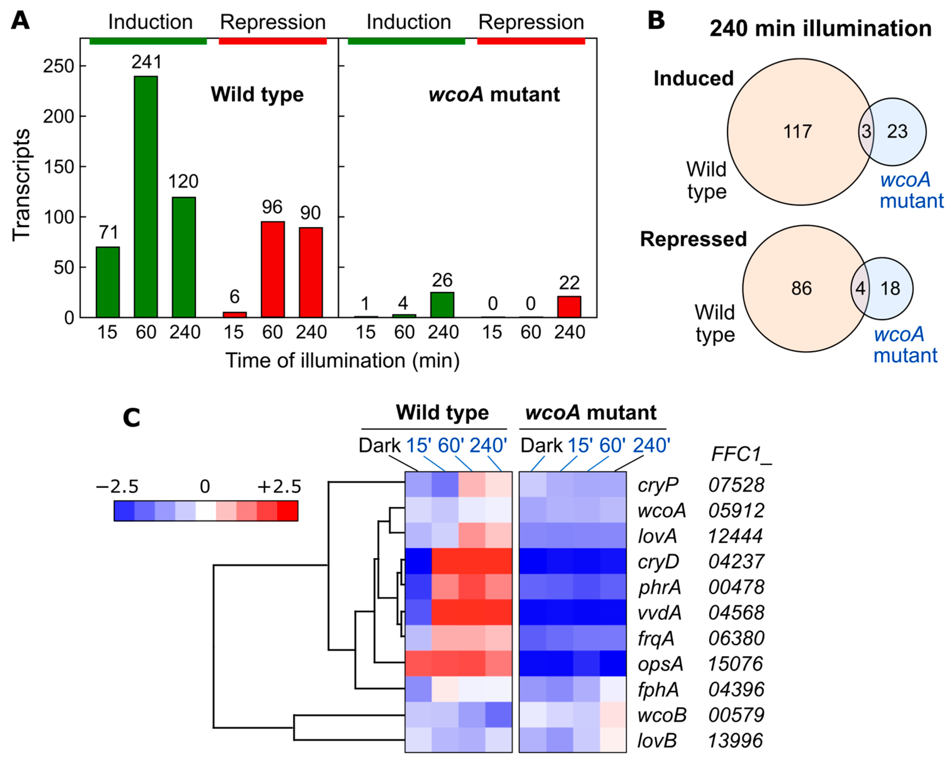
8.1. Effect of wcoA Mutation on the Fusarium Transcriptome

As shown in Section 2.2, the most investigated light response in Fusarium is the photoinduction of carotenoid synthesis [35,63]. The loss of function of the wcoA gene produces a drastic decrease in the mRNA levels of the carotenogenesis genes, both under light and in the dark, but it does not prevent their induction by light (Figure 5). The phenotype resulting from the mutation is, however, highly pleiotropic. Diverse morphological and metabolic changes are observed, including alterations in the synthesis of different secondary metabolites, suggesting the complex regulatory role of WcoA. To further investigate the diversity of functions of this photoreceptor in this fungus, the impact of the wcoA mutation on the transcriptome was recently analyzed by RNA-seq. Total RNA samples of a wild-type and a wcoA mutant were compared either in the dark or under illumination for 15, 60, or 240 min [138]. Using changes in mRNA levels greater than 4-fold as part of the criteria for effects on regulation, 298 light-activated genes and 160 light-repressed genes were observed after at least one of the three illumination intervals (Figure 8A). Photoactivated genes were distributed in those with fast, intermediate, and slow responses, while only intermediate and slow responses were found among photorepressed genes. Most of the responses to light were lost in the wcoA mutant. Interestingly, 41 genes that were not regulated by light in the wild type showed slow light-dependent responses in the mutant (Figure 8B). Therefore, WcoA is the main photoreceptor for responses to light at the transcriptional level in this fungus.
WcoA is required for the photoinduction of other regulatory proteins, including several photoreceptors, confirming previously obtained results (Figure 8C). The strong photoinduction of the genes for cryptochrome CryD, flavoprotein VvdA, rhodopsin CarO, and photolyase PhrA is lost in the wcoA mutant. WcoA also participates in the photoinduction of the F. fujikuroi ortholog of the frq (frequency) gene of N. crassa, which is involved in the circadian rhythm and not yet studied in Fusarium. The genes for the other F. fujikuroi cryptochrome, which we call CryP genes, and flavoprotein LovA also show that photoinduction is lost in wcoA the mutant. However, lovB is hardly affected by light or by the wcoA mutation. Thus, WcoA plays a central role in Fusarium photobiology, not only through its direct action on many photoregulated genes, but also through its regulation of other photoreceptor genes. Of particular interest is the case of the OpsA rhodopsin gene, the mRNA levels of which are barely affected by light but drop considerably in the wcoA mutant both in the light and in the dark.
The influence of the wcoA mutation on the opsA mRNA content draws attention to the most striking feature of the effect of WcoA on the transcriptome, which is that the most extensive effects of its mutation are light-independent (Figure 9). In approximately 16% of the F. fujikuroi genes, mRNA levels changed at least fourfold when comparing the wild type to the wcoA mutant in the dark or after different light exposures. The genes with decreased mRNA levels in the wcoA mutant (2843 genes in the sum of all conditions) predominated over those with increased mRNA levels (1297). As would be expected from the enormous number of them, the affected genes are associated with a wide variety of functions, including the production of different secondary metabolites and hydrophobins. Secondary metabolite pathway genes are normally clustered in the genome. Most of the genes of the clusters for the synthesis of fusaric acid, fusarin, gibberellin, and echisetin/trichotecin were found at much lower levels in the wcoA mutant than in the wild type. However, the opposite pattern was observed in the bikaverin synthesis cluster. Therefore, WcoA acts, directly or indirectly, as an activator or as a repressor of several secondary metabolism clusters.
The massive transcriptomic change found in the wcoA mutant in the dark, in addition to its central role as a photoreceptor responsible for most transcriptional responses to light in this fungus, points to the role of WcoA as a master regulatory protein in F. fujikuroi. These results were obtained with a wcoA mutant obtained from the wild-type strain FKMC1995. Ongoing experiments with a different wild-type strain, IMI58289, point to regulatory differences even between strains of the same species (J. Marente, personal communication).

8.2. Effect of cryD Deletion on the Fusarium Transcriptome

In contrast to the effect of wcoA mutation, the cryD mutant exhibited patterns of photoinduction of the genes carRA and carB that were similar to those of the wild type (Figure 5). The influence of the CryD photoreceptor on the transcriptome was analyzed by studying a cryD mutant in comparison to the wild-type strain FKMC1995 in the dark or after 1h of illumination [137]. Experiments were performed in parallel to those on the wcoA mutant, summarized in Section 8.1. The number of photoregulated transcripts decreased in the cryD mutant (Figure 9A,D). Only 206 genes were photoinduced at least 2-fold in contrast to 884 genes in the wild-type strain under the same conditions (Figure 10A). The reduction in photorepression was more noticeable, with lower mRNA levels being found for 56 genes after illumination in the mutant versus 556 genes in the wild-type strain. By analyzing the 1549 photoregulated transcripts in either of the two strains on a heatmap, we concluded that most of the genes that showed photoinduction or photorepression in the wild-type strain also showed photoinduction or photorepression in the cryD mutant, but that many of them exhibited changes in response intensity (Figure 10B). This is not unexpected, considering that the major photoreceptor responsible for light regulation is WcoA. However, the data suggest the role of CryD as an accessory photoreceptor that modulates the degree of photoresponsivity of many genes.
Using the two-fold change (log2 = 1) observed as a threshold, 230 genes were differentially expressed in the dark in the cryD mutant compared to the wild-type strain, 154 of which were upregulated and 76 of which were downregulated. Similar numbers of affected genes were observed after one hour of illumination with 216 differentially expressed genes (DETs), of which 135 were upregulated and 81 were downregulated. When the effect of the cryD mutation was compared to that of the wcoA mutation under the same threshold, the number of WcoA-dependent DETs was much higher, this number being 3333, compared to the number of CryD-dependent DETs, which was 382. Approximately half of them (138 in the dark and 114 in the light) were shared between the mutants for the two photoreceptors. Nevertheless, 92 and 102 of the transcripts were specific for the cryD mutation.
Previous phenotypic analyses of the cryD mutant suggested the participation of this photoreceptor in the control of secondary metabolism, since they indicated alterations in the coloration pattern under light, which were mainly due to the accumulation of bikaverin [51]. However, although significant changes in mRNA levels were observed for some genes involved in secondary metabolism, those of the bikaverin cluster did not reach the two-fold change in transcript levels in the dark or after 60 min of exposure to light. The discovery of RNA molecules bound to this protein [68], unlike the other cryptochromes described, was proposed as the basis for the possible mechanism of non-transcriptional regulation.
Although CryD is presumably not a DNA binding protein, the number of affected genes in the mRNA content of the cryD mutant is notable. This could be due to the indirect effects on other regulatory proteins or due to changes in the stability or half-life of the mRNA. However, the drop in the number of photoinduced genes in the cryD mutant consolidates its role as a light-dependent regulatory protein, which is consistent with its function as a photoreceptor. Moreover, many genes that maintain their photoregulation exhibit a less prominent response [137]. Therefore, the data suggest that CryD modulates the intensity of the photoresponse of many genes. The opposite effect was also observed: an analysis of the clustering of mean values revealed a group of genes with higher photoinduction in the cryD mutant than in the wild-type strain. This group included several genes related to photoreception and the gene frq, indicating a similar possible influence of CryD on the circadian rhythm to that in N. crassa [132].

9. Concluding Remarks

The abundance of photoreceptor genes in fungi reveals the importance of light as a modulating signal for different activities. Species of the Fusarium genus are no exception, and the evolutionary conservation of photoreceptor genes indicates their relevant roles in the life cycle of these species and in their ability to compete and survive in environment.
As already observed in N. crassa, a well-known model in fungal photobiology, the White Collar system plays a central role in light regulation in Fusarium and also modulates other photoresponses. Noteworthily, it controls light induction of gene transcription for four of the other photoreceptors, the CarO rhodopsin, the VvdA flavoprotein, the CryD cryptochrome, and the PhrA photolyase (Figure 11). Sun exposure is accompanied by collateral damage, including exposure to UV radiation, which makes biological sense in terms of the light induction of the photolyase gene, which is responsible for repairing DNA damage caused by UV rays. With respect to the rest of the photoreceptors, progress has been made in assigning their functions, but much remains to be discovered. In the case of OpsA, CryP, or FphA, no data are available yet, although according to information on other fungi, the latter may be part of a Velvet complex, for which Fusarium has all the components.
The list of photoreceptors described in Table 1 is not necessarily exhaustive. Another type of blue-light photoreceptors, BLUF proteins (from “blue-light using FAD”), have been found in Ustilago and related species, but their functions have not yet been studied [100], and we did not find orthologs in the available Fusarium genomes.
Carotenoids are easy to trace externally, which explains the abundance of studies on the photoinduction of carotenogenesis in Fusarium. In addition, these pigments are of biotechnological interest, since they protect against oxidative damage, with neurosporaxanthin being especially effective in this regard [173]. A side effect of light is the generation of photooxidative damage, which gives biological significance to the light regulation of the synthesis of these pigments. The available information reveals a regulatory network that is depicted in Figure 11. Like in N. crassa, the main regulator of the light induction of carotenogenesis is the White Collar complex, with the counter involvement of VvdA. However, the available data indicate the participation of at least the CryD cryptochrome, and the participation of other photoreceptors currently under investigation, such as the CryP cryptochrome, is not ruled out. For reasons yet to be elucidated, Fusarium shows unexpected complexity in the photoregulation of this pathway, which most likely extends to other less externally visible light-regulated processes, such as the controlling of the sexual cycle or sporulation.

Author Contributions

Data analysis, J.P.-M.; writing, review, and editing, J.P.-M., M.C.L. and J.A.; funding acquisition, M.C.L. and J.A. All authors have read and agreed to the published version of the manuscript.

Funding

Grants BIO 2015–69613-R and 2018-101902-B-I00 funded by MCIN/AEI/10.13039/501100011033 and by “ERDF A way of making Europe”. Grants P10-CTS-6638 and P20-01243 funded by Junta de Andalucía (Plan Andaluz de Investigación, Desarrollo e Innovación). Grants included support from the European Regional Development Fund.

Data Availability Statement

RNA-seq data in Figure 8, Figure 9 and Figure 10 are available in NCBI’s Gene Expression Omnibus repository under GEO Series accession numbers GSE159533 and GSE223375.

Acknowledgments

J.P.-M. received grants FPU014/0079 funded by MCIN/AEI/10.13039/501100011033 and Margarita Salas 89/2021-20257 funded by MCIN, Universidad de Sevilla, and NextGenerationEU/PRTR. M.C.L. and J.A. are members of the CaRed Excellence Network (BIO2017-90877-REDT) and European COST Action CA15113 EUROCAROTEN. We thank the referees for their valuable suggestions, which have improved the review.

Conflicts of Interest

The authors declare no conflict of interest. The funders had no role in the design of the study; in the collection, analyses, or interpretation of data; in the writing of the manuscript; in the decision to publish the results.

References

  1. Fischer, R.; Aguirre, J.; Herrera-Estrella, A.; Corrochano, L.M. The Complexity of Fungal Vision. Microbiol. Spectr. 2016, 4, 1–22. [Google Scholar] [CrossRef]
  2. Tisch, D.; Schmoll, M. Light Regulation of Metabolic Pathways in Fungi. Appl. Microbiol. Biotechnol. 2010, 85, 1259–1277. [Google Scholar] [CrossRef]
  3. Yu, Z.; Fischer, R. Light Sensing and Responses in Fungi. Nat. Rev. Microbiol. 2019, 17, 25–36. [Google Scholar] [CrossRef] [PubMed]
  4. Chen, C.-H.; Ringelberg, C.S.; Gross, R.H.; Dunlap, J.C.; Loros, J.J. Genome-Wide Analysis of Light-Inducible Responses Reveals Hierarchical Light Signalling in Neurospora. EMBO J. 2009, 28, 1029–1042. [Google Scholar] [CrossRef] [PubMed]
  5. Ruger-Herreros, C.; Rodríguez-Romero, J.; Fernández-Barranco, R.; Olmedo, M.; Fischer, R.; Corrochano, L.M.; Canovas, D. Regulation of Conidiation by Light in Aspergillus nidulans. Genetics 2011, 188, 809–822. [Google Scholar] [CrossRef]
  6. Ruger-Herreros, M.; Parra-Rivero, O.; Pardo-Medina, J.; Romero-Campero, F.J.; Limón, M.C.; Avalos, J. Comparative Transcriptomic Analysis Unveils Interactions between the Regulatory CarS Protein and Light Response in Fusarium. BMC Genom. 2019, 20, 67. [Google Scholar] [CrossRef]
  7. Tisch, D.; Schmoll, M. Targets of Light Signalling in Trichoderma reesei. BMC Genom. 2013, 14, 657. [Google Scholar] [CrossRef] [PubMed]
  8. Avalos, J.; Limón, M.C. Fungal Secondary Metabolism. Encyclopedia 2022, 2, 1–13. [Google Scholar] [CrossRef]
  9. Adrio, J.L.; Demain, A.L. Fungal Biotechnology. Int. Microbiol. 2003, 6, 191–199. [Google Scholar] [CrossRef]
  10. Gmoser, R.; Ferreira, J.A.; Lennartsson, P.R.; Taherzadeh, M.J. Filamentous Ascomycetes Fungi as a Source of Natural Pigments. Fungal Biol. Biotechnol. 2017, 4, 4. [Google Scholar] [CrossRef]
  11. Lin, L.; Xu, J. Fungal Pigments and Their Roles Associated with Human Health. J. Fungi 2020, 6, 280. [Google Scholar] [CrossRef] [PubMed]
  12. Janik, E.; Niemcewicz, M.; Ceremuga, M.; Stela, M.; Saluk-Bijak, J.; Siadkowski, A.; Bijak, M. Molecular Aspects of Mycotoxins—A Serious Problem for Human Health. Int. J. Mol. Sci. 2020, 21, 8187. [Google Scholar] [CrossRef]
  13. Fisher, M.C.; Gurr, S.J.; Cuomo, C.A.; Blehert, D.S.; Jin, H.; Stukenbrock, E.H.; Stajich, J.E.; Kahmann, R.; Boone, C.; Denning, D.W.; et al. Threats Posed by the Fungal Kingdom to Humans, Wildlife, and Agriculture. mBio 2020, 11, e00449-20. [Google Scholar] [CrossRef] [PubMed]
  14. Cerón-Bustamante, M.; Balducci, E.; Beccari, G.; Nicholson, P.; Covarelli, L.; Benincasa, P. Effect of Light Spectra on Cereal Fungal Pathogens, a Review. Fungal Biol. Rev. 2022, 43, 100291. [Google Scholar] [CrossRef]
  15. Moss, M.O.; Smith, J.E. The Applied Mycology of Fusarium; Cambridge University Press: Cambridge, UK, 1982; ISBN 978-0-521-27924-6. [Google Scholar]
  16. Leslie, J.F.; Summerell, B.A. The Fusarium Laboratory Manual; Blackwell Professional: Ames, IA, USA, 2006; ISBN 0-8138-1919-9. [Google Scholar]
  17. Geiser, D.M.; Aoki, T.; Bacon, C.W.; Baker, S.E.; Bhattacharyya, M.K.; Brandt, M.E.; Brown, D.W.; Burgess, L.W.; Chulze, S.; Coleman, J.J.; et al. One Fungus, One Name: Defining the Genus Fusarium in a Scientifically Robust Way That Preserves Longstanding Use. Phytopathology 2013, 103, 400–408. [Google Scholar] [CrossRef]
  18. Rau, W. Über Den Einfluss Der Temperatur Auf Die Lichtabhängige Carotinoidbildung von Fusarium aquaeductuum. Planta 1962, 59, 123–137. [Google Scholar] [CrossRef]
  19. Ajmal, M.; Hussain, A.; Ali, A.; Chen, H.; Lin, H. Strategies for Controlling the Sporulation in Fusarium Spp. J. Fungi 2022, 9, 10. [Google Scholar] [CrossRef]
  20. Crous, P.W.; Lombard, L.; Sandoval-Denis, M.; Seifert, K.A.; Schroers, H.-J.; Chaverri, P.; Gené, J.; Guarro, J.; Hirooka, Y.; Bensch, K.; et al. Fusarium: More than a Node or a Foot-Shaped Basal Cell. Stud. Mycol. 2021, 98, 100116. [Google Scholar] [CrossRef]
  21. Zemánková, M.; Lebeda, A. Fusarium Species, Their Taxonomy, Variability and Significance in Plant Pathology–a Review. Plant Protect. Sci. 2001, 37, 25–42. [Google Scholar] [CrossRef]
  22. Gordon, T.R. Fusarium oxysporum and the Fusarium Wilt Syndrome. Annu. Rev. Phytopathol. 2017, 55, 23–39. [Google Scholar] [CrossRef]
  23. Egel, D.S.; Martyn, R.D. Fusarium Wilt of Watermelon and Other Cucurbits. Plant Health Instructor 2007, 10, 1094. [Google Scholar] [CrossRef]
  24. Avalos, J.; Estrada, A.F. Regulation by Light in Fusarium. Fungal Genet. Biol. 2010, 47, 930–938. [Google Scholar] [CrossRef]
  25. Fanelli, F.; Schmidt-Heydt, M.; Haidukowski, M.; Susca, A.; Geisen, R.; Logrieco, A.; Mulè, G. Influence of Light on Growth, Conidiation and Fumonisin Production by Fusarium verticillioides. Fungal Biol. 2012, 116, 241–248. [Google Scholar] [CrossRef]
  26. Niehaus, E.-M.; Kim, H.-K.; Münsterkötter, M.; Janevska, S.; Arndt, B.; Kalinina, S.A.; Houterman, P.M.; Ahn, I.-P.; Alberti, I.; Tonti, S.; et al. Comparative Genomics of Geographically Distant Fusarium fujikuroi Isolates Revealed Two Distinct Pathotypes Correlating with Secondary Metabolite Profiles. PLoS Pathog. 2017, 13, e1006670. [Google Scholar] [CrossRef]
  27. Costa, T.P.C.; Rodrigues, E.M.; Dias, L.P.; Pupin, B.; Ferreira, P.C.; Rangel, D.E.N. Different Wavelengths of Visible Light Influence the Conidial Production and Tolerance to Ultra-Violet Radiation of the Plant Pathogens Colletotrichum acutatum and Fusarium fujikuroi. Eur. J. Plant Pathol. 2020, 159, 105–115. [Google Scholar] [CrossRef]
  28. Son, H.; Kim, M.-G.; Min, K.; Seo, Y.-S.; Lim, J.Y.; Choi, G.J.; Kim, J.-C.; Chae, S.-K.; Lee, Y.-W. AbaA Regulates Conidiogenesis in the Ascomycete Fungus Fusarium graminearum. PLoS ONE 2013, 8, e72915. [Google Scholar] [CrossRef] [PubMed]
  29. Niehaus, E.-M.; Schumacher, J.; Burkhardt, I.; Rabe, P.; Spitzer, E.; Münsterkötter, M.; Güldener, U.; Sieber, C.M.K.; Dickschat, J.S.; Tudzynski, B. The GATA-Type Transcription Factor Csm1 Regulates Conidiation and Secondary Metabolism in Fusarium fujikuroi. Front. Microbiol. 2017, 8, 1175. [Google Scholar] [CrossRef]
  30. Janevska, S.; Güldener, U.; Sulyok, M.; Tudzynski, B.; Studt, L. Set1 and Kdm5 Are Antagonists for H3K4 Methylation and Regulators of the Major Conidiation-Specific Transcription Factor Gene ABA1 in Fusarium fujikuroi. Environ. Microbiol. 2018, 20, 3343–3362. [Google Scholar] [CrossRef] [PubMed]
  31. Li, S.; Myung, K.; Guse, D.; Donkin, B.; Proctor, R.H.; Grayburn, W.S.; Calvo, A.M. FvVE1 Regulates Filamentous Growth, the Ratio of Microconidia to Macroconidia and Cell Wall Formation in Fusarium verticillioides. Mol. Microbiol. 2006, 62, 1418–1432. [Google Scholar] [CrossRef] [PubMed]
  32. Kim, H.; Kim, H.-K.; Lee, S.; Yun, S.-H. The White Collar Complex Is Involved in Sexual Development of Fusarium graminearum. PLoS ONE 2015, 10, e0120293. [Google Scholar] [CrossRef]
  33. Tschanz, A.T.; Horst, R.K.; Nelson, P.E. The Effect of Environment on Sexual Reproduction of Gibberella zeae. Mycologia 1976, 68, 327. [Google Scholar] [CrossRef]
  34. Trail, F.; Xu, H.; Loranger, R.; Gadoury, D. Physiological and Environmental Aspects of Ascospore Discharge in Gibberella zeae (Anamorph Fusarium graminearum). Mycologia 2002, 94, 181–189. [Google Scholar] [CrossRef] [PubMed]
  35. Avalos, J.; Pardo-Medina, J.; Parra-Rivero, O.; Ruger-Herreros, M.; Rodríguez-Ortiz, R.; Hornero-Méndez, D.; Limón, M. Carotenoid Biosynthesis in Fusarium. J. Fungi 2017, 3, 39. [Google Scholar] [CrossRef]
  36. Rau, W. Untersuchungen Über Die Lichtabhängige Carotinoidsynthese: II. Ersatz Der Lichtinduktion Durch Mercuribenzoat. Planta 1967, 74, 263–277. [Google Scholar] [CrossRef]
  37. Rau, W. Untersuchungen über die lichtabhängige Carotinoidsynthese. VII. Reversible Unterbrechung der Reaktionskette durch Cycloheximid und Anaerobe Bedingungen. Planta 1971, 101, 251–264. [Google Scholar] [CrossRef]
  38. Rau, W.; Feuser, B.; Rau-Hund, A. Substitution of P-Chloro- or p-Hydroxymercuribenzoate for Light in Carotenoid Synthesis by Fusarium aquaeductuum. Biochim. Biophys. Acta 1967, 136, 589–590. [Google Scholar] [CrossRef]
  39. Theimer, R.R.; Rau, W. Untersuchungen über die Lichtabhängige Carotinoidsynthese V. Aufhebung der Lichtinduktion Dutch Reduktionsmittel und Ersatz des Lichts Durch Wasserstoffperoxid. Planta 1970, 92, 129–137. [Google Scholar] [CrossRef]
  40. Theimer, R.R.; Rau, W. Untersuchungen über die lichtabhängige Carotinoidsynthese. VIII. Die unterschiedlichen Wirkungsmechanismen von Licht und Mercuribenzoat. Planta 1972, 106, 331–343. [Google Scholar] [CrossRef] [PubMed]
  41. Ávalos, J.; Cerdá-Olmedo, E. Chemical Modification of Carotenogenesis in Gibberella fujikuroi. Phytochemistry 1986, 25, 1837–1841. [Google Scholar] [CrossRef]
  42. Rodríguez-Ortiz, R.; Michielse, C.; Rep, M.; Limón, M.C.; Avalos, J. Genetic Basis of Carotenoid Overproduction in Fusarium oxysporum. Fungal Genet. Biol. 2012, 49, 684–696. [Google Scholar] [CrossRef] [PubMed]
  43. Adám, A.L.; García-Martínez, J.; Szucs, E.P.; Avalos, J.; Hornok, L. The MAT1-2-1 Mating-Type Gene Upregulates Photo-Inducible Carotenoid Biosynthesis in Fusarium verticillioides. FEMS Microbiol. Lett. 2011, 318, 76–83. [Google Scholar] [CrossRef] [PubMed]
  44. Díaz-Sánchez, V.; Limón, M.C.; Schaub, P.; Al-Babili, S.; Avalos, J. A RALDH-like Enzyme Involved in Fusarium verticillioides Development. Fungal Genet. Biol. 2016, 86, 20–32. [Google Scholar] [CrossRef]
  45. Mende, K.; Homann, V.; Tudzynski, B. The Geranylgeranyl Diphosphate Synthase Gene of Gibberella fujikuroi: Isolation and Expression. Mol. Genet. Genom. 1997, 255, 96–105. [Google Scholar] [CrossRef] [PubMed]
  46. Díaz-Sánchez, V.; Estrada, A.F.; Trautmann, D.; Al-Babili, S.; Avaard, J. The Gene carD Encodes the Aldehyde Dehydrogenase Responsible for Neurosporaxanthin Biosynthesis in Fusarium fujikuroi. FEBS J. 2011, 278, 3164–3176. [Google Scholar] [CrossRef] [PubMed]
  47. Wiemann, P.; Brown, D.W.; Kleigrewe, K.; Bok, J.W.; Keller, N.P.; Humpf, H.-U.; Tudzynski, B. FfVel1 and FfLae1, Components of a Velvet-like Complex in Fusarium fujikuroi, Affect Differentiation, Secondary Metabolism and Virulence. Mol. Microbiol. 2010, 77, 972–994. [Google Scholar] [CrossRef]
  48. Bayram, O.; Krappmann, S.; Ni, M.; Bok, J.W.; Helmstaedt, K.; Valerius, O.; Braus-Stromeyer, S.; Kwon, N.-J.; Keller, N.P.; Yu, J.-H.; et al. VelB/VeA/LaeA Complex Coordinates Light Signal with Fungal Development and Secondary Metabolism. Science 2008, 320, 1504–1506. [Google Scholar] [CrossRef]
  49. Niehaus, E.-M.; Rindermann, L.; Janevska, S.; Münsterkötter, M.; Güldener, U.; Tudzynski, B. Analysis of the Global Regulator Lae1 Uncovers a Connection between Lae1 and the Histone Acetyltransferase HAT1 in Fusarium fujikuroi. Appl. Microbiol. Biotechnol. 2018, 102, 279–295. [Google Scholar] [CrossRef] [PubMed]
  50. Castrillo, M.; Avalos, J. The Flavoproteins CryD and VvdA Cooperate with the White Collar Protein WcoA in the Control of Photocarotenogenesis in Fusarium fujikuroi. PLoS ONE 2015, 10, e0119785. [Google Scholar] [CrossRef] [PubMed]
  51. Castrillo, M.; García-Martínez, J.; Avalos, J. Light-Dependent Functions of the Fusarium fujikuroi CryD DASH Cryptochrome in Development and Secondary Metabolism. Appl. Environ. Microbiol. 2013, 79, 2777–2788. [Google Scholar] [CrossRef]
  52. Audhya, T.K.; Russell, D.W. Production of Enniatins by Fusarium sambucinum: Selection of High-Yield Conditions from Liquid Surface Cultures. J. Gen. Microbiol. 1974, 82, 181–190. [Google Scholar] [CrossRef] [PubMed]
  53. Bayram, Ö.S.; Dettmann, A.; Karahoda, B.; Moloney, N.M.; Ormsby, T.; McGowan, J.; Cea-Sánchez, S.; Miralles-Durán, A.; Brancini, G.T.P.; Luque, E.M.; et al. Control of Development, Secondary Metabolism and Light-Dependent Carotenoid Biosynthesis by the Velvet Complex of Neurospora crassa. Genetics 2019, 212, 691–710. [Google Scholar] [CrossRef] [PubMed]
  54. Bayram, O.; Braus, G.H.; Fischer, R.; Rodriguez-Romero, J. Spotlight on Aspergillus nidulans Photosensory Systems. Fungal Genet. Biol. 2010, 47, 900–908. [Google Scholar] [CrossRef] [PubMed]
  55. Gil-Sánchez, M.D.M.; Cea-Sánchez, S.; Luque, E.M.; Cánovas, D.; Corrochano, L.M. Light Regulates the Degradation of the Regulatory Protein VE-1 in the Fungus Neurospora crassa. BMC Biol. 2022, 20, 149. [Google Scholar] [CrossRef]
  56. Jiang, J.; Liu, X.; Yin, Y.; Ma, Z. Involvement of a Velvet Protein FgVeA in the Regulation of Asexual Development, Lipid and Secondary Metabolisms and Virulence in Fusarium graminearum. PLoS ONE 2011, 6, e28291. [Google Scholar] [CrossRef] [PubMed]
  57. Merhej, J.; Urban, M.; Dufresne, M.; Hammond-Kosack, K.E.; Richard-Forget, F.; Barreau, C. The Velvet Gene, FgVe1, Affects Fungal Development and Positively Regulates Trichothecene Biosynthesis and Pathogenicity in Fusarium graminearum. Mol. Plant Pathol. 2012, 13, 363–374. [Google Scholar] [CrossRef]
  58. López-Berges, M.S.; Hera, C.; Sulyok, M.; Schäfer, K.; Capilla, J.; Guarro, J.; Di Pietro, A. The Velvet Complex Governs Mycotoxin Production and Virulence of Fusarium oxysporum on Plant and Mammalian Hosts. Mol. Microbiol. 2013, 87, 49–65. [Google Scholar] [CrossRef] [PubMed]
  59. Beattie, G.A.; Hatfield, B.M.; Dong, H.; McGrane, R.S. Seeing the Light: The Roles of Red- and Blue-Light Sensing in Plant Microbes. Annu. Rev. Phytopathol. 2018, 56, 41–66. [Google Scholar] [CrossRef]
  60. Purschwitz, J.; Müller, S.; Kastner, C.; Schöser, M.; Haas, H.; Espeso, E.A.; Atoui, A.; Calvo, A.M.; Fischer, R. Functional and Physical Interaction of Blue- and Red-Light Sensors in Aspergillus nidulans. Curr. Biol. 2008, 18, 255–259. [Google Scholar] [CrossRef]
  61. Schumacher, J. How Light Affects the Life of Botrytis. Fungal Genet. Biol. 2017, 106, 26–41. [Google Scholar] [CrossRef]
  62. Corrochano, L.M. Light in the Fungal World: From Photoreception to Gene Transcription and Beyond. Annu. Rev. Genet. 2019, 53, 149–170. [Google Scholar] [CrossRef]
  63. Estrada, A.F.; Avalos, J. The White Collar Protein WcoA of Fusarium fujikuroi Is Not Essential for Photocarotenogenesis, but Is Involved in the Regulation of Secondary Metabolism and Conidiation. Fungal Genet. Biol. 2008, 45, 705–718. [Google Scholar] [CrossRef]
  64. Kim, H.; Son, H.; Lee, Y.-W. Effects of Light on Secondary Metabolism and Fungal Development of Fusarium graminearum. J. Appl. Microbiol. 2014, 116, 380–389. [Google Scholar] [CrossRef]
  65. Ruiz-Roldán, M.C.; Garre, V.; Guarro, J.; Mariné, M.; Roncero, M.I. Role of the White Collar 1 Photoreceptor in Carotenogenesis, UV Resistance, Hydrophobicity, and Virulence of Fusarium oxysporum. Eukaryot. Cell 2008, 7, 1227–1230. [Google Scholar] [CrossRef]
  66. Tang, Y.; Zhu, P.; Lu, Z.; Qu, Y.; Huang, L.; Zheng, N.; Wang, Y.; Nie, H.; Jiang, Y.; Xu, L. The Photoreceptor Components FaWC1 and FaWC2 of Fusarium asiaticum Cooperatively Regulate Light Responses but Play Independent Roles in Virulence Expression. Microorganisms 2020, 8, 365. [Google Scholar] [CrossRef] [PubMed]
  67. Castrillo, M.; Avalos, J. Light-Mediated Participation of the VIVID-like Protein of Fusarium fujikuroi VvdA in Pigmentation and Development. Fungal Genet. Biol. 2014, 71, 9–20. [Google Scholar] [CrossRef] [PubMed]
  68. Castrillo, M.; Bernhardt, A.; Avalos, J.; Batschauer, A.; Pokorny, R. Biochemical Characterization of the DASH-Type Cryptochrome CryD from Fusarium fujikuroi. Photochem. Photobiol. 2015, 91, 1356–1367. [Google Scholar] [CrossRef]
  69. Alejandre-Durán, E.; Roldán-Arjona, T.; Ariza, R.R.; Ruiz-Rubio, M. The Photolyase Gene from the Plant Pathogen Fusarium oxysporum f. Sp. lycopersici Is Induced by Visible Light and Alpha-Tomatine from Tomato Plant. Fungal Genet. Biol. 2003, 40, 159–165. [Google Scholar] [CrossRef]
  70. Adam, A.; Deimel, S.; Pardo-Medina, J.; García-Martínez, J.; Konte, T.; Limón, M.C.; Avalos, J.; Terpitz, U. Protein Activity of the Fusarium fujikuroi Rhodopsins CarO and OpsA and Their Relation to Fungus-Plant Interaction. Int. J. Mol. Sci. 2018, 19, 215. [Google Scholar] [CrossRef]
  71. Prado, M.M.; Prado-Cabrero, A.; Fernández-Martín, R.; Avalos, J. A Gene of the Opsin Family in the Carotenoid Gene Cluster of Fusarium fujikuroi. Curr. Genet. 2004, 46, 47–58. [Google Scholar] [CrossRef] [PubMed]
  72. Estrada, A.F.; Avalos, J. Regulation and Targeted Mutation of opsA, Coding for the NOP-1 Opsin Orthologue in Fusarium fujikuroi. J. Mol. Biol. 2009, 387, 59–73. [Google Scholar] [CrossRef]
  73. Iwata, T.; Masuda, S. Photoreaction Mechanisms of Flavoprotein Photoreceptors and Their Applications. Adv. Exp. Med. Biol. 2021, 1293, 189–206. [Google Scholar] [CrossRef]
  74. Harding, R.W.; Turner, R.V. Photoregulation of the Carotenoid Biosynthetic Pathway in Albino and White Collar Mutants of Neurospora crassa. Plant Physiol. 1981, 68, 745–749. [Google Scholar] [CrossRef]
  75. Ballario, P.; Vittorioso, P.; Magrelli, A.; Talora, C.; Cabibbo, A.; Macino, G. White Collar-1, a Central Regulator of Blue Light Responses in Neurospora, Is a Zinc Finger Protein. EMBO J. 1996, 15, 1650–1657. [Google Scholar] [CrossRef] [PubMed]
  76. Linden, H.; Macino, G. White Collar 2, a Partner in Blue-Light Signal Transduction, Controlling Expression of Light-Regulated Genes in Neurospora crassa. EMBO J. 1997, 16, 98–109. [Google Scholar] [CrossRef] [PubMed]
  77. Degli-Innocenti, F.; Russo, V.E. Isolation of New White Collar Mutants of Neurospora crassa and Studies on Their Behavior in the Blue Light-Induced Formation of Protoperithecia. J. Bacteriol. 1984, 159, 757–761. [Google Scholar] [CrossRef]
  78. van der Horst, M.A.; Hellingwerf, K.J. Photoreceptor Proteins, “Star Actors of Modern Times”: A Review of the Functional Dynamics in the Structure of Representative Members of Six Different Photoreceptor Families. Acc. Chem. Res. 2004, 37, 13–20. [Google Scholar] [CrossRef]
  79. Crosson, S.; Moffat, K. Photoexcited Structure of a Plant Photoreceptor Domain Reveals a Light-Driven Molecular Switch. Plant Cell 2002, 14, 1067–1075. [Google Scholar] [CrossRef]
  80. Pfeifer, A.; Majerus, T.; Zikihara, K.; Matsuoka, D.; Tokutomi, S.; Heberle, J.; Kottke, T. Time-Resolved Fourier Transform Infrared Study on Photoadduct Formation and Secondary Structural Changes within the Phototropin LOV Domain. Biophys. J. 2009, 96, 1462–1470. [Google Scholar] [CrossRef]
  81. Briggs, W.R. The LOV Domain: A Chromophore Module Servicing Multiple Photoreceptors. J. Biomed. Sci. 2007, 14, 499–504. [Google Scholar] [CrossRef]
  82. Taylor, B.L.; Zhulin, I.B. PAS Domains: Internal Sensors of Oxygen, Redox Potential, and Light. Microbiol. Mol. Biol. Rev. 1999, 63, 479–506. [Google Scholar] [CrossRef] [PubMed]
  83. Wu, C.; Yang, F.; Smith, K.M.; Peterson, M.; Dekhang, R.; Zhang, Y.; Zucker, J.; Bredeweg, E.L.; Mallappa, C.; Zhou, X.; et al. Genome-Wide Characterization of Light-Regulated Genes in Neurospora crassa. G3 Genes Genomes Genet. 2014, 4, 1731–1745. [Google Scholar] [CrossRef]
  84. Sancar, C.; Ha, N.; Yilmaz, R.; Tesorero, R.; Fisher, T.; Brunner, M.; Sancar, G. Combinatorial Control of Light Induced Chromatin Remodeling and Gene Activation in Neurospora. PLoS Genet. 2015, 11, e1005105. [Google Scholar] [CrossRef] [PubMed]
  85. Malzahn, E.; Ciprianidis, S.; Káldi, K.; Schafmeier, T.; Brunner, M. Photoadaptation in Neurospora by Competitive Interaction of Activating and Inhibitory LOV Domains. Cell 2010, 142, 762–772. [Google Scholar] [CrossRef]
  86. Brenna, A.; Grimaldi, B.; Filetici, P.; Ballario, P. Physical Association of the WC-1 Photoreceptor and the Histone Acetyltransferase NGF-1 Is Required for Blue Light Signal Transduction in Neurospora crassa. Mol. Biol. Cell 2012, 23, 3863–3872. [Google Scholar] [CrossRef]
  87. Grimaldi, B.; Coiro, P.; Filetici, P.; Berge, E.; Dobosy, J.R.; Freitag, M.; Selker, E.U.; Ballario, P. The Neurospora crassa White Collar-1 Dependent Blue Light Response Requires Acetylation of Histone H3 Lysine 14 by NGF-1. Mol. Biol. Cell 2006, 17, 4576–4583. [Google Scholar] [CrossRef] [PubMed]
  88. Káldi, K.; González, B.H.; Brunner, M. Transcriptional Regulation of the Neurospora Circadian Clock Gene wc-1 Affects the Phase of Circadian Output. EMBO Rep. 2006, 7, 199–204. [Google Scholar] [CrossRef]
  89. Schwerdtfeger, C.; Linden, H. Localization and Light-Dependent Phosphorylation of White Collar 1 and 2, the Two Central Components of Blue Light Signaling in Neurospora crassa. Eur. J. Biochem. 2000, 267, 414–422. [Google Scholar] [CrossRef]
  90. He, Q.; Liu, Y. Molecular Mechanism of Light Responses in Neurospora: From Light-Induced Transcription to Photoadaptation. Genes Dev. 2005, 19, 2888–2899. [Google Scholar] [CrossRef]
  91. Schafmeier, T.; Haase, A.; Káldi, K.; Scholz, J.; Fuchs, M.; Brunner, M. Transcriptional Feedback of Neurospora Circadian Clock Gene by Phosphorylation-Dependent Inactivation of Its Transcription Factor. Cell 2005, 122, 235–246. [Google Scholar] [CrossRef]
  92. Dunlap, J.C.; Loros, J.J. Making Time: Conservation of Biological Clocks from Fungi to Animals. In The Fungal Kingdom; Heitman, J., Howlett, B.J., Crous, P.W., Stukenbrock, E.H., James, T.Y., Gow, N.A., Eds.; American Society of Microbiology: Washington, DC, USA, 2017; pp. 515–534. ISBN 978-1-55581-957-6. [Google Scholar]
  93. Franchi, L.; Fulci, V.; Macino, G. Protein Kinase C Modulates Light Responses in Neurospora by Regulating the Blue Light Photoreceptor WC-1. Mol. Microbiol. 2005, 56, 334–345. [Google Scholar] [CrossRef]
  94. Cheng, P.; Yang, Y.; Gardner, K.H.; Liu, Y. PAS Domain-Mediated WC-1/WC-2 Interaction Is Essential for Maintaining the Steady-State Level of WC-1 and the Function of Both Proteins in Circadian Clock and Light Responses of Neurospora. Mol. Cell. Biol. 2002, 22, 517–524. [Google Scholar] [CrossRef]
  95. Schafmeier, T.; Diernfellner, A.C.R. Light Input and Processing in the Circadian Clock of Neurospora. FEBS Lett. 2011, 585, 1467–1473. [Google Scholar] [CrossRef] [PubMed]
  96. Idnurm, A.; Verma, S.; Corrochano, L.M. A Glimpse into the Basis of Vision in the Kingdom Mycota. Fungal Genet. Biol. 2010, 47, 881–892. [Google Scholar] [CrossRef] [PubMed]
  97. Sanz, C.; Rodríguez-Romero, J.; Idnurm, A.; Christie, J.M.; Heitman, J.; Corrochano, L.M.; Eslava, A.P. Phycomyces MADB Interacts with MADA to Form the Primary Photoreceptor Complex for Fungal Phototropism. Proc. Natl. Acad. Sci. USA 2009, 106, 7095–7100. [Google Scholar] [CrossRef]
  98. Silva, F.; Torres-Martínez, S.; Garre, V. Distinct White Collar-1 Genes Control Specific Light Responses in Mucor circinelloides. Mol. Microbiol. 2006, 61, 1023–1037. [Google Scholar] [CrossRef]
  99. Idnurm, A.; Heitman, J. Light Controls Growth and Development via a Conserved Pathway in the fungal Kingdom. PLoS Biol. 2005, 3, e95. [Google Scholar] [CrossRef]
  100. Brych, A.; Mascarenhas, J.; Jaeger, E.; Charkiewicz, E.; Pokorny, R.; Bölker, M.; Doehlemann, G.; Batschauer, A. White Collar 1-induced Photolyase Expression Contributes to UV -tolerance of Ustilago maydis. Microbiologyopen 2016, 5, 224–243. [Google Scholar] [CrossRef]
  101. Qi, Y.; Sun, X.; Ma, L.; Wen, Q.; Qiu, L.; Shen, J. Identification of Two Pleurotus ostreatus Blue Light Receptor Genes (PoWC-1 and PoWC-2) and in Vivo Confirmation of Complex PoWC-12 Formation through Yeast Two Hybrid System. Fungal Biol. 2020, 124, 8–14. [Google Scholar] [CrossRef]
  102. Castellanos, F.; Schmoll, M.; Martínez, P.; Tisch, D.; Kubicek, C.P.; Herrera-Estrella, A.; Esquivel-Naranjo, E.U. Crucial Factors of the Light Perception Machinery and Their Impact on Growth and Cellulase Gene Transcription in Trichoderma reesei. Fungal Genet. Biol. 2010, 47, 468–476. [Google Scholar] [CrossRef]
  103. Canessa, P.; Schumacher, J.; Hevia, M.A.; Tudzynski, P.; Larrondo, L.F. Assessing the Effects of Light on Differentiation and Virulence of the Plant Pathogen Botrytis cinerea: Characterization of the White collar Complex. PLoS ONE 2013, 8, e84223. [Google Scholar] [CrossRef] [PubMed]
  104. Corrochano, L.M.; Garre, V. Photobiology in the Zygomycota: Multiple Photoreceptor Genes for Complex Responses to Light. Fungal Genet. Biol. 2010, 47, 893–899. [Google Scholar] [CrossRef]
  105. Silva, F.; Navarro, E.; Peñaranda, A.; Murcia-Flores, L.; Torres-Martínez, S.; Garre, V. A RING-Finger Protein Regulates Carotenogenesis via Proteolysis-Independent Ubiquitylation of a White Collar-1-like Activator. Mol. Microbiol. 2008, 70, 1026–1036. [Google Scholar] [CrossRef]
  106. Brandhoff, B.; Simon, A.; Dornieden, A.; Schumacher, J. Regulation of Conidiation in Botrytis cinerea Involves the Light-Responsive Transcriptional Regulators BcLTF3 and BcREG1. Curr. Genet. 2017, 63, 931–949. [Google Scholar] [CrossRef]
  107. Schumacher, J.; Simon, A.; Cohrs, K.C.; Viaud, M.; Tudzynski, P. The Transcription Factor BcLTF1 Regulates Virulence and Light Responses in the Necrotrophic Plant Pathogen Botrytis cinerea. PLoS Genet. 2014, 10, e1004040. [Google Scholar] [CrossRef] [PubMed]
  108. Calvo, A.M. The VeA Regulatory System and Its Role in Morphological and Chemical Development in Fungi. Fungal Genet. Biol. 2008, 45, 1053–1061. [Google Scholar] [CrossRef] [PubMed]
  109. De Fabo, E.C.; Harding, R.W.; Shropshire, W. Action Spectrum between 260 and 800 Nanometers for the Photoinduction of Carotenoid Biosynthesis in Neurospora crassa. Plant Physiol. 1976, 57, 440–445. [Google Scholar] [CrossRef]
  110. Rau, W. Untersuchungen Über Die Lichtabhängige Carotinoidsynthese. I. Das Wirkungsspektrum von Fusarium aquaeductuum. Planta 1967, 72, 14–28. [Google Scholar] [CrossRef]
  111. Schwerdtfeger, C.; Linden, H. Blue Light Adaptation and Desensitization of Light Signal Transduction in Neurospora crassa. Mol. Microbiol. 2001, 39, 1080–1087. [Google Scholar] [CrossRef]
  112. Zoltowski, B.D.; Crane, B.R. Light Activation of the LOV Protein Vivid Generates a Rapidly Exchanging Dimer. Biochemistry 2008, 47, 7012–7019. [Google Scholar] [CrossRef]
  113. Schwerdtfeger, C.; Linden, H. VIVID Is a Flavoprotein and Serves as a Fungal Blue Light Photoreceptor for Photoadaptation. EMBO J. 2003, 22, 4846–4855. [Google Scholar] [CrossRef] [PubMed]
  114. Zoltowski, B.D.; Schwerdtfeger, C.; Widom, J.; Loros, J.J.; Bilwes, A.M.; Dunlap, J.C.; Crane, B.R. Conformational Switching in the Fungal Light Sensor Vivid. Science 2007, 316, 1054–1057. [Google Scholar] [CrossRef]
  115. Chen, C.-H.; DeMay, B.S.; Gladfelter, A.S.; Dunlap, J.C.; Loros, J.J. Physical Interaction between VIVID and White Collar Complex Regulates Photoadaptation in Neurospora. Proc. Natl. Acad. Sci. USA 2010, 107, 16715–16720. [Google Scholar] [CrossRef]
  116. Hunt, S.M.; Thompson, S.; Elvin, M.; Heintzen, C. VIVID Interacts with the WHITE COLLAR Complex and FREQUENCY-Interacting RNA Helicase to Alter Light and Clock Responses in Neurospora. Proc. Natl. Acad. Sci. USA 2010, 107, 16709–16714. [Google Scholar] [CrossRef]
  117. Dasgupta, A.; Chen, C.-H.; Lee, C.; Gladfelter, A.S.; Dunlap, J.C.; Loros, J.J. Biological Significance of Photoreceptor Photocycle Length: VIVID Photocycle Governs the Dynamic VIVID-White Collar Complex Pool Mediating Photo-Adaptation and Response to Changes in Light Intensity. PLoS Genet. 2015, 11, e1005215. [Google Scholar] [CrossRef]
  118. Ruger-Herreros, C.; Gil-Sánchez, M.D.M.; Sancar, G.; Brunner, M.; Corrochano, L.M. Alteration of Light-Dependent Gene Regulation by the Absence of the RCO-1/RCM-1 Repressor Complex in the Fungus Neurospora crassa. PLoS ONE 2014, 9, e95069. [Google Scholar] [CrossRef] [PubMed]
  119. Ruesch, C.E.; Ramakrishnan, M.; Park, J.; Li, N.; Chong, H.S.; Zaman, R.; Joska, T.M.; Belden, W.J. The Histone H3 Lysine 9 Methyltransferase DIM-5 Modifies Chromatin at Frequency and Represses Light-Activated Gene Expression. G3 Genes Genomes Genet. 2014, 5, 93–101. [Google Scholar] [CrossRef] [PubMed]
  120. Schmoll, M.; Franchi, L.; Kubicek, C.P. Envoy, a PAS/LOV Domain Protein of Hypocrea jecorina (Anamorph Trichoderma reesei), Modulates Cellulase Gene Transcription in Response to Light. Eukaryot. Cell 2005, 4, 1998–2007. [Google Scholar] [CrossRef] [PubMed]
  121. Schmoll, M.; Zeilinger, S.; Mach, R.L.; Kubicek, C.P. Cloning of Genes Expressed Early during Cellulase Induction in Hypocrea jecorina by a Rapid Subtraction Hybridization Approach. Fungal Genet. Biol. 2004, 41, 877–887. [Google Scholar] [CrossRef] [PubMed]
  122. Li, Y.; Sun, T.; Guo, D.; Gao, J.; Zhang, J.; Cai, F.; Fischer, R.; Shen, Q.; Yu, Z. Comprehensive Analysis of the Regulatory Network of Blue-Light-Regulated Conidiation and Hydrophobin Production in Trichoderma guizhouense. Environ. Microbiol. 2021, 23, 6241–6256. [Google Scholar] [CrossRef]
  123. Schmoll, M. Light, Stress, Sex and Carbon–The Photoreceptor ENVOY as a Central Checkpoint in the Physiology of Trichoderma reesei. Fungal Biol. 2018, 122, 479–486. [Google Scholar] [CrossRef]
  124. Tong, S.-M.; Zhang, A.-X.; Guo, C.-T.; Ying, S.-H.; Feng, M.-G. Daylight Length-Dependent Translocation of VIVID Photoreceptor in Cells and Its Essential Role in Conidiation and Virulence of Beauveria bassiana. Environ. Microbiol. 2018, 20, 169–185. [Google Scholar] [CrossRef] [PubMed]
  125. Zhang, H.; Tang, W.; Liu, K.; Huang, Q.; Zhang, X.; Yan, X.; Chen, Y.; Wang, J.; Qi, Z.; Wang, Z.; et al. Eight RGS and RGS-like Proteins Orchestrate Growth, Differentiation, and Pathogenicity of Magnaporthe oryzae. PLoS Pathog. 2011, 7, e1002450. [Google Scholar] [CrossRef]
  126. Ozturk, N. Phylogenetic and Functional Classification of the Photolyase/Cryptochrome Family. Photochem. Photobiol. 2017, 93, 104–111. [Google Scholar] [CrossRef] [PubMed]
  127. Sancar, A. Photolyase and Cryptochrome Blue-Light Photoreceptors. Adv. Protein Chem. 2004, 69, 73–100. [Google Scholar] [CrossRef]
  128. Kiontke, S.; Göbel, T.; Brych, A.; Batschauer, A. DASH-Type Cryptochromes–Solved and Open Questions. Biol. Chem. 2020, 401, 1487–1493. [Google Scholar] [CrossRef]
  129. Navarro, E.; Niemann, N.; Kock, D.; Dadaeva, T.; Gutiérrez, G.; Engelsdorf, T.; Kiontke, S.; Corrochano, L.M.; Batschauer, A.; Garre, V. The DASH-Type Cryptochrome from the Fungus Mucor circinelloides Is a Canonical CPD-Photolyase. Curr. Biol. 2020, 30, 4483–4490.e4. [Google Scholar] [CrossRef]
  130. Tagua, V.G.; Pausch, M.; Eckel, M.; Gutiérrez, G.; Miralles-Durán, A.; Sanz, C.; Eslava, A.P.; Pokorny, R.; Corrochano, L.M.; Batschauer, A. Fungal Cryptochrome with DNA Repair Activity Reveals an Early Stage in Cryptochrome Evolution. Proc. Natl. Acad. Sci. USA 2015, 112, 15130–15135. [Google Scholar] [CrossRef]
  131. Bayram, O.; Biesemann, C.; Krappmann, S.; Galland, P.; Braus, G.H. More than a Repair Enzyme: Aspergillus nidulans Photolyase-like CryA Is a Regulator of Sexual Development. Mol. Biol. Cell 2008, 19, 3254–3262. [Google Scholar] [CrossRef] [PubMed]
  132. Froehlich, A.C.; Chen, C.-H.; Belden, W.J.; Madeti, C.; Roenneberg, T.; Merrow, M.; Loros, J.J.; Dunlap, J.C. Genetic and Molecular Characterization of a Cryptochrome from the Filamentous Fungus Neurospora crassa. Eukaryot. Cell 2010, 9, 738–750. [Google Scholar] [CrossRef] [PubMed]
  133. Olmedo, M.; Ruger-Herreros, C.; Luque, E.M.; Corrochano, L.M. A Complex Photoreceptor System Mediates the Regulation by Light of the Conidiation Genes con-10 and con-6 in Neurospora crassa. Fungal Genet. Biol. 2010, 47, 352–363. [Google Scholar] [CrossRef]
  134. Cohrs, K.C.; Schumacher, J. The Two Cryptochrome/Photolyase Family Proteins Fulfill Distinct Roles in DNA Photorepair and Regulation of Conidiation in the Gray Mold Fungus Botrytis cinerea. Appl. Environ. Microbiol. 2017, 83, e00812-17. [Google Scholar] [CrossRef]
  135. Veluchamy, S.; Rollins, J.A. A CRY-DASH-Type Photolyase/Cryptochrome from Sclerotinia sclerotiorum Mediates Minor UV-A-Specific Effects on Development. Fungal Genet. Biol. 2008, 45, 1265–1276. [Google Scholar] [CrossRef]
  136. Wang, F.; Song, X.; Dong, X.; Zhang, J.; Dong, C. DASH-Type Cryptochromes Regulate Fruiting Body Development and Secondary Metabolism Differently than CmWC-1 in the Fungus Cordyceps militaris. Appl. Microbiol. Biotechnol. 2017, 101, 4645–4657. [Google Scholar] [CrossRef] [PubMed]
  137. Pardo-Medina, J. Regulatory Roles of Photoreceptors and Non-Coding RNA in Fusarium fujikuro. Ph.D. Thesis, Universidad de Sevilla, Seville, Spain, 2021. [Google Scholar]
  138. Pardo-Medina, J.; Gutiérrez, G.; Limón, M.C.; Avalos, J. Impact of the White Collar Photoreceptor WcoA on the Fusarium fujikuroi Transcriptome. Front. Microbiol. 2021, 11, 619474. [Google Scholar] [CrossRef]
  139. Milo-Cochavi, S.; Adar, S.; Covo, S. Developmentally Regulated Oscillations in the Expression of UV Repair Genes in a Soilborne Plant Pathogen Dictate UV Repair Efficiency and Survival. mBio 2019, 10, e02623-19. [Google Scholar] [CrossRef] [PubMed]
  140. Nagata, T.; Inoue, K. Rhodopsins at a Glance. J. Cell Sci. 2021, 134, jcs258989. [Google Scholar] [CrossRef]
  141. Brown, L.S. Fungal Rhodopsins and Opsin-Related Proteins: Eukaryotic Homologues of Bacteriorhodopsin with Unknown Functions. Photochem. Photobiol. Sci. 2004, 3, 555–565. [Google Scholar] [CrossRef]
  142. Wang, Z.; Wang, J.; Li, N.; Li, J.; Trail, F.; Dunlap, J.C.; Townsend, J.P. Light Sensing by Opsins and Fungal Ecology: NOP-1 Modulates Entry into Sexual Reproduction in Response to Environmental Cues. Mol. Ecol. 2018, 27, 216–232. [Google Scholar] [CrossRef] [PubMed]
  143. García-Martínez, J.; Brunk, M.; Avalos, J.; Terpitz, U. The CarO Rhodopsin of the Fungus Fusarium fujikuroi Is a Light-Driven Proton Pump That Retards Spore Germination. Sci. Rep. 2015, 5, 7798. [Google Scholar] [CrossRef]
  144. Rodriguez-Romero, J.; Hedtke, M.; Kastner, C.; Müller, S.; Fischer, R. Fungi, Hidden in Soil or up in the Air: Light Makes a Difference. Annu. Rev. Microbiol. 2010, 64, 585–610. [Google Scholar] [CrossRef]
  145. Panzer, S.; Brych, A.; Batschauer, A.; Terpitz, U. Opsin 1 and Opsin 2 of the Corn Smut Fungus Ustilago maydis Are Green Light-Driven Proton Pumps. Front. Microbiol. 2019, 10, 735. [Google Scholar] [CrossRef] [PubMed]
  146. Keszthelyi, A.; Jeney, A.; Kerényi, Z.; Mendes, O.; Waalwijk, C.; Hornok, L. Tagging Target Genes of the MAT1-2-1 Transcription Factor in Fusarium verticillioides (Gibberella fujikuroi MP-A). Antonie Leeuwenhoek 2007, 91, 373–391. [Google Scholar] [CrossRef]
  147. Rensing, S.A.; Sheerin, D.J.; Hiltbrunner, A. Phytochromes: More Than Meets the Eye. Trends Plant Sci. 2016, 21, 543–546. [Google Scholar] [CrossRef]
  148. Blumenstein, A.; Vienken, K.; Tasler, R.; Purschwitz, J.; Veith, D.; Frankenberg-Dinkel, N.; Fischer, R. The Aspergillus nidulans Phytochrome FphA Represses Sexual Development in Red Light. Curr. Biol. 2005, 15, 1833–1838. [Google Scholar] [CrossRef]
  149. Froehlich, A.C.; Noh, B.; Vierstra, R.D.; Loros, J.; Dunlap, J.C. Genetic and Molecular Analysis of Phytochromes from the Filamentous Fungus Neurospora crassa. Eukaryot. Cell 2005, 4, 2140–2152. [Google Scholar] [CrossRef]
  150. Schumacher, J.; Gorbushina, A.A. Light Sensing in Plant- and Rock-Associated Black Fungi. Fungal Biol. 2020, 124, 407–417. [Google Scholar] [CrossRef] [PubMed]
  151. Rodríguez-Pires, S.; Espeso, E.A.; Rasiukevičiūtė, N.; Melgarejo, P.; De Cal, A. Light-Photoreceptors and Proteins Related to Monilinia laxa Photoresponses. J. Fungi 2021, 7, 32. [Google Scholar] [CrossRef]
  152. Streng, C.; Hartmann, J.; Leister, K.; Krauß, N.; Lamparter, T.; Frankenberg-Dinkel, N.; Weth, F.; Bastmeyer, M.; Yu, Z.; Fischer, R. Fungal Phytochrome Chromophore Biosynthesis at Mitochondria. EMBO J. 2021, 40, e108083. [Google Scholar] [CrossRef] [PubMed]
  153. Röhrig, J.; Kastner, C.; Fischer, R. Light Inhibits Spore Germination through Phytochrome in Aspergillus nidulans. Curr. Genet. 2013, 59, 55–62. [Google Scholar] [CrossRef] [PubMed]
  154. Yu, Z.; Armant, O.; Fischer, R. Fungi Use the SakA (HogA) Pathway for Phytochrome-Dependent Light Signalling. Nat. Microbiol. 2016, 1, 16019. [Google Scholar] [CrossRef]
  155. Hedtke, M.; Rauscher, S.; Röhrig, J.; Rodríguez-Romero, J.; Yu, Z.; Fischer, R. Light-Dependent Gene Activation in Aspergillus nidulans Is Strictly Dependent on Phytochrome and Involves the Interplay of Phytochrome and White Collar-Regulated Histone H3 Acetylation. Mol. Microbiol. 2015, 97, 733–745. [Google Scholar] [CrossRef] [PubMed]
  156. Rauscher, S.; Pacher, S.; Hedtke, M.; Kniemeyer, O.; Fischer, R. A Phosphorylation Code of the Aspergillus nidulans Global Regulator VelvetA (VeA) Determines Specific Functions. Mol. Microbiol. 2016, 99, 909–924. [Google Scholar] [CrossRef]
  157. Igbalajobi, O.; Yu, Z.; Fischer, R. Red- and Blue-Light Sensing in the Plant Pathogen Alternaria alternata Depends on Phytochrome and the White-Collar Protein LreA. mBio 2019, 10, e00371-19. [Google Scholar] [CrossRef]
  158. Igbalajobi, O.; Gao, J.; Fischer, R. The HOG Pathway Plays Different Roles in Conidia and Hyphae during Virulence of Alternaria alternata. Mol. Plant Microbe Interact. 2020, 33, 1405–1410. [Google Scholar] [CrossRef]
  159. Hayes, S.; Schachtschabel, J.; Mishkind, M.; Munnik, T.; Arisz, S.A. Hot Topic: Thermosensing in Plants. Plant. Cell Environ. 2021, 44, 2018–2033. [Google Scholar] [CrossRef] [PubMed]
  160. Njimona, I.; Lamparter, T. Temperature Effects on Agrobacterium Phytochrome Agp1. PLoS ONE 2011, 6, e25977. [Google Scholar] [CrossRef]
  161. Yu, Z.; Ali, A.; Igbalajobi, O.A.; Streng, C.; Leister, K.; Krauß, N.; Lamparter, T.; Fischer, R. Two Hybrid Histidine Kinases, TcsB and the Phytochrome FphA, Are Involved in Temperature Sensing in Aspergillus nidulans. Mol. Microbiol. 2019, 112, 1814–1830. [Google Scholar] [CrossRef]
  162. Yu, Z.; Streng, C.; Seibeld, R.F.; Igbalajobi, O.A.; Leister, K.; Ingelfinger, J.; Fischer, R. Genome-Wide Analyses of Light-Regulated Genes in Aspergillus nidulans Reveal a Complex Interplay between Different Photoreceptors and Novel Photoreceptor Functions. PLoS Genet. 2021, 17, e1009845. [Google Scholar] [CrossRef] [PubMed]
  163. Casal, J.J. Photoreceptor Signaling Networks in Plant Responses to Shade. Annu. Rev. Plant Biol. 2013, 64, 403–427. [Google Scholar] [CrossRef]
  164. Virtanen, O.; Constantinidou, E.; Tyystjärvi, E. Chlorophyll Does Not Reflect Green Light–How to Correct a Misconception. J. Biol. Educ. 2022, 56, 552–559. [Google Scholar] [CrossRef]
  165. Fuller, K.K.; Dunlap, J.C.; Loros, J.J. Fungal Light Sensing at the Bench and Beyond. Adv. Genet. 2016, 96, 1–51. [Google Scholar] [CrossRef]
  166. Fuller, K.K.; Ringelberg, C.S.; Loros, J.J.; Dunlap, J.C. The Fungal Pathogen Aspergillus fumigatus Regulates Growth, Metabolism, and Stress Resistance in Response to Light. MBio 2013, 4, e00142-13. [Google Scholar] [CrossRef] [PubMed]
  167. Tagua, V.G.; Navarro, E.; Gutiérrez, G.; Garre, V.; Corrochano, L.M. Light Regulates a Phycomyces blakesleeanus Gene Family Similar to the Carotenogenic Repressor Gene of Mucor circinelloides. Fungal Biol. 2020, 124, 338–351. [Google Scholar] [CrossRef] [PubMed]
  168. Cetz-Chel, J.E.; Balcázar-López, E.; Esquivel-Naranjo, E.U.; Herrera-Estrella, A. The Trichoderma atroviride Putative Transcription Factor Blu7 Controls Light Responsiveness and Tolerance. BMC Genom. 2016, 17, 327. [Google Scholar] [CrossRef]
  169. Brenna, A.; Talora, C. WC-1 and the Proximal GATA Sequence Mediate a Cis-/Trans-Acting Repressive Regulation of Light-Dependent Gene Transcription in the Dark. Int. J. Mol. Sci. 2019, 20, 2854. [Google Scholar] [CrossRef] [PubMed]
  170. Pola-Sánchez, E.; Villalobos-Escobedo, J.M.; Carreras-Villaseñor, N.; Martínez-Hernández, P.; Beltrán-Hernández, E.B.; Esquivel-Naranjo, E.U.; Herrera-Estrella, A. A Global Analysis of Photoreceptor-Mediated Transcriptional Changes Reveals the Intricate Relationship between Central Metabolism and DNA Repair in the Filamentous Fungus Trichoderma atroviride. Front. Microbiol. 2021, 12, 724676. [Google Scholar] [CrossRef]
  171. Krobanan, K.; Liang, S.-W.; Chiu, H.-C.; Shen, W.-C. The Blue-Light Photoreceptor sfwc-1 Gene Regulates the Phototropic Response and Fruiting-Body Development in the Homothallic Ascomycete Sordaria fimicola. Appl. Environ. Microbiol. 2019, 85, e02206-18. [Google Scholar] [CrossRef]
  172. Idnurm, A.; Heitman, J. Ferrochelatase Is a Conserved Downstream Target of the Blue Light-Sensing White Collar Complex in Fungi. Microbiology 2010, 156, 2393–2407. [Google Scholar] [CrossRef]
  173. Parra-Rivero, O.; Paes de Barros, M.; Prado, M.D.M.; Gil, J.V.; Hornero-Méndez, D.; Zacarías, L.; Rodrigo, M.J.; Limón, M.C.; Avalos, J. Neurosporaxanthin Overproduction by Fusarium fujikuroi and Evaluation of Its Antioxidant Properties. Antioxidants 2020, 9, 528. [Google Scholar] [CrossRef]
Figure 1. Photoregulation of carotenogenesis in Fusarium. (A) Carotenoid biosynthetic pathway. The colors of the main carotenoids that contribute to pigmentation in the mycelium are indicated in the background of their names. FPP: farnesyl diphosphate, GGPP: geranylgeranyl diphosphate. GGS1 is a prenyl transferase, CarRA is a bifunctional phytoene synthase/carotene cyclase, CarB is a desaturase, CarT and CarX are cleaving dioxygenases, CarD is an aldehyde dehydrogenase. (B) Genomic organization of the indicated genes in the pathway. The carO gene encodes a rhodopsin using retinal as chromophore. (C) Colonies of F. fujikuroi IMI58289 grown for one week in the dark or under illumination at 30 °C on minimal medium. (D) Light inductions of mRNA levels of the carRA and carB genes in three Fusarium species. Data from left to right adapted from [50] and from [42,44] (Copyright Elsevier, 2012 and 2016), respectively.
Jof 09 00319 g001
Figure 2. Simplified scheme of the mode of action of the White Collar complex (WCC) in N. crassa. (A) The WCC, consisting of WC-1 and WC-2, binds to the promoters of light-regulated genes in the dark. (B) Upon receipt of blue light, the interaction between WC-1 proteins forms WCC dimers that activate transcription through chromatin modifications (H3K14ac, small green circles) by histone acetyltransferase NGF-1. The transcription of many genes, including vvd, frq, and sub-1, is stimulated. (C) VVD protein accumulates in dimers in the nucleus and interacts with WC-1 in the WCC. New histone modification (H3K9me3, small red circles) by DIM-5 further contributes to stopping WCC activation. (D) WCCs dissociate from promoters and are transiently phosphorylated through PKC activity (not shown). WC-1 is partially degraded. After dephosphorylation or synthesis of new WC proteins in the dark, the cycle restarts. Adapted with permission from [1]; copyright 2016 American Society for Microbiology.
Jof 09 00319 g002
Figure 3. Flavin photoreceptors in Fusarium proteomes. Protein sizes and the major protein domains in F. fujikuroi are shown. The big blue arrow indicates the domain in which the chromophore, depicted on the right, is bound. WcoB is involved, as a partner of WcoA, in the WC complex. The LOV domains in LovA and LovB were identified through the NCBI domain search tool. Right: alignment of a 15-aa segment in the LOV domain of Phot1 from Arabidopsis thaliana (At3g45780), BcLov3 from B. cinerea (Bcin10g03870.1), and F. fujikuroi photoreceptors WcoA, VvdA, LovA, and LovB (Table 1). The conserved flavin-binding cysteine residue is shaded in blue and pointed with a small arrow. Numbers indicate the position of the last residue of the displayed segment in the corresponding protein.
Jof 09 00319 g003
Figure 4. Photoreceptors of the cryptochrome/photolyase family in Fusarium. Protein sizes and chromophore binding domains in F. fujikuroi are shown. The arrows indicate the binding of the chromophores that are depicted on the right. MTHF: 5,10-methenyltetrahydrofolate.
Jof 09 00319 g004
Figure 5. Effect of wcoA or cryD mutations on light induction of mRNA levels of the carRA and carB genes in F. fujikuroi. Cultures grown in shake flasks in the dark were transferred to Petri dishes, adapted to these conditions for 4 h, and then illuminated for the indicated intervals. mRNA levels are referred to those of the wild type (WT) in the dark. Adapted from [137,138].
Jof 09 00319 g005
Figure 6. Rhodopsins in Fusarium proteomes. Protein sizes and transmembrane domains in F. fujikuroi rhodopsins are shown. The two proteins bind to the retinal chromophore. The arrow indicates the transmembranal (TM) domain in which the chromophore is covalently attached to a conserved lysine residue.
Jof 09 00319 g006
Figure 7. Chromophore and domain structure of the Fusarium phytochrome. The F. fujikuroi coding gene fphA (FFUJ_05887) was identified due to its sequence similarity with F. graminearum Fgfph (FGSG_08608), which has been previously described [32]. The arrow indicates the domain in which the chromophore is expected to bind to a conserved cysteine residue.
Jof 09 00319 g007
Figure 8. Effect of illumination on wild-type (FKMC1995) and wcoA mutant transcriptomes. (A) Numbers of genes upregulated (green) or downregulated (red) after 15, 60, or 240 min of illumination using a four-fold threshold. (B) Venn diagrams of genes induced (above) or repressed (below) after 240 min illumination in the wild type and wcoA mutant. The intersection shows the coincident genes in both strains. (C) Hierarchical heatmap for the effect of different illumination intervals on transcript levels for photoreceptor genes in wild type and wcoA mutant. The FFC1_ numbers of the genes in the genome annotation are indicated on the right. The N. crassa frequency protein orthologous gene (frqA) is also included because of its connection to light regulation. Data adapted from [138].
Jof 09 00319 g008
Figure 9. Scatter plot representations of the effect of wcoA or cryD mutations on the transcriptomic response under 60 min illumination. Differentially expressed genes according to the DESeq2 analysis of the Seqmonk program are indicated in blue. Genes that exceed log2 values of ±2 are indicated in red. (A) Effect of light on the wild type. (B) Effect of wcoA mutation in the dark. (C) Effect of light on the wcoA mutant. (D) Effect of cryD mutation after 60 min of illumination. (E) Effect of cryD mutation in the dark. (F) Effect of light on the cryD mutant. Data from [137,138].
Jof 09 00319 g009
Figure 10. Effect of illumination on wild-type and cryD mutant transcriptomes. (A) Venn diagrams of the genes induced (above) or repressed (below) after 60 min illumination in the wild-type strain and in the cryD mutant (SG237) using a two-fold threshold. The intersection shows the coincident genes in both strains. (B) Hierarchical heatmap of genes induced or repressed after 60 min of illumination in the wild strain or in the cryD mutant. On the right are the groups, shown according to the effect of the cryD mutation. Data from [137].
Jof 09 00319 g010
Figure 11. Summary of the regulatory relationships between photoreceptors and carotenogenic genes in F. fujikuroi according to transcriptomic and experimental data from previous works. The intensity of the arrows represents the impact of each element on their targets. CarS is a RING-finger protein that downregulates the carotenoid pathway. Dashed lines indicate post-transcriptional effects. The grey element corresponds to a hypothetical regulator that is not yet known.
Jof 09 00319 g011
Table 1. Genes for photoreceptor proteins in the genomes of three Fusarium species.
GroupChromophore 1Protein 2References 3F. fujikuroi 4F. oxysporum 5F. graminearum 6
FlavoproteinsFlavin (blue)WCC 7WcoA[32,63,64,65,66]FFUJ_13691FOXG_03727FGSG_07941
WcoB[32,65,66]FFUJ_00530FOXG_01037FGSG_00710
VvdA[50,67]FFUJ_06055FOXG_03254FGSG_08456
LovA-FFUJ_11713FOXG_12253FGSG_02972
LovB-FFUJ_08848FOXG_09176FGSG_04991
Cryptochrome
photolyase
family
MTHF/flavin (blue)CryD[50,51,68]FFUJ_05732FOXG_03570FGSG_08852
CryP-FFUJ_03105FOXG_02060FGSG_06765
PhrA[69]FFUJ_00436FOXG_01134FGSG_00797
RhodopsinsRetinal (green)CarO[70,71]FFUJ_11804FOXG_12142FGSG_03064
OpsA[70,72]FFUJ_02352FOXG_15406FGSG_07554
PhytochromesBiliverdin (red)FphA[32]FFUJ_05887FOXG_03424FGSG_08608
1 Color of absorbed light is indicated in parentheses. 2 Protein denomination in F. fujikuroi. 3 References to studies in which the gene or protein was investigated in a Fusarium species. 4,5,6 Gene denominations in genome annotations. 4 Strain IMI58289. 5 Strain 4287. Formae specialis lycopersici. 6 Strain PH-1. 7 WCC: White Collar complex. WcoA forms a complex with WcoB, in which WcoA is the light-detecting component.
Disclaimer/Publisher’s Note: The statements, opinions and data contained in all publications are solely those of the individual author(s) and contributor(s) and not of MDPI and/or the editor(s). MDPI and/or the editor(s) disclaim responsibility for any injury to people or property resulting from any ideas, methods, instructions or products referred to in the content.

