Article

Facile Synthesis and Characterization of Cupric Oxide Loaded 2D Structure Graphitic Carbon Nitride (g-C3N4) Nanocomposite: In Vitro Anti-Bacterial and Fungal Interaction Studies

by
Rajendran Lakshmi Priya
1,
Bheeranna Kariyanna
2,
Sengodan Karthi
3,
Raja Sudhakaran
4,
Sundaram Ganesh Babu
1,* and
Radhakrishnan Vidya
2,*
1
Department of Chemistry, School of Advanced Sciences, Vellore Institute of Technology, Vellore 632014, Tamil Nadu, India
2
VIT School of Agricultural Innovations and Advanced Learning, Vellore Institute of Technology, Vellore 632014, Tamil Nadu, India
3
Department of Entomology, College of Agriculture, Food and Environment, University of Kentucky, Lexington, KY 40503, USA
4
School of Biosciences and Technology, Vellore Institute of Technology, Vellore 632014, Tamilnadu, India
*
Authors to whom correspondence should be addressed.
J. Fungi 2023, 9(3), 310; https://doi.org/10.3390/jof9030310
Submission received: 6 February 2023 / Revised: 17 February 2023 / Accepted: 18 February 2023 / Published: 28 February 2023
(This article belongs to the Special Issue Fungal Nanotechnology 2.0)

Abstract

:
The active and inexpensive catalyst cupric oxide (CuO) loaded foliar fertilizer of graphitic carbon nitride (g-C3N4) is investigated for biological applications due to its low cost and easy synthesis. The synthesized CuO NPs, bulk g-C3N4, exfoliated g-C3N4, and different weight percentages of 30 wt%, 40 wt%, 50 wt%, 60 wt%, and 70 wt% CuO-loaded g-C3N4 are characterized using different analytical techniques, including powder X-ray diffraction, scanning electron microscopy, energy dispersive X-ray analysis, and ultraviolet-visible spectroscopy. The nanocomposite of CuO NPs loaded g-C3N4 exhibits antibacterial activity against Gram-positive bacteria (Staphylococcus aureus and Streptococcus pyogenes) and Gram-negative bacteria (Escherichia coli and Pseudomonas aeruginosa). The 20 μg/mL of 70 wt% CuO/g-C3N4 nanocomposite showed an efficiency of 98% for Gram-positive bacteria, 80% for E. Coli, and 85% for P. aeruginosa. In the same way, since the 70 wt% CuO/g-C3N4 nanocomposite showed the best results for antibacterial activity, the same compound was evaluated for anti-fungal activity. For this purpose, the fungi Fusarium oxysporum and Trichoderma viride were used. The anti-fungal activity experiments were not conducted in the presence of sunlight, and no appreciable fungal inhibition was observed. As per the literature, the presence of the catalyst g-C3N4, without an external light source, reduces the fungal inhibition performance. Hence, in the future, some modifications in the experimental conditions should be considered to improve the anti-fungal activity.

1. Introduction

Different types of microorganisms cause disease in human beings. Recent years have seen an increase in the number of alternative antimicrobial agents or drugs (such as metal nanoparticles, polymers, and peptides) for the treatment of antibiotic-resistant infections [1]. In this study, we focused on the synthesis, characterization, antimicrobial activity, and interactions of fungi. Microorganisms produce infections in humans, animals, and plants. These infections can cause unexpected death in humans. Major causes of death, such as those related to the respiratory system, the gastrointestinal system, the central nervous system, etc., are frequently caused by bacterial infections. One of the bacteria of P. aeruginosa can cause serious lung problems, and it can be difficult to treat the disease due to resulting bloodstream infections. A few studies reported a rate of P. aeruginosa infection of up to 61%. While most studies consider plant diseases to be of great importance, bacteria-induced diseases in humans, such as cholera, diphtheria, dysentery, plague, pneumonia, tuberculosis (TB), and typhoid, among many others [2], can be deadly.
On the other hand, plant diseases have intensive effects on human civilizations because plant diseases affect food, ornamentals, and natural environments. These plant diseases are caused by pathogens or microorganisms. These pathogens cause plant diseases to spread through fungi, bacteria, and mycoplasmas, and they can easily disperse disease from an infected plant to a healthy plant. Pathogenic fungi cause plant infections such as anthracnose, leaf spot, rust, wilt, blight, coils, scab, gall, canker, damping-off, root rot, mildew, and dieback [3]. Some of the fungi (such as Clubroot, Pythium species, Fusarium species, Rhizoctonia species, and Sclerotium species) are responsible for foliar diseases and soilborne diseases [2]. Fusarium pathogens mostly enter through root wounds caused by cultivation. These diseases are effectively controlled by some medicines, but these fungicides are costly and not eco-friendly, motivating the search to find non-conventional alternatives to control plant pathogens.
Different types of metal oxide and metallic nanoparticles were fabricated and used for their antimicrobial activity. Metal oxide in the nano range contains crucial properties that vary by size, chemical composition, and surface chemistry. Transition metal oxide nanoparticles, such as CuO NPs, have been broadly used for various applications, including as sensors, catalysts, semiconductors, antimicrobial agents, supercapacitors, etc. [4,5,6,7]. Additionally, CuO nanoparticles have recently been used in biomedical and cancer applications owing to their attractive chemical properties. Currently, one of the most attractive and active metals is copper, which has a long history in biological chemistry [8,9]. CuO nanoparticles showed excellent antimicrobial activity against different bacterial strains. Previous studies described that CuO nanoparticles exhibited high potential activity for Gram-positive and Gram-negative bacteria, with high efficiency surpassing the accepted therapeutic indices. Researchers have used nanotechnology in agriculture for applications involving agrochemicals, nutrients, pesticides, etc. The uses of nanomaterials are easily adaptable to soil climates and crop conditions. Copper is an essential plant nutrient. It aids in plant growth and disease resistance. In addition, copper is necessary for the creation of key plant defense proteins, peroxidase, and copper multiple oxidases in response to pathogen diseases. The CuO NPs can affect plant nutrition, as demonstrated for the first time by Elmer et al. The Cu2O NPs can be used as a nano-fertilizer in plant diseases, effectively suppressing the disease.
Graphitic carbon nitride (g-C3N4) is a stacked morphology of two-dimensional (2D) layered polymeric material composed of sp2-hybridized nitrogen-substituted graphene of tris-triazine-based patterns with a small amount of hydrogen. g-C3N4 has a carbon-nitrogen bond, a covalent bond between carbon and nitrogen, and is one of the most plentiful bonds in organic chemistry and biochemistry [10,11]. These materials were prepared by the calcination method. Prepared using the exfoliated g-C3N4 process, the bulk carbon nitride forms by self-condensation of the exfoliation process [12,13]. It exhibits remarkable features including bio-friendliness, tunable electronic structures, good thermal and chemical stability, low cost, simple preparation, and large surface area [14,15]. It can be used for biocompatible medical coatings, chemically inert coatings, drug delivery, catalysis, degradation, sensing, insulators, and supercapacitors [16]. As an antibacterial agent, g-C3N4 nanosheets possess the ability to produce reactive oxygen species (ROS), followed by metal nanoparticles reacting to the reactive oxygen species (·O2- and ·OH), and under low-intensity light irradiation, bacteria were killed [17,18].
In the present study, the synthesis of CuO-loaded g-C3N4 nanocomposites is achieved using dry synthesis methods. Moreover, the antibacterial activity of the compound is investigated, and the fungal interactions with the compound are also discussed.

2. Materials and Methods

2.1. Materials

Copper acetate Cu(CH3COO)2, H2SO4, and melamine were acquired from Sisco Research Laboratories Pvt. Ltd., Mumbai, India. All the chemicals used in the experimental process were of pure analytical grade and used as such, without any further processing being performed.

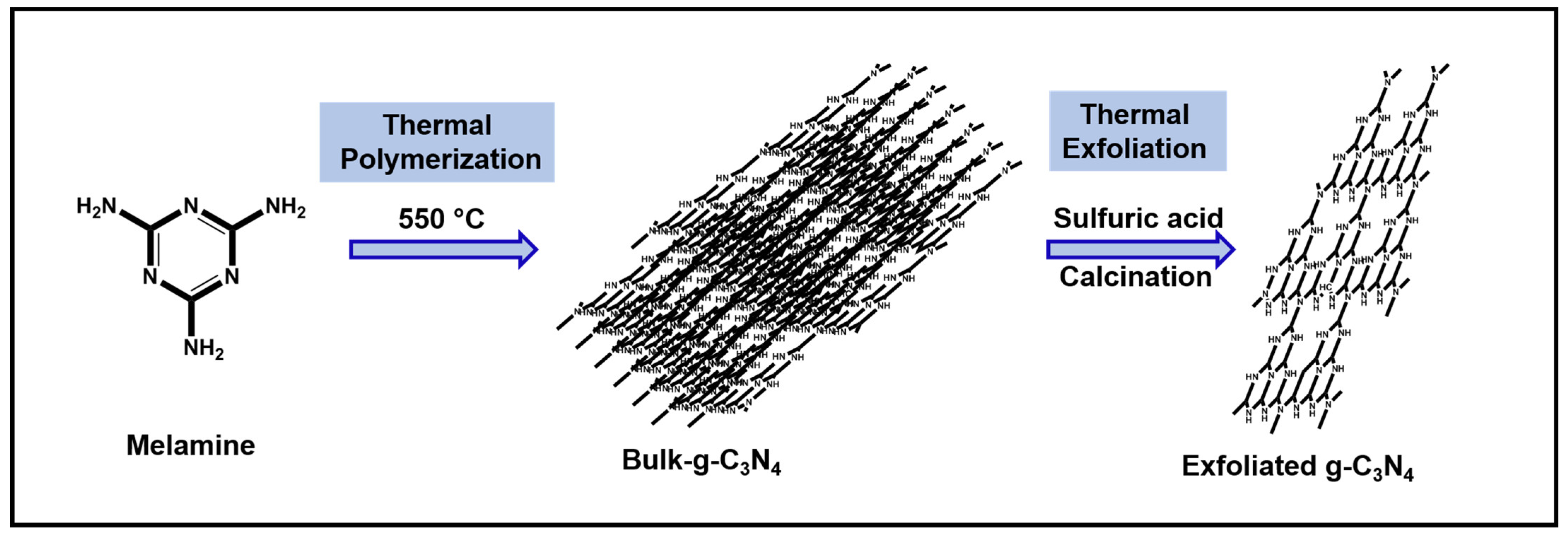
2.2. Synthesis of g-C3N4

Initially, the bulk-graphitic carbon nitride (Bulk g-C3N4) was prepared under atmospheric air conditions by the thermal polymerization of melamine. The temperature was set to 550 °C in a muffle furnace and 6 g of melamine was placed in an alumina crucible for 4 h. After that, the sample was cooled at room temperature. Next, the obtained solid material was ground using a mortar, forming a yellow-colored powder, and the final yield was 3.94 g. During the thermal treatment, due to the decomposition of ammonia, the color changed from intense white to pale yellow. The resultant sample was named bulk-C3N4. Secondly, the exfoliated graphitic carbon nitride sheets (g-C3N4) were prepared via the thermal exfoliation method using prepared bulk g-C3N4. In detail, to the 2 g of prepared bulk g-C3N4, 20 mL of H2SO4 was added and stirred for 12 h. Then 200 mL of deionized water was added to the mixture. After that, the resultant mixture was washed with deionized water until it reached a neutral pH. The faint yellow-colored sample was dried at 60 °C for 12 h in a hot air oven. Finally, the faint yellow-colored powder was poured into an alumina boat, and this boat was placed into a tubular furnace at 550 °C (2 °C/min heating rate, for 4 h, under a nitrogen gas flow), and the porous sheet of exfoliated g-C3N4 was obtained [14] (Figure 1).

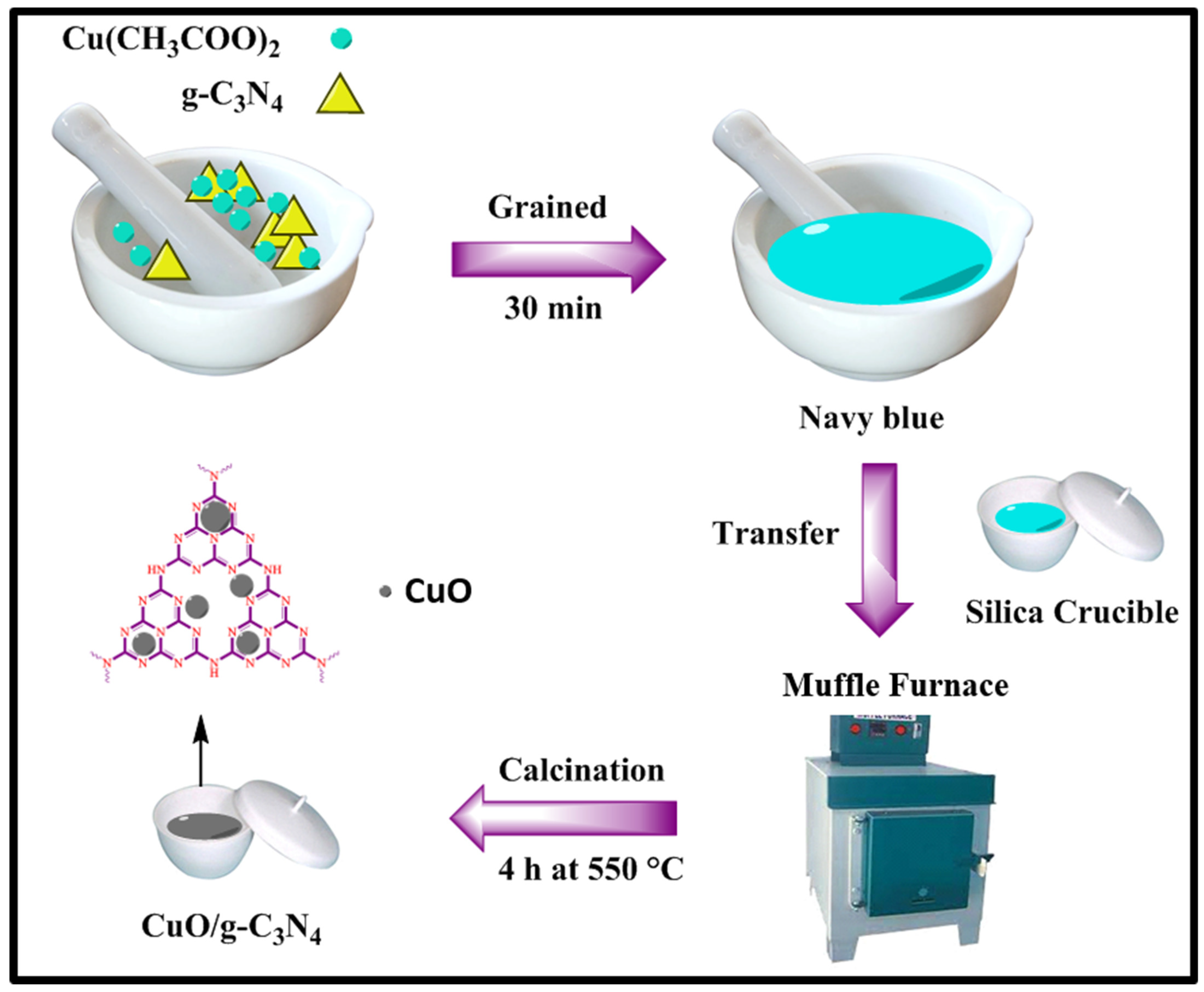
2.3. Synthesis of Cupric Oxide Loaded g-C3N4 Nanocomposites

Synthesis of cupric oxide (CuO) loaded graphitic carbon nitride (g-C3N4) nanocomposites by the direct heating process of the dry synthesis method. In the detailed protocol, various amounts of g-C3N4 and copper acetate compounds were grained for 30 min. No solvent or capping agents were used in this method. The grained mixture was navy-blue in color. Then, the solid mixture was taken in a silica crucible and placed in a furnace for 3 h at 550 °C. The mixture was then cooled to room temperature, and we obtained a brown-colored mixture of CuO/g-C3N4 nanocomposites as shown in Figure 2: CuO, 30 wt% CuO/g-C3N4, 40 wt% CuO/g-C3N4, 50 wt% CuO/g-C3N4, 60 wt% CuO/ g-C3N4, and 70 wt% CuO/g-C3N4, respectively. The various compounds were compared to the results of the antimicrobial analysis.

2.4. Characterization Techniques

The phase and crystallographic structure of as-prepared nanocrystals were observed by a Powder X-ray diffractometer (Bruker, D8 Advance). The diffractograms were recorded for 2Ɵ in the range of 5–80°, with a time of 6 s. The SEM images were captured by scanning electron microscope SEM-ZEISS (EVO18)), Carl Zeiss Microscopy GmbH, 07745 Jena, Germany, before applying spectral coating using gold platinum metal (Quorum). The elemental analysis of the catalyst was analyzed by energy dispersive X-ray (EDX; VEGA3 XUM/TESCAN). UV-Visible absorption spectra were analyzed on JASCO (V-670 PC) equipment, with a wavelength range of 200–1000 nm at room temperature. The optical band gap of these materials was estimated using the Tauc equation.

2.5. Anti-Bacterial Activity

2.5.1. Preparation of Inoculum

The antimicrobial properties of the given samples were tested against Gram-positive bacteria (Staphylococcus aureus ATCC 25923, Streptococcus pyogenes ATCC 19615) and Gram-negative bacteria (Escherichia coli ATCC 25922, Pseudomonas aeruginosa ATCC 27853). All bacteria were pre-cultured in Mueller Hinton Broth (MHB) in a rotary shaker at 37 °C for 18 h. Next, each strain was modified at a concentration of 108 cells/mL using the 0.5 McFarland standard [19].

2.5.2. Agar Well Diffusion Method

The fresh bacterial culture was pipetted in a sterile petri dish. Molten-cooled Mueller Hinton Agar (MAH) was then poured into the petri dish and blended well. Upon solidification, wells were made using a sterile cork borer (6 mm in diameter) into the agar plates containing the inoculums. Then, 50 μL of the sample (20 μg/μL concentration) was added to the respective wells. These plates were then incubated at 37 °C for 18 h. After the incubation period, the antibacterial activity was obtained by measuring the zone of inhibition (including the diameter of the wall). Saline was applied as a negative control.

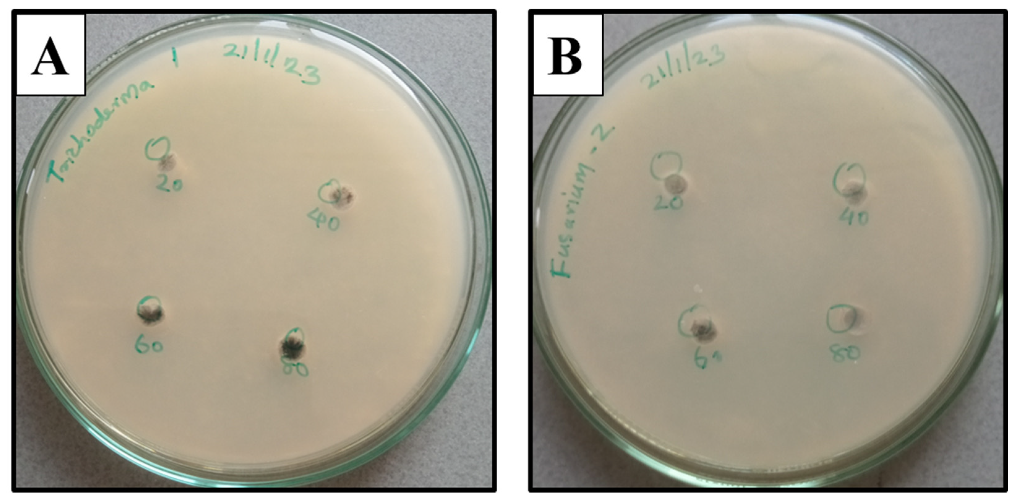
2.6. Fungal Activity

The antifungal activity of the nanocomposites prepared from a facile dry synthesis method was tested against 2 fungi, namely Fusarium oxysporum and Trichoderma viride.

Agar Well Diffusion Method

The fungal cultures Fusarium oxysporum and Trichoderma viride were cultured in potato dextrose broth. PDA agar plates were prepared. Four wells were bored in each plate. Overnight fungal cultures were swabbed into the PDA plates. The prepared compounds were pipetted into the wells in volumes of 20, 40, 60, and 80 μL. The plates were incubated at room temperature for 2–3 days to check the formation of the zone around the well. The plates maintained were kept in triplicate, and zones around the wells were closely monitored.

3. Results

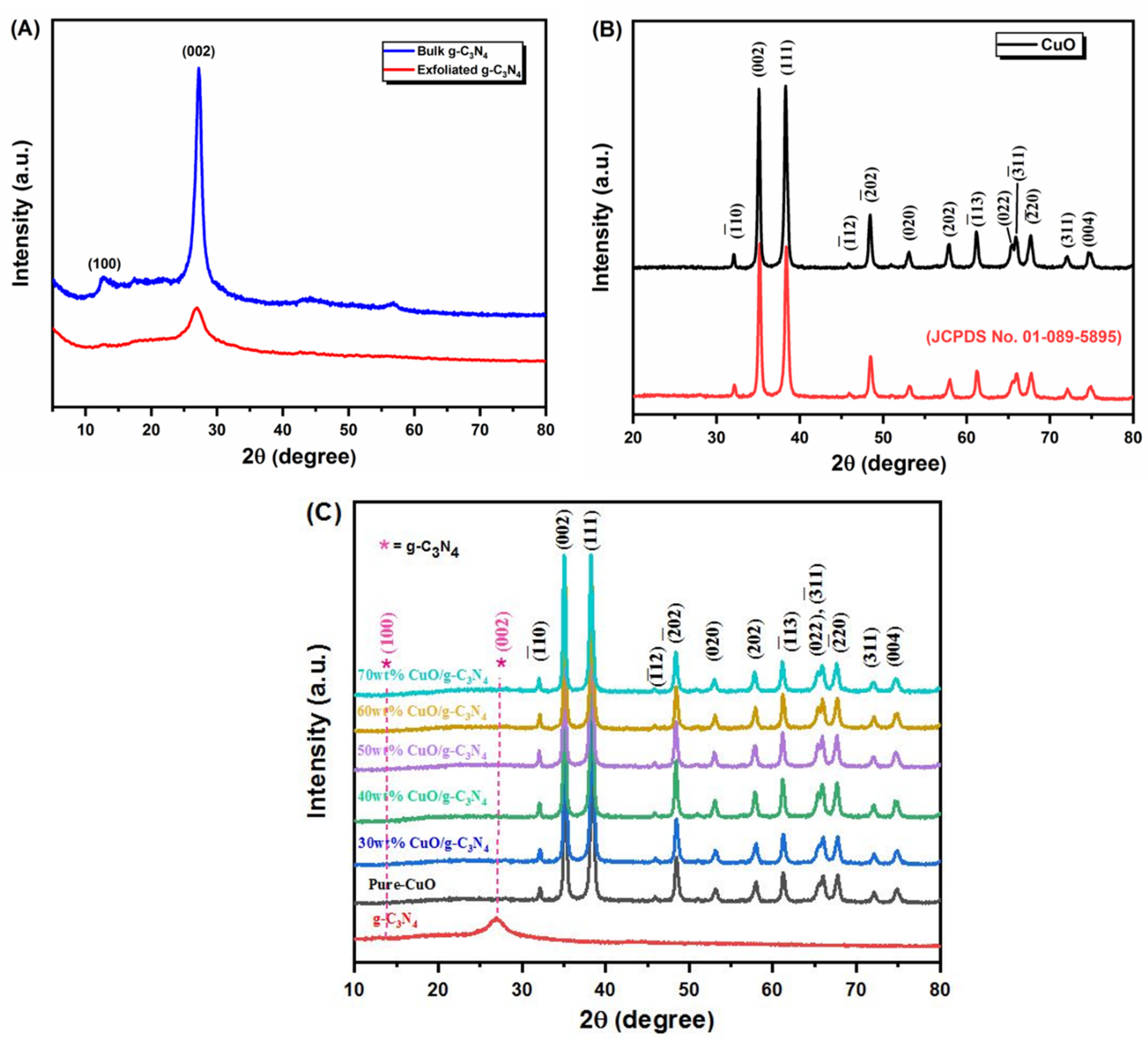
3.1. X-ray Diffraction Method (XRD)

To confirm the structural properties of the synthesized cupric oxide nanoparticles (CuO NPs), bulk and exfoliated g-C3N4, and 30 wt%, 40 wt%, 50 wt%, 60 wt%, 70 wt% of CuO loaded g-C3N4 nanocomposites were evaluated by powder X-ray diffraction (P-XRD) analysis. The XRD pattern of g-C3N4, with hexagonal symmetry, is also presented in Figure 3A. The diffraction weak peak at 13.1° (110) and the strong peak at 27.4° (200) represent the g-C3N4 surfaces. Figure 3B represents the XRD spectra of the CuO nanoparticles. The diffraction spectra of pure CuO NPs at 32.35°, 35.26°, 39.35°, 48.97°, 53.15°, 58.90°, 61.78°, 66.35°, 68.88°, and 72.13° corresponded to the (-110), (111), (111), (202), (020), (202), (113), (004), (220), and (311) crystal facets and the lattice parameters a = 4.68 Å, b = 3.43 Å, c = 5.13 Å, β = 99.47°. Both diffracted peaks of the g-C3N4 and CuO NPs could be seen clearly for all different weight percentages of CuO-loaded g-C3N4 nanocomposites (Figure 3C).

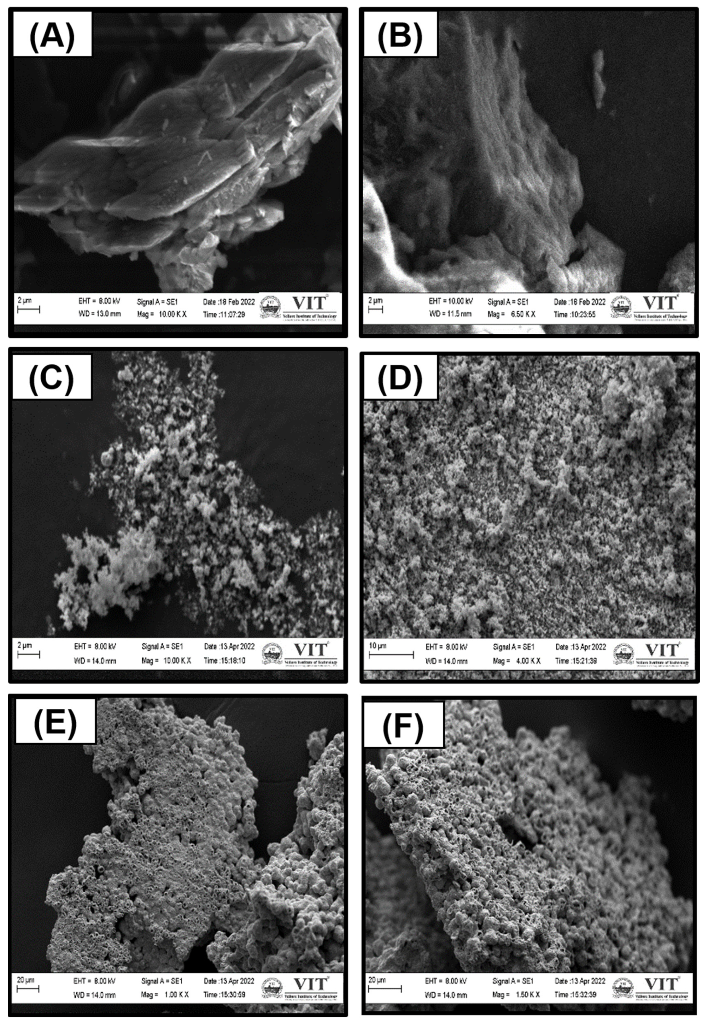
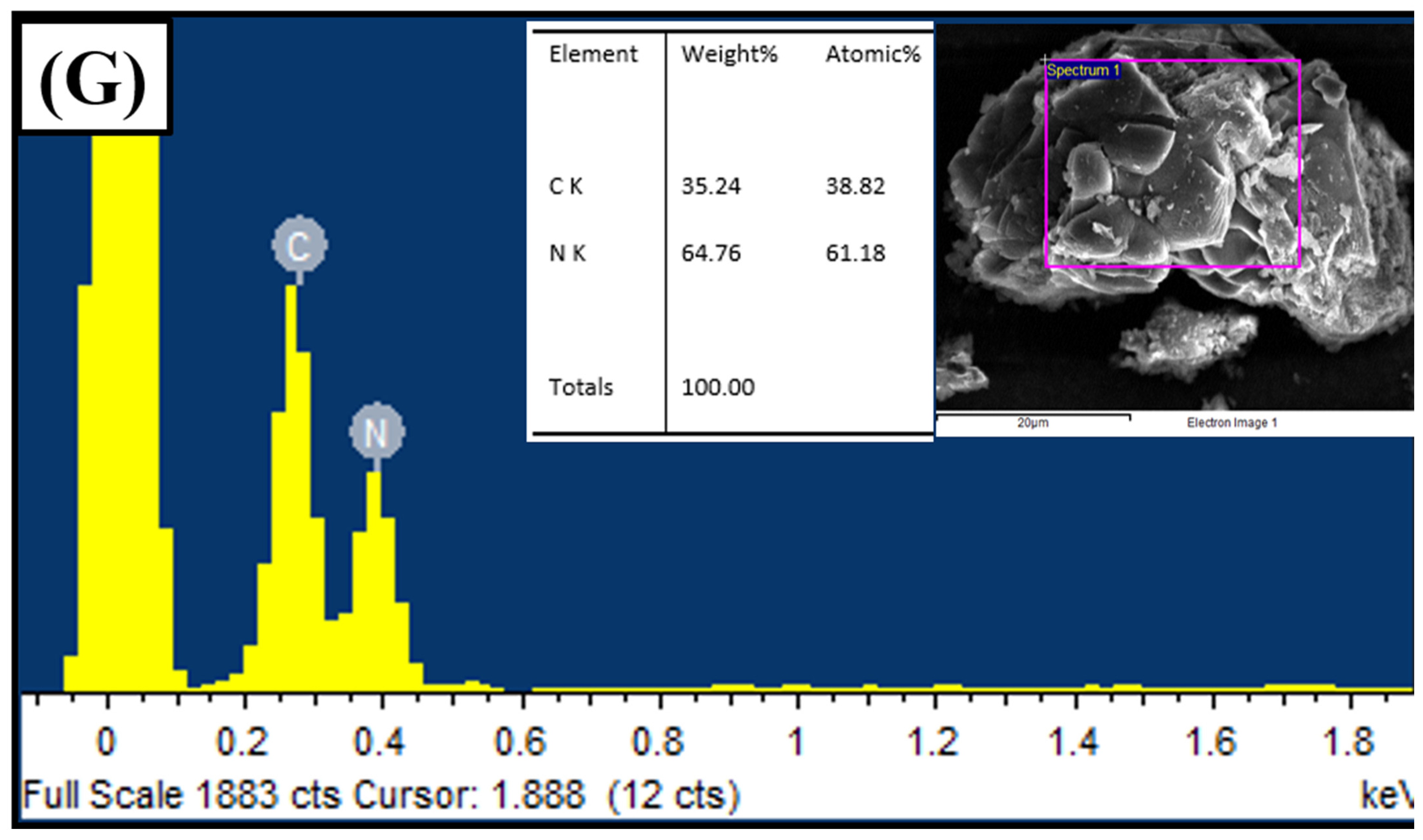
3.2. Scanning Electron Microscopy (SEM)

The morphology and elemental composition of the products were analyzed through SEM and EDS. Figure 4 displays the SEM images of the obtained CuO sample from the dry synthesis method of the copper acetate precursor, the bulk g-C3N4, the exfoliated g-C3N4, and the CuO-loaded g-C3N4 nanocomposite. From Figure 4A,B, the sheet-like structure of bulk g-C3N4 (Figure 4A) and exfoliated g-C3N4 (Figure 4B) is observed. Moreover, thermal treatment resulted in a sheet-like g-C3N4 structure. Figure 4C–F shows the SEM images of the CuO nanoparticles and CuO loaded onto the g-C3N4 nanocomposites. This analysis confirms that the shape of as-prepared CuO NPs shows solid agglomeration, as seen in Figure 4C,D. Furthermore, the successful loading of CuO Nps on exfoliated g-C3N4 is confirmed in Figure 4E,F. The SEM-EDS measurements of the presence of C and N atoms in the nanosheets are shown in Figure 4G.

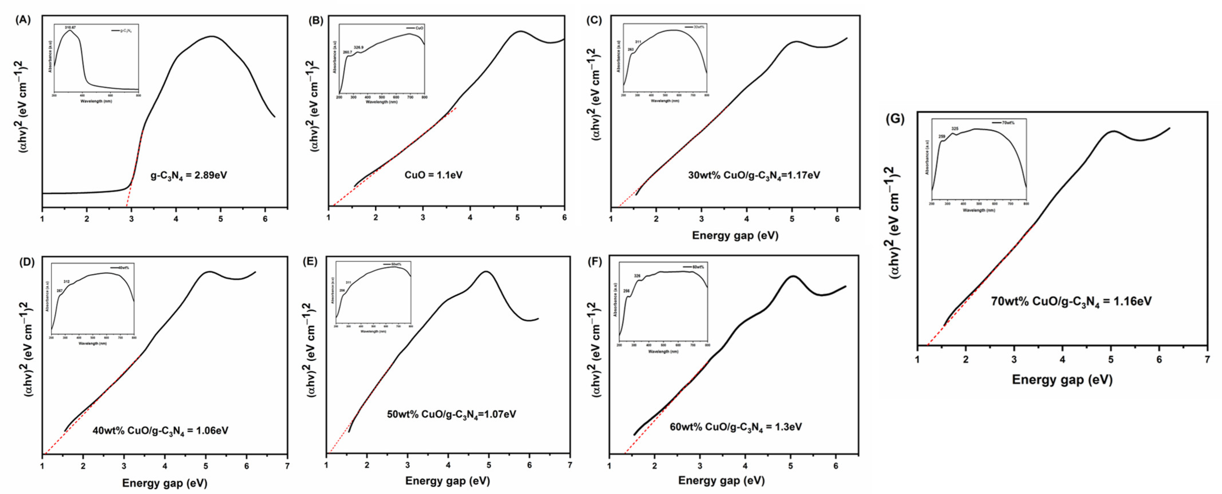
3.3. UV-Visible Diffuse Reflectance Spectroscopy (UV-Vis DRS)

The optical absorption properties of CuO, g-C3N4, and CuO-loaded g-C3N4 are presented in Figure 5. The estimated band gap is obtained by extrapolating the straight portion of (αhv)2 against the hv plot to the point α = 0, i.e., 1.10, 2.89, 1.17, 1.06, 1.03, 1.30, and 1.16 eV for the bare CuO, g-C3N4, 30 wt% CuO loaded g-C3N4, 40 wt% CuO loaded g-C3N4, 50 wt% CuO loaded g-C3N4, 60 wt% CuO loaded g-C3N4, and 70 wt% CuO loaded g-C3N4, respectively, as shown in Figure 5A–G. The UV-Vis spectrum of the sphere-like structured CuO nanoparticles (inset of Figure 5B) showed two absorptions at 290 and 355 nm. The g-C3N4 and 30 wt% CuO/g-C3N4, 40 wt% CuO/g-C3N4, 50 wt% CuO/g-C3N4, 60 wt% CuO/g-C3N4, 70 wt% CuO/g-C3N4 manifest the absorption maxima in the region of 310 nm and 230–380 nm, respectively (inset of Figure 5A,C–G).

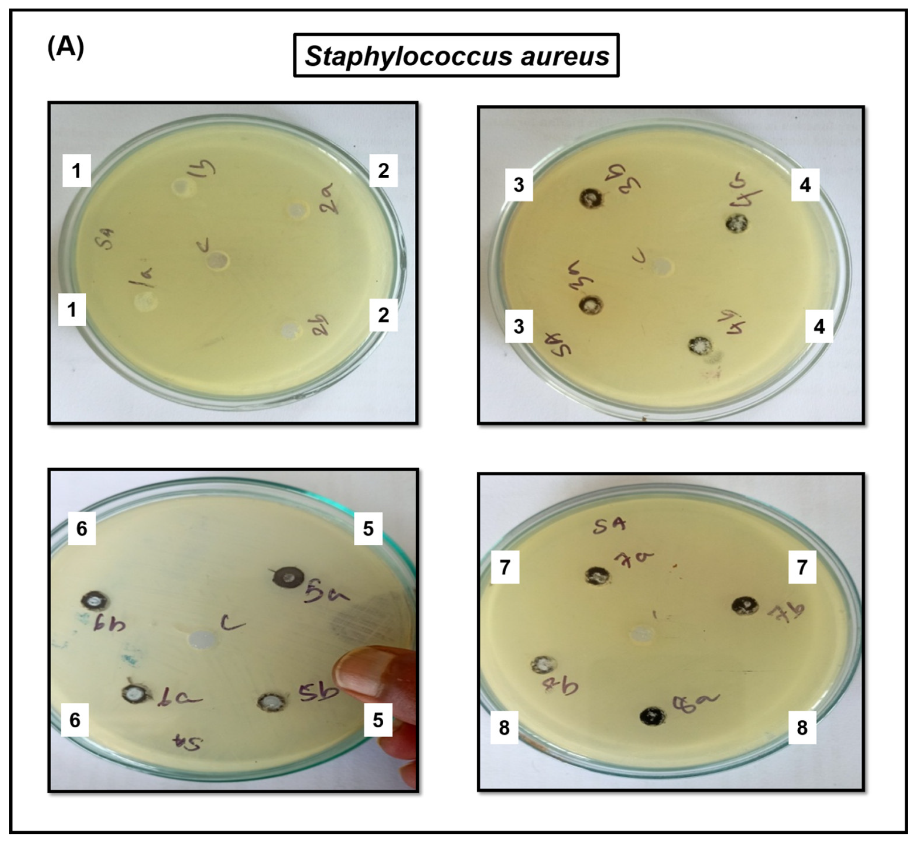
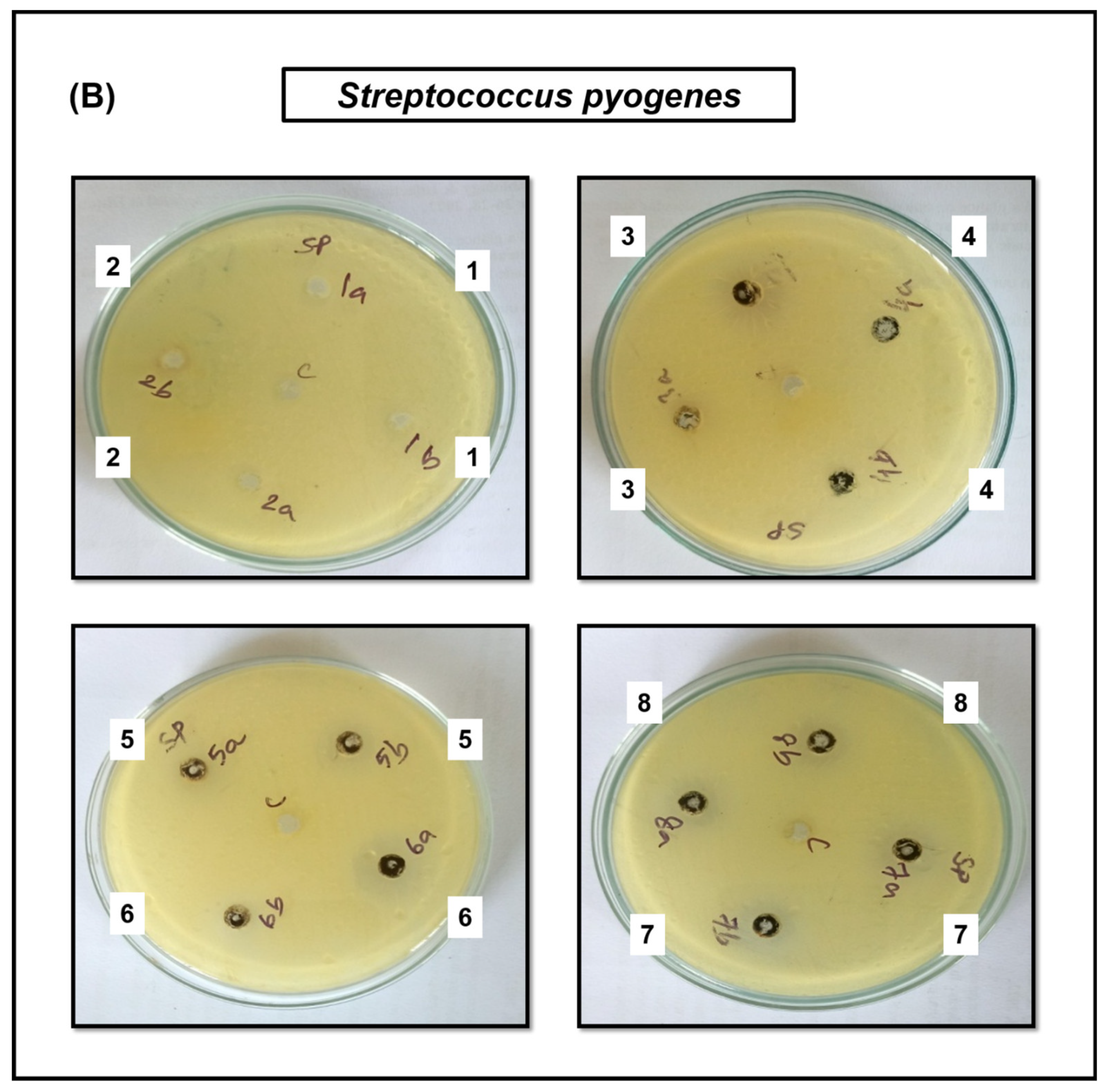
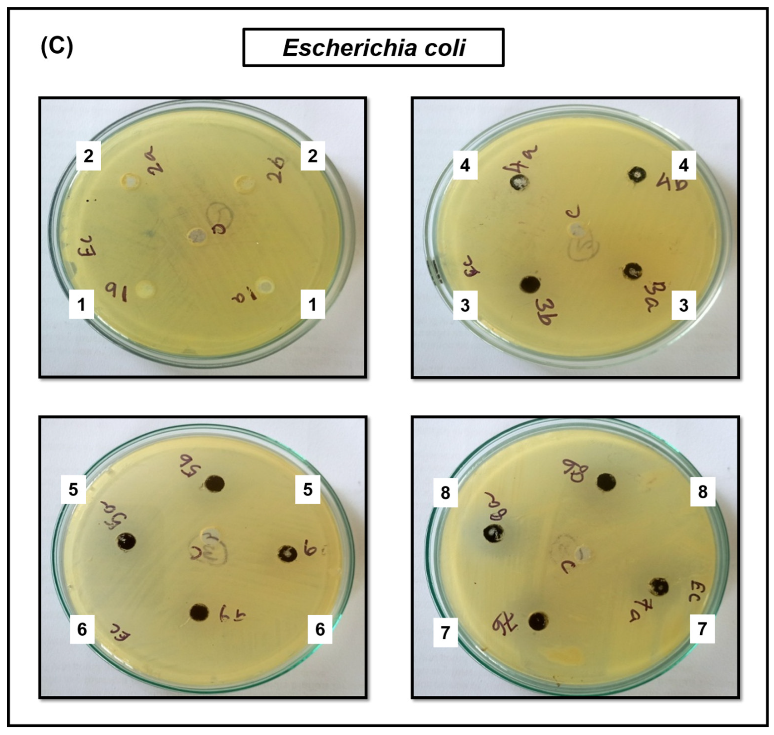
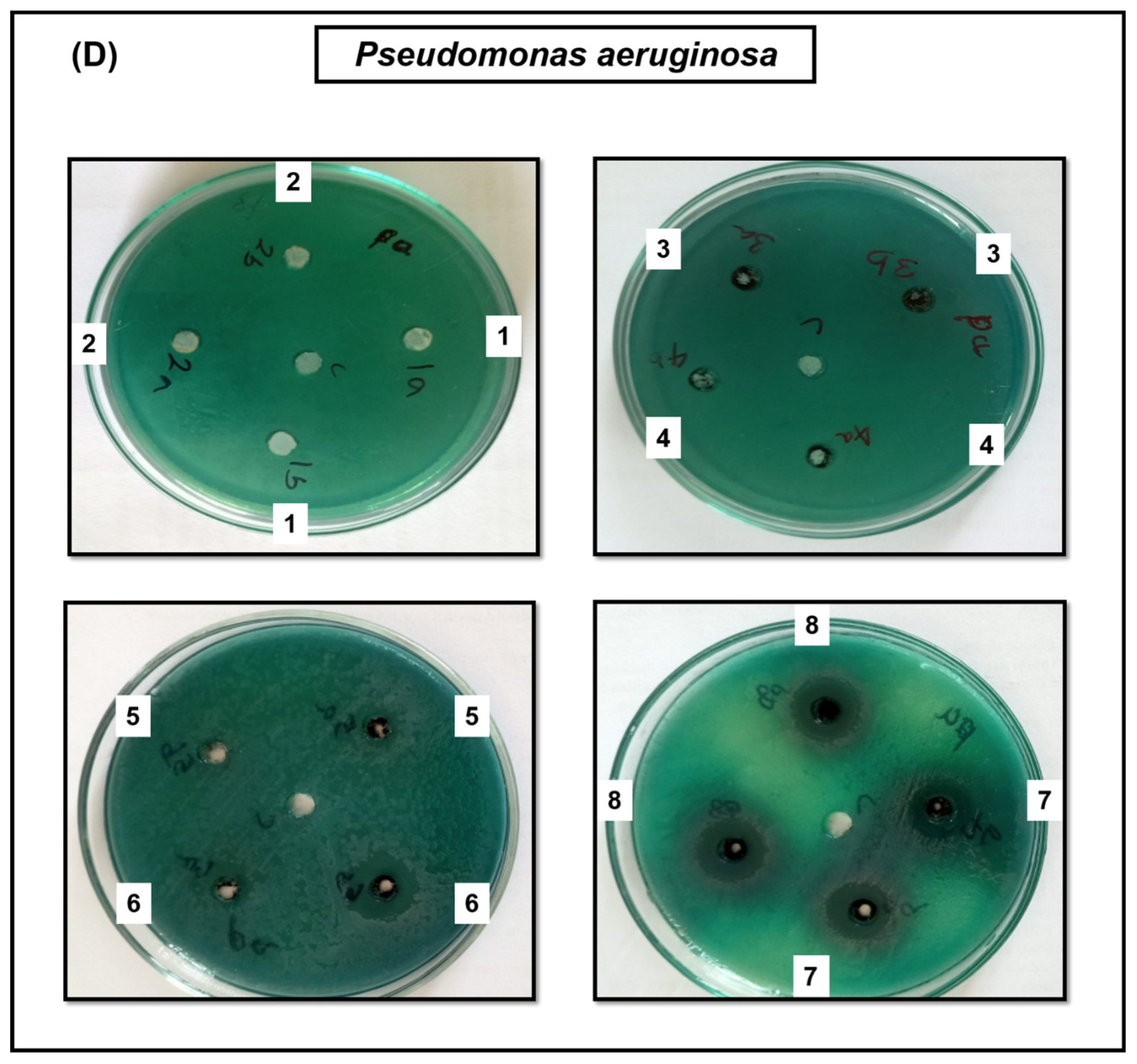
3.4. Antimicrobial Activity
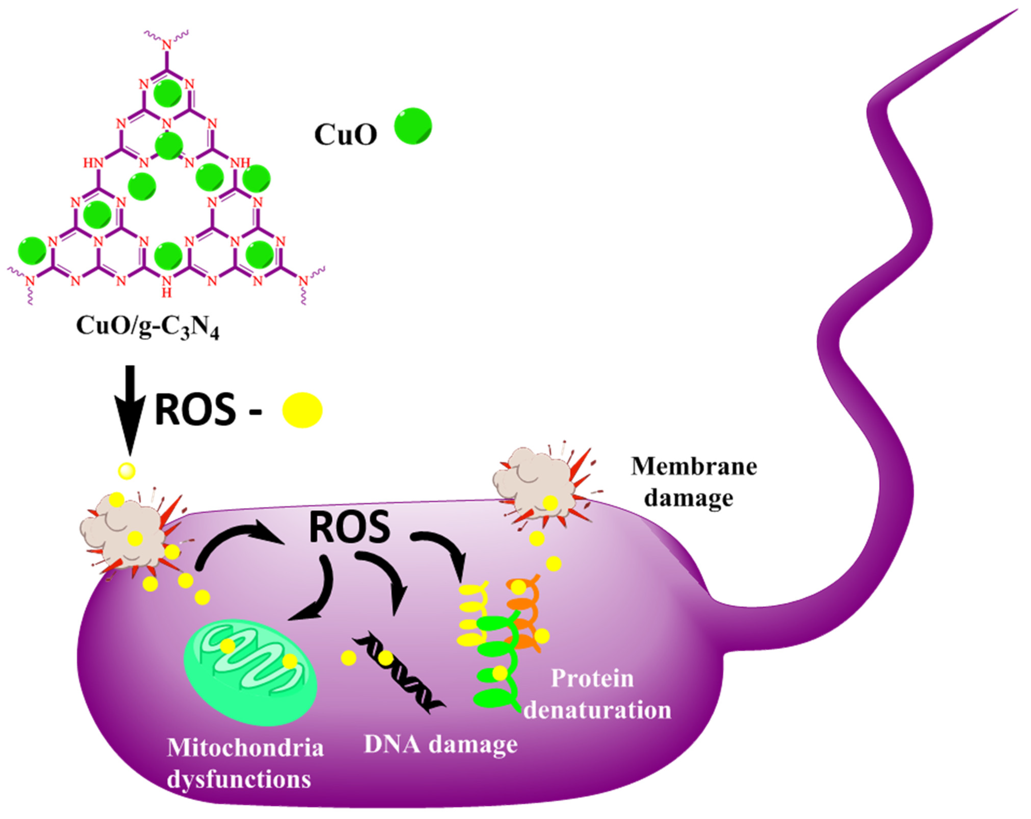
The antibacterial activity of bare CuO, bulk g-C3N4, exfoliated g-C3N4, and CuO-loaded g-C3N4 was analyzed against various bacterial strains of Gram-positive bacteria (Staphylococcus aureus ATCC 25923, Streptococcus pyogenes ATCC 19615) and Gram-negative bacteria (Escherichia coli ATCC 25922, Pseudomonas aeruginosa ATCC 27853). The CuO NPs strains manifest a larger zone of inhibition, and a smaller zone of inhibition is manifested by the resistant strains. In regards to the zone of inhibition, the Gram-positive bacteria exhibited the highest activity toward CuO, while the Gram-negative bacteria manifested the lowest activity [1]. The inactivation rates of Staphylococcus aureus and Streptococcus pyogenes are not significantly different from each other; however, compared to that of E. coli, they are significantly higher. The antibacterial activity exhibited by CuO NPs and different weight percentages of 40 wt%, 50 wt%, 60 wt%, and 70 wt% cupric oxide-loaded g-C3N4 were effective against both Gram-positive and Gram-negative bacteria (Figure 6).
The effect of 70 wt% CuO-loaded g-C3N4 nanocomposites on the development of Fusarium oxysporum and Trichoderma viride as determined by the diameter of the fungal colonies on the samples, non-nanocomposite samples, and other samples of 1 mg/10 mL 70 wt% of CuO loaded g-C3N4 solution with the various concentrations of 20, 40, 60, 80 μL, respectively, after 4 days of incubation. In addition, Figure 7 shows the change in the diameter of fungal colonies for various concentrations of CuO/g-C3N4 solutions. These results show that 70 wt% of CuO-loaded g-C3N4 nanocomposite does not satisfactorily inhibit the fungal species.

4. Discussion

In this study, for the first time, cupric (II) oxide (CuO) nanoparticles loaded with exfoliated graphitic carbon nitride (g-C3N4) nanocomposite also showed antimicrobial activity. The current trending field of nanotechnology is critical and requires environmentally safe methods for the synthesis of nanoparticles. Here, a facile, rapid route and a low-cost approach for the preparation of stable cupric oxide (CuO NPs) nanoparticles and graphitic carbon nitride using a dry synthesis method is reported. The characteristics of the formed cupric (II) oxide loaded on graphitic carbon nitride nanocomposites were confirmed using XRD, UV-Vis (DRS), EDX, and SEM analyses.
The XRD patterns of CuO-loaded g-C3N4 nanosheets demonstrated their sheet-like structure [20,21]. The XRD patterns of CuO-loaded g-C3N4 nanocomposite revealed their crystalline structure. The XRD pattern of g-C3N4 was also presented in Figure 3A. The diffraction weak peak at 13.1° (110) and the strong peak at 27.4° (200) represent the g-C3N4 surface, arising from the in-plane structure of triazine, with the typical interplanar staking peaks of the inner layer structural packing. Figure 3B represents the XRD spectra of the CuO nanospheres particles. The diffraction spectra of pure CuO NPs at 32.35°, 35.26°, 39.35°, 48.97°, 53.15°, 58.90°, 61.78°, 66.35°, 68.88°, and 72.13° corresponded to the (-110), (111), (111), (202), (020), (202), (113), (004), (220), and (311) crystal facets of CuO [22]. In the diffractogram, all the strong and sharp peaks were in accord with the standard JCPDS card no 01-089-5895. The lattice parameters a = 4.68 Å, b = 3.43 Å, c = 5.13 Å, and β = 99.47° of the materials are indicated by a purely monoclinic phase and are additionally confirmed, without any phase impurity present in the synthesis materials. Both diffracted peaks of the g-C3N4 and CuO NPs could be seen clearly for all different weight percentages of the CuO-loaded g-C3N4 nanocomposites. The peak pertaining to g-C3N4 can be found as the mass ratio of g-C3N4 in CuO-loaded g-C3N4 nanocomposites of 30 wt%. While low intensities of g-C3N4 diffraction peaks are observed, this can be attributed to the low content of g-C3N4, which confirms the presence of g-C3N4 and CuO in the CuO-loaded g-C3N4 nanocomposites (Figure 3C).
The morphology and structure of bulk, exfoliated g-C3N4, CuO, and CuO-loaded g-C3N4 nanocomposites were analyzed by SEM. From Figure 3A,B, the sheet-like structure of bulk g-C3N4 (Figure 3A) and exfoliated g-C3N4 (Figure 3B) is clearly observed. Besides, thermal treatment resulted in a sheet-like g-C3N4 structure, and the layers became thinner, with a more detached and several-layer structure characteristic of two-dimensional exfoliated materials in the thermally exfoliated processed nanosheets. Furthermore, scanning electron microscopy analysis confirmed that the as-prepared CuO NPs exhibit nanoparticles of uniform size, as shown in Figure 3C,D. In CuO NPs loaded g-C3N4, Figure 3E,F shows that the nanoparticles were loaded onto the crumpled and rippled surface of the g-C3N4 sheets.
One of the most important electronic parameters for metal oxide nanomaterials is band gap energy. The band gap energy impacts the activity of the molecular adsorption sites and affects adsorption activity. The band gap energy for these samples was calculated from the optical absorption experiments using the Tauc equation, in which
hv) = A × (hvEg)
where the optical absorption coefficient is indicated by α. A is the constant, hv is an incident photon energy, and Eg is the energy gap, respectively. α can be replaced with the absorbance of the sample. The estimated band gap was obtained by extrapolating the straight portion of (αhv)2 against the hv plot to the point α = 0, i.e., 1.10, 2.89, 1.17, 1.06, 1.03, 1.30, and 1.16 eV for the samples of bare CuO NSs, g-C3N4, 30 wt% CuO loaded g-C3N4, 40 wt% CuO loaded g-C3N4, 50 wt% CuO loaded g-C3N4, 60 wt% CuO loaded g-C3N4, and 70 wt% CuO loaded g-C3N4, respectively, as shown in Figure 5A–G. The reason for the absorption was that the 3p state was located at the conduction band in the g-C3N4, resulting in a decrease in the band gap. Its band gap energy is 2.89 eV, as shown in Figure 5A. This indicates that g-C3N4 enhances the visible light utilization ability. The UV-Vis spectrum of the sphere-like structured CuO nanoparticles (inset of Figure 5B) showed two absorptions at 290 and 355 nm. The g-C3N4 and 30 wt% CuO/g-C3N4, 40 wt% CuO/g-C3N4, 50 wt% CuO/g-C3N4, 60 wt% CuO/g-C3N4, and 70 wt% CuO/g-C3N4 manifest the absorption maxima in the region of 310 nm and 230–380 nm, respectively (inset of Figure 5A,C–G).
The antibacterial activity of various nanoparticles for various bacteria using a well diffusion assay is shown in Figure 6A–D. The results illustrated an increase in CuO weight percentage directly proportionally to the antibacterial activity. The CuO NPs strains manifest a larger zone of inhibition, and a smaller zone of inhibition is manifested by the resistant strains. In regards to the zone of inhibition, the Gram-positive bacteria exhibited the highest activity toward CuO, while the Gram-negative bacteria manifest the lowest activity. The inactivation rates of Staphylococcus aureus and Streptococcus pyogenes are not significantly different from each other; however, compared to that of E. coli, they are significantly higher. These results indicated that Gram-positive bacteria are more resistant to the antibacterial activity of CuO-loaded g-C3N4 than Gram-negative bacteria, such as Escherichia coli and Pseudomonas aeruginosa [21]. The antibacterial activity results for CuO-loaded g-C3N4 indicated a superior resistance to Gram-positive bacteria compared to Gram-negative bacteria such as Escherichia coli and Pseudomonas aeruginosa [1,23,24,25]. These results demonstrated that susceptibility to inactivation is dependent upon the microorganism, as shown in Table 1. Future work is necessary to draw firm conclusions about the improved g-C3N4 inactivation performance for Gram-negative over Gram-positive microorganisms.
The photocatalytic activities are depended on the amount of active radical species (such as h+, e-, ·OH, ·O2-) produced in the reactions [21]. Previous studies confirmed that radicals play important roles in photoreactions. Figure 8 shows an increase in active metal oxide, indicating that g-C3N4 under light irradiation can easily produce ·OH, due to the oxidation between H+ and H2O. Subsequently, the ·OH reacts with the bacterial cell membrane and cleaves the linkages. It is commonly known that g-C3N4 is a photocatalytic agent; therefore, the application of light and well-produced radicals, such as ·O2- and ·OH, play important roles in the photocatalytic process [14,15,16,20,26]. This compound is highly active under light irradiation, but in this study, dark conditions were maintained. However, it is still moderately active against Gram-positive and Gram-negative bacteria, compared to metal oxide nanoparticles [27,28], but CuO/g-C3N4 nanocomposites exhibit good antibacterial activity. Finally, the bacteria cell was distorted. If its cell membrane is damaged, the intracellular contents (such as DNA, protein, and mitochondria) can leak from the extracellular suspensions of the cell, resulting in the destruction of the cell structure.
In previous reports, g-C3N4 exhibited excellent activity in visible light irradiation due to the production of ·O2- and ·OH active radical species in photocatalytic disinfection [27]. The g-C3N4 exhibits good photoelectric properties and band structure. In our case, however, since this study was performed under room conditions in vitro, the results showed that fungal growth was not inhibited [21,29,30]. Thus, future studies should be conducted in such a way that a fungal species should be subjected to photocatalytic irradiation [1]. Here, this study was not performed in the presence of sunlight. Under lab conditions, the pathogenic fungi (Fusarium oxysporum and Trichoderma viride) were allowed to interact with 1 mg/mL in different concentrations of 20, 40, 60, and 80 μL (triplicates), and the compounds at these particular concentrations did not inhibit the fungi in the presence of this CuO/g-C3N4 nanocomposite [31,32]. This is because g-C3N4 masks the effect of the CuO molecules and does not inhibit fungi. According to Lin et al., g-C3N4 responds to nitrogen fixation and acts as a foliar fertilizer [26]. Since effective nitrogen fixation results in prominent nitrogen fixations for a good yield, as well as predominant protein synthesis, the result is the growth of fungi, rather than fungal inhibition. Therefore, further studies should be conducted in such a way that the CuO/g-C3N4 compounds are subjected to photocatalytic exposure in the presence of sunlight. In this way, the inhibition of fungi may be possible for these molecules.

5. Conclusions

The various biological processes, including apoptosis, anti-angiogenesis, oxidative stress, chemotherapy, and inflammation, are modulated by inorganic nanoparticles. In this study, cupric (II) oxide (CuO) nanoparticles loaded with exfoliated graphitic carbon nitride (g-C3N4) nanocomposite assisted in the suppression or inhibition of bacterial growth and the interaction of fungi. The current trending field of nanotechnology is critical and requires environmentally safe methods for the synthesis of nanoparticles. Here, a facile, rapid route and a low-cost approach for the preparation of stable cupric oxide (CuO) nanospheres and graphitic carbon nitride using a dry synthesis method was reported. The characteristics of the formed copper (II) oxide loaded on graphitic carbon nitride nanocomposites were confirmed using XRD, UV-Vis (DRS), EDX, and SEM analyses. The XRD patterns of CuO-loaded g-C3N4 nanosheets demonstrated the sheet-like structure. The XRD patterns of CuO-loaded g-C3N4 nanocomposite revealed the crystalline structure. The SEM images of the synthesis CuO-loaded g-C3N4 nanocomposite morphology shows a two-dimensional sheet-like structure. The antibacterial activity was exhibited by CuO NPs, with different weight percentages of 40 wt%, 50 wt%, 60 wt%, and 70 wt% cupric oxide-loaded g-C3N4 against both Gram-positive and Gram-negative bacteria. The synthesized bulk g-C3N4, exfoliated g-C3N4, and lower weight percentages of the nanocomposite (30 wt%) showed low antibacterial activity. The optimum dose of 70wt % CuO-loaded g-C3N4 showed high antibacterial activity but did not exhibit satisfactory activity in antifungal studies conducted in dark fields. This is because g-C3N4 masks the effect of CuO molecules and does not inhibit fungi. The scope of using CuO-loaded g-C3N4 nanocomposites as antimicrobials needs to be further explored in the presence of sunlight.

Author Contributions

R.L.P.—methodology, analysis, writing—original draft preparation; S.K.—methodology, writing—review and editing; B.K.—methodology, writing—review and editing; R.S.—methodology, writing—review and editing; R.V.—methodology, antibacterial activity, anti-angiogenesis analysis, and writing—review, and editing; S.G.B.—design, conceptualization, writing—review, and editing. All authors have read and agreed to the published version of the manuscript.

Funding

This work was supported by the VIT School of Agricultural Innovations and Advanced Learning (VAIAL), Vellore Institute of Technology, Vellore, India.

Institutional Review Board Statement

The article was reviewed by the institutions.

Informed Consent Statement

Not applicable.

Data Availability Statement

Not applicable.

Acknowledgments

The authors wish to thank VIT management for characterization support and infrastructure. S.G.B. thanks VIT Seed Grand (SG20210159) for financial support. V.R. acknowledges VIT for financial support to perform biology studies. R.L.P. acknowledges VIT for providing the fellowship grant.

Conflicts of Interest

The authors declare no competing financial support.

References

  1. Shinde, R.S.; More, R.A.; Adole, V.A.; Koli, P.B.; Pawar, T.B.; Jagdale, B.S.; Desale, B.S.; Sarnikar, Y.P. Design, fabrication, antitubercular, antibacterial, antifungal and antioxidant study of silver doped ZnO and CuO nano candidates: A comparative pharmacological study. Curr. Opin. Green Sustain. Chem. 2021, 4, 100138. [Google Scholar] [CrossRef]
  2. Peng, Y.; Li, S.J.; Yan, J.; Tang, Y.; Cheng, J.P.; Gao, A.J.; Yao, X.; Ruan, J.J.; Xu, B.L. Research progress on phytopathogenic fungi and their role as biocontrol agents. Front. Microbiol. 2021, 12, 670135. [Google Scholar] [CrossRef]
  3. Gomes, E.V.; Costa, M.d.N.; de Paula, R.G.; Ricci de Azevedo, R.; da Silva, F.L.; Noronha, E.F.; José Ulhoa, C.; Neves Monteiro, V.; Elena Cardoza, R.; Gutiérrez, S. The Cerato-Platanin protein Epl-1 from Trichoderma harzianum is involved in mycoparasitism, plant resistance induction and self cell wall protection. Sci. Rep. 2015, 5, 17998. [Google Scholar] [CrossRef] [Green Version]
  4. Verma, A.; Anand, P.; Kumar, S.; Fu, Y.-P. Cu-cuprous/cupric oxide nanoparticles towards dual application for nitrophenol conversion and electrochemical hydrogen evolution. Appl. Surf. Sci. 2022, 578, 151795. [Google Scholar] [CrossRef]
  5. Giridasappa, A.; Rangappa, D.; Shanubhoganahalli Maheswarappa, G.; Marilingaiah, N.R.; Kagepura Thammaiah, C.; Shareef, I.M.; Kanchugarakoppal Subbegowda, R.; Doddakunche Shivaramu, P. Phytofabrication of cupric oxide nanoparticles using Simarouba glauca and Celastrus paniculatus extracts and their enhanced apoptotic inducing and anticancer effects. Appl. Nanosci. 2021, 11, 1393–1409. [Google Scholar] [CrossRef]
  6. Jithul, K.; Samra, K.S. Cupric Oxide based Supercapacitors: A Review. In Proceedings of the Journal of Physics: Conference Series, Wuhan, China, 18 September 2022; p. 012120. [Google Scholar]
  7. Sumathi, S.; Nehru, M.; Vidya, R. Synthesis, characterization and effect of precipitating agent on the antibacterial properties of cobalt ferrite nanoparticles. Trans. Indian Ceram. Soc. 2015, 74, 79–82. [Google Scholar] [CrossRef]
  8. Cuong, H.N.; Pansambal, S.; Ghotekar, S.; Oza, R.; Hai, N.T.T.; Viet, N.M.; Nguyen, V.-H. New frontiers in the plant extract mediated biosynthesis of copper oxide (CuO) nanoparticles and their potential applications: A review. Environ. Res. 2022, 203, 111858. [Google Scholar] [CrossRef]
  9. Rangarajan, S.; Verekar, S.; Deshmukh, S.K.; Bellare, J.R.; Balakrishnan, A.; Sharma, S.; Vidya, R.; Chimote, G. Evaluation of anti-bacterial activity of silver nanoparticles synthesised by coprophilous fungus PM0651419. IET Nanobiotechnol. 2018, 12, 106–115. [Google Scholar] [CrossRef]
  10. Hafeez, H.Y.; Lakhera, S.K.; Bellamkonda, S.; Rao, G.R.; Shankar, M.; Bahnemann, D.; Neppolian, B. Construction of ternary hybrid layered reduced graphene oxide supported g-C3N4-TiO2 nanocomposite and its photocatalytic hydrogen production activity. Int. J. Hydrog. Energy 2018, 43, 3892–3904. [Google Scholar] [CrossRef]
  11. Moradi, M.; Hasanvandian, F.; Isari, A.A.; Hayati, F.; Kakavandi, B.; Setayesh, S.R. CuO and ZnO co-anchored on g-C3N4 nanosheets as an affordable double Z-scheme nanocomposite for photocatalytic decontamination of amoxicillin. Appl. Catal. 2021, 285, 119838. [Google Scholar] [CrossRef]
  12. Thiyagarajan, K.; Samuel, S.; Kumar, P.S.; Babu, S.G.J.B.o.M.S. C3N4 supported on chitosan for simple and easy recovery of visible light active efficient photocatalysts. Bull. Mater. Sci. 2020, 43, 1–7. [Google Scholar] [CrossRef]
  13. Liu, Q.; Wang, X.; Yang, Q.; Zhang, Z.; Fang, X. Mesoporous g-C3N4 nanosheets prepared by calcining a novel supramolecular precursor for high-efficiency photocatalytic hydrogen evolution. Appl. Surf. Sci. 2018, 450, 46–56. [Google Scholar] [CrossRef]
  14. Bhuvaneswari, C.; Babu, S.G. Nanoarchitecture and surface engineering strategy for the construction of 3D hierarchical CuS-rGO/g-C3N4 nanostructure: An ultrasensitive and highly selective electrochemical sensor for the detection of furazolidone drug. J. Electroanal. Chem. 2022, 907, 116080. [Google Scholar] [CrossRef]
  15. Liu, Y.; Shen, S.; Li, Z.; Ma, D.; Xu, G.; Fang, B. Mesoporous g-C3N4 nanosheets with improved photocatalytic performance for hydrogen evolution. Mater. Charact. 2021, 174, 111031. [Google Scholar] [CrossRef]
  16. Wen, J.; Xie, J.; Chen, X.; Li, X. A review on g-C3N4-based photocatalysts. Appl. Surf. Sci. 2017, 391, 72–123. [Google Scholar] [CrossRef]
  17. Li, R.; Ren, Y.; Zhao, P.; Wang, J.; Liu, J.; Zhang, Y. Graphitic carbon nitride (g-C3N4) nanosheets functionalized composite membrane with self-cleaning and antibacterial performance. J. Hazard. Mater. 2019, 365, 606–614. [Google Scholar] [CrossRef] [PubMed]
  18. Sher, M.; Shahid, S.; Javed, M. Synthesis of a novel ternary (g-C3N4 nanosheets loaded with Mo doped ZnO nanoparticles) nanocomposite for superior photocatalytic and antibacterial applications. J. Photochem. Photobiol. B Biol. 2021, 219, 112202. [Google Scholar] [CrossRef]
  19. Gonelimali, F.D.; Lin, J.; Miao, W.; Xuan, J.; Charles, F.; Chen, M.; Hatab, S.R. Antimicrobial properties and mechanism of action of some plant extracts against food pathogens and spoilage microorganisms. Front. Microbiol. 2018, 9, 1639. [Google Scholar] [CrossRef] [PubMed]
  20. Induja, M.; Sivaprakash, K.; Karthikeyan, S.J.M.R.B. Facile green synthesis and antimicrobial performance of Cu2O nanospheres decorated g-C3N4 nanocomposite. Mater. Res. Bull. 2019, 112, 331–335. [Google Scholar] [CrossRef]
  21. Kublik, N.; Gomes, L.E.; Plaça, L.F.; Lima, T.H.; Abelha, T.F.; Ferencz, J.A.; Caires, A.R.; Wender, H.J.P. Metal-free g-C3N4/nanodiamond heterostructures for enhanced photocatalytic pollutant removal and bacteria photoinactivation. Photochem. 2021, 1, 302–318. [Google Scholar] [CrossRef]
  22. Bai, B.; Saranya, S.; Dheepaasri, V.; Muniasamy, S.; Alharbi, N.S.; Selvaraj, B.; Undal, V.S.; Gnanamangai, B.M. Biosynthesized copper oxide nanoparticles (CuO NPs) enhances the anti-biofilm efficacy against K. pneumoniae and S. aureus. J. King Saud Univ.-Sci. 2022, 34, 102120. [Google Scholar] [CrossRef]
  23. Selvaraj, S.P. Enhanced surface morphology of copper oxide (CuO) nanoparticles and its antibacterial activities. Mater. Today Proc. 2022, 50, 2865–2868. [Google Scholar] [CrossRef]
  24. Du, B.D.; Phu, D.V.; Quoc, L.A.; Hien, N.Q. Synthesis and investigation of antimicrobial activity of Cu2O nanoparticles/zeolite. J. Nanoparticles Res. 2017, 2017, 705684. [Google Scholar] [CrossRef] [Green Version]
  25. Salem, S.S.; Ali, O.M.; Reyad, A.M.; Abd-Elsalam, K.A.; Hashem, A.H. Pseudomonas indica-mediated silver nanoparticles: Antifungal and antioxidant biogenic tool for suppressing mucormycosis fungi. J. Fungus. 2022, 8, 126. [Google Scholar] [CrossRef]
  26. Liu, Y.; Huang, X.; Yu, Z.; Yao, L.; Guo, S.; Zhao, W.J.C. Environmentally Friendly Non-Metal Solar Photocatalyst C3N4 for Efficient Nitrogen Fixation as Foliar Fertilizer. ChemistrySelect 2020, 5, 7720–7727. [Google Scholar] [CrossRef]
  27. Kalia, A.; Abd-Elsalam, K.A.; Kuca, K. Zinc-based nanomaterials for diagnosis and management of plant diseases: Ecological safety and future prospects. J. Fungus 2020, 6, 222. [Google Scholar] [CrossRef] [PubMed]
  28. Chen, J.; Mao, S.; Xu, Z.; Ding, W. Various antibacterial mechanisms of biosynthesized copper oxide nanoparticles against soilborne Ralstonia solanacearum. RSC Adv. 2019, 9, 3788–3799. [Google Scholar] [CrossRef] [Green Version]
  29. Soni, A.; Kaushal, D.; Kumar, M.; Sharma, A.; Maurya, I.K.; Kumar, S. Synthesis, characterizations and antifungal activities of copper oxide and differentially doped copper oxide nanostructures. Mater. Today Proc. 2022, in press. [Google Scholar] [CrossRef]
  30. Kumar, P.; Inwati, G.K.; Mathpal, M.C.; Ghosh, S.; Roos, W.; Swart, H.C. Defects induced enhancement of antifungal activities of Zn doped CuO nanostructures. Appl. Surf. Sci. 2021, 560, 150026. [Google Scholar] [CrossRef]
  31. Zaki, S.A.; Ouf, S.A.; Albarakaty, F.M.; Habeb, M.M.; Aly, A.A.; Abd-Elsalam, K.A. Trichoderma harzianum-mediated ZnO nanoparticles: A green tool for controlling soil-borne pathogens in cotton. J. Fungus 2021, 7, 952. [Google Scholar] [CrossRef] [PubMed]
  32. Alghuthaymi, M.A.; Abd-Elsalam, K.A.; AboDalam, H.M.; Ahmed, F.K.; Ravichandran, M.; Kalia, A.; Rai, M.J. Trichoderma: An eco-friendly source of nanomaterials for sustainable agroecosystems. J. Fungus 2022, 8, 367. [Google Scholar] [CrossRef] [PubMed]
Figure 1. Simplified representation of the production of exfoliated g-C3N4.
Jof 09 00310 g001
Figure 2. CuO/g-C3N4 nanocomposites prepared by dry synthesis method.
Jof 09 00310 g002
Figure 3. XRD diffractogram of (A) bulk g-C3N4 and exfoliated g-C3N4, (B) bare CuO and standard peak, (C) bare CuO and exfoliated g-C3N4, and various weight percentages of CuO loaded g-C3N4.
Jof 09 00310 g003
Figure 4. SEM figure of (A) bulk g-C3N4, (B) exfoliated g-C3N4, (C,D) CuO, (E,F) CuO/g-C3N4 nanocomposites, and (G) EDS spectra of bulk g-C3N4.
Jof 09 00310 g004aJof 09 00310 g004b
Figure 5. UV-DRS spectra exhibit band gap energy of exfoliated g-C3N4 (A), CuO (B), and CuO loaded g-C3N4 (CG); (inset: absorption peak). (Bold black line represented by UV-DRS and dotted line noted by bandgap measurement line).
Jof 09 00310 g005
Figure 6. Antimicrobial activity of bulk g-C3N4 (1), exfoliated g-C3N4 (2), CuO (3), and different weight percentages of CuO loaded g-C3N4 nanocomposite, 30 wt% (4), 40 wt% (5), 50 wt% (6), 60 wt% (7), and 70 wt% (8) against Staphylococcus aureus (A), Streptococcus pyogenes (B), Escherichia coli (C), and Pseudomonas aeruginosa (D).
Jof 09 00310 g006aJof 09 00310 g006bJof 09 00310 g006cJof 09 00310 g006d
Figure 7. Antifungal activity of 70 wt% of CuO loaded g-C3N4 nanocomposite solution with the various concentrations of 20, 40, 60, and 80 μL (A), Trichoderma viride, and (B) Fusarium oxysporum.
Jof 09 00310 g007
Figure 8. Plausible mechanisms of antimicrobial activity.
Jof 09 00310 g008
Table 1. Antimicrobial activity of cupric oxide/g-C3N4nanocomposite.
SamplesS. aureus (mm)S. pyogenes (mm)E. coli (mm)P. aeruginosa (mm)
Bulk-g-C3N4RRRR
Exfoliated g-C3N4RRRR
CuO81268
30 wt% CuO/g-C3N4RRRR
40 wt% CuO/g-C3N416161012
50 wt% CuO/g-C3N416161014
60 wt% CuO/g-C3N418181214
70 wt% CuO/g-C3N418181214
(R)—resistant.
Disclaimer/Publisher’s Note: The statements, opinions and data contained in all publications are solely those of the individual author(s) and contributor(s) and not of MDPI and/or the editor(s). MDPI and/or the editor(s) disclaim responsibility for any injury to people or property resulting from any ideas, methods, instructions or products referred to in the content.

