Article

Pseudomonas aeruginosa Strain 91: A Multifaceted Biocontrol Agent against Banana Fusarium Wilt

1
Institute of Biological Sciences and Technology, Guangxi Academy of Sciences, Nanning 530007, China
2
Guangxi Key Laboratory of Sugarcane Genetic Improvement, Sugarcane Research Institute, Guangxi Academy of Agricultural Sciences, Nanning 530007, China
3
State Key Laboratory of Mycology, Institute of Microbiology, Chinese Academy of Sciences, Beijing 100101, China
*
Authors to whom correspondence should be addressed.
These authors contributed equally to this work.
J. Fungi 2023, 9(11), 1047; https://doi.org/10.3390/jof9111047
Submission received: 10 October 2023 / Revised: 20 October 2023 / Accepted: 23 October 2023 / Published: 25 October 2023
(This article belongs to the Special Issue Plant-Pathogenic Fusarium Species 2.0)

Abstract

:
Banana Fusarium wilt (BFW), caused by the soil-borne fungus Fusarium oxysporum f. sp. cubense (Foc), poses significant threats to banana cultivation. Currently, effective control methods are lacking, and biological control has emerged as a possible strategy to manage BFW outbreaks. In this investigation, 109 bacterial strains were isolated from the rhizospheric soil surrounding banana plants in search of potent biological agents against Foc. Strain 91 exhibited the highest antifungal activity against the causal agent of Foc and was identified as Pseudomonas aeruginosa through 16S rRNA gene sequencing and scanning electron microscopy (SEM). Elucidation of strain 91’s inhibitory mechanism against Foc revealed a multifaceted antagonistic approach, encompassing the production of bioactive compounds and the secretion of cell wall hydrolytic enzymes. Furthermore, strain 91 displayed various traits associated with promoting plant growth and showed adaptability to different carbon sources. By genetically tagging with constitutively expressing GFP signals, effective colonization of strain 91 was mainly demonstrated in root followed by leaf and stem tissues. Altogether, our study reveals the potential of P. aeruginosa 91 for biocontrol based on inhibition mechanism, adaptation, and colonization features, thus providing a promising candidate for the control of BFW.

1. Introduction

Banana (Musa spp.) is a significant fruit crop within the Musaceae family, extensively cultivated in developing countries. Banana Fusarium wilt (BFW), mainly caused by Fusarium oxysporum f. sp. cubense tropical race 4 (Foc TR4), stands as one of the most devastating diseases, significantly hampering global banana production [1,2]. The pathogen was first identified in Southeast Asia in the early 1990s. By the early 21st century, an endemic outbreak of Foc TR4 had extended to substantial areas in Australia’s northern territories, China, Indonesia, and Malaysia [3]. The fungus is highly stress-resistant and can endure in soil for up to 30 years, resulting in widespread orchard devastation [4]. Foc initially infects the roots and then invades the plant’s vascular tissue, resulting in wilting and eventual plant death [3].
Chemical control methods are not only costly and inefficient but also pose potential risks to public health and the environment. Conversely, biological control, specifically the use of plant growth-promoting rhizobacteria (PGPR), offers a promising alternative to chemical control while circumventing the challenges associated with conventional plant protection systems [5]. In recent years, biological control has garnered significant attention in various pathosystems. The discovery of microorganism-based biological control agents for banana diseases is highly expected [6]. Effective colonization of the rhizosphere and the ability to maintain a substantial population are considered essential prerequisites for the efficacy of PGPR [7]. Thus, it is important to find indigenous biocontrol strains with not only the efficiency of suppressing local pathogenic strains but also the adaptability to local conditions and the ability to compete with in situ microorganisms [8].
The rhizosphere is the region where soil particles come into contact with plant roots, constituting a dynamic and highly complex microbial ecosystem [9]. This environment facilitates intricate interactions between plants and microorganisms, resulting in a distinctive ecosystem characterized by carbon and water cycling, as well as the sequestration of nutrients and minerals. The examination of plant–microbe interactions and the diverse metabolites produced through their co-metabolism is of significant interest. These metabolites serve various functions, such as acting as energy sources and signaling molecules [10,11]. Rhizobacteria inhabiting the rhizosphere have been shown to synthesize a broad array of beneficial compounds [12]. Several studies have demonstrated that certain rhizobacteria possess antimicrobial properties against causal agents of plant diseases [13,14,15,16]. Healthy plant rhizosphere soil serves as a valuable source of PGPR [17], which readily colonize plant roots and promote plant development through various mechanisms [18]. These mechanisms include phosphate solubilization, the production of plant growth regulators, nitrogen fixation, ethylene metabolism, and the indirect enhancement of disease resistance via the production of antimicrobial metabolites or siderophores that inhibit harmful microorganisms [19]. Consequently, PGPR is gaining recognition as an environmentally friendly alternative to agrochemicals for sustainable agriculture. PGPR offers several advantages. It can serve as a substitute for non-leguminous agricultural fertilizers, enhance nutrient uptake by banana plants, and efficiently colonize banana root surfaces, where microbial cell density surpasses that in the root hair growth zone [20].
Antifungal rhizobacteria have received extensive attention for the biological control of plant diseases. The Pseudomonas genus encompasses over a hundred species [21], with many native to plant rhizosphere, endosphere, and phyllosphere environments, establishing commensal relationships therein. Some Pseudomonas strains have found application as plant inoculants due to their ability to mitigate the detrimental effects of specific phytopathogens, thereby promoting plant growth and health [22,23]. Several Pseudomonas species, including P. aeruginosa, P. putida, P. chlororaphis, P. syringe, and P. fluorescens, are well-recognized for their capacity to enhance plant development and suppress various plant diseases [23,24]. In Chinese biofertilizers, the primary strains are Bacillus subtilis, Paenibacillus mucilaginosus, B. amyloliquefaciens, B. licheniformis, and B. megaterium [25]. Conversely, various countries employ Pseudomonas-based biofertilizers, including P. fluorescens and P. putida in Vietnam, P. fluorescens in Cuba and Sri Lanka, P. striata in India, and P. azotoformans and P. chlororaphis in Sweden [26]. In recent years, a diverse array of Pseudomonas spp. strains has been explored as antagonists against Foc, offering new insights into the biological management of BFW. Most research in this field has focused on P. fluorescens, while our understanding of P. aeruginosa strains remains limited.
In this study, we isolated Pseudomonas aeruginosa strain 91 from banana rhizospheric soil due to its antagonistic activity against Foc. We investigated its inhibition mechanism, PGP traits, and colonization properties, affirming the potential of strain 91 as a promising candidate for the biological management of BFW.

2. Materials and Methods

2.1. Collection of Soil Samples and Bacteria Isolation

Rhizospheric soil samples were randomly collected from six different banana fields in Long’an county, Nanning city, Guangxi zhuang autonomous region, China. In each field, three healthy banana plants, five months old, were selected, and soil adhering to their roots was carefully gathered upon uprooting, with subsequent removal of root debris through a 2 mm mesh sieve. Samples were stored at 4 °C and processed within 24 h of collection. To isolate bacteria, 10 grams of soil from each sample were individually mixed with 90 mL of saline water (0.85% NaCl) in separate flasks. These suspensions were placed on an orbital shaker at 100 rpm and incubated at 32 °C for 1 h. After incubation, the soil suspensions of all six samples were serially diluted up to 10−6 and 100 µL of each dilution was spread on the Nutrient Agar (NA), Luria-Bertani (LB) Agar, Yeast Mannitol Agar, and Pikovskaya’s Agar media (Table S1) and incubated for 2 days at 32 °C. A total of 109 bacterial colonies were purified for further studies. All pure cultures were preserved in 20% glycerol at −80 °C.

2.2. Antifungal Assay of Isolated Bacterial Strains

The antifungal activity of all isolated strains was evaluated against the pathogenic fungus Foc (isolated by our group and stored in our lab) using a dual-culture method on Potato Dextrose Agar (PDA):Nutrient Agar (NA) at 1:1 (Table S1). Initially, a 10 μL aliquot of pathogen spores at a concentration of 106 spores/mL was aseptically dispensed at the central region of the Petri dish. Subsequently, the experimental strain was inoculated at an approximate distance of 1 cm from the plate’s edge. Incubation was then conducted at 28 °C for 7 days, or until complete mycelial growth was detected on the control plate. The inhibition percentage was calculated using the following formula: [(R1 − R2)/R1] × 100, where R1 represents the radial growth of the fungal pathogen in the control plate and R2 represents the radial growth of the fungal pathogen in the presence of the tested strain [27].
Subsequently, strain 91 was cultured in nutrient broth (NB) medium for 7 days in an orbital incubator shaker at 80 rpm at 32 °C. The culture filtrate was obtained by centrifugation (10,000 rpm, 10 min, 20 °C) and subsequent filter sterilization (0.2 µm pore size). Spores of Foc were suspended in sterile distilled water and diluted into a concentration of 105 spores·mL−1. A volume of 0.1 mL of diluted spores was spread on PDA Petri dishes (90 mm diameter). Wells of 5 mm diameter were punched into the inoculated plates and filled with 100 and 150 µL of cell-free culture filtrate. The plates were kept at 28 °C for 7 days.

2.3. Extraction of Crude Metabolites from Strain 91

Strain 91 was subjected to solid-state fermentation yeast extract medium (YAG) plates, with a total fermentation volume of 9 L at a temperature of 25 °C for 9 days. Bacterial cultures were cut into small pieces and extracted exhaustively using a solution of ethyl acetate (EtOAc):methanol (MeOH):acetic acid (HAc) at a ratio of 80:15:5 (v/v/v) three times to generate a crude extract. The extracts were dissolved in water and first extracted three times with EtOAc, then extracted three times with n-butanol. A total of 15.779 g of EtOAc extract and 3.127 g of n-butanol extract was obtained after condensation and evaporation with a rotary evaporator.
Antifungal activity against Foc was assessed using the disk diffusion method [28]. The crude extracts were dissolved in methanol and added to paper disks (6 mm in diameter) at the concentrations of 2, 4, and 6 mg/disk, respectively. The dried paper disks were applied onto the surface of the assay plates seeded with Foc spores for 48 h at 28 °C. Each treatment was repeated three times. The diameter of the fungal inhibition zone was then determined.

2.4. Fractionation of Crude Extracts

The EtOAc extracts (15.779 g) were subjected to a silica gel G column (200–300 mesh, Qingdao Marine Chemical Factory, Qingdao, China) using petroleum ether: EtOAc at 90:10, 80:20, 70:30, 60:40 and chloroform (CHCl3): MeOH at 90:10 and 0:100 gradient solvent system to produce 11 fractions (Fr.1–Fr.11). After the antifungal activity assay, Fr.7 was purified by Sephadex LH-20 (MeOH, 2 × 100 cm, 1 mL/min, Amersham Pharmacia, Uppsala, Sweden) to produce 5 fractions (Fr.7.1–Fr.7.5) for further activity assay.

2.5. Morphology and Interaction Study of Isolate 91 with Foc

Scanning electron microscopy (SEM, HITACHI, SU8100, Tokyo, Japan) was used to investigate the morphology and interaction of isolate 91 with Foc. For testing interactions, a 5 mm mycelial disc was taken from the interaction region and transferred to glass coverslips. After washing with phosphate buffer (PB) (0.1 M; pH 7.4), fixing with glutaraldehyde (2.5 %) and PB (0.1 M; pH 7.4) for 2 h at room temperature, and subsequently post-fixing by OsO4 (1%) in PB (0.1 M; pH 7.4), the coverslips were desiccated in a critical point dryer (EMITECH model K850 Hitachi, Tokyo, Japan). The samples were placed on stubs and sputter-coated with 10 nm Au before being examined using SEM. Foc without interaction was used as a control.

2.6. Enzymatic Assay of Cell Wall-Degrading Enzymes Produced by Isolate 91

To test whether strain 91 produced cell wall-degrading enzymes, the isolate was streaked on an NA plate and incubated for 36 h at 32 °C. A single pure bacterial colony was inoculated in NB medium (10 mL) and cultured for 36 h at 32 °C in an orbital shaker (180 rpm) and centrifuged for 5 min at 4 °C and at 12,000 rpm to obtain the supernatant. The supernatant was filtered to detect the activities of chitinase (kit no. MM1062O1), cellulase (kit no. MM91502O1), β-1,3 glucanase (kit no. MM91504O1), and protease (kit no. MM1206O1) using enzyme-linked immune sorbent assays (ELISA) (Wuhan Colorful Gene Biological Technology Co., Ltd., Wuhan, China).

2.7. Plant Growth-Promoting Characteristics of Isolate 91

Isolate 91 was cultivated on NB medium for 36 h at 32 °C in an orbital shaker (120 rpm) to examine PGP characteristics. Standard techniques for estimating qualitative and quantitative production of siderophore [29,30], ammonia [31,32], and P-solubilization [33,34] were used. A qualitative technique was used to determine the production of hydrogen cyanide (HCN) [35]. The synthesis of indole-3-acetic acid (IAA) was measured using a spectrophotometer at 530 nm in the presence (0.5%) and absence of tryptophan in the medium [36].
The 1-Aminocyclopropane-1-carboxylate (ACC) deaminase activity of isolate 91 was measured using the [37] method with nitrogen-free Dworkin and Foster (DF) salts minimal medium [38] (Table S1). Medium lacking ACC was utilized as a negative control, whereas medium with (0.2% w/v) ammonium sulfate (NH4)2SO4 or 3 mM ACC was utilized as a positive control. Growth of 91 was detected after 5 days of incubation at 32 °C.

2.8. BIOLOG(R) GENIII Phenotypic Assay

BIOLOG Phenotype Micro-ArrayTM GENIII plate (Biolog Inc., Hayward, CA, USA) was used to investigate the prospective carbon (C) consumption profile of isolate 91. After cultivation on NA medium at 32 °C for 48 h, isolate 91 was suspended in inoculation fluid (IF) to achieve a transmittance of 90–98%. A total of 100 µL of cell suspension was placed into each well of Micro Plate’s 96 wells and incubated at 35 °C for 48 h to produce the phenotypic fingerprint. During incubation, the wells experience enhanced respiration, allowing the cells to use various carbon sources to grow. The tetrazolium dye is reduced by increased respiration, resulting in purple color. Following incubation, readings were recorded using an automated BIOLOG(R) Micro-Station Reader (Biolog Inc., Hayward, CA, USA)by the manufacturer’s recommendations.

2.9. GFP Tagging of Isolate 91 and Colonization in Banana Plantlets

Plasmid GFP-pPROBE-pTetr-TT encoding green fluorescent protein (GFP) was collected from Guangxi University, Nanning, China. Freshly cultivated 91 and Escherichia coli strain containing GFP-pPROBE-pTetr-TT plasmid were cultured in LB broth media and combined at 1:2 then kept in an orbital shaker (160 rpm) at 35 °C for 48 h. After the incubation, 100 µL of bacterial broth was dispersed on LB agar plates and left overnight to test for the presence of the tagged strain. Tagging was further confirmed by confocal laser scanning microscopy (CLSM).
Tissue cultures of banana plantlets were collected from Guangxi Academy of Agricultural Sciences, Nanning, China, and washed with autoclaved distilled water before being incubated with bacteria. Plantlets were grown in a growth chamber at 30 °C in an autoclaved cylindrical glass container (V = 200 mL) with 50 mL of MS liquid medium (sucrose and basal salt mixture). The 500 μL tagged bacterial suspension (~2.0 × 10⁵ mL−1) was carefully transferred into the plantlets in bottles and then the bottles were placed back into a growth chamber set at 30 °C with a 14 h photoperiod and a photon flux density of 60 µ moL m−2 s−1. After 72 h of growth, plantlets were removed, cleaned with autoclaved water, and CLSM was performed to check the colonization of isolate 91. The root, stem, and leaf tissues of both inoculated and uninoculated banana plantlets were chopped into small pieces (50 to 150 µm) and placed on the bridge slide using a 10% (v/v) glycerol solution. The CLSM (Leica DMI 6000, Leica Microsystems, Mannheim, Germany) was used to detect all plant sections [39,40].

2.10. Identification of Isolate 91

To amplify the 16S rRNA gene in isolate 91, DNA was utilized as a template with primer pairs PA-F (AGAGTTTGATCCTGGCTCAG) and PH-R (AAGGAGGTGATCCAGCCGCA) [41], following PCR conditions of initial denaturation at 95 °C for 5 min, 30 cycles of denaturation at 95 °C for 1 min, annealing at 55 °C for 1 min, extension at 72 °C for 1 min, and final extension at 72 °C for 5 min. The amplified product was purified by a PCR purification kit (BioFlux, Hangzhou, China) and sequenced (Sangon Biotech, Shanghai, China).

2.11. Phylogenetic Study

The 16S rRNA gene sequence of 91 was compared to essential reference sequences in the NCBI GenBank database to confirm its identity and evolutionary relationships. ClustalW [42] was used to align the various sequences and the sequences were compared using the BlastN search tool (NCBI, USA, https://blast.ncbi.nlm.nih.gov/Blast.cgi, accessed on 15 February 2021). MEGA X (molecular evolutionary genetics analysis, 10.0.2) software was used to perform a phylogenetic analysis of isolate 91 [43].

3. Results

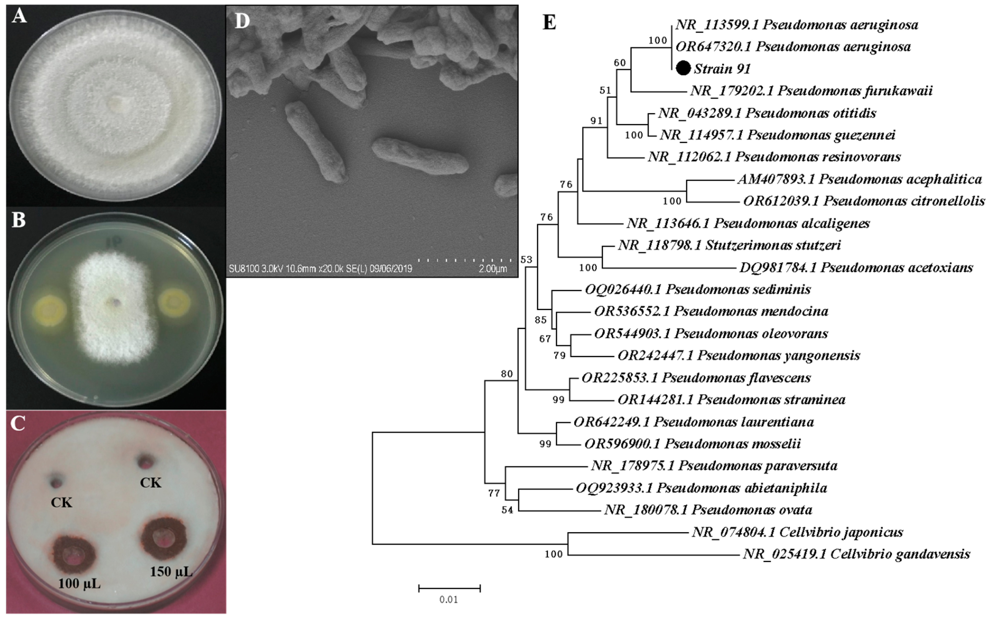
3.1. Isolation, Screening, and Identification of Effective Antagonistic Bacteria from Banana Rhizosphere

From six rhizospheric soil samples of banana plants, we isolated 109 bacterial strains. We evaluated all these isolates for their antifungal activity against Foc. Isolate 91 exhibited the highest antifungal activity, inhibiting 69% of Foc growth in the dual-culture plate assay (Figure 1A,B). Moreover, the cell-free fermentation supernatant of isolate 91 demonstrated inhibition of the Foc pathogen when compared to the control (Figure 1C).
Isolate 91’s identification involved initial examination under light microscopy, revealing its motile, rod-shaped cells. This identification was further validated through scanning electron microscopy (SEM) (Figure 1D) and 16S rRNA sequencing analysis. The 16S rRNA sequence of isolate 91 was compared to nucleotide sequences in the NCBI GenBank database using the BlastN program. A phylogenetic tree was constructed using representative strains obtained from BlastN, confirming isolate 91 as P. aeruginosa (Figure 1E). The accession number OL658832 was assigned to P. aeruginosa 91 and submitted to the NCBI GenBank database.

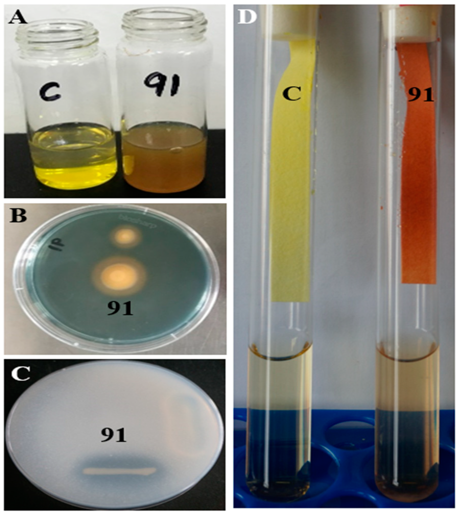
3.2. Isolate 91 Displays a Multivariate Mode of Antagonism Mechanism

We initially hypothesized that the antagonistic activity of isolate 91 stemmed from its secondary metabolites. To investigate this, we conducted separate extractions of the solid fermentation material on YAG plates using ethyl acetate and n-butanol. The EtOAc extract, at concentrations of 2 mg/disk, 4 mg/disk, and 6 mg/disk, exhibited inhibition zone diameters of 11 mm, 14 mm, and 17 mm, respectively (Figure 2A). Conversely, the n-butanol extract, at concentrations of 2 mg/disk, 4 mg/disk, and 6 mg/disk, displayed inhibition zone diameters of 7 mm, 8 mm, and 9 mm, respectively (Figure 2B). These results indicate that both EtOAc and n-butanol extracts contain bioactive compounds capable of inhibiting the growth of Foc.
The EtOAc extract was further separated into 11 fractions (Fr.1–Fr.11), which were incubated at 1.5 mg/disk to detect inhibition activity. Among the 11 fractions, only Fr.7 displayed a transparent inhibition zone at 7 mm (Figure 2C,D). Further fractionation of Fr.7 was conducted and an activity test revealed a 7 mm inhibition zone of Fr.7.4 at 1.5 mg/disk. It was noticed that step-by-step purification of the EtOAc failed to show increased antifungal activity. Therefore, it is reasonable to speculate that instead of a single bioactive compound displaying antifungal activity, multiple components in the EtOAc extract exhibit synergistic antifungal effects.
Microorganisms can inhibit fungal growth directly by secreting various cell wall-degrading enzymes [44]. To determine whether isolate 91 produces these enzymes, we conducted an activity assay, which revealed cellulase activity at 451.23 ± 5.37 IU·mL−1, glucanase activity at 665.76 ± 10.84 IU·mL−1, protease activity at 143.56 ± 1.82 IU·mL−1, and chitinase activity at 495.43 ± 5.32 IU·mL−1 under in vitro conditions (Table 1). Additionally, SEM images showed distorted and ruptured Foc mycelia when incubated with isolate 91 (Figure 2F), confirming the secretion of cell wall-degrading enzymes by isolate 91 to disrupt the fungal cell wall. In conjunction with its multiple bioactive metabolites, isolate 91 exhibits a multifaceted antagonistic mechanism.

3.3. Isolate 91 Possesses Various Plant Growth-Promoting (PGP) Traits

PGP is an attractive trait for biocontrol microorganisms [45]. Isolate 91 exhibits a diverse range of PGP traits (Table 1 and Figure 3). It produces substantial levels of ammonia (5.53 ± 0.07 µmoL mL−1) (Figure 3A). On Chrome Azurol S Agar medium, it forms an orange halo zone, indicating siderophore production (85.21 ± 0.06 PSU) (Figure 3B). When grown on Pikovskaya’s Agar medium, it creates a clear zone, suggestive of phosphate solubilization capability (1.38 ± 0.03 PSI) (Figure 3C). Isolate 91 also exhibits a positive HCN production test (Figure 3D).
Isolate 91 exhibited significant IAA synthesis capacity, producing 175.96 ± 1.63 µg·mL−1 of IAA in a medium containing 0.5% tryptophan, while 49.53 ± 1.48 µg·mL−1 of IAA was generated in tryptophan-free media (Table 1). Furthermore, in DF-ACC medium with 3 mM ACC as the sole nitrogen source, isolate 91 displayed ACC deaminase enzyme activity by consuming ACC, yielding an estimated rate of 519.28 ± 6.58 nmoL α-ketobutyrate mg−1·h−1 after 48 h of incubation (Table 1).

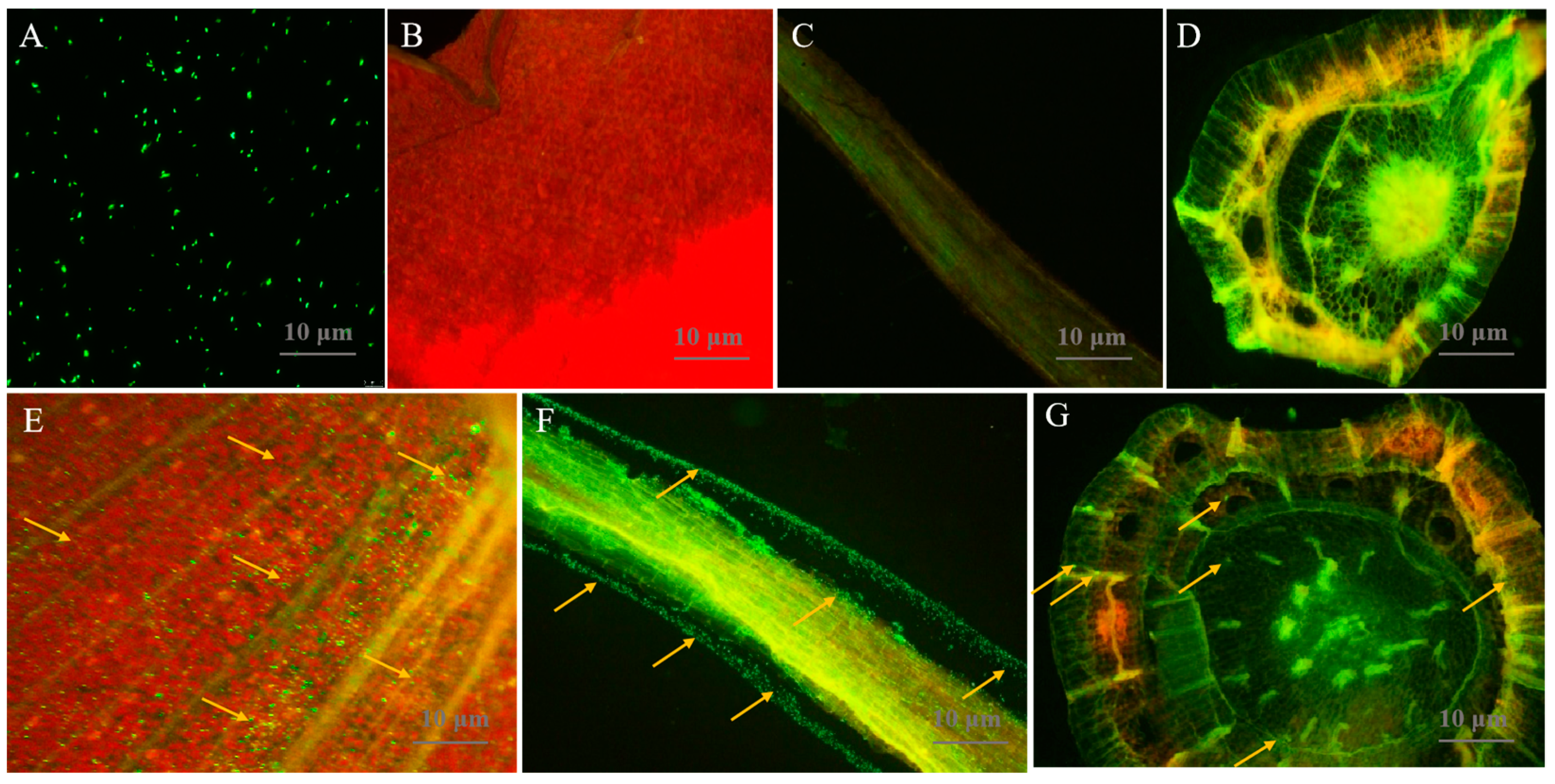
3.4. Isolate 91 Colonizes in All Tissues of Banana Plants

Biocontrol microorganisms must possess effective colonization abilities for plant disease management and growth promotion. Therefore, we assessed the colonization of GFP-tagged isolate 91 in banana tissue culture plantlets after 3 days of inoculation using confocal laser scanning microscopy (CLSM) (Figure 4). In comparison to the control (Figure 4B–D), GFP-tagged isolate 91 cells (Figure 4A) efficiently colonized all plant tissues, appearing as green spots, with a primary distribution in the root tissue, followed by the leaf and stem tissues (Figure 4E–G).

3.5. Isolate 91 Utilizes Various Carbon Sources

The GNIII BiologR technique was applied to study the carbon substrate utilization pattern of microorganisms. Metabolic characteristics are crucial for biocontrol microorganisms to adapt to specific environments, such as soil and plant tissues [46]. Excitingly, isolate 91 demonstrates a versatile carbon source utilization capacity, encompassing a wide spectrum of substrates. These substrates include dextrin, d-turanose, d-raffinose, d-salicin, N-acetyl-d-glucosamine, N-acetyl-d-galactosamine, N-acetyl neuraminic acid, d-fructose, d-galactose, 3-methyl glucose, d-fucose, l-fucose, inosine, d-sorbitol, d-mannitol, d-arabitol, myo-inositol, glycerol, d-glucose-6-PO4, d-fructose-6-PO4, d-aspartic acid, d-serine, glycyl-l-proline, l-alanine, l-arginine, l-aspartic acid, l-glutamic acid, l-histidine, l-serine, lincomycin, niaproof 4, pectin, d-gluconic acid, d-glucuronic acid, glucuronamide, mucic acid, quinic acid, tetrazolium violet, tetrazolium blue, and l-lactic acid on GNIII BiologR plate (Figure S1 and Table S2).

4. Discussion

The rhizosphere, housing both beneficial and pathogenic microorganisms, acts as the primary defense against soil-borne diseases [47]. Plant growth-promoting rhizobacteria (PGPR) play a dual role in enhancing nutrient uptake by plants and acting as biocontrol agents, reducing soil-borne diseases [48]. Consequently, the screening of rhizosphere-competent bacteria represents an effective strategy for managing fungal pathogens. This study aimed to isolate highly effective antagonistic rhizobacteria against the BFW pathogen, Foc. A total of 109 bacterial strains were isolated from rhizospheric soil samples of banana plants in Guangxi, China. Among them, P. aeruginosa strain 91 exhibited the most potent antagonistic activity, prompting its selection for further investigation. Due to their proven biocontrol efficacy against various diseases, P. aeruginosa strains are recognized as valuable tools for disease management in tropical regions [49]. However, P. aeruginosa is commonly characterized as an opportunistic pathogen with a broad host range, and our understanding of P. aeruginosa strains remains limited. Therefore, a thorough risk assessment of isolate P. aeruginosa 91 will be conducted before field trials, establishing a robust risk assessment framework for biofertilizer products in the future.
PGPR support plant growth through various mechanisms, including the production of IAA, P-solubilization, secretion of siderophores, and enhancing plant resilience against biotic and abiotic stresses [50]. Siderophores improve iron acquisition and inhibit plant pathogens through iron competition [51]. Phosphate-solubilizing microorganisms make previously immobile phosphorus in the soil available to plants [52]. Several Pseudomonas strains, including P. aeruginosa, P. fluorescens, and P. brassicacearum, have demonstrated phosphate solubilization capabilities in previous studies [23,53,54,55,56]. IAA production has been associated with the ability of many Pseudomonas and other strains to promote plant growth [23,39,40,57,58]. In this study, isolate 91 produced a substantial amount of IAA, both in the presence and absence of tryptophan (Table 1). This result indicates that isolate 91 has the ability to promote plant growth, which is consistent with previous studies. Several bacterial strains produce secondary metabolites like HCN and ammonia, which play crucial roles in preventing fungal infections in various plants [19] or increasing plant nutrient availability [59]. Similarly, isolate 91 exhibited a high production of HCN and ammonia, suggesting its involvement in suppressing Foc or facilitating plant growth.
Biocontrol strategies involve competition for resources, the production of inhibitory chemicals and cell wall-degrading enzymes, and the induction of systemic resistance [60]. Pseudomonas, a diverse genus comprising numerous species, has been effectively employed as a plant inoculant to enhance plant growth and health [21]. While various Pseudomonas spp. strains have been explored as antagonists against Foc, most research has focused on P. fluorescens, with limited attention given to P. aeruginosa [6]. Prior studies have examined the potential of different P. aeruginosa strains to suppress Foc growth, exhibit plant growth-promoting (PGP) traits, and secrete hydrolytic enzymes in vitro [61,62,63]. An isolated strain P. aeruginosa BG from seawater demonstrated the ability to produce IAA (19 μg mL−1) and ammonia (27 μg mL−1), alongside the production of cyanide, iron carriers, and hydrogen peroxidase; the authors of the study believe that this strain holds significant potential for promoting plant growth and controlling plant diseases [61]. Our strain 91 exhibited higher levels of IAA and ammonia production (Table 1) compared to the marine P. aeruginosa BG, and it displayed similar inhibitory effects on Foc. Thus, it is undeniable that strain 91 has substantial application prospects. The previous research findings reveal the P. aeruginosa capability to produce a wide array of antifungal substances, including enzymes such as catalase, chitin-binding protein, and protease [64], alongside small molecule compounds like 3,4-dihydroxy-N-methyl-4-(4-oxochroman-2-yl)butanamide, pyocyanin, rhamnolipids, phenazine-1-carboxylic acid, and phenazine-1-carboxamide [65,66,67,68,69]. These components exhibit significant impacts on fungal hyphal growth, resulting in abnormal growth, unusual bending, or hyphal breakage, closely mirroring our experimental outcomes (Figure 2F). This strongly suggests that strain 91 may produce analogous compounds capable of inhibiting the growth of Foc.
An organism’s metabolic traits are vital for fostering plant growth and forming a successful host community [70]. Numerous Pseudomonas strains, including P. aeruginosa and P. koreensis, have previously showcased diverse carbon utilization patterns, contributing to enhanced plant development and fungal infection protection [23,39]. In this investigation, isolate 91 demonstrated diverse carbon utilization patterns on GNIII Biolog plateR (Figure S1), highlighting its potential advantages in promoting plant growth and development. Furthermore, this observation underscores its robust adaptability to various environments and its capacity to compete effectively with other microorganisms in diverse ecological settings.
Competitive rhizosphere colonization is a crucial aspect of PGPR–plant interactions [71]. Gaining insights into the molecular mechanisms underlying banana–rhizobacteria interactions can pave the way for technical enhancements in banana cultivation. Our observation of the GFP-tagged isolate 91 exhibiting robust colonization across all tissues of cultivated banana plantlets (Figure 4) reaffirms its substantial potential for the management of BFW. On one hand, in comparison to conventional biocontrol bacteria, it appears to possess heightened efficacy in controlling BFW. On the other hand, it can more readily fulfill its function in promoting plant growth.

5. Conclusions

Employing efficient biocontrol bacteria with PGP traits holds promise for safeguarding crops against diseases and augmenting crop yields. This study highlights Pseudomonas aeruginosa 91, isolated from the banana rhizosphere, as a potent antifungal agent against Foc, employing a multifaceted mechanism encompassing the production of bioactive compounds and the secretion of cell wall-degrading enzymes. Isolate 91 also demonstrates diverse PGP traits, a broad carbon source utilization spectrum, and successful colonization within all banana plant tissues. In summary, isolate 91 emerges as a prospective biocontrol strain for mitigating BFW. However, its performance in controlling Foc and enhancing banana growth necessitates assessment through future field trials.

Supplementary Materials

The following supporting information can be downloaded at: https://www.mdpi.com/article/10.3390/jof9111047/s1, Table S1: Media used in this study; Table S2: List of carbon sources utilized by Pseudomonas aeruginosa 91 in GENIII BIOLOGR plate; Figure S1: Carbon substrate utilization pattern of isolate 91 on GNIII BiologR plate.

Author Contributions

W.F. and B.W. conceived the study. J.X. conducted crude extraction and separation. P.S. performed strain isolation, screening, and PGP trait characterization. Y.Q. collected soil samples. R.K.S. helped in PGP trait and colonization study. Q.Q. and C.J. are involved in data curation. P.S., J.X., B.W. and W.F. analyzed and interpreted the data and wrote the manuscript with input from all authors. All authors have read and agreed to the published version of the manuscript.

Funding

This work was supported by Guangxi Natural Science Foundation (2023GXNSFBA026235) to J.X. Guangxi Science and Technology Base and Talent Special Project (AD23026030) and Guangxi Key Research and Development Plan (AB21220030) to B.W. Guangxi Natural Science Foundation (2023GXNSFFA026011) to W.F. Research Start-up Funding of Guangxi Academy of Sciences (2021YBJ704) to J.X.

Institutional Review Board Statement

Not applicable.

Informed Consent Statement

Not applicable.

Data Availability Statement

The data presented in this study are available in the article.

Acknowledgments

We are very thankful to Sugarcane Research Center, Guangxi Academy of Agricultural Sciences, Nanning, China for a colonization study using confocal laser scanning microscopy (CLSM).

Conflicts of Interest

The authors declare no conflict of interest.

References

  1. Pegg, K.G.; Coates, L.M.; O’Neill, W.T.; Turner, D.W. The epidemiology of Fusarium wilt of banana. Front. Plant Sci. 2019, 10, 1395. [Google Scholar] [CrossRef] [PubMed]
  2. Thangavelu, R.; Edwin Raj, E.; Loganathan, M.; Pushpakanth, P.; Uma, S. Draft genome of Fusarium oxysporum f. sp. cubense strain Tropical Race-4 infecting Cavendish (AAA) group of banana in India. Plant Dis. 2020, 105, 481–483. [Google Scholar] [CrossRef]
  3. Ploetz, R.C. Management of Fusarium wilt of banana: A review with special reference to tropical race 4. Crop Prot. 2015, 73, 7–15. [Google Scholar] [CrossRef]
  4. Wang, Y.; Xia, Q.; Wang, G.; Zhang, H.; Lu, X.; Sun, J.; Zhang, X. Differential gene expression in banana roots in response to Fusarium wilt. Can. J. Plant Pathol. 2017, 39, 163–175. [Google Scholar] [CrossRef]
  5. Arinaitwe, I.K.; Teo, C.H.; Kayat, F.; Tumuhimbise, R.; Uwimana, B.; Kubiriba, J.; Swennen, R.; Harikrishna, J.A.; Othman, R.Y. Evaluation of banana germplasm and genetic analysis of an F1 population for resistance to Fusarium oxysporum f. sp. cubense race 1. Euphytica 2019, 215, 175. [Google Scholar] [CrossRef]
  6. Bubici, G.; Kaushal, M.; Prigigallo, M.I.; Gómez-Lama Cabanás, C.; Mercado-Blanco, J. Biological control agents against Fusarium wilt of banana. Front. Microbiol. 2019, 10, 616. [Google Scholar] [CrossRef]
  7. Fliessbach, A.; Winkler, M.; Lutz, M.P.; Oberholzer, H.R.; Mäder, P. Soil amendment with Pseudomonas fuorescens CHA0: Lasting effects on soil biological properties in soils low in microbial biomass and activity. Microb. Ecol. 2009, 57, 611–623. [Google Scholar] [CrossRef] [PubMed]
  8. Ahmad, F.; Ahmad, I.; Khan, M.S. Screening of free-living rhizospheric bacteria for their multiple plant growth promoting activities. Microbiol. Res. 2008, 163, 173–181. [Google Scholar] [CrossRef] [PubMed]
  9. Dessaux, Y.; Grandclément, C.; Faure, D. Engineering the rhizosphere. Trends Plant Sci. 2016, 21, 266–278. [Google Scholar] [CrossRef] [PubMed]
  10. Prashar, P.; Kapoor, N.; Sachdeva, S. Rhizosphere: Its structure, bacterial diversity and significance. Rev. Environ. Sci. Biotechnol. 2014, 13, 63–67. [Google Scholar] [CrossRef]
  11. Estabrook, E.M.; Yoder, J.I. Plant-plant communications: Rhizosphere signaling between parasitic angiosperms and their hosts. Plant Physiol. 1998, 116, 1–7. [Google Scholar] [CrossRef]
  12. Lugtenberg, B.; Kamilova, F. Plant-growth-promoting rhizobacteria. Annu. Rev. Microbiol. 2009, 63, 541–556. [Google Scholar] [CrossRef] [PubMed]
  13. Bhattacharyya, P.N.; Jha, D.K. Plant growth-promoting rhizobacteria (PGPR): Emergence in agriculture. World J. Microbiol. Biotechnol. 2012, 28, 1327–1350. [Google Scholar] [CrossRef] [PubMed]
  14. Compant, S.; Duffy, B.; Nowak, J.; Clément, C.; Barka, E.A. Use of plant growth-promoting bacteria for biocontrol of plant diseases: Principles, mechanisms of action, and future prospects. Appl. Environ. Microbiol. 2005, 71, 4951–4959. [Google Scholar] [CrossRef] [PubMed]
  15. Singh, R.K.; Singh, P.; Li, H.; Guo, D.; Song, Q.; Yang, L.; Malviya, M.K.; Song, X.; Li, Y. Plant-PGPR interaction study of plant growth-promoting diazotrophs Kosakonia radicincitans BA1 and Stenotrophomonas maltophilia COA2 to enhance growth and stress-related gene expression in Saccharum spp. J. Plant Interact. 2020, 15, 427–445. [Google Scholar] [CrossRef]
  16. Singh, R.K.; Singh, P.; Li, H.; Song, Q.; Li, Y. Diversity of nitrogen-fixing rhizobacteria associated with sugarcane, a comprehensive study of plant-microbe interactions for growth enhancement in Saccharum spp. BMC Plant Biol. 2020, 20, 220. [Google Scholar] [CrossRef]
  17. Yang, W.; Xu, Q.; Liu, H.; Wang, Y.; Wang, Y.; Yang, H.; Guo, J. Evaluation of biological control agents against Ralstonia wilt on ginger. Biol. Control. 2012, 62, 144–151. [Google Scholar] [CrossRef]
  18. Farina, R.; Beneduzi, A.; Ambrosini, A.; Campos, S.B.; Lisboa, B.B.; Wendisch, V.; Vargas, L.K.; Passaglia, L.M.P. Diversity of plant growth-promoting rhizobacteria communities associated with the stages of canola growth. Appl. Soil Ecol. 2012, 55, 44–52. [Google Scholar] [CrossRef]
  19. Olanrewaju, O.; Glick, B.R.; Babalola, O.O. Mechanisms of action of plant growth promoting bacteria. World J. Microbiol. Biotechnol. 2017, 33, 197. [Google Scholar] [CrossRef]
  20. Mia, M.; Zulkifli, S.; Maziah, M. Use of plant growth promoting bacteria in banana: A new insight for sustainable banana production. Int. J. Agric. Biol. 2010, 12, 459–467. [Google Scholar]
  21. Hesse, C.N.; Schulz, F.; Bull, C.T.; Shaffer, B.T.; Yan, Q.; Shapiro, N.; Hassan, K.A.; Varghese, N.; Elbourne, L.D.H.; Paulsen, I.T.; et al. Genome-based evolutionary history of Pseudomonas spp. Environ. Microbiol. 2018, 20, 2142–2159. [Google Scholar] [CrossRef] [PubMed]
  22. Mendes, R.; Pizzirani-Kleiner, A.A.; Araujo, W.L.; Raaijmakers, J.M. Diversity of cultivated endophytic bacteria from sugarcane, genetic and biochemical characterization of Burkholderia cepacia complex isolates. Appl. Environ. Microbiol. 2007, 73, 7259–7267. [Google Scholar] [CrossRef]
  23. Singh, P.; Singh, R.K.; Guo, D.; Sharma, A.; Singh, R.N.; Li, D.; Malviya, M.K.; Song, X.; Lakshmanan, P.; Yang, L.; et al. Whole genome analysis of sugarcane root-associated endophyte Pseudomonas aeruginosa B18-a plant growth-promoting bacterium with antagonistic potential against Sporisorium scitamineum. Front. Microbiol. 2021, 12, 628376. [Google Scholar] [CrossRef] [PubMed]
  24. Raaijmakers, J.M.; Mazzola, M. Diversity and natural functions of antibiotics produced by beneficial and pathogenic soil bacteria. Annu. Rev. Phytopathol. 2012, 50, 403–424. [Google Scholar] [CrossRef] [PubMed]
  25. Ma, M.C.; Jiang, X.; Cao, M.F.; Li, J. Risk analysis and management measure on microorganism in microbial organic fertilizers. Qual. Saf. AgroProducts 2019, 6, 57–61. (In Chinese) [Google Scholar]
  26. Singh, P.; Singh, R.K.; Zhou, Y.; Wang, J.; Jiang, Y.; Shen, N.; Wang, Y.; Yang, L.; Jiang, M. Unlocking the strength of plant growth-promoting Pseudomonas in improving crop productivity in normal and challenging environments: A review. J. Plant Interact. 2022, 17, 220–238. [Google Scholar] [CrossRef]
  27. Singh, R.K.; Kumar, D.P.; Singh, P.; Solanki, M.K.; Srivastava, S.; Kashyap, P.L.; Kumar, S.; Srivastava, A.K.; Singhal, P.K.; Arora, D.K. Multifarious plant growth promoting characteristics of chickpea rhizosphere associated Bacilli help to suppress soil-borne pathogens. Plant Growth Regul. 2013, 73, 91–101. [Google Scholar] [CrossRef]
  28. Espinel-Ingroff, A.; Pfaller, M.; Messer, S.A.; Knapp, C.C.; Killian, S.; Norris, H.A.; Ghannoum, M.A. Multicenter comparison of the sensititre Yeast One Colorimetric Antifungal Panel with the National Committee for Clinical Laboratory standards M27-A reference method for testing clinical isolates of common and emerging Candida spp., Cryptococcus spp., and other yeasts and yeast-like organisms. J. Clin. Microbiol. 1999, 37, 591–595. [Google Scholar]
  29. Schwyn, B.; Neilands, J.B. Universal chemical assay for the detection and determination of siderophores. Anal. Biochem. 1987, 160, 47–56. [Google Scholar] [CrossRef]
  30. Hu, Q.P.; Xu, J.G. A simple double-layered chrome azurol S agar (SDCASA) plate assay to optimize the production of siderophores by a potential biocontrol agent Bacillus. Afr. J. Microbiol. Res. 2011, 5, 4321–4327. [Google Scholar]
  31. Dey, R.; Pal, K.K.; Bhatt, D.M.; Chauhan, S.M. Growth promotion and yield enhancement of peanut (Arachis hypogaea L.) by application of plant growth promoting rhizobacteria. Microbiol. Res. 2004, 159, 391–394. [Google Scholar] [CrossRef] [PubMed]
  32. Goswami, D.; Dhandhukia, P.; Patel, P.; Thakker, J.N. Screening of PGPR from saline desert of Kutch: Growth promotion in Arachis hypogea by Bacillus licheniformis A2. Microbiol. Res. 2014, 169, 66–75. [Google Scholar] [CrossRef] [PubMed]
  33. Pikovskaya, R.I. Mobilization of phosphorus in soil in connection with the vital activity of some of the microbial species. Microbiology 1948, 17, 362–370. [Google Scholar]
  34. Mehta, S.; Nautiyal, C.S. An efficient method for qualitative screening of phosphate-solubilizing bacteria. Curr. Microbiol. 2001, 43, 51–56. [Google Scholar] [CrossRef] [PubMed]
  35. Lorck, H. Production of hydrocyanic acid by bacteria. Physiol. Plant. 1948, 1, 142–146. [Google Scholar] [CrossRef]
  36. Glickmann, E.; Dessaux, Y.A. Critical examination of the specificity of the salkowski reagent for indolic compounds produced by phytopathogenic bacteria. Appl. Environ. Microbiol. 1995, 61, 793–796. [Google Scholar] [CrossRef] [PubMed]
  37. Honma, M.; Shimomura, T. Metabolism of 1-aminocyclopropane-1-carboxylic acid. Agric. Biol. Chem. 1978, 42, 1825–1831. [Google Scholar]
  38. Jacobson, C.B.; Pasternak, J.J.; Glick, B.R. Partial purification and characterization of 1-aminocyclopropane-1-carboxylate deaminase from the plant growth promoting rhizobacterium Pseudomonas putida GR12-2. Can. J. Microbiol. 1994, 40, 1019–1025. [Google Scholar] [CrossRef]
  39. Li, H.; Singh, R.K.; Singh, P.; Song, Q.; Xing, Y.; Yang, L.; Li, Y. Genetic diversity of nitrogen-fixing and plant growth promoting Pseudomonas species isolated from sugarcane rhizosphere. Front. Microbiol. 2017, 8, 1268. [Google Scholar] [CrossRef]
  40. Singh, P.; Singh, R.K.; Li, H.; Guo, D.; Sharma, A.; Lakshmanan, P.; Malviya, M.K.; Song, X.; Solanki, M.K.; Verma, K.K.; et al. Diazotrophic bacteria Pantoea dispersa and Enterobacter asburiae promote sugarcane growth by inducing nitrogen uptake and defense-related gene expression. Front. Microbiol. 2021, 11, 600417. [Google Scholar] [CrossRef]
  41. Edwards, U.; Rogall, T.H.; Blocker, H.; Emde, M.; Bottger, E.C. Isolation and direct complete nucleotide determination of entire genes. Characterization of a gene coding for 16S ribosomal RNA. Nucleic Acids Res. 1989, 17, 7843–7853. [Google Scholar] [CrossRef]
  42. Saitou, N.; Nei, M. The neighbor-joining method, a new method for reconstructing phylogenetic trees. Mol. Biol. Evol. 1987, 4, 406–425. [Google Scholar]
  43. Kumar, S.; Stecher, G.; Tamura, K. MEGA7, molecular evolutionary genetics analysis version 7.0 for bigger datasets. Mol. Biol. Evol. 2016, 33, 1870–1874. [Google Scholar] [CrossRef]
  44. Husson, E.; Hadad, C.; Huet, G.; Laclef, S.; Lesur, D.; Lambertyn, V.; Jamali, A.; Gottis, S.; Sarazin, C.; Nguyen, V.N.A. The effect of room temperature ionic liquids on the selective biocatalytic hydrolysis of chitin via sequential or simultaneous strategies. Green. Chem. 2017, 19, 4122–4131. [Google Scholar] [CrossRef]
  45. Orozco-Mosqueda, M.; del Carmen, R.G.M.; Glick, B.R.; Santoyo, G. Microbiome engineering to improve biocontrol and plant growth-promoting mechanisms. Microbiol. Res. 2018, 208, 25–31. [Google Scholar] [CrossRef] [PubMed]
  46. Wielbo, J.; Marek-Kozaczuk, M.; Kubik-Komar, A.; Skorupska, A. Increased metabolic potential of Rhizobium spp. is associated with bacterial competitiveness. Can. J. Microbiol. 2007, 53, 957–967. [Google Scholar] [CrossRef]
  47. Weller, D.M. Biological control of soilborne plant pathogens in the rhizosphere with bacteria. Annu. Rev. Phytopathol. 1988, 26, 379–407. [Google Scholar] [CrossRef]
  48. Lucy, M.; Reed, E.; Glick, B.R. Applications of free living plant growth-promoting rhizobacteria. Antonie Van Leeuwenhoek 2004, 86, 1–25. [Google Scholar] [CrossRef] [PubMed]
  49. Kumar, A.; Munder, A.; Aravind, R.; Eapen, S.J.; Tümmler, B.; Raaijmakers, J.M. Friend or foe: Genetic and functional characterization of plant endophytic Pseudomonas aeruginosa. Environ. Microbiol. 2013, 15, 764–779. [Google Scholar] [CrossRef]
  50. Costa, P.B.D.; Granada, C.E.; Ambrosini, A.; Moreira, F.; de Souza, R.; dos Passos, J.F.M.; Arruda, L.; Passaglia, L.M.P. A model to explain plant growth promotion traits: A multivariate analysis of 2211 bacterial isolates. PLoS ONE 2014, 9, e116020. [Google Scholar] [CrossRef]
  51. Zhang, L.; Chen, W.; Jiang, Q.; Fei, Z.; Xiao, M. Genome analysis of plant growth-promoting rhizobacterium Pseudomonas chlororaphis subsp. aurantiaca JD37 and insights from comparison of genomics with three Pseudomonas strains. Microbiol. Res. 2020, 237, 126483. [Google Scholar] [CrossRef] [PubMed]
  52. Joe, M.M.; Devaraj, S.; Benson, A.; Sa, T. Isolation of phosphate solubilizing endophytic bacteria from Phyllanthus mares Schum & Thonn: Evaluation of plant growth promotion and antioxidant activity under salt stress. J. Appl. Res. Med. Aromat. Plants 2016, 3, 71–77. [Google Scholar]
  53. Chandra, H.; Kumari, P.; Bisht, R.; Prasad, R.; Yadav, S. Plant growth promoting Pseudomonas aeruginosa from Valeriana wallichii displays antagonistic potential against three phytopathogenic fungi. Mol. Biol. Rep. 2020, 47, 6015–6026. [Google Scholar] [CrossRef]
  54. Chenniappan, C.; Narayanasamy, M.; Daniel, G.M.; Ramaraj, G.B.; Ponnusamy, P.; Sekar, J.; Vaiyapuri, R.P. Biocontrol efficiency of native plant growth promoting rhizobacteria against rhizome rot disease of turmeric. Biol. Control. 2019, 129, 55–64. [Google Scholar] [CrossRef]
  55. Durairaj, K.; Velmurugan, P.; Park, J.H.; Chang, W.S.; Park, Y.J.; Senthilkumar, P.; Choi, K.M.; Lee, J.H.; Oh, B.T. Potential for plant biocontrol activity of isolated Pseudomonas aeruginosa and Bacillus stratosphericus strains against bacterial pathogens acting through both induced plant resistance and direct antagonism. FEMS Microbiol. Lett. 2017, 364, fnx225. [Google Scholar] [CrossRef] [PubMed]
  56. Nelkner, J.; Tejerizo, G.T.; Hassa, J.; Lin, T.W.; Witte, J.; Verwaaijen, B.; Winkler, A.; Bunk, B.; Spröer, C.; Overmann, J.; et al. Genetic potential of the biocontrol agent Pseudomonas brassicacearum (Formerly P. trivialis) 3Re2-7 unraveled by genome sequencing and mining, comparative genomics and transcriptomics. Genes 2019, 10, 601. [Google Scholar] [CrossRef]
  57. Guo, D.; Singh, R.K.; Singh, P.; Li, D.; Sharma, A.; Xing, Y.; Song, X.; Yang, L.; Li, Y. Complete genome sequence of Enterobacter roggenkampii ED5, a nitrogen fixing plant growth promoting endophytic bacterium with biocontrol and stress tolerance properties, isolated from sugarcane root. Front. Microbiol. 2020, 11, 580081. [Google Scholar] [CrossRef]
  58. Singh, P.; Xie, J.; Qi, Y.; Qin, Q.; Jin, C.; Wang, B.; Fang, W. A thermotolerant marine Bacillus amyloliquefaciens S185 producing iturin A5 for antifungal activity against Fusarium oxysporum f. sp. cubense. Mar. Drugs 2021, 19, 516. [Google Scholar] [CrossRef]
  59. Rijavec, T.; Lapanje, A. Hydrogen cyanide in the rhizosphere: Not suppressing plant pathogens, but rather regulating availability of phosphate. Front. Microbiol. 2016, 7, 1785. [Google Scholar] [CrossRef]
  60. Hayat, R.; Ahmed, I.; Sheirdil, R.A. An overview of plant growth promoting rhizobacteria (PGPR) for sustainable agriculture. Crop Prod. Agric. Improv. 2012, 27, 557–579. [Google Scholar]
  61. Goswami, D.; Patel, K.; Parmar, S.; Vaghela, H.; Muley, N.; Dhandhukia, P.; Thakker, J.N. Elucidating multifaceted urease producing marine Pseudomonas aeruginosa BG as a cogent PGPR and bio-control agent. Plant Growth Regul. 2015, 75, 253–263. [Google Scholar] [CrossRef]
  62. Sekhar, A.C.; Pious, T. Isolation and identification of shoot-tip associated endophytic bacteria from banana cv. Grand Naine and testing for antagonistic activity against Fusarium oxysporum f. sp. cubense. Am. J. Plant Sci. 2015, 6, 943–954. [Google Scholar] [CrossRef]
  63. Yu, C.; Xiao, R.; Liu, B.; Lin, N.; Chen, L. Endophytic colonization of biocontrol bacterium FJAT-346-PA and its efficiency against banana Fusarium wilt. Acta Phytophylacica Sin. 2010, 37, 493–498. [Google Scholar]
  64. Sowanpreecha, R.; Rerngsamran, P. Biocontrol of Orchid-pathogenic Mold, Phytophthora palmivora, by Antifungal Proteins from Pseudomonas aeruginosa RS1. Mycobiology 2018, 46, 129–137. [Google Scholar] [CrossRef] [PubMed]
  65. Illakkiam, D.; Ponraj, P.; Shankar, M.; Muthusubramanian, S.; Rajendhran, J.; Gunasekaran, P. Identification and structure elucidation of a novel antifungal compound produced by Pseudomonas aeruginosa PGPR2 against Macrophomina phaseolina. Appl. Biochem. Biotechnol. 2013, 171, 2176–2185. [Google Scholar] [CrossRef]
  66. Jayaseelan, S.; Ramaswamy, D.; Dharmaraj, S. Pyocyanin: Production, applications, challenges and new insights. World J. Microbiol. Biotechnol. 2014, 30, 1159–1168. [Google Scholar] [CrossRef] [PubMed]
  67. Rekadwad, B.; Maske, V.; Khobragade, C.N.; Kasbe, P.S. Production and evaluation of mono- and di-rhamnolipids produced by Pseudomonas aeruginosa VM011. Data Brief 2019, 3, 24. [Google Scholar] [CrossRef]
  68. Simionato, A.S.; Navarro, M.O.P.; Jesus, M.L.A.; Barazetti, A.R.; Silva, C.S.; Simões, G.C.; Balbi-Peña, M.I.; Mello, J.C.P.; Panagio, L.A.; Almeida, R.S.C.; et al. The Effect of Phenazine-1-Carboxylic Acid on Mycelial Growth of Botrytis cinerea Produced by Pseudomonas aeruginosa LV Strain. Front. Microbiol. 2017, 14, 8. [Google Scholar] [CrossRef]
  69. Shanmugaiah, V.; Mathivanan, N.; Varghese, B. Purification, crystal structure and antimicrobial activity of phenazine-1-carboxamide produced by a growth-promoting biocontrol bacterium, Pseudomonas aeruginosa MML2212. J. Appl. Microbiol. 2010, 108, 703–711. [Google Scholar] [CrossRef]
  70. Mazur, A.; Stasiak, G.; Wielbo, J.; Koper, P.; Kubik-Komar, A.; Skorupska, A. Phenotype profiling of Rhizobium leguminosarum bv. trifolii clover nodule isolates reveal their both versatile and specialized metabolic capabilities. Arch. Microbiol. 2013, 195, 255–267. [Google Scholar] [CrossRef]
  71. Timmusk, S.; Grantcharova, N.; Wagner, G.H. Paenibacillus polymyxa invades plant roots and forms biofilms. Appl. Environ. Microbiol. 2005, 71, 7292–7300. [Google Scholar] [CrossRef] [PubMed]
Figure 1. Screening and identification of isolate 91 for antifungal activity against Fusarium oxysporum f. sp. cubense (Foc). (A) Foc control plate after 7 days’ incubation; (B) dual-culture plate assay showing inhibition of Foc mycelia by isolate 91 after 7 days’ incubation; (C) cell-free fermentation supernatant displayed growth inhibition zone against Foc compared to control (CK); (D) scanning electron microscopy showing rod-shaped isolate 91; (E) phylogenetic tree showing isolate 91’s position compared to other Pseudomonas strains.
Jof 09 01047 g001
Figure 2. Isolate 91 secrets bioactive metabolites and cell wall-degrading enzymes for antagonistic activity against Foc. (A) EtOAc extract displayed inhibition activity at 2 mg/disk, 4 mg/disk, and 6 mg/disk, and CK is methanol. (B) n-butanol extract displayed inhibition activity at 2 mg/disk, 4 mg/disk, and 6 mg/disk, and CK is methanol. (C) Fr.1–Fr.5 of EtOAc extract displayed no inhibition activity, and CK is methanol. (D) Inhibition activity of Fr.6–Fr.11 of EtOAc extract. (E) Foc mycelia under SEM, red arrows showing straight, intact hyphae. (F) Foc mycelia under SEM after incubating with isolate 91, red arrows showing distorted, ruptured hyphae.
Jof 09 01047 g002
Figure 3. Plant growth-promoting traits of isolate 91. (A) Ammonia production on peptone water broth medium; (B) siderophore production on Chrome Azurol S Agar plate medium; (C) phosphate solubilization on Pikovskaya’s Agar plate medium; (D) HCN production in Luria-Bertani broth medium containing glycine.
Jof 09 01047 g003
Figure 4. Confocal laser scanning microscopy (CLSM) presenting morphology and colonization of P. aeruginosa 91 in banana plants. (A) GFP-tagged 91 strain; (BD) are tissues of leaf, root, and stem of untreated banana plantlets; (EG) are tissues of leaf, root, and stem of banana plantlets inoculated with isolate 91. Yellow arrow marks show isolate 91 cells colonized as small green dots in all tissues of a banana plant.
Jof 09 01047 g004
Table 1. Functional characteristics of antifungal isolate Pseudomonas aeruginosa 91 isolated from the banana rhizosphere.
ParametersP. aeruginosa 91
Plant Growth-Promoting Traits
Siderophore production85.21 ± 0.06 PSU
Phosphate solubilization1.38 ± 0.03 PSI
Ammonia production5.53 ± 0.07 µmoL mL−1
HCN production+
1-Aminocyclopropane-1-carboxylic deaminase activity519.28 ± 6.58 nmoL α-ketobutyrate mg−1 h−1
Indole Acetic Acid (µg mL−1)
Absence of Tryptophan49.53 ± 1.48
Presence of Tryptophan (0.5 %)175.96 ± 1.63
Hydrolytic Enzyme Production (IU mL−1)
Cellulase451.23 ± 5.37
Protease143.56 ± 1.82
Chitinase 495.43 ± 5.32
Glucanase665.76 ± 10.84
PSU, percent siderophore unit; PSI, phosphate solubilization index; +, positive.
Disclaimer/Publisher’s Note: The statements, opinions and data contained in all publications are solely those of the individual author(s) and contributor(s) and not of MDPI and/or the editor(s). MDPI and/or the editor(s) disclaim responsibility for any injury to people or property resulting from any ideas, methods, instructions or products referred to in the content.

