
Paecilomyces/Purpureocillium Infection in Children, Case Report, and Review of the Literature

Abstract
:1. Introduction
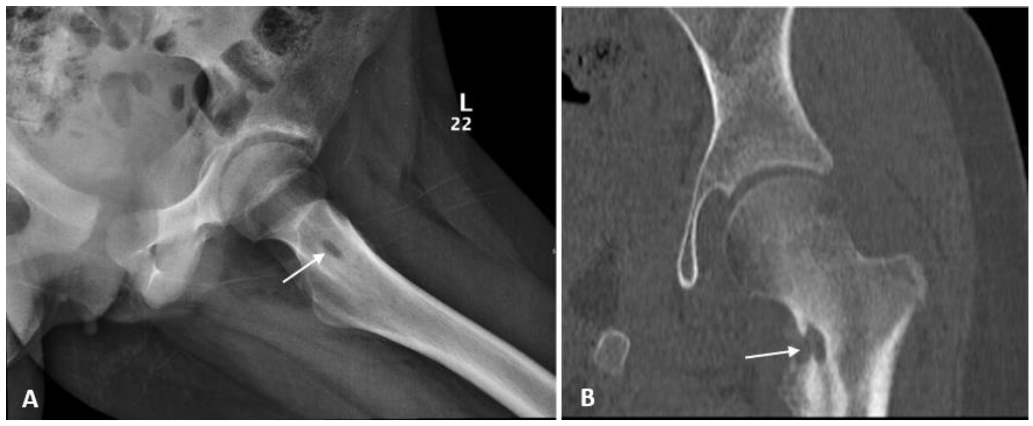
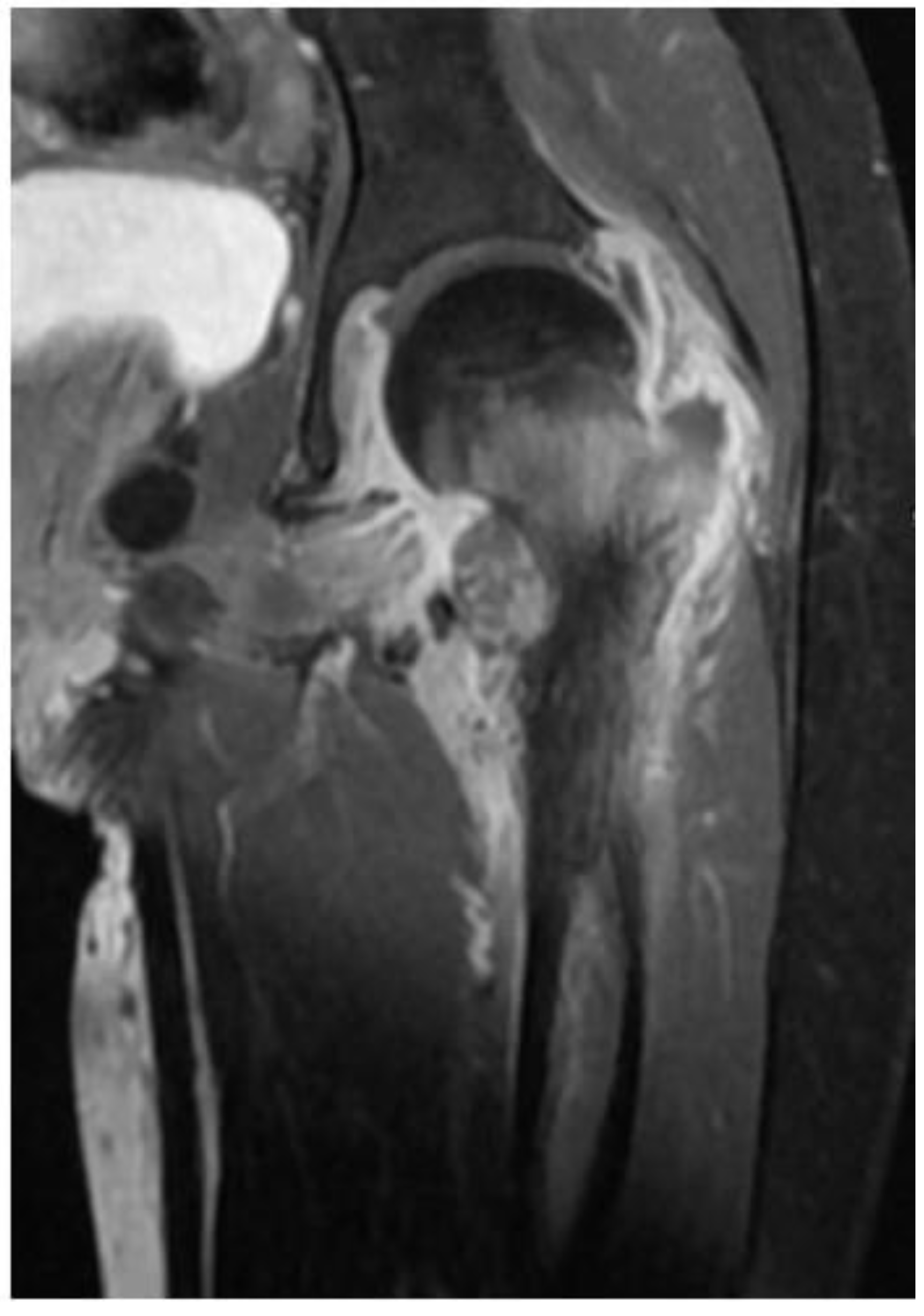
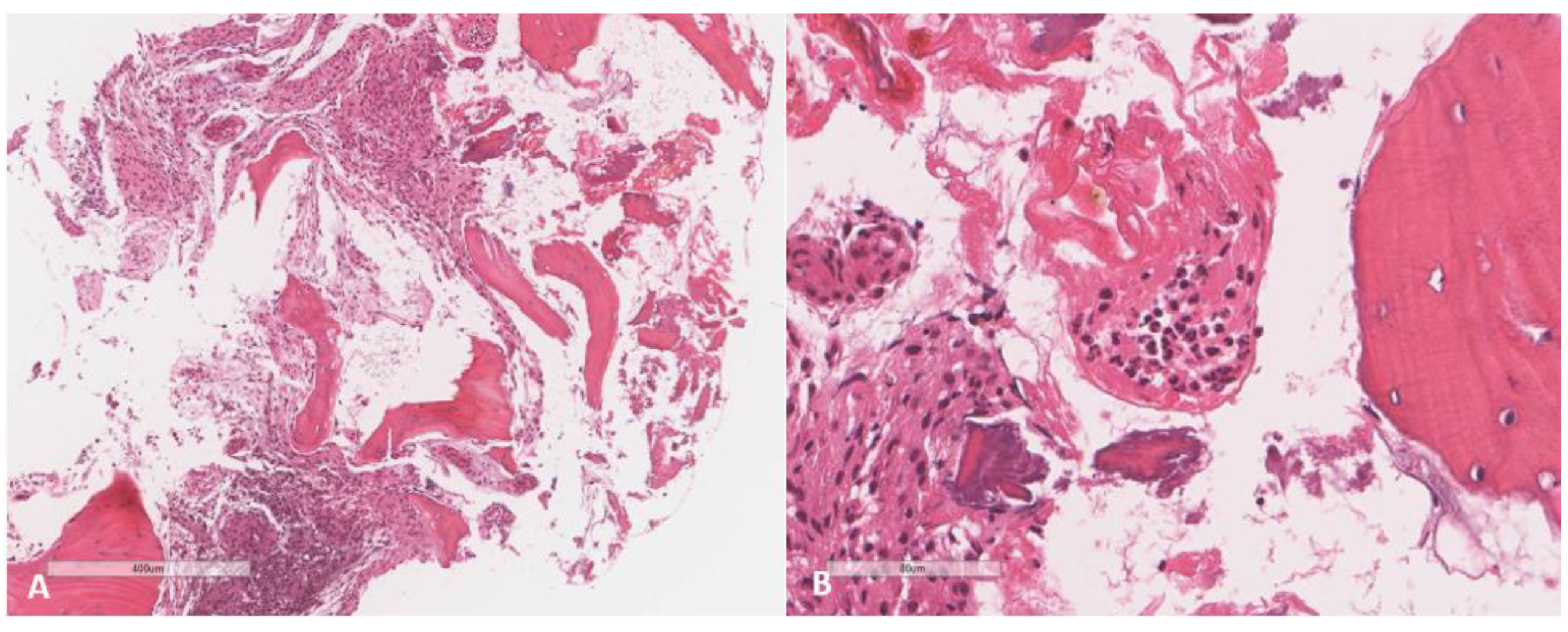
2. Case
3. Discussion
4. Conclusions
Author Contributions
Funding
Institutional Review Board Statement
Informed Consent Statement
Data Availability Statement
Acknowledgments
Conflicts of Interest
References
- Pastor, F.J.; Guarro, J. Clinical manifestations, treatment and outcome of Paecilomyces lilacinus infections. Clin. Microbiol. Infect. 2006, 12, 948–960. [Google Scholar] [CrossRef] [PubMed]
- Sprute, R.; Salmanton-García, J.; Sal, E.; Malaj, X.; Falces-Romero, I.; Hatvani, L.; Heinemann, M.; Klimko, N.; López-Soria, L.; Meletiadis, J.; et al. Characterization and outcome of invasive infections due to Paecilomyces variotii: Analysis of patients from the FungiScope® registry and literature reports. J. Antimicrob. Chemother. 2021, 76, 765–774. [Google Scholar] [CrossRef] [PubMed]
- Houbraken, J.; Verweij, P.E.; Rijs, A.J.; Borman, A.M.; Samson, R.A. Identification of Paecilomyces variotii in clinical samples and settings. J. Clin. Microbiol. 2010, 48, 2754. [Google Scholar] [CrossRef] [PubMed]
- Luangsa-Ard, J.; Houbraken, J.; van Doorn, T.; Hong, S.B.; Borman, A.M.; Hywel-Jones, N.L.; Samson, R.A. Purpureocillium, a new genus for the medically important Paecilomyces lilacinus. FEMS Microbiol. Lett. 2011, 321, 141–149. [Google Scholar] [CrossRef]
- Batarseh, R.Y.; Shehata, M.; Becker, M.D.; Sigdel, S.; He, P.; Shweihat, Y.R. Paecilomyces in an immune competent host. IDCases 2020, 21, e00885. [Google Scholar] [CrossRef]
- Cesaro, S.; Tridello, G.; Castagnola, E.; Calore, E.; Carraro, F.; Mariotti, I.; Colombini, A.; Perruccio, K.; Decembrino, N.; Russo, G.; et al. Retrospective study on the incidence and outcome of proven and probable invasive fungal infections in high-risk pediatric onco-hematological patients. Eur. J. Haematol. 2017, 99, 240–248. [Google Scholar] [CrossRef]
- de Hoog, G.S.; Guarro, J.; Gené, J.; Figueras, M.J. Atlas of Clinical Fungi; Centraalbureau voor Schimmelcultures (CBS): Utrecht, The Netherlands, 2000. [Google Scholar]
- Larone, D.H.; Larone, D.H. Medically Important Fungi: A Guide to Identification; Elsevier: New York, NY, USA, 1987. [Google Scholar]
- St-Germain, G.; Summerbell, R. Identifying Filamentous Fungi: A Clinical Laboratory Handbook; Star Publishing Company: Singapore, 1996. [Google Scholar]
- Wong, G.; Nash, R.; Barai, K.; Rathod, R.; Singh, A. Paecilomyces lilacinus causing debilitating sinusitis in an immunocompetent patient: A case report. J. Med. Case Rep. 2012, 6, 1–5. [Google Scholar] [CrossRef]
- Rodrigues, M.M.; MacLeod, D. Exogenous fungal endophthalmitis caused by Paecilomyces. Am. J. Ophthalmol. 1975, 79, 687–690. [Google Scholar] [CrossRef]
- Murciano, A.; Domer, J.; Cohen, I. Paecilomyces lilacinus infection in an immunocompromised patient. J. La. State Med. Soc. Off. Organ La. State Med. Soc. 1990, 142, 35. [Google Scholar]
- Crompton, C.H.; Balfe, J.W.; Summerbell, R.C.; Silver, M.M. Peritonitis with Paecilomyces complicating peritoneal dialysis. Pediatric Infect. Dis. J. 1991, 10, 869–871. [Google Scholar]
- Williamson, P.R.; won-Chung, K.J.; Gallin, J.I. Successful treatment of Paecilomyces varioti infection in a patient with chronic granulomatous disease and a review of Paecilomyces species infections. Clin. Infect. Dis. 1992, 14, 1023–1026. [Google Scholar] [CrossRef] [PubMed]
- Tan, T.Q.; Ogden, A.K.; Tillman, J.; Demmler, G.J.; Rinaldi, M.G. Paecilomyces lilacinus catheter-related fungemia in an immunocompromised pediatric patient. J. Clin. Microbiol. 1992, 30, 2479. [Google Scholar] [CrossRef] [PubMed] [Green Version]
- Silliman, C.C.; Lawellin, D.W.; Lohr, J.A.; Rodgers, B.M.; Donowitz, L.G. Paecilomyces lilacinus infection in a child with chronic granulomatous disease. J. Infect. 1992, 24, 191–195. [Google Scholar] [CrossRef]
- Bernacer, M.; Gadea, I.; Esteban, J.; Gegundez, M.I.; Kamal, K.; Soriano, F. Catheter-related fungemia due to Paecilomyces lilacinus in a leukemic child. Med. Microbiol. Lett. 1992, 1, 207–212. [Google Scholar]
- Marzec, A.N.; Heron, L.G.; Pritchard, R.C.; Butcher, R.H.; Powell, H.R.; Disney, A.P.; Tosolini, F.A. Paecilomyces variotii in peritoneal dialysate. J. Clin. Microbiol. 1993, 31, 2392. [Google Scholar] [CrossRef]
- Cohen-Abbo, A.; Edwards, K.M. Multifocal osteomyelitis caused by Paecilomyces varioti in a patient with chronic granulomatous disease. Infection 1995, 23, 55–57. [Google Scholar] [CrossRef]
- Shing, M.M.; Ip, M.; Li, C.K.; Chik, K.W.; Yuen, P.M. Paecilomyces varioti fungemia in a bone marrow transplant patient. Bone Marrow Transplant. 1996, 17, 281–283. [Google Scholar]
- Orth, B.; Frei, R.; Itin, P.H.; Rinaldi, M.G.; Speck, B.; Gratwohl, A.; Widmer, A.F. Outbreak of invasive mycoses caused by Paecilomyces lilacinus from a contaminated skin lotion. Ann. Intern. Med. 1996, 125, 799–806. [Google Scholar] [CrossRef]
- Smitt, J.S.; Leusen, J.H.; Stas, H.G.; Teeuw, A.H.; Weening, R.S. Chronic bullous disease of childhood and a paecilomyces lung infection in chronic granulomatous disease. Arch. Dis. Child. 1997, 77, 150–152. [Google Scholar] [CrossRef]
- Itina, P.H.; Freib, R.; Lautenschlagera, S.; Buechnera, S.A.; Surberc, C.; Gratwohld, A.; Widmere, A.F. Cutaneous manifestations of Paecilomyces lilacinus infection induced by a contaminated skin lotion in patients who are severely immunosuppressed. J. Am. Acad. Dermatol. 1998, 39, 401–409. [Google Scholar] [CrossRef]
- Rinaldi, S.; Fiscarelli, E.; Rizzoni, G. Paecilomyces variotii peritonitis in an infant on automated peritoneal dialysis. Pediatric Nephrol. 2000, 14, 365–366. [Google Scholar] [CrossRef]
- Nayak, D.R.; Balakrishnan, R.; Nainani, S.; Siddique, S. Paecilomyces fungus infection of the paranasal sinuses. Int. J. Pediatric Otorhinolaryngol. 2000, 52, 183–187. [Google Scholar] [CrossRef]
- Das, A.; MacLaughlin, E.F.; Ross, L.A.; Monforte, H.L.; Horn, M.V.; Lam, G.L.; Mason, W.H. Paecilomyces variotii in a pediatric patient with lung transplantation. Pediatric Transplant. 2000, 4, 328–332. [Google Scholar] [CrossRef]
- Roque, J.; Navarro, M.; Toro, G.; González, I.; Pimstein, M.; Venegas, E. Paecilomyces lilacinus systemic infection in an immunocompromised child. Rev. Med. Chile 2003, 131, 77–80. [Google Scholar]
- Chamilos, G.; Kontoyiannis, D.P. Voriconazole-resistant disseminated Paecilomyces variotii infection in a neutropenic patient with leukaemia on voriconazole prophylaxis. J. Infect. 2005, 51, 225–228. [Google Scholar] [CrossRef]
- Wang, S.M.; Shieh, C.C.; Liu, C.C. Successful treatment of Paecilomyces variotii splenic abscesses: A rare complication in a previously unrecognized chronic granulomatous disease child. Diagn. Microbiol. Infect. Dis. 2005, 53, 149–152. [Google Scholar] [CrossRef]
- Jackson, S.T. Paecilomyces lilacinus fungemia in a Jamaican neonate. West Indian Med. J. 2006, 55, 361. [Google Scholar] [CrossRef]
- Chang, B.P.; Sun, P.L.; Huang, F.Y.; Tsai, T.C.; Lin, C.C.; Lee, M.D.; Chen, Y.C.; Sheu, J.C.; Tsai, J.D. Paecilomyces lilacinus peritonitis complicating peritoneal dialysis cured by oral voriconazole and terbinafine combination therapy. J. Med. Microbiol. 2008, 57, 1581–1584. [Google Scholar] [CrossRef]
- Yuan, X.; Wilhelmus, K.R.; Matoba, A.Y.; Alexandrakis, G.; Miller, D.; Huang, A.J. Pathogenesis and outcome of Paecilomyces keratitis. Am. J. Ophthalmol. 2009, 147, 691–696. [Google Scholar] [CrossRef]
- Bogomolova, T.S.; Nikitina, E.A.; Boychenko, E.G.; Kolbin, A.S.; Ignatieva, S.M.; Pitsik, E.V.; Mikhaylova, Y.V.; Avdeenko, Y.L.; Vasilyeva, N.V.; Klimko, N.N. The case of successful treatment of Paecilomyces lilacinus infection in a pediatric patient with acute lymphoblastic leukemia. In Mycoses. Diagnosis, Therapy and Prophylaxis of Fungal Diseases; Blackwell Science: Hoboken, NJ, USA, 2012; p. 208. [Google Scholar]
- Polat, M.; Kara, S.S.; Tapısız, A.; Demirtaş, Z.; Sarı, S.; Kalkancı, A.; Tezer, H.; Dalgıç, B. Successful treatment of Paecilomyces variotii peritonitis in a liver transplant patient. Mycopathologia 2015, 179, 317–320. [Google Scholar] [CrossRef]
- Kuboi, T.; Okazaki, K.; Inotani, M.; Sugino, M.; Sadamura, T.; Nakano, A.; Kobayashi, S.; Ota, A.; Nishimura, K.; Yaguchi, T. A case of cutaneous Paecilomyces formosus infection in an extremely premature infant. J. Infect. Chemother. 2016, 22, 339–341. [Google Scholar] [CrossRef]
- Toker, E.; Ziyade, N.; Atici, S.; Eda, K.K.; Türel, Ö.; Toprak, D.; Oray, M.; Cerikcioglu, N.; Soysal, A.; Bakir, M. Postoperative keratitis due to Paecilomyces: A rare pediatric case. Pan Afr. Med. J. 2016, 24, 317. [Google Scholar]
- Çolakoglu, S.; Durmaz, S.; Poyrazoğlu, H.; Tekinsen, F.F.; Atalay, M.A.; Koc, A.N. Urinary System Infection Caused by Paecilomyces variotti. Eur. J. Gen. Med. 2016, 13, 168–170. [Google Scholar]
- Anand, S.; Vaidyanathan, S.; Janeel, M.; Solomon, N.A. Fungal infective endocarditis of polytetrafluoroethylene pulmonary valve with Paecilomyces species following tetralogy of Fallot correction. Int. Surg. J. 2020, 7, 1666–1668. [Google Scholar] [CrossRef]
- Tiwari, P.; Hazarika, N.; Ahlawat, S.; Sen, I.B.; Shirshi, N. Paecilomyces as a Cause of Lymph Nodes Enlargement in Hodgkin’s Lymphoma. Indian J. Pediatrics 2017, 84, 881–882. [Google Scholar] [CrossRef]
- Chen, Y.T.; Yeh, L.K.; Ma, D.H.; Lin, H.C.; Sun, C.C.; Tan, H.Y.; Chen, H.C.; Chen, S.Y.; Sun, P.L.; Hsiao, C.H. Paecilomyces/Purpureocillium keratitis: A consecutive study with a case series and literature review. Med. Mycol. 2020, 58, 293–299. [Google Scholar] [CrossRef]
- Monpierre, L.; Aït-Ammar, N.; Valsecchi, I.; Normand, A.C.; Guitard, J.; Riat, A.; Huguenin, A.; Bonnal, C.; Sendid, B.; Hasseine, L.; et al. Species Identification and In Vitro Antifungal Susceptibility of Paecilomyces/Purpureocillium Species Isolated from Clinical Respiratory Samples: A Multicenter Study. J. Fungi 2022, 8, 684. [Google Scholar] [CrossRef]
- Aguilar, C.; Pujol, I.; Sala, J.; Guarro, J. Antifungal susceptibilities of Paecilomyces species. Antimicrob. Agents Chemother. 1998, 42, 1601. [Google Scholar] [CrossRef]
- Lamoth, F.; Alexander, B.D. Antifungal activities of SCY-078 (MK-3118) and standard antifungal agents against clinical non-Aspergillus mold isolates. Antimicrob. Agents Chemother. 2015, 59, 4308–4311. [Google Scholar] [CrossRef]
- Drogari-Apiranthitou, M.; Mantopoulou, F.D.; Skiada, A.; Kanioura, L.; Grammatikou, M.; Vrioni, G.; Mitroussia-Ziouva, A.; Tsakris, A.; Petrikkos, G. In vitro antifungal susceptibility of filamentous fungi causing rare infections: Synergy testing of amphotericin B, posaconazole and anidulafungin in pairs. J. Antimicrob. Chemother. 2012, 67, 1937–1940. [Google Scholar] [CrossRef]
- Castelli, M.V.; Alastruey-Izquierdo, A.; Cuesta, I.; Monzon, A.; Mellado, E.; Rodriguez-Tudela, J.L.; Cuenca-Estrella, M. Susceptibility testing and molecular classification of Paecilomyces spp. Antimicrob. Agents Chemother. 2008, 52, 2926. [Google Scholar] [CrossRef]
- Cuenca-Estrella, M.; Gomez-Lopez, A.; Mellado, E.; Buitrago, M.J.; Monzon, A.; Rodriguez-Tudela, J.L. Head-to-head comparison of the activities of currently available antifungal agents against 3378 Spanish clinical isolates of yeasts and filamentous fungi. Antimicrob. Agents Chemother. 2006, 50, 917. [Google Scholar] [CrossRef]
- Hoenigl, M.; Salmanton-García, J.; Walsh, T.J.; Nucci, M.; Neoh, C.F.; Jenks, J.D.; Lackner, M.; Sprute, R.; Al-Hatmi, A.M.; Bassetti, M.; et al. Global guideline for the diagnosis and management of rare mould infections: An initiative of the European Confederation of Medical Mycology in cooperation with the International Society for Human and Animal Mycology and the American Society for Microbiology. Lancet Infect. Dis. 2021, 21, e246–e257. [Google Scholar]
- Ortoneda, M.; Capilla, J.; Pastor, F.J.; Pujol, I.; Yustes, C.; Serena, C.; Guarro, J. In vitro interactions of approved and novel drugs against Paecilomyces spp. Antimicrob. Agents Chemother. 2004, 48, 2727. [Google Scholar] [CrossRef] [Green Version]
- Lazarus, J.E.; Branda, J.A.; Gandhi, R.G.; Barshak, M.B.; Zachary, K.C.; Barczak, A.K. Disseminated Intravascular Infection Caused by Paecilomyces variotii: Case Report and Review of the Literature. In Open Forum Infectious Diseases; Oxford University Press: Oxford, UK, 2020; Volume 7, p. ofaa166. [Google Scholar]


| Report | Age/Gender | Underlying Condition | Diagnosis | Site of Culture | Organism | Management | Outcome |
|---|---|---|---|---|---|---|---|
| Rodrigues et al. [11] | 17Y/M | S/P trauma by large nail from barn | Endophthalmitis | Corneal scraping | P. viridis | Pimaricin topical † Enucleation | Enucleation of the eye |
| Murciano et al. [12] | 6Y/F | ALL, prolong neutropenia on chemotherapy | Cutaneous lesions disseminated to lungs | Skin biopsy | P. lilacinus | AMB plus 5-FC † | Death |
| Crompton et al. [13] | 13Y/M | Bilateral Wilms tumor, S/P resection, chemoradiation, on PD | Peritonitis | Peritoneal fluid | P. variotii | Removal of catheter AMB until clinical improvement, then discharged on FLC for 4 weeks | Resolution |
| Williamson et al. [14] | 8Y/M | CGD | Purulent cellulitis | Culture from debridement | P. variotii | AMB for 7 weeks, then ITC for 1 year | Resolution |
| Tan TQ et al. [15] | 18mo/M | Rhabdomyosarcoma on chemotherapy | Catheter-related fungemia | Central line blood culture | P. lilacinus | Removal of port-A-cathAMB for short course † | Resolution |
| Silliman et al. [16] | 4Y/M | CGD | Abdominal wall abscess | Fine needle aspirate | P. lilacinus | AMB for 2 months | Resolution |
| Bernacer et al. [17] | 7Y/M | ALL, neutropenia on chemotherapy | Catheter-related fungemia | Central line blood culture | P. lilacinus | Removal of central line AMB † | Resolution |
| Marzec et al. [18] | 12Y/M | ESRD, on PD | Peritonitis | Peritoneal fluid | P. variotii | -Failed the initial treatment with intraperitoneal AMB for 10 days -Catheter was removed and symptoms resolved without further antifungal therapy | Resolution |
| Cohen-Abbo et al. [19] | 18Y/M | CGD | Multifocal osteomyelitis, pneumonia | Bone and lung biopsy | P. variotii | Total dose of AMB, IFN-γ, then ITC for 1 year | Resolution |
| Shing et al. [20] | NM | S/P bone marrow transplant | Catheter-related fungemia | Central line blood culture | P. variotii | Removal of central line AMB plus ITC for 3 months | Resolution |
| Orth et al. [21] | 14Y/F | AML, S/P bone marrow transplant, GVHD | Disseminated necrotizing skin eruption | Skin biopsy | P. lilacinus | AMB, ITC, FLC, GSV, TBF, 5-FC † | Death |
| Smitt et al. [22] | 12Y/M | CGD | Pneumonia | Lung biopsy | P. species | AMB for 4 weeks, then ITC | Resolution |
| Itina et al. [23] | 15Y/F | Hematological malignancy, S/P bone marrow transplant, GVHD, neutropenic | Cutaneous lesions | Skin swab | P. lilacinus | AMB, ITC, FLC, GSV, TBF, 5-FC † | Death |
| Rinaldi et al. [24] | 14mo/M | Congenital bilateral renal hypoplasia, on PD | Peritonitis | Peritoneal fluid | P. variotii | FLC IV and intraperitoneal for 4 weeks | Resolution |
| Nayak et al. [25] | 8Y/M | Healthy child, S/P polypectomy | Sinusitis | Tissue from ethmoid, maxillary and sphenoid sinuses | P. lilacinus | Fronto-spheno-ethmoidectomy with maxillary clearance ITC for 6 months | Resolution |
| Das et al. [26] | 9.5Y/F | CF, S/P bilateral lobar-lung transplant | Pneumonia | Bronchoalveolar lavage | P. variotii A.fumigatus A. niger | AMB † | Death |
| Roque et al. [27] | 5Y/M | AML, neutropenia on chemotherapy | Fungemia and cutaneous lesions | Blood and bone marrow cultures | P. lilacinus | AMB, FLC, and ITC † | Resolution |
| Chamilos et al. [28] | 14Y/M | ALL, on chemotherapy and steroids, prolong neutropenia On VRC as prophylaxis | Fungemia disseminated to lungs and skin | Blood culture Skin biopsy | P. variotii | Removal of central line AMB for 2 months Kept on ITC as prophylaxis | Resolution |
| Wang et al. [29] | 21mo/M | CGD | Splenic abscess | Culture of the abscess | P. variotii | Partial splenectomy, FLC and 5-FC for total 14 months | Resolution |
| Jackson et al. [30] | 14D/F | 35 week gestation, down syndrome with NEC | Fungemia | Blood culture | P. lilacinus | AMB and discharged on FLC maintenance † | Resolution |
| Chang et al. [31] | 15Y/M | Reflux nephropathy, S/P bilateral nephrectomy and renal transplant complicated with rejection, on PD | Peritonitis | Peritoneal fluid | P. lilacinus | -Failed the initial treatment with AMB and FLC-Catheter was removed and switched to oral VRC with no improvement -Then added TBF every other day with VRC for three months | Resolution |
| Yuan et al. [32] | 17Y/F | Extended-wear soft contact lens | Keratitis | Corneal scraping | P. lilacinus | Natamycin 5% topical † | Resolution |
| Bogomolova et al. [33] | 13Y/M | ALL, neutropenic | Invasive mycosis with destruction of the septal cartilage | Nasal swab culture, Histopathological findings from damaged cartilage | P. lilacinus | VRC for 80 days | Resolution |
| Polat et al. [34] | 16Y/M | Wilson disease, S/P liver transplant, with peritoneal drainage | Peritonitis | Peritoneal fluid | P. variotii | AMB and VRC for 10 days Then discharged on VRC for 4 weeks | Resolution |
| Kuboi et al. [35] | 6D/M | Premature 23-week gestation, part of twin | Cutaneous lesions | Skin culture | P. formosus | IV micafungin and topical lanoconazole for 22 days | Resolution |
| Toker et al. [36] | 14Y/M | S/P keratoplasty | Keratitis | Corneal scraping | P. species | -Failed one week therapy on IV FLC and topical 2% FLC plus intracameral injection AMB and topical 0.3% AMB -Switched to TBF and topical 1% VRC and IV VRC † | Resolution |
| Çolakoglu et al. [37] | 14mo/M | Ureteropelvic obstruction, Symptomatic for 10 months since double –J (D-J) catheter was removed | Urinary tract infection | Tissue fragments from urine | P. variotii | AMB for 15 days | Resolution |
| Anand et al. [38] | 15Y/F | S/P TOF repair and prosthetic pulmonary valve | Infective endocarditis | Tissue culture from pulmonary valve | P. species | Pulmonary valve excision IV AMB plus oral VRC † | Resolution |
| Tiwari et al. [39] | 3Y/M | Hodgkin’s lymphoma | Lymphadenopathy | Lymph node biopsy | P. species | VRC for 3 weeks | Resolution |
| Chen et al. [40] | 11Y/F | Extended-wear soft contact lens | Keratitis | Corneal scraping | P. species | Ciprofloxacin † | Resolution |
| Index case | 12Y/F | Healthy child | Chronic osteomyelitis | Tissue culture | P. species | VRC total 6 months with surgical debridement | Resolution |
Publisher’s Note: MDPI stays neutral with regard to jurisdictional claims in published maps and institutional affiliations. |
© 2022 by the authors. Licensee MDPI, Basel, Switzerland. This article is an open access article distributed under the terms and conditions of the Creative Commons Attribution (CC BY) license (https://creativecommons.org/licenses/by/4.0/).
Share and Cite
Alharbi, M.; Alruqaie, N.; Alzahrani, A.; Almuneef, M. Paecilomyces/Purpureocillium Infection in Children, Case Report, and Review of the Literature. J. Fungi 2022, 8, 930. https://doi.org/10.3390/jof8090930
Alharbi M, Alruqaie N, Alzahrani A, Almuneef M. Paecilomyces/Purpureocillium Infection in Children, Case Report, and Review of the Literature. Journal of Fungi. 2022; 8(9):930. https://doi.org/10.3390/jof8090930
Chicago/Turabian StyleAlharbi, Musaed, Nourah Alruqaie, Ahmed Alzahrani, and Maha Almuneef. 2022. "Paecilomyces/Purpureocillium Infection in Children, Case Report, and Review of the Literature" Journal of Fungi 8, no. 9: 930. https://doi.org/10.3390/jof8090930