Review

Postantifungal Effect of Antifungal Drugs against Candida: What Do We Know and How Can We Apply This Knowledge in the Clinical Setting?

1
Department of Pharmacology, Faculty of Medicine and Nursing, University of the Basque Country (UPV/EHU), 48940 Bilbao, Spain
2
Department of Immunology, Microbiology and Parasitology, Faculty of Medicine and Nursing, University of the Basque Country (UPV/EHU), 48940 Bilbao, Spain
*
Author to whom correspondence should be addressed.
J. Fungi 2022, 8(7), 727; https://doi.org/10.3390/jof8070727
Submission received: 30 May 2022 / Revised: 29 June 2022 / Accepted: 6 July 2022 / Published: 12 July 2022
(This article belongs to the Special Issue Pharmacodynamics of Antifungal Drugs)

Abstract

:
The study of the pharmacological properties of an antifungal agent integrates the drug pharmacokinetics, the fungal growth inhibition, the fungicidal effect and the postantifungal activity, laying the basis to guide optimal dosing regimen selection. The current manuscript reviews concepts regarding the postantifungal effect (PAFE) of the main classes of drugs used to treat Candida infections or candidiasis. The existence of PAFE and its magnitude are highly dependent on both the fungal species and the class of the antifungal agent. Therefore, the aim of this article was to compile the information described in the literature concerning the PAFE of polyenes, azoles and echinocandins against the Candida species of medical interest. In addition, the mechanisms involved in these phenomena, methods of study, and finally, the clinical applicability of these studies relating to the design of dosing regimens were reviewed and discussed. Additionally, different factors that could determine the variability in the PAFE were described. Most PAFE studies were conducted in vitro, and a scarcity of PAFE studies in animal models was observed. It can be stated that the echinocandins cause the most prolonged PAFE, followed by polyenes and azoles. In the case of the triazoles, it is worth noting the inconsistency found between in vitro and in vivo studies.

1. Introduction

The therapeutic approach of systemic candidiasis is focused on clinical, microbiological and pharmacological criteria, including the immune status of the patient, specific characteristics of the infectious disease and the pharmacokinetic (PK) and pharmacodynamic (PD) features of the antifungal agent [1]. In contrast to antibacterial drugs, the toolbox for dealing with invasive mycoses is not so varied. There are only four main classes of compounds that can have a systemic antifungal effect: the polyenes (amphotericin B), the azoles that comprise the largest number of drugs (fluconazole, itraconazole, posaconazole, voriconazole and isavuconazole), the echinocandins (anidulafungin, caspofungin and micafungin) and 5-fluorocytosine.
Due to the relatively scarce options to treat invasive fungal infections and the emergence of resistant strains and new fungal species, such as Candida auris, there is an urgent need to enhance the therapeutic options. There are different strategies available to combat the emergence of antimicrobial resistance and the scarcity of therapeutic options, such as the development of novel agents, drug repositioning and the more effective use of existing drugs. In addition, the development of a new drug is a process that consumes significant resources in terms of time and cost. Moreover, without financial incentives, many pharmaceutical companies would not develop or market these drugs. In this scenario, it is absolutely essential to focus antimicrobial therapeutics on the best knowledge of existing treatments and use this knowledge to design better antimicrobial treatment protocols. In this regard, by optimizing the dosing of currently available antimicrobials, the clinical efficacy is enhanced. The knowledge of the PK/PD or the study of the time evolution of antimicrobial therapy improves the efficacy of antimicrobial drugs and facilitates the selection of the optimal therapy for drug resistant infections.
There are two main characteristics that determine the time course of antifungal effect, and consequently, the selection of dose interval. Firstly, how the increasing drug concentrations, whose evolution varies depending on drug PK properties as half-life, impact on the rate and extent of the fungal killing or growth inhibition. Secondly, the absence or presence of antifungal effect which persists after drug concentrations fall below the minimum inhibitory concentration (MIC). In fact, the length of the anti-infective action after a drug clears from the site of infection impacts both on the dose interval choice and the outcome of the infectious disease [2]. Whereas antifungal drugs that exhibit long postantifungal effect (PAFE) may be given less frequently, those drugs with short PAFE may require frequent administration [3].
PAFE is the persistent activity of the drug once the isolates are exposed to it, despite the absence of measurable concentrations of the drug in contact with the fungus. It is determined as the continuation of the drug effect of suppression of fungal growth after it has been removed from the fungal suspension or the infection site [3]. It is known that the existence of PAFE depends, among other factors, on both the fungal species and the class of the antifungal drug. This review examines the post-drug exposure effects induced by polyenes (amphotericin B and nystatin), azoles, echinocandins and 5-fluorocytosine against the main Candida species, including the most prevalent in human disease as well as emerging species. In addition, the review aims to compile information regarding the observed or suggested mechanisms involved in these phenomena, the methods to study the PAFE and, finally, the clinical applicability of these studies relating to the design of dosing regimens.
It should be highlighted that the determination of the PAFE and, therefore, its results and interpretation, are subject to great variability, as summarized in Figure 1. All these factors will be reviewed in this paper and will be taken into account when interpreting the results of the different studies.

2. In Vitro and In Vivo Methods for PAFE Study

As PAFE is an important characteristic of antifungal drugs that may influence their PD, it is essential to study this effect both in vitro and in vivo. Factors that may influence the variability of the PAFE include the methodology employed, the drug exposure time, the antifungal class and drug concentrations or fungal species and strains evaluated (Figure 1).
Despite the importance that PAFE may have on the drug PD, few studies have analyzed this effect both in vitro and in vivo.

2.1. In Vitro Methods for PAFE Study

There are mainly two methodologies for the in vitro study of PAFE, which are carried out in most of the works reviewed; those based on the measurement of optical density (OD) and those based on the counting of colonies (CC), the latter carried out with killing curves experiments. Chryssanthou et al. [4], on the other hand, analyzed the PAFE employing an automated BacT/Alert system, which is based on the colorimetric detection of CO2.

2.1.1. PAFE Determination by Optical Density Measurement

The research group from Kuwait University has been studying the PAFE of different antifungal drugs on Candida since 1998. These authors, among others, have measured OD, by using the principle of periodic turbidimetric assessment of growth rates [5,6,7,8,9,10,11,12,13,14].
This methodology follows this protocol: Candida strains are subcultured into Sabouraud dextrose agar (SDA) plates at 37 °C for 24 h prior to testing. The starting inoculum is usually 106 Candida colony forming units (CFU)/mL in RPMI 1640 (control) and RPMI 1640/drug (test). The concentrations of antifungal drugs used are those corresponding to between two or three times the MIC values, depending on the study [10].
Following 30 or 60 min of exposure, drugs are removed by two or three cycles of dilution using sterile phosphate buffered saline (PBS). After resuspending the cells in PBS and incubating at 37 °C, the optical density of the different samples is measured every 15 min for 5 or 6 h [10,11,12,13,14]. In other studies conducted by these authors, the optical density is measured at 30 min intervals for 8 or 18 h [6,7,9] using a computerized spectrophotometric incubator. All experiments are carried out in triplicate.

Data Analysis

PAFE is calculated according to the equation: PAFE = T − C; where T is the time required for the relative OD of the drug-exposed cell suspension to reach the 0.05 absorbance value at 520 nm after drug removal and C is the time required for the relative OD of the drug-free control cell suspension to reach the same absorbance value. Therefore, T − C refers to the time (h) that the effect of drugs against Candida lasts after a given exposure [5,6,7,9,10,11,12,13,14].

Determination of Carry-Over Effect

The carry-over effect refers to the possibility that the drug has not been effectively removed after the drug washing or removal technique. After removal of the drug by washing with PBS and resuspending the pellets, viable cells from the control experiment and the test are counted to check that the drug has been removed correctly and to exclude any carry-over effect [5,6,7,9,10,11,12,13,14].

2.1.2. PAFE Determination by Colony Counting

From the PK/PD point of view, the determination of PAFE by measuring absorbance provides limited information on the kinetics of drug action. In this sense, the measurement of absorbance at a fixed point provides information at a given time, without taking into account the rate of drug activity (fungicidal or fungistatic) or whether increases in drug concentration can lead to an increase in this rate. These factors are important to determine the PAFE more closely in the in vivo scenario.
The methodology carried out to evaluate the PAFE by colony counting methodology is similar in all studies, with slight differences. The following experiments are carried out to obtain time-kill (TK) curves and PAFE curves [3,15,16,17,18,19,20,21,22,23,24,25,26,27,28,29,30,31,32].

Time-Kill Assays

TK experiments are performed to assess whether studied antifungal drugs are fungistatic or fungicidal. For this purpose, Candida strains are subcultured into SDA plates from 30–37 °C for between 24 and 48 h prior to testing. The suspensions are prepared in RPMI 1640 medium (RPMI) to achieve a starting inoculum of 105–106 CFU/mL. TK curve assays are carried out on tubes or microtiter plates in RPMI. The drug concentrations are selected according to pharmacological properties, toxicity and on the basis of the MIC values [3,15,16,17,18,19,20,21,22,23,24,25,26,27,28,29,30,31,32].
At predetermined time points (0–48 h), the samples are collected (control and test), serially diluted in PBS and plated into SDA. After incubation of the plates from 35–37 °C for between 24 and 48 h Candida colonies are counted. The lower limit of accurate and reproducible detectable CFU varies from 30 to 200 CFU/mL, depending on the study [3,15,16,17,18,19,20,21,22,23,24,25,26,27,28,29,30,31,32,33].

PAFE Assays

In most PAFE studies, 105–106 CFU/mL of Candida are exposed to the same drug concentrations of the aforementioned TK studies for between 1 and 2 h [3,15,16,17,18,19,20,21,22,23,24,25,26,27,28,29,30,31,32]. Other authors have tested the PAFE at lower exposition times of 5, 15, 30 and 60 min [25].
After antifungal drug exposition, cells are washed three times with PBS; then, the fungal pellet is suspended in RPMI and samples are incubated on tubes or on microtiter plates from 35–37 °C for between 24 and 48 h. At the same predetermined time points as TK, samples are serially diluted in PBS and inoculated into an SDA plate for CFU counting. TK and PAFE assays are performed at least in duplicate [3,15,16,17,18,19,20,21,22,23,24,25,26,27,28,29,30,31,32].
Although in most studies PAFE assays are conducted at between 35 °C and 37 °C, García et al. evaluated the influence of temperature (22 °C, 35 °C and 37 °C) and observed that the PAFE duration of some antifungal drugs increased with temperature [34].

Data Analysis

Fungicidal activity is described as a ≥3 log10 (99.9%) reduction, and fungistatic activity is defined as a <99.9% reduction in CFU from the starting inoculum size. Plots of averaged colony counts (log10 CFU/mL) versus time are constructed and compared against a growth control. In addition, the ratios of the log killing during PAFE experiments to the log killing during time-kill experiments can also be calculated. TK and PAFE experiments are performed simultaneously [3,15,21,23,24,29,30,32].
PAFE is calculated as the difference in time required for the control (in the absence of drug) and treated isolates to grow 1 log10 following drug removal using the following equation: PAFE = T − C, where T is the time required for counts in treated cultures to increase by 1 log10 unit above that seen following drug removal and C is the time required for counts in control to increase by 1 log10 unit above that following the last washing [3,15,16,17,18,19,20,21,22,23,24,25,26,27,28,29,30,31,32].

Determination of Carry-Over Effect

It is important to determine the carry-over effect before performing lethality curves. If not assessed, a concentration of antifungal that is carried over with the sample can be considered fungistatic or fungicide. Carry-over is determined by spreading into SDA plates a determinate volume of antifungal drug and counting the CFU, then, the drug is compared with the control plates. Carry-over is considered to be absent if the difference in CFU between the control plate and the plate containing the antifungal drug is <25% [21,23,26,29,30,32].

In Vitro PAFE of the Combinations between Antifungal Drugs

The PAFE of drug–drug combinations has not been extensively studied. Oz et al. studied the in vitro PAFE of the combinations of antifungal drugs by colony counting methodology [3]. The evaluation of PAFE and the interpretation was made according to the explanation above.
Scalarone et al. evaluated the in vitro PAFE of flucytosine with fluconazole [35,36] and flucytosine with amphotericin B on Candida albicans [37]. The methodology they followed was based on the measurement of the turbidity of tubes above explained, with slight differences in inoculums, employed media and temperature [35,36,37]. The interpretation of the PAFE was made according to the explanation above.

In Vitro PAFE in Presence of Serum

Most studies that analyzed PAFE in Candida were performed in protein-free RPMI. However, this medium does not reflect in vivo conditions as Candida is rarely found in a protein-free environment in vivo [38,39]. Therefore, PD parameters of antifungal drugs measured in RPMI likely do not correctly predict efficacy in the case of highly protein-bound antifungal drugs. After absorption, most drugs bind to plasma proteins. They can also leave the plasma and enter the tissues. Protein binding is relevant because only the unbound drug will be available to exert a pharmacological effect. Tissue distribution of drugs is important as infections affect different tissues [38,39]. The influence of serum on the action of antifungal drugs by TK methodology has been extensively studied [40,41,42,43], however, few studies have analyzed the PAFE of these drugs on Candida in the presence of human serum [16,28,31]. The methodology used to evaluate this influence is based on the methods explained above, measuring PAFE in media with and without 50% of human serum. The main conclusion, in this regard, is that PD parameters of antifungal drugs, especially those with high plasma protein binding, cannot be correctly predicted in the presence of RPMI. In general, antifungal agents with high protein binding exhibit reduced activity in the presence of serum compared to RPMI alone [16,28,31,44,45].
So far, the influence of serum on PAFE has only been analyzed on C. albicans [16,28,44,45] and recently on four strains of Candida dubliniensis and two of Candida africana [31].

2.2. In Vivo Methods for PAFE Study

The in vitro studies are essential to determine the antimicrobial properties of antifungal agents and to elucidate their mechanisms of action; nevertheless, in vivo studies are indispensable to assess whether antimicrobial activity persists in a complex organism.
The in vivo models most commonly used to study the properties of antimicrobials are mice [46]. In fact, in all in vivo studies in which the PAFE was analyzed, murine models of infection were employed to determinate the PD characteristics of the antifungal drugs against Candida [47,48,49,50,51,52].
The in vivo PAFE of anidulafungin, posaconazole and ravuconazole on C. albicans, Candida glabrata and Candida tropicalis has been reported in different studies [47,49,50]. The studied animals were ICR/Swiss specific-pathogen-free female, CD1 male [51] and DBA/2 male mice [52].

2.2.1. Murine Models of Disseminated Candidiasis

In order to carry out these experiments, neutropenic murine disseminated candidiasis models were used.
Candida strains were subcultured on SDA 24 h prior to infection. The Candida inoculum to create the infection in the neutropenic mice was 104–106 CFU/mL. Disseminated infection was achieved injecting a determinate inoculum via the lateral tail vein from 2–5 h prior to the start of drug therapy [47,49,50,51,52].

2.2.2. In Vivo Time-Kill and PAFE Assays

From 1–2 h after infection with Candida, mice were treated with intraperitoneal doses of anidulafungin, posaconazole, ravuconazole [47,49,50], isavuconazole, [51], fluconazole, itraconazole or ketoconazole [52]. Groups of three or four treated and control mice were sacrificed at determinate sampling intervals over a total period of between 3 and 4 days. Kidneys were removed at each time point and processed for the determination of numbers of CFU [47,49,50,51,52]. Warn et al. [51] calculated the duration of the in vivo PAFE with a mathematical model.

Data Analysis

The duration of the in vivo PAFE is defined following the equation: PAFE = T − C; where C was the time it took for the organism burden in controls to increase by 1 log10 CFU/kidney and T was the value from the amount of time it took organism burdens in the treated animals to increase by 1 log10 CFU/kidney after serum drug levels fell below the MIC for the organism [47,49,50,51,52].

3. PAFE of Polyenes

Polyenes or polyene macrolides are the oldest family of antifungal drugs since they were introduced in the late 1950s. Their potent antifungal activity is due to interaction with fungal cell membrane sterols. The binding of polyenes to ergosterol creates channels in the membrane leading to the leakage of cellular contents through the pores formed. This negatively affects membrane permeability and fluidity, as well as other cellular functions, and leads to cell collapse [53]. Polyenes include several effective antifungal agents, such as amphotericin B and nystatin.
Polyenes have a broad spectrum of antifungal activity that includes yeasts, filamentous fungi and endemic dimorphic fungi. Candida, Cryptococcus, Aspergillus, Mucor, Rhizopus, Rhizomucor, Histoplasma, Coccidioides and Blastomyces are susceptible to polyene action [1,54]. To date, few yeast species present resistance to polyenes, although this resistance is higher among filamentous fungi. Amphotericin B lacks activity against a few filamentous fungi, such as Aspergillus terreus, Fusarium, Lomentospora prolificans, Scedosporium apiospermum and Sporothrix schenckii. As for yeasts, Candida lusitaniae, the Candida haemulonii species complex and C. auris are considered resistant to this antifungal agent [53].
Amphotericin B is widely used in the treatment of invasive mycoses because of its excellent clinical and pharmacological action, in addition to its broad spectrum of activity and low resistance rate. However, this drug is not water-soluble, must be administered intravenously and has serious side effects that are moderated with liposomal formulations. Regarding preclinical assays, one of the aspects to be taken into account is the degradation of amphotericin B by light during drug incubation.
Polyenes have shown a prolonged PAFE against Candida in some studies, as well as an increase in PAFE with increasing concentrations of the drug (Table 1).
Early studies were performed to determine the in vitro PAFE of amphotericin B against C. albicans. Ernst et al. [15] assessed the PAFE of several antifungal drugs by testing two isolates of C. albicans: a clinical isolate and the reference isolate from the American Type Culture Collection (ATCC), ATCC 90028. In this work, fungal suspensions were exposed to drug concentrations ranging from 0.125- to 4-times the MIC. Amphotericin B showed prolonged PAFE (>12 h) after exposure for 1 h to concentrations greater than or equal to the MIC of the organism. For that same exposure time and concentrations below the MIC, the PAFE of amphotericin B ranged from 2 to >12 h. In addition, these authors investigated the effect of drug exposure time and observed a prolonged PAFE of >12 h after exposure for only 0.25 h at concentrations above the MIC. These PAFE values were concordant with those of previous work by Turnidge et al. [55] in which the duration of the PAFE of amphotericin B ranged from 0.5–10.4 h for Candida. Chryssanthou et al. [4] analyzed the PAFE of amphotericin B, also testing C. albicans ATCC 90028, which obtained lower values. Concentrations equal to and above the MIC were used. The PAFE of amphotericin B increased significantly with the increasing concentrations tested. After 0.25 h exposure with concentrations from 1- to 20-times the MIC, the PAFE values ranged from 0.96 h to 10.67 h, whereas after 0.5 h exposure of concentrations between 1- and 10-times, the MIC ranged from 4.04 h to 11.54 h. A third study including C. albicans ATCC 90028 analyzed the PAFE of amphotericin B after exposure for 1 h [20]. The results showed a PAFE of 5.3 h after exposure to the drug in a concentration 8-times the MIC. Moreover, a greater effect was detected as the MIC of the tested isolate increased. Thus, results obtained by Ozkutuk et al. [8] indicated that amphotericin B induced lower PAFE values on a C. albicans isolate that was inhibited by a minimum concentration of 0.125 mg/L compared to others whose growths were inhibited at concentrations of 0.25 mg/L, 0.50 mg/L and 1 mg/L for all concentrations tested (1-, 2-, 4- and 8-times the MIC). For example, for exposure at the concentration of twice the MIC, the values were for the four isolates 4.66 h, 7.23 h, 14.71 h and 19.45 h, respectively.
Other studies analyzed the PAFE of amphotericin B and nystatin using a larger number of C. albicans isolates with different results. For the study of polyene induced PAFE Egusa et al. [56] tested 20 oral isolates of C. albicans. These authors observed, after 1 h of exposure with a concentration of 2-times the MIC of the drugs, a PAFE of 8.73 h for amphotericin B and 5.99 h for nystatin. With the same experimental conditions and testing 10 oral isolates of C. albicans, Anil et al. [6] obtained similar results for amphotericin B and considerably higher results for nystatin, with mean values of 9.93 h and 12.31 h, respectively. Subsequently, the same group obtained lower PAFE values for both polyenes using a larger number of isolates. Thus, Ellepola et al. [57] analyzed the PAFE of polyenes testing 50 oral isolates of C. albicans. The drug concentration was 2-times the MIC and the drug exposure lasted for 1 h. The mean PAFE of the 50 isolates was similar for the two polyenes, with values of 2.18 h for amphotericin B and 2.20 h for nystatin.
The in vitro PAFE of polyenes has been less well studied on other Candida species, with some variation in effect compared to that observed on C. albicans. Some studies have detected a greater effect on Candida species other than C. albicans [6,58]. These differences could be related to slight changes in the structure of the different species and to the higher virulence of C. albicans, which would allow a faster recovery of growth after transient exposure to drugs. Thus, Ellepola et al. [58] analyzed the effect after 1 h of exposure to nystatin on five oral isolates of six different species. The mean duration of PAFE caused by nystatin was shorter on C. albicans (6.85 h) compared to the other species tested, which were C. glabrata (8.51 h), Candida guilliermondii (8.68 h), Candida krusei (11.58 h), Candida parapsilosis (15.17 h) and C. tropicalis (12.73 h). In this study, in addition to interspecies variations, they found statistically significant intraspecific variation in the PAFE on each of the six Candida species examined. Moreover, a significantly longer PAFE on C. tropicalis compared to C. albicans for both amphotericin B and nystatin, as well as other drugs, were detected by Anil et al. [6]. The results of the assay with 10 oral isolates of C. tropicalis after exposure for 1 h to a drug concentration twice the MIC were 12.42 h for amphotericin B and 14.83 h for nystatin, compared with 9.93 h and 12.31 h, respectively, obtained on C. albicans. Chryssanthou et al. [4] tested, apart from C. albicans ATCC 90028, the reference strains C. glabrata ATCC 90030 and C. krusei ATCC 6258. An increase in PAFE was also observed at the higher concentrations studied, in addition to some variation in PAFE duration on C. glabrata and C. krusei strains compared to C. albicans. After 0.25 h exposure with concentrations of 5- and 20-times the MIC, the PAFE values were 3.19 h and 5.02 h on C. glabrata, while after 0.5 h exposure, the values were 4.18 h and 6.65 h, respectively. In the case of C. krusei, the PAFE values at concentrations of 2.5- and 10-times the MIC were 3.33 h and 9.65 h after 0.25 h exposure, and 5.27 h and 14.24 h after 0.5 h exposure, respectively. Samaranayake et al. [59] reported that the most effective antifungal drugs in producing a prolonged PAFE on 14 isolates of C. glabrata were the two polyenes, amphotericin B and nystatin, compared to ketoconazole and 5-fluorocytosine. The results indicated that the PAFE values depended on the MIC value and the antifungal drug tested. These values after 1 h of exposure and subsequent drug elimination varied for amphotericin B and nystatin, with significant intraspecific variations in the PAFE values observed. The duration of the PAFE of amphotericin B at concentrations for 1-, 2- and 4-times the MIC ranged from 5.92–10.5 h, 6.42–18.5 h and 8.67–22 h, respectively. On the other hand, the PAFE values of nystatin were much lower compared to the results obtained with amphotericin B, the values ranged from 0 to 9.36 h, 3.60 to 17.83 h and 0 to 13.57 h for 1-, 2- and 4-times the MIC, respectively.
The effect of amphotericin B and nystatin on 20 oral isolates of C. dubliniensis after brief exposure resulted in PAFE values of 2.21 h for amphotericin B and 2.17 h for nystatin [10,12]. These results are comparable to those published by the same group on C. albicans, a closely related species [57].
Two bloodstream isolates each of C. guilliermondii, Candida kefyr and C. lusitaniae were tested by Di Bonaventura et al. [19]. These authors found that amphotericin B induced significant effect on all of the isolates tested. The effect, as described above, was drug dose-dependent and ranged from 1.3 to 9.4 h, 3.6 to 10 h and 9.2 to 14.9 h at 0.125-, 0.25- and 1-times the MIC, respectively. Results by species were not detailed; however, it is noted that at 4- and 8-times the MIC, amphotericin B produced a PAFE of approximately 13 h on one of the C. lusitaniae isolates.
In general, the studies analyze the in vitro effect using short exposure times between 0.5 and 2 h; however, longer times, up to 12 h, have also been tested. The study of Mínguez et al. [44], in addition to using long exposure times (12 h), analyzed the effect of human serum on PAFE. Two isolates of C. albicans were tested to determine the effect of amphotericin B after 12 h of exposure. The PAFE lasted up to 4.1 h on one of the isolates at the maximum concentration tested, 1 mg/L. When the experiments were carried out in the presence of 10% human serum, the PAFE values were significantly prolonged. García et al. [18] highlighted the importance of drug concentration and the influence of exposure time on PAFE duration. Amphotericin B-induced PAFE was greater for the longer antifungal exposures. They evaluated the PAFE on C. albicans ATCC 10231 and C. glabrata ATCC 2001 after exposure for 1.5, 3 and 12 h at drug concentrations of 1-, 4- and 8-times the MIC. PAFE values increased with exposure time, e.g., from 3.5 h to 6.5 h and 8 h for the 4-times the MIC concentration on C. albicans with the three exposure times, respectively.
Few studies have assessed the in vitro PAFE of antifungal drug combinations. Oz et al. [3] evaluated the PAFE of caspofungin, voriconazole and amphotericin B, and combinations of caspofungin and voriconazole and caspofungin and amphotericin B. These authors tested the effect on 30 clinical isolates of C. krusei because of its known intrinsic resistance to fluconazole and lower susceptibility to other drugs. The concentrations used were 0.25-, 1- and 4-times the MIC of each of them individually and exposure lasted 1 h. The drug that produced the longest PAFE was caspofungin and the combination of caspofungin with amphotericin B at 4-times their MICs resulted in a synergistic interaction against most isolates.

4. PAFE of Azoles

Azole antifungal drugs, or azoles, are a heterogeneous group of synthetic fungistatic drugs characterized by a five-atom azole ring linked to other aromatic rings. The azole ring may contain two or three nitrogens and depending on the latter characteristic, the azoles are divided into imidazoles and triazoles. The imidazoles, which have two nitrogens in the main ring structure, include miconazole, ketoconazole and clotrimazole. The triazoles, on the other hand, present three nitrogens in the azole ring and include fluconazole, itraconazole, voriconazole, posaconazole and isavuconazole [1,54].
The azoles act by inhibiting the enzyme 14-α demethylase (Erg11), an important enzyme in ergosterol biosynthesis. This inhibition occurs by complexing the azole with part of the fungal cytochrome P-450. The function of the enzyme is the conversion of lanosterol to ergosterol and its blockage results in an accumulation of toxic precursor molecules, which ultimately leads to a structural and functional alteration of the membrane.
The azoles show, in general terms, a broad spectrum of action, but it depends on each drug. Imidazoles show good activity against yeasts, dermatophytes, endemic fungi and some filamentous fungi. However, their toxicity is higher than that of the triazoles and they are mainly used topically.
As for the triazoles, fluconazole has activity against most Candida species, Cryptococcus, dermatophytes and endemic fungi, although it is inactive against filamentous fungi. In addition, C. krusei shows intrinsic resistance, C. glabrata shows reduced sensitivity and secondary resistance is common. Itraconazole, voriconazole, posaconazole and isavuconazole have a broader spectrum, with activity against yeasts and many filamentous fungi.
Ketoconazole is the imidazole whose in vitro PAFE has been most extensively studied in vitro. It has been evaluated mainly against oral Candida isolates (Table 2). Testing 10 and 50 isolates of C. albicans under similar experimental conditions, with 1 h of exposure at drug concentrations of 2-times the MIC, Anil et al. [6] obtained a PAFE of 1.14 h and Ellepola, et al. [57] of 0.62 h. The study by Anil et al. [6] also included 10 isolates of C. tropicalis and obtained a significantly longer PAFE (2.03 h) than on C. albicans. Exposure for 1 h at concentrations 3-times the MIC of ketoconazole induced a PAFE of 0.6 h on 20 isolates of C. dubliniensis [12]. In contrast to the polyenes evaluated in the study by Samaranayake et al. [59], the PAFE of ketoconazole had a shorter duration for most C. glabrata isolates after exposure to the different drug concentrations. Finally, in the study by García et al. [18], ketoconazole was not able to induce significant PAFE against C. albicans ATCC 10231 and C. glabrata ATCC 2001.
Concerning triazoles, fluconazole is not able to induce PAFE against different Candida species in the vast majority of published in vitro studies [6,12,15,18,19,44,57]. Manavathu et al. [20] evaluated the effect on C. albicans 90028 of seven drugs, amphotericin B, itraconazole, voriconazole, posaconazole, ravuconazole, and the echinocandins caspofungin and micafungin. Unlike amphotericin B and echinocandins that gave rise to between 5 and 5.6 h PAFEs, triazoles produced a short PAFE (≤0.5 h) and summarized that triazoles and fungistatic drugs produced a short PAFE unlike fungicidal drugs.
Most in vitro PAFE studies with fluconazole have not detected remarkable effects, however, the in vivo PAFE results of Andes et al. [60] demonstrated significant and persistent effects after treatment with this azole. A murine model of disseminated C. albicans infection was used to characterize the activity of fluconazole. Treatment with the two studied doses (3.125 and 12.5 mg/kg) significantly flattened Candida growth curves, suppressing growth for between 4 and 21 h after serum levels had fallen below the MIC compared to growth in the controls. The study by Maki et al. [52] analyzed in the in vivo model the activity of three azoles, fluconazole, itraconazole and ketoconazole, on disseminated C. albicans ATCC 90028 infection. Significant effect of the azoles was also detected in this study. The PAFE detected was similar for all three drugs with values of 10 h for fluconazole, 10.5 h for itraconazole and 11.2 h for ketoconazole. In a murine model of disseminated C. albicans infection, Andes et al. [49] characterized the activity of posaconazole. Twelve clinical strains of C. albicans, including susceptible and fluconazole-resistant strains, were used. Studies of the PAFE of a single dose demonstrated the suppression of microbial growth after serum levels of posaconazole had fallen below the MIC from 20 h to 30 h. Warn et al. [51] studied the PK and PD of isavuconazole in a murine model of disseminated C. albicans candidiasis and detected a higher PAFE value in vivo than in vitro. In the in vitro study, at concentrations of less than or equal to the MIC of isavuconazole, no PAFE was observed, while at concentrations of 2-times the MIC the PAFE was 2 h and at higher concentrations (5-, 10-, 40-, and 100-times the MIC) the PAFE was 5 h. As for the results of the in vivo model, the PAFE was 8.41 h. Andes et al. [47] reported similar results for ravuconazole and for the rest of the triazoles. Ravuconazole suppressed the regrowth of organisms at each of the doses studied. The growth of microorganisms was suppressed for 9.8 and 2.9 h at the 10 and 40 mg/kg doses, respectively.
These studies propose one of the probable reasons why in vivo studies of triazoles demonstrate prolonged PAFE durations and in vitro studies do not. In vivo determinations cannot differentiate between persistent growth suppression due to initial concentrations above the MIC in serum and those potentially due to effects below the MIC [47,49,60].

5. PAFE of Echinocandins

The echinocandins are the most recently incorporated group of systemic antifungal drugs, comprising the agents anidulafungin, caspofungin, micafungin and rezafungin, the latter under development [61]. Nowadays, anidulafungin, caspofungin and micafungin are considered the first choice for invasive candidiasis treatment, and they are administered as single daily intravenous doses with minimal side effects [62].
They share a structure based on amphiphilic cyclic hexapeptide with an N-linked acyl-lipid side chain, variable in each of the compounds [54]. The mechanism of action of echinocandins is based on the inhibition of the synthesis of 1,3-β-D-glucan, an essential component of the fungal cell wall responsible for maintaining its stability, which confers a fungicidal effect against Candida. Mutations in the FKS1 and FKS2 genes cause alterations in the enzyme complex responsible for the synthesis of 1,3-β-D-glucan, which is less susceptible to the activity of echinocandins, leading to an increase in the MICs and a decrease in the therapeutic effect [1,54].
The compounds included in this group have a similar broad spectrum of in vitro and in vivo activity against most Candida, although species of the C. parapsilosis complex and C. guilliermondii present higher MICs.
The echinocandins PAFE length is dependent on the concentration of these antifungal drugs administered and varies between the different Candida species (Table 3).
With respect to anidulafungin, the experiments carried out in vitro on C. albicans species, revealed a PAFE in the different studies that varied from values >12 h, reported by Ernst et al. [15] and Nguyen et al. [23], after 1 h of exposure to concentrations both below and above the MIC; to values above 33.6 h, and above 37.7 h reported by Gil-Alonso et al. [32] after exposure to anidulafungin for 1 h at 2 mg/L and ≤ 0.5 mg/L. In this latter work, which also analyzed the PAFE on closely related species such as C. dubliniensis and C. africana, a similar effect (although slightly lower) was detected [32].
On the C. parapsilosis complex (including C. parapsilosis, C. metapsilosis and C. orthopsilosis), exposure to anidulafungin (8 mg/L) for 1 h produced a PAFE greater than 42 h, with no differences observed between the different species of the complex [32]. Prolonged PAFE higher than 24 h was also observed in the work by Smith et al. [26] when analyzing one isolate of C. parapsilosis and C. glabrata. In view of these initial results, these authors increased the time of analysis, and observed prolonged PAFE ranging from >48 h to >144 h. Nguyen et al. [23] also described a PAFE longer than 12 h on C. parapsilosis, C. glabrata and C. krusei.
The PAFE after the exposure to caspofungin has been more variable in the different studies, with values of 2.14 and 2.17 h on C. albicans and C. dubliniensis, respectively, reported by Ellepola et al. [13], using concentrations 3-the MIC, to values of 5.6 h [20], >12 h [15] and >24 h [21] detected in other works. Gil-Alonso et al. [30] observed effect for 38.41 h on isolates belonging to the C. albicans clade, such as C. albicans, C. dubliniensis and C. africana.
The PAFE of caspofungin has also been assessed on other species such as C. guilliermondii, C. kefyr and C. lusitaniae. In the study carried out by Di Bonaventura et al. (2004), PAFE was only detected on one isolate of C. lusitaniae [19]. In the case of C. parapsilosis, PAFE of 13.93 h [30], >24 h, including C. parapsilosis and C. glabrata [21], and even >48 h to >144 h on one strain of C. parapsilosis and one of C. glabrata [26], have been described. In the case of C. krusei, a concentration-dependent PAFE was detected with maximum values of >45 h [3].
It is worth highlighting the work by Shields et al. [25], as these authors assessed the PAFE of caspofungin after short periods of exposition to this antifungal agent (5, 15, 30 and 60 min). After exposures for only 5 min at concentrations 4-times the MIC, a PAFE of >24 h on 2 of 4 strains was achieved. In the report of Ernst et al. [15], they also observed that exposure to concentrations above the MIC for 0.25 h resulted in a PAFE of >12 h and that even exposure to concentrations below the MIC for 0.5 h resulted in a PAFE from 0 to 2 h.
As mentioned in Section 2 on methods for PAFE studies, the influence of the medium seems to be a key factor when measuring the PAFE, as can be seen in the work carried out by Kovacs et al. [28], since, although a PAFE was observed after exposure to caspofungin when the experiments were carried out in RPMI, these values decreased when 50% serum was added. Thus, at the minimum concentration tested, 4 mg/L, the PAFE decreased from 4.89–>19.34 to 0–2.27 h in the presence of serum; at the maximum concentration tested, 32 mg/L, the PAFE decreased from 7.65–>19.88 h to 0.24–>18.79 h in the presence of serum. Interestingly, in this work, they analyzed the effect on an echinocandin-resistant Candida strain, and no PAFE was observed, irrespective of the medium used.
Variable PAFE duration has also been reported for micafungin. The highest PAFE values were obtained on C. albicans or related species, as some studies have observed PAFEs of ≥12 h [24] or even ≥37.5 h after exposure to micafungin at 2 mg/L [29]. In the case of micafungin, the relationship of the presence of serum with a decrease in the time of PAFE was also evident, as for example, in the work of Kardos et al. [31]. Values ranged from 1.5 to 20.1 h when performing the experiments in RPMI, through values ≤1.7 h and even to no PAFE being detected [31]. Experiments against C. parapsilosis revealed lower PAFE of micafungin than against C. albicans, with values ranging from 2.8–15.7 h [29] or ≥12 h [24].
Micafungin exhibited prolonged PAFE on C. krusei, both in experiments carried out in the absence of serum, ≥12 h [24], and in the presence of serum, >19.5 and >20.1 h [63]. In the case of C. glabrata, although some authors reported higher values of PAFE, >12 h and >24 h [24,26], experiments in the presence of serum again showed a reduction in the effect with values of 2.6 and 3.4 h [63].
The only in vivo study carried out characterized the PD effect of anidulafungin in a neutropenic murine model [50]. Anidulafungin presented prolonged PAFE ranging from 56 to >96 h on C. albicans and from 19 to 67 h on C. glabrata infection.
With regard to rezafungin, to date, no PAFE studies have been reported. The appropriate PK characteristics of this new echinocandin, such as its high elimination half-life and tissue penetration, would contribute to its potent antifungal activity and allow for a once-weekly administration schedule, as observed in animal studies [64,65].

6. PAFE of 5-Fluorocytosine

5-fluorocytosine is an antifungal drug that interferes with the biosynthesis of nucleic acids. Susceptible cells import the molecule through the enzyme cytosine permease. Inside the fungal cell, 5-fluorocytosine exerts its effect after conversion to its active form, 5-fluorouracil, by cytosine deaminase. The molecules generated by this conversion disrupt multiple cellular processes, including RNA, protein and DNA synthesis.
This drug acts mainly against Cryptococcus, Aspergillus, Candida and some dematiaceous fungi. Fungal resistance to 5-fluorocytosine can be intrinsic or acquired. It is rarely used in monotherapy due to its proven synergism with other agents and to avoid the development of acquired resistance [1,54].
The PAFE of 5-fluorocytosine has been less extensively studied (Table 4). The different studies find a significant PAFE of 5-fluorocytosine of up to more than 10 h [6,18,55,59]. Differences have been observed depending on drug concentration and exposure time. As with other antifungal drugs, some studies have observed a lower effect value on C. albicans than on other Candida species [6,18].
Regarding the analysis of in vivo PAFE, the study by Andes et al., using the murine model and infection by a clinical isolate of C. albicans, showed that the values increased with the dose of 5-fluorocytosine. These values increased from 3.3 to 15.1 h with drug doses between 6.25 and 100 mg/kg [66].

7. Mechanisms Involved in PAFE

The mechanism of action of PAFE is unknown, although it has been suggested that many factors could be involved, such as persistence of the drug at the site of action, the time that the microorganism requires to recover the synthesis of ergosterol in the cell membrane, or the synthesis of 1,3-β-D-glucan in the cell wall once exposed to an antifungal drug and withdrawn [23]. The duration of the PAFE and the differences in this parameter among fungal species might be related to the growing features of the cell, the inoculum size, the affinity of the drug for the target or the amount of glucan in the fungal wall, as well as the concentration of the antifungal drug. In this regard, echinocandins are rapidly associated with their target, the 1,3-β-D-glucan synthase, on which they exert a prolonged effect. Alternatively, echinocandins, large lipopeptides, could be rapidly intercalated in the phospholipid bilayer of the membrane of Candida, and access its target progressively over time. Moreover, Nguyen et al. [24] observed that micafungin PAFE inhibited the growth of C. albicans, C. glabrata, C. krusei and C. parapsilosis, and induced cell wall disturbances detected by electron microscopy that were observable 32 h after drug exposure. These isolates were more susceptible to echinocandins, suggesting the rapid association of these drugs with the target and their maintained activity. Cell wall disturbances induced by micafungin PAFE led to a reduced adherence to buccal epithelial cells and a higher susceptibility to killing by phagocytes. However, other authors propose an additional explanation. Louie et al. [67] suggested that the prolonged residual antifungal activity of caspofungin could be related to the long tissue half-life of caspofungin (59 h vs. serum half-life of 20.5 h), as determined in a murine model of systemic candidiasis.
Regarding differences in PAFE among the three echinocandins, the different lipophilicities of the compounds or the affinity of binding to the target may explain the shorter PAFE of micafungin observed against certain isolates, compared to the PAFE observed for anidulafungin or caspofungin [63].
In contrast to echinocandins, the PAFE of amphotericin B may be explained by the effect of this drug on the fungal cell by physically creating pores. This physical damage makes the effect quick and short and requires only brief exposure to induce a prolonged disabling effect on the fungal cell, as suggested by Manavathu et al. [20]. In addition, several in vitro and in vivo studies have suggested that amphotericin B enhances immune response [68,69], which could contribute to the therapeutical success in in vivo models.
The PAFE of nystatin was determined for oral C. dubliniensis isolates, obtaining suppression of growth of 2 h. It was assumed that drug-induced cell wall structure disturbances would be responsible for this PAFE, as well as the suppression of adhesion to epithelial cells and germ tube formation observed [10].
Finally, in vitro studies have evidenced that triazoles do not induce a significant PAFE in Candida. However, Warn et al. [51] evaluated the PK and PD of isavuconazole in a murine model of disseminated candidiasis and assessed the in vivo and in vitro PAFE of this newest triazole. These authors obtained in vitro and in vivo PAFEs from 2–5 h and 8.4 h, respectively, suggesting that the persistence of this triazole in the kidney may be linked to the PAFE observed. Significant persistent effects in in vivo PAFE studies for other triazoles, such as fluconazole, were estimated to be from 4–21 h. Different factors are consistent with this in vivo PAFE of triazoles, including the sub-MIC effects, the time for fungal cell to recover from disrupted ergosterol synthesis, and the persistence of the drug at the effect site [47,49].

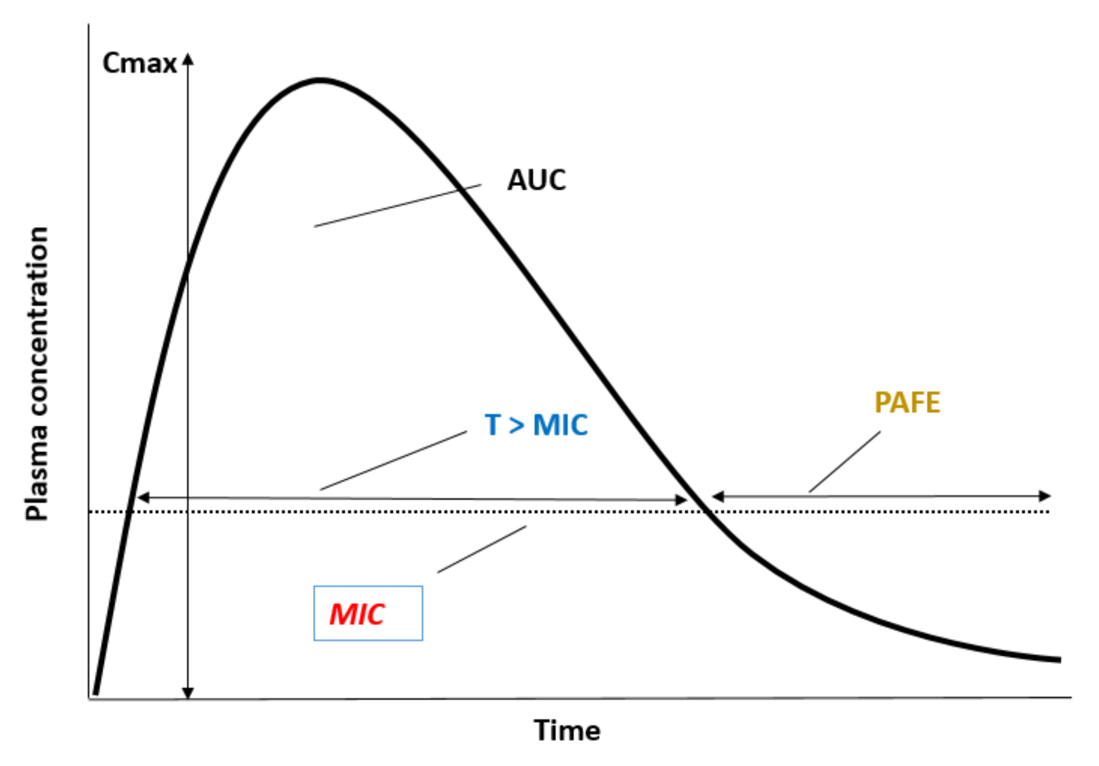
8. Relevance of PAFE in the Design of Dosing Therapeutical Regimens: Clinical Applicability

Successful treatment of candidiasis requires the choice of the most suitable antifungal agent and dose regimen [62]. Optimally dosing antifungal therapy is dependent on several factors, such as pathophysiological characteristics of the patients and their immune status, the infecting organism, the site of infection and the PK/PD properties of the antifungal drug (Figure 2) [70,71,72].
PK includes the factors affecting drug absorption, distribution, metabolism and elimination, which determine the evolution of the drug concentration in the body [70]. Considering PK aspects of antifungal therapy in mucocutaneous and invasive/disseminated candidiasis, the site of infection is the extracellular fluid compartment, and, therefore, serum or plasma drug concentrations correlate well with drug concentrations at the site of infection [49]. The antifungal exposure in plasma can be measured as peak plasma concentration (Cmax) and area under the concentration–time curve over a period of 24 h (AUC0–24h), PK variables that are dependent on the dose used and the PK properties of each antifungal drug. Additionally, only the unbound drug concentrations are able to access to the fungal target and, thus, are microbiologically active [70,71]. At the site of infection, antifungal drugs act according to their MIC and PAFE [71,73]. In the context of antifungal drugs, the clinical relevance of plasma protein binding changes has been assessed for both triazoles and echinocandins [74,75,76].
Echinocandins are administered as single intravenous daily doses [72]. However, other dosing strategies merit study, such as larger doses less frequently administered with the same cumulative doses as the standard daily divided ones, as suggested by Gumbo et al. [2], Andes et al. [77] and Prépost et al. [78]. Although the clinical evidence that supports the usefulness of larger single echinocandin doses is still very scarce, high single doses of caspofungin and micafungin have been shown to be effective in murine models of candidiasis [2,77,78]. Prepost et al. [78] reported excellent in vivo efficacy of high single doses of caspofungin (40 mg/kg) against echinocandin-resistant isolates. They proposed potential explanations for this higher efficacy, such as the increased unbound drug fraction of caspofungin in patients with invasive candidiasis (7.6% vs. 3.5% in healthy persons). This fact leads to higher free-caspofungin AUC/MIC and Cmax/MIC values than what is recommended. Other possible reasons include the rapid increase and higher peak concentrations following large doses [2,79]. These authors highlighted the importance of free echinocandin penetration into the site of infection related to the clinical outcomes. Additionally, it has been suggested that elevated single doses of caspofungin can be used safely [80,81]. Apart from the PK explanations aforementioned, the persistent effect of echinocandins could also be related to the prolonged PAFE observed, both in vitro and in vivo.
Careful consideration of the factors affecting PK/PD should allow the selection of the most appropriate antifungal drug when treating candidiasis and establishing the dosage with a better risk: benefit ratio in terms of efficacy, safety and development of resistance [73,82]. The interindividual variability in PK or PD processes is one of the main contributors to the variability in the antifungal dose–exposure-response relation [72]. Physiopathological factors associated with interindividual variability of the patient, such as age [83,84], renal disease or replacement therapy [85,86], hepatic disease [87,88], transplantation [89,90] and critical illnesses [91,92,93,94], may be responsible for the interindividual variability of the antifungal PK processes. In particular, critically ill patients have pathophysiological changes that are responsible for antifungal PK alterations, such as organ failure, reduced protein binding, capillary leakage resulting in an altered drug volume of distribution and use of organ support [82]. Moreover, interacting co-medications may result in the variable PK of antifungal drugs [95,96].
When considering the factors responsible for PD variability, antifungal susceptibility rates can vary widely among different Candida species [97]. MIC distribution could vary according to whether the species is susceptible, intermediate, susceptible dose-dependent or resistant to the antifungal drug. Interregional, time-dependent or species differences in MIC distribution associated with local epidemiology and resistance patterns [72,98], and factors associated with the variability range of PAFE values summarized in Figure 1, can contribute to the PD variability of the antifungal drug [99]. All these factors that explain the antifungal drug dose–response relationship support the optimization of adequate dosing regimens in clinical practice (Figure 2).
An in-depth understanding of the relationship between antifungal exposure and clinical response is required to establish useful threshold values for clinical outcome and adverse effects. PK/PD analysis integrates both the PK and PD information for drug to enhance the possibility of success of the antifungal therapy [100,101,102]. The maximal concentration (Cmax), the ratio of drug area under the concentration–time curve (AUC) to MIC over a 24 h period (AUC0–24h/MIC) or the time (expressed as a percentage of the dosing interval) that drug concentrations are expected to exceed the MIC (T > MIC) have been used extensively as a PK/PD index that link the kinetics of antimicrobial disposition, MIC and PAFE values and antifungal clinical efficacy. The value of the PK/PD index associated with antimicrobial efficacy varies according to the chosen endpoint, such as stasis, maximal kill or resistance suppression (for preclinical studies), and microbiological or clinical cure (for clinical studies and clinical trials) [70]. Traditionally, Cmax/MIC and AUC0–24h/MIC are the optimal drivers that predict the dosing regimens for concentration-dependent antimicrobials, and T > MIC for time-dependent antimicrobials (Figure 3).
The PAFE, as a PD characteristic, can influence the dose–response relationships (Figure 2) [73,103]. Extended PAFE periods allow dosing intervals to be lengthened. For time-dependent antifungal drugs (%T > MIC), the PAFE reduces reliance on concentrations above the MIC and increases relevance of the total drug exposure or AUC/MIC index. For example, triazoles exhibit prolonged in vivo PAFEs, and it has been shown that AUC/MIC is the optimal predictive index [104], whereas flucytosine exhibits limited PAFE, and, in this case, %T > MIC is the optimal efficacy index [66,102]. For polyenes, Cmax/MIC is the PK/PD driver of efficacy. For concentration-dependent drugs, as echinocandins which exhibit a prolonged PAFE, AUC/MIC and Cmax/MIC can both be optimal predictive drivers of efficacy [101,105,106]. Thus, antifungal drugs, depending on whether they are concentration-dependent or time-dependent drugs and whether their PAFE is prolonged or short, have been shown to have different PK/PD clinical efficacy objectives [72].
These PK/PD indexes can be used as a tool to guide the selection of dosing regimens in the studied population, increasing the probability of selecting clinically successful treatments, identifying clinical breakpoints and preventing the emergence of resistance [70,77,98]. Population PK/PD analyses in combination with Monte Carlo simulation [107], incorporating antifungal PK and PD variability, can be combined to computationally estimate the likelihood of a given drug dose to attain a predefined value of a PK/PD target previously defined for antimicrobial drugs [108]. With this clinical translational tool, several clinical objectives can be achieved as dosing regimens expected to have success for a special group of patients, identify drug exposures that may be inadequate, or identify alternative dosing strategies to enhance efficacy (such as dose escalation or increased frequency according PAFE) [108,109,110].
In the case of invasive candidiasis, the clinical validation of PK/PD targets is a final and necessary step to harness the full translational potential of these studies [103]. Although knowledge of the PK/PD of antifungal drugs is increasing due to the many studies in vitro and in experimental animal models being conducted, the practical application of these concepts to individualized dosing at the point of care is still limited. Studies on the PK/PD of 5-fluorocytosine and amphotericin B are quite limited in experimental models concerning invasive candidiasis [102]. Regarding the azole group, fluconazole is the most extensively studied drug using experimental animal models with invasive candidiasis. Its PK/PD index related to efficacy, evaluated in experimental animal models, has been validated in invasive candidiasis through clinical practice. However, data on voriconazole and posaconazole regarding this topic are quite limited. Although a robust PK/PD index of echinocandins have been identified in the laboratory, the translation to the clinical treatment of invasive candidiasis is currently limited [102,106]. Patients with oesophageal candidiasis treated with the standard daily dosing schedule of micafungin showed no difference with a large dosing at prolonged intervals, consistent with animal studies providing AUC/MIC as a predictor of efficacy [77]. The practical application of these concepts to individualized dosing at the point of care is still limited. PAFE as a PD feature and its variability should be considered.

9. Conclusions and Proposals for Further Studies

Invasive candidiasis is increasing; highly related to the increase in patients at risk of these infections, such as critically ill patients. These infections are often refractory to the currently available antifungal therapies, as fungal resistance becomes an increasing obstacle to therapy. In order to expand therapeutic options, the selection of the optimal antifungal administration is an important tool in dealing with these infections. In this respect, PAFE has been studied for its potential impact in antifungal drug dosing.
The PAFE attempts to capture the persistent activity of the drug once the isolates are exposed to it, despite the absence of measurable concentrations of the drug in contact with the fungus. In this work, various in vitro and in vivo studies that have determined the PAFE of the main antifungal agents have been compiled. Each model, in vitro and in vivo, has its pros and cons in their ability to assess the postantifungal activity of the drugs, especially when trying to reproduce the results in human infections. Variations in the in vitro/in vivo correlation results may be due to different factors leading to in vitro methods not being able to reproduce the conditions at the site of infection. These factors or conditions include the inoculum, the interaction between drug, pathogen and host immune system, and the PK properties of the drug, among others. In addition, there is no standard method for PAFE determination, hence, standard methods should be defined in order to reduce inter-study variability. Among the issues identified that would require a higher degree of standardization are the medium in which in vitro experiments are carried out. This medium should mimic the conditions of the dynamic environment in the body. Although most PAFE results are obtained in protein-free RPMI medium, several studies have demonstrated that drug exposures in the presence of serum mimic in vivo conditions more reliably. The ideal situation would certainly be to complete and verify in vitro studies with in vivo models, which are closer to human candidiasis. The infection site most commonly used when determining the PAFE in murine models is the kidney. However, in these neutropenic mouse models, when interpreting PAFE results from drugs with a wide tissue distribution, caution should be exercised. In the case of amphotericin B, its high distribution to the kidney is widely known and concentrations reached in kidneys can be much higher than in serum. Hence, it may appear that the antifungal effect continues despite no measurable drug in serum, whereas drug concentrations at the infection site may persist above the MIC and continue to show activity. In these in vivo studies, the state of free drug in the tissues should be verified in order to be able to attribute persistent drug activity to a true PAFE. Additionally, this review has detected a scarcity of PAFE studies in animal models. More in vivo studies should be conducted to explore their translational relevance as a tool that helps to establish the clinical dosing rationale.
After comparing the PAFE of the main groups of antifungal drugs, it can be stated that the echinocandins cause the most prolonged PAFE, followed by polyenes and azoles. In the case of the triazoles, it is worth noting the inconsistency found between in vitro and in vivo studies for the same drug, possibly due to difficulty in differentiating between continued growth suppression related to serum concentrations above MIC and sub-MIC effect. In vivo PAFE studies of triazoles have revealed a longer PAFE. On the other hand, the newest oral and intravenous antifungal drug, ibrexafungerp, should be mentioned. Ibrexafungerp is the first triterpenoid class antifungal agent labeled for the treatment of vulvovaginitis [111]. Although it has been shown that ibrexafungerp displays potent in vitro and in vivo activity against the most clinically relevant species of Candida [112], so far, no PAFE studies have been published for this new drug. This semi-synthetic derivative of the terpenoid enfumafungin, acts by inhibiting glucan synthase, decreasing 1,3-β-D-glucan polymers and weakening fungal cell wall [113]. This mechanism of action, similar to that previously described for the echinocandin group, might suggest that ibrexafungerp could also exert a PAFE. However, this should be studied both in vitro and in vivo. In general, such PAFE studies should be carried out for all drugs to be approved by regulatory agencies, given their possible implications for the design of dosing regimens.
On the other hand, considering that there might be species-specific variability in PAFE results, it would be interesting to further evaluate the PAFE of the different antifungal drugs against non-C. albicans species, as most PAFE studies have been carried out on C. albicans. Some studies have detected a greater PAFE of polyenes on Candida species other than C. albicans. These differences could be explained by slight changes in the structure of the different species and to the higher virulence of C. albicans, which would allow a faster recovery of growth after short exposure to drugs. On the other hand, differences in methodological issues among studies, such as the drug concentrations used, preclude species-dependent PAFE comparisons among studies. Moreover, PAFE has been studied, but not extensively, for fungi other than Candida, such as Aspergillus, other filamentous fungi and zygomizetes. As remarked in those studies, PAFE appears to be dependent on several factors, such as the concentration of the drug and class of drug, exposure time or media used [114,115,116].
The PAFE, as an exposure–response factor, can impact on the PD relationships. 5-Fluorocytosine exhibits limited or short PAFE and T > MIC is the PK/PD index best related to efficacy, whereas triazoles show prolonged in vivo PAFE and concentration-independent action, suggesting that AUC/MIC is the optimal predictive driver of efficacy. In contrast, for polyenes, Cmax/MIC is the PK/PD driver of efficacy. For echinocandins, with concentration-dependent activity and prolonged PAFE, both Cmax/MIC and AUC/MIC are optimal predictors of efficacy. Additionally, the dosing intervals impact on the treatment efficacy and can determine the PK/PD index. It has been demonstrated that when higher doses are administered with larger dosing intervals and treatment outcome improves, then the Cmax/MIC is the optimal efficacy index. When treatment effect is similar among several dosing intervals, the AUC/MIC index is the predictive index. From a clinical point of view, this could have applicability for echinocandins, concentration-dependent and prolonged PAFE drugs. When the standard daily dosing regimen of micafungin was compared to an extended interval large dose regimen in patients with esophageal candidiasis, no statistically different outcomes were obtained between both dosing schedules, in agreement with animal studies providing AUC/MIC as the efficacy driver.
In conclusion, PAFE is a relevant exposure–response variable to understand and optimize antifungal efficacy in vivo, and it should be taken into account in therapeutic decisions related to dosage regimens. However, further in vivo studies are warranted, including experiments in animal models of candidiasis. These could be taken as a starting point for the study of extended interval dosing regimens, as is the case of echinocandins for treating mycoses.

Author Contributions

Conceptualization, N.J., G.Q. and E.E.; writing—original draft preparation, N.J., S.G.-A., E.S. (Elena Suárez), E.S. (Elena Sevillano) and E.E.; writing—review and editing, N.J., G.Q., S.G.-A., E.S. (Elena Suárez), E.S. (Elena Sevillano) and E.E.; supervision, N.J., G.Q. and E.E.; funding acquisition, N.J., G.Q. and E.E. All authors have read and agreed to the published version of the manuscript.

Funding

The research group was funded by the Consejería de Educación, Universidades e Investigación of Gobierno Vasco-Eusko Jaurlaritza (GIC15/78 IT-990-16/IT1607-22) and by the Spanish Ministry of Science and Innovation (PID2020-117983RB-I00).

Institutional Review Board Statement

Not applicable.

Informed Consent Statement

Not applicable.

Data Availability Statement

Not applicable.

Conflicts of Interest

Outside the current study, we declare the following potential conflicts: Guillermo Quindós has received research grants from Astellas Pharma, Pfizer, Merck Sharp & Dohme, and Scynexis. Guillermo Quindós has served on advisory/consultant boards for Merck, Sharp & Dohme, and Scynexis, and he has received speaker honoraria from Abbvie, Astellas Pharma, Merck Sharp & Dohme, Pfizer, and Scynexis. All authors declare no other competing interests.

References

  1. Quindós, G. Micología Médica, 1st ed.; Elsevier: Barcelona, Spain, 2015. [Google Scholar]
  2. Gumbo, T.; Drusano, G.L.; Liu, W.; Kulawy, R.W.; Fregeau, C.; Hsu, V.; Louie, A. Once-weekly micafungin therapy is as effective as daily therapy for disseminated candidiasis in mice with persistent neutropenia. Antimicrob. Agents Chemother. 2007, 51, 968–974. [Google Scholar] [CrossRef] [PubMed] [Green Version]
  3. Oz, Y.; Kiremitci, A.; Dag, I.; Metintas, S.; Kiraz, N. Postantifungal effect of the combination of caspofungin with voriconazole and amphotericin B against clinical Candida krusei isolates. Med. Mycol. 2013, 51, 60–65. [Google Scholar] [CrossRef] [PubMed] [Green Version]
  4. Chryssanthou, E.; Cars, O.; Sjölin, J. New automated method for determining postantifungal effect of amphotericin B against Candida species: Effects of concentration, exposure time, and area under the curve. Antimicrob. Agents Chemother. 2002, 46, 4016–4018. [Google Scholar] [CrossRef] [PubMed] [Green Version]
  5. Ellepola, A.N.; Samaranayake, L.P. The postantifungal effect (PAFE) of antimycotics on oral C. albicans isolates and its impact on candidal adhesion. Oral Dis. 1998, 4, 260–267. [Google Scholar] [CrossRef] [PubMed]
  6. Anil, S.; Ellepola, A.N.; Samaranayake, L.P. Post-antifungal effect of polyene, azole and DNA-analogue agents against oral Candida albicans and Candida tropicalis isolates in HIV disease. J. Oral Pathol. Med. 2001, 30, 481–488. [Google Scholar] [CrossRef] [PubMed]
  7. Shu, M.; Ellepola, A.N.; Samaranayake, L.P. Effects of two different growth media on the postantifungal effect induced by polyenes on Candida species. J. Clin. Microbiol. 2001, 39, 2732–2735. [Google Scholar] [CrossRef] [PubMed] [Green Version]
  8. Ozkutuk, A.; Dogan, Y.; Kirdar, S.; Ozkutuk, N. Changes in some Candida albicans virulence factors due to amphotericin-B post-antifungal effect. J. Chemother. 2007, 19, 101–103. [Google Scholar] [CrossRef]
  9. Ellepola, A.N. Amphotericin B-induced in vitro postantifungal effect on Candida species of oral origin. Med. Princ. Pract. 2012, 21, 442–446. [Google Scholar] [CrossRef]
  10. Ellepola, A.N.; Joseph, B.K.; Chandy, R.; Khan, Z.U. The postantifungal effect of nystatin and its impact on adhesion attributes of oral Candida dubliniensis isolates. Mycoses 2014, 57, 56–63. [Google Scholar] [CrossRef]
  11. Ellepola, A.N.; Chandy, R.; Khan, Z.U. In vitro postantifungal effect, adhesion traits and haemolysin production of Candida dubliniensis isolates following exposure to 5-fluorocytosine. Mycoses 2015, 58, 536–543. [Google Scholar] [CrossRef]
  12. Ellepola, A.N.; Chandy, R.; Khan, Z.U. Post-antifungal effect and adhesion to buccal epithelial cells of oral Candida dubliniensis isolates subsequent to limited exposure to amphotericin B, ketoconazole and fluconazole. J. Investig. Clin. Dent. 2015, 6, 186–192. [Google Scholar] [CrossRef]
  13. Ellepola, A.N.; Chandy, R.; Khan, Z.U.; Samaranayake, L.P. Caspofungin-induced in-vitro post-antifungal effect and its impact on adhesion related traits of oral Candida dubliniensis and Candida albicans isolates. Microbiol. Immunol. 2016, 60, 160–167. [Google Scholar] [CrossRef] [PubMed] [Green Version]
  14. Ellepola, A.N.B.; Dassanayake, R.S.; Khan, Z. In vitro Post-Antifungal Effect of Posaconazole and Its Impact on Adhesion-Related Traits and Hemolysin Production of Oral Candida dubliniensis isolates. Med. Princ. Pract. 2019, 28, 552–558. [Google Scholar] [CrossRef] [PubMed]
  15. Ernst, E.J.; Klepser, M.E.; Pfaller, M.A. Postantifungal effects of echinocandin, azole, and polyene antifungal agents against Candida albicans and Cryptococcus neoformans. Antimicrob. Agents Chemother. 2000, 44, 1108–1111. [Google Scholar] [CrossRef] [PubMed] [Green Version]
  16. Zhanel, G.G.; Saunders, D.G.; Hoban, D.J.; Karlowsky, J.A. Influence of human serum on antifungal pharmacodynamics with Candida albicans. Antimicrob. Agents Chemother. 2001, 45, 2018–2022. [Google Scholar] [CrossRef] [Green Version]
  17. García, M.T.; Llorente, M.T.; Mínguez, F.; Prieto, J. Postantifungal effect and effects of sub-MIC concentrations on previously treated Candida sp. Influence of growth phase. J. Infect. 2002, 45, 263–267. [Google Scholar] [CrossRef]
  18. García, M.T.; Llorente, M.T.; Mínguez, F.; Prieto, J. Post-antifungal effect and effects of sub-MIC concentrations on previously treated Candida spp.: Influence of exposure time and concentration. Scand. J. Infect Dis. 2002, 34, 197–200. [Google Scholar] [CrossRef]
  19. Di Bonaventura, G.; Spedicato, I.; Picciani, C.; D’Antonio, D.; Piccolomini, R. In vitro pharmacodynamic characteristics of amphotericin B, caspofungin, fluconazole, and voriconazole against bloodstream isolates of infrequent Candida species from patients with hematologic malignancies. Antimicrob. Agents Chemother. 2004, 48, 4453–4456. [Google Scholar] [CrossRef] [Green Version]
  20. Manavathu, E.K.; Ramesh, M.S.; Baskaran, I.; Ganesan, L.T.; Chandrasekar, P.H. A comparative study of the post-antifungal effect (PAFE) of amphotericin B, triazoles and echinocandins on Aspergillus fumigatus and Candida albicans. J. Antimicrob. Chemother. 2004, 53, 386–389. [Google Scholar] [CrossRef] [Green Version]
  21. Clancy, C.J.; Huang, H.; Cheng, S.; Derendorf, H.; Nguyen, M.H. Characterizing the effects of caspofungin on Candida albicans, Candida parapsilosis, and Candida glabrata isolates by simultaneous time-kill and postantifungal-effect experiments. Antimicrob. Agents Chemother. 2006, 50, 2569–2572. [Google Scholar] [CrossRef] [Green Version]
  22. Li, Y.; Nguyen, M.H.; Derendorf, H.; Cheng, S.; Clancy, C.J. Measurement of voriconazole activity against Candida albicans, C. glabrata, and C. parapsilosis isolates using time-kill methods validated by high-performance liquid chromatography. Antimicrob. Agents Chemother. 2007, 51, 2985–2987. [Google Scholar] [CrossRef] [PubMed] [Green Version]
  23. Nguyen, K.T.; Ta, P.; Hoang, B.T.; Cheng, S.; Hao, B.; Nguyen, M.H.; Clancy, C.J. Anidulafungin is fungicidal and exerts a variety of postantifungal effects against Candida albicans, C. glabrata, C. parapsilosis, and C. krusei isolates. Antimicrob. Agents Chemother. 2009, 53, 3347–3352. [Google Scholar] [CrossRef] [PubMed] [Green Version]
  24. Nguyen, K.T.; Ta, P.; Hoang, B.T.; Cheng, S.; Hao, B.; Nguyen, M.H.; Clancy, C.J. Characterising the post-antifungal effects of micafungin against Candida albicans, Candida glabrata, Candida parapsilosis and Candida krusei isolates. Int. J. Antimicrob. Agents 2010, 35, 80–84. [Google Scholar] [CrossRef] [PubMed]
  25. Shields, R.K.; Nguyen, M.H.; Press, E.G.; Clancy, C.J. Five-minute exposure to caspofungin results in prolonged postantifungal effects and eliminates the paradoxical growth of Candida albicans. Antimicrob. Agents Chemother. 2011, 55, 3598–3602. [Google Scholar] [CrossRef] [Green Version]
  26. Smith, R.P.; Baltch, A.; Bopp, L.H.; Ritz, W.J.; Michelsen, P.P. Post-antifungal effects and time-kill studies of anidulafungin, caspofungin, and micafungin against Candida glabrata and Candida parapsilosis. Diagn. Microbiol. Infect Dis. 2011, 71, 131–138. [Google Scholar] [CrossRef]
  27. Moriyama, B.; Henning, S.A.; Penzak, S.R.; Walsh, T.J. The postantifungal and paradoxical effects of echinocandins against Candida spp. Future Microbiol. 2012, 7, 565–569. [Google Scholar] [CrossRef]
  28. Kovacs, R.; Gesztelyi, R.; Perlin, D.S.; Kardos, G.; Doman, M.; Berenyi, R.; Majoros, L. Killing rates for caspofungin against Candida albicans after brief and continuous caspofungin exposure in the presence and absence of serum. Mycopathologia 2014, 178, 197–206. [Google Scholar] [CrossRef] [Green Version]
  29. Gil-Alonso, S.; Jauregizar, N.; Eraso, E.; Quindós, G. Postantifungal effect of micafungin against the species complexes of Candida albicans and Candida parapsilosis. PLoS ONE 2015, 10, e0132730. [Google Scholar] [CrossRef]
  30. Gil-Alonso, S.; Jauregizar, N.; Eraso, E.; Quindós, G. Postantifungal effect of caspofungin against the Candida albicans and Candida parapsilosis clades. Diagn. Microbiol. Infect. Dis. 2016, 86, 172–177. [Google Scholar] [CrossRef]
  31. Kardos, T.; Saleh, Q.; Kovacs, R.; Gesztelyi, R.; Kardos, G.; Bozo, A.; Majoros, L. Postantifungal effect of micafungin against Candida albicans, Candida dubliniensis and Candida Africana in the presence and absence of serum. New Microbiol. 2017, 40, 286–288. [Google Scholar]
  32. Gil-Alonso, S.; Quindós, G.; Eraso, E.; Jauregizar, N. Postantifungal effect of anidulafungin against Candida albicans, Candida dubliniensis, Candida africana, Candida parapsilosis, Candida metapsilosis and Candida orthopsilosis. Rev. Esp. Quimioter. 2019, 32, 183–188. [Google Scholar] [PubMed]
  33. Gowri, M.; Jayashree, B.; Jeyakanthan, J.; Girija, E.K. Sertraline as a promising antifungal agent: Inhibition of growth and biofilm of Candida auris with special focus on the mechanism of action in vitro. J. Appl. Microbiol. 2020, 128, 426–437. [Google Scholar] [CrossRef] [PubMed]
  34. García, M.T.; Llorente, M.T.; Mínguez, F.; Prieto, J. Influence of temperature and concentration on the postantifungal effect and the effects of sub-MIC concentrations of four antifungal agents on previously treated Candida species. Chemotherapy 2000, 46, 245–252. [Google Scholar] [CrossRef] [PubMed]
  35. Scalarone, G.M.; Mikami, Y.; Kurita, N.; Yazawa, K.; Uno, J.; Miyaji, M. In vitro comparative evaluations of the postantifungal effect: Synergistic interaction between flucytosine and fluconazole against Candida albicans. Mycoses 1991, 34, 405–410. [Google Scholar] [CrossRef]
  36. Mikami, Y.; Scalarone, G.M.; Kurita, N.; Yazawa, K.; Uno, J.; Miyaji, M. Synergistic postantifungal effect of flucytosine and fluconazole on Candida albicans. J. Med. Vet. Mycol. 1992, 30, 197–206. [Google Scholar] [CrossRef]
  37. Scalarone, G.M.; Mikami, Y.; Kurita, N.; Yazawa, K.; Miyaji, M. Comparative studies on the postantifungal effect produced by the synergistic interaction of flucytosine and amphotericin B on Candida albicans. Mycopathologia 1992, 120, 133–138. [Google Scholar] [CrossRef] [PubMed]
  38. Hoffman, A.; Stepensky, D. Pharmacodynamic aspects of modes of drug administration for optimization of drug therapy. Crit. Rev. Ther. Drug Carrier Syst. 1999, 16, 571–639. [Google Scholar] [CrossRef]
  39. Venisse, N.; Gregoire, N.; Marliat, M.; Couet, W. Mechanism-based pharmacokinetic-pharmacodynamic models of in vitro fungistatic and fungicidal effects against Candida albicans. Antimicrob. Agents Chemother. 2008, 52, 937–943. [Google Scholar] [CrossRef] [Green Version]
  40. Foldi, R.; Kovacs, R.; Gesztelyi, R.; Kardos, G.; Berenyi, R.; Juhasz, B.; Szilágyi, J.; Mózes, J.; Majoros, L. Comparison of in vitro and vivo efficacy of caspofungin against Candida parapsilosis, C. orthopsilosis, C. metapsilosis and C. albicans. Mycopathologia 2012, 174, 311–318. [Google Scholar] [CrossRef]
  41. Spreghini, E.; Orlando, F.; Tavanti, A.; Senesi, S.; Giannini, D.; Manso, E.; Barchiesi, F. In vitro and in vivo effects of echinocandins against Candida parapsilosis sensu stricto, Candida orthopsilosis and Candida metapsilosis. J. Antimicrob. Chemother. 2012, 67, 2195–2202. [Google Scholar] [CrossRef] [Green Version]
  42. Tóth, Z.; Kardos, T.; Kovács, R.; Kardos, G.; Nagy, F.; Prépost, E.; Barta, Z.; Takacs, I.; Majoros, L. comparison of killing activity of micafungin against six candida species isolated from peritoneal and pleural cavities in RPMI-1640, 10 and 30% serum. Mycopathologia 2018, 183, 905–912. [Google Scholar] [CrossRef] [PubMed]
  43. Kovács, R.; Tóth, Z.; Locke, J.B.; Forgács, L.; Kardos, G.; Nagy, F.; Borman, A.; Majoros, L. comparison of in vitro killing activity of rezafungin, anidulafungin, caspofungin, and micafungin against four Candida auris clades in RPMI-1640 in the absence and presence of human serum. Microorganisms 2021, 9, 863. [Google Scholar] [CrossRef] [PubMed]
  44. Mínguez, F.; García, M.T.; Lima, J.E.; Llorente, M.T.; Prieto, J. Postantifungal effect and effects of low concentrations of amphotericin B and fluconazole on previously treated Candida albicans. Scand. J. Infect. Dis. 1996, 28, 503–506. [Google Scholar] [CrossRef] [PubMed]
  45. Mínguez, F.; Lima, J.E.; García, M.T.; Prieto, J. Influence of human serum on the postantifungal effect of four antifungal agents on Candida albicans. Chemotherapy 1996, 42, 273–279. [Google Scholar] [CrossRef]
  46. Jota Baptista, C.V.; Faustino-Rocha, A.I.; Oliveira, P.A. Animal models in pharmacology: A brief history awarding the nobel prizes for physiology or medicine. Pharmacology 2021, 106, 356–368. [Google Scholar] [CrossRef]
  47. Andes, D.; Marchillo, K.; Stamstad, T.; Conklin, R. In vivo pharmacodynamics of a new triazole, ravuconazole, in a murine candidiasis model. Antimicrob. Agents Chemother. 2003, 47, 1193–1199. [Google Scholar] [CrossRef] [Green Version]
  48. Andes, D.; Marchillo, K.; Lowther, J.; Bryskier, A.; Stamstad, T.; Conklin, R. In vivo pharmacodynamics of HMR 3270, a glucan synthase inhibitor, in a murine candidiasis model. Antimicrob. Agents Chemother. 2003, 47, 1187–1192. [Google Scholar] [CrossRef] [Green Version]
  49. Andes, D.; Marchillo, K.; Conklin, R.; Krishna, G.; Ezzet, F.; Cacciapuoti, A.; Loebenberg, D. Pharmacodynamics of a new triazole, posaconazole, in a murine model of disseminated candidiasis. Antimicrob. Agents Chemother. 2004, 48, 137–142. [Google Scholar] [CrossRef] [Green Version]
  50. Andes, D.; Diekema, D.J.; Pfaller, M.A.; Prince, R.A.; Marchillo, K.; Ashbeck, J.; Hou, J. In vivo pharmacodynamic characterization of anidulafungin in a neutropenic murine candidiasis model. Antimicrob. Agents Chemother. 2008, 52, 539–550. [Google Scholar] [CrossRef] [Green Version]
  51. Warn, P.A.; Sharp, A.; Parmar, A.; Majithiya, J.; Denning, D.W.; Hope, W.W. Pharmacokinetics and pharmacodynamics of a novel triazole, isavuconazole: Mathematical modeling, importance of tissue concentrations, and impact of immune status on antifungal effect. Antimicrob. Agents Chemother. 2009, 53, 3453–3461. [Google Scholar] [CrossRef] [Green Version]
  52. Maki, K.; Kaneko, S. Effective concentration-based serum pharmacodynamics for antifungal azoles in a murine model of disseminated Candida albicans infection. Eur. J. Drug Metab. Pharmacokinet. 2013, 38, 261–268. [Google Scholar] [CrossRef] [PubMed] [Green Version]
  53. Cavassin, F.B.; Baú-Carneiro, J.L.; Vilas-Boas, R.R.; Queiroz-Telles, F. Sixty years of amphotericin B: An overview of the main antifungal agent used to treat invasive fungal infections. Infect. Dis. Ther. 2021, 10, 115–147. [Google Scholar] [CrossRef] [PubMed]
  54. Johnson, M.D. Antifungals in clinical use and the pipeline. Infect. Dis. Clin. North Am. 2021, 35, 341–371. [Google Scholar] [CrossRef] [PubMed]
  55. Turnidge, J.D.; Gudmundsson, S.; Vogelman, B.; Craig, W.A. The postantibiotic effect of antifungal agents against common pathogenic yeasts. J. Antimicrob. Chemother. 1994, 34, 83–92. [Google Scholar] [CrossRef]
  56. Egusa, H.; Ellepola, A.N.; Nikawa, H.; Hamada, T.; Samaranayake, L.P. Sub-therapeutic exposure to polyene antimycotics elicits a post-antifungal effect (PAFE) and depresses the cell surface hydrophobicity of oral Candida albicans isolates. J. Oral Pathol. Med. 2000, 29, 206–213. [Google Scholar] [CrossRef] [PubMed]
  57. Ellepola, A.N.; Khajah, R.; Jayatilake, S.; Samaranayake, L.; Sharma, P.; Khan, Z. Impact of brief exposure to antifungal agents on the post-antifungal effect and hemolysin activity of oral Candida albicans. J. Appl. Oral Sci. 2015, 23, 412–418. [Google Scholar] [CrossRef]
  58. Ellepola, A.N.; Samaranayake, L.P. The in vitro post-antifungal effect of nystatin on Candida species of oral origin. J. Oral Pathol. Med. 1999, 28, 112–116. [Google Scholar] [CrossRef]
  59. Samaranayake, Y.H.; Yau, J.Y.; Thein, Z.M.; Jayatilake, J.A.; Yeung, K.W.; Samaranayake, L.P. The post-antifungal effect (PAFE) of amphotericin B, nystatin, ketoconazole and 5-fluorocytosine and its impact on the colonization traits of Candida glabrata. Med. Mycol. 2010, 48, 725–734. [Google Scholar] [CrossRef] [Green Version]
  60. Andes, D.; van Ogtrop, M. Characterization and quantitation of the pharmacodynamics of fluconazole in a neutropenic murine disseminated candidiasis infection model. Antimicrob. Agents Chemother. 1999, 43, 2116–2120. [Google Scholar] [CrossRef] [Green Version]
  61. ClinicalTrial.gov Rezafungin. Available online: https://clinicaltrials.gov/ct2/results?cond=rezafungin&term=&cntry=&state=&city=&dist= (accessed on 16 May 2022).
  62. Pappas, P.G.; Kauffman, C.A.; Andes, D.R.; Clancy, C.J.; Marr, K.A.; Ostrosky-Zeichner, L.; Reboli, A.C.; Schuster, M.G.; Vazquez, J.A.; Walsh, T.J.; et al. Clinical Practice guideline for the management of candidiasis: 2016 Update by the Infectious Diseases Society of America. Clin. Infect. Dis. 2016, 62, 1. [Google Scholar] [CrossRef]
  63. Ernst, E.J.; Roling, E.E.; Petzold, C.R.; Keele, D.J.; Klepser, M.E. In vitro activity of micafungin (FK-463) against Candida spp.: Microdilution, time-kill, and postantifungal-effect studies. Antimicrob. Agents Chemother. 2002, 46, 3846–3853. [Google Scholar] [CrossRef] [PubMed] [Green Version]
  64. Lepak, A.J.; Zhao, M.; Andes, D.R. Pharmacodynamic Evaluation of rezafungin (CD101) against Candida auris in the neutropenic mouse invasive candidiasis model. Antimicrob Agents Chemother. 2018, 62, e01572-18. [Google Scholar] [CrossRef] [PubMed] [Green Version]
  65. Miesel, L.; Lin, K.Y.; Ong, V. Rezafungin treatment in mouse models of invasive candidiasis and aspergillosis: Insights on the PK/PD pharmacometrics of rezafungin efficacy. Pharmacol. Res. Perspect. 2019, 7, e00546. [Google Scholar] [CrossRef] [PubMed]
  66. Andes, D.; van Ogtrop, M. In vivo characterization of the pharmacodynamics of flucytosine in a neutropenic murine disseminated candidiasis model. Antimicrob. Agents Chemother. 2000, 44, 938–942. [Google Scholar] [CrossRef] [PubMed] [Green Version]
  67. Louie, A.; Deziel, M.; Liu, W.; Drusano, M.F.; Gumbo, T.; Drusano, G.L. Pharmacodynamics of caspofungin in a murine model of systemic candidiasis: Importance of persistence of caspofungin in tissues to understanding drug activity. Antimicrob. Agents Chemother. 2005, 12, 5058–5068. [Google Scholar] [CrossRef] [Green Version]
  68. Khan, M.A.; Khan, A.; Khan, S.H.; Azam, M.; Khan, M.M.U.; Khalilullah, H.; Younus, H. Coadministration of liposomal methylglyoxal increases the activity of amphotericin B against Candida albicans in leukopoenic mice. J. Drug Target. 2021, 29, 78–87. [Google Scholar] [CrossRef]
  69. Mesa-Arango, A.C.; Rueda, C.; Román, E.; Quintin, J.; Terrón, M.C.; Luque, D.; Netea, M.G.; Pla, J.; Zaragoza, O. Cell wall changes in amphotericin B-resistant strains from Candida tropicalis and relationship with the immune responses elicited by the host. Antimicrob. Agents Chemother. 2016, 60, 2326–2335. [Google Scholar] [CrossRef] [Green Version]
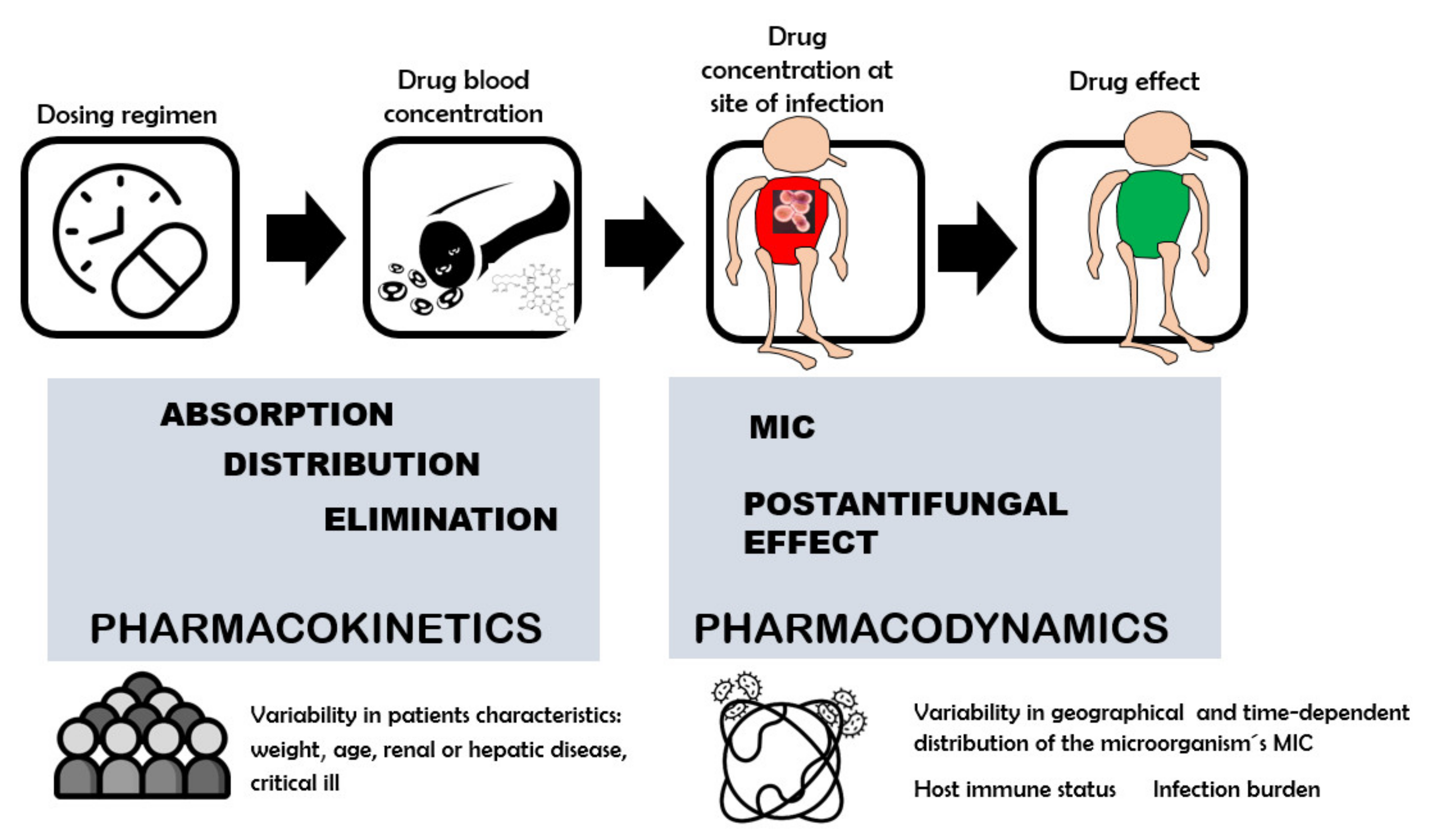
  70. Asín-Prieto, E.; Rodríguez-Gascón, A.; Isla, A. Applications of the pharmacokinetic/pharmacodynamic (PK/PD) analysis of antimicrobial agents. J. Infect. Chemother. 2015, 21, 319–329. [Google Scholar] [CrossRef]
  71. Sy, S.K.; Zhuang, L.; Derendorf, H. Pharmacokinetics and pharmacodynamics in antibiotic dose optimization. Expert Opin. Drug Metab. Toxicol. 2016, 12, 93–114. [Google Scholar] [CrossRef]
  72. Lewis, R.E.; Andes, D.R. Managing uncertainty in antifungal dosing: Antibiograms, therapeutic drug monitoring and drug-drug interactions. Curr. Opin. Infect. Dis. 2021, 34, 288–296. [Google Scholar] [CrossRef]
  73. McAleenan, A.; Ambrose, P.G.; Bhavnani, S.M.; Drusano, G.L.; Hope, W.W.; Mouton, J.W.; Higgins, J.P.T.; MacGowan, A.P. Methodological features of clinical pharmacokinetic-pharmacodynamic studies of antibacterials and antifungals: A systematic review. J. Antimicrob. Chemother. 2020, 75, 1374–1389. [Google Scholar] [CrossRef]
  74. Arredondo, G.; Suárez, E.; Calvo, R.; Vazquez, J.A.; García-Sanchez, J.; Martinez-Jordá, R. Serum protein binding of itraconazole and fluconazole in patients with diabetes mellitus. J. Antimicrob. Chemother. 1999, 43, 305–307. [Google Scholar] [CrossRef] [PubMed] [Green Version]
  75. Florent, A.; Gandia, P.; Seraissol, P.; Chatelut, E.; Houin, G. Determination of plasma unbound fraction of voriconazole in patients treated with a prophylactic or a curative treatment. Ther. Drug Monit. 2014, 36, 752–758. [Google Scholar] [CrossRef] [PubMed]
  76. Yamasaki, K.; Sakurama, K.; Nishi, K.; Tsukigawa, K.; Seo, H.; Otagiri, M.; Taguchi, K. An in-vitro comparative study of the binding of caspofungin and micafungin to plasma proteins. J. Pharm. Pharmacol. 2022, 74, 88–93. [Google Scholar] [CrossRef] [PubMed]
  77. Andes, D. Optimizing antifungal choice and administration. Curr. Med. Res. Opin. 2013, 29 (Suppl. 4), 13–18. [Google Scholar] [CrossRef] [Green Version]
  78. Prépost, E.; Tóth, Z.; Perlin, D.S.; Gesztelyi, R.; Kardos, G.; Kovács, R.; Nagy, F.; Forgács, L.; Majoros, L. Efficacy of humanized single large doses of caspofungin on the lethality and fungal tissue burden in a deeply neutropenic murine model against Candida albicans and Candida dubliniensis. Infect. Drug Resist. 2019, 12, 1805–1814. [Google Scholar] [CrossRef] [Green Version]
  79. Zhao, Y.; Prideaux, B.; Nagasaki, Y.; Lee, M.H.; Chen, P.Y.; Blanc, L.; Ho, H.; Clancy, C.J.; Nguyen, M.H.; Dartois, V.; et al. Unraveling drug penetration of echinocandin antifungals at the site of infection in an intra-abdominal abscess model. Antimicrob. Agents Chemother. 2017, 61, e01009-17. [Google Scholar] [CrossRef] [Green Version]
  80. CANCIDAS, INN-Caspofungin (as Acetate)–Europa EU. Available online: https://www.ema.europa.eu/en/documents/product-information/cancidas-epar-product-information_en.pdf (accessed on 18 May 2022).
  81. Cornely, O.A.; Vehreschild, J.J.; Vehreschild, M.J.; Würthwein, G.; Arenz, D.; Schwartz, S.; Heussel, C.P.; Silling, G.; Mahne, M.; Franklin, J.; et al. Phase II dose escalation study of caspofungin for invasive aspergillosis. Antimicrob. Agents Chemother. 2011, 55, 5798–5803. [Google Scholar] [CrossRef] [Green Version]
  82. Chatelon, J.; Cortegiani, A.; Hammad, E.; Cassir, N.; Leone, M. Choosing the right antifungal agent in ICU patients. Adv. Ther. 2019, 36, 3308–3320. [Google Scholar] [CrossRef] [Green Version]
  83. Hope, W.W.; Kaibara, A.; Roy, M.; Arrieta, A.; Azie, N.; Kovanda, L.L.; Benjamin, D.K., Jr. Population pharmacokinetics of micafungin and its metabolites M1 and M5 in children and adolescents. Antimicrob. Agents Chemother. 2015, 59, 905–913. [Google Scholar] [CrossRef] [Green Version]
  84. Yanni, S.B.; Smith, P.B.; Benjamin, D.K., Jr.; Augustijns, P.F.; Thakker, D.R.; Annaert, P.P. Higher clearance of micafungin in neonates compared with adults: Role of age-dependent micafungin serum binding. Biopharm. Drug Dispos. 2011, 32, 222–232. [Google Scholar] [CrossRef] [PubMed] [Green Version]
  85. Ittel, T.H.; Legler, U.F.; Polak, A.; Glöckner, W.M.; Sieberth, H.G. 5-Fluorocytosine kinetics in patients with acute renal failure undergoing continuous hemofiltration. Chemotherapy 1987, 33, 77–84. [Google Scholar] [CrossRef] [PubMed]
  86. Weiler, S.; Seger, C.; Pfisterer, H.; Stienecke, E.; Stippler, F.; Welte, R.; Joannidis, M.; Griesmacher, A.; Bellmann, R. Pharmacokinetics of caspofungin in critically ill patients on continuous renal replacement therapy. Antimicrob. Agents Chemother. 2013, 57, 4053–4057. [Google Scholar] [CrossRef] [PubMed] [Green Version]
  87. Undre, N.; Pretorius, B.; Stevenson, P. Pharmacokinetics of micafungin in subjects with severe hepatic dysfunction. Eur. J. Drug Metab. Pharmacokinet. 2015, 40, 285–293. [Google Scholar] [CrossRef] [Green Version]
  88. Desai, A.; Schmitt-Hoffmann, A.H.; Mujais, S.; Townsend, R. Population pharmacokinetics of isavuconazole in subjects with mild or moderate hepatic impairment. Antimicrob. Agents Chemother. 2016, 60, 3025–3031. [Google Scholar] [CrossRef] [Green Version]
  89. Oshima, K.; Kanda, Y.; Kako, S.; Ohno, K.; Kishino, S.; Kurokawa, M. Pharmacokinetics of micafungin in patients undergoing allogeneic hematopoietic stem cell transplantation. Transpl. Infect. Dis. 2013, 15, 323–327. [Google Scholar] [CrossRef]
  90. Würthwein, G.; Young, C.; Lanvers-Kaminsky, C.; Hempel, G.; Trame, M.N.; Schwerdtfeger, R.; Ostermann, H.; Heinz, W.J.; Cornely, O.A.; Kolve, H.; et al. Population pharmacokinetics of liposomal amphotericin B and caspofungin in allogeneic hematopoietic stem cell recipients. Antimicrob. Agents Chemother. 2012, 56, 536–543. [Google Scholar] [CrossRef] [Green Version]
  91. Ray, J.; Campbell, L.; Rudham, S.; Nguyen, Q.; Marriott, D. Posaconazole plasma concentrations in critically ill patients. Ther. Drug Monit. 2011, 33, 387–392. [Google Scholar] [CrossRef]
  92. Maseda, E.; Grau, S.; Hernandez-Gancedo, C.; Suarez-de-la-Rica, A.; Aguilar, L.; Gilsanz, F. Pharmacokinetics/pharmacodynamics of micafungin in a surgical critically ill patient during extracorporeal carbon dioxide removal and continuous renal replacement therapy. J. Crit. Care 2015, 30, 1129–1130. [Google Scholar] [CrossRef]
  93. Sinnollareddy, M.G.; Roberts, J.A.; Lipman, J.; Akova, M.; Bassetti, M.; De Waele, J.J.; Kaukonen, K.M.; Koulenti, D.; Martin, C.; Montravers, P.; et al. Pharmacokinetic variability and exposures of fluconazole, anidulafungin, and caspofungin in intensive care unit patients: Data from multinational Defining Antibiotic Levels in Intensive care unit (DALI) patients Study. Crit. Care 2015, 19, 33. [Google Scholar] [CrossRef] [Green Version]
  94. Roger, C.; Wallis, S.C.; Muller, L.; Saissi, G.; Lipman, J.; Brüggemann, R.J.; Lefrant, J.Y.; Roberts, J.A. Caspofungin population pharmacokinetics in critically ill patients undergoing continuous veno-venous haemofiltration or haemodiafiltration. Clin. Pharmacokinet. 2017, 56, 1057–1068. [Google Scholar] [CrossRef] [PubMed]
  95. Bellmann, R.; Smuszkiewicz, P. Pharmacokinetics of antifungal drugs: Practical implications for optimized treatment of patients. Infection 2017, 45, 737–779. [Google Scholar] [CrossRef] [PubMed]
  96. Andes, D.; Azie, N.; Yang, H.; Harrington, R.; Kelley, C.; Tan, R.D.; Wu, E.Q.; Franks, B.; Kristy, R.; Lee, E.; et al. Drug-drug interaction associated with mold-active triazoles among hospitalized patients. Antimicrob. Agents Chemother. 2016, 60, 3398–3406. [Google Scholar] [CrossRef] [PubMed] [Green Version]
  97. European Committee on Antimicrobial Susceptibility Testing. Antimicrobial Wild Type Distribution of Microorganism. Available online: https://mic.eucast.org/Eucast2/ (accessed on 15 May 2022).
  98. Heffernan, A.J.; Sime, F.B.; Lipman, J.; Roberts, J.A. Individualising therapy to minimize bacterial multidrug resistance. Drugs 2018, 78, 621–641. [Google Scholar] [CrossRef] [PubMed]
  99. Srimani, J.K.; Huang, S.; LopaCCin, A.J.; You, L. Drug detoxification dynamics explain the postantibiotic effect. Mol. Syst. Biol. 2017, 13, 948. [Google Scholar] [CrossRef] [PubMed]
  100. Andes, D.; Ambrose, P.G.; Hammel, J.P.; Van Wart, S.A.; Iyer, V.; Reynolds, D.K.; Buell, D.N.; Kovanda, L.L.; Bhavnani, S.M. Use of pharmacokinetic-pharmacodynamic analyses to optimize therapy with the systemic antifungal micafungin for invasive candidiasis or candidemia. Antimicrob. Agents Chemother. 2011, 55, 2113–2121. [Google Scholar] [CrossRef] [Green Version]
  101. Tascini, C.; Sozio, E.; Di Paolo, A.; Tintori, G.; Leonildi, A.; Bertolino, G.; Carmassi, F.; Tagliaferri, E.; Menichetti, F.; Barchiesi, F. Fungicidal activity and PK/PD of caspofungin as tools to guide antifungal therapy in a fluconazole-resistant C. parapsilosis candidemia. J. Chemother. 2017, 29, 376–379. [Google Scholar] [CrossRef] [Green Version]
  102. Pea, F. From bench to bedside: Perspectives on the utility of pharmacokinetics/pharmacodynamics in predicting the efficacy of antifungals in invasive candidiasis. Mycoses 2020, 63, 854–858. [Google Scholar] [CrossRef]
  103. Lepak, A.J.; Andes, D.R. Antifungal pharmacokinetics and pharmacodynamics. Cold Spring Harb Perspect. Med. 2014, 5, a019653. [Google Scholar] [CrossRef]
  104. Perez-Pitarch, A.; Guglieri-Lopez, B.; Ferriols-Lisart, R.; Pérez, A.; Ezquer-Garín, C.; Hernández-Boluda, J.C.; Piñana, J.L.; Navarro, D.; Solano, C.; Alós-Almiñana, M. Pharmacokinetic/Pharmacodynamic analysis of voriconazole against Candida spp. and Aspergillus spp. in allogeneic stem cell transplant recipients. Ther. Drug Monit. 2019, 41, 740–747. [Google Scholar] [CrossRef]
  105. Andes, D.; Diekema, D.J.; Pfaller, M.A.; Bohrmuller, J.; Marchillo, K.; Lepak, A. In vivo comparison of the pharmacodynamic targets for echinocandin drugs against Candida species. Antimicrob. Agents Chemother. 2010, 54, 2497–2506. [Google Scholar] [CrossRef] [PubMed] [Green Version]
  106. Bader, J.C.; Bhavnani, S.M.; Andes, D.R.; Ambrose, P.G. We can do better: A fresh look at echinocandin dosing. J. Antimicrob. Chemother. 2018, 73, 831. [Google Scholar] [CrossRef] [PubMed] [Green Version]
  107. De Velde, F.; Mouton, J.W.; de Winter, B.C.M.; van Gelder, T.; Koch, B.C.P. Clinical applications of population pharmacokinetic models of antibiotics: Challenges and perspectives. Pharmacol. Res. 2018, 134, 280–288. [Google Scholar] [CrossRef]
  108. Trang, M.; Dudley, M.N.; Bhavnani, S.M. Use of Monte Carlo simulation and considerations for PK-PD targets to support antibacterial dose selection. Curr. Opin. Pharmacol. 2017, 36, 107–113. [Google Scholar] [CrossRef] [PubMed]
  109. Rayner, C.R.; Smith, P.F.; Andes, D.; Andrews, K.; Derendorf, H.; Friberg, L.E.; Hanna, D.; Lepak, A.; Mills, E.; Polasek, T.M.; et al. Model-informed drug development for anti-infectives: State of the art and future. Clin. Pharmacol. Ther. 2021, 109, 867–891. [Google Scholar] [CrossRef]
  110. Gómez-López, A. Antifungal therapeutic drug monitoring: Focus on drugs without a clear recommendation. Clin. Microbiol. Infect. 2020, 26, 1481–1487. [Google Scholar] [CrossRef]
  111. McCarthy, M.W. Pharmacokinetics and Pharmacodynamics of Ibrexafungerp. Drugs R D 2022, 22, 9–13. [Google Scholar] [CrossRef]
  112. Quindós, G.; Miranda-Cadena, K.; San-Millán, R.; Borroto-Esoda, K.; Cantón, E.; Linares-Sicilia, M.J.; Hamprecht, A.; Montesinos, I.; Tortorano, A.M.; Prigitano, A.; et al. In Vitro Antifungal Activity of Ibrexafungerp (SCY-078) Against Contemporary Blood Isolates From Medically Relevant Species of Candida: A European Study. Front Cell Infect. Microbiol. 2022, 16, 906563. [Google Scholar] [CrossRef]
  113. Davis, M.R.; Donnelley, M.A.; Thompson, G.R. Ibrexafungerp: A novel oral glucan synthase inhibitor. Med. Mycol. 2020, 1, 579–592. [Google Scholar] [CrossRef]
  114. Chryssanthou, E.; Loebig, A.; Sjölin, J. Post-antifungal effect of amphotericin B and voriconazole against germinated Aspergillus fumigatus conidia. J. Antimicrob. Chemother. 2008, 61, 1309–1311. [Google Scholar] [CrossRef]
  115. D’Agostino, M.; Tesse, N.; Lavergne, R.; Pape, P.L.; Bouchara, J.P.; Frippiat, J.P.; Machouart, M.; Debourgogne, A. Characterisation of the antifungal effects of a plant-based compound, CIN-102, on the main septal filamentous fungi involved in human pathology. J. Glob. Antimicrob. Resist. 2021, 25, 171–180. [Google Scholar] [CrossRef] [PubMed]
  116. Vitale, R.; Meis, J.F.G.M.; Mouton, J.W.; Verweij, P.E. Evaluation of the post-antifungal effect (PAFE) of amphotericin B and nystatin against 30 zygomycetes using two different media. J. Antimicrob. Chemother. 2003, 52, 65–70. [Google Scholar] [CrossRef] [PubMed]
Figure 1. Different factors that determine the variability in PAFE. TK: Time-kill; OD: Optical density.
Jof 08 00727 g001
Figure 2. Dose–antifungal response relation. PK and PD properties of antifungal drugs and main sources of variability.
Jof 08 00727 g002
Figure 3. The maximal concentration (Cmax), the ratio of drug area under the concentration–time curve (AUC) to MIC (dotted line) over a 24 h period (AUC0–24h/MIC) or the time (expressed as a percentage of the dosing interval) that drug concentrations are expected to exceed the MIC (%T > MIC) as PK/PD index that link the kinetics of antifungal disposition, MIC and PAFE values with the antifungal clinical efficacy.
Jof 08 00727 g003
Table 1. PAFE in vitro of polyenes.
AntifungalSpecies (Strains)MIC (mg/L)ExposureConcentrationPAFE (h)Methodology *Reference
Amphotericin BC. albicans (2)0.25–112 h0.5, 1 mg/L0.9–2.5, 2.4–4.1CC, bYNBg[44]
Amphotericin BC. albicans (12)0.191 h2 × MIC4.69–13.44OD, RPMI[56]
Amphotericin BC. albicans (2)0.5–11 h0.125–4 × MIC2–>12CC, RPMI[15]
Amphotericin BC. albicans (10)0.19–0.381 h2 × MIC9.93OD, RPMI[6]
Amphotericin BC. albicans (1)0.50.25–0.5 h1, 2.5, 5, 10, 20 × MIC0.96–4.04, 2.54–7.68, 3.92–9.13, 7.67–11.54, 10.67CO2, RPMI[4]
Amphotericin BC. albicans (1)11.5–12 h1, 4, 8 × MIC0.8–4.9, 3.5–8.0, 4.6–12CC, bYNBg[18]
Amphotericin BC. albicans (1)0.1251 h1–16 mg/L5.3CC, RPMI[20]
Amphotericin BC. albicans (1)0.125–11.5 h1, 2, 4, 8 × MIC0.54–16.35, 3.48–19.31, 9.11–19.86, 16.25–21.54OD, RPMI[8]
Amphotericin BC. albicans (50)0.004–0.191 h2 × MIC2.18 OD, RPMI[57]
Amphotericin BC. dubliniensis (20)0.002–0.1251 h3 × MIC1.92–2.41OD, RPMI[12]
Amphotericin BC. glabrata (1)0.50.25 h5, 20 × MIC3.19, 5.02CO2, RPMI[4]
Amphotericin BC. glabrata (1)0.50.5 h2.5, 10 × MIC4.18, 6.65CO2, RPMI[4]
Amphotericin BC. glabrata0.51.5–12 h1, 4, 8 × MIC1.3–5.3, 3.5–8.6, 4.8–13CC, bYNBg[18]
Amphotericin BC. glabrata (14)0.256–0.5121 h1, 2, 4 xMIC5.92–10.50, 6.42–18.50, 8.67–22.00OD, RPMI[59]
Amphotericin BC. guilliermondii (2), C. kefyr (2), C. lusitaniae (2)0.5–41 h0.125, 0.25, 1 × MIC1.3–9.4, 3.6–10, 9.2–14.9CC, RPMI[19]
Amphotericin BC. krusei (1)0.50.25 h5, 20 × MIC3.33, 9.65CO2, RPMI[4]
Amphotericin BC. krusei (1)0.50.5 h2.5, 10 × MIC5.27, 14.24CO2, RPMI[4]
Amphotericin BC. tropicalis (10)0.25–0.381 h2 × MIC12.42OD, RPMI[6]
NystatinC. albicans (5)0.78–1.561 h1 × MIC6.85OD, RPMI[58]
NystatinC. albicans (12)0.78–1.561 h2 × MIC1.91–7.99 OD, RPMI[56]
NystatinC. albicans (10)0.78–1.561 h2 × MIC12.31OD, RPMI[6]
NystatinC. albicans (50)0.78–1.561 h2 × MIC2.20OD, RPMI[57]
NystatinC. dubliniensis (20)0.09–0.781 h3 × MIC1.92–2.41OD, RPMI[10]
NystatinC. glabrata (5)0.78–1.561 h1 × MIC8.51OD, RPMI[58]
NystatinC. glabrata (14)0.39–1.561 h1, 2, 4 × MIC0–9.36, 3.60–17.83, 0–13.57OD, RPMI[59]
NystatinC. guilliermondii (5)0.39–0.781 h1 × MIC8.68OD, RPMI[58]
NystatinC. krusei (5)3.121 h1 × MIC11.58OD, RPMI[58]
NystatinC. parapsilosis (5)1.56–3.121 h1 × MIC15.17OD, RPMI[58]
NystatinC. tropicalis (5)1.56–3.121 h1 × MIC12.73OD, RPMI[58]
NystatinC. tropicalis (10)0.781 h2 × MIC14.83OD, RPMI[6]
* The methodology for analyzing the PAFE: by determining the turbidity of the different samples, measuring the optical density (OD) or counting colonies (CC). Culture medium: RPMI 1640 (Roswell Park Memorial Institute) and bYNBg (Yeast Nitrogen Base-Glucose, modified liquid medium of Shadomy).
Table 2. PAFE in vitro of azoles.
AntifungalSpecies (Strains)MIC (mg/L)ExposureConcentrationPAFE (h)Methodology *Reference
FluconazoleC. albicans (2)2–412 h4, 8 mg/LNo measurableCC, bYNBg[44]
FluconazoleC. albicans (2)0.251 h1, 2, 4 × MICNo measurableCC, RPMI[15]
FluconazoleC. albicans (10)0.125–0.381 h2 × MICNo measurableOD, RPMI[6]
FluconazoleC. albicans (1)41.5–12 h1, 4, 8 × MICNo measurableCC, bYNBg[18]
FluconazoleC. albicans (50)0.047–0.1251 h2 × MICNo measurableOD, RPMI[57]
FluconazoleC. dubliniensis (20)0.016–0.381 h3 × MICNo measurableOD, RPMI[12]
FluconazoleC. guilliermondii (2), C. kefyr (2), C. lusitaniae (2)0.12–21 h0.125–8 × MICNo measurableCC, RPMI[19]
FluconazoleC. tropicalis (10)0.25–0.501 h2 × MICNo measurableOD, RPMI[6]
Itraconazole, Voriconazole, Posaconazole, RavuconazoleC. albicans (1)0.06–0.251 h1–16 mg/L≤0.5CC, RPMI[20]
Ketoconazole C. albicans (10)0.012–0.0161 h2 × MIC1.14OD, RPMI[6]
KetoconazoleC. albicans (1)11.5–12 h1, 4, 8 × MICNo measurableCC, bYNBg[18]
Ketoconazole C. albicans (50)0.004–0.0321 h2 × MIC0.62OD, RPMI[57]
Ketoconazole C. dubliniensis (20)0.002–0.0121 h3 × MIC0.50–0.75OD, RPMI[12]
KetoconazoleC. glabrata (1)11.5–12 h1, 4, 8 × MICNo measurable, 0–0.3CC, bYNBg[18]
Ketoconazole C. glabrata (14)100–5201 h1, 2, 4 xMIC1.8–5.72, 1.04–5.54, 0.69–5.7OD, RPMI[59]
Ketoconazole C. tropicalis (10)0.064–0.1251 h2 × MIC2.03OD, RPMI[6]
VoriconazoleC. guilliermondii (2), C. kefyr (2), C. lusitaniae (2)0.12–11 h0.125–8 × MICNo measurableCC, RPMI[19]
* The methodology for analyzing the PAFE: by determining the turbidity of the different samples, measuring the optical density (OD) or counting of colonies (CC). Culture medium: RPMI (Roswell Park Memorial Institute) and bYNBg (Yeast Nitrogen Base-Glucose, modified liquid medium of Shadomy).
Table 3. PAFE in vitro of echinocandins.
AntifungalSpecies (Strains)MIC (mg/L)ExposureConcentrationPAFE (h)Methodology *Reference
AnidulafunginC. africana (2)0.003–0.0061 h0.12, 0.5, 2 mg/L0.7–2.8, 2–>37.7, 36.6–>37.7CC, RPMI[32]
AnidulafunginC. albicans (2)0.0151 h0.125–4 × MIC>12CC, RPMI[15]
AnidulafunginC. albicans (4) 0.008–0.031 h1, 4, 16 × MIC≥12CC, RPMI[23]
AnidulafunginC. albicans (7)0.003–0.0061 h0.12, 0.5, 2 mg/L0–>43, 0–>42, 39, 1–>44 CC, RPMI[32]
AnidulafunginC. dubliniensis (5)0.003–0.0061 h0.12, 0.5, 2 mg/L0. 0–>42, 18–>44CC, RPMI[32]
AnidulafunginC. glabrata (3)0.03–0.061 h1, 4, 16 × MIC≥12CC, RPMI[23]
AnidulafunginC. glabrata (2)0.061 h0.5, 2, 16 × MIC>24CC, RPMI[26]
AnidulafunginC. krusei (2)0.03–0.061 h1, 4, 16 × MIC≥12CC, RPMI[23]
AnidulafunginC. metapsilosis (2)11 h0.25, 2, 8 mg/L0, 0–2, >24CC, RPMI[32]
AnidulafunginC. orthopsilosis (2)11 h0.25, 2, 8 mg/L0, 0–2, 42–>44CC, RPMI[32]
AnidulafunginC. parapsilosis (2)0.5, 11 h0.5, 2, 16 × MIC9, 17, >24, >24CC, RPMI[26]
AnidulafunginC. parapsilosis (3)1.0–2.0 1 h0.25, 2, 8 mg/L0, 0–5.7, 5.2–42CC, RPMI[32]
AnidulafunginC. parapsilosis (3)1.0–2.01 h1, 4, 16 × MIC≥12CC, RPMI[23]
CaspofunginC. africana (2)0.51 h0.12, 0.5, 2 mg/L0–0.7, 0–0.8, 13.5–37.7CC, RPMI[30]
CaspofunginC. albicans (2) 0.031 h0.125–4 × MIC0–>12CC, RPMI[15]
CaspofunginC. albicans (1) 0.031 h0.25 mg/L5.6CC, RPMI[20]
CaspofunginC. albicans (4) 0.03–0.251 h1, 4, 16 × MIC>24 CC, RPMI[21]
CaspofunginC. albicans (5)0.03–0.1255 min0.25, 1, 8 mg/L1.4–>24, 1.4–>24, 1.7–3.6CC, RPMI[25]
CaspofunginC. albicans (5)0.03–0.12515 min0.25, 1, 8 mg/L0.09–1.8, 1.2–2.9, 0.8–2.7CC, RPMI[25]
CaspofunginC. albicans (5)0.03–0.12530 min0.25, 1, 8 mg/L0.07–1.9, 0.5–2.1, 0.8–2.5CC, RPMI[25]
CaspofunginC. albicans (5)0.03–0.12560 min 0.25, 1, 8 mg/L1.2–>24, 0.02–>24,
1.2–3.0
CC, RPMI[25]
CaspofunginC. albicans (5)0.015–3.01 h4, 16, 32 mg/L4.9–>19.3, 13.8–>19.9, 7.7–>19.9CC, RPMI[28]
CaspofunginC. albicans (5)0.125–0.51 h4, 16, 32 mg/L0–2.3, 0.3–10.1,
0.2–>18.8
CC, RPMI
+ 50% serum
[28]
CaspofunginC. albicans (1)41 h4, 16, 32 mg/L0CC, RPMI[28]
CaspofunginC. albicans (1)>321 h4, 16, 32 mg/L0CC, RPMI
+ 50% serum
[28]
CaspofunginC. albicans (7)0.25–0.51 h0.12, 0.5, 2 mg/L0–2.9, 0–2.3, >39.5–>44CC, RPMI[30]
CaspofunginC. albicans (20)0.004–0.1251 h3 × MIC2.14OD, RPMI[13]
CaspofunginC. dubliniensis (20)0.003–0.191 h3 × MIC2.17OD, RPMI[13]
CaspofunginC. dubliniensis (5)0.25–0.51 h0.12, 0.5, 2 mg/L0, 0, 20–>42CC, RPMI[30]
CaspofunginC. glabrata (2)0.251 h1, 4, 16 × MIC>24 CC, RPMI[21]
CaspofunginC. glabrata (2)0.51 h0.5, 2, 16 × MIC>24, >24, >24CC, RPMI[25]
CaspofunginC. guillermondii (2)1281 h4, 8 × MICNo measurableCC, RPMI[19]
CaspofunginC. kefyr (2)0.251 h4, 8 × MICNo measurableCC, RPMI[19]
CaspofunginC. krusei (30)0.25–0.1251 h0.25, 1.0, 4.0 × MIC12->45, 10–>45, 20->45CC, RPMI[3]
CaspofunginC. lusitaniae (2)0.51 h4, 8 × MIC**CC, RPMI[19]
CaspofunginC. metapsilosis (2)1–21 h0.25, 2, 8 mg/L0, 0, 6.6–>42CC, RPMI[30]
CaspofunginC. orthopsilosis (2)1–21 h0.25, 2, 8 mg/L0, 0–2, 9–20CC, RPMI[30]
CaspofunginC. parapsilosis (2)0.06–0.51 h1, 4, 16 × MIC>24 CC, RPMI[21]
CaspofunginC. parapsilosis (2)1–0.51 h0.5, 2, 16 × MIC12, 17, >24, >24CC, RPMI[25]
CaspofunginC parapsilosis (3)1–21 h0.25, 2, 8 mg/L0, 0, 3.7–11.6CC, RPMI[30]
MicafunginC. africana (2)0.06–0.121 h0.12, 0.5, 2 mg/L0 h, 0–3 h, >37.5->37.7CC, RPMI[29]
MicafunginC. africana (3)0.0151 h4, 16, 32 mg/L9.7->19.2, >19.2–19.9, >19.20–>20.1CC, RPMI[31]
MicafunginC. albicans (4)0.0312–0.1251 h0.25, 1, 4 × MIC0–0.5,−0.30–4.7, 0.90–>16.6CC, RPMI[63]
MicafunginC. albicans (1)0.1251 h0.25 mg/L5.0CC, RPMI[20]
MicafunginC. albicans (4) 0.008–0.1251 h1, 4, 16 × MIC≥ 12CC, RPMI[24]
MicafunginC. albicans (7)0.12–0.251 h0.12, 0.5, 2 mg/L0–2.4, 00–>43, >39.50–>44CC, RPMI[29]
MicafunginC. albicans (3)0.03–11 h4, 16, 32 mg/L1.50–>19.3, >18.20–>19.4, >18.2–>19.4CC, RPMI[31]
MicafunginC. dubliniensis (5)0.06–0.251 h0.12, 0.5, 2 mg/L0 h, 0–20, 42–>44 CC, RPMI[29]
MicafunginC. dubliniensis (4)0.015–0.631 h4, 16, 32 mg/L>15.9–> 18.5, >15.9–19.9, >15.9–18.5CC, RPMI[31]
MicafunginC. glabrata (2)0.0156–0.06251 h0.25, 1, 4 × MIC019, 0.09, 0.12, 0.45,
3.4, 2.6
CC, RPMI[63]
MicafunginC. glabrata (2)0.015–0.1251 h1, 4, 16 × MIC≥12CC, RPMI[24]
MicafunginC. glabrata (2)0.031 h0.5, 2, 16 × MIC0.3–0.8, 7, > 24,
>24
CC, RPMI[26]
MicafunginC. krusei (2)0.51 h0.25, 1, 4 × MIC2.4–4.1, ≥4.5, ≥19.5CC, RPMI[63]
MicafunginC. krusei (2)0.03–0.061 h1, 4, 16 × MIC≥ 12CC, RPMI[24]
MicafunginC. metapsilosis (2)21 h0.25, 2, 8 mg/L0, 0, 5.4–9.3CC, RPMI[29]
MicafunginC. orthopsilosis (2)11 h0.25, 2, 8 mg/L0–2, 0–2, 3.8–11CC, RPMI[29]
MicafunginC. parapsilosis (2)0.5–11 h1, 4, 16 × MIC≥12CC, RPMI[24]
MicafunginC. parapsilosis (2)11 h0.5, 2, 16 × MIC0.2, 3, 11, 10, >24CC, RPMI[26]
MicafunginC parapsilosis (3)1–21 h0.25, 2, 8 mg/L0, 0, 5.3–15.7CC, RPMI[29]
MicafunginC. tropicalis (2)0.51 h0.25, 1, 4 × MIC<0.2, 5, 0.4, ≥11.6, 2.6CC, RPMI[63]
* The methodology for analyzing the PAFE: by determining the turbidity of the different samples, measuring the optical density (OD) or counting of colonies (CC). Culture medium: RPMI (Roswell Park Memorial Institute) and bYNBg (Yeast Nitrogen Base-Glucose, modified liquid medium of Shadomy). ** Measurable PAFE in 1 isolate regardless of concentration; data not shown.
Table 4. PAFE in vitro of 5-fluorocytosine.
AntifungalSpecies (Strains)MIC (mg/L)ExposureConcentrationPAFE (h)Methodology *Reference
5-FluorocytosineC. albicans (10)0.25–0.501 h2 × MIC2.37OD, RPMI[6]
5-FluorocytosineC. albicans (1)0.51.5–12 h1, 4, 8 × MIC0.6–2.6, 2.2–5.2, 4.1–6.8CC, bYNBg[18]
5-FluorocytosineC. glabrata (1)0.06251.5–12 h1, 4, 8 × MIC1–3, 3–6.2, 4.8–10.8CC, bYNBg[18]
5-FluorocytosineC. glabrata (14)0.0008–0.0251 h1, 2, 4 × MIC2.2–9.5, 2.5–16.6, 2.8–17.5OD, RPMI[59]
5-FluorocytosineC. tropicalis (10)0.094–0.1251 h2 × MIC4.41OD, RPMI[6]
* The methodology for analyzing the PAFE: by determining the turbidity of the different samples, measuring the optical density (OD) or counting of colonies (CC). Culture medium: RPMI (Roswell Park Memorial Institute) and bYNBg (Yeast Nitrogen Base-Glucose, modified liquid medium of Shadomy).
Publisher’s Note: MDPI stays neutral with regard to jurisdictional claims in published maps and institutional affiliations.

