Article

Three New Species, Two New Records and Four New Collections of Tubeufiaceae from Thailand and China

1
Center for Yunnan Plateau Biological Resources Protection and Utilization, College of Biological Resource and Food Engineering, Qujing Normal University, Qujing 655011, China
2
Centre for Mountain Futures, Kunming Institute of Botany, Kunming 650201, China
3
School of Food and Pharmaceutical Engineering, Guizhou Institute of Technology, Guiyang 550003, China
4
Center of Excellence in Fungal Research, Mae Fah Luang University, Chiang Rai 57100, Thailand
5
School of Science, Mae Fah Luang University, Chiang Rai 57100, Thailand
6
CIFOR-ICRAF China Program, World Agroforestry (ICRAF), Kunming 650201, China
7
Yunnan Key Laboratory of Fungal Diversity and Green Development, Kunming Institute of Botany, Kunming 650201, China
8
Research Center of Microbial Diversity and Sustainable Utilization, Faculty of Science, Chiang Mai University, Chiang Mai 50200, Thailand
9
College of Agriculture & Biological Science, Dali University, Dali 671003, China
*
Authors to whom correspondence should be addressed.
J. Fungi 2022, 8(2), 206; https://doi.org/10.3390/jof8020206
Submission received: 29 January 2022 / Revised: 15 February 2022 / Accepted: 16 February 2022 / Published: 20 February 2022

Abstract

:
Tubeufiaceae, a cosmopolitan family with a worldwide distribution, is mostly reported as saprobic on decaying woody materials from both aquatic and terrestrial habitats. The family is commonly found as helicosporous hyphomycetes, while some are chlamydosporous and phragmosporous. In this study, thirteen helicosporous hyphomycetes were collected from Thailand and China. The phylogenetic analyses of combined ITS, LSU, TEF1-α, and RPB2 sequence data placed them in Dematiohelicomyces, Helicoma, Helicotruncatum, Neohelicosporium, Parahelicomyces, and Tubeufia within Tubeufiaceae. Three new species, Tubeufia cocois, Parahelicomyces chiangmaiensis, and Neohelicosporium bambusicola, one new host record, Tubeufia laxispora, and one new geographic record, T. longihelicospora, are introduced based on both morphological characteristics and phylogenetic analyses. In addition, Dematiohelicomyces helicosporus, Helicoma guttulatum, Helicotruncatum palmigenum, and Tubeufia cylindrothecia are described with detailed descriptions and color photo plates.

1. Introduction

The order Tubeufiales was introduced by Boonmee et al. [1] to accommodate a single family Tubeufiaceae based on phylogenic evidence. Later, the other two families, Bezerromycetaceae and Wiesneriomycetaceae, were accepted into Tubeufiales by Liu et al. [2] based on phylogenetic analysis and divergence time estimates. Tubeufiales currently includes three families viz. Bezerromycetaceae, Tubeufiaceae, and Wiesneriomycetaceae, of which Bezerromycetaceae and Wiesneriomycetaceae are known for only sexual morphs, while Tubeufiaceae has been reported to have both sexual and asexual morphs, and the asexual morphs are mostly found as helicosporous hyphomycetes [2,3,4].
Tubeufiaceae was introduced by Barr [5] with Tubeufia as the generic type. The sexual morph of Tubeufiaceae is characterized by superficial ascomata, pseudoparaphysate hamathecium, and multiseptate, which are hyaline to pale brown cylindrical ascospores [5,6,7,8,9,10], while the asexual morph is hyphomycetous, mostly helicosporous, and some are chlamydosporous and phragmosporous [3,11,12]. Tubeufiaceae was recently revised by Lu et al. [3], providing an updated multi-gene phylogenetic tree for Tubeufiales with 13 new genera in this family. Currently, the family comprises 46 genera viz: Acanthohelicospora, Acanthophiobolus, Acanthostigma, Acanthostigmina, Acanthotubeufia, Aquaphila, Artocarpomyces, Berkleasmium, Bifrontia, Boerlagiomyces, Camporesiomyces, Chaetosphaerulina, Chlamydotubeufia, Dematiohelicoma, Dematiohelicomyces, Dematiohelicosporum, Dematiotubeufia, Dictyospora, Discotubeufia, Helicangiospora, Helicoarctatus, Helicodochium, Helicohyalinum, Helicoma, Helicomyces, Helicosporium, Helicotruncatum, Helicotubeufia, Kamalomyces, Kevinhydea, Manoharachariella, Muripulchra, Neoacanthostigma, Neochlamydotubeufia, Neohelicoma, Neohelicomyces, Neohelicosporium, Neotubeufia, Pleurohelicosporium, Podonectria, Pseudohelicomyces, Pseudohelicoon, Tamhinispora, Thaxteriella, Thaxteriellopsis, and Tubeufia [3,4]. Tubeufiaceae is cosmopolitan with a worldwide distribution in both tropical and temperate regions [1,3,5,12].
Previously, the taxonomic placement of Tubeufiaceae was uncertain, thus it has been discussed by several mycologists. First, Barr [5] accommodated Tubeufiaceae in Pleosporales based on the generic type Tubeufia and later it was followed by various mycologists [6,7,13,14,15]. Eriksson and Winka [16], and Eriksson [17,18] preferred to accommodate Tubeufiaceae within the Dothideales, while Eriksson [19], as well as Lumbsch and Huhndorf [20], preferred to accommodate Tubeufiaceae within Dothideomycetes and Chaetothyriomycetes incertae sedis. Based on 28S rDNA sequence data, Kodsueb et al. [21] preferred to keep Tubeufiaceae in Pleosporales (as the natural placement), which also follows the ordinal circumscription of Barr [6,7], Sivanesan [13], Rossman, Crane et al. [14], and Kirk et al. [15]. Species of Tubeufiaceae are commonly reported as saprobes on woody substrates or submerged decaying wood in terrestrial and aquatic habitats [1,2,3,10,12,22]. Several studies reported that Tubeufiaceae species are able to produce active secondary metabolites, which have potential anti-fungal, anti-bacterial, anti-diabetic, and anti-cancer properties [3,23,24,25]. Several Tubeufiaceae studies have been carried out based on descriptions, illustrations, and phylogenetic evidence in Asia, especially in China, India, Japan, and Thailand [1,2,3,4,12,26].
In this study, thirteen helicosporous hyphomycetes were collected from Thailand and China. Phylogenetic analyses of combined ITS, LSU, TEF1-α, and RPB2 sequence data place them in Dematiohelicomyces, Helicoma, Helicotruncatum, Neohelicosporium, Parahelicomyces, and Tubeufia. Three new species, Tubeufia cocois, Parahelicomyces chiangmaiensis, and Neohelicosporium bambusicola are introduced with morphological and phylogenic evidence. One new host record, Tubeufia laxispora, from Cocos nucifera in Thailand, as well as one new geographic record, T. longihelicospora, in China, are introduced. In addition, four known species, Dematiohelicomyces helicosporus, Helicoma guttulatum, Helicotruncatum palmigenum, and Tubeufia cylindrothecia are also described. Full descriptions, color photographs, and a phylogenetic tree to show the placement of nine taxa are provided.

2. Materials and Methods

2.1. Sample Collection, Isolation, and Specimen Examination

Decaying wood, leaves, and culms were collected from Chiang Rai and Chiang Mai Provinces of Thailand from May 2020 to March 2021 and Yunnan Province of China in September 2021. Specimens were brought to a mycology laboratory for observation. Tian et al. [27,28] and Senanayake et al. [29] were followed for the morphological study and single spore isolation. Morphological characteristics were examined under a stereomicroscope (Motic SMZ-171, Wetzlar, Germany). Conidiomata were observed and photographed using a Nikon ECLIPSE Ni-U compound microscope connected with a Nikon camera series DS-Ri2. Germinating conidia were transferred aseptically to a potato dextrose agar (PDA) medium, incubated at 28 °C for 2–4 weeks, and the morphological characteristics of cultures were recorded.
Herbarium specimens were deposited at the herbarium of the Mae Fah Luang University (MFLU) and Kunming Institute of Botany (HKAS), while the living cultures were deposited at Mae Fah Luang University Culture Collection (MFLUCC) and Kunming Institute of Botany Culture Collection (KUMCC). Faces of Fungi and Index Fungorum numbers were registered as outlined in Index Fungorum [30] and Jayasiri et al. [31].

2.2. DNA Extraction, PCR Amplification, and Sequencing

Genomic DNA was extracted from two-week-old living pure cultures grown on PDA using the Biospin Fungus Genomic DNA extraction Kit (BioFlux, Kun Ming, P.R. China) following the manufacturer’s protocol. DNA was subjected to PCR amplification to amplify the genes ITS, LSU TEF1-α, and RPB2, while internal transcribed spacer (ITS) with the primer pair of ITS4/ITS5 [32], the partial large subunit nuclear rDNA (LSU) with the primer pair of LR0R/LR5 [33], the translation elongation factor 1-alpha gene (TEF1-α) with the primer pair of EF1–983F/EF1–2218R [34], and RNA polymerase II second largest subunit (RPB2) with the primer pair of RPB2–5f/7cR [35]. The PCR was carried out using the method described by Tian et al. [28]. ITS, LSU, and TEF1-α amplification reactions were set using the method described by Cai et al. [36] and Lu et al. [37]. RPB2 amplification reaction was set using the method described by Lu et al. [3]. PCR products were checked and purified in 1% agarose gels and were sequenced at TsingKe Biological Technology (Kunming) Co., China.

2.3. Phylogenetic Analyses

The raw sequences (ITS, LSU, TEF1-α, and RPB2) were spliced using SeqMan and subjected to BLAST in GenBank to find closely related taxa. Sequences of four genes downloaded from NCBI GenBank are listed in Table 1. A single gene sequence alignment was generated with MAFFT v.7.110 online application [38,39] and trimmed using trimAl v 1.2 with the ‘gappyout’ option [40]. Multiple genes were concatenated by Sequence Matrix. Multigene phylogenetic analyses of the concatenated genes were reconstructed from maximum likelihood (ML) and Bayesian inference (BI) analyses. Maximum likelihood was performed using the online RAxML-HPC on XSEDE tool on CIPRES under the GTRGAMMA substitution model and 1000 bootstrap replicates [38,41,42]. Bayesian inference analysis was performed using the MrBayes on XSEDE tool on CIPRES [42]. The best-fit models were selected as GTR+I+G for ITS, LSU, TEF1-α, and RPB2 for the Bayesian posterior probability analysis. Two parallel runs were conducted using the default settings, six simultaneous Markov chains were run for 50,000,000 generations, and trees were sampled every 500th generation. The alignment generated in this study was submitted to TreeBASE (https://treebase.org/treebase-web/home.html, accessed on 10 January 2022) under the submission number ID29068. Trees were visualized with FigTree v1.4.4, and layouts were carried out with Adobe Illustrator CS5 v. 16.0.0.

3. Results

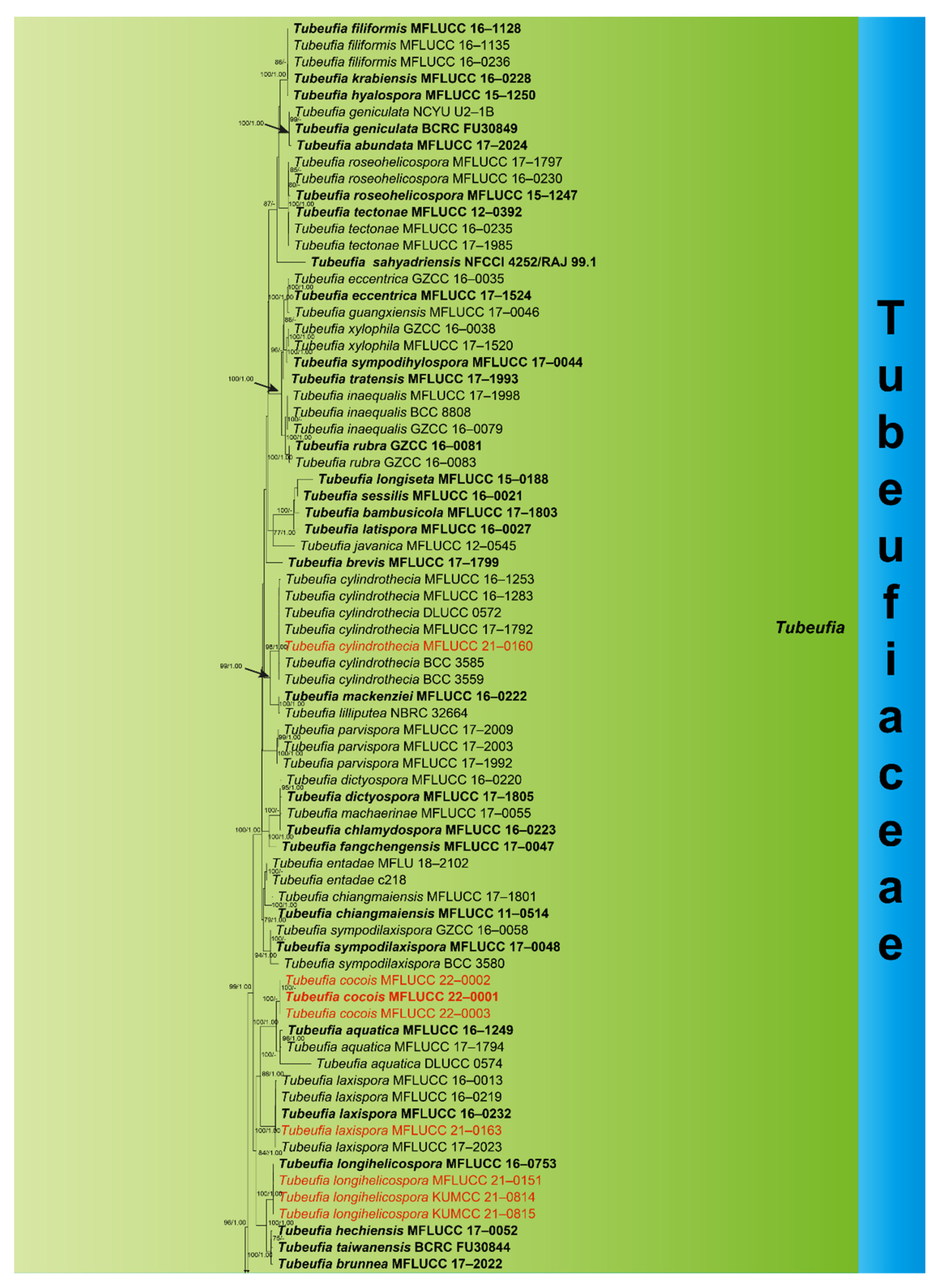
3.1. Phylogenetic Analyses

The combined ITS, LSU, TEF1-α, and RPB2 dataset comprised thirteen newly sequenced strains, with Botryosphaeria dothidea (CBS 115476) and B. agaves (MFLUCC 10–0051) as outgroup taxa. Multiple genes were concatenated, which comprised 3425 nucleotide characters, including gaps (ITS: 1–606 bp, LSU: 607–1471 bp, RPB2: 1472–2514 bp, TEF1-α: 2515–3425 bp). The RAxML analysis of the combined dataset yielded the best-scoring tree (Figure 1) with a final ML optimization likelihood value of −50085.613741. The matrix had 1671 distinct alignment patterns, with 26.72% undetermined characters or gaps. Estimated base frequencies were as follows: A = 0.244864, C = 0.251768, G = 0.258885, T = 0.244483; substitution rates AC = 1.177463, AG = 5.880242, AT = 2.143397, CG = 0.867985, CT = 9.022451, GT = 1.000000; gamma distribution shape parameter α = 0.224491.
Phylogenetic analyses showed that our thirteen collections were placed within Tubeufiaceae viz: Dematiohelicomyces, Helicoma, Helicotruncatum, Neohelicosporium, Parahelicomyces, and Tubeufia. Eight collections clustered within Tubeufia; the new strain Tubeufia cylindrothecia (MFLUCC 21–0160) was nested with six strains of T. cylindrothecia with strong bootstrap support (98% ML/1.00 PP). Three strains of Tubeufia cocois (MFLUCC 22–0001, MFLUCC 22–0002, and MFLUCC 22–0003) clustered together and formed a branch at the basal clades of T. aquatica with strong bootstrap support (100% ML/1.00 PP). The new strain T. laxispora (MFLUCC 21–0163) nested with four strains of T. laxispora with strong bootstrap support (100% ML/1.00 PP), and T. longihelicospora (MFLUCC 21–0151, KUMCC 21–0478, and KUMCC 21–0479) clustered together within the same clade as T. longihelicospora (MFLUCC 16–0753). Parahelicomyces chiangmaiense (MFLUCC 21–0159) formed a single branch at the basal clades of Parahelicomyces members with strong support (97% ML). Newly obtained strain Helicotruncatum palmigenum (KUMCC 21–0474) nested with two strains of H. palmigenum (NMRC 32,663 and MFLUCC 15–0093) strong bootstrap support (100% ML/1.00 PP). Neohelicosporium bambusicola (MFLUCC 21–0160) was placed as a sister taxon to N. ellipsoideum (MFLUCC 16–0229) and N. acrogenisporum (MFLUCC 17–2019). New strain Dematiohelicomyces helicosporus (KUMCC 21–0473) clustered with three strains of Dematiohelicomyces helicosporus with strong bootstrap support (100% ML/1.00 PP), and Helicoma guttulatum (MFLUCC 21–0152) clustered with its ex-type strain of H. guttulatum (MFLUCC 16–0022) with high support (100% ML/1.00 PP).

3.2. Taxonomy

3.2.1. Dematiohelicomyces Y.Z. Lu, Boonmee, and K.D. Hyde, Fungal Diversity 92: 159 (2018)

Index Fungorum, IF 554824; Facesoffungi number, FoF 04701
Type species: Dematiohelicomyces helicosporus (Boonmee, Y.Z. Lu, and K.D. Hyde) Y.Z. Lu
The monotypic genus Dematiohelicomyces was introduced by Lu et al. [3], with D. helicosporus as the type species based on morphology and phylogeny. Dematiohelicomyces are saprobic on submerged decaying wood in a freshwater stream in Thailand. Dematiohelicomyces is characterized by short conidiophores that are brown, 0–3-septate, and helicoid conidia, with a spathulate basal end cell. In this paper, Dematiohelicomyces helicosporus was collected from submerged decaying wood in a freshwater river in Thailand.
  • Dematiohelicomyces helicosporus (Boonmee, Y.Z. Lu, and K.D. Hyde) Y.Z. Lu, Fungal Diversity 92: 159 (2018) (Figure 2).
Chlamydotubeufia helicospora Boonmee, Y.Z. Lu, and K.D. Hyde, Fungal Diversity 80: 123 (2016)
Index Fungorum, IF 554825; Facesoffungi number, FoF 04702
Saprobic on submerged decaying wood in a freshwater stream. Sexual morph Undetermined. Asexual morph Hyphomycetous, helicosporous. Colonies are superficial, effuse, gregarious, white, and shiny. Mycelium is mostly immersed, composed of branched, septate hyphae, brown, with masses of glistening, crowded conidia. Conidiophores (16.5–)30–65.5(–80.5) × 4–5 μm ( x ¯ = 47 × 4.5 μm, n = 25) are macronematous, erect, cylindrical, branched, 0–4-septate, hyaline to pale brown, arising as lateral branches from creeping hyphae, and smooth-walled. Conidiogenous cells (9–)14–24.5(–30) × 4–5 μm ( x ¯ = 19.5 × 4.5 μm, n = 30) are holoblastic, monoblastic, integrated, terminal, cylindrical, truncate at the apex after conidial secession, hyaline, and smooth-walled. Conidia are solitary, acrogenous, helicoid, rounded at tip, with the basal cells broadly spathulate and bearing a flattened attachment scar, guttulate, hyaline, with a (59–)80–139(–158) μm ( x ¯ = 110 μm, n = 25) diam. and conidial filament 5–6.5 μm ( x ¯ = 5.5 μm, n = 25) wide in the broadest part, tapering towards the ends, 328–482.5 μm ( x ¯ = 405.5 μm, n = 25) long, multi-septate, coiled 1–2 ½times, tightly to loosely coiled in water, smooth-walled, and contain granules.
Culture characteristics: conidia germinating on PDA within 12 h; colonies growing on PDA, reaching 20 mm in 2 weeks at 28 °C, circular, with a flat surface, edge entire, pale brown to dark brown in PDA medium; mycelium partially immersed, branched, multi-septate, hyaline to pale brown, smooth.
Material examined: Thailand, Chiang Rai Province, Mae Fah Luang University, on submerged decaying wood, 22 May 2020, R. J. Xu, MD38 (MFLU 21–0184), living culture, KUMCC 21–0473.
Notes: There are some differences between our new isolate (KUMCC 21–0473) and D. helicosporus morphologically, such as conidiogenous cells in our new isolate are monoblastic, while in D. helicosporus (MFLUCC 16–0003), they are mono- to polyblastic. In addition, the new isolate (KUMCC 21–0473) differs from D. helicosporus (MFLUCC 16–0003) in having larger (80–139 vs. 70–100 μm) and shorter conidial filaments (328–482.5 vs. 400–600 μm) [3,43]. However, our phylogenetic results show that the new isolate D. helicosporus (KUMCC 21–0473) clusters with three strains of D. helicosporus (MFLUCC 16–0003, MFLUCC 16–0007, and MFLUCC 16–0213) with high statistical supports (100% ML/1.00 PP, Figure 1). Therefore, we identify our new isolate as Dematiohelicomyces helicosporus. Chlamydotubeufia helicospora was collected on decaying wood in a flowing freshwater stream in Uttaradit Province, Thailand [43], and later, based on morphology and phylogeny, Lu et al. [3] synonymized this taxon under Dematiohelicomyces helicosporus. In this study, our new isolate was also collected from a submerged decaying wood in Chiang Rai Province, Thailand, which is a little far from the original collection location, meaning that this species still prefers similar environmental conditions.

3.2.2. Helicoma Corda, Icon. fung. (Prague) 1: 15 (1837)

Index Fungorum: IF 8473
Type species: Helicoma muelleri Corda, Icon.
Helicoma was introduced by Corda [44], with H. muelleri as a type species. Two types of asexual morphs have been observed in Helicoma: the first asexual morphs are characterized by conidiogenous cells that are cylindrical, with denticles, intercalary, arising laterally from the lower portion of conidiophores, and conidia are pleurogenous, tapering towards the apex and rounded at the tip, helicoid, hygroscopic, and become loosely coiled in water [12]. Another asexual morph is characterized by conidia that are acrogenous or acropleurogenous, helicoid, circinate, dry, tapering towards the apex, truncating at the base, coiled 1¼–¾ times, and not becoming loose in the water. There are 97 records listed in Index Fungorum (2021), however, most of them are lacking sequence data in GenBank. The last treatment of Helicoma was provided by Lu et al. [3], and they accepted 57 species within the genus while introducing 10 new species and 11 new combinations. In this study, the new isolate is identified as H. guttulatum based on both phylogenetic analysis and morphological characteristics.
  • Helicoma guttulatum Y.Z. Lu, Boonmee, and K.D. Hyde, Fungal Diversity 80: 125 (2016) (Figure 3).
Index Fungorum, IF 552218; Facesoffungi, FoF 02358
Saprobic on submerged decaying wood in a freshwater stream. Sexual morph Undetermined. Asexual morph Hyphomycetous, helicosporous. Colonies are superficial, effuse, gregarious, brown to dark brown. Mycelium is mostly immersed, partly superficial, composed of branched, septate, brown hyphae. Conidiophores (65–)93–156.5 × 4.5–6 μm ( x ¯ = 125 × 5 μm, n = 20) are macronematous, mononematous, cylindrical, septate, erect, unbranched, pale brown to brown at the apex and dark brown at the base, and smooth-walled. Conidiogenous cells (9–)12–24(–30.5) × 4–5.5 μm ( x ¯ = 18 × 4.5 μm, n = 20) are holoblastic, mono- to polyblastic, integrated, terminal, cylindrical, brown, and smooth-walled. Conidia 22–26.5 μm ( x ¯ = 24 μm, n = 25) have a diam. and conidial filament 7–8.5 μm ( x ¯ = 8 μm, n = 25) wide and 49–58 μm ( x ¯ = 53.5 μm, n = 25) long, are integrated, terminal, helicoid, tightly coiled 1–1½ times, guttulate, do not become loose in the water, 8-septate, straight constricted at the septa, subhyaline to yellowish, rounded at the apex, and smooth-walled.
Culture characteristics: conidia germinated on PDA within 12 h; colonies growing on PDA, reaching 25 mm in 2 weeks at 28 °C, circular, with a flat surface, edge entire, and pale brown to brown in PDA medium; mycelium were partially immersed, branched, multi-septate, hyaline to pale brown, and smooth.
Material examined: Thailand, Chiang Rai Province, Mueang, Nang Lae on submerged decaying wood, 14 August 2020, R. J. Xu, MD106 (MFLU 21–0183), living culture, MFLUCC 21–0152.
Notes: Helicoma guttulatum was introduced by Hyde et al. [43] on submerged decaying wood from a freshwater stream in Thailand. In our phylogenetic analyses, the newly obtained isolate (MFLUCC 21–0152) clustered with the ex-type strain of H. guttulatum (MFLUCC 16–0022) with high statistical support (100% ML/1.00 PP, Figure 1). Morphologically, the new isolate was indistinguishable from the holotype of H. guttulatum [43]. Therefore, we identify the new isolate as Helicoma guttulatum based on morphological and phylogenetic data.

3.2.3. Helicotruncatum Y.Z. Lu, J.C. Kang, and K.D. Hyde, Fungal Diversity 92: 220 (2018)

Index Fungorum, IF 554859; Facesoffungi number, FoF 04,730
Type species: Helicotruncatum palmigenum (Penz. and Sacc.) Y.Z. Lu and K.D. Hyde
The monotypic genus Helicotruncatum was established by Lu et al. [3] with H. palmigenum as the type species, and it is the only species accepted in the genus [3,12]. Helicotruncatum palmigenum was originally placed in Helicoma, based on morphological characters [12,45,46]. Phylogenetic analysis of Lu et al. [3] showed that H. palmigenum formed an independent lineage and was distant from Helicoma. Morphologically, H. palmigenum can be distinguished from other helicosporous hyphomycetes by the distinctively thickened lateral cell wall of the conidiophore and basal cell of the conidium. Thus, Lu et al. [3] introduced a new genus, Helicotruncatum, to accommodate H. palmigenum based on both phylogeny and morphology. In this paper, Helicotruncatum palmigenum was collected from dead Cocos nucifera leaves in Thailand.
  • Helicotruncatum palmigenum (Penz. and Sacc.) Y.Z. Lu and K.D. Hyde, Fungal Diversity 92: 220 (2018) (Figure 4).
Helicosporium intermedium var. palmigenum Penz. and Sacc., Malpighia 15(7–9): 249 (1902)
Helicoma palmigenum (Penz. and Sacc.) Linder, Ann. Mo. bot. Gdn 16: 306 (1929)
=Helicoma westonii Linder [as ‘westoni’], Ann. Mo. bot. Gdn 18: 12 (1931)
Index Fungorum: IF 554860; Facesoffungi number: FoF 04800
Saprobic on dead leaves of Cocos nucifera. Sexual morph: Undetermined. Asexual morph Hyphomycetous, helicosporous. Colonies on the substratum are superficial, effuse, gregarious, and velvety black. Mycelium is composed of brown, septate hyphae. Conidiophores 165.5–283.5 × 6.5–10 µm ( x ¯ = 224.5 × 8.5 µm, n =10) are macronematous, mononematous, cylindrical, stout, septate, erect, unbranched, pale brown to subhyaline at the apex and dark brown at the base, and smooth-walled. Conidiogenous cells 22–34.5 × 6–7.5 µm ( x ¯ = 28 × 6.5 µm, n =15) are holoblastic, monoblastic, integrated, determinate, cylindrical, terminal, smooth-walled, and truncate at the apex after conidial secession. Conidia 32–44 μm ( x ¯ = 38 μm, n = 20) and conidial filament are 10.5–15 μm ( x ¯ = 12.5 μm, n = 20) wide, 82–108.5 μm ( x ¯ = 95.5 μm, n = 20) long, solitary, terminal, smooth-walled, helicoid, coiled 1½–2¾ times, do not become loose in the water, septate, not constricted at septa, dilute fuliginously, and the basal cell is truncated with thickened lateral walls.
Culture characteristics: conidia germinating on PDA within 12 h; colonies reaching 40 mm in 2 weeks at 28 °C, irregular, dark brown from above and pale brown from below; mycelium are slow-growing, thin, and effuse brownish grey.
Material examined: Thailand, Chiang Rai Province, on decaying leaves of Cocos nucifera, 16 January 2021, X. G. Tian, C6–6, (MFLU 21–0185), living culture, KUMCC 21–0474.
Notes: In the phylogenetic analyses, our new collection KUMCC 21–0474 clusters with two strains of H. palmigenum (NBRC 32663, MFLUCC 15–0993) with high statistical supports (100% ML/1.00 PP, Figure 1). Morphologically, our new isolate is almost identical to H. palmigenum except for the size of the conidiogenous cells (22–34.5 vs. 17–25 µm long) and the conidia (82–108.5 vs. 50–60 µm long). The nucleotide comparisons show 2 bp and 1 bp of ITS and LSU differences between the new isolate (KUMCC 21–0474) and H. palmigenum (NBRC 32663). Thus, we identify the new isolate as H. palmigenum based on both phylogenetic analyses and morphological characteristics.
Helicotruncatum palmigenum was introduced as Helicoma palmigenum by Linder [45] on decaying petioles of palms that were collected from Australia, Brazil, China, Indonesia, Japan, Mexico, New Guinea, Seychelles, Thailand, Trinidad, and the USA [3,45,46]. In addition, Helicotruncatum palmigenum has been reported on leaves and husks of Cocos nucifera [34]. Our new isolate was also collected on dead leaves of Cocos nucifera from Thailand.

3.2.4. Neohelicosporium Y.Z. Lu, J.C. Kang, and K.D. Hyde, Mycological Progress 17: 637 (2017)

Index Fungorum: IF 822045
Type species: Neohelicosporium parvisporum Y.Z. Lu, J.C. Kang, and K.D. Hyde
Neohelicosporium was introduced by Lu et al. [10], with five new species. The taxonomic revision of the genus was recently provided by Lu et al. [3]; eight Helicosporium, two Helicoma, and one Tubeufia species were transferred to Neohelicosporium based on both phylogeny and morphology. The genus is characterized by superficial, ellipsoidal to subglobose, ostiolate ascomata, bitunicate, cylindrical, pedicellate asci and fusiform, straight or slightly curved, multi-septate, guttulate, hyaline, smooth-walled ascospores; macronematous, mononematous, branched or unbranched, septate, pale brown to brown conidiophores, holoblastic, mono- to polyblastic, integrated, sympodial, intercalary or terminal conidiogenous cells with denticles and solitary, acrogenous and/or acropleurogenous, helicoid, multi-septate, guttulate, hyaline to pale brown conidia. Species of the genus are saprobic on decaying woody substrates from both aquatic and terrestrial habitats [47]. In this study, the new species Neohelicosporium bambusicola is introduced based on both phylogenetic analysis and morphological characters.
  • Neohelicosporiumbambusicola X.G. Tian and Tibpromma, sp. nov. (Figure 5).
Index Fungorum number, IF 555045; Facesoffungi number, FoF 10571
Etymology: Referring to the host plant bamboo, on which the fungus was collected.
Saprobic on terrestrial dead culms of bamboo. Sexual morph Undetermined. Asexual morph Hyphomycetous, helicosporous. Colonies on the substratum are superficial, effuse, and white. Mycelium is composed of partly immersed, hyaline to brown, septate, branched hyphae with glistening conidia. Conidiophores 21–76 × 3–5 µm ( x ¯ = 48.5 × 4 µm, n = 10) are macronematous, mononematous, cylindrical, unbranched or branched, septate, subhyaline to brown, and smooth-walled. Conidiogenous cells 8.5–16 × 3–4.5 µm ( x ¯ = 12 × 4 µm, n = 15) are holoblastic, mono to ployblastic, integrated, sympodial, terminal or intercalary, cylindrical, truncate at apex after conidial secession, pale brown, smooth-walled. Conidia are solitary, acropleurogenous, helicoid, multi-septate, guttulate, hyaline when young and become brown when mature, smooth-walled, and do not become loose in water, with a 24–30 μm ( x ¯ = 27 μm, n = 20) diam. and a conidial filament 3–4.5 μm ( x ¯ = 3.9 μm, n = 20) wide, 100.5–128 μm ( x ¯ = 114 μm, n = 20) long, and coiled 2–2 ¾ times.
Culture characteristics: conidia germinated on PDA within 12 h; colonies on PDA reach 20 mm in 2 weeks at 28 °C, and are superficial, effuse, and brown; mycelium is composed of partly immersed, hyaline to brown, septate, branched, smooth hyphae.
Material examined: Thailand, Chiang Mai Province, on dead culms of bamboo, 16 December 2020, X. G. Tian, U4–10 (MFLU 21–0189 holotype), ex-type culture, MFLUCC 21–0156.
Notes: In the phylogenetic analyses, the new isolate Neohelicosporium bambusicola (MFLUCC 21–0156) formed a distinct lineage sister to N. ellipsoideum (MFLUCC 16–0229) and N. acrogenisporum (MFLUCC 17–2009). Neohelicosporium bambusicola resembles N. ellipsoideum and N. acrogenisporum in having macronematous, mononematous, unbranched or branched, septate conidiophores, holoblastic, mono- to ployblastic conidiogenous cells, and helicoid, septate conidia. However, Neohelicosporium bambusicola is distinct from N. ellipsoideum and N. acrogenisporum as it has shorter and narrower conidiophores (21–76 × 3–5 vs. 50–230 × 5–6 vs. 45–150 × 6–7 µm), smaller conidiogenous cells (8.5–16 × 3–4.5 vs. 15–25 × 5–6 vs. 12–15 × 4–6 µm), and narrower conidia (3–4.5 vs. 5–6 vs. 4.5–7.5 µm) [3]. Pairwise nucleotide comparisons revealed that the new isolate Neohelicosporium bambusicola is different from N. ellipsoideum (MFLUCC 16–0229) in 64/544 bp (11.76%) of the ITS, 9/814 (1.1%) of the LSU, 20/1045 bp (1.91%) of RPB2, and 15/894 bp (1.68%) of TEF1-α, while Neohelicosporium bambusicola is different from N. acrogenisporum (MFLUCC 17–2009) in 2/370 bp (0.54%) of the ITS, 6/831 (0.72%) of the LSU, 27/1045 bp (2.58%) of RPB2, and 16/894 bp (1.79%) of TEF1-α. Both phylogenetic analyses and morphological characteristics support this species as a distinct new species.

3.2.5. Parahelicomyces Goh, in Hsieh, and Goh and Kuo, Mycological Progress 20 (2): 182 (2021)

=Pseudohelicomyces Y.Z. Lu, J.K. Liu, and K.D. Hyde
Index Fungorum, IF 554886; Facesoffungi number, FoF 04745
Type species: Parahelicomyces talbotii (Goos) S.Y. Hsieh, Goh, and C.H. Kuo
Parahelicomyces is a well-studied genus, introduced as Pseudohelicomyces by Lu et al. [3] with Pseudohelicomyces talbotii as the type species [3,48]. Pseudohelicomyces was renamed Parahelicomyce by Hsieh et al. [49] because Parahelicomyces was a homonym and illegitimate. Currently, seven species are accepted in the genus, and all the species have sequence data available in the GenBank database. The genus is characterized by superficial, subglobose, ellipsoidal-ovate, coriaceous, ostiolate ascomata, bitunicate, cylindrical, apically thickened and rounded asci, and fusiform, multi-septate, hyaline, smooth-walled ascospores [3], as well as macronematous, mononematous, hyaline to brown, branched, septate conidiophores, holoblastic, mono- to polyblastic, integrated, intercalary or terminal, determinate or sympodial conidiogenous cells with denticles and pleurogenous or acropleurogenous, helicoid, multi-septate, hyaline to pale brown conidia. Species of the genus Parahelicomyces are found from both terrestrial and freshwater habitats in China, Japan, Mexico, South Africa, and Thailand [3,50]. In this study, we introduced a new Parahelicomyces species from Thailand.
  • Parahelicomyceschiangmaiensis X.G. Tian and Tibpromma, sp. nov. (Figure 6).
Index Fungorum, IF 555060; Facesoffungi number, FoF 10570
Etymology: Referring to Chiangrai Province, Thailand, where the fungus was collected.
Saprobic on a terrestrial woody substrate. Sexual morph Undetermined. Asexual morph Hyphomycetous, helicosporous. Colonies on the substratum are superficial, effuse, gregarious, and white. Mycelium is composed of partly superficial, hyaline to pale brown, branched hyphae, with masses of crowded, glistening conidia. Conidiophores 85–180 × 2.9–3.7 µm ( x ¯ = 132 × 3 µm, n =10) are macronematous, mononematous, cylindrical, pale brown to brown, paler towards the apex, straight or flexuous, branched, septate, and smooth-walled. Conidiogenous cells 6–10 × 2.5–3.5 µm ( x ¯ = 8 × 3 µm, n =20) are holoblastic, mono- to polyblastic, integrated, sympodial, terminal or intercalary, cylindrical, with denticles, hyaline to pale brown, and smooth-walled. Conidia have a 21–33.5 μm ( x ¯ = 27 μm, n = 20) diam. and a conidial filament 2–3 μm ( x ¯ = 2.5 μm, n = 20) wide, 73–130 μm ( x ¯ = 101.5 μm, n = 20) long, and coiled 1¼–3 times, and are acropleurogenous, solitary, multi-septate, helicoid, rounded at the tip, hyaline to pale brown, guttulate, tightly to loosely coiled in water, and smooth-walled.
Culture characteristics: conidia germinated on PDA within 12 h; colonies adpressed reaching 30 mm in 2 weeks at 28 °C, amd were circular, brown to dark brown, reverse brown, and slow-growing; mycelium was superficial and partially immersed, branched, septate, hyaline to pale brown, and smooth.
Material examined: Thailand, Chiang Mai Province, on the dead terrestrial woody substrate, 16 December 2020, X. G. Tian, U4–8 (MFLU 21–0188 holotype), ex-type culture, MFLUCC 21–0159.
Notes: Phylogenetic analyses of combined LSU, ITS, RPB2, and TEF1-α sequence data showed that our new isolate Parahelicomyces chiangmaiensis (MFLUCC 21–0159) formed an independent lineage within the genus with strong support (97% ML). Parahelicomyces chiangmaiensis is phylogenetically closely related to P. talbotii (MFLUCC 17–2021), however, P. chiangmaiensis can be distinguished from P. talbotii by the size (21–33.5 vs. 7–16 μm diam.) of conidia and the size (6–10 vs. 7–16 μm long) of conidiogenous cells. Parahelicomyces chiangmaiensis is morphologically closely related to P. indicus, however, P. chiangmaiensis can be distinguished from P. indicus by the colour (pale brown to brown vs. dark to yellowish-brown) and size (85–180 × 2.9–3.7 vs. 47–145× 3–7.5 μm) of the conidiophores [51]. Both phylogenetic analyses and morphological characteristics support Parahelicomyces chiangmaiensis as a distinct new species.

3.2.6. Tubeufia Penz. and Sacc., Malpighia 11(11–12): 517 (1898)

Index Fungorum: IF 5635
Type species: Tubeufia javanica Penz. and Sacc., Malpighia 11(11–12): 517 (1898)
Tubeufia, the type genus of Tubeufiaceae, was established by Penzig and Saccardo [52]. Currently, 88 records are listed in the Index Fungorum (2021); however, most of the species are lacking sequence data in the GenBank. While morphologies of Tubeufia species are quite similar, using morphology alone presents difficulties for identification; thus, sequence data are required to resolve taxonomic confusions. The last treatment of Tubeufia was provided by Lu et al. [3], and they introduced seventeen new species and six new combinations in the genus, accepting fifty species in the genus based on both phylogenic analysis and morphological characters. In this paper, we introduced two novel species, one new record species, and a new isolate of known species in Tubeufia.
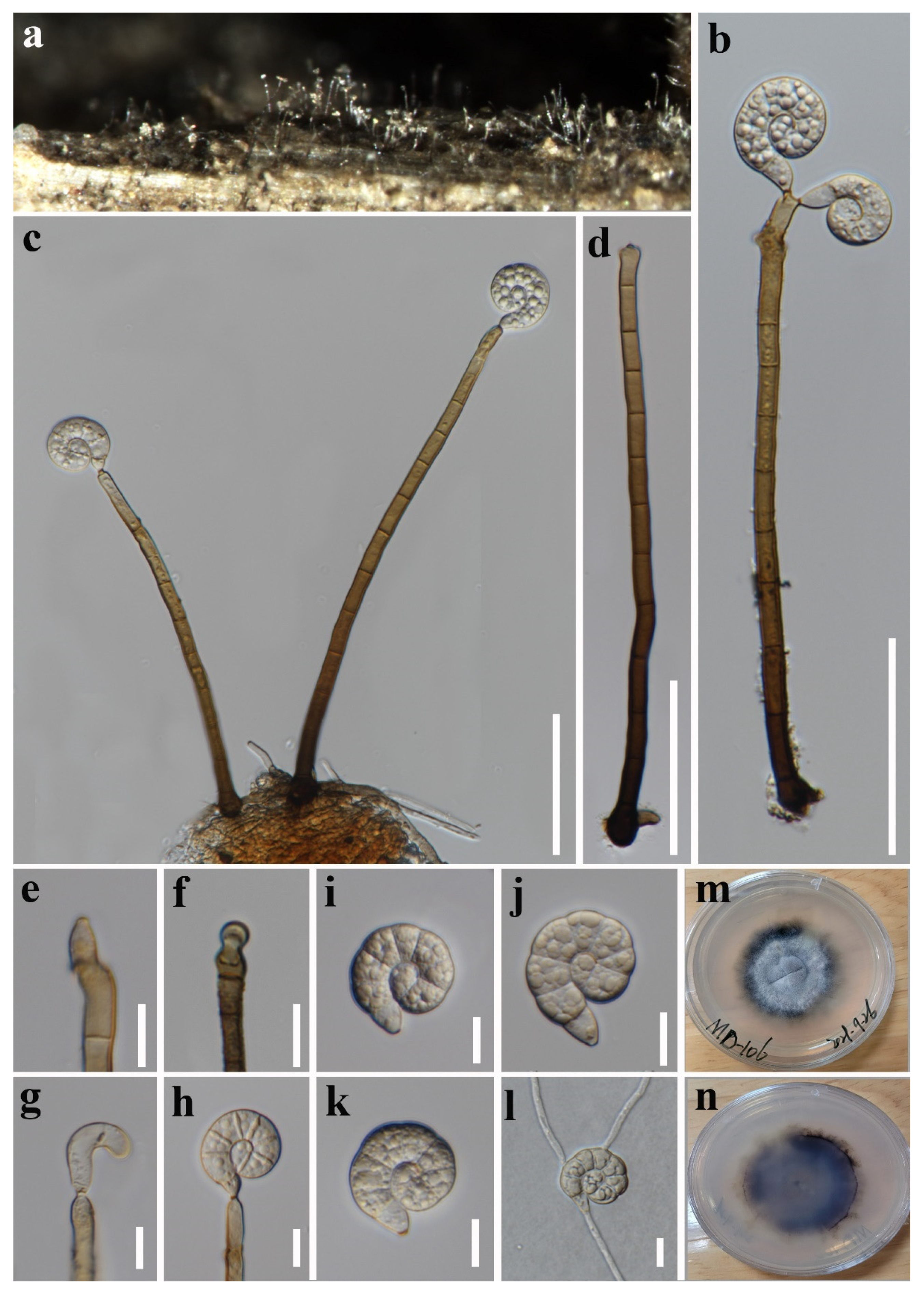
  • Tubeufia cocois X.G. Tian and Tibpromma, sp. nov. (Figure 7).
Index Fungorum number, IF 555070; Facesoffungi number, FoF 10576
Etymology: Referring to the host plant Cocos nucifera, on which the fungus was collected.
Saprobic on the decaying leaves of Cocos nucifera. Sexual morph Undetermined. Asexual morph Hyphomycetous, helicosporous. Colonies on the substratum are superficial, effuse, gregarious, and white to pale brown. Mycelium is partly immersed, partly superficial, hyaline to brown, septate, branched, and with glistening conidia. Conidiophores 38–123 × 4.5–6 µm ( x ¯ = 80.5 × 5.5 µm, n = 20) are macronematous, mononematous, straight or slightly flexuous, cylindrical, branched, septate, orange brown to dark brown, paler towards the apex, and smooth-walled. Conidiogenous cells 8–17.5 × 4–5.5 µm ( x ¯ = 13 × 5 µm, n =25) are holoblastic, mono- to polyblastic, integrated, sympodial, terminal or intercalary, irregular cylindrical, hyaline to pale brown, and smooth-walled, with most of them being denticulate protrusions. Conidia are solitary, acropleurogenous, helicoid, rounded at tip, with a 26–32.5 μm ( x ¯ = 29 μm, n = 20) diam. and a conidial filament 3.5–5 μm ( x ¯ = 4 μm, n = 20) wide, 116–136 μm ( x ¯ = 126 μm, n = 20) long, and coiled 2¼–2¾ times, and they do not become loose in water, are indistinctly multi-septate, guttulate, hyaline when young, pale brown to brown at maturity, and smooth-walled.
Culture characteristics: conidia germinated on PDA within 12 h; colonies grow on PDA, reach 30 mm in 2 weeks at 28 °C, are irregular, with a flat surface, edge undulate, and brown to dark brown in PDA medium; mycelium are superficial and partially immersed, branched, septate, hyaline to brown, and smooth.
Material examined: Thailand, Chiang Rai Province, on decaying leaves of Cocos nucifera, 16 January 2021, X. G. Tian, C6–15 (MFLU 21–0192, holotype), ex-type culture, MFLUCC 22–0001; ibid, C6–8 (MFLU 21–0186, paratype), ex-paratype, MFLUCC 22–0002; ibid, C6–20 (MFLU 21–0187, paratype), ex-paratype, MFLUCC 22–0003.
Notes: Tubeufia cocois is introduced as a distinct new species from Cocos nucifera in Thailand. In the phylogenetic analyses, three newly obtained strains of T. cocois (MFLUCC 22–0001, MFLUCC 22–0002, and MFLUCC 22–0003) clustered together and were sister to three T. aquatica strains with strong statistical support values (100% ML/1.00 PP, Figure 1). Morphologically, T. cocois can be easily distinguished from T. aquatica by the shape and size of the conidiophores, conidiogenous cells, and conidia. Tubeufia cocois has branched or unbranched, multi-septate, and longer conidiophores (38–123 vs. 18–40 μm), while the conidiophores of T. aquatica are unbranched, 0–1-septate, and shorter than those of T. cocois. The conidiogenous cells of T. cocois are terminal or intercalary and conidia are acropleurogenous, whereases T. aquatica has terminal conidiogenous cells and conidia are acrogenous [53]. Based on pairwise nucleotide comparisons, the new strain (MFLUCC 22-0001) is different from T. aquatica (MFLUCC 16–1249) in 16/413 bp (3.87%) of the ITS, 2/845 (0.24%) of the LSU, 23/919 bp (2.5%) of RPB2, and 10/617 bp (1.62%) of TEF1-α.
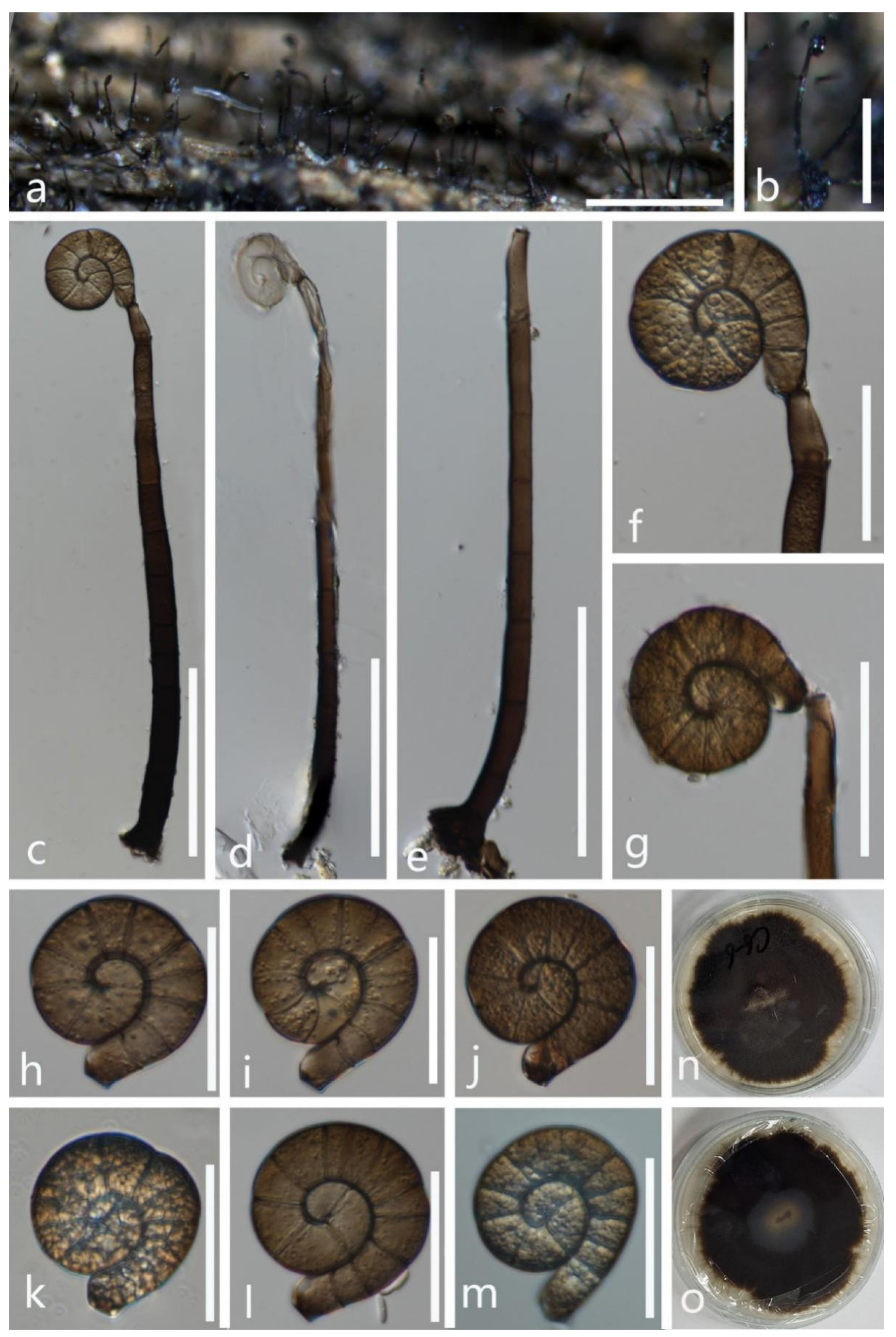
  • Tubeufia laxispora Y.Z. Lu, Boonmee, and K.D. Hyde, Mycological Progress 16: 409 (2017) (Figure 8).
Index Fungorum, IF 818987; Facesoffungi number, FoF 02694
Saprobic on the decaying leaves of Cocos nucifera. Sexual morph Undetermined. Asexual morph Hyphomycetous, helicosporous. Colonies on the substratum are superficial, effuse, gregarious, and range from white to brown. Mycelium is partly immersed, partly superficial, pale brown, septate, sparsely branched hyphae, with masses of crowded conidia. Conidiophores 26–53 × 3.5–5 µm ( x ¯ = 39.5 × 4 µm, n =15) are hyaline to brown, macronematous, erect, short, and smooth-walled. Conidiogenous cells 8–16 × 3–4.5 µm ( x ¯ = 12 × 4 µm, n =20) are monoblastic, holoblastic, integrated, and each have a single conidium. Conidia are solitary, acropleurogenous, helicoid, and rounded at the tip, with a 17.5–29 μm ( x ¯ = 23 μm, n = 20) diam. and a conidial filament 2–3.5 μm ( x ¯ = 2.5 μm, n = 20) wide, 63–94.5 μm ( x ¯ = 78.5 μm, n = 20) long, loosely coiled 1–2½ times in the water, indistinctly multi-septate, hyaline, and smooth-walled.
Culture characteristics: conidia germinating on PDA within 12 h; colonies growing on PDA, reaching 20 mm in 2 weeks at 28 °C—they are circular, with a flat surface, edge undulate, and brown to dark brown in PDA medium; mycelium are superficial and partially immersed, branched, septate, hyaline to brown, and smooth.
Material examined: Thailand, Chiang Rai Province, on decaying leaves of Cocos nucifera, 9 March 2021, X. G. Tian, C7–10 (MFLU 21–0191), living culture, MFLUCC 21–0163.
Notes: In our phylogenetic analyses, the newly obtained isolate (MFLUCC 21–0163) clustered with four strains of T. laxispora with high statistical supports (100% ML/1.00 PP, Figure 1). Based on pairwise nucleotide comparisons, the new strain (MFLUCC 21–0163) almost overlapped with the ex-type strain of T. laxispora (MFLUCC 16–0232), except TEF1-α 1 bp out of 878 bp (<1%). Morphologically, our new isolate fits well with the description of T. laxispora, except for the conidial size (17.5–40 μm diam., 63–94.5 μm long vs. 17.5–29 μm diam., 111–182 μm long) [37]. Hence, we identify our new isolate as T. laxispora. Tubeufia laxispora was described by Lu et al. [37] on submerged wood in Thailand, while our new isolate was collected on decaying leaves of Cocos nucifera in Thailand, and this is the first report of Tubeufia laxispora associated with a coconut tree from Thailand.
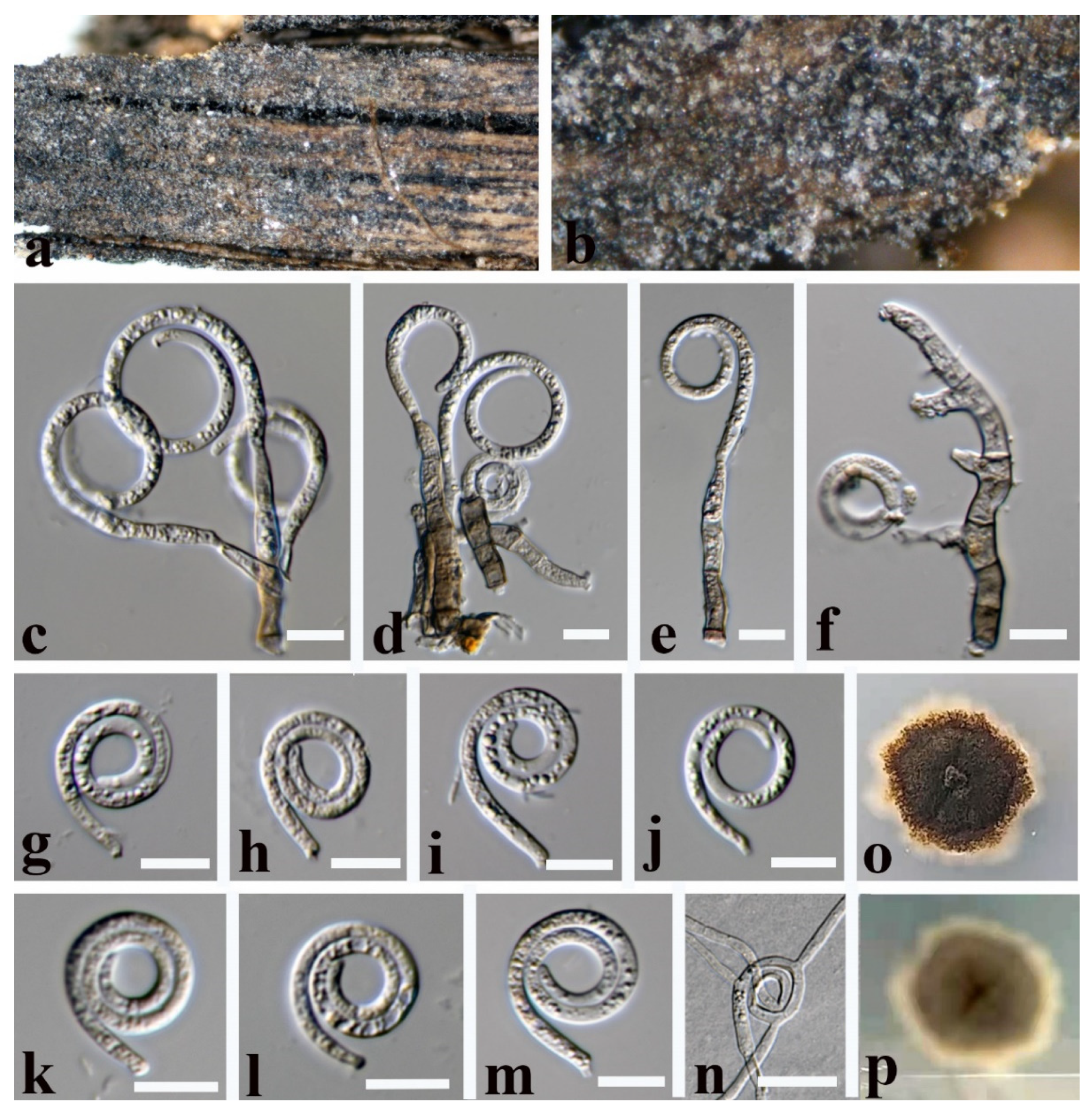
  • Tubeufia cylindrothecia (Seaver) Höhn Sber. Akad. Wiss. Wien, Math. -naturw. Kl., Abt. 1 128: 562 (1919) (Figure 9).
Index Fungorum, IF 340543; Facesoffungi number, FoF 02650
Saprobic on submerged decaying wood. Sexual morph See Seaver [54]. Asexual morph Colonies on the substratum are superficial, effuse, gregarious, and white to pale brown. Mycelium is partly immersed, partly superficial, hyaline to brown, septate, and with masses of conidia. Conidiophores 57–95 × 4–7 µm ( x ¯ = 76 × 5.5 µm, n =10) are pale brown, macronematous, mononematous, septate, cylindrical, unbranched, erect, and smooth-walled. Conidiogenous cells 6.5–16 × 3.5–5 µm ( x ¯ = 11 × 4 µm, n =15) are holoblastic, monoblastic, integrated, smooth, terminal or intercalary, and cylindrical. Conidia are acropleurogenous, 40.5–82 μm ( x ¯ = 61.5 μm, n = 20) in diam. and with a conidial filament that is 3.5–8 μm ( x ¯ = 5.5 μm, n = 20) wide, 220–321 μm ( x ¯ = 270.5 μm, n = 20) long, helicoid, with conidial loosely coiled 1–3½ times in the water, hyaline to brown, indistinctly multi-septate, guttulate, and smooth.
Culture characteristics: conidia germinating on PDA within 12 h; colonies growing on PDA, reaching 25 mm in 2 weeks at 28 °C, irregular, with a flat surface, edge undulate, and brown to dark brown in PDA medium; mycelium are partially immersed, branched, septate, hyaline to brown, and smooth.
Material examined: Thailand, Chiang Rai Province, on decaying submerged wood, 11 November 2020, X. G. Tian, W1–10 (MFLU 21–0190), living culture, MFLUCC 21–0160.
Notes: Tubeufia cylindrothecia was originally introduced with both sexual and asexual morphs that link to Helicomyces roseus based on morphological studies [6,55], while phylogenetic analyses showed that Tubeufia cylindrotheci and Helicomyces roseus can be recognized as two different species [1,8,53,56]. Luo et al. [53] first reported its asexual morph as collected from a freshwater habitat in China. Our phylogenetic results show that the newly obtained isolate (MFLUCC 21–0160) clustered with six strains of T. cylindrothecia with high bootstrap support (98% ML/1.00 PP). Morphologically, our new isolate is almost identical to T. cylindrothecia, except for the conidiogenous cells of the new isolate (MFLUCC 21–0160) that are terminal or intercalary, while the conidiogenous cells are terminal in T. cylindrothecia (MFLU 16–2547). Thus, based on morphological and molecular data, we identify the new isolate as Tubeufia cylindrothecia.
  • Tubeufia longihelicospora Boonmee, Promputtha, and K.D. Hyde, in Boonmee et al., Fungal Diversity 111: 133 (2021) (Figure 10).
Index Fungorum number, IF 558543; Facesoffungi number, FoF 09195
Saprobic on submerged decaying wood in a freshwater stream. Sexual morph Undetermined. Asexual morph Hyphomycetous, helicosporous. Colonies on the substratum are superficial, effuse, gregarious, and white. Mycelium is composed of partly immersed, hyaline to pale brown, septate, branched hyphae with masses of glistening, crowded conidia. Stalked sclerotia are often present and are medium brown, spherical, and muriform. Conidiophores (10.5–)13.5–35.5(–55.5) × 3.5–5(–6) μm ( x ¯ = 24.5 × 4 μm, n = 15) are macronematous, mononematous, arise as lateral branches from creeping hyphae, cylindrical, branched, 0–4-septate, hyaline to pale brown, and smooth-walled. Conidiogenous cells (7–)9–19(–24.5) × 3.5–4.5 μm ( x ¯ = 14 × 4 μm, n = 15) are holoblastic, monoblastic, integrated, terminal or intercalary, cylindrical, truncate at the apex after conidial secession, hyaline to pale brown, and smooth-walled. Conidia are solitary, acrogenous, holoblastic, helicoid, rounded at the tip, hyaline to pale brown, (40–)55.5–86.5(–100.5) μm ( x ¯ = 71 μm, n = 15) in diam. and with a conidial filament 6.5–8.5 μm ( x ¯ = 7.5 μm, n = 15) wide in the broadest part and tapering towards the ends, (99.5–)240.5–355.5 μm ( x ¯ = 298 μm, n = 15) long, loosely coiled 1–2 times, multi-septate, tightly to loosely coiled in the water, constricted at the septa, guttulate, hyaline to pale brown, rough-walled, and bearing conidiola. Conidiola are globose, unicellular, and rough-walled.
Culture characteristics: conidia germinated on PDA within 12 h; colonies growing on PDA reach 20 mm in 2 weeks at 28 °C, are irregular, with a flat surface, edge undulate, and brown to dark brown in PDA medium; mycelium are superficial and partially immersed, branched, septate, hyaline to brown, and smooth.
Material examined: Thailand, Chiang Rai Province, Mueang, Ban Du, on decaying submerged wood, 15 August 2020, R. J. Xu, MD77 (MFLU 21–0182), living culture, MFLUCC 21–0151; China, Yunnan Province, Xishuangbanna, on decaying submerged wood, 13 September 2021, X. G. Tian, WB12 (HKAS 122173), living culture, KUMCC 21–0814; ibid NWBB9 (HKAS 122169), living culture, KUMCC 21–0815.
Notes: In the phylogenetic analyses, our three new strains (MFLUCC 21–0151, KUMCC 21–0478, and KUMCC 21–0479) are clustered together within the same clade as T. longihelicospora (MFLUCC 16–0753). Based on pairwise nucleotide comparisons, our three new strains almost overlap with the ex-type strain of Tubeufia longihelicospora (MFLUCC 16–0753). Morphologically, our new isolate (MFLU 21–0182) is almost identical to Tubeufia longihelicospora, except for the size of the conidia (55.5–86.5 vs. 36–52 µm diam.). Therefore, we identify the three new isolates as Tubeufia longihelicospora based on morphological and phylogenetic data. Tubeufia longihelicospora was introduced by Boonmee et al. [57] on a submerged decaying wood in a small freshwater stream in Thailand. Our isolate Tubeufia longihelicospora (MFLUCC 21–0151) was also collected in Thailand, while the other two isolates of Tubeufia longihelicospora (KUMCC 21–0478 and KUMCC 21–0479) were collected in China, which is a new geographical record.

4. Discussion

Tubeufiaceae is an interesting family with diverse morphologies, habitats, and a worldwide distribution [1,3,12,24]. The asexual morph of the Tubeufiaceae species is reported as helicosporous, chlamydosporous, and phragmosporous conidia, of which helicosporous conidia are the most common morphology in Tubeufiaceae. However, species with helicosporous conidia are not only found in Tubeufiaceae; for example, Helicoascotaiwania also has helicosporous conidia but it is phylogenetically distinct from Tubeufiaceae. Helicoascotaiwania is placed in Pleurotheciaceae and Sordariomycetes [58], while Tubeufiaceae is placed in Pleosporales. The interesting finding is that the asexual morphs of some genera were reported with two different morphologies. For example, Tubeufia and Berkleasmium produce both dictyosporous and helicosporous conidia, while Helicoma produces both helicosporous and phragmosporous conidia, confirmed by phylogenetic analyses [3].
The morphology of helicosporous hyphomycetes is quite similar; thus, the phylogenetic analyses are efficient to identify the helicosporous hyphomycetes at the species level [59,60,61]. With the availability of molecular data, some species were revised and transferred to other genera based on phylogenetic analyses. For example, Helicomyces roseus (BCC 3381) and Helicoma perelegans (ATCC 22621) were transferred to Tubeufia [3]. However, many helicosporous species lack sequence data in the GenBank, and the taxonomy of helicosporous hyphomycetes needs revisions based on phylogenetic analyses. For example, species in the genera of Tubeufia, Helicoma, and Helicomyces were introduced based on morphological characteristics, but many of them lack sequence data in the GenBank; thus, further study into herbarium specimens is necessary to resolve taxonomic problems in the three genera.
In this study, nine Tubeufiaceae species were collected from terrestrial and freshwater habitats in Thailand, of which three were introduced as new species, while six were identified as existing species based on phylogenetic analyses and morphological characteristics. The nine species were placed in Dematiohelicomyces, Helicoma, Helicotruncatum, Neohelicosporium, Parahelicomyces, and Tubeufia, respectively, of which the genera Dematiohelicomyces, Helicotruncatum, Neohelicosporium, and Parahelicomyces are well studied, and all species in these genera have sequence data available in the GenBank. Helicoma and Tubeufia were recently revised by Lu et al. [3]. In our phylogenetic analyses, Helicoma and Tubeufia formed well-supported and monophyletics clades within the family. The morphologies of Tubeufia and Helicoma are quite similar; thus, morphology alone is not enough to identify species in Tubeufia and Helicoma, and phylogenetic analyses are necessary. However, earlier studies identified the two genera only based on morphological characteristics, and sequence data of many species are not available in the GenBank, so, it is entirely possible that some species were incorrectly identified; accordingly, fresh collections and molecular data are required to clarify their taxonomic status. Even though Dematiohelicomyces helicosporus, Helicotruncatum palmigenum, Helicoma guttulatum, Neohelicosporium bambusicola, Tubeufia cylindrothecia, T. laxispora, and T. longihelicospora are known species and were collected again, some species are known as new hosts and new geographical records. In addition, it is also better to provide the full descriptions and color plates of the micro-characteristics of new isolates to understand some fine morphological differences.

Author Contributions

Conceptualization, X.T.; formal analysis, X.T.; funding acquisition, S.C.K. and S.T.; methodology, D.B. and R.X.; resources, S.C.K. and S.T.; supervision, S.C.K.; writing—original draft, X.T.; writing—review and editing, X.T., J.X., A.M., S.C.K., S.T., N.S. and Y.L. All authors have read and agreed to the published version of the manuscript.

Funding

The APC was funded by editorial board member Samantha C. Karunarathna’s free APC facility in 2021. Xingguo Tian thanks Kevin D. Hyde for reviewing and editing the manuscript. Saowaluck Tibpromma thanks the International Postdoctoral Exchange Fellowship Program (number Y9180822S1), the CAS President’s International Fellowship Initiative (PIFI) (number 2020PC0009), the China Postdoctoral Science Foundation, and the Yunnan Human Resources and Social Security Department Foundation for funding her postdoctoral research. Samantha C. Karunarathna thanks the CAS President’s International Fellowship Initiative (PIFI) young staff under the grant number: 2020FYC0002, and the National Science Foundation of China (NSFC) project code31851110759 for funding. Yong-Zhong Lu would like to thank the National Natural Science Foundation of China (NSFC 31900020) and the Science and Technology Foundation of Guizhou Province ([2020]1Y058). Ausana Mapook would like to thank the MFU foundation grant and Mae Fah Luang University Fund (Grant No. 651A16029), entitled “Taxonomy, phylogeny, risk assessment, and potential impact of fungi on Siam weed in northern Thailand”. Austin Smith at World Agroforestry (ICRAF), Kunming Institute of Botany, China, is thanked for English editing. This work was partially supported by Chiang Mai University.

Institutional Review Board Statement

Not applicable.

Informed Consent Statement

Not applicable.

Data Availability Statement

Not applicable.

Conflicts of Interest

The authors declare no conflict of interest.

References

  1. Boonmee, S.; Rossman, A.Y.; Liu, J.K.; Li, W.J.; Dai, D.Q.; Bhat, J.D.; Jones, E.B.G.; McKenzie, E.H.C.; Xu, J.C.; Hyde, K.D. Tubeufiales, ord. nov., integrating sexual and asexual generic names. Fungal Divers. 2014, 68, 239–298. [Google Scholar] [CrossRef]
  2. Liu, J.K.; Hyde, K.D.; Jeewon, R.; Phillips, A.J.; Maharachchikumbura, S.S.; Ryberg, M.; Liu, Z.Y.; Zhao, Q. Ranking higher taxa using divergence times: A case study in Dothideomycetes. Fungal Divers. 2017, 84, 75–99. [Google Scholar] [CrossRef]
  3. Lu, Y.Z.; Liu, J.K.; Hyde, K.D.; Jeewon, R.; Kang, J.C.; Fan, C.; Boonmee, S.; Bhat, D.J.; Luo, Z.L.; Lin, C.G.; et al. A taxonomic reassessment of Tubeufiales based on multi-locus phylogeny and morphology. Fungal Divers. 2018, 92, 131–344. [Google Scholar] [CrossRef]
  4. Hongsanan, S.; Hyde, K.D.; Phookamsak, R.; Wanasinghe, D.N.; McKenzie, E.H.; Sarma, V.V.; Lücking, R.; Boonmee, S.; Bhat, J.D.; Liu, N.G.; et al. Refined families of Dothideomycetes: Orders and families incertae sedis in Dothideomycetes. Fungal Divers. 2020, 105, 17–318. [Google Scholar] [CrossRef]
  5. Barr, M.E. A classification of Loculoascomycetes. Mycologia 1979, 71, 935–957. [Google Scholar] [CrossRef]
  6. Barr, M.E. On the family Tubeufiaceae (Pleosporales). Mycotaxon 1980, 12, 137–167. [Google Scholar]
  7. Rossman, A.Y. The Tubeufiaceae and Similar Loculoascomycetes; CAB International Mycological Institute: London, UK, 1987. [Google Scholar]
  8. Boonmee, S.; Zhang, Y.; Chomnunti, P.; Chukeatirote, E.; Tsui, C.K.; Bahkali, A.H.; Hyde, K.D. Revision of lignicolous Tubeufiaceae based on morphological reexamination and phylogenetic analysis. Fungal Divers. 2011, 51, 63–102. [Google Scholar] [CrossRef]
  9. Brahmanage, R.S.; Lu, Y.Z.; Bhat, D.J.; Wanasinghe, D.N.; Yan, J.Y.; Hyde, K.D.; Boonmee, S. Phylogenetic investigations on freshwater fungi in Tubeufiaceae (Tubeufiales) reveals the new genus Dictyospora and new species Chlamydotubeufia aquatica and Helicosporium flavum. Mycosphere 2017, 8, 917–933. [Google Scholar] [CrossRef]
  10. Lu, Y.Z.; Boonmee, S.; Liu, J.K.; Hyde, K.D.; McKenzie, E.H.; Eungwanichayapant, P.D.; Kang, J.C. Multi-gene phylogenetic analyses reveals Neohelicosporium gen. nov. and five new species of helicosporous hyphomycetes from aquatic habitats. Mycol. Prog. 2018, 17, 631–646. [Google Scholar] [CrossRef]
  11. Rajeshkumar, K.C.; Sharma, R. Tamhinispora, a new genus belongs to family Tubeufiaceae from the western Ghats, India based on morphology and phylogenetic analysis. Mycosphere 2013, 4, 165–174. [Google Scholar] [CrossRef]
  12. Dong, W.; Wang, B.; Hyde, K.D.; McKenzie, E.H.C.; Raja, H.A.; Tanaka, K.; Abdel-Wahab, M.A.; Abdel-Aziz, F.A.; Doilom, M.; Phookamsak, R.; et al. Freshwater Dothideomycetes. Fungal Divers. 2020, 105, 319–575. [Google Scholar] [CrossRef]
  13. Sivanesan, A. The Bitunicate Ascomycetes and Their Anamorphs; CAB International: Wallingford, UK, 1984; p. 701. [Google Scholar]
  14. Crane, J.L.; Shearer, C.J.; Barr, M.E. A revision of Boerlagiomyces with notes and a key to the saprobic genera of Tubeufiaceae. Can. J. Bot. 1998, 76, 602–612. [Google Scholar]
  15. Kirk, P.M.; Cannon, P.F.; David, J.C.; Stalpers, J.A. Ainsworth & Bisby’s Dictionary of the Fungi, 9th ed.; CABI International: Wallingford, UK, 2001; pp. 1–655. [Google Scholar]
  16. Eriksson, O.E.; Winka, K. Families and higher taxa of Ascomycota. Myconet 1998, 1, 17–24. [Google Scholar]
  17. Eriksson, O.E. Outline of Ascomycota—1999. Myconet 1999, 3, 1–88. [Google Scholar]
  18. Eriksson, O.E. SSU rDNA sequences from Ascomycota. Myconet 2001, 6, 27–76. [Google Scholar]
  19. Eriksson, O.E. Outline of Ascomycota—2005. Myconet 2005, 11, 1–113. [Google Scholar]
  20. Lumbsch, H.T.; Huhndorf, S.M. Myconet volume 14. Part One. Outline of Ascomycota—2009. Part Two. Notes on Ascomycete systematics. Nos. 4751–5113. Fieldiana Life Earth Sci. 2010, 2010, 1–64. [Google Scholar] [CrossRef]
  21. Kodsueb, R.; Jeewon, R.; Vijaykrishna, D.; McKenzie, E.H.C.; Lumyong, P.; Lumyong, S.; Hyde, K.D. Systematic revision of Tubeufiaceae based on morphological and molecular data. Fungal Divers. 2006, 21, 105–130. [Google Scholar]
  22. Chaiwan, N.; Lu, Y.Z.; Tibpromma, S.; Bhat, D.J.; Hyde, K.D.; Boonmee, S. Neotubeufia gen. nov. and Tubeufia guangxiensis sp. nov. (Tubeufiaceae) from freshwater habitats. Mycosphere 2017, 8, 1443–1456. [Google Scholar] [CrossRef]
  23. Hanada, T.; Sato, T.; Arioka, M.; Uramoto, M.; Yamasaki, M. Purification and characterization of a 15 kDa protein (p15) produced by Helicosporium that exhibits distinct effects on neurite outgrowth from cortical neurons and PC12 cells. Biochem. Biophys. Res. Commun. 1996, 228, 209–215. [Google Scholar] [CrossRef] [PubMed]
  24. Hu, H.; Guo, H.; Li, E.; Liu, X.; Zhou, Y.; Che, Y. Decaspirones F–I, bioactive secondary metabolites from the saprophytic fungus Helicoma viridis. J. Nat. Prod. 2006, 69, 1672–1675. [Google Scholar] [CrossRef] [PubMed]
  25. Itazaki, H.; Nagashima, K.; Sugita, K.; Yoshida, H.; Kawamura, Y.; Yasuda, Y.; Matsumoto, K.; Ishii, K.; Uotani, N.; Nakai, H. Solation and structural elucidation of new cyclotetrapeptides, trapoxins A and B, having detransformation activities as antitumor agents. J. Antibiot. 1990, 43, 1524–1532. [Google Scholar] [CrossRef] [Green Version]
  26. Tibpromma, S.; Hyde, K.D.; Jeewon, R.; Maharachchikumbura, S.S.; Liu, J.K.; Bhat, D.J.; Jones, E.G.; McKenzie, E.H.; Camporesi, E.; Bulgakov, T.S.; et al. Fungal diversity notes 491–602: Taxonomic and phylogenetic contributions to fungal taxa. Fungal Divers. 2017, 83, 1–261. [Google Scholar] [CrossRef]
  27. Tian, X.G.; Karunarathna, S.C.; Mapook, A.; Xu, J.C.; Bao, D.F.; Promputtha, I.; Tibpromma, S. Koorchaloma oryzae sp. nov. (Stachybotryaceae, Sordariomycetes), from Oryza sativa (Poaceae) in northern Thailand. Phytotaxa 2021, 524, 283–292. [Google Scholar] [CrossRef]
  28. Tian, X.G.; Karunarathna, S.C.; Mapook, A.; Promputtha, I.; Xu, J.C.; Bao, D.F.; Tibpromma, S. One new species and two new host records of Apiospora from bamboo and maize in northern Thailand with thirteen new combinations. Life 2021, 11, 1071. [Google Scholar] [CrossRef] [PubMed]
  29. Senanayake, I.C.; Rathnayaka, A.R.; Marasinghe, D.S.; Calabon, M.S.; Gentekaki, E.; Lee, H.B.; Hurdeal, V.G.; Pem, D.; Dissanayake, L.S.; Wijesinghe, S.N.; et al. Morphological approaches in studying fungi: Collection, examination, isolation, sporulation and preservation. Mycosphere 2020, 11, 2678–2754. [Google Scholar] [CrossRef]
  30. Index Fungorum. 2021. Available online: http://www.indexfungorum.org/Names/Names.asp (accessed on 30 November 2021).
  31. Jayasiri, S.C.; Hyde, K.D.; Ariyawansa, H.A.; Bhat, J.; Buyck, B.; Cai, L.; Dai, Y.C.; Abd-Elsalam, K.A.; Ertz, D.; Hidayat, I.; et al. The faces of fungi database: Fungal names linked with morphology, phylogeny and human impacts. Fungal Divers. 2015, 74, 3–18. [Google Scholar] [CrossRef]
  32. White, T.J.; Bruns, T.; Lee, S.; Taylor, J. Amplification and direct sequencing of fungal ribosomal RNA genes for phylogenetics. In PCR Protocols: A Guide to Methods and Applications; Innis, G.M., Shinsky, D., White, T., Eds.; Academic: New York, NY, USA, 1990; pp. 315–322. [Google Scholar]
  33. Vilgalys, R.; Hester, M. Rapid genetic identification and mapping of enzymatically amplified ribosomal DNA from several Cryptococcus species. J. Bacteriol. 1990, 172, 4238–4246. [Google Scholar] [CrossRef] [Green Version]
  34. Rehner, S.A.; Buckley, E. A Beauveria phylogeny inferred from nuclear ITS and EF1-α sequences: Evidence for cryptic diversification and links to Cordyceps teleomorphs. Mycologia 2005, 97, 84–98. [Google Scholar] [CrossRef]
  35. Liu, Y.J.; Whelen, S.; Hall, B.D. Phylogenetic relationships among ascomycetes: Evidence from an RNA polymerse II subunit. Mol. Biol. Evol. 1999, 16, 1799–1808. [Google Scholar] [CrossRef] [PubMed]
  36. Cai, L.; Jeewon, R.; Hyde, K.D. Phylogenetic evaluation and taxonomic revision of Schizothecium based on ribosomal DNA and protein coding genes. Fungal Divers. 2005, 19, 1–21. [Google Scholar]
  37. Lu, Y.Z.; Boonmee, S.; Dai, D.Q.; Liu, J.K.; Hyde, K.D.; Bhat, D.J.; Ariyawansa, H.; Kang, J.C. Four new species of Tubeufia (Tubeufiaceae, Tubeufiales) from Thailand. Mycol. Prog. 2017, 16, 403–417. [Google Scholar] [CrossRef]
  38. Dissanayake, A.J.; Bhunjun, C.S.; Maharachchikumbura, S.S.N.; Liu, J.K. Applied aspects of methods to infer phylogenetic relationships amongst fungi. Mycosphere 2020, 11, 2652–2676. [Google Scholar] [CrossRef]
  39. Katoh, K.; Rozewicki, J.; Yamada, K.D. MAFFT online service: Multiple sequence alignment, interactive sequence choice and visualization. Brief. Bioinform. 2019, 20, 1160–1166. [Google Scholar] [CrossRef] [Green Version]
  40. Capella-Gutiérrez, S.; Silla-Martínez, J.M.; Gabaldón, T. TrimAl: A tool for automated alignment trimming in large-scale phylogenetic analyses. Bioinformatics 2009, 25, 1972–1973. [Google Scholar] [CrossRef]
  41. Stamatakis, A.; Hoover, P.; Rougemont, J. A rapid bootstrap algorithm for the RAxML web servers. Syst. Biol. 2008, 57, 758–771. [Google Scholar] [CrossRef]
  42. Miller, M.A.; Pfeiffer, W.; Schwartz, T. Creating the CIPRES Science Gateway for inference of large phylogenetic trees. In Proceedings of the 2010 Gateway Computing Environments Workshop (GCE), New Orleans, LA, USA, 14 November 2010; pp. 1–8. [Google Scholar]
  43. Hyde, K.D.; Hongsanan, S.; Jeewon, R.; Bhat, D.J.; McKenzie, E.H.C.; Jones, E.B.G.; Phookamsak, R.; Ariyawansa, H.A.; Boonmee, S.; Zhao, Q.; et al. Fungal diversity notes 367–490: Taxonomic and phylogenetic contributions to fungal taxa. Fungal Divers. 2016, 80, 1–270. [Google Scholar] [CrossRef]
  44. Corda, A.K.J. Incones Fungorum Hucusque Cognitorum; J.G. Calve: Prague, Czech Republic, 1837; Volume 1, pp. 1–32. [Google Scholar]
  45. Linder, D.H. A monograph of the helicosporous fungi imperfecti. Ann. Mo. Bot. Gard. 1929, 16, 227–388. [Google Scholar] [CrossRef]
  46. Goos, R.D. A review of the anamorph genus Helicoma. Mycologia 1986, 78, 744–761. [Google Scholar] [CrossRef]
  47. Li, W.J.; McKenzie, E.H.C.; Liu, J.K.; Bhat, D.J.; Dai, D.Q.; Camporesi, E.; Tian, Q.; Maharachchikumbura, S.S.N.; Luo, Z.L.; Shang, Q.J.; et al. Taxonomy and phylogeny of hyaline-spored coelomycetes. Fungal Divers. 2020, 100, 279–801. [Google Scholar] [CrossRef]
  48. Lu, Y.Z.; Liu, J.K.; Hyde, K.D. Proposal to conserve Pseudohelicomyces YZ Lu & al. (Tubeufiaceae) against Pseudohelicomyces Garnica & E. Valenz. (Hymenogastraceae). Taxon 2020, 69, 615–616. [Google Scholar]
  49. Hsieh, S.Y.; Goh, T.K.; Kuo, C.H. New species and records of Helicosporium sensu lato from Taiwan, with a reflection on current generic circumscription. Mycol. Prog. 2021, 20, 169–190. [Google Scholar] [CrossRef]
  50. Rossman, A.Y. The genus Ophionectria (Euascomycetes, Hypocreales). Mycologia 1977, 69, 355–391. [Google Scholar] [CrossRef]
  51. Rao, P.R.; Rao, D. Some helicosporae from Hyderabad-II. Mycopathol. Mycol. Appl. 1964, 24, 27–34. [Google Scholar] [CrossRef]
  52. Penzig, O.A.J.; Saccardo, P.A. Diagnoses fungorum novorum in Insula Java collectorum. Series secunda. Malpighia 1897, 11, 491–530. [Google Scholar]
  53. Luo, Z.L.; Bhat, D.J.; Jeewon, R.; Boonmee, S.; Bao, D.F.; Zhao, Y.C.; Chai, H.M.; Su, H.Y.; Su, X.J.; Hyde, K.D. Molecular phylogeny and morphological characterization of asexual fungi (Tubeufiaceae) from freshwater habitats in Yunnan, China. Cryptogam. Mycol. 2017, 38, 27–53. [Google Scholar] [CrossRef]
  54. Seaver, F.J. The Hypocreales of north America–I. Mycologia 1909, 1, 41–76. [Google Scholar]
  55. Seaver, F.J.; Waterston, J.M. Contributions to the mycoflora of Bermuda–I. Mycologia 1940, 32, 388–407. [Google Scholar]
  56. Zhao, G.Z.; Liu, X.Z.; Wu, W.P. Helicosporous hyphomycetes from China. Fungal Divers. 2007, 26, 313–524. [Google Scholar]
  57. Boonmee, S.; Wanasinghe, D.N.; Calabon, M.S.; Huanraluek, N.; Chandrasiri, S.K.; Jones, G.E.; Rossi, W.; Leonardi, M.; Singh, S.K.; Rana, S.; et al. Fungal diversity notes 1387–1511: Taxonomic and phylogenetic contributions on genera and species of fungal taxa. Fungal Divers. 2021, 111, 1–335. [Google Scholar] [CrossRef] [PubMed]
  58. Dayarathne, M.C.; Maharachchikumbura, S.S.N.; Jones, E.B.G.; Dong, W.; Devadatha, B.; Yang, J.; Ekanayaka, A.H.; De Silva, W.; Sarma, V.V.; Al-Sadi, A.M.; et al. Phylogenetic revision of Savoryellaceae and evidence for its ranking as a subclass. Front. Microbiol. 2019, 10, 840. [Google Scholar] [CrossRef] [PubMed] [Green Version]
  59. Chethana, K.W.T.; Manawasinghe, I.S.; Hurdeal, V.G.; Bhunjun, C.S.; Appadoo, M.A.; Gentekaki, E.; Raspé, O.; Promputtha, I.; Hyde, K.D. What are fungal species and how to delineate them. Fungal Divers. 2021, 109, 1–25. [Google Scholar] [CrossRef]
  60. Pem, D.; Jeewon, R.; Chethana, K.W.T.; Hongsanan, S.; Doilom, M.; Suwannarach, N.; Hyde, K.D. Species concepts of Dothideomycetes: Classification, phylogenetic inconsistencies and taxonomic standardization. Fungal Divers. 2021, 109, 283–319. [Google Scholar] [CrossRef]
  61. Hyde, K.D.; Jeewon, R.; Chen, Y.J.; Bhunju, C.S.; Calabon, M.S.; Jiang, H.B.; Lin, C.G.; Norphanphoun, C.; Sysouphanthong, P.; Pem, D.; et al. The numbers of fungi: Is the descriptive curve flattening? Fungal Divers. 2020, 103, 219–271. [Google Scholar] [CrossRef]
Figure 1. Phylogenetic tree generated from a maximum likelihood analysis based on a concatenated alignment of ITS, LSU, TEF1-α, and RPB2 sequences data in Tubeufiaceae. The tree is rooted with Botryosphaeria dothidea (CBS 115476) and B. agaves (MFLUCC 10–0051). Bootstrap support values equal to or higher than 75% ML (left) or posterior probability values equal to or higher than 0.95 Bayesian PP (right) are indicated on the nodes. Newly generated sequences are in red. Ex-type strains are in black/red bold.
Jof 08 00206 g001aJof 08 00206 g001bJof 08 00206 g001c
Figure 2. Dematiohelicomyces helicosporus (MFLU 21–0184). (a,b) Colony on decaying wood; (c,d) conidiophores and conidia; (eg) conidiophores and conidiogenous cells; (hl) conidia; (m) germinated conidium; (n,o) colony cultures on PDA (observe and reverse). Scale bars: (j) = 80 µm, (d) = 60 µm, (c,h,i,km) = 40 µm, (eg) = 20 µm.
Jof 08 00206 g002
Figure 3. Helicoma guttulatum (MFLU 21–0183). (a) Colony on decaying wood; (b,c) conidiophores and conidia; (d) conidiophore; (e,f) tip of conidiogenous cells; (g,h) conidiogenous cells and conidia; (ik) conidia; (l) germinated conidium; (m,n) colony cultures on PDA (observe and reverse). Scale bars: (b) = 40 µm, (c,d) = 60 µm, (el) = 20 µm.
Jof 08 00206 g003
Figure 4. Helicotruncatum palmigenum (MFLU 21–0185). (a,b) Colony on decaying leaves; (c,d) conidiophores, conidiogenous cells, and conidia; (e) conidiophores and conidiogenous cells; (f,g) conidiogenous cells and conidia; (hm) conidia; (n,o) colony cultures on PDA (observe and reverse). Scale bars: (a) = 500 µm, (b) = 200 µm, (ce) = 80 µm, (fm) = 30 µm.
Jof 08 00206 g004
Figure 5. Neohelicosporiumbambusicola (MFLU 21–0189, holotype). (a,b) Colony on culms of bamboo; (c,eg,i) conidiogenous cells and conidia; (d) conidiophores; (h,k,l) conidia; (j) germinated conidium; (m,n) colony cultures on PDA (observe and reverse). Scale bars: (a) = 500 µm, (b) = 200 µm, (e,f,k,l) = 20 µm, (c,d,gj) = 10 µm.
Jof 08 00206 g005
Figure 6. Parahelicomyces chiangmaiensis (MFLU 21–0188, holotype). (a,b) Colony on decaying wood; (cg) conidiophores, conidiogenous cells, and conidia; (hm) conidia; (n) germinated conidium; (o) colony cultures on PDA (observe and reverse). Scale bars: (a) = 500 µm, (b) = 200 µm, (e,f,km) = 20 µm, (c,d,gj,n) = 10 µm.
Jof 08 00206 g006
Figure 7. Tubeufia cocois (MFLU 21–0192, holotype). (a) Colony on decaying leaves of Cocos nucifera; (b,c,e) conidiophores and conidiogenous cells; (d) conidia and conidiogenous cells; (fl) conidia; (m) germinated conidium; (n,o) colony cultures on PDA (observe and reverse). Scale bars: (bm) = 20 µm.
Jof 08 00206 g007
Figure 8. Tubeufia laxispora (MFLU 21–0191). (a,b) Colony on decaying leaves of Cocos nucifera; (cf) conidiogenous cells and conidia; (gm) conidia; (n) germinated conidium; (o,p) colony cultures on PDA (observe and reverse). Scale bars: (cn) = 20 µm.
Jof 08 00206 g008
Figure 9. Tubeufia cylindrothecia (MFLU 21–0190). (a,b) Colony on decaying wood; (c,e,f) conidiophores with attached conidia; (d,g) conidiophores and conidiogenous cells; (hk,m,n) conidia; (l) germinated conidium; (o,p) colony cultures on PDA (observe and reverse). Scale bars: (a) = 1000 µm, (b) = 200 µm, (l) = 50 µm, (cf,h) = 30 µm, (ik,m,n) = 20 µm, (g) = 10 µm.
Jof 08 00206 g009
Figure 10. Tubeufia longihelicospora (MFLU 21–0182). (a,b) Colony on decaying wood; (cf) conidiophores, conidiogenous cells, and conidia; (g,h) stalked sclerotia; (im) conidia; (n) germinated conidium; (o,p) colony cultures on PDA (observe and reverse). Scale bars: (n) = 40 µm, (cm) = 20 µm.
Jof 08 00206 g010
Table 1. Names, culture collection accession numbers, and corresponding GenBank accession numbers of the fungal taxa used in this study.
TaxaStrain NumbersGenBank Accession Numbers
ITSLSUTEF1-αRPB2
Aquaphila albicansBCC 3543DQ341096DQ341101
Aquaphila albicansMFLUCC 16–0010KX454165KX454166KY117034MF535255
Berkleasmium concinnumILLS 80803KY582485
Berkleasmium fusiformeMFLUCC 17–1979MH558694MH558821MH550885MH551008
Berkleasmium longisporumMFLUCC 17–1990MH558697MH558824MH550888MH551011
Botryosphaeria agavesTMFLUCC 10–0051JX646790JX646807JX646855
Botryosphaeria dothideaCBS 115476NG_027577
Chlamydotubeufia cylindricaTMFLUCC 16–1130MH558702MH558830MH550893MH551018
Chlamydotubeufia huaikangplaensisTMFLUCC10–0926JN865210JN865198
Chlamydotubeufia krabiensisTMFLUCC 16–1134KY678767KY678759KY792598MF535261
Dematiohelicomyces helicosporusTMFLUCC 16–0213KX454169KX454170KY117035MF535258
Dematiohelicomyces helicosporusMFLUCC 16–0003MH558703MH558831MH550894MH551019
Dematiohelicomyces helicosporusMFLUCC 16–0007MH558704MH558832MH550895MH551020
Dematiohelicomyces helicosporusKUMCC 21–0473OM331856OL985958OM355487
Dictyospora thailandicaMFLUCC 16–0215KY873628KY873623KY873287
Dictyospora thailandicaMFLUCC 11–0512KF301528KF301536
Helicoarctatus aquaticusTMFLUCC 17–1996MH558707MH558835MH550898MH551024
Helicodochium aquaticumMFLUCC 16–0008MH558708MH558836MH550899MH551025
Helicodochium aquaticumTMFLUCC 17–2016MH558709MH558837MH550900MH551026
Helicohyalinum aquaticumTMFLUCC 16–1131KY873625KY873620KY873284MF535257
Helicohyalinum infundibulumTMFLUCC 16–1133MH558712MH558840MH550903MH551029
Helicoma ambiensUAMH 10533AY916451AY856916
Helicoma ambiensUAMH 10534AY916450AY856869
Helicoma aquaticumTMFLUCC 17–2025MH558713MH558841MH550904MH551030
Helicoma brunneisporumTMFLUCC 17–1983MH558714MH558842MH550905MH551031
Helicoma dennisiiNBRC 30667AY916455AY856897
Helicoma fusiformeTMFLUCC 17–1981MH558715MH550906
Helicoma guttulatumTMFLUCC 16–0022KX454171KX454172MF535254MH551032
Helicoma guttulatumMFLUCC 21–0152OL545456OL606150OL964521OL964527
Helicoma hongkongenseMFLUCC 17–2005MH558716MH558843MH550907MH551033
Helicoma inthanonenseTMFLUCC 11–0003JN865211JN865199
Helicoma khunkornensisTMFLUCC 10–0119JN865203JN865191KF301559_
Helicoma linderiNBRC 9207AY916454AY856895
Helicoma longisporumMFLUCC 16–0002MH558717MH558844MH550908MH551034
Helicoma longisporumMFLUCC 16–0005MH558718MH550909MH551035
Helicoma longisporumMFLUCC 16–0211MH558719MH558845MH550910MH551036
Helicoma longisporumTMFLUCC 17–1997MH558720MH558846MH550911MH551037
Helicoma miscanthiTMFLUCC 11–0375KF301525KF301533KF301554
Helicoma muelleriCBS 964.69AY916453AY856877
Helicoma muelleriUBC F13877AY916452AY856917
Helicoma multiseptatumTGZCC 16–0080MH558721MH558847MH550912MH551038
Helicoma nematosporumTMFLUCC 16–0011MH558722MH558848MH550913MH551039
Helicoma rubriappendiculatumTMFLUCC 18–0491MH558723MH558849MH550914MH551040
Helicoma rufumTMFLUCC 17–1806MH558724MH558850MH550915
Helicoma rugosumANM 196GQ856138GQ850482
Helicoma rugosumANM 953GQ856139GQ850483
Helicoma rugosumANM 1169GQ850484
Helicoma rugosumJCM 2739AY856888
Helicoma septoconstrictumMFLUCC 17–1991MH558725MH558851MH550916MH551041
Helicoma septoconstrictumTMFLUCC 17–2001MH558726MH558852MH550917MH551042
Helicoma siamenseTMFLUCC 10–0120JN865204JN865192KF301558_
Helicoma sp.HKUCC 9118AY849966
Helicoma tectonaeTMFLUCC 12–0563KU144928KU764713KU872751_
Helicoma vacciniiCBS 216.90AY916486AY856879__
Helicomyces chiayiensisTBCRC FU30842LC316604
Helicomyces colligatusMFLUCC 16–1132MH558727MH558853MH550918MH551043
Helicomyces hyalosporusGZCC 16–0070MH558728MH558854MH550919MH551044
Helicomyces hyalosporusMFLUCC 17–0051MH558731MH558857MH550922MH551047
Helicomyces torquatusMFLUCC 16–0217MH558732MH558858MH550923MH551048
Helicosporium flavumTMFLUCC 16–1230KY873626KY873621KY873285
Helicosporium luteosporumTMFLUCC 16–0226KY321324KY321327KY792601MH551056
Helicosporium vesicariumTMFLUCC 17–1795MH558739MH558864MH550930MH551055
Helicotruncatum palmigenumNBRC 32663AY916480AY856898
Helicotruncatum palmigenumMFLUCC 15–0993MT627685MN913690
Helicotruncatum palmigenumKUMCC 21–0474OM102542OL985959OM355488OM355492
Helicotubeufia guangxiensisTMFLUCC 17–0040MH290018MH290023MH290028MH290033
Helicotubeufia hydeiTMFLUCC 17–1980MH290021MH290026MH290031MH290036
Helicotubeufia jonesiiTMFLUCC 17–0043MH290020MH290025MH290030MH290035
Muripulchra aquaticaDLUCC 0571KY320531KY320548
Muripulchra aquaticaKUMCC 15–0245KY320533KY320550KY320563MH551057
Muripulchra aquaticaKUMCC 15–0276KY320534KY320551KY320564MH551058
Muripulchra aquaticaTMFLUCC 15–0249KY320532KY320549
Neoacanthostigma fusiformeTMFLUCC 11–0510KF301529KF301537
Neochlamydotubeufia fusiformisTMFLUCC 16–0016MH558740MH558865MH550931MH551059
Neochlamydotubeufia fusiformisMFLUCC 16–0214MH558741MH558866MH550932MH551060
Neochlamydotubeufia khunkornensisTMFLUCC 10–0118JN865202JN865190KF301564
Neochlamydotubeufia khunkornensisMFLUCC 16–0025MH558742MH558867MH550933MH551061
Neohelicomyces aquaticusKUMCC 15–0463KY320529KY320546KY320562MH551065
Neohelicomyces grandisporusTKUMCC 15–0470KX454173KX454174MH551067
Neohelicomyces submersusTMFLUCC 16–1106KY320530KY320547MH551068
Neohelicosporium abuenseCBS 101688AY916470
Neohelicosporium acrogenisporumTMFLUCC 17–2019MH558746MH558871MH550937MH551069
Neohelicosporium aquaticumTMFLUCC 17–1519MF467916MF467929MF535242MF535272
Neohelicosporium astrictumTMFLUCC 17–2004MH558747MH558872MH550938MH551070
Neohelicosporium aurantiellumANM 718GQ856140GQ850485
Neohelicosporium bambusicolaTMFLUCC 21–0156OL606157OL606146OL964517OL964523
Neohelicosporium ellipsoideumTMFLUCC 16–0229MH558748MH558873MH550939MH551071
Neohelicosporium fusisporumTMFUCC 16–0642MG017612MG017613MG017614
Neohelicosporium griseumCBS 961.69AY916474AY856884
Neohelicosporium griseumCBS 113542AY916475AY916088
Neohelicosporium guangxienseGZCC 16–0042MF467920MF467933MF535246MF535276
Neohelicosporium guangxienseMFLUCC 17–0054MH558750MH558875MH550941MH551073
Neohelicosporium hyalosporumGZCC 16–0063MH558751MH558876MH550942MH551074
Neohelicosporium hyalosporumTGZCC 16–0076MF467923MF467936MF535249MF535279
Neohelicosporium irregulareTMFLUCC 17–1796MH558752MH558877MH550943MH551075
Neohelicosporium irregulareMFLUCC 17–1808MH558753MH558878MH550944MH551076
Neohelicosporium krabienseTMFLUCC 16–0224MH558754MH558879MH550945MH551077
Neohelicosporium laxisporumTMFLUCC 17–2027MH558755MH558880MH550946MH551078
Neohelicosporium morganiiCBS 281.54AY916468AY856876
Neohelicosporium morganiiCBS 222.58AY916469AY856880
Neohelicosporium ovoideumTGZCC 16–0064MH558756MH558881MH550947MH551079
Neohelicosporium ovoideumGZCC 16–0066MH558757MH558882MH550948MH551080
Neohelicosporium panacheumCBS 257.59AY916471AY916087
Neohelicosporium parvisporumGZCC 16–0078MF467924MF467937MF535250MF535280
Neohelicosporium parvisporumMFLUCC 17–2010MH558763MH558888MH550954MH551086
Neohelicosporium sp.CBS 189.95AY916472AY856882
Neohelicosporium sp.HKUCC 10235AY849942
Neohelicosporium submersumMFLUCC 17–2376MT627738MN913738
Neohelicosporium taiwanenseTBCRC FU30841LC316603
Neohelicosporium thailandicumTMFLUCC 16–0221MF467928MF467941MF535253MF535283
Neotubeufia krabiensisTMFLUCC 16–1125MG012031MG012024MG012010MG012017
Parahelicomyces aquaticusTMFLUCC 16–0234MH558766MH558891MH550958MH551092
Parahelicomyces chiangmaiensisTMFLUCC 21–0159OL697884OL606145OL964516OL964522
Parahelicomyces hyalosporusCBS 283.51AY916464AY856881DQ677928DQ677981
Parahelicomyces hyalosporusKUMCC 15–0411KY320527KY320544KY320560
Parahelicomyces hyalosporusTMFLUCC 15–0343KY320523KY320540
Parahelicomyces indicusCBS 374.93AY916477AY856885
Parahelicomyces menglunicusTHKAS 85793MK335914MK335916
Parahelicomyces paludosusCBS 120503DQ341095DQ341103
Parahelicomyces quercusMFLU 18-2091MK360077MK434906
Parahelicomyces quercusMFUCC 17-0895MK347720MK347934
Parahelicomyces talbotiiMUCL 33010AY916465AY856874
Parahelicomyces talbotiiMFLUCC 17–2021MH558765MH558890MH550957MH551091
Pseudohelicoon gigantisporumBCC 3550AY916467AY856904
Pseudohelicoon subglobosumTBCRC FU30843LC316607LC316610
Thaxteriellopsis lignicolaMFLUCC 10–0123JN865207JN865195KF301562_
Thaxteriellopsis lignicolaMFLUCC 10–0124JN865208JN865196KF301561_
Tubeufia abundataTMFLUCC 17–2024MH558769MH558894MH550961MH551095
Tubeufia aquaticaMFLUCC 17–1794MH558770MH558895MH550962MH551096
Tubeufia aquaticaTMFLUCC 16–1249KY320522KY320539KY320556MH551142
Tubeufia aquaticaDLUCC 0574KY320521KY320538KY320555MH551141
Tubeufia bambusicolaTMFLUCC 17–1803MH558771MH558896MH550963MH551097
Tubeufia brevisTMFLUCC 17–1799MH558772MH558897MH550964MH551098
Tubeufia brunneaTMFLUCC 17–2022MH558773MH558898MH550965MH551099
Tubeufia chiangmaiensisMFLUCC 17–1801MH558774MH558899MH550966MH551100
Tubeufia chiangmaiensisTMFLUCC 11–0514KF301530KF301538KF301557
Tubeufia chlamydosporaTMFLUCC 16–0223MH558775MH558900MH550967MH551101
Tubeufia cocoisTMFLUCC 22–0001OM102541OL985957OM355486OM355491
Tubeufia cocoisMFLUCC 22–0002OM102543OL985960OM355489OM355493
Tubeufia cocoisMFLUCC 22–0003OM102544OL985961OM355490OM355494
Tubeufia cylindrotheciaBCC 3559AY849965
Tubeufia cylindrotheciaBCC 3585AY916482AY856908
Tubeufia cylindrotheciaDLUCC 0572KY320520KY320537KY320554
Tubeufia cylindrotheciaMFLUCC 16–1253KY320519KY320536KY320553
Tubeufia cylindrotheciaMFLUCC 16–1283KY320518KY320535KY320552MH551143
Tubeufia cylindrotheciaMFLUCC 21–0160OL545365OL606147OL964518OL964524
Tubeufia cylindrotheciaMFLUCC 17–1792MH558776MH558901MH550968MH551102
Tubeufia dictyosporaTMFLUCC 17–1805MH558778MH558903MH550970
Tubeufia dictyosporaMFLUCC 16–0220MH558777MH558902MH550969MH551103
Tubeufia eccentricaTMFLUCC 17–1524MH558782MH558907MH550974MH551108
Tubeufia eccentricaGZCC 16–0035MH558779MH558904MH550971MH551105
Tubeufia entadaeMFLU 18–2102NR_163323
Tubeufia entadaeMFLU 18-2102MK347727MK347943
Tubeufia fangchengensisTMFLUCC 17–0047MH558783MH558908MH550975MH551109
Tubeufia filiformisTMFLUCC 16–1128KY092407KY117028MF535284
Tubeufia filiformisMFLUCC 16–1135KY092416KY092411KY117032MF535285
Tubeufia filiformisMFLUCC 16–0236MH558938MH550976MH551110
Tubeufia geniculataTBCRC FU30849LC335817
Tubeufia geniculataNCYU U2–1BLC335816
Tubeufia guangxiensisMFLUCC 17–0046MH558784MH558909MH550977MH551111
Tubeufia hechiensisTMFLUCC 17–0052MH558785MH558910MH550978MH551112
Tubeufia hyalosporaTMFLUCC 15–1250MH558786MH558911MH550979
Tubeufia inaequalisGZCC 16–0079MH558787MH558912MH550980MH551113
Tubeufia inaequalisMFLUCC 17–1998MH558791MH558916MH550984MH551117
Tubeufia inaequalisBCC 8808AY916481AY856910
Tubeufia javanicaMFLUCC 12–0545KJ880034KJ880036KJ880037
Tubeufia krabiensisTMFLUCC 16–0228MH558792MH558917MH550985MH551118
Tubeufia latisporaTMFLUCC 16–0027KY092417KY092412KY117033MH551119
Tubeufia laxisporaMFLUCC 16–0013MH558793MH558918MH550986MH551120
Tubeufia laxisporaMFLUCC 16–0219KY092414KY092409KY117030MF535286
Tubeufia laxisporaTMFLUCC 16–0232KY092413KY092408KY117029MF535287
Tubeufia laxisporaMFLUCC 17–2023MH558794MH558919MH550987MH551121
Tubeufia laxisporaMFLUCC 21–0163OL545455OL606148OL964519OL964525
Tubeufia lilliputeaNBRC 32664AY916483AY856899
Tubeufia longihelicosporaTMFLUCC 16–0753MZ538531MZ538565MZ567106
Tubeufia longihelicosporaMFLUCC 21–0151OL606156OL606149OL964520OL964526
Tubeufia longihelicosporaKUMCC 21–0814OM331690OM331688OM355484
Tubeufia longihelicosporaKUMCC 21–0815OM331691OM331705OM355485
Tubeufia longisetaTMFLUCC 15–0188KU940133
Tubeufia mackenzieiTMFLUCC 16–0222KY092415KY092410KY117031MF535288
Tubeufia parvisporaMFLUCC 17–1992MH558796MH558921MH550989MH551123
Tubeufia parvisporaMFLUCC 17–2003MH558797MH558922MH550990MH551124
Tubeufia parvisporaMFLUCC 17–2009MH558798MH558923MH550991MH551125
Tubeufia roseohelicosporaMFLUCC 16–0230MH558799MH558924MH550992MH551126
Tubeufia roseohelicosporaMFLUCC 17–1797MH558800MH558925MH550993MH551127
Tubeufia roseohelicosporaTMFLUCC 15–1247KX454177KX454178MH551144
Tubeufia rubraGZCC 16–0083MH558802MH558927MH550995MH551129
Tubeufia rubraTGZCC 16–0081MH558801MH558926MH550994MH551128
Tubeufia sahyadriensisTNFCCI 4252MH033849MH033850MH033851
Tubeufia sessilisTMFLUCC 16–0021MH558803MH550996MH551130
Tubeufia sympodihylosporaTMFLUCC 17–0044MH558806MH558930MH550999MH551133
Tubeufia sympodilaxisporaBCC 3580DQ296554
Tubeufia sympodilaxisporaGZCC 16–0058MH558807MH558931MH551000MH551134
Tubeufia sympodilaxisporaTMFLUCC 17–0048MH558808MH558932MH551001MH551135
Tubeufia taiwanensisTBCRC FU30844LC316605
Tubeufia tectonaeMFLUCC 16–0235MH558809MH558933MH551002MH551136
Tubeufia tectonaeMFLUCC 17–1985MH558810MH558934MH551003MH551137
Tubeufia tectonaeTMFLUCC 12–0392KU144923KU764706KU872763
Tubeufia tratensisTMFLUCC 17–1993MH558811MH558935MH551004MH551138
Tubeufia xylophilaMFLUCC 17–1520MH558813MH558937MH551006MH551140
Tubeufia xylophilaGZCC 16–0038MH558812MH558936MH551005MH551139
Notes: Ex-type strains are indicated by T after the species name. Newly generated sequences are in black bold. The symbol “–” indicates information not available. Abbreviations: ANM, A.N. Miller; BCC, Biotec Culture Collection, Thailand; BCRC, Bioresearch Collection and Research Centre; CBS, Westerdijk Fungal Biodiversity Institute; DLUCC, Culture collection of Dali University; GUCC, Guizhou University Culture Collection; HKAS, the herbarium of Cryptogams Kunming Institute of Botany Academia Sinica; HKUCC, Hong Kong University Culture Collection; JCM, Japan Collection of Microorganisms; KUMCC, Culture collection of Kunming Institute of Botany; MFLU, the herbarium of the Mae Fah Luang University; MFLUCC, Mae Fah Luang University Culture Collection; NBRC, NITE Biological Resource Center; NCYU, National Chiayi University; NFCCI, National Fungal Culture Collection of India; UAMH, the University of Alberta Microfungus Collection and Herbarium; UBC F, University of British Columbia Herbarium.
Publisher’s Note: MDPI stays neutral with regard to jurisdictional claims in published maps and institutional affiliations.

