Article

Coniochaeta massiliensis sp. nov. Isolated from a Clinical Sampl28

by
Jihane Kabtani
1,
Muriel Militello
1,2 and
Stéphane Ranque
1,3,*
1
Institut Hospitalo-Universitaire Méditerranée Infection, 19-21 Boulevard Jean Moulin, 13005 Marseille, France
2
Faculty of Medical and Paramedical Sciences, Aix-Marseille Université, AP-HM, IRD, SSA, MEPHI, 13005 Marseille, France
3
Department of Mycology and Parasitology, Aix-Marseille Université, AP-HM, IRD, SSA, VITROME, 13005 Marseille, France
*
Author to whom correspondence should be addressed.
J. Fungi 2022, 8(10), 999; https://doi.org/10.3390/jof8100999
Submission received: 16 June 2022 / Revised: 14 September 2022 / Accepted: 15 September 2022 / Published: 23 September 2022
(This article belongs to the Special Issue Novel, Emerging and Neglected Fungal Pathogens for Humans and Animals)

Abstract

:
The genus Coniochaeta belongs to the class Ascomycota and the family Coniochaetaceae. Some of the Coniochaeta species are plant and animal pathogens, while others are known to be primarily involved in human diseases. In the last few decades, case reports of human infections with Coniochaeta have increased, mainly in immunocompromised hosts. We have described and characterised a new species in the genus Coniochaeta, here named Coniochaeta massiliensis (PMML0158), which was isolated from a clinical sample. Species identification and thorough description were based on apposite and reliable phylogenetic and phenotypic approaches. The phylogenetic methods included multilocus phylogenetic analyses of four genomic regions: ITS (rRNA Internal Transcribed Spacers 1 and 2), TEF-1α (Translation Elongation Factor-1alpha), B-tub2 (β-tubulin2), and D1/D2 domains (28S large subunit rRNA). The phenotypic characterisation consisted, first, of a physiological analysis using both EDX (energy-dispersive X-ray spectroscopy) and BiologTM advanced phenotypic technology for fixing the chemical mapping and carbon-source oxidation/assimilation profiles. Afterwards, morphological characteristics were highlighted by optical microscopy and scanning electron microscopy. The in vitro antifungal susceptibility profile was characterised using the E-testTM exponential gradient method. The molecular analysis revealed the genetic distance between the novel species Coniochaeta massiliensis (PMML0158) and other known taxa, and the phenotypic analysis confirmed its unique chemical and physiological profile when compared with all other species of this genus.

1. Introduction

Coniochaeta spp. are pleomorphic ascomycetous fungi belonging to the family Coniochaetaceae [1]. Some of these yeasts are also classified within dematiaceous fungi due to the presence of melanin in cell walls, known for emitting dark pigments in culture, which is perceived as a virulence factor [2]. Coniochaeta species are ubiquitously distributed in the environment. They have been isolated from several natural substrates, including soil [3,4], wood [5], plants [6], water [7], and food [8]. The previously given name Coniochaeta has been retained for the Lecythophora genus, following the “one fungus, one name” nomenclature change in January 2013. Therefore, all six species of the Lecythophora genus (L. decumbens, L. fasciculata, L. hoffmannii, L. lignicola, L. luteoviridis, and L. mutabilis) were transferred to the Coniochaeta genus [9,10]. C. hoffmannii and C. mutabilis are considered the most frequent species of the Coniochaeta genus found in clinical samples. These yeasts are known to be human pathogens, causing invasive fungal infections, occasionally with fatal outcomes, particularly in immunocompromised patients [11]. C. hoffmannii has been described as a plant pathogen [12]. It has also been implicated in emerging human fungal infections, including subcutaneous abscess [12], keratitis [13], and sinusitis [14], and in canine osteomyelitis in a dog [15]. However, Coniochaeta mutabilis [12] is known to be more frequently involved in severe infections. It has been involved in human peritonitis [16], endocarditis [17,18], septic shock [19], endophthalmitis [20,21], and keratomycosis [22]. The present study aimed to properly describe and characterise, genetically and phenotypically, a new species of Coniochaeta isolated from a human abscess.

2. Materials and Methods

2.1. Fungal Strains

The phenotypic features of Coniochaeta massiliensis PMML0158 were compared with those of two reference strains, C. mutabilis DSM 10716 and C. hoffmannii DSM 2693. The genotypic characteristics of these three strains were also compared with those of seven other reference strains in the genus Coniochaeta, including C. fasciculate CBS 205.38, C. lignicola CBS 267.33, C. luteoviridis CBS 206.38, C. hoffmannii CBS 245.38, C. mutabilis CBS 157.44, C. lignaria DWS9m2/SMH2569/95.605, and C. cateniformis UTHSC 01-1644. Phialemonium obovatum CBS 279.76 was used as an outgroup in the phylogenetic analyses.

2.2. Macroscopic Characterisation

The temperature growth profiles and macroscopic features, including colony time of growth, aspect, and surface/reverse colour, for all strains were determined with five-day-old colonies cultivated on Sabouraud Dextrose Agar (SDA) plates supplemented with Gentamycin and Chloramphenicol (GC). Colonies were inoculated on other, new plates of SDA GC at 4, 25, 30, 33, 37, 40, and 45 °C.

2.3. Microscopic Characterisation

Microscopic features were first analysed with optical microscopy. Microscopic slides were prepared using the cellophane adhesive-tape method with lactophenol cotton blue (LCB). Photographs were taken with the DM 2500 (LEICA CAMERA SARL, Paris, France) camera. Further observation was performed using scanning electron microscopy. A fragment of fungal colonies was fixed on a microscope slide with 400 μL of glutaraldehyde 2.5% in 0.1 M sodium cacodylate buffer and stored at 30 °C for at least 30 min. Photographs were taken with the TM4000 Plus microscope (Hitachi High-Technologies, Tokyo, Japan) adjusted to 15 KeV lens mode 4 with a back-scattered electron detector (BSE).

2.4. MALDI-TOF MS Identification

The fungi were incubated on SDA GC at 30 °C for five to eight days. Once grown, colonies were identified with matrix-assisted laser desorption/ionization–time of flight mass spectrometry (MALDI-TOF MS), using the procedure described in Cassagne et al. (2016) [23]. The Microflex LTTM instrument and the MALDI BiotyperTM system (Bruker Daltonics GmbH, Bremen, Germany) were used, along with the manufacturer’s and in-house reference spectra databases, as described in Normand et al. (2017) [24].

2.5. Antifungal Susceptibility Testing (AFST)

The in vitro sensitivity of nine antifungal drugs was tested against the three Coniochaeta strains using the Sensititre YeastOneTM (Thermo Fisher Scientific, Dardilly, France) microdilution system, following the supplier’s recommendations. Briefly, isolates were grown on SDA GC until maturity, the inoculum was suspended in 2 mL of saline, and turbidity was adjusted to 0.5 McFarland, to obtain an inoculum of ~1.5 × 10⁸ CFU/mL. Next, 20 µL of this solution was added to 10 mL of YeastBrothTM (Thermo Fisher Scientific, Illkirch, France) before 100 µL of this final solution was transferred into each Sensititre YeastOneTM YO09 (Thermo Fisher Scientific) plate well. MICs were read after 48 h incubation at 35 °C.

2.6. Physiological Analyses

2.6.1. Energy-Dispersive X-ray Spectroscopy (EDX) Analysis

Fresh cultures of the three strains were fixed in 1 mL of 2.5% Glutaraldehyde in 0.1 M Sodium Cacodylate buffer for at least 1 h. A cytospin was performed with 200 μL of the fixed fungal solution and centrifugation at 800 rpm for eight minutes. EDX was achieved with an INCA X-Stream-2 detector (Oxford Instruments) associated with TM4000 Plus scanning electron microscopy and AztecOne software (Oxford Instruments, Pasadena, CA, USA). The chemical mapping was performed blindly and took into consideration all chemical elements. The results for the weights and atomic percentages of chemical elements for each strain were subjected to principal component analysis with the XLSTAT (Addinsoft, Paris, France) software.

2.6.2. BiologTM Phenotypic Analysis

Biolog’s advanced phenotypic technology was used for the phenotypic analysis. YT MicroPlatesTM (Gen III) (Biolog catalogue no. 1005) were used for oxidation tests and carbon-source assimilation. The selected carbon sources can discriminate between the different profiles of fungal phenotypes [25]. The 96-well YT MicroPlateTM contains a patented Redox tetrazolium dye that changes colour when cellular respiration occurs, conferring a metabolic fingerprint. All strains were cultivated on Biolog Universal YeastTM (BUY) Agar medium (Biolog catalogue no. 70005). Colonies must be fresh and well-developed. Incubation time depends on the genus. For Coniochaeta spp., maximal growth was observed after four to six days of incubation. A fungal suspension was prepared by swabbing some conidia into YT Inoculating FluidTM (Biolog catalogue no. 72501) adjusted to a 47% transmittance level with the Biolog TurbidimeterTM (Biolog catalogue no. 3587). Then, 100 μL of this suspension was pipetted into each YT MicroPlatesTM well. The plates were incubated at 26 °C for one week. The results are shown in the form of a heat map generated by XLSTAT.

2.7. DNA Extraction and Sequencing

After five days of incubation on SDA GC at 30 °C, DNA was extracted from the fungal colonies with the QiagenTM Tissue kit after mechanical lysis using the FastPrepTM-24 instrument in bead tubes with G2 lysis buffer (provided with the QiagenTM Tissue kit). The extraction was performed with the EZ1 Advanced XLTM instrument, following the manufacturer’s instructions.
Four genomic regions were amplified: the rRNA internal transcribed spacers 1 and 2 (ITS1-2), a fragment of the β-tubulin gene (B-tub2), a fragment of the translation elongation factor 1-alpha gene (TEF-1-α), and the D1/D2 domains of the rRNA large subunit (LSU) (Table 1). PCR mixes included 5 μL of DNA extract to 20 μL of mix (12.5 μL ATG (Ampli Taq GoldTM 360 Master Mix, Applied Biosystems), 6 μL sterile water (DNase/RNase-free), 0.75 μL forward/reverse primer) to a total volume of 25 μL/well. The PCR program for all gene amplifications was as follows: an initial denaturation step at 95 °C for 15 min, followed by 39 cycles of: 1 min denaturation at 95 °C, 30 s annealing at 56 °C, 1 min extension at 72 °C, and a final 5 min extension at 72 °C. The amplicons were visualized on 2% agarose gel with Sybr Safe DNATM gel stain (Invitrogen, Waltham, MA, USA) using the Safe Imager 2.0 Blue-Light TransilluminatorTM (Invitrogen). Sequencing was performed on a 3500 Genetic AnalyserTM (Applied Biosystems, Inc.). The sequences were assembled and corrected using Chromas Pro 2.0 and analysed using the BLASTn tool with the reference data available from the GenBank database of the National Center for Biotechnology Information (NCBI).

2.8. Phylogenetic Analyses

Five phylogenetic trees were built. The first one was a multilocus tree constructed after concatenating the ITS, B-tub2, and D1/D2 nucleotide sequences of the clinical isolate, two reference strains, and sequences of other Coniochaeta species obtained from the GenBank database. The four other trees were each built with only one locus and included several Coniochaeta species also collected from GenBank database (accession numbers are detailed in Table 2). All sequences were aligned with Muscle (a tool available in MEGA 11 software). Phialemonium obovatum CBS 279.76 was used as the outgroup. The maximum-parsimony phylogenetic trees were constructed with the default settings and branch-robustness estimation was tested using 1000 bootstrap replications with the molecular evolutionary genetics analysis (MEGA) software version 11.

3. Results

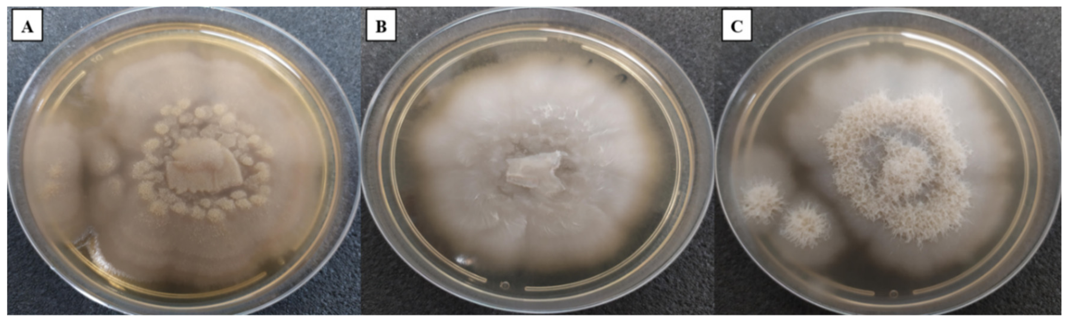
3.1. Macroscopic Characterisation

Macroscopic features confirmed the rapid growth time of Coniochaeta species on SDA GC medium at an optimal temperature of 25 °C for all species (Figure 1). However, none of the three yeasts was able to grow at 4 and 45 °C. Colonies of the three isolates were initially white to beige, both on the surface and on the reverse. After four to five days of incubation, Coniochaeta massiliensis turned light orange to salmon. All colonies were flat and moist. Coniochaeta hoffmannii DSM 2693 and the newly isolated yeast (PMML0158) presented a glabrous aspect, while Coniochaeta mutabilis DSM 10716 was typified by an aerial mycelial growth.

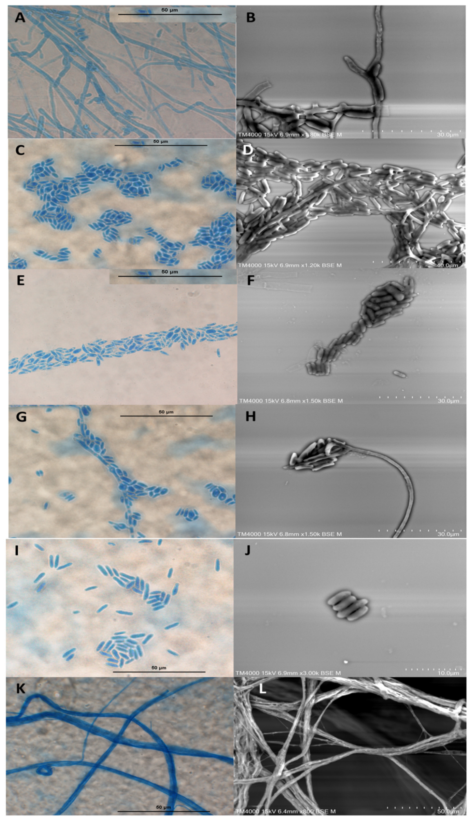
3.2. Microscopic Characterisation

The microscopic observation of the three strains revealed wide septate hyphae, numerous cylindrical adelophialides (short phialides without septum), discrete phialides with conical tips exhibiting ellipsoidal to cylindrical and rarely curved conidia, and nonseptate with thin and smooth conidial walls (2 to 3 by 6 to 10 μm). Several conidia were observed aggregating on the hyphae’s sides and most often in clusters. Collarettes were only found in both Coniochaeta massiliensis and C. mutabilis. No chlamydospore was observed (Figure 2).

3.3. Antifungal Susceptibility Testing (AFST)

The minimum inhibitory concentration (MIC) values for the three species for all nine antifungal drugs are shown in (Table 3). The MIC endpoints for all strains were determined as the lowest concentrations inhibiting the growth of 90% of the strains and were determined as described in Perdomo et al. (2011), since there are no validated AFST guidelines for this genus. The MICs of AMB, 5-FC, ITC, POS, and VOR were low for the three isolates. The new species of Coniochaeta displayed low echinocandin (AND and CAS) MICs and a high FL MIC, while C. hoffmannii and C. mutabilis demonstrated the opposite for these four antifungal drugs.

3.4. MALDI-TOF MS Identification

The MALDI-TOF MS identification score (log score < 1.90) was below the limit required for obtaining a good identification. The isolate was identified at the genus level as Coniochaeta sp. identification score values were generated from MALDI-TOF MS spectra of the new isolate and the two other reference strains from the DSMZ collection, Coniochaeta mutabilis DSM 10716 and Coniochaeta hoffmannii DSM 2693, which were collected and have been entered in the MALDI-TOF MS database.

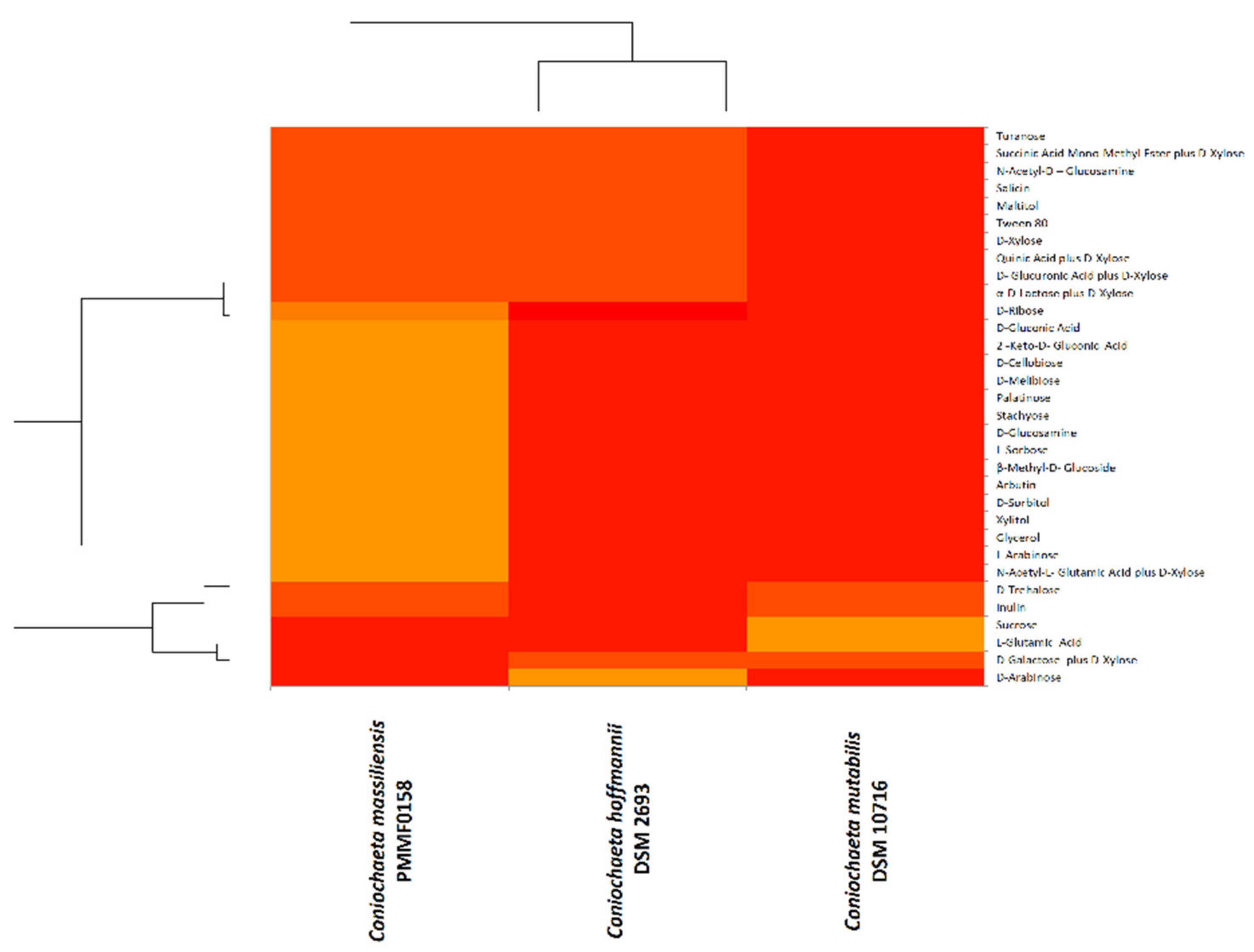
3.5. Physiological Analysis

3.5.1. EDX (Energy-Dispersive X-ray Spectroscopy)

The weights and atomic percentages of chemical elements resulting from the chemical mapping performed on the three species of the Coniochaeta genus displayed three distinct profiles. In the principal component analysis (Figure 3), each Coniochaeta species was very distant from the others, demonstrating highly heterogeneous chemical profiles.

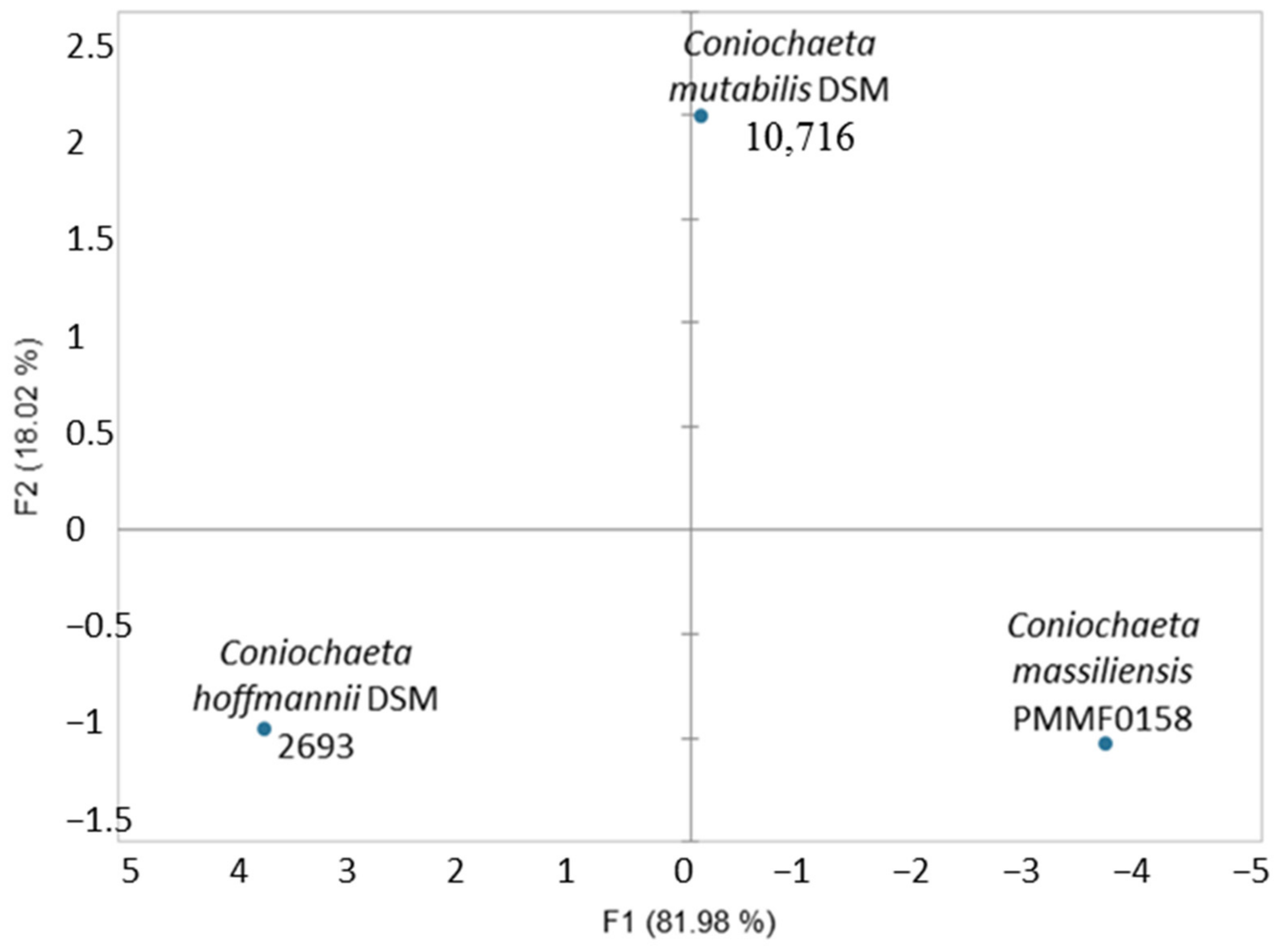
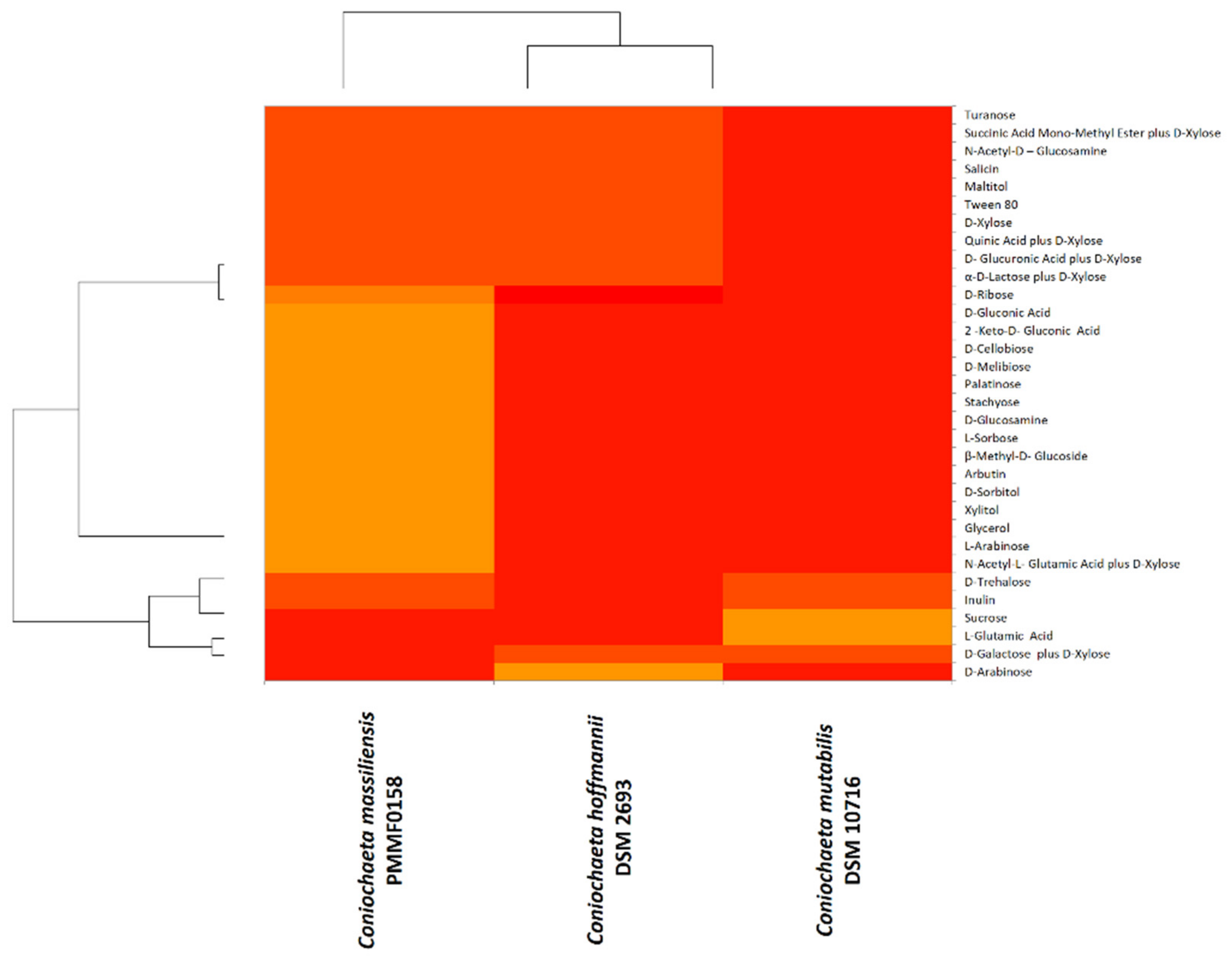
3.5.2. BiologTM System

BiologTM advanced phenotypic technology was very useful for the phenotypic characterisation. The oxidation and assimilation test results were illustrated using heat maps (Figure 4 and Figure 5). The majority of the substrates were not oxidized/assimilated. Each heat map was quite heterogeneous, and both demonstrated similar findings: Coniochaeta mutabilis DSM 10716 appeared more divergent from Coniochaeta massiliensis (PMML0158) than from Coniochaeta hoffmannii DSM 2693. However, Coniochaeta massiliensis (PMML0158) appeared to be closely related to Coniochaeta hoffmannii DSM 2693.

3.6. DNA Sequencing

The sequences were analysed using the BLASTn tool with the reference data available from the GenBank database of NCBI. The BLASTn of the new isolate showed a percent identity ≤97% for the four gene markers. Notably, the best-match pairwise identity was 96.51% for C. rhopalochaeta CBS 109872 with ITS (NR172554.1), 96.41% for C. deborreae BE19 001008 with TEF-1a (MW890087.1), 97.45% for C. deborreae CBS 147215 with D1/D2 (NR076709.1), and 94.44% for Cosmospora inonoticola 9361 with B-tub2 (KU563621.1).

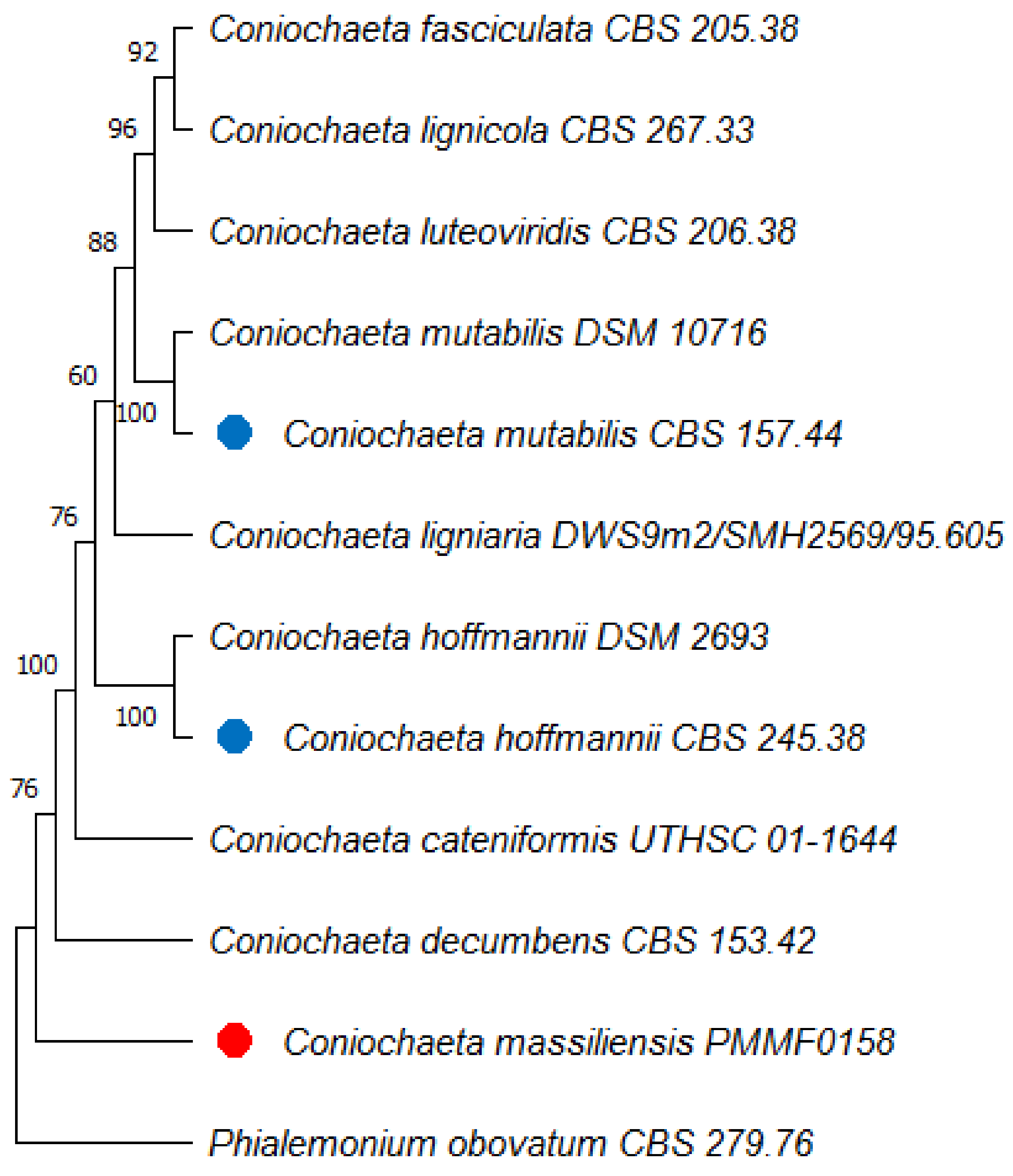
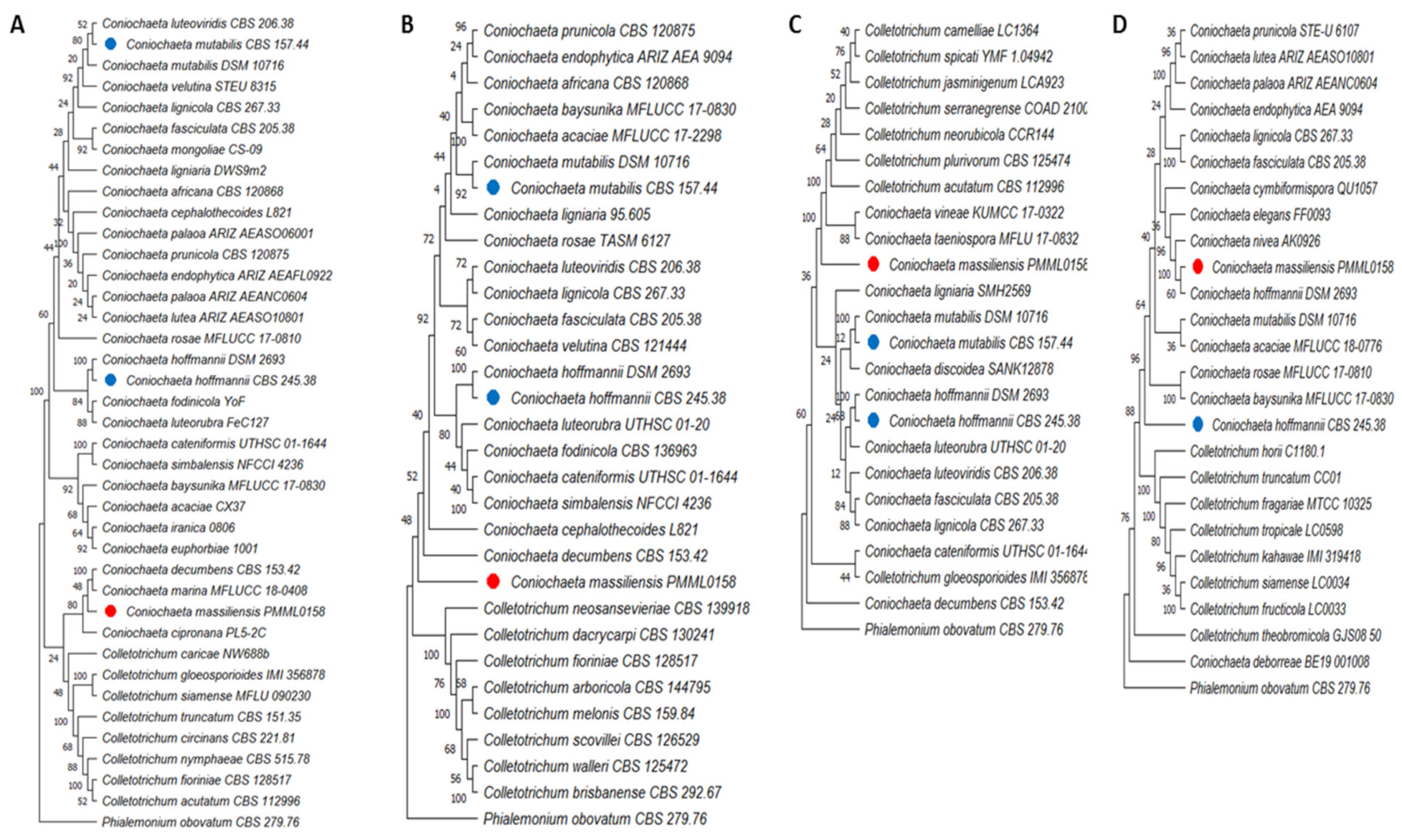
3.7. Phylogenetic Analyses

We built phylogenetic trees using MEGA 11 software. The multilocus analysis of the first tree (Figure 6) based on the assembled sequences of the ITS, B-tub2, and D1/D2 genetic regions revealed that the new isolate Coniochaeta massiliensis (PMML0158) clustered apart and seemed quite distinct from all other Coniochaeta species. Then, we constructed four other trees (Figure 7) with only one locus each, including supplementary Coniochaeta and Colletotrichum species. Within both ITS and TEF-1a trees, Coniochaeta massiliensis PMML0158 clustered with other Coniochaeta species. However, the D1/D2 and B-tub2 trees could not clearly position the new species PMML0158 in the Coniochaeta or Colletotrichum genus. However, the TEF-1a tree (Figure 7D) clearly positioned Coniochaeta massiliensis (PMML0158) as a distinct species within the Coniochaeta genus, which was unambiguously separated from the Colletotrichum genus.

3.8. Taxonomy

Coniochaeta massiliensis Kabtani J. & Ranque S. sp. nov.
MycoBank: MB843839
(Figure 2A–L).
Etymology: Named in honour of Marseille (France), the city where it was isolated.
Diagnosis: Closely similar to the other two Coniochaeta species examined, displaying the same flat and moist colonies aspect, as well as the absence of the dematiaceous appearance. However, relying on macroscopic features, it differs from C. mutabilis in that it lacks aerial growth. On the other hand, C. massiliensis presented the same microscopic structures as other species, such as the presence of several adelophialides, discrete phialides, and cylindrical or curved conidia. In this respect, C. massiliensis is closer to C. mutabilis, due to the decisive presence of the collarette.
Type: France: Marseille. Human body (abscess of the hand), 15 July 2020. (Holotype IHEM 28559/PMML0158, stored in a metabolically inactive state.) GenBank: OM366153 (ITS), ON000097 (B-tub2), OM640093 (TEF-1a), OM366268 (D1/D2).
Description: the macroscopic features were characterised by a rapid growth time on SDA GC medium at an optimal temperature of 25 °C. However, Coniochaeta massiliensis was not able to grow at 4 and 45 °C. Colonies were first white to beige, both on the surface and on the reverse after four to five days of incubation, then turned light orange to salmon. Colonies were flat and moist, with a glabrous aspect. There was no aerial mycelial growth. The microscopic features were characterised by the presence of wide septate hyphae, numerous cylindrical adelophialides (short phialides without septum), discrete phialides with conical tips, exhibiting ellipsoidal to cylindrical and rarely curved conidia, nonseptate with thin and smooth conidial walls (2 to 3 by 6 to 10 μm). Several conidia were observed aggregating on the side of the hyphae and most often in clusters. Collarettes were present, but chlamydospores formation was not observed.
The BiologTM carbon-source assimilation profile showed that C. massiliensis PMML0158 can assimilate different carbon substrates, such as 2-keto-D-gluconic acid, D-gluconic acid, D-ribose, D-xylose, D-glucosamine, D-cellobiose, D-melibiose, Palatinose, Turanose, L-sorbose, and β-methyl-D-glucoside. Based on this phenotypic analysis, C. massiliensis PMMFL0158 appears similar to C. hoffmannii DSM 2693.

Host: Human

Additional specimen examined: C. Hofmann: Reference: Country of origin unknown. Treated pine stake, before 8 July 1983. (DSM 2693-ATCC 34158–SP33-4.) GenBank: OM366155 (ITS), ON000099 (B-tub2), OM640095 (TEF-1a), OM366270 (D1/D2).
Additional specimen examined: C. mutabilis: Reference: Sweden. Origin of isolation unknown, before 14 June 1996. (DSM 10716-EMPA 573, S24 E.) GenBank: OM366154 (ITS), ON000098 (B-tub2), OM640094 (TEF-1a), OM366269 (D1/D2).

4. Discussion

Coniochaeta mutabilis and Coniochaeta hoffmannii are the most familiar human pathogens of the Coniochaeta genus and the most widespread and commonly encountered in human samples and severe infections. The MALDI-TOF MS identifications for these two species were relevant, with log scores >2.0. However, this tool was not able to perform a species identification for the new isolate, which led to a molecular analysis targeting four relevant genes: the internal transcribed spacer (ITS1/ITS2) in the RNA ribosomal small subunit (SSU), a fragment of the translation elongation factor 1-alpha gene (TEF-1-α), a fragment of the β-tubulin gene (B-tub2), and the D1/D2 domains of the ribosomal DNA large subunit (LSU).
The multilocus phylogenetic tree (Figure 6) constructed with the concatenated sequences of ITS, B-tub2, and D1D2 showed that the type strains C. mutabilis CBS 157.44 and C. hoffmannii CBS 245.38 clustered within distinct clades and that both were distant from Coniochaeta massiliensis (PMML0158). Indeed, this newly isolated yeast was divergent from all other Coniochaeta species present in the tree. However, in the single-locus trees for ITS and B-tub2, Coniochaeta massiliensis (PMML0158) seemed closer to Coniochaeta species than in other single-locus trees for D1/D2 and TEF-1a, where it appeared much more distant from Coniochaeta species than the genus Colletotrichum.
Based on phylogeny, the new isolate seems divergent from the group’s other known taxa of Coniochaeta. de Hoog et al. (2000) described C. lignaria as being genetically closely related to C. hoffmannii. However, in our phylogenetic tree, these yeasts clustered into two different clades, indicating a large distance between the two species. These findings are in line with those of both Perdomo et al. (2011) and Weber and Begerow (2002), who reported significant differences between these species.
Antifungal susceptibility testing showed relatively low MICs for AMB, 5-FC, ITC, POS, and VOR in all strains. These results are in line with those of Perdomo et al. (2011) but contrast with those of other authors [9,13,20] who reported relatively high MICs for AMB and ITC against C. hoffmannii and C. mutabilis.
We macroscopically observed a fast-growing phenotype in Coniochaeta species. This finding contrasts with those of Ahmad et al. (1985), who reported slow colony growth. All species displayed the same flat and moist colonies. Only C. mutabilis additionally displayed low aerial growth, as reported by Drees et al. (2007). The primary colony colour remained unchanged (white/beige to orange/salmon). None of the colonies turned brown/dark in culture even after one month of incubation. This aspect has been well described in C. hoffmanii, a species that lacks the dematiaceous aspect, whereas C. mutabilis is classified within dematiaceous fungi, which are particularly typified by the presence of melanin in hyphae and conidia cell walls, which is responsible for dark pigment emission. Occasionally, certain C. mutabilis species lack the melanin property, as mentioned in Khan et al. (2013), Dress et al. (2007), and Perdomo et al. (2011). This was noted for the C. mutabilis DSM 10716 strain. Furthermore, in this study, the three species lacked the dematiaceous aspect.
The most remarkable morphological characteristics of the Coniochaeta species that have been described were the presence of several adelophialides, discrete phialides, and cylindrical or curved conidia, in addition to the presence or absence of collarettes and chlamydospores, depending on the species [9,14,18,29]. Most of these specific characteristics were observed in the newly described species (PMML0158). Based on this microscopic analysis, we infer that Coniochaeta massiliensis is closer to C. mutabilis, due to the decisive presence of a collarette.
Morphological characterisation helped for species differentiation, and physiological analysis was more convincing in distinguishing the three species, with the aim of describing the new one as thoroughly as possible. EDX (energy-dispersive X-ray spectroscopy) and BiologTM phenotypic technology revealed divergent chemical mapping and carbon-source oxidation/assimilation profiles between the new isolate and the two main species of Coniochaeta genus. Moreover, the BiologTM system findings were more relevant, as they revealed that the physiological profile of Coniochaeta massiliensis was closer to C. hoffmannii DSM 2693 than to C. mutabilis DSM 10716.
In conclusion, the clinical yeast isolate PMML0158 is herein described as Coniochaeta massiliensis, a new species that can be easily discriminated from the other species in the Coniochaeta genus owing to distinct genomic sequences and chemical and physiological profiles.

Author Contributions

Conceptualisation, J.K. and S.R.; methodology, S.R., J.K., and M.M.; formal analysis, J.K., M.M., and S.R.; writing—original draft preparation, J.K.; writing—review and editing, S.R. and M.M. All authors have read and agreed to the published version of the manuscript.

Funding

This research was funded by the Institut Hospitalo-Universitaire (IHU) Méditerranée Infection and by the French government under the “Investissements d’avenir” (Investments for the Future) programme managed by the Agence Nationale de la Recherche (ANR, fr: National Agency for Research), (reference: Méditerranée Infection 10-IAHU-03) and the Région Provence-Alpes-Côte d’Azur, European ERDF funding (European Regional Development Fund) and PRIMMI (Plateformes de Recherche et d’Innovation Mutualisées Méditerranée Infection).

Institutional Review Board Statement

Not applicable.

Informed Consent Statement

Not applicable.

Data Availability Statement

The Coniochaeta massiliensis holotype is available in the IHU MI (no. PMML0158) and IHEM (no. 28559) strain collections. The nucleotide sequences are available in GenBank (accession nos. OM366153, ON000097, OM640093, and OM366268). The datasets analysed in the current study are available from the corresponding author upon reasonable request.

Acknowledgments

The authors gratefully acknowledge wholehearted and fruitful discussions with Pierre Pontarotti concerning phylogenetic analysis and the technical support of Anthony Fontanini for electron microscopy investigations. We also sincerely thank Takashi Irie, Kyoko Imai, Shigeki Matsubara, Taku Sakazume, Toshihide Agemura, and the Hitachi team in Japan for the collaborative study conducted together with IHU Méditerranée Infection and for the installation of TM4000 Plus microscopes at the IHU Méditerranée Infection facility.

Conflicts of Interest

The authors have no conflicts of interest to declare regarding this study.

Abbreviations

5-FC5-fluorocytosine
AMBAmphotericin B
ANDAnidulafungin
CASCaspofungin
DNADeoxyribonucleic acid
EDXEnergy-dispersive X-ray spectroscopy
FLFluconazole
GCGentamycin and chloramphenicol
IHU Méditerranée InfectionInstitut Hospitalo-Universitaire Méditerranée Infection
ITCItraconazole
ITSInternal transcribed spacers of the rRNA
SDASabouraud Dextrose Agar
LSULarge subunit of rRNA
MALDI-TOF MSMatrix-assisted laser desorption/ionization–time of flight mass spectrometry
MICsMinimum inhibitory concentrations
MIFMicafungin
NANot available
POSPosaconazole
rRNARibosomal ribonucleic acid
SSUSmall subunit of rRNA
TEF-1αPartial translation elongation factor 1-alpha gene
B-tub2Partial β-tubulin 2 gene
VORVoriconazole

References

  1. Weber, E.G. The Lecythophora-Coniochaeta Complex: I. Morphological Studies on Lecythophora Species Isolated from Picea Abies. Nova Hedwig. 2002, 74, 85–159. [Google Scholar] [CrossRef]
  2. Nosanchuk, J.D.; Casadevall, A. Impact of Melanin on Microbial Virulence and Clinical Resistance to Antimicrobial Compounds. Antimicrob. Agents Chemother. 2006, 50, 28–3519. [Google Scholar] [CrossRef] [PubMed]
  3. Guarro, J.; Gené, J.; Ai-Bader, S.M.; Abdullah, S.K. A New Species of Coniochaetidium from Soil. Mycoscience 1997, 38, 25–123. [Google Scholar] [CrossRef]
  4. Kamiya, S.; Uchiyama, S.; Udagawa, S. Two New Species of Coniochaeta with a Cephaiothecoid Peridium Wall. Mycoscience 1995, 36, 83–377. [Google Scholar] [CrossRef]
  5. Yuan, Z.Q.; Mohamed, C. New species and new records of Ascomycetes of stems of Eucalyptus from Tasmania, Australia. Mycotaxon 1997, 63, 9–23. [Google Scholar]
  6. Mahoney, D.P.; Lafavre, J.S. Coniochaeta extramundana, with a synopsis of other Coniochaeta species. Mycologia 1981, 73, 931–952. [Google Scholar] [CrossRef]
  7. Crane, J.L.; Shearer, C.A. A new Coniochaeta from fresh water. Mycotaxon 1995, 54, 107–110. [Google Scholar]
  8. Perdomo, H.D.; García, D.; Gené, J.; Cano, J.; Sutton, D.A.; Summerbell, R.C.; Guarro, J. Phialemoniopsis, a New Genus of Sordariomycetes, and New Species of Phialemonium and Lecythophora. Mycologia 2013, 105, 398–421. [Google Scholar] [CrossRef]
  9. Khan, Z.; Gené, J.; Ahmad, S.; Cano, J.; Al-Sweih, N.; Joseph, L.; Chandy, R.; Guarro, J. Coniochaeta Polymorpha, a New Species from Endotracheal Aspirate of a Preterm Neonate, and Transfer of Lecythophora Species to Coniochaeta. Antonie Van Leeuwenhoek 2013, 104, 52–243. [Google Scholar] [CrossRef]
  10. Weber, E.G.; Begerow, D. The Lecythophora-Coniochaeta Complex: II. Molecular Studies Based on Sequences of the Large Subunit of Ribosomal DNA. Nova Hedwig. 2002, 74, 187–200. [Google Scholar] [CrossRef]
  11. De Hoog, G.S.; Guarro, J.; Gené, J.; Figueras, M.J. Atlas of Clinical Fungi, 2nd ed.; Universitat Rovira i Virgili Reus: Tarragona, Spain; Centraalbureau voor Schimmelcultures: Utrecht, The Netherlands, 2000. [Google Scholar]
  12. Gams, W.; McGinnis, M.R. Phialemonium, A New Anamorph Genus Intermediate Between Phialophora and Acremonium. Mycologia 1983, 75, 977–987. [Google Scholar] [CrossRef]
  13. Fintelmann, R.E. Recurrent Lecythophora mutabilis Keratitis and Endophthalmitis after Deep Anterior Lamellar Keratoplasty. Arch. Ophthalmol. 2011, 129, 106–110. [Google Scholar] [CrossRef] [PubMed] [Green Version]
  14. Marriott, D.J.; Wong, K.H.; Aznar, E.; Harkness, J.L.; Cooper, D.A.; Muir, D. Scytalidium dimidiatum and Lecythophora hoffmannii: Unusual Causes of Fungal Infections in a Patient with AIDS. J. Clin. Microbiol. 1997, 35, 2949–2952. [Google Scholar] [CrossRef] [PubMed]
  15. Sakaeyama, S.; Sano, A.; Murata, Y.; Kamei, K.; Nishimura, K.; Hatai, K. Lecythophora hoffmannii Isolated from a Case of Canine Osteomyelitis in Japan. Med. Mycol. 2007, 45, 72–267. [Google Scholar] [CrossRef] [PubMed]
  16. Ahmad, S.; Johnson, R.J.; Hillier, S.; Shelton, W.R.; Rinaldi, M.G. Fungal Peritonitis Caused by Lecythophora mutabilis. J. Clin. Microbiol. 1985, 22, 86–182. [Google Scholar] [CrossRef]
  17. Slifkin, M.; Bowers, H.M. Phialophora mutabilis Endocarditis. Am. J. Clin. Pathol. 1975, 63, 30–120. [Google Scholar] [CrossRef]
  18. Drees, M.; Wickes, B.L.; Gupta, M.; Hadley, S. Lecythophora mutabilis Prosthetic Valve Endocarditis in a Diabetic Patient. Med. Mycol. 2007, 45, 67–463. [Google Scholar] [CrossRef]
  19. Taniguchi, Y.; Taketani, T.; Moriyama, H.; Moriki, S.; Nishimura, K.; Sato, E.; Notsu, Y.; Higuchi, T.; Sugitani, Y.; Yasuda, K.; et al. Septic Shock Induced by Lecythophora mutabilis in a Patient with Mitochondrial Encephalomyopathy. J. Med. Microbiol. 2009, 58, 58–1255. [Google Scholar] [CrossRef]
  20. Scott, I.U.; Cruz-Villegas, V.; Flynn, H.W.; Miller, D. Delayed-Onset, Bleb-Associated Endophthalmitis Caused by Lecythophora Mutabilis. Am. J. Ophthalmol. 2004, 3, 85–583. [Google Scholar] [CrossRef]
  21. Marcus, D.M.; Hull, D.S.; Rubin, R.M.; Newman, C.L. Lecythophora mutabilis endophthalmitis after long-term corneal cyanoacrylate. Retina 1999, 19, 351–353. [Google Scholar] [CrossRef]
  22. Ho, R.H.T.; Bernard, P.J.; McClellan, K.A. Phialophora mutabilis Keratomycosis. Am. J. Ophthalmol. 1991, 112, 29–728. [Google Scholar] [CrossRef]
  23. Cassagne, C.; Normand, A.N.; L’Ollivier, C.; Ranque, S.; Piarroux, R. Performance of MALDI-TOF MS Platforms for Fungal Identification. Mycoses 2016, 59, 90–678. [Google Scholar] [CrossRef] [PubMed]
  24. Normand, A.C.; Cassagne, C.; Gautier, M.; Becker, P.; Ranque, S.; Hendrickx, M.; Piarroux, R. Decision Criteria for MALDI-TOF MS-Based Identification of Filamentous Fungi Using Commercial and in-House Reference Databases. BMC Microbiol. 2017, 17, 17–25. [Google Scholar] [CrossRef]
  25. Bochner, B.R. Phenotype MicroArrays for High-Throughput Phenotypic Testing and Assay of Gene Function. Genome Res. 2001, 11, 55–1246. [Google Scholar] [CrossRef] [PubMed]
  26. White, T.J.; Bruns, T.; Lee, S.; Taylor, J. Amplificarion and direct sequencing of fungal ribosomal RNA genes for phylogenetics. PCR Protoc. 1990, 18, 22–315. [Google Scholar]
  27. Glass, N.L.; Donaldson, G.C. Development of Primer Sets Designed for Use with the PCR to Amplify Conserved Genes from Filamentous Ascomycetes. Appl. Environ. Microbiol. 1995, 61, 30–1323. [Google Scholar] [CrossRef] [PubMed]
  28. Carbone, I.; Kohn, L.M. A Method for Designing Primer Sets for Speciation Studies in Filamentous Ascomycetes. Mycologia 1999, 91, 553–556. [Google Scholar] [CrossRef]
  29. Perdomo, H.D.; Sutton, A.; Garcia, D.; Fothergill, A.W.; Gené, J.; Cano, J.; Summerbell, R.C.; Rinaldi, M.G.; Guarro, J. Molecular and Phenotypic Characterization of Phialemonium and Lecythophora Isolates from Clinical Samples. J. Clin. Microbiol. 2011, 49, 1209–1216. [Google Scholar] [CrossRef] [Green Version]
Figure 1. Culture growth on Sabouraud Dextrose Agar + gentamicin and chloramphenicol after five days of incubation at 25 °C. The colour of both the recto and verso of the colonies was white/beige to salmon. (A) Coniochaeta massiliensis PMML0158. (B) Coniochaeta hoffmannii DSM 2693. (C) Coniochaeta mutabilis DSM 10716.
Jof 08 00999 g001
Figure 2. Morphology of Coniochaeta massiliensis (PMML0158). (A,B) Formation of conidiogenous cells (adelophialides) on hyphae and presence of collarette (arrow). (CF) Conidia aggregating in clusters. (G) Conidia aggregating along the sides of the hyphae. (H) Phialoconidia assembled at the phialide tip. (I,J) Cylindrical conidia with thin and smooth walls. (K,L) Wide hyphae. Optical microscopy (magnification ×1000). Scale bars: 50 μm. Scanning electron microscopy (15 KeV lens mode 4). Scale bars: J = 10 μm; B,F,H = 30 μm; D = 40 μm; L = 50 μm.
Jof 08 00999 g002
Figure 3. Principal component analysis (PCA) processed with the XLSTAT software of the energy-dispersive X-ray spectroscopy chemical mapping profile, performed for the novel species Coniochaeta massiliensis (PMML0158) and two reference strains in the genus. The principal components F1 and F2 explained 100% of the chemical mapping profile variance.
Jof 08 00999 g003
Figure 4. Heat map computed with the XLSTAT software for carbon-source oxidation by the BiologTM system for the novel species Coniochaeta massiliensis (PMML0158) and two reference strains in the genus. Colour-gradient interpretation: the most-oxidized substrates are shown in light orange and the least-oxidized substrates are shown in red.
Jof 08 00999 g004
Figure 5. Heat map computed with the XLSTAT software for carbon-source assimilation by the BiologTM system for the novel species Coniochaeta massiliensis (PMML0158) and two reference strains. Colour-gradient interpretation: the most-assimilated substrates are shown in light orange and the least-assimilated substrates are shown in red.
Jof 08 00999 g005
Figure 6. Multilocus phylogenetic tree of the newly isolated species Coniochaeta massiliensis PMML0158 (indicated with red dots) and 11 reference strains (type strains are indicated with blue dots), based on the concatenated ITS, B-tub2, and D1/D2 sequences. Phialemonium obovatum CBS 279.76 was used as an outgroup. The maximum-parsimony tree was generated using the MEGA 11 software, with 1000-replication bootstrap values.
Jof 08 00999 g006
Figure 7. Four single-locus phylogenetic trees using the ITS (A), D1/D2 (B), B-tub2 (C), and TEF-1α (D) genomic regions. The species included in each tree differ because the sequences for each locus were not available for all strains. The red dots indicate the new species Coniochaeta massiliensis PMML0158 and blue dots indicate type strains. Phialemonium obovatum CBS 279.76 was used as the outgroup. The maximum-parsimony tree was generated using the MEGA 11 software, with 1000-replication bootstrap values.
Jof 08 00999 g007
Table 1. Sets of primers used for amplifying the ITS, B-tub2, TEF1, and D1/D2 genetic regions.
PrimersSequencesTargeted RegionsReferences
ITS1TCCGTAGGTGAACCTGCGG18S-5.8S[26]
ITS2GCTGCGTTCTTCATCGATGC18S-5.8S[26]
ITS3GCATCGATGAAGAACGCAGC5.8S-28S[26]
ITS4TCCTCCGCTTATTGATATGC5.8S-28S[26]
ITS1TCCGTAGGTGAACCTGCGG18S-5.8S, 5.8S-28S[26]
ITS4TCCTCCGCTTATTGATATGC18S-5.8S, 5.8S-28S[26]
Bt-2aGGTAACCAAATCGGTGCTGCTTTCB-tub2[27]
Bt-2bACCCTCAGTGTAGTGACCCTTGGCB-tub2[27]
EF1-728FCATCGAGAAGTTCGAGAAGGTEF1[28]
EF1-986RTACTTGAAGGAACCCTTACCTEF1[28]
D1AACTTAAGCATATCAATAAGCGGAGGA28S[11]
D2GGT CCG TGT TTC AAG ACG G28S[11]
Table 2. GenBank accession numbers of the reference strains used for the phylogenetic analyses.
SpeciesCollection IDGenBank Accession Numbers
ITSB-tub2D1/D2TEF1
Coniochaeta massiliensisPMML0158OM366153ON000097OM366268OM640093
Coniochaeta mutabilisDSM 10716OM366154ON000098OM366269OM640094
Coniochaeta hoffmanniiDSM 2693OM366155ON000099OM366270OM640095
Coniochaeta fasciculataCBS 205.38HE610336HE610350FR691988MK693152
Coniochaeta lignicolaCBS 267.33HE610335HE610353FR691986MK693154
Coniochaeta luteoviridisCBS 206.38HE610333HE610351FR691987NA *
Coniochaeta hoffmanniiCBS 245.38HE610332HE610352FR691982MK693150
Coniochaeta mutabilisCBS 157.44HE610334HE610349AF353604NA
Coniochaeta lignariaDWS9m2/SMH2569/95.605KJ188673AY780113AF353584NA
Coniochaeta cateniformisUTHSC 01-1644HE610331HE610347HE610329NA
Coniochaeta decumbensCBS 153.42HE610337HE610348HE610463NA
Coniochaeta velutinaCBS 121444NANAGQ154605NA
Coniochaeta velutinaSTEU 8315KY312638NANANA
Coniochaeta simbalensisNFCCI 4236MG825743NANG_068555NA
Coniochaeta fodinicolaYoF/CBS 136963JQ904607NANG_064287NA
Coniochaeta prunicolaCBS 120875GQ154540NANG_066151NA
Coniochaeta prunicolaSTE-U 6107NA *NANAMK693162
Coniochaeta africanaCBS 120868GQ154539NANG_066150NA
Coniochaeta palaoaARIZ AEASO06001MZ241149NANANA
Coniochaeta palaoaARIZ AEANC0604MK458764NANAMZ241188
Coniochaeta marinaMFLUCC 18-0408MF422164NANANA
Coniochaeta cipronanaPL5-2CMG828883NANANA
Coniochaeta rosaeMFLUCC/17-0810MG828880NANAMG829197
Coniochaeta rosaeTASM 6127NANANG_066204NA
Coniochaeta baysunikaMFLUCC/17-0830MW750761NAMG828996MG829196
Coniochaeta acaciaeCX37MW750756NANANA
Coniochaeta acaciaeMFLUCC 18-0776NANANAMT503199
Coniochaeta acaciaeMFLUCC 17-2298NANAMG062737NA
Coniochaeta fibrosaeCX04D1MW077645NANANA
Coniochaeta mongoliaeCS-09KP941078NANANA
Coniochaeta iranica806KP941076NANANA
Coniochaeta euphorbiae1001KY064029NANANA
Coniochaeta cephalothecoidesL821MW447035NAKY064030NA
Coniochaeta luteorubraFeC127MZ241160NANANA
Coniochaeta luteorubraUTHSC 01-20NAHE610346HE610328NA
Coniochaeta luteaAEASO10801MZ241150NANAMZ241193
Coniochaeta endophyticaARIZ AEAFL0922MZ241147NANANA
Coniochaeta endophyticaARIZ AEA 9094NANANG_075158NA
Coniochaeta vineaeKUMCC 17-0322NAMN485898NANA
Coniochaeta taeniosporaMFLU 17-0832NAMN509784NANA
Coniochaeta discoideaSANK12878NAAY780134NANA
Coniochaeta elegansFF0093NANANAMZ267815
Coniochaeta niveaAK0926NANANAMZ267793
Coniochaeta deborreaeBE19_001008NANANAMW890087
Coniochaeta cymbiformisporaQU1057NANANAMZ267839
Coniochaeta endophyticaAEA 9094NANANG_075158MK693159
Colletotrichum nymphaeaeCBS 515.78NR_111736NANANA
Colletotrichum caricaeNW688bNR_111736NANANA
Colletotrichum fioriniaeCBS 151.35NR_111458NANG_069002NA
Colletotrichum circinansCBS 128517NR_111747NANANA
Colletotrichum gloeosporioidesCBS 221.81NR_111457NANANA
Colletotrichum gloeosporioidesIMI 356878NAAJ409291NANA
Colletotrichum acutatumIMI 356878NR_160754NANANA
Colletotrichum acutatumCBS 112996NAAJ409296NANA
Colletotrichum siamenseCBS 112996NR_144794NANANA
Colletotrichum siamenseLC0034NANANAJQ071904
Colletotrichum truncatumMFLU 090230NR_144784NANANA
Colletotrichum truncatumCC01NANANAMW030430
Colletotrichum plurivorumCBS 125474NAMG600985NANA
Colletotrichum jasminigenumLCA923NAHM153770NANA
Colletotrichum serranegrenseCOAD 2100NAKY407896NANA
Colletotrichum neorubicolaCCR144NAMN186400NANA
Colletotrichum spicatiYMF 1.04942NAOL981226NANA
Colletotrichum fructicolaLC0033NANANAJQ071903
Colletotrichum theobromicolaGJS08_50NANANAGU994506
Colletotrichum horiiC1180.1NANANAGQ329693
Colletotrichum tropicaleLC0598NANANAJQ071909
Colletotrichum kahawaeIMI 319418NANANAJQ071908
Colletotrichum fragariaeMTCC 10325NANANAJQ071906
Colletotrichum camelliaeLC1364NAJN936976NANA
Colletotrichum dacrycarpiCBS 130241NANANG_073638NA
Colletotrichum neosansevieriaeCBS 139918NANANG_070628NA
Colletotrichum arboricolaCBS 144795NANANG_070064NA
Colletotrichum scovilleiCBS 126529NANANG_070041NA
Colletotrichum walleriCBS 125472NANANG_070040NA
Colletotrichum melonisCBS 159.84NANANG_070037NA
Colletotrichum brisbanenseCBS 292.67NANANG_070034NA
Phialemonium obovatumCBS 279.76HE610365HE599334FR691997LT634003
* NA, not available.
Table 3. Results of in vitro antifungal susceptibility testing.
MIC * (mg/L) Read at 48 h
AMBANDCAS5-FCFLITCMIFPOSVOR **
Coniochaeta massiliensis PMML01580.250.51280.2510.250.12
Coniochaeta hoffmannii DSM 26930.1244140.06>320.120.12
Coniochaeta mutabilis DSM 107160.1248120.015>320.030.03
* MIC, minimum inhibitory concentration; ** AMB, amphotericin B; AND, anidulafungin; CAS, caspofungin; 5-FC, 5-fluorocytosine; FL, fluconazole; ITC, itraconazole; MIF, micafungin; POS, posaconazole; VOR, voriconazole.
Publisher’s Note: MDPI stays neutral with regard to jurisdictional claims in published maps and institutional affiliations.

