


Design and Validation of qPCR-Specific Primers for Quantification of the Marketed Terfezia claveryi and Terfezia crassiverrucosa in Soil

Abstract
:1. Introduction
2. Materials and Methods
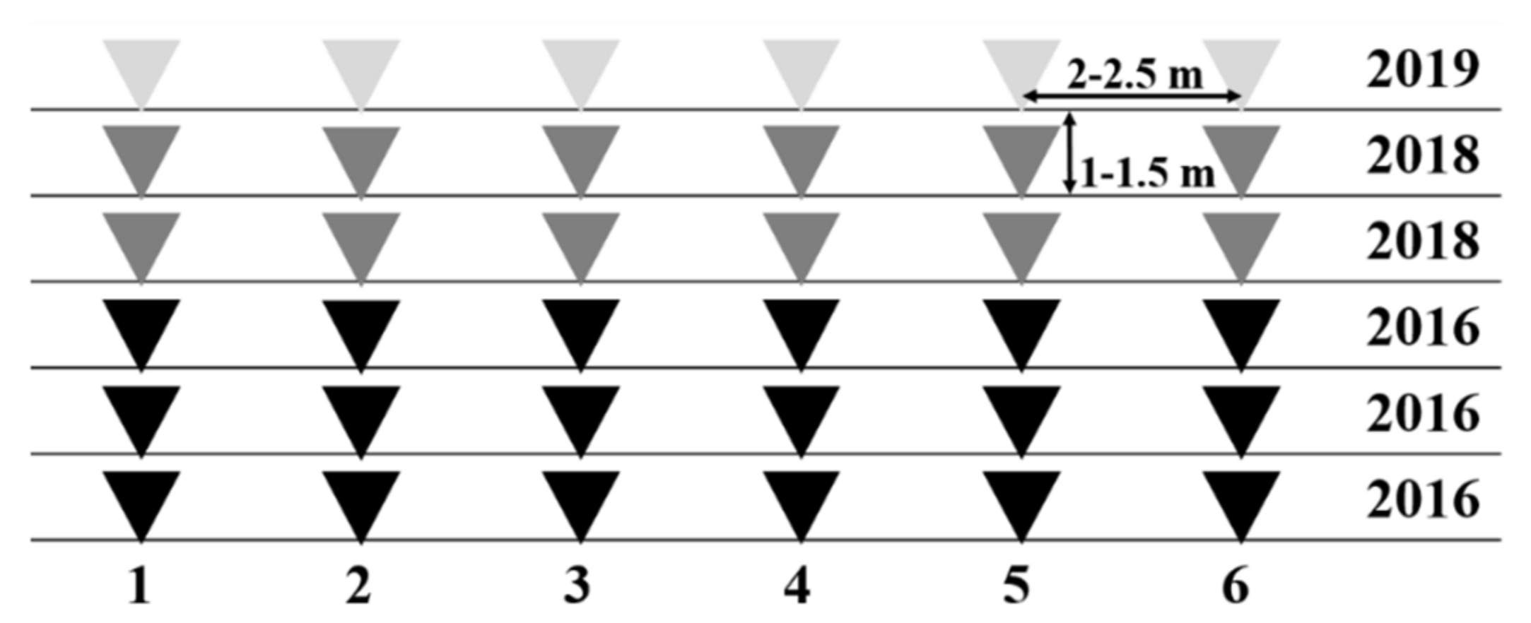
2.1. Environmental Sampling
2.2. Soil DNA Extraction
2.3. Design of Specific Primers for Turmas
2.4. Quantitative Real-Time PCR Conditions
2.5. Statistical Analysis
3. Results and Discussion
3.1. In Silico Primer Screening
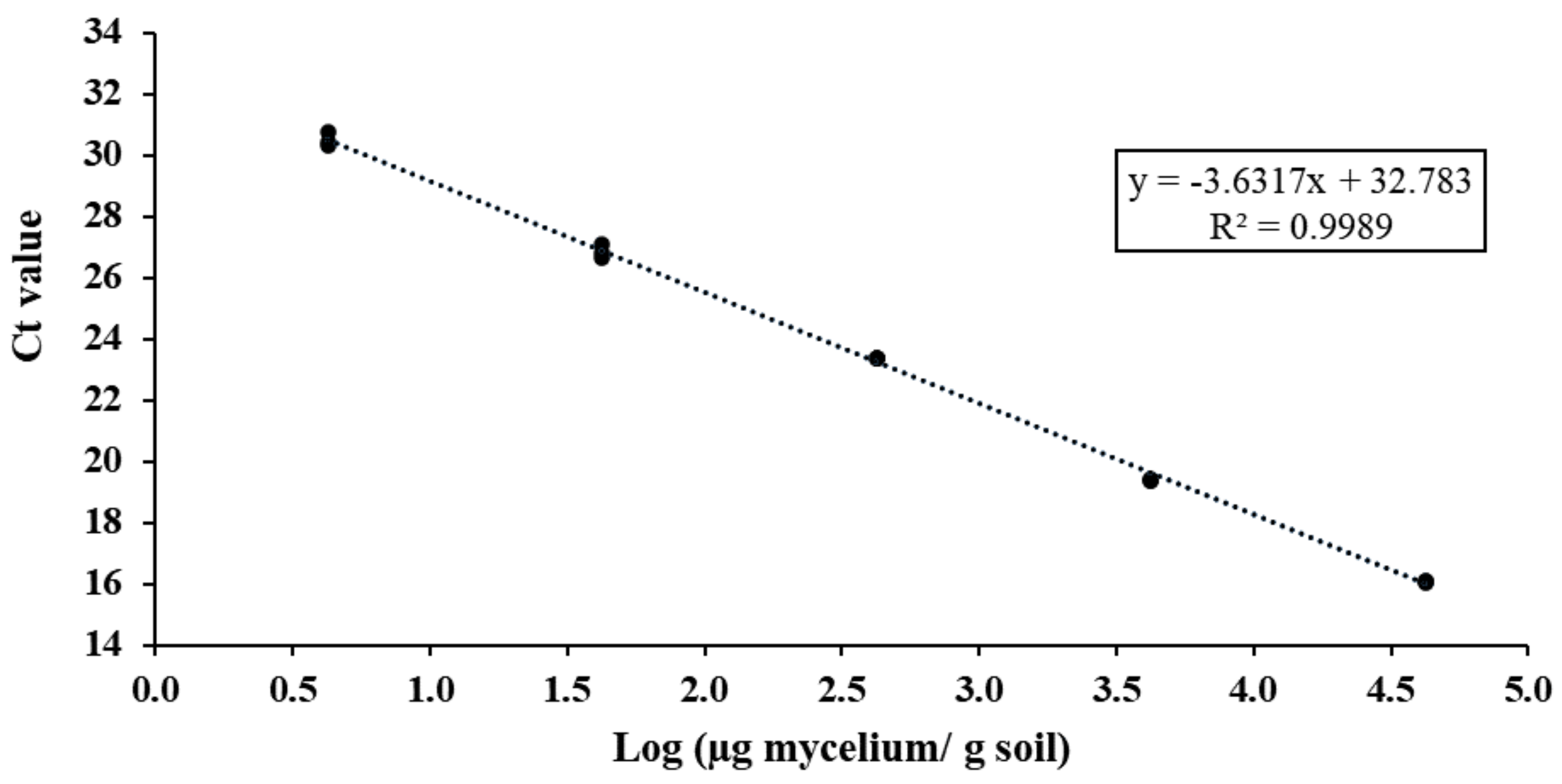
3.2. Selection and Validation of qPCR-Specific Primers
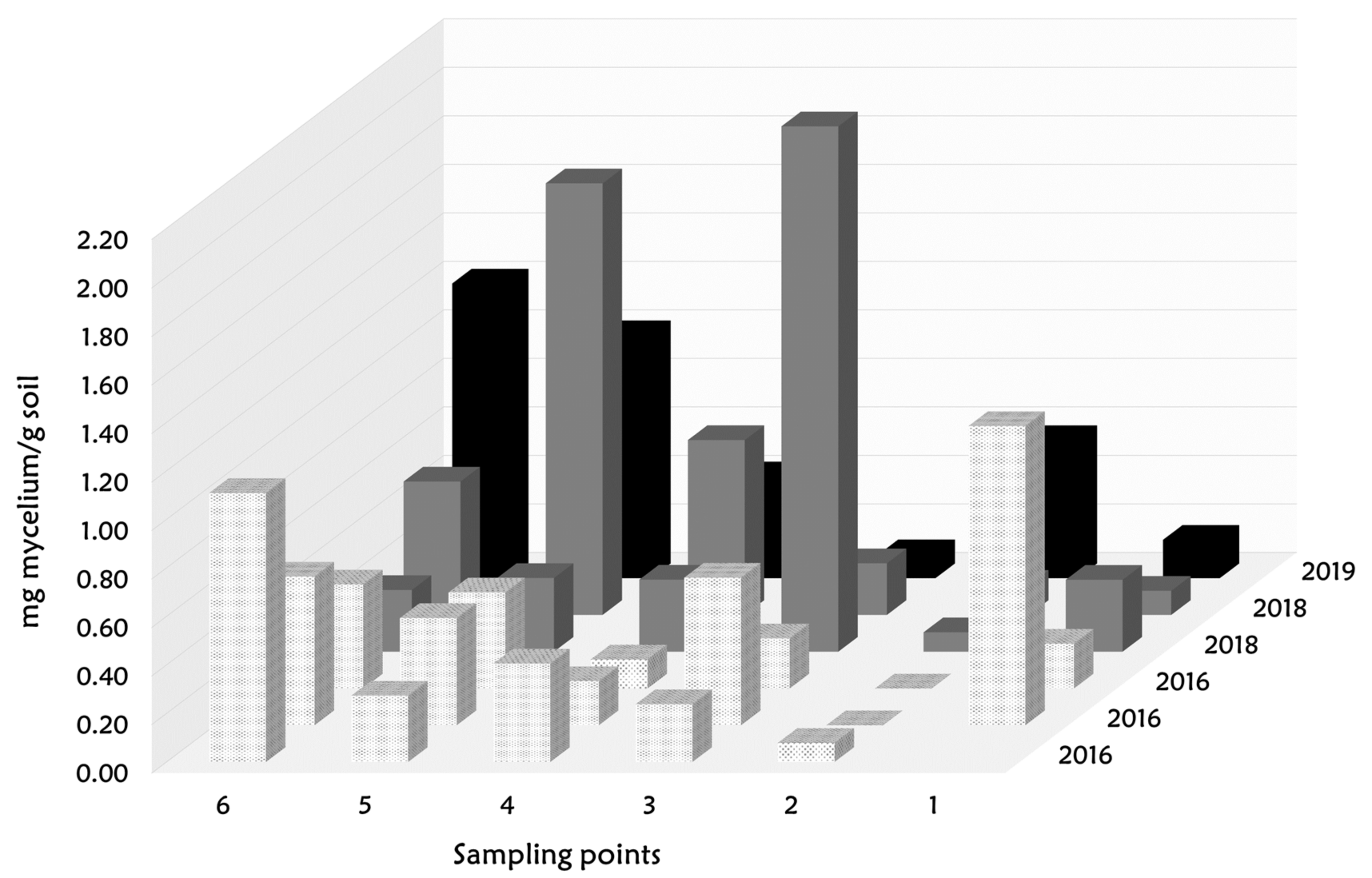
3.3. Spatial Dynamic of Turmas Mycelium in a Desert Truffle Orchard
4. Conclusions
Supplementary Materials
Author Contributions
Funding
Data Availability Statement
Acknowledgments
Conflicts of Interest
References
- Morte, A.; Pérez-Gilabert, M.; Gutiérrez, A.; Arenas, F.; Marqués-Gálvez, J.E.; Bordallo, J.J.; Rodríguez, A.; Berná, L.M.; Lozano-Carrillo, C.; Navarro-Ródenas, A. Basic and Applied Research for Desert Truffle Cultivation. In Mycorrhiza-Eco-Physiology, Secondary Metabolites, Nanomaterials; Varma, A., Prasad, R., Tuteja, N., Eds.; Springer: Cham, Switzerland, 2017; pp. 23–42. [Google Scholar]
- Bradai, L.; Bissati, S.; Chenchouni, H. Desert Truffles of the North Algerian Sahara: Diversity and Bioecology. Emir. J. Food Agric. 2014, 26, 425–435. [Google Scholar] [CrossRef] [Green Version]
- Shavit, E. The History of Desert Truffle Use. In Desert Truffles. Soil Biology; Kagan-Zur, V., Roth-Bejerano, N., Sitrit, Y., Morte, A., Eds.; Springer: Berlin/Heidelberg, Germany, 2014; pp. 217–241. [Google Scholar]
- Honrubia, M.; Gutiérrez, A.; Morte, A. Desert Truffle Plantation from South-East Spain. In Proceedings of the Edible Mycorrhizal Mushrooms and Their Cultivation: Proceedings of the Second International Conference on Edible Mycorrhizal Mushrooms, Christchurch, New Zealand, 3–6 July 2001; pp. 3–5. [Google Scholar]
- Morte, A.; Gutiérrez, A.; Ródenas, A.N. Advances in Desert Truffle Mycorrhization and Cultivation. In Mushrooms, Humans and Nature in a Changing World. Perspectives from Ecological, Agricultural and Social Sciences; Pérez-Moreno, J., Guerin-Laguette, A., Arzú, R.F., Yu, F.-Q., Eds.; Springer: Cham, Switzerland, 2020; pp. 205–219. [Google Scholar]
- Volpato, G.; Rossi, D.; Dentoni, D. A Reward for Patience and Suffering: Ethnomycology and Commodification of Desert Truffles among Sahrawi Refugees and Nomads of Western Sahara. Econ. Bot. 2013, 67, 147–160. [Google Scholar] [CrossRef]
- Murcia, M.A.; Martínez-Tomé, M.; Jiménez, A.M.; Vera, A.M.; Honrubia, M.; Parras, P. Antioxidant Activity of Edible Fungi (Truffles and Mushrooms): Losses during Industrial Processing. J. Food Prot. 2002, 65, 1614–1622. [Google Scholar] [CrossRef] [PubMed]
- Murcia, M.A.; Martínez-Tomé, M.; Vera, A.; Morte, A.; Gutierrez, A.; Honrubia, M.; Jiménez, A.M. Effect of Industrial Processing on Desert Truffles Terfezia claveryi Chatin and Picoa juniperi Vittadini): Proximate Composition and Fatty Acids. J. Sci. Food Agric. 2003, 83, 535–541. [Google Scholar] [CrossRef]
- Wang, S.; Marcone, M.F. The Biochemistry and Biological Properties of the World’s Most Expensive Underground Edible Mushroom: Truffles. Food Res. Int. 2011, 44, 2567–2581. [Google Scholar] [CrossRef]
- Martínez-Tomé, M.; Maggi, L.; Jiménez-Monreal, A.M.; Murcia, M.A.; Marí, J.A.T. Nutritional and Antioxidant Properties of Terfezia and Picoa. In Desert Truffles. Soil Biology; Kagan-Zur, V., Roth-Bejerano, N., Sitrit, M.A., Eds.; Springer: Berlin/Heidelberg, Germany, 2014; pp. 261–273. [Google Scholar]
- Patel, S.; Rauf, A.; Khan, H.; Khalid, S.; Mubarak, M.S. Potential Health Benefits of Natural Products Derived from Truffles: A Review. Trends Food Sci. Technol. 2017, 70, 1–8. [Google Scholar] [CrossRef]
- Dahham, S.S.; Al-Rawi, S.S.; Ibrahim, A.H.; Abdul Majid, A.S.; Abdul Majid, A.M.S. Antioxidant, Anticancer, Apoptosis Properties and Chemical Composition of Black Truffle Terfezia claveryi. Saudi J. Biol. Sci. 2018, 25, 1524–1534. [Google Scholar] [CrossRef] [Green Version]
- Morte, A.; Arenas, F.; Marqués-Gálvez, J.E.; Berna, L.M.; Guarnizo-Serrudo, Á.L.; Gutierrez, A.; Rodriguez, A.; Navarro-Ródenas, A. Turmiculture Project: Desert Truffle Crop against Climate Change and for Rural Development. In Proceedings of the X International Workshop of Edible Mycorrhizal Mushrooms (IWEMM10), Suwa City, Japan, 20–29 October 2019. [Google Scholar]
- Andrino, A.; Navarro-Ródenas, A.; Marqués-Gálvez, J.E.; Morte, A. The Crop of Desert Truffle Depends on Agroclimatic Parameters during Two Key Annual Periods. Agron. Sustain. Dev. 2019, 39, 51. [Google Scholar] [CrossRef]
- Marqués-Gálvez, J.E.; Morte, A.; Navarro-Ródenas, A. Spring Stomatal Response to Vapor Pressure Deficit as a Marker for Desert Truffle Fruiting. Mycorrhiza 2020, 30, 503–512. [Google Scholar] [CrossRef]
- Marqués-Gálvez, J.E.; Navarro-Ródenas, A.; Peguero-Pina, J.J.; Arenas, F.; Guarnizo, A.L.; Gil-Pelegrín, E.; Morte, A. Elevated Atmospheric CO2 Modifies Responses to Water-Stress and Flowering of Mediterranean Desert Truffle Mycorrhizal Shrubs. Physiol. Plant 2020, 170, 537–549. [Google Scholar] [CrossRef]
- Morte, A.; Arenas, F.; Marqués-Gálvez, J.E.; Andrino, A.; Guarnizo, Á.L.; Gutiérrez, A.; Berná, L.M.; Pérez-Gilabert, M.; Rodríguez, A.; Navarro-Ródenas, A. Desert Truffles (Terfezia spp.) Breeding. In Advances in Plant Breeding Strategies: Vegetable Crops; Al-Khayri, J.M., Jain, S.M., Johnson, D.V., Eds.; Springer: Cham, Switzerland, 2021; pp. 479–504. ISBN 978-3-030-66969-0. [Google Scholar]
- Arenas, F.; Navarro-Ródenas, A.; Marqués-Gálvez, J.E.; Ghignone, S.; Mello, A.; Morte, A. Different Patterns in Root and Soil Fungal Diversity Drive Plant Productivity of the Desert Truffle Terfezia claveryi in Plantation. Environ. Microbiol. 2021, 23, 5917–5933. [Google Scholar] [CrossRef]
- Hall, I.R.; Yun, W.; Amicucci, A. Cultivation of Edible Ectomycorrhizal Mushrooms. Trends Biotechnol. 2003, 21, 433–438. [Google Scholar] [CrossRef]
- Zambonelli, A.; Iotti, M.; Boutahir, S.; Lancellotti, E.; Perini, C.; Pacioni, G. Ectomycorrhizal Fungal Communities of Edible Ectomycorrhizal Mushrooms. In Edible Ectomycorrhizal Mushrooms: Current Knowledge and Future Prospects; Zambonelli, A., Bonito, G.M., Eds.; Springer: Berlin/Heidelberg, Germany, 2012; pp. 105–124. [Google Scholar]
- Navarro-Ródenas, A.; Berná, L.M.; Lozano-Carrillo, C.; Andrino, A.; Morte, A. Beneficial Native Bacteria Improve Survival and Mycorrhization of Desert Truffle Mycorrhizal Plants in Nursery Conditions. Mycorrhiza 2016, 26, 769–779. [Google Scholar] [CrossRef]
- Anderson, I.C.; Cairney, J.W.G. Diversity and Ecology of Soil Fungal Communities: Increased Understanding through the Application of Molecular Techniques. Environ. Microbiol. 2004, 6, 769–779. [Google Scholar] [CrossRef]
- Séjalon-Delmas, N.; Roux, C.; Martins, M.; Kulifaj, M.; Bécard, G.; Dargent, R. Molecular Tools for the Identification of Tuber melanosporum in Agroindustry. J. Agric. Food Chem. 2000, 48, 2608–2613. [Google Scholar] [CrossRef]
- Zarivi, O.; Cesare, P.; Ragnelli, A.M.; Aimola, P.; Leonardi, M.; Bonfigli, A.; Colafarina, S.; Poma, A.M.; Miranda, M.; Pacioni, G. Validation of Reference Genes for Quantitative Real-Time PCR in Périgord Black Truffle (Tuber melanosporum) Developmental Stages. Phytochemistry 2015, 116, 78–86. [Google Scholar] [CrossRef]
- Leonardi, M.; Ascione, S.; Pacioni, G.; Cesare, P.; Pacioni, M.L.; Miranda, M.; Zarivi, O. The Challenge for Identifying the Fungi Living inside Mushrooms: The Case of Truffle Inhabiting Mycelia. Plant Biosyst. 2018, 152, 1002–1010. [Google Scholar] [CrossRef]
- Rizzello, R.; Zampieri, E.; Vizzini, A.; Autino, A.; Cresti, M.; Bonfante, P.; Mello, A. Authentication of Prized White and Black Truffles in Processed Products Using Quantitative Real-Time PCR. Food Res. Int. 2012, 48, 792–797. [Google Scholar] [CrossRef]
- Jomura, M.; Kuwayama, T.; Soma, Y.; Yamaguchi, M.; Komatsu, M.; Maruyama, Y. Mycelial Biomass Estimation and Metabolic Quotient of Lentinula edodes Using Species-specific QPCR. PLoS ONE 2020, 15, e0232049. [Google Scholar] [CrossRef]
- Suz, L.M.; Martín, M.P.; Colinas, C. Detection of Tuber melanosporum DNA in Soil. FEMS Microbiol. Lett. 2006, 254, 251–257. [Google Scholar] [CrossRef] [Green Version]
- Bertini, L.; Rossi, I.; Zambonelli, A.; Amicucci, A.; Sacchi, A.; Cecchini, M.; Gregori, G.; Stocchi, V. Molecular Identification of Tuber magnatum Ectomycorrhizae in the Field. Microbiol. Res. 2006, 161, 59–64. [Google Scholar] [CrossRef]
- Hortal, S.; Pera, J.; Galipienso, L.; Parladé, J. Molecular Identification of the Edible Ectomycorrhizal Fungus Lactarius deliciosus in the Symbiotic and Extraradical Mycelium Stages. J. Biotechnol. 2006, 126, 123–134. [Google Scholar] [CrossRef]
- Suz, L.M.; Martín, M.P.; Oliach, D.; Fischer, C.R.; Colinas, C. Mycelial Abundance and Other Factors Related to Truffle Productivity in Tuber melanosporum-Quercus ílex Orchards. FEMS Microbiol. Lett. 2008, 285, 72–78. [Google Scholar] [CrossRef] [Green Version]
- Parladé, J.; De la Varga, H.; De Miguel, A.M.; Sáez, R.; Pera, J. Quantification of Extraradical Mycelium of Tuber melanosporum in Soils from Truffle Orchards in Northern Spain. Mycorrhiza 2013, 23, 99–106. [Google Scholar] [CrossRef]
- Queralt, M.; Parladé, J.; Pera, J.; De Miguel, A.M. Seasonal Dynamics of Extraradical Mycelium and Mycorrhizas in a Black Truffle (Tuber melanosporum) Plantation. Mycorrhiza 2017, 27, 565–576. [Google Scholar] [CrossRef]
- Iotti, M.; Leonardi, M.; Oddis, M.; Salerni, E.; Baraldi, E.; Zambonelli, A. Development and Validation of a Real-Time PCR Assay for Detection and Quantification of Tuber magnatum in Soil. BMC Microbiol. 2012, 12, 93. [Google Scholar] [CrossRef] [Green Version]
- Iotti, M.; Leonardi, M.; Lancellotti, E.; Salerni, E.; Oddis, M.; Leonardi, P.; Perini, C.; Pacioni, G.; Zambonelli, A. Spatio-Temporal Dynamic of Tuber magnatum Mycelium in Natural Truffle Grounds. PLoS ONE 2014, 9, e115921. [Google Scholar] [CrossRef] [Green Version]
- Iotti, M.; Leonardi, P.; Vitali, G.; Zambonelli, A. Effect of Summer Soil Moisture and Temperature on the Vertical Distribution of Tuber magnatum Mycelium in Soil. Biol. Fertil. Soils 2018, 54, 707–716. [Google Scholar] [CrossRef]
- Gryndler, M.; Trilčová, J.; Hršelová, H.; Streiblová, E.; Gryndlerová, H.; Jansa, J. Tuber aestivum Vittad. Mycelium Quantified: Advantages and Limitations of a QPCR Approach. Mycorrhiza 2013, 23, 341–348. [Google Scholar] [CrossRef]
- Todesco, F.; Belmondo, S.; Guignet, Y.; Laurent, L.; Fizzala, S.; Le Tacon, F.; Murat, C. Soil Temperature and Hydric Potential Influences the Monthly Variations of Soil Tuber aestivum DNA in a Highly Productive Orchard. Sci. Rep. 2019, 9, 12964. [Google Scholar] [CrossRef]
- Parladé, J.; Hortal, S.; Pera, J.; Galipienso, L. Quantitative Detection of Lactarius deliciosus Extraradical Soil Mycelium by Real-Time PCR and Its Application in the Study of Fungal Persistence and Interspecific Competition. J. Biotechnol. 2007, 128, 14–23. [Google Scholar] [CrossRef] [PubMed]
- Hortal, S.; Pera, J.; Parladé, J. Tracking Mycorrhizas and Extraradical Mycelium of the Edible Fungus Lactarius deliciosus under Field Competition with Rhizopogon spp. Mycorrhiza 2008, 18, 69–77. [Google Scholar] [CrossRef] [PubMed]
- Hortal, S.; Pera, J.; Parladé, J. Field Persistence of the Edible Ectomycorrhizal Fungus Lactarius deliciosus: Effects of Inoculation Strain, Initial Colonization Level, and Site Characteristics. Mycorrhiza 2009, 19, 167–177. [Google Scholar] [CrossRef] [PubMed]
- De la Varga, H.; Águeda, B.; Ágreda, T.; Martínez-Peña, F.; Parladé, J.; Pera, J. Seasonal Dynamics of Boletus edulis and Lactarius deliciosus Extraradical Mycelium in Pine Forests of Central Spain. Mycorrhiza 2013, 23, 391–402. [Google Scholar] [CrossRef] [PubMed]
- Yamaguchi, M.; Narimatsu, M.; Fujita, T.; Kawai, M.; Kobayashi, H.; Ohta, A.; Yamada, A.; Matsushita, N.; Neda, H.; Shimokawa, T.; et al. A QPCR Assay That Specifically Quantifies Tricholoma matsutake Biomass in Natural Soil. Mycorrhiza 2016, 26, 847–861. [Google Scholar] [CrossRef]
- De la Varga, H.; Águeda, B.; Martínez-Peña, F.; Parladé, J.; Pera, J. Quantification of Extraradical Soil Mycelium and Ectomycorrhizas of Boletus edulis in a Scots Pine Forest with Variable Sporocarp Productivity. Mycorrhiza 2011, 22, 59–68. [Google Scholar] [CrossRef] [PubMed]
- Parladé, J.; Martínez-Peña, F.; Pera, J. Effects of Forest Management and Climatic Variables on the Mycelium Dynamics and Sporocarp Production of the Ectomycorrhizal Fungus Boletus edulis. For. Ecol. Manag. 2017, 390, 73–79. [Google Scholar] [CrossRef]
- Aviram, S.; Roth-Bejerano, N.; Kagan-Zur, V. Two ITS Forms Co-Inhabiting a Single Genet of an Isolate of Terfezia boudieri (Ascomycotina), a Desert Truffle. Antonie Van Leeuwenhoek Int. 2004, 85, 169–174. [Google Scholar] [CrossRef]
- Bordallo, J.-J.; Rodríguez, A.; Kaounas, V.; Camello, F.; Honrubia, M.; Morte, A. Two New Terfezia Species from Southern Europe. Phytotaxa 2015, 230, 239–249. [Google Scholar] [CrossRef] [Green Version]
- Bordallo, J.J.; Rodríguez, A.; Santos-Silva, C.; Louro, R.; Muñoz-Mohedano, J.; Morte, A. Terfezia lusitanica, a New Mycorrhizal Species Associated to Tuberaria guttata (Cistaceae). Phytotaxa 2018, 357, 141–147. [Google Scholar] [CrossRef]
- Zitouni-Haouar, F.E.H.; Carlavilla, J.R.; Moreno, G.; Manjón, J.L.; Fortas, Z. Genetic Diversity of the Genus Terfezia (Pezizaceae, Pezizales): New Species and New Record from North Africa. Phytotaxa 2018, 334, 183–194. [Google Scholar] [CrossRef] [Green Version]
- Crous, P.W.; Wingfield, M.J.; Burgess, T.I.; Hardy, G.E.S.J.; Gené, J.; Guarro, J.; Baseia, I.G.; García, D.; Gusmão, L.F.P.; Souza-Motta, C.M.; et al. Fungal Planet Description Sheets: 716–784. Pers. Mol. Phylogeny Evol. Fungi 2018, 40, 240–393. [Google Scholar] [CrossRef]
- Crous, P.W.; Wingfield, M.J.; Lombard, L.; Roets, F.; Swart, W.J.; Alvarado, P.; Carnegie, A.J.; Moreno, G.; Luangsa-Ard, J.; Thangavel, R.; et al. Fungal Planet Description Sheets: 951–1041. Pers. Mol. Phylogeny Evol. Fungi 2019, 43, 223–425. [Google Scholar] [CrossRef]
- Moreno, G.; Manjón, J.L.; Alvarado, P. A New Terfezia from Spain. Bol. Soc. Micol. Madr. 2019, 43, 55–60. [Google Scholar]
- Rodríguez, A.; Navarro-Ródenas, A.; Arenas, F.; Muñoz-Mohedano, J.M.; Morte, A. Solving the Identity of Terfezia trappei (Pezizaceae, Ascomycota). Phytotaxa 2019, 411, 230–236. [Google Scholar] [CrossRef]
- Vizzini, A.; Arenas, F.; Rodríguez, A.; Mello, A.; Lainé, P.; Muñoz-Mohedano, J.M.; Morte, A. Typification of Terfezia fanfani (Ascomycota, Pezizaceae). Phytotaxa 2019, 387, 73–76. [Google Scholar] [CrossRef]
- Kovács, G.M.; Balázs, T.K.; Calonge, F.D.; Martín, M.P. The Diversity of Terfezia Desert Truffles: New Species and a Highly Variable Species Complex with Intrasporocarpic NrDNA ITS Heterogeneity. Mycologia 2011, 103, 841–853. [Google Scholar] [CrossRef] [Green Version]
- Bordallo, J.-J.; Rodríguez, A. Cryptic and New Species. In Desert Truffles. Soil Biology; Kagan-Zur, V., Roth-Bejerano, N., Sitrit, Y., Morte, A., Eds.; Springer: Berlin/Heidelberg, Germany, 2014; pp. 39–53. ISBN 978-3-642-40095-7. [Google Scholar]
- Louro, R.; Santos-Silva, C.; Nobre, T. What Is in a Name? Terfezia Classification Revisited. Fungal. Biol. 2019, 123, 267–273. [Google Scholar] [CrossRef] [Green Version]
- Schoch, C.L.; Seifert, K.A.; Huhndorf, S.; Robert, V.; Spouge, J.L.; Levesque, C.A.; Chen, W.; Consortium, F.B. Nuclear Ribosomal Internal Transcribed Spacer (ITS) Region as a Universal DNA Barcode Marker for Fungi. Proc. Natl. Acad. Sci. USA 2012, 109, 6241–6246. [Google Scholar] [CrossRef] [Green Version]
- Oliach, D.; Morte, A.; Sánchez, S.; Navarro-Ródenas, A.; Marco, P.; Gutiérrez, A.; Martín- Santafé, M.; Fischer, C.; Albisu, L.M.; García-Barreda, S.; et al. Las Trufas y Las Turmas. In Los Productos Forestales No Madereros en España: Del Monte a La Industria; Sánchez-González, M., Calama, R., Bonet, J.A., Eds.; INIA, Ministerio de Economía Industria y Competitividad: Madrid, Spain, 2020; pp. 283–324. ISBN 9788474985856. [Google Scholar]
- Arenas, F.; Navarro-Ródenas, A.; Chávez, D.; Gutiérrez, A.; Pérez-Gilabert, M.; Morte, A. Mycelium of Terfezia claveryi as Inoculum Source to Produce Desert Truffle Mycorrhizal Plants. Mycorrhiza 2018, 28, 691–701. [Google Scholar] [CrossRef]
- Edgar, R.C. MUSCLE: Multiple Sequence Alignment with High Accuracy and High Throughput. Nucleic Acids Res. 2004, 32, 1792–1797. [Google Scholar] [CrossRef]
- Kumar, S.; Stecher, G.; Li, M.; Knyaz, C.; Tamura, K. MEGA X: Molecular Evolutionary Genetics Analysis across Computing Platforms. Mol. Biol. Evol. 2018, 35, 1547–1549. [Google Scholar] [CrossRef]
- Thornton, B.; Basu, C. Rapid and Simple Method of QPCR Primer Design. In PCR Primer Design. Methods in Molecular Biology; Basu, C., Ed.; Humana Press Inc.: Totowa, NJ, USA, 2015; Volume 1275, pp. 173–179. [Google Scholar]
- Rodríguez, A.; Rodríguez, M.; Córdoba, J.J.; Andrade, M.J. Design of Primers and Probes for Quantitative Real-Time PCR Methods. In PCR Primer Design. Methods in Molecular Biology; Basu, C., Ed.; Humana Press Inc.: Totowa, NJ, USA, 2015; Volume 1275, pp. 31–56. [Google Scholar]
- Bustin, S.A.; Benes, V.; Garson, J.A.; Hellemans, J.; Huggett, J.; Kubista, M.; Mueller, R.; Nolan, T.; Pfaffl, M.W.; Shipley, G.L.; et al. The MIQE Guidelines: Minimum Information for Publication of Quantitative Real-Time PCR Experiments. Clin. Chem. 2009, 55, 611–622. [Google Scholar] [CrossRef] [Green Version]
- Morgulis, A.; Coulouris, G.; Raytselis, Y.; Madden, T.L.; Agarwala, R.; Schäffer, A.A. Database Indexing for Production MegaBLAST Searches. Bioinformatics 2008, 24, 1757–1764. [Google Scholar] [CrossRef] [Green Version]
- Bonito, G. Fast DNA-Based Identification of the Black Truffle Tuber melanosporum with Direct PCR and Species-Specific Primers. FEMS Microbiol. Lett. 2009, 301, 171–175. [Google Scholar] [CrossRef] [Green Version]
- White, T.J.; Bruns, T.; Lee, S.; Taylor, J. Amplification and Direct Sequencing of Fungal Ribosomal RNA Genes for Phylogenetics. In PCR Protocols: A Guide to Methods and Applications.; Innis, M.A., Gelfand, D.H., Sninsky, J.J., White, T.J., Eds.; Academic Press: New York, NY, USA, 1990; Volume 18, pp. 315–322. [Google Scholar]
- Gardes, M.; Bruns, T.D. ITS Primers with Enhanced Specificity for Basidiomycetes-Application to the Identification of Mycorrhizae and Rusts. Mol. Ecol. 1993, 2, 113–118. [Google Scholar] [CrossRef]
- Kralik, P.; Ricchi, M. A Basic Guide to Real Time PCR in Microbial Diagnostics: Definitions, Parameters, and Everything. Front. Microbiol. 2017, 8, 108. [Google Scholar] [CrossRef] [Green Version]
- R Core Team. R: A Language and Environment for Statistical Computing. R Foundation for Statistical Computing. 2021. Available online: https://Www.R-Project.Org/ (accessed on 20 May 2022).
- Ogle, D.H.; Doll, J.C.; Wheeler, P.; Dinno, A.; FSA: Fisheries Stock Analysis. R Package Version 0.9.3. 2022. Available online: https://Github.Com/FishR-Core-Team/FSA (accessed on 20 May 2022).
- Nilsson, R.H.; Kristiansson, E.; Ryberg, M.; Hallenberg, N.; Larsson, K.H. Intraspecific ITS Variability in the Kingdom Fungi as Expressed in the International Sequence Databases and Its Implications for Molecular Species Identification. Evol. Bioinform. 2008, 4, EBO-S653. [Google Scholar] [CrossRef]
- Chemidlin Prévost-Bouré, N.; Christen, R.; Dequiedt, S.; Mougel, C.; Lelièvre, M.; Jolivet, C.; Shahbazkia, H.R.; Guillou, L.; Arrouays, D.; Ranjard, L. Validation and Application of a PCR Primer Set to Quantify Fungal Communities in the Soil Environment by Real-Time Quantitative PCR. PLoS ONE 2011, 6, e24166. [Google Scholar] [CrossRef] [Green Version]
- Singh, A.; Pandey, G.K. Primer Design Using Primer Express ® for SYBR Green- Based Quantitative PCR. In PCR Primer Design. Methods in Molecular Biology; Basu, C., Ed.; Humana Press Inc.: Totowa, NJ, USA, 2015; Volume 1275, pp. 153–164. [Google Scholar]
- Bordallo, J.J.; Rodríguez, A.; Muñoz-Mohedano, J.M.; Suz, L.M.; Honrubia, M.; Morte, A. Five New Terfezia Species from the Iberian Peninsula. Mycotaxon 2013, 124, 189–208. [Google Scholar] [CrossRef]
- Hall, I.R.; Zambonelli, A.; Wang, Y. The Cultivation of Mycorrhizal Mushrooms-Success and Failure. In Proceedings of the Internation Conference on Mushroom Biology and Mushroom Products, Nantong, China, 18–21 June 2009. [Google Scholar]
- Landeweert, R.; Veenman, C.; Kuyper, T.W.; Fritze, H.; Wernars, K.; Smit, E. Quantification of Ectomycorrhizal Mycelium in Soil by Real-Time PCR Compared to Conventional Quantification Techniques. FEMS Microbiol. Ecol. 2003, 45, 283–292. [Google Scholar] [CrossRef]
- Johnson, G.; Nolan, T.; Bustin, S.A. Real-Time Quantitative PCR, Pathogen Detection and MIQE. Methods Mol. Biol. 2013, 943, 1–16. [Google Scholar] [CrossRef] [PubMed]
- Bustin, S.; Huggett, J. QPCR Primer Design Revisited. Biomol. Detect. Quantif. 2017, 14, 19–28. [Google Scholar] [CrossRef] [PubMed]
- Tajadini, M.; Panjehpour, M.; Javanmard, S. Comparison of SYBR Green and TaqMan Methods in Quantitative Real-Time Polymerase Chain Reaction Analysis of Four Adenosine Receptor Subtypes. Adv. Biomed. Res. 2014, 3, 85. [Google Scholar] [CrossRef]
- Marqués-Gálvez, J.E.; Miyauchi, S.; Paolocci, F.; Navarro-Ródenas, A.; Arenas, F.; Pérez-Gilabert, M.; Morin, E.; Auer, L.; Barry, K.W.; Kuo, A.; et al. Desert Truffle Genomes Reveal Their Reproductive Modes and New Insights into Plant–Fungal Interaction and Ectendomycorrhizal Lifestyle. New Phytol. 2021, 229, 2917–2932. [Google Scholar] [CrossRef]
- Arenas, F.; López-García, Á.; Berná, L.M.; Morte, A.; Navarro-Ródenas, A. Desert Truffle Mycorrhizosphere Harbors Organic Acid Releasing Plant Growth–Promoting Rhizobacteria, Essentially during the Truffle Fruiting Season. Mycorrhiza 2022, 32, 193–202. [Google Scholar] [CrossRef]
- Selosse, M.A.; Schneider-Maunoury, L.; Taschen, E.; Rousset, F.; Richard, F. Black Truffle, a Hermaphrodite with Forced Unisexual Behaviour. Trends Microbiol. 2017, 25, 784–787. [Google Scholar] [CrossRef]
- Martin, F.; Kohler, A.; Murat, C.; Balestrini, R.; Coutinho, P.M.; Jaillon, O.; Montanini, B.; Morin, E.; Noel, B.; Percudani, R.; et al. Périgord Black Truffle Genome Uncovers Evolutionary Origins and Mechanisms of Symbiosis. Nature 2010, 464, 1033–1038. [Google Scholar] [CrossRef] [Green Version]
- Chen, J.; De la Varga, H.; Todesco, F.; Beacco, P.; Martino, E.; Le Tacon, F.; Murat, C. Frequency of the Two Mating Types in the Soil under Productive and Non-Productive Trees in Five French Orchards of the Périgord Black Truffle (Tuber melanosporum Vittad.). Mycorrhiza 2021, 31, 361–369. [Google Scholar] [CrossRef]
- Zampieri, E.; Rizzello, R.; Bonfante, P.; Mello, A. The Detection of Mating Type Genes of Tuber melanosporum in Productive and Non Productive Soils. Appl. Soil Ecol. 2012, 57, 9–15. [Google Scholar] [CrossRef]
- Splivallo, R.; Vahdatzadeh, M.; MacIá-Vicente, J.G.; Molinier, V.; Peter, M.; Egli, S.; Uroz, S.; Paolocci, F.; Deveau, A. Orchard Conditions and Fruiting Body Characteristics Drive the Microbiome of the Black Truffle Tuber aestivum. Front. Microbiol. 2019, 10, 1437. [Google Scholar] [CrossRef]
- Oliach, D.; Colinas, C.; Castaño, C.; Fischer, C.R.; Bolaño, F.; Bonet, J.A.; Oliva, J. The Influence of Forest Surroundings on the Soil Fungal Community of Black Truffle (Tuber melanosporum) Plantations. For. Ecol. Manag. 2020, 469, 118119. [Google Scholar] [CrossRef]
- Olivera, A.; Fischer, C.R.; Bonet, J.A.; de Aragón, J.M.; Oliach, D.; Colinas, C. Weed Management and Irrigation Are Key Treatments in Emerging Black Truffle (Tuber melanosporum) Cultivation. New For. 2011, 42, 227–239. [Google Scholar] [CrossRef]
- Piñuela, Y.; Alday, J.G.; Oliach, D.; Castaño, C.; Bolaño, F.; Colinas, C.; Bonet, J.A. White Mulch and Irrigation Increase Black Truffle Soil Mycelium When Competing with Summer Truffle in Young Truffle Orchards. Mycorrhiza 2021, 31, 371–382. [Google Scholar] [CrossRef]
- Fischer, C.; Oliach, D.; Bonet, A.; Colinas, C. Best Practices for Cultivation of Truffles; Forest Sciences Centre of Catalonia: Solsona, Spain; Yaşama Dair Vakıf: Antalaya, Turkey, 2017; ISBN 978-84-697-8163-0. [Google Scholar]


| Taxon | Specimen ID 1 | GenBank Accession Number |
|---|---|---|
| Terfezia albida Ant. Rodr., Muñoz-Mohedano & Bordallo | j574 | OP458226 |
| Terfezia eliocrocae Bordallo, Morte & Honrubia | j579 | OP458228 |
| Terfezia olbiensis (Tul. & C. Tul.) Sacc. | j588 | OP458229 |
| Terfezia claveryi Chatin | j592 | OP458224 |
| Terfezia claveryi Chatin | j596 | OP458223 |
| Terfezia claveryi Chatin | j597 | OP458222 |
| Terfezia claveryi Chatin | j216 | OP458220 |
| Terfezia claveryi Chatin | j73 | OP458219 |
| Terfezia crassiverrucosa Zitouni-Haouar, G. Moreno, Manjón, Fortas, & Carlavilla | j53 | OP458218 |
| Terfezia crassiverrucosa Zitouni-Haouar, G. Moreno, Manjón, Fortas, & Carlavilla | j235 | OP458221 |
| Tirmania pinoyi (Maire) Malençon | j601 | MG920185.1 |
| Tirmania nivea (Desf.) Trappe | j590 | OP458225 |
| Terfezia grisea Bordallo, V. Kaounas & Ant. Rodr. | j485 | KP189333 |
| Terfezia fanfani Mattir. | j484 | OP458230 |
| Terfezia pseudoleptoderma Bordallo, Ant. Rodr. & Muñoz-Mohedano | j478 | OP458231 |
| Terfezia arenaria (Moris) Trappe | j466 | OP458227 |
| Terfezia boudieri Chatin | j371 | OP458234 |
| Tirmania honrubiae Morte, Bordallo & Ant. Rodr. | j366 | OP458233 |
| Terfezia fanfani Mattir. | L14 | HM056219 |
| Terfezia extremadurensis Muñoz-Mohedano, Ant. Rodr. & Bordallo | j96 | OP458232 |
| Terfezia pini Bordallo, Ant. Rodr. & Muñoz-Mohedano | j151 | OP458235 |
| Picoa sp. Vittad. | j442 | OP458217 |
| Picoa sp. Vittad. | j17 | OP458215 |
| Picoa sp. Vittad. | j59 | OP458214 |
| Picoa sp. Vittad. | j41 | OP458213 |
| Picoa sp. Vittad. | j45 | OP458212 |
| Picoa sp. Vittad. | j20 | OP458216 |
| Geopora sp. Harkn. | R21b | OP458210 |
| Geopora sp. Harkn. | R23 | OP458209 |
| Geopora sp. Harkn. | j121 | OP458211 |
| Primer Set | Sequence (5′ → 3′) | Length (nt) | Tm (°C) | GC (%) | Amplicon (nt) |
|---|---|---|---|---|---|
| TerclaF1 TerclaR1 | ATAGGGCATGCCTGTCTGAG | 20 | 60.0 | 55 | 106 |
| TGGAGGGCAACTTAATACACAGT | 23 | 59.2 | 43 | ||
| TerclaF2 TerclaR2 | TAACTGTGTATTAAGTTGCCCTCCAG | 26 | 59.0 | 42 | 120 |
| GAGTTGAGGCAAGTACAATCAATCATAC | 28 | 59.2 | 39 | ||
| TerclaF3 TerclaR1 | GCTCCCCCTCACTCAAGTAT | 20 | 59.1 | 55 | 79 |
| TGGAGGGCAACTTAATACACAGT | 23 | 59.2 | 43 |
| Variable | Samples (N) | Mean Fungal Biomass (Mg Mycelium/g Soil) | SD | Significance Level (p-Value < 0.05) |
|---|---|---|---|---|
| Year 2016 | 18 | 0.386 | 0.350 | a |
| Year 2018 | 12 | 0.574 | 0.684 | a |
| Year 2019 | 6 | 0.577 | 0.451 | a |
| SP-1 | 5 | 0.394 | 0.474 | ab |
| SP-2 | 6 | 0.142 | 0.215 | a |
| SP-3 | 6 | 0.588 | 0.792 | ab |
| SP-4 | 6 | 0.358 | 0.214 | ab |
| SP-5 | 6 | 0.700 | 0.592 | b |
| SP-6 | 6 | 0.695 | 0.383 | b |
Publisher’s Note: MDPI stays neutral with regard to jurisdictional claims in published maps and institutional affiliations. |
© 2022 by the authors. Licensee MDPI, Basel, Switzerland. This article is an open access article distributed under the terms and conditions of the Creative Commons Attribution (CC BY) license (https://creativecommons.org/licenses/by/4.0/).
Share and Cite
Arenas, F.; Morte, A.; Navarro-Ródenas, A. Design and Validation of qPCR-Specific Primers for Quantification of the Marketed Terfezia claveryi and Terfezia crassiverrucosa in Soil. J. Fungi 2022, 8, 1095. https://doi.org/10.3390/jof8101095
Arenas F, Morte A, Navarro-Ródenas A. Design and Validation of qPCR-Specific Primers for Quantification of the Marketed Terfezia claveryi and Terfezia crassiverrucosa in Soil. Journal of Fungi. 2022; 8(10):1095. https://doi.org/10.3390/jof8101095
Chicago/Turabian StyleArenas, Francisco, Asunción Morte, and Alfonso Navarro-Ródenas. 2022. "Design and Validation of qPCR-Specific Primers for Quantification of the Marketed Terfezia claveryi and Terfezia crassiverrucosa in Soil" Journal of Fungi 8, no. 10: 1095. https://doi.org/10.3390/jof8101095