Article

Characterization and Evaluation of Metarhizium spp. (Metsch.) Sorokin Isolates for Their Temperature Tolerance

by
Viswakethu Velavan
1,2,*,
Rajendran Dhanapal
3,4,*,
Govindaraju Ramkumar
5,
Sengodan Karthi
5,
Sengottayan Senthil-Nathan
5,*,
Osmund A. Ndomba
6 and
Eliningaya J. Kweka
6,7
1
ICAR-National Bureau of Agricultural Insect Resources, Bengaluru 560024, India
2
Ashoka Trust for Research in Ecology and the Environment (ATREE), Bengaluru 560064, India
3
Adhiparasakthi Horticultural College, Tamil Nadu Agricultural University, Ranipet 632506, India
4
Department of Entomology, Banaras Hindu University, Varanasi 221005, India
5
Sri Paramakalyani Centre for Excellence in Environmental Sciences, Division of Bio pesticides and Environmental Toxicology, Manonmaniam Sundaranar University, Tirunelveli 627012, India
6
Division of Livestock and Human Diseases Vector Control, Tropical Pesticides Research Institute, P.O. Box 3024, Arusha 23xxx, Tanzania
7
Department of Medical Parasitology and Entomology, School of Medicine, Catholic University of Health and Allied Sciences, P.O. Box 1464, Mwanza 33xxx, Tanzania
*
Authors to whom correspondence should be addressed.
J. Fungi 2022, 8(1), 68; https://doi.org/10.3390/jof8010068
Submission received: 30 September 2021 / Revised: 29 November 2021 / Accepted: 13 December 2021 / Published: 10 January 2022
(This article belongs to the Special Issue Fungal Pathogen as Potent Toxin for Pest and Disease Control)

Abstract

:
A field survey was done in teak (Tectona grandis F.) forests in South India to explore the entomopathogenic effect of Metarhizium anisopliae (Ascomycota: Sordariomycetes) against teak defoliator, Hyblaea puera (Lepidoptera: Hyblaeidae). About 300 soils and infected insect samples were collected during the survey and thirty-six fungal isolates were isolated from soil and insect samples and characterized. The fungi were cultured on PDAY with dodine and antibiotics. Generally, the EPF culture was incubated at 27 °C in darkness for 15 days. Virulence of the Entomopathogenic Fungi (EPF) ability to germinate under cold and heat temperatures was assessed in a culture impregnated with conidia. In the experiment, it was found that for the first time Metarhizium quizhouense, Metarhizium robertsii, and Metarhizium majus species caused significantly higher mortality to hosts. These isolates of M. anisopliae, M. robertsii, M. majus, and M. quizhouense were all considered to be effective virulent and environmentally adaptive. The Metarhizium isolates were recommended as effective bio-control agents through the field investigation of teak defoliator Hyblaea puera from South India forest. This study paves the way to utilize the indigenous isolates of EPF for the control of teak defoliator and to combat the pests thatare resistant to insecticide.

1. Introduction

Entomopathogenic fungi (EPF) are widely used as bio-agents against various insect pests in agriculture, forest, other crop pests, and aquatic invertebrates. For about 100 years, myco-insecticides have been established [1] as one of the most promising pest biocontrol options in important insects or other arthropods [2,3]. However, it increases the low growth rate of opportunistic fungi [4,5,6].
The use of EPF as a pest management tool is affected by many biotic and abiotic factors. During conidia germination, they are affected by enhanced activity of the secondary metabolism [7,8,9,10,11]. Thermotolerance is an important factor that influences the survival, growth, development, and pathogenicity of EPF [12]. The Metarhizium sp., culture showed delayed conidial germination and mycelia growth when it was exposed to lower light [13,14]. Conversely, the EPF tolerated heat temperature during mycelia growth, which induces the high-protein metabolism [15,16].
The EPF isolates have been an asset in pest control because of their contact mode of action, the production of toxins in the insect body. When the conidia makes contact with the insect integument, they can directly penetrate insects’ pest bodies through the cuticle degradation enzyme [17,18,19] by the development of endo-chitinase, chitin deacetylase, chitosanase, alkaline protease, and lipase [20,21], which facilitate infection. In previous studies, it was described that M. anisopliae isolates were one of the major virulent entomopathogenic microorganisms that induced large amounts of extracellular enzymes (chitinase, lipase, and protease) for penetrating host cuticles [22,23].
The effect of the biocontrol agents was tested against various insect orders, with the case study being an important forest pest called teak defoliator, Hyblaea puera Cramer (Hyblaeidae). The defoliator is considered as a very serious pest in both forest plantations and natural forests [24]. It is a host specific moth that is native to Southeast Asia [25]. First described by Pieter Cramer, caterpillars of the species have been recently reported to feed on teak and other high value trees [26,27] in India. In vitro screening of an entomopathogenic fungus called Metarhizium spp. was done against Hyblaea puera. Out of the nine isolates of the EPF (Metarhizium sp.), four species of M. anisopliae, M. robertsii, M. majus, and M. quizhouense were analyzed by testing conidia produced under light and dark conditions to detect any differences in conidia thermo-tolerances. The best performing EPF isolate was described as being the potential in pest control, which plays an important role in secondary metabolism, caused by several insect groups. In the current research, an investigation of EPF isolates found in forest soils and infected insects and their pathogenicity against teak defoliator was done. This is one of the pioneer experiments investigating a novel way to use M. robertsii, M. majus, and M. quizhouense for the biological control of teak defoliator Hyblaea puera.

2. Materials and Methods

2.1. Fungal Isolates and Culture Conditions

During the survey, 300 soil samples were collected from 20 different climatic locations of South India, and 16 infected cadavers were collected [28] (Figure 1). The fungus was isolated from the soil using the Galleria bait method. The fungal infected was subsequently purified on Veen’s medium containing [29] Dodine, subjected to selective media PDAY supplemented with antibiotics with the following composition (1% yeast extract (0.6 g), Chloramphenicol (100 μg/mL), Streptomycin (50 μg/mL), crystal violet (2 mg) in Potato Dextrose Agar with Yeast extract (PDAY), and then incubated at 27 ± 2 °C for up to 5 to 14 days with RH 80% to evaluate the fungal viability. EPF isolates were continuously sub-cultured on Sabouraud Dextrose Agar with Yeast extract (SDAY) slants at 28 °C and maintained at 4 °C. Further, microscopic permanent slide cultures were prepared for the identification of the Metarhizium sp. [30].

2.2. Insect Culture

The insect cultures of teak defoliator (Hyblaea puera) were maintained at ICFRE-Institute of Wood Science and Technology, Bengaluru, Karnataka, India. The culture was maintained on the natural host plant (teak) for two generations. For establishing the insect culture, larvae were grown on fresh tender teak leaf. The leaves were collected and stored in the plastic box that contained moist filter paper disc. The leaves were changed every 10 days. After establishment of the culture under laboratory conditions, the larvae were maintained on artificial media. The larvae were reared on ingredients of artificial diet (Kabuligram flour, Multivitamin and mineral mixture, Vitamin E, Streptomycin sulphate, Tetracycline hydrochloride, Sorbic acid, Ascorbic acid, and Formaldehyde) under laboratory conditions [31]. Individual larvae were then reared separately in sterilized plastic tubes (7 cm × 2.5 cm) [25]. The cups were covered with aluminum foil to prevent the larvae from escaping. The culture was maintained at the temperature at 28 ± 2 °C and 75 ± 5% RH.

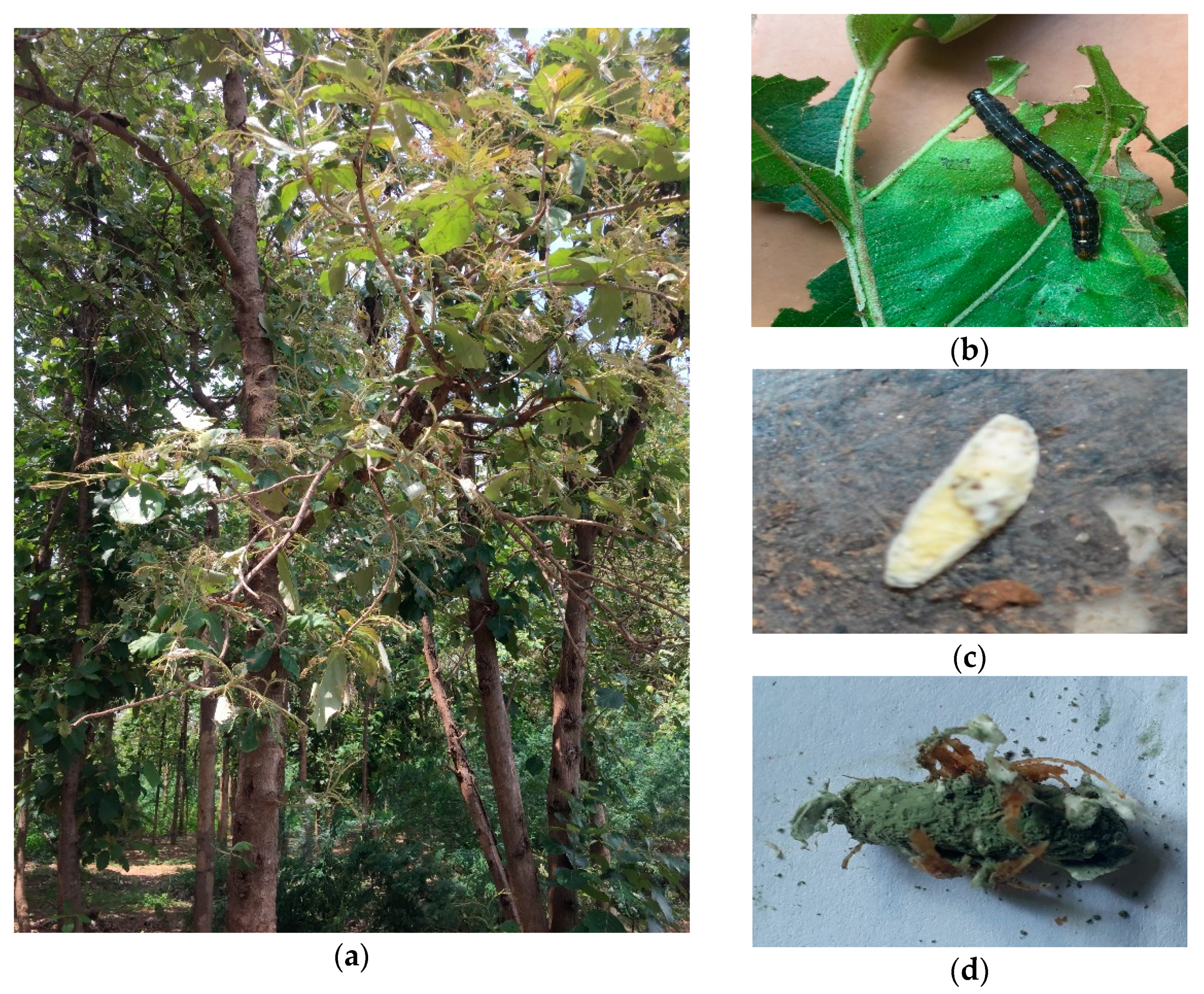
2.3. Bioassay of Metarhizium spp. against Hyblaea puera

Out of the 36 isolates that were screened, nine EPF isolates were evaluated through the YPD medium (0.2% yeast extract, 1% peptone, 2% dextrose with 1% casein/10 g chitin purified powder (25 ± 2 °C, 75 ± 5% RH) incubated at 15 days post inoculation and some modification in Pontecorvo [32]. Based on the preliminary pathogenicity studies, the virulent isolates of nine Metarhizium sp. were used for the bioassay against 3rd instar larvae of H. puera, using for the concentration of 1 × 10³ to 1 × 10⁶ conidial/mL to determine their pathogenicity. Each experiment was replicated three times. The conidial suspension was prepared using the 15 days old culture by scrapping the mycelial growth in sterile water and then filtering in double layer muslin cloth. The stock conidial suspension was prepared by counting the conidia using the improved Neubauer Heamocytometer. Uniform dispersion of conidial suspension was achieved by adding the wetting agents (0.2% Tween-80) to the stock solution. The teak leaf was dipped in the conidial suspension and provided for feeding to the 3rd instars larvae. In each replication, 15 larvae were used. The mortality counts were taken daily for 10 days. The dead larvae were placed on moist filter paper in Petri plates.

2.4. Production of Cuticle-Degradation Enzymes

To obtain the insect cuticle, infected 3rd insect larvae of the H. puera cuticles were used. The internal materials of the dead insect larvae were removed and dried to a constant weight in an oven at 80 °C. Then, the exoskeleton was macerated. The resulting powder was sieved and stored at −20 °C. At the time of experiment, the powder was suspended in an aqueous solution of potassium tetraborate (1%). The extract was subjected to a flowing steam for 20 min [33], dried in a sterile-air-flow cabinet to avoid contamination, and inoculated with 50 µL distilled water, which contained 5000 conidia. Following incubation (60 h) at 27 ± 5 °C, the cuticles were checked microscopically to ensure there was no bacterial contamination and enzymes were extracted as described [33]. The cuticle extract was placed on performed enzyme specific medium and incubated at 27 °C for 15 days after growth of the EPF species.

2.5. Estimation of Extracellular Enzyme Activity on H. puera

Among the nine isolates selected, Metarhizium spp. was grown in a selective medium for 12 days as described above. Protease activity was measured using casein as a substrate 0.6% (w/v). Casein solution (prepared by mixing 6 mg/mL casein with the reaction mixture) contained 100 µL of Tris-HCl buffer (20 mM, pH 8) and was inoculated with 1 mL of the enzyme at 30 °C for 10 min [22,23]. A measure of 0.5 mL of the supernatant was added (1.25 mL of 0.4 M Na2CO3 and 0.25 mL of 2N Folin). It was then added to 0.4 M trichloroacetic acid (TCA) solution and the absorbance was measured at 650 nm in a spectrophotometer, which was incubated at 30 °C for 30 min. The amount of amino acids released was calculated from a standard curve plotted against a range of known concentrations of tyrosine. Afterwards, the product was centrifuged for 5 min at 10,000 rpm. One unit of enzyme was defined as the amount of enzyme that released 1 μg of tyrosine mL−1 of crude enzyme.
Chitinase enzyme activity of the culture supernatant was estimated using the substrate acid-swollen chitin [20,34]. To prepare the acid-swollen chitin, 10 g of chitin (purified powder from crab shells (HiMedia, Mumbai, Maharashtra) was suspended in 100 mL of HCl (35%, w/v) and the mixture was stirred at 5 min intervals for 1 h at room temperature in a fume hood. One unit of chitins was defined as the amount of enzyme that released N-acetyl D-glucosamine, or 100 µL dinitrosalicylic acid was added with incubation for 10 min inboiling water. Absorbance of the reaction mixture at 582 nm (A582) was measured after cooling the room temperature [35]. The procedure is described above, and the bioassay was conducted according to [32].

2.6. Temperature Optimization of Fungal Isolates Growth

2.6.1. Effect of Heat Temperature on Relative Germination (RG)

The relative germination of EPF at high temperature (45 °C & 48 °C) was checked on the 9 virulent isolates of Metarhizium spp., Out of 36 isolates, nine isolates has been found to be virulent isolates. The EPF was cultured on PDAY medium and kept in the dark at 27 ± 1 °C for 15 days. Mycelium of the fungus was harvested with a microbiological loop in sterile water and immediately suspended in Tween 80 solution. The suspension was filtered out through double layer muslin cloth to get conidia. The conidial concentration was prepared by counting the conidia under the Neubauer hemocytometer and the concentration of 106 conidia/mL was made. The 2 mL filtered suspension was transferred to a Borosil glass screw cap tube and kept in water bath at 45 °C and 48 °C for 5 ± 1 min. After 2, 4, 6, 8, 10, 12, 24, 36, 48, and 60 h of exposure with 45 °C and 48 °C, a 20 µL aliquot was placed on 4 mL of medium (PDAY) and one drop of lactophenol methyl blue was added, after which a coverslip was placed on it [36]. The germination was observed at 400× magnification. For the control, the conidial suspension was not exposed at 45 °C and 48 °C temperatures. All of the culture was incubated at 27 ± 2 °C. The observation was taken on the 300 conidia and the germination percentage was calculated by comparing the germination of the heated conidia with the control [37]. Only germinated isolates were taken for assessment.

2.6.2. Effect of Cold Temperature on Relative Germination (RG)

To know the effect of cold temperature (5 °C, 10 °C, and 20 °C) on relative germination, the 2 mL of filtered suspension was transferred to a (Borosil glass) screw cap tube and exposed to cold temperature at 5 °C, 10 °C, and 20 °C. After 5, 10, and 15 days of exposure, 20 µL of conidial suspension of each isolate was poured on 4 mL of medium by following the procedure that was used for the effect of heat temperature. For the control, the conidia were grown in ambient temperature (28 ± 2 °C) without exposure to cold temperature [38].

2.7. Analysis of Results

Significant mortality of the larvae (percent) was recorded from the bioassay and corrected according to Abbott’s formula. Two-way ANOVA was used to compare the effects of the experimental and control treatments on the larval mortality. Prior to statistical analysis, data expressed as proportions were subjected to a regular arc transformation (one way analysis p < 0.001). The data were then subjected to analysis of variance (ANOVA), and the means were compared by Tukey test at the (p < 0.001) probability level using (Graph was generated using Origin Lab Professional Version 2021b software; URL link: www.OriginLab.com/2021b, accessed on 29 September 2021).

3. Results

3.1. Isolation and Identification of Fungi

A total 36 isolates were obtained from forest soils and infected cadavers, which were collected from various locations [39]. There were four species of entomopathogenic fungi, viz., M. robertsii, M. quizhouense, M. majus, and M. anisopliae, from the soil and infected cadaver sample (Table 1). At the same time, other entomopathogenic fungi were grown in culture medium but failed to infect the host insects and were, therefore, not taken for further study. Moreover, during the isolation on PDAY culture medium, opportunistic fungi, viz., Aspergillus, Penicillium, Colletotrichum, and Scopulariopsis, were dominant and these fungi were not considered for further study in this identification. All these isolates were molecularly characterized using ribosomal internal transcribed spacer region (ITS) and DNA-directed RNA polymerase II subunit (RPB1) [39], and the Elongation factor 1-alpha (EP1α) genes sequence was analyzed as well, although it could not yet be submitted (NCBI).

3.2. Bioassay Tested Metarhiziumsp. Isolates against H. puera

Overall, these results revealed that the three isolates of M. anisopliae (DhMz4R), M. quizhouense (ArMz1W), and M. majus (VjMz1W) showed higher mortality, viz., 94.4 ± 2.03, 92 ± 4.19, and 90.8 ± 3.2 per cent, respectively, against the larvae of H. puera. However, there was no significant difference in the pathogenicity of isolates during 10 days after treatment (Figure 2). At 8 days after treatment, there was a significant difference in the pathogenicity among the isolates. The isolate of M. quizhouense (ArMz1W) showed significant percent mortality against the 3rd instar larvae of H. puera (Figure 2).

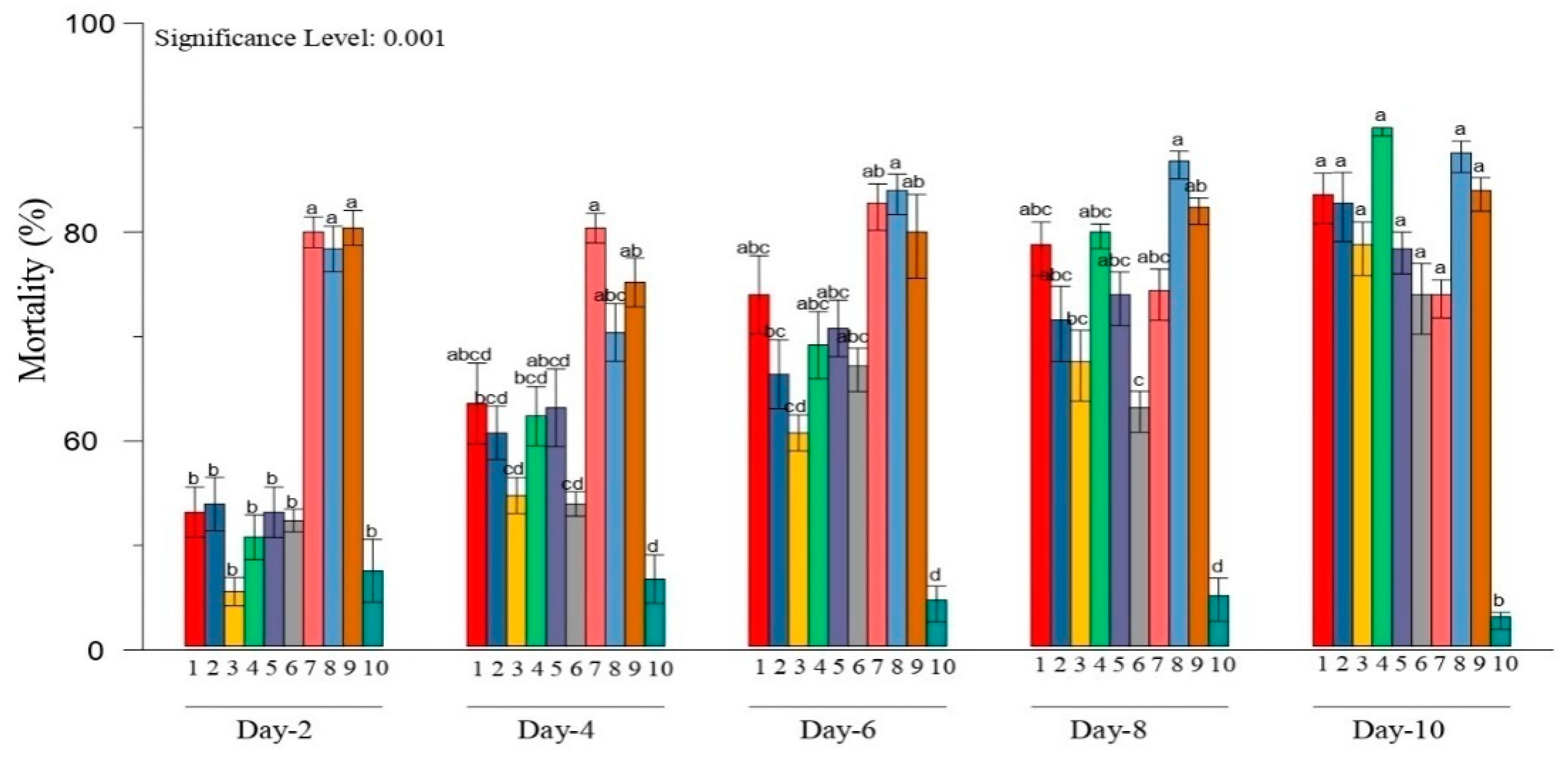
3.3. Evaluation of Metarhizium sp. Isolates Effected Non-Extracellur Enzymes

The four species of Metarhizium sp., viz., M. robertsii, M. quizhouense, M. majus and M. anisopliae, were previously screened for pathogenicity [28]. The larvae were evaluated in four concentrations spanning from 1 × 10³ to 1 × 10⁶ conidia/mL (Table 2). It was observed that the mortality was increased as the concentration increased. Protease response during the observation (1 × 10⁶ conidia/mL), M. robertsii (ArMz3S), M. quizhouense (ArMz1W), M. majus (VjMz1W), M.anisopliae (WnMz1S), and M. anisopliae (NlMz2S)showed significantly higher pathogenicity and their percent morality was on par with each other (27.807, 27.063, 27.489, 27.943, and 27.618 percent) (CD = 1.050).The lowest percent mortality was shownby the M. anisopliae (BgMz2S) isolate.
The percent mortality caused by different isolates of entomopathogenic fungi due to chitinase was significantly different (CD = 1.414). M. quizhouense (ArMz1W) isolate showed highest percent mortality (28.214%), followed by ArMz3S (27.618%) and ArMz6W (27.489%) at 106 conidia/mL (Table 2).
The results indicate that M. quizhouense (ArMz1W) was most effective in causing the mortality against the 3rd instar larvae of H. puera through the production of chitinase and protease.

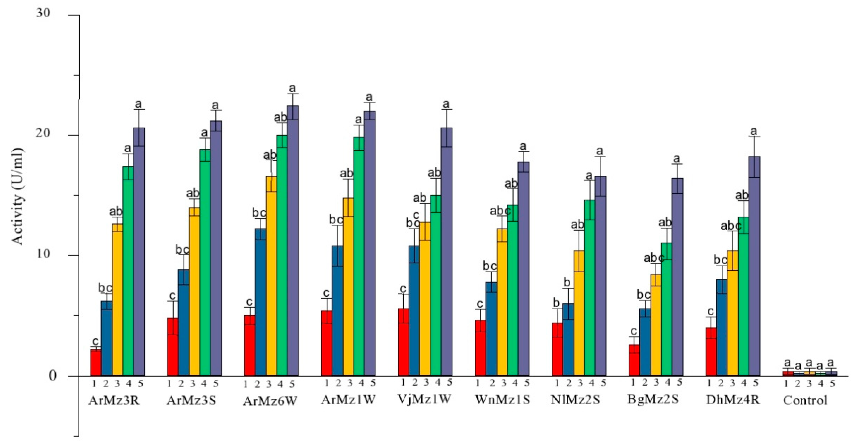
3.4. Extracellur Protease Enzymes Activity

During the experiment, the effect of protease enzymes on virulence levels was evaluated. Protease activities reached a maximum after 5 days of incubation and decreased thereafter for the control. The protease activity of nine isolates of Metarhizium sp. was significantly different from each other (Table 3; Figure 3). Through comparative analysis, the present study demonstrated that the isolates, viz., M. quizhouense (ArMz1W) and M. robertsii (ArMz6W), showed the highest significant on par protease activity compared with other isolates (22.4 ± 1.0 and 22 ± 0.7 U/mL), respectively, whereas the isolate of M. anisopliae (BgMz2S) showed the lowest protease activity.

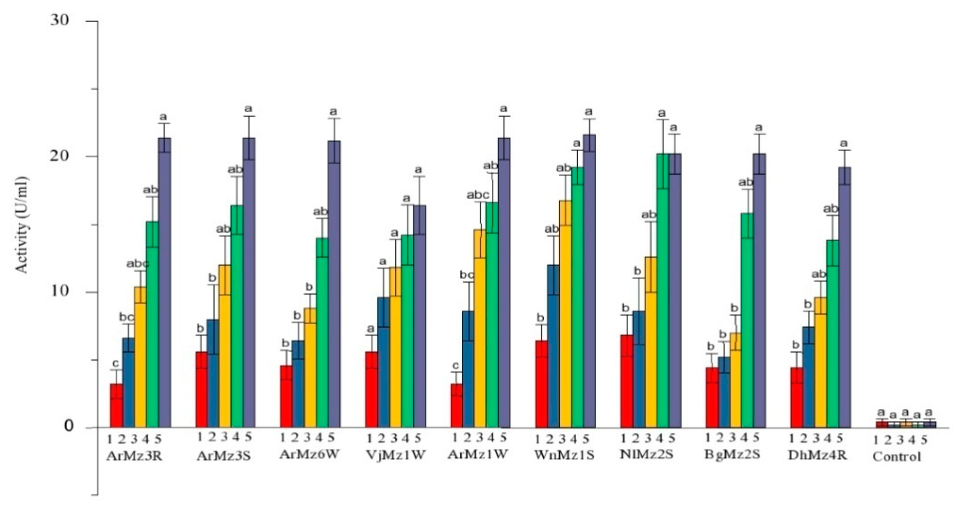
3.5. Extracellur-Chitinases Enzymes Activity

Chitinase activity of M. anisopliae, M. majus, M. robertsii, and M. quizhouense was varied significantly between the nine pathogenic isolates on 5 days post inoculation in chitin medium (CD(0.01) = 2.684). The comparative analysis demonstrated that the isolates, viz., M. robertsii (ArMz3S, ArMz3R, and ArMz1W) and M. anisopliae (WnMz1S), showed statistically on par chitinase activity. The chitinase activity was found to be at its maximum during the 5th day of inoculation (Table 4; Figure 4).

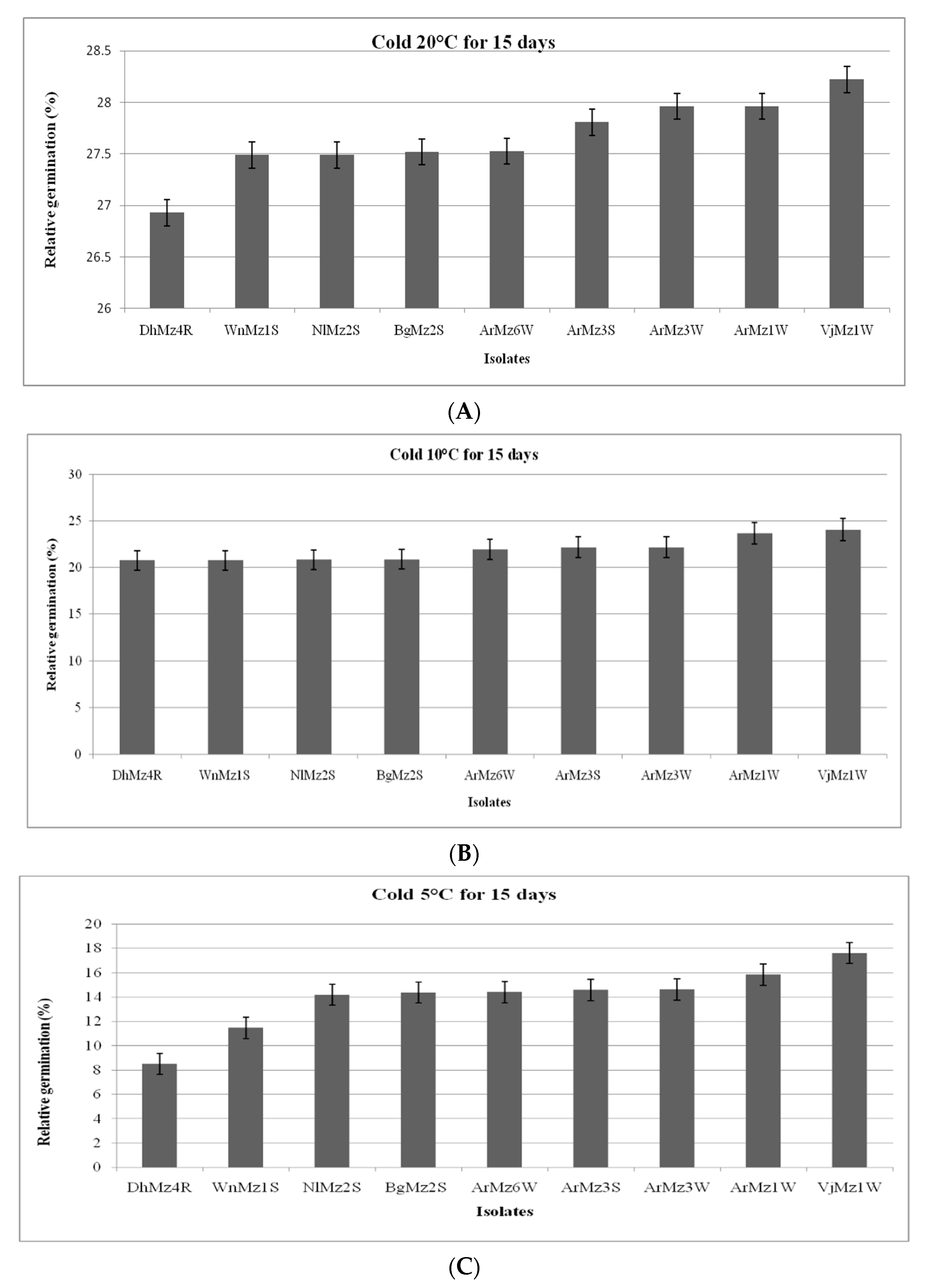
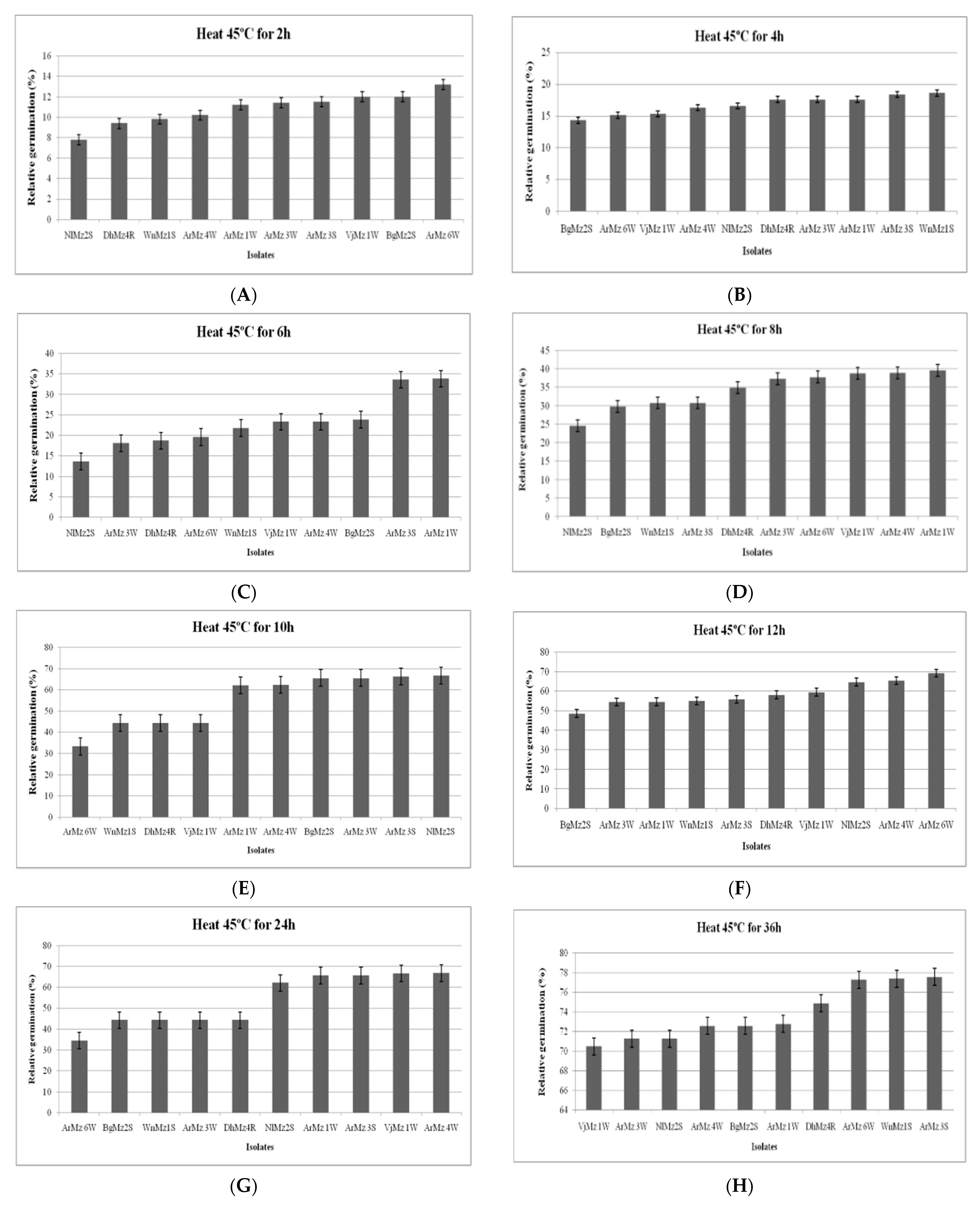
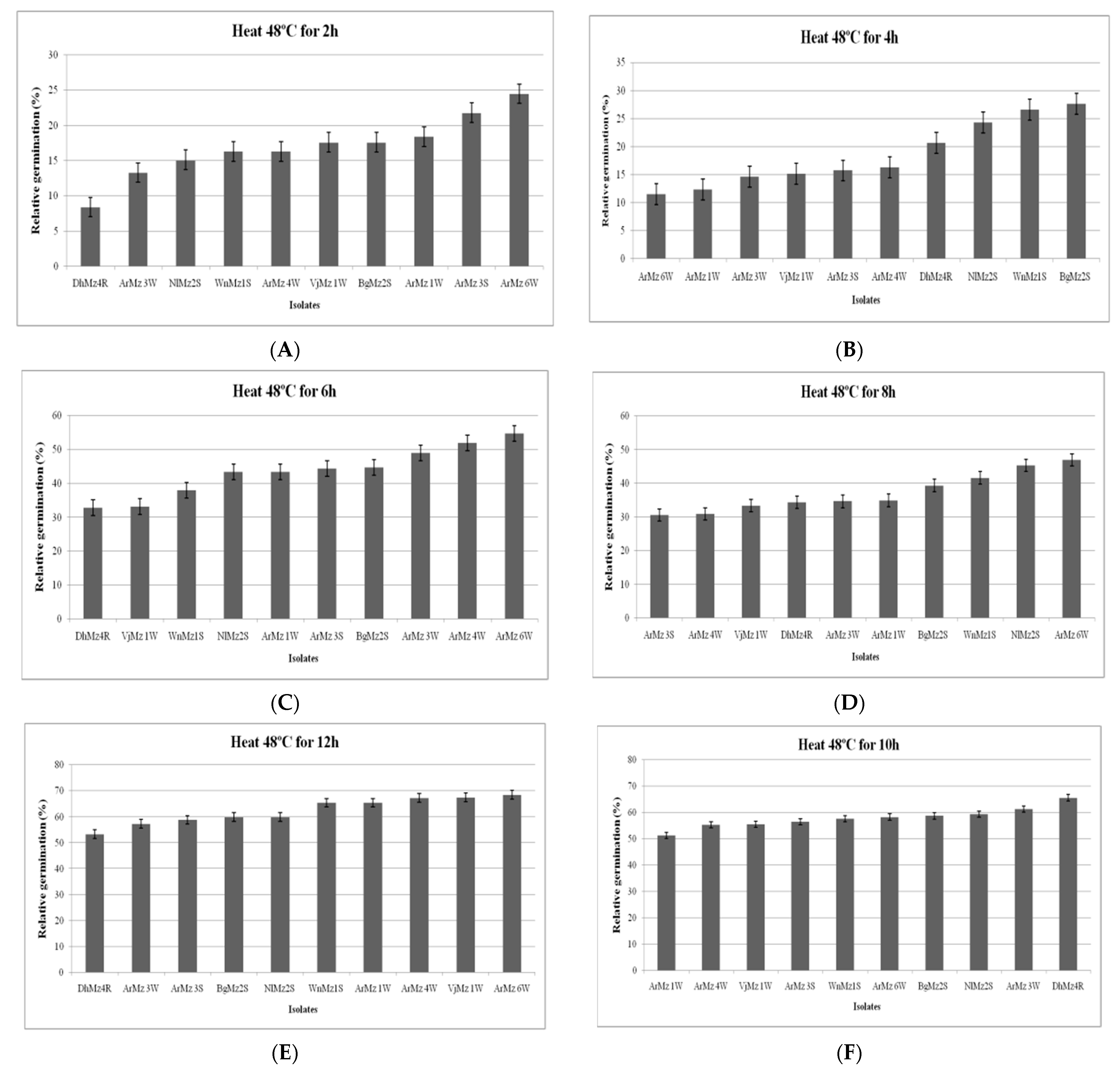
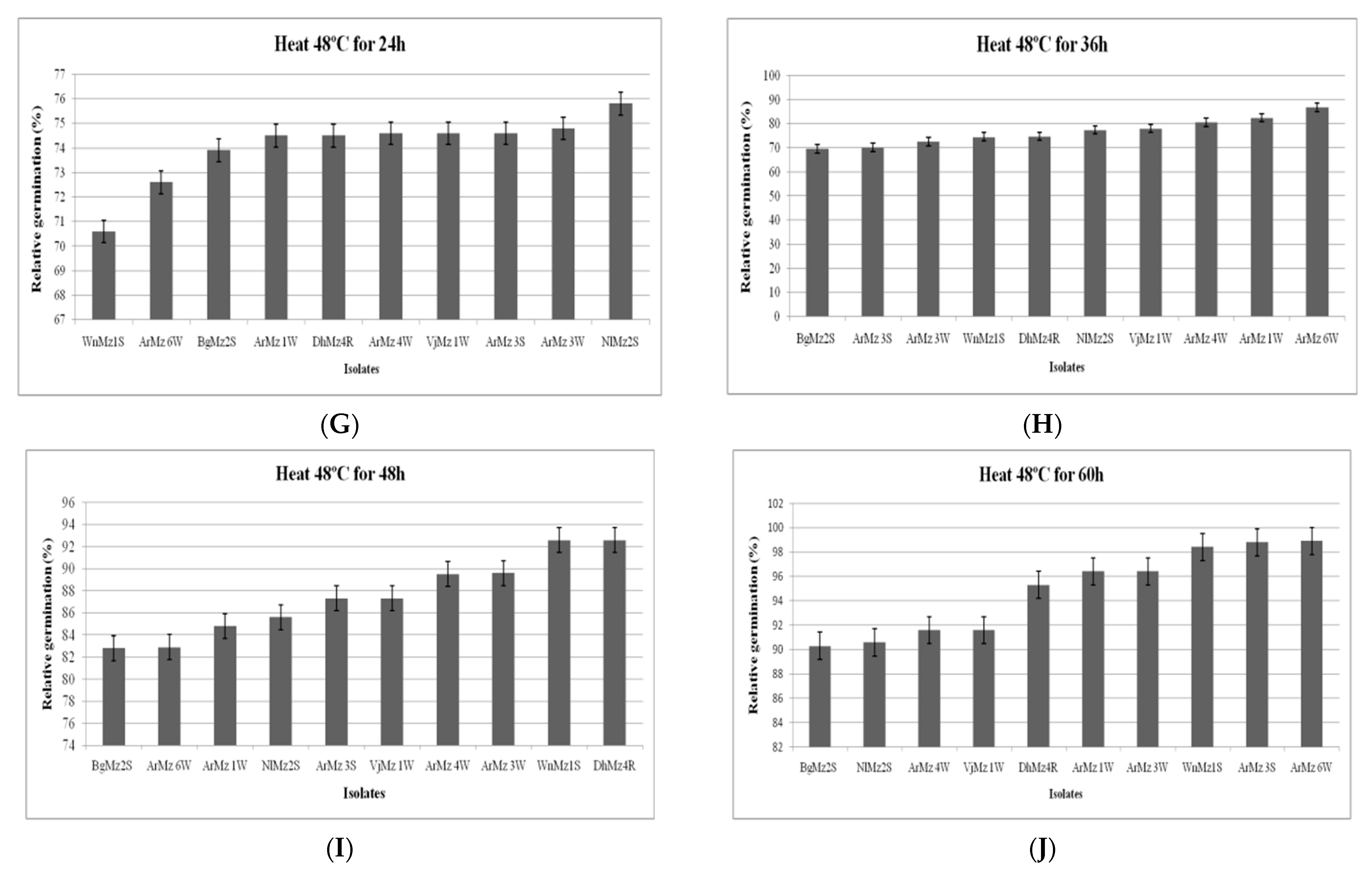
3.6. Heat Tolerance and Cold Activity

High variability in conidia thermo-tolerance was observed among the Metarhizium isolates. However, Metarhizium sp. isolates had a maximum germination after being exposed to cold germination temperatures at 20 °C for 15 days in-between (26.92 to 28.22%) for M. anisopliae (Dhz14R) and M. majus (VjMz1W), respectively. In contrast, the second high variability in cold activity was observed when conidia wasexplored at 10 °C for 15 days, with results ranging from (20.77 to 24.05%) in M. anisopliae (DhMz4R) and M. majus (VjMz1W). Similarly conidial germination was greatly reduced at 5 °C for 15 days with the exceptions of M. anisopliae (DhMz4R) and M. majus (VjMz1W), which had approximately lower germination (Figure 5A–C).
Among the 9 Metarhizium spp., which were exposed at 45 °C and 48 °C for 2 h, 4 h, 6 h, 8 h, and 10 h, the isolates of Metarhizium sp. varied in their conidial germination (Figure 6A–J and Figure 7A–J). All the isolates showed progressive germination in 12 h, 24 h, 36 h, 48 h, and 60 h after exposure at 45 °C. However, at highest exposure (60 h), the isolates, viz., ArMz3S and ArMz6W, showed the highest conidial germination (Figure 6J).It was observed that the conidial germination was high when the incubation time increasedto 48 °C (Figure 7). At the highest exposure (60 h), the isolates, viz., M. robertsii (ArMz6W), M. quizhouense (ArMz1W), and M. majus (VjMz1W), showed the highest conidial germination (Figure 7J).

4. Discussion

The identification was done on the basis of morphotaxonomic characters revealed by microscopically inspecting the conidia forming mycelia for conidigenious structure. Each isolate was put into regular host passages using the natural hosts on three species of M. anisopliae, M. robertsii, and M. majus isolate [2,3]. The effect of visible light during mycelia growth factor on the stress tolerance may exhibit an enhanced protein metabolism as suggested by the conidia [7,11,38].
In the present study, nine isolates of Metarhizium sp. were characterized for thermal tolerance and cold activity.For cold activity, the isolates were exposed to low temperature, viz., 5, 10, and 20 °C. M. majus (VjMz1W) showed higher conidial germination in all the cold temperatures. An earlier study reported that the Metarhizium sp., viz., M. flavoviridae and M. frigidium, were able to tolerate the cold temperature and produced conidia at the lowest temperature of their experiments (5 °C).
The heat tolerance of isolates of Metarhizium sp. was tested at the higher temperatures of 45 and 48 °C in different exposure times (2, 4, 6, 8, 10, 12, 24, 36, 48 and 60 h). The temperature tolerance was increased when the exposure time increased. In both of the heat temperatures, M. robertsii (ArMz6W) showed high conidial germination at 60 h after exposure. Earlier studies reported that the isolates of M. anisopliae var. acridium (ARSEF 6421, ARSEF 3614, ARSEF 5736, ARSEF 324, and ARSEF 2341) were most conidial heat tolerant at higher temperatures (45 °C) [6,7,15,40,41].
In the present investigation, chitinase and protease were found to show high correlation with mortality. Earlier literature has been reported that chitinases and proteases were the virulence factors for entomopathogenicity [19,42]. The hydrophobicity of conidia is one of the important parameters influencing fungus-insect interaction as it helps in adhesion of the fungus to the insect cuticle. This plays an important role in providing nutrients before and after the cuticle is penetrated [20,21]. However, chitinase is required only for a brief period during penetration of the host cuticle and is tightly regulated by chitin degradation products [22,23,33]. The fungus produces spores, which, upon germination, produce young germ tubes that penetrate through the cuticle, physically as well as chemically through the enzymes produced, such as proteases, chitinases, and lipases [43,44,45,46,47].
The insecticidal mechanism underlying in the pathogenicity of isolated fungi lies in presence of cuticle degrading enzymes which make paves way for the entry of EPF in the insect body [48,49,50,51,52]. To invade the insect cuticle, which is mainly composed of chitin and several other proteins, entomopathogens must secrete chitinase and protease enzymes [53,54,55,56,57,58]. Mechanism of enzymatic degradation of larval cuticle for hyphal penetrance by this fungus inside larval body is confirmed by the detection of chitinase and protease enzymes thatare secreted by the fungi.
Our study showed that the isolates of Metarhizium sp., viz., M.anisopliae (DhMz4R), M. quizhouense (ArMz1W), and M. majus (VjMz1W), showed higher pathogenicity (94.4 ± 2.03, 92 ± 4.19, and 90.8 ± 3.2 percent mortality, respectively) at 106 conidia/mL. Similarly, Ramadevi et al. [59] conducted the bioassay experiment to test virulence of M. anisopliae (MIS2 and MIS7) against H. puera. They found less percent mortality compared to the present study isolates.
The four species of Metarhizium spp., viz., M. anisopliae, M. robertsii, M. quizhouense, and M. majus, were obtained from insects infected and soil baiting methods. Therefore, EPF isolates revealed that for different parts of South India, indigenous isolates showed thebest mortality against 3rd instar larvae H. puera in the range of 84–92% within 5 days. Our results have indicated that three species, viz., M. robertsii, M. quizhouense, and M. majus, isolates were recorded for the first time and they had comparatively significantly higher mortality rates.

5. Conclusions

This study has revealed that M. anisopliae, M. robertsii, M. quizhouense, and M. majus are exceptionally effective and can be considered some of the best EPF isolates. Further investigation to control the infestation of teak defoliator H. puera should be performed on plantations in South India. The development of a possible new bio-control agent forthe invasive teak defoliator H. puera pest is now possible with a better understanding ofthe potential use of indigenous entomopathogenic fungi.

Author Contributions

Conceptualization, V.V. and R.D.; methodology, validation, V.V. and O.A.N. and G.R., S.K. and S.S.-N.; formal analysis; V.V.; writing—review and editing, E.J.K., O.A.N., R.D., S.K., S.S.-N. and G.R. All authors have read and agreed to the published version of the manuscript.

Funding

This research received no external funding.

Institutional Review Board Statement

Not applicable.

Informed Consent Statement

Not applicable.

Data Availability Statement

All the relevant data are included within the manuscript.

Acknowledgments

The authors are highly thankful to R. Rangeshwaran, G. Sivakumar Principal Scientist, (ICAR-National Bureau of Agricultural Insect Resources), T.O. Sasidharan and R. Sundararaj, Scientist G (Institute of Wood Science and Technology, Forest and wood protection) for valuable suggestion for identification of entomopathogenic fungi. The author grateful to the R.Karthi at ICAR-National Research Center for Banana, for Tukey analysis (Graph was generated using OriginLab Professional Version 2021b software; URL link: https://www.originlab.com/, accessed on 29 September 2021).

Conflicts of Interest

The authors declare no conflict of interest.

Abbreviations

EPFEntomopathogenic Fungi
PDAYPotato Dextrose Agar with Yeast extract
SDAYSabouraud Dextrose Agar with Yeast extract
ITSribosomal internal transcribed spacer
RPB1DNA-directed RNA polymerase II subunit

References

  1. Metchnikoff, E. Diseases of the larva of the grain weevil. In Insects Harmful to Agriculture; Odessa Zemstvo Office: Odessa, Ukraine, 1879. (In Russian) [Google Scholar]
  2. Bischoff, J.F.; Rehner, S.A.; Humber, R.A. A multilocus phylogeny of the Metarhizium anisopliae lineage. Mycologia 2009, 101, 512–530. [Google Scholar] [CrossRef] [Green Version]
  3. Kepler, R.M.; Humber, R.A.; Bischoff, J.F.; Rehner, S.A. Clarification of generic and species boundaries for Metarhizium and related fungi through multigenephylogenetics. Mycologia 2014, 106, 811–829. [Google Scholar] [CrossRef]
  4. Chandra Teja, K.N.; Pand Rahman, S.J. Characterisation and evaluation of Metarhizium anisopliae (Metsch.) Sorokin strains for their temperature tolerance. Mycology 2016, 7, 171–179. [Google Scholar] [CrossRef] [PubMed]
  5. Keyser, C.A.; Fernandes, É.K.K.; Rangel, D.E.N.; Roberts, D.W. Heat-induced post-stress growth delay: A biological trait of many Metarhizium isolates reducing biocontrol efficacy. J. Invertebr. Pathol. 2014, 120, 67–73. [Google Scholar] [CrossRef] [PubMed]
  6. Rangel, D.E.N.; Fernandes, É.K.K.; Dettenmaier, S.J.; Roberts, D.W. Thermotolerance of germlings and mycelium of the insect-pathogenic fungus Metarhizium spp. and mycelial recovery after heat stress. J. Basic. Microbiol. 2010, 50, 344–350. [Google Scholar] [CrossRef]
  7. Fernandes, E.K.K.; Keyser, C.A.; Chong, J.P.; Rangel, D.E.N.; Miller, M.P.; Roberts, D.W. Characterization of Metarhizium species and varieties based on molecular analysis, heat tolerance and cold activity. J. App. Microbiol. 2010, 108, 115–128. [Google Scholar] [CrossRef] [PubMed]
  8. Hunt, V.L.; Zhong, W.; McClure, C.D.; Mlynski, D.T.; Duxbury, E.M.L.; Keith Charnley, A.; Priest, N.K. Cold-seeking behaviour mitigates reproductive losses from fungal infection in Drosophila. J. Anim. Ecol. 2016, 85, 178–186. [Google Scholar] [CrossRef] [Green Version]
  9. Ibrahim, L.; Butt, T.M.; Jenkinson, P. Effect of Artificial Culture Media on Germination, Growth, Virulence and Surface Properties of the Entomopathogenic Hyphomycete (Metarhizium anisopliae). Myco. Res. 2002, 106, 705715. [Google Scholar] [CrossRef]
  10. Inglis, G.D.; Duke, G.M.; Kawchuk, L.M.; Goettel, M.S. Influence of oscillating temperatures on the competitive infection and colonization of the migratory grasshopper by Beauveria bassiana and Metarhizium flavoviride. Biol. Control. 1999, 14, 111–120. [Google Scholar] [CrossRef]
  11. Nishi, O.; Hasegawa, K.; Iiyama, K.; Yasunaga-Aoki, C.; Shimizu, S. Phylogenetic analysis of Metarhizium spp. isolated from soil in Japan. Appl. Entomol. Zool. 2011, 46, 301–309. [Google Scholar] [CrossRef]
  12. Lekime, M.; Focant, C.; Farnir, F.; Mignon, B.; Losson, B. Pathogenicity and Theromotolerance of Entomopathogenic Fungi for the Control of the Scab Mite, Psoroptesovis. Exp. Appl. Acarol. 2008, 46, 95–104. [Google Scholar] [CrossRef] [PubMed]
  13. Bidochka, M.J.; Kamp, A.M.; Lavender, T.M.; Dekoning, J.; De Croos, J.N. Habitat association in two genetic groups of the insect-pathogenic fungus Metarhizium anisopliae: Uncovering cryptic species. Appl. Environ. Microbiol. 2001, 67, 1335–1342. [Google Scholar] [CrossRef] [PubMed] [Green Version]
  14. Braga, G.U.L.; Flint, S.D.; Messias, C.L.; Anderson, A.J.; Roberts, D.W. Effect of UV-B on conidia and germlings of the entomopathogenic hyphomycete Metarhizium anisopliae. Mycol. Res. 2001, 105, 874–882. [Google Scholar] [CrossRef]
  15. Rangel, D.E.N.; Anderson, A.J.; Roberts, D.W. Evaluating physical and nutritional stress during mycelial growth as inducers of tolerance to heat and UV-B radiation in Metarhizium anisopliae conidia. Mycol. Res. 2008, 112, 1362–1372. [Google Scholar] [CrossRef]
  16. Liu, H.; Skinner, M.; Brownbridge, M.; Parker, B.L. Characterization of Beauveria bassiana and Metarhizium anisopliae isolates for management of tarnished plant bug, Lygus lineolaris (Hemiptera: Miridae). J. Invertebr. Pathol. 2003, 82, 139–147. [Google Scholar] [CrossRef]
  17. Hegedus, D.D.; Khachatourians, G.G. The impact of biotechnology on hyphomycetous fungal insect biocontrol agents. Biotechnol. Adv. 1995, 13, 455–490. [Google Scholar] [CrossRef]
  18. Krieger de Moraes, C.; Schrank, A.; Vainstein, M.H. Regulation of extracellular chitinases and proteases in the entomopathogen and acaricides Metarhizium anisopliae. Curr. Microbiol. 2003, 46, 205–210. [Google Scholar] [CrossRef]
  19. St. Leger, R.J.; Bidochka, M.J.; Roberts, D.W. Construction of an improved mycoinsecticide over expressing a toxic protease. Proc. Natl. Acad. Sci. USA 1996, 93, 6349–6354. [Google Scholar] [CrossRef] [Green Version]
  20. Nahar, P.; Ghormade, V.; Deshpande, M.V. The extracellular constitutive production of chitin deacetylase in Metarhizium anisopliae: Possible edge to entomopathogenic fungi in the biological control of insect pests. J. Invertebr. Pathol. 2004, 85, 80–88. [Google Scholar] [CrossRef]
  21. Revathi, N.; Ravikumar, G.; Kalaiselvi, M.; Gomathi, D.; Uma, C. Pathogenicity of three entomopathogenic fungi against Helicoverpa armigera. J. Plant Pathol. Microbiol. 2011, 2, 114. [Google Scholar] [CrossRef] [Green Version]
  22. Sapna Bai, N.; Remadevi, O.K.; Sasidharan, T.O.; Balachander, M.; Priyadarsanan, D.R. Cuticle degrading enzyme production by some isolates of the Entomopathogenic fungus, Metarhizium anisopliae (Metsch.). J. Bio-Sci. 2012, 20, 25–32. [Google Scholar]
  23. Ramanujam, B.; Balachander, M.; Roopa, G.; Rangeshwaran, R.; Karmakar, P. Chitinase activity and virulence of different isolates of Beauveria bassiana, Metarhizium anisopliae and Lecanicillium spp. J. Biol. Control 2011, 25, 223–228. [Google Scholar]
  24. Beeson, C.F.C. The Ecology and Control of the Forest Insects of India and the Neighboring Countries; Government of India: New Delhi, India, 1941; p. 767. [Google Scholar]
  25. Sudheendrakumar, V.V.; Mohammed Ali, I.M.; Varma, R.V. Nuclear Polyhedrosis Virus of the teak defoliator Hyblaeapuera. J. Invert. Pathol. 1988, 51, 307–308. [Google Scholar] [CrossRef]
  26. Senthil-Nathan, S.; Saehoon, K. Behavioral and physiological effects of Melia azedarach L. extract on the teak defoliator Hyblaeapuera Cramer (Lepidoptera: Hyblaeidae). Crop Prot. 2006, 25, 287–291. [Google Scholar] [CrossRef]
  27. Senthil-Nathan, S. A Review of Bio Pesticides and Their Mode of Action against Insect Pests. In Environmental Sustainability-Role of Green Technologies; Springer: Dordrecht, The Netherlands, 2015; pp. 49–63. [Google Scholar]
  28. Velavan, V.; Sivakumar, G.; Rangeswaran, R.; Sasidharan, T.O.; Sundararaj, R. Metarhiziummajus and Metarhiziumrobertsii show enhanced activity against the coleopteran pests Holotricha serrata L. and Oryctes rhinoceros L. J. Biol. Control 2017, 31, 135–145. [Google Scholar] [CrossRef] [Green Version]
  29. Veen, K.H.; Ferron, P. A selective medium for the isolation of Beauveria tenella and of Metarhiziumanisopliae. J. Invertebr. Pathol. 1966, 8, 268–269. [Google Scholar] [CrossRef]
  30. Riddell, R.W. Permanent stained mycological preparations obtained by slide culture. Mycologia 1950, 42, 265–270. [Google Scholar] [CrossRef]
  31. Senthil-Nathan, S.; Choi, M.Y.; Paik, C.H.; Seo, H.Y. Food consumption, utilization, and detoxification enzyme activity of the rice leaffolder larvae after treatment with Dysoxylum triterpenes. Pest. Biochem. Physiol. 2007, 88, 260–267. [Google Scholar] [CrossRef]
  32. Remadevi, O.K.; Sasidharan, T.O.; Balachander, M.; Sapna Bai, N. Prospects of developing Metarhizium based mycoinsecticide for pest management in forestry. J. Biopestic. 2010, 3, 38–41. [Google Scholar]
  33. Dhar, P.; Kaur, G. Cuticle-Degrading Proteases Produced by Metarhizium anisopliae and Their Induction in Different Media. Indian J. Microbiol. 2010, 50, 449–455. [Google Scholar] [CrossRef] [Green Version]
  34. Kulkarni, S.A.; Ghormade, V.; Kulkarni, G.; Kapoor, M.; Chavan, S.B.; Rajendran, A.; Patil, S.K.; Shouche, Y.; Deshpande, M.V. Comparison of Metarhizium isolates for biocontrol of Helicoverpa armigera (Lepidoptera: Noctuidae) in chickpea. Biocontrol. Sci. Technol. 2008, 18, 809–828. [Google Scholar] [CrossRef]
  35. Senthil-Nathan, S. Physiological and biochemical effect of Neem and other Meliaceae plants secondary metabolites against Lepidopteran insects. Front. Physiol. 2013, 4, 359. [Google Scholar] [CrossRef] [PubMed] [Green Version]
  36. Jha, S.C. Modi Comparative Analysis of Chitinase Activity by Four Different Assays from Soil Born Actinomycetes. In Proceedings of the 4th International Conference on Multidisciplinary Research & Practice (4ICMRP) Gujarat University, Ahmedabad, India, 22 December 2017; pp. 185–190. [Google Scholar]
  37. Milner, R.J.; Huppatz, R.J.; Swaris, S.C. A new method for assessment of germination of Metarhizium conidia. J. Invertebr. Pathol. 1991, 57, 121–123. [Google Scholar] [CrossRef]
  38. Braga, G.U.L.; Flint, S.D.; Miller, C.D.; Anderson, A.J.; Roberts, D.W. Variability in response to UV-B among species and strains of Metarhizium isolated from sites at latitudes from 61N to 54 S. J. Invertebr. Pathol. 2001, 78, 98–108. [Google Scholar] [CrossRef]
  39. Velavan, V.; Rangeshwaran, R.; Sivakumar, G.; Sasidharan, T.O.; Sundararaj, R.; Kandan, A. Occurrence of Metarhizium spp. isolated from forest soils in South India and their potential in biological control of banana stem weevil Odoiporus longicollis Oliver. Egypt. J. Biol. Pest Control 2021, 31, 131–143. [Google Scholar] [CrossRef]
  40. Lee, W.W.; Shin, T.Y.; Bae, S.M.; Woo, S.D. Screening and evaluation of entomopathogenic fungi against the green peach aphid, Myzus persicae, using multiple tools. J. Asia Pac. Entomol. 2015, 18, 607–615. [Google Scholar] [CrossRef]
  41. Li, J.; Feng, M.G. Intra specific tolerance of Metarhizium anisopliae conidia to the upper thermal limits of summer with a description of a quantitative assay system. Mycol. Res. 2009, 113, 93–99. [Google Scholar] [CrossRef] [PubMed]
  42. Rangel, D.E.N.; Anderson, A.J.; Roberts, D.W. Growth of Metarhizium anisopliae on non-preferred carbon sources yields conidia with increased UV-B tolerance. J. Invertebr. Pathol. 2006, 93, 127–134. [Google Scholar] [CrossRef]
  43. Vasantha-Srinivasan, P.; Karthi, S.; Chellappandian, M.; Ponsankar, A.; Thanigaivel, A.; Senthil-Nathan, S.; Chandramohan, D.; Ganesan, R. Aspergillus flavus (Link) toxins reduces the fitness of dengue vector Aedes aegypti (Linn.) and their non-target toxicity against aquatic predator. Microb. Pathog. 2019, 128, 281–287. [Google Scholar] [CrossRef] [PubMed]
  44. Karthi, S.; Vasantha-Srinivasan, P.; Ganesan, R.; Ramasamy, V.; Senthil-Nathan, S.; Khater, H.F.; Radhakrishnan, N.; Amala, K.; Kim, T.J.; El-Sheikh, M.A.; et al. Target Activity of Isaria tenuipes (Hypocreales: Clavicipitaceae) Fungal Strains against Dengue Vector Aedes aegypti (Linn.) and Its Non-Target Activity Against Aquatic Predators. J. Fungi. 2020, 6, 196. [Google Scholar] [CrossRef]
  45. Nishi, O.; Iiyama, K.; Yasunaga-Aoki, C.; Shimizu, S. Comparison of the germination rates of Metarhizium spp. conidia from Japan at high and low temperatures. Lett. Appl. Microbiol. 2013, 57, 554–560. [Google Scholar] [CrossRef]
  46. Gurvinder, K.; Padmaja, V. Relationships among activities of extracellular enzyme production and virulence against Helicoverpa armigera in Beauveria bassiana. J. Basic Microbiol. 2009, 49, 264–274. [Google Scholar]
  47. Charnley, A.K.; St. Leger, R.J. The Role of Cuticle Degrading Enzymes in Fungal Pathogenesis of Insects. In The Fungal Spore and Disease Initiation in Plants and Animals; Plenum Publishing Co.: New York, NY, USA, 1991; pp. 267–286. [Google Scholar]
  48. St. Leger, R.J.; Cooper, R.M.; Charnley, A.K. Cuticle-degrading enzymes of entomopathogenic fungi: Cuticle degradationin vitro by enzymes from entomopathogens. J. Invertebr. Pathol. 1986, 47, 167–177. [Google Scholar] [CrossRef]
  49. Dhanapal, R.; Kumar, D.V.S.R.; Lakshmipathy, R.; Sandhya Rani, C.; Manoj Kumar, R. Pathogenicity testing of indigenous isolates of entomopathogenic fungus, Lecanicillium lecanii against tobacco caterpillar, Spodoptera litura. J. Experi. Zool. India 2019, 22, 753–756. [Google Scholar]
  50. Dhanapal, R.; Kumar, D.V.S.R.; Lakshmipathy, R.; Sandhya Rani, C.; Manoj Kumar, R. Exploration of indigenous strains of the green muscardine fungus from soils and their pathogenicity against the tobacco caterpillar, Spodoptera litura (Fabricius) (Lepidoptera: Noctuidae). Egypt. J. Biol. Pest Control 2020, 30, 34. [Google Scholar] [CrossRef]
  51. Dhanapal, R.; Kumar, D.V.S.R.; Lakshmipathy, R.; Sandhya Rani, C.; Manoj Kumar, R. Isolation of indigenous strains of the white halo fungus as a biological control agent against 3rd instar larvae of tobacco caterpillar, Spodoptera litura (Fabricius) (Lepidoptera: Noctuidae). Egypt. J. Biol. Pest Control 2020, 30, 89. [Google Scholar] [CrossRef]
  52. Karthi, S.; Vaideki, K.; Shivakumar, M.S.; Ponsankar, A.; Thanigaivel, A.; Chellappandian, M.; Vasantha-Srinivasan, P.; Chanthini, K.M.; Hunter, W.B.; Senthil-Nathan, S. Effect of on the mortality of Aspergillus flavus and activity of antioxidant enzymes of Spodoptera litura Fab. (Lepidoptera: Noctuidae) larvae. Pestic. Biochem. Physiol. 2018, 149, 54–60. [Google Scholar] [CrossRef] [PubMed]
  53. Ghosh, S.K.; Podder, D.; Mukherjee, A. An insight of anopheline larvicidal mechanism of Trichoderma asperellum (TaspSKGN2). Sci. Rep. 2021, 11, 16029. [Google Scholar] [CrossRef] [PubMed]
  54. Kirubakaran, S.A.; Senthil-Nathan, S.; Revathi, K.; Chandrasekaran, R.; Senthil-Nathan, S. Effect of oil-formulated Metarhiziumanisopliae and Beauveria bassiana against the rice leaffolder Cnaphalocrocis medinalis Guenée (Lepidoptera: Pyralidae). Arch. Phytopathol. Plant Prot. 2014, 47, 977–992. [Google Scholar] [CrossRef]
  55. Sangeetha, A.; Arivudainambi, S. Studies on the efficacy of bio—products in the management of teak skeletonizer Eutectonamachaeralis Walker (Pyralidae: Lepidoptera). J. Biopest. 2012, 5, 129–134. [Google Scholar]
  56. Loganathan, J.; David, P.M.M. Laboratory and field evaluation of Bacillus thuriengiensis Berliner products against the teak defoliator Hyblaea puera Cramer. Insect. Sci. Appl. 2000, 20, 61–65. [Google Scholar]
  57. Biji, C.P.; Sudheendrakumar, V.V.; Sajeev, T.V. Quantitative estimation of Hyblaea puera NPV production in three larval stages of the teak defoliator, Hyblaea puera (Cramer). J. Virol. Meth. 2006, 136, 78–82. [Google Scholar] [CrossRef]
  58. Narayanan, K. Insect defence: Its impact on microbial control of insect pests. Curr. Sci. 2004, 86, 800–814. [Google Scholar]
  59. Remadevi, O.K.; SapnaBai, N.; Sasidharan, T.O.; Balachander, M.; Dharmarajan, P. Attempts at controlling Teak Defoliator (Hyblaea puera Cramer, Lepidoptera, Hyblaeidae) with the entomopathogenic fungus, Metarhizium anisopliae (Metsch.): Laboratory, nursery and field trials. Int. J. Pest Manag. 2013, 59, 236–242. [Google Scholar] [CrossRef]
Figure 1. (a) Teak forest with H. puera infestation in field, (b) H. puera feeding on leaves, (c) Dead larvae showing the symptoms of M. quizhouense infection, and (d) Insect mummified covered M. robertsii infected cadaver with spores.
Jof 08 00068 g001
Figure 2. Virulence of entomopathogenic fungi isolates against the H. puera larvae, and statistical differences are shown by different letters (Tukey’s test, p < 0.001). (1–4) M. anisopliae (WnMz1S, NlMz2S, BgMz2S, DhMz4R); (5–7) M. robertsii (ArMz3R, ArMz3S, ArMz6W); (8), M. quizhouense (ArMz1W); (9), M. majus (VjMz1W); (10), Control.
Jof 08 00068 g002
Figure 3. Activities of the proteases (U/mL), Mean±SE in the liquid culture media of the entomopathogenic fungi in the presence of H. puera cuticle. Statistical differences are shown by different letters (Tukey’s test, p < 0.001). Each color indicating different days of incubation (1st to 5th days).
Jof 08 00068 g003
Figure 4. Activities of the chitinases (U/mL), Mean ± SE in the liquid culture media of the entomopathogenic fungi in the presence of H. puera cuticle. Statistical differences are shown by different letters (Tukey’s test, p < 0.001). Each color indicating different days of incubation (1st to 5th days).
Jof 08 00068 g004
Figure 5. (AC): Relative germination (RG) of Metarhizium spp. conidia after incubation at 5 °C, 10 °C, and 20 °C for 15 days. (A) At 20 °C; (B) At 10 °C; (C) At 5 °C.
Jof 08 00068 g005
Figure 6. Relative germination of different isolates of Metarhizium sp., exposed at 45 °C. Relative germination (RG) of Metarhizium spp. conidia exposed for (A) 2 h, (B) 4 h, (C) 6 h, (D) 8 h, (E) 10 h, (F) 12 h, (G) 24 h, (H) 36 h, (I) 48 h, and (J) 60 h. RG was calculated in relation to non-heated controls.
Jof 08 00068 g006aJof 08 00068 g006b
Figure 7. Relative germination of different isolates of Metarhizium sp. exposed at 48 °C. Relative germination (RG) of Metarhizium spp. conidia exposed for (A) 2 h, (B) 4 h, (C) 6 h, (D) 8 h, (E) 10 h, (F) 12 h, (G) 24 h, (H) 36 h, (I) 48 h, and (J) 60 h. RG was calculated in relation to non-heated controls.
Jof 08 00068 g007aJof 08 00068 g007b
Table 1. List of Metarhizium spp. Isolates obtained and sequenced for phylogenetic analysis.
FungiType of ForestSourceRegionGeographical OriginAccession Number
ITSRPB1
M.robertsii ArMz3RWEFInsect(Protaetia aurichalcea)Aralam11′99° N 75′76° EKU983799KU680339
M.robertsii ArMz3SWEFInsect(P. aurichalcea)Aralam11′99° N 75′76° EKU983775KU680335
M.robertsii ArMz6WWEFInsect (P. aurichalcea)Aralam11′99° N 75′76° EKU983797KU680341
M.quizhouense ArMz1WWEFInsect(Eutectona machaeralis)Aralam11′99° N 75′76° EKU870314Not yet
M.majus VjMz1WWEFInsect(Melolontha guttigera)Coorg12′20° N 75′80° EKU983771KU680342
M.anisopliae WnMz1SWEFsoilAralam11′60° N 76′08° EKU983788KU680323
M.anisopliae NlMz2SMDFsoilBengaluru12′97° N 77′59° EKU983785KU680325
M.anisopliae BgMz2SDDFsoilBengaluru12′80° N 77′57° EKU983780KU680320
M.anisopliae DhMz4RMDFsoilPalakkad10′77° N 76′65° EKU983784KU680328
Wet Evergreen Forest (WEF), Moist Deciduous (MDF), and Dry Deciduous Forest (DDF). ITS—ribosomal internal transcribed spacer; RPB1—DNA-directed RNA polymerase II subunit.
Table 2. Virulence of Entomopathogenic fungi isolates against the third-instar larvae of H. puera.
Isolates Name% Mortality of H. puera Due Protease% Mortality of H. puera Due Chitinase
103 *104 *105 *106 *103 *104 *105 *106 *
M. robertsii ArMz3R8.499 ab13.400 ab24.121 a26.929 b9.862 b12.826 e19.355 de23.619 c
M. robertsii ArMz3S11.465 a14.771 a24.539 a27.807 a8.879 b19.594 a20.270 cd27.618 ab
M. robertsii ArMz6W8.702 ab13.285 ab22.481 b26.371 b8.350 b15.728 cd20.518 cd27.489 ab
M. quizhouense ArMz1W8.280 ab14.503 a24.850 a27.063 a13.241 a19.575 a23.607 ab28.214 a
M. majus VjMz1W5.476 bc13.067 ab22.160 bc27.489 a13.241 a19.234 ab25.673 a27.083 ab
M. anisopliae WnMz1S3.965 c13.341 ab21.524 cd27.943 a8.813 b12.109 ef24.057 ab26.648 b
M. anisopliae NlMz2S2.573 cd12.947 ab20.997 d27.618 a8.501 b14.019 de17.910 ef26.916 b
M. anisopliae BgMz2S2.639 cd12.419 abc19.333 e23.333 f8.879 b14.111 de19.965 de22.704 c
M. anisopliae DhMz4R4.151 c10.899 bc19.218 e25.284 e9.912 b17.039 bc22.265 bc26.772 b
Control0.573 d1.440 d6.957 f10.184 g1.606 c2.639 g4.151 g9.873 d
SE18.7017.8493.1862.34827.27311.3938.0323.159
CD(0.01)4.4592.4781.1841.0504.4003.0192.8971.414
CD(0.05)3.0721.8490.8830.7833.2822.2522.1611.055
Within the same column, mean ± S.E.M. followed by different letters are significantly different from each other followed by adjusted Tukey test. * Spores/mL.
Table 3. Activities of protease (U/mL) (Mean ± SE) in the liquid culture media of the entomopathogenic fungi in the presence of H. puera cuticle.
IsolatesProteases Enzyme Activity (U/mL)Compatative Analysis
Day-1Day-2Day-3Day-4Day-5
ArMz3R2.2 ± 0.2 c6.2 ± 0.6 bc12.6 ± 0.6 ab17.4 ± 1.1 a20.6 ± 1.5 a20.6 ± 3.4 b
ArMz3S4.8 ± 1.3 c8.8 ± 1.2 bc14 ± 0.7 ab18.8 ± 0.9 a21.2 ± 0.8 a21.2 ± 1.7 ab
ArMz6W5 ± 0.7 c12.2 ± 0.8 bc16.6 ± 1.3 ab20 ± 1 ab22.4 ± 1.0 a22.4 ± 3.3 a
ArMz1W5.4 ± 1.0 c10.8 ± 1.7 bc14.8 ± 1.5 ab19.8 ± 1.0 a22 ± 0.7 a22.0 ± 2.4 a
VjMz1W5.6 ± 1.2 c10.8 ± 1.4 bc12.8 ± 1.5 abc15 ± 1.41 ab20.6 ± 1.5 a20.6 ± 3.1 b
WnMz1S4.6 ± 0.9 c7.8 ± 0.8 bc12.2 ± 1.0 abc14.2 ± 1.3 ab17.8 ± 0.8 a17.8 ± 3.1 cd
NlMz2S4.4 ± 1.2 b6 ± 1.3 b10.4 ± 1.7 ab14.6 ± 1.6 a16.6 ± 1.6 a16.6 ± 1.7 de
BgMz2S2.6 ± 0.6 c5.6 ± 0.6 bc8.4 ± 0.92 abc11 ± 1.30 ab16.4 ± 1.2 a16.4 ± 2.2 e
DhMz4R4 ± 0.8 c8 ± 1.1 bc10.4 ± 1.6 abc13.2 ± 1.3 ab18.2 ± 1.7 a18.2 ± 1.4 c
Control0.4 ± 0.2 a0.2 ± 0.2 a0.4 ± 0.24 a0.2 ± 0.2 a0.4 ± 0.2 a0.4 ± 0.9 f
CV 5.714
CD(0.01)1.745
Within the same row, mean ± S.E.M. followed by different letters are significantly different from each other followed by adjusted Tukey test.
Table 4. Activities of chitinase (U/mL) (Mean ± SE) in the liquid culture media of the entomopathogenic fungi in the presence of H. puera cuticle.
IsolatesChitinases Enzyme Activity (U/mL)Comparative Analysis
Day-1Day-2Day-3Day-4Day-5
ArMz3R3.2 ± 1.0 c6.6 ± 1.0 bc10.4 ± 1.2 abc15.2 ± 1.8 ab21.4 ± 1.0 a21.4 ± 2.4 a
ArMz3S5.6 ± 1.2 b8 ± 2.5 b12 ± 2.1 ab16.4 ± 2.1 ab21.4 ± 1.6 a21.4 ± 3.6 a
ArMz6W4.6 ± 1.0 b6.4 ± 1.3 b8.8 ± 1.0 b14 ± 1.4 ab21.2 ± 1.6 a21.2 ± 3.7 ab
VjMz1W5.6 ± 1.2 a9.6 ± 2.1 a11.8 ± 2.0 a14.2 ± 2.2 a16.4 ± 2.1 a16.4 ± 4.8 c
ArMz1W3.2 ± 0.8 c8.6 ± 2.1 bc14.6 ± 2.0 abc16.6 ± 2.2 ab21.4 ± 1.6 a21.4 ± 3.6 a
WnMz1S6.4 ± 1.2 b12 ± 2.1 ab16.8 ± 1.8 ab19.2 ± 1.2 a21.6 ± 1.2 a21.6 ± 2.7 a
NlMz2S6.8 ± 1.5 b8.6 ± 2.4 b12.6 ± 2.6 ab20.2 ± 2.5 a21.2 ± 1.4 a20.2 ± 3.3 ab
BgMz2S4.4 ± 1.0 b5.2 ± 1.1 b7 ± 1.3 b15.8 ± 1.8 ab20.2 ± 1.4 a20.2 ± 3.3 ab
DhMz4R4.4 ± 1.1 b7.4 ± 1.2 b9.6 ± 1.2 ab13.8 ± 1.8 ab19.2 ± 1.2 a19.2 ± 2.9 b
Control0.4 ± 0.2 a0.2 ± 0.2 a0.4 ± 0.2 a0.2 ± 0.2 a0.4 ± 0.2 a0.4 ± 0.8 d
CV 8.429
CD(0.01)2.684
Within the same row, mean ± S.E.M. followed by different letters are significantly different from each other followed by adjusted Tukey test.
Publisher’s Note: MDPI stays neutral with regard to jurisdictional claims in published maps and institutional affiliations.

