Article

Genomic Analyses of Penicillium Species Have Revealed Patulin and Citrinin Gene Clusters and Novel Loci Involved in Oxylipin Production

1
Hainan Key Laboratory for Biosafety Monitoring and Molecular Breeding in Off-Season Reproduction Regions, Institute of Tropical Bioscience and Biotechnology, Chinese Academy of Tropical Agricultural Sciences, Haikou 571101, China
2
Department of Plant Biology and Pathology, Rutgers, The State University of New Jersey, New Brunswick, NJ 08901, USA
3
Toxicology and Mycotoxin Research Unit, U.S. Department of Agriculture, Agricultural Research Service (USDA ARS), Athens, GA 30605, USA
4
Food Quality Laboratory, U.S. Department of Agriculture, Agricultural Research Service (USDA ARS), Beltsville, MD 20705, USA
5
Center for Evolutionary & Theoretical Immunology (CETI), Department of Biology, University of New Mexico, Albuquerque, NM 87131, USA
*
Authors to whom correspondence should be addressed.
These authors contributed equally to this work.
Current Address: National Center for Agricultural Utilization Research, Mycotoxin Prevention and Applied Microbiology Research Unit, U.S. Department of Agriculture, Agricultural Research Service (USDA ARS), 1815 N University St., Peoria, IL 61604, USA.
§
Current Address: Joint Institute for Food Safety and Applied Nutrition, University of Maryland, College Park, MD 20742, USA.
J. Fungi 2021, 7(9), 743; https://doi.org/10.3390/jof7090743
Submission received: 30 July 2021 / Revised: 7 September 2021 / Accepted: 7 September 2021 / Published: 9 September 2021
(This article belongs to the Special Issue Molecular Processes of Fungi)

Abstract

:
Blue mold of apple is caused by several different Penicillium species, among which P. expansum and P. solitum are the most frequently isolated. P. expansum is the most aggressive species, and P. solitum is very weak when infecting apple fruit during storage. In this study, we report complete genomic analyses of three different Penicillium species: P. expansum R21 and P. crustosum NJ1, isolated from stored apple fruit; and P. maximae 113, isolated in 2013 from a flooded home in New Jersey, USA, in the aftermath of Hurricane Sandy. Patulin and citrinin gene cluster analyses explained the lack of patulin production in NJ1 compared to R21 and lack of citrinin production in all three strains. A Drosophila bioassay demonstrated that volatiles emitted by P. solitum SA and P. polonicum RS1 were more toxic than those from P. expansum and P. crustosum strains (R27, R11, R21, G10, and R19). The toxicity was hypothesized to be related to production of eight-carbon oxylipins. Putative lipoxygenase genes were identified in P. expansum and P. maximae strains, but not in P. crustosum. Our data will provide a better understanding of Penicillium spp. complex secondary metabolic capabilities, especially concerning the genetic bases of mycotoxins and toxic VOCs.

1. Introduction

As one of the largest and most ubiquitous fungal genera, Penicillium currently contains 483 accepted species with varying lifestyles that include saprobes, pathogens, and endophytes capable of growing in a wide range of habitats [1,2,3]. The penicillia are commonly known as blue or green molds because of the distinctive color that the spores of many species give to their macroscopic colonies, easily visible to the naked eye. A large study of buildings and outdoor environments in different seasons and regions in the United States demonstrated that Penicillium spp. ranked amongst the most common airborne fungi [4]. The best-known species in the genus have positive economic impacts and include Penicillium chrysogenum (formerly P. notatum), which produces the famous antibiotic penicillin [5]; P. griseofulvum, which produces the antifungal drug griseofulvin [6]; and P. camemberti and P. roqueforti, which are important in cheese fermentation [7].
Many other Penicillium species have negative economic effects. In addition to causing allergies in humans [8], these psychrophilic fungal pathogens are the most common causes of spoilage of stored fruits and vegetables and capable of growing at very cold temperatures. Furthermore, many species produce mycotoxins on the various substrates including foods intended for human consumption [9]. For example, P. italicum and P. digitatum are economically impactful pathogens of citrus fruit, while P. expansum decays apples and produces patulin, a toxic secondary metabolite [10,11,12,13]. Other Penicillium species are exceptional causes of biodeterioration, especially on water-logged building materials, walls and wallpaper, floors, carpets, mattresses, and upholstered furniture such as those found after hurricane events, and can be used as indicator organisms for indoor dampness [14,15].
Through reduced price and higher throughputs, next-generation sequencing technology has advanced fungal genomic research, especially on fungal secondary metabolite biosynthesis [16]. Many Penicillium genomes including P. chrysogenum [5], P. digitatum [17,18] P. expansum [12,13,19], P. solitum [20], P. roqueforti [21], P. griseofulvum [22], and P. marneffei [23] have been sequenced, assembled, and annotated. For a summary of 30 Penicillium genomes from 18 species and a complete review of the diversity and mechanisms of genomic adaption, see Ropars et al. [24].
Blue mold of apples can be caused by many different Penicillium species, among which P. expansum and P. solitum are usually considered to be the most important species [25]. Nevertheless, recent studies show that P. crustosum can also cause blue mold decay of pome and stone fruits and produce penitrem A, roquefortine C, terrestric acid, and cyclopenol [26]. P. expansum regularly occurs on apples, pears, and quince [27,28]. In addition to pome fruits, this species also has been reported to cause bulb rot on Lilium in India [29], postharvest decay on stored kiwifruit (Actinidia argute) in China [30], and blue mold in fresh date palm fruit (Phoenix dactylifera) in Spain [31]. Other Penicillium species able to cause similar decay include P. italicum, P. digitatum, P. commune, P. verrucosum, P. rugulosum, P. polonicum, and P. chrysogenum [25,32,33,34,35]. Of particular concern is the fact that P. expansum produces patulin, a potential carcinogenic mycotoxin that can contaminate fruit juices, compotes, and other processed apple products [36]. A recent review presented the detailed biosynthetic mechanisms used for secondary metabolite production in P. expansum, especially focused on patulin biosynthesis [37,38].
Penicillium species not only produce mycotoxins but also a variety of volatile organic compounds (VOCs) [39]. In particular, the eight-carbon oxylipin volatile 1-octen-3-ol is very toxic in a Drosophila eclosion bioassay [14,40,41]. In order to explore the possible fungal virulence factors and to devise novel strategies to mitigate blue mold contamination in stored fruits, several genomes of Penicillium species (P. expansum R19 and R21, P. polonicum RS1 and P. crustosum NJ1) have been reported from our group [19,42,43,44]. In this study, we further analyzed the genomes of P. expansum R21, P. crustosum NJ1, and P. maximae P113, comparing them with other Penicillium and Aspergillus species with sequenced genomes. Our long-term goal is to use genomic analyses to elucidate the toxicity of targeted mycotoxins and volatile organic compounds produced by Penicillium species. Our specific aims were: (1) to use Illumina sequencing technology to characterize the complete genomes of two Penicillium species (P. expansum R21 and P. crustosum NJ1) isolated from infected pome fruits, and one Penicillium species (P. maximae 113) isolated from an environmental sample; (2) to compare the mycotoxin gene clusters from these three species with other Penicillium and Aspergillus spp.; (3) to analyze the toxigenic potential of VOCs from seven Penicillium strains including four P. expansum strains (R11, R19, R21, and R27), one P. crustosum (G10), one P. polonicum (RS1), and one P. solitum (Stanley A, labeled as SA), in a Drosophila bioassay; and (4) to identify the lipoxygenase genes involved in VOCs production from P. expansum, P. crustosum, and P. maximae.

2. Materials and Methods

2.1. Fungal Preparation and Sequencing

P. expansum R21 (labeled as R21) was isolated from red delicious apple fruit in Pennsylvania in 2011 and deposited in NCBI with accession number MJGF02000000 [42]; P. solitum NJ1 (labeled as NJ1) was a subculture of Penicillium strain NRRL2159A, renamed as P. crustosum and deposited in NCBI with accession number MJCB01000000 [45,46]; P. sclerotiorum 113 (labeled as 113) was a strain isolated from a home in Manasquan, NJ, flooded with marine water after Hurricane Sandy in 2013, renamed as P. maximae and deposited in NCBI with accession number MJCA01000000 [47,48]. Fresh spores (final concentration at 104/mL) of these three strains were inoculated in potato dextrose broth and incubated with shaking at 200 rpm at 25 °C for seven days in the dark. The total genomic DNA was extracted using the E.Z.N.A. ®fungal DNA mini kit (Omega Bio-Tek Inc., Norcross, GA, USA). Sequencing was performed using an Illumina MiSeq Benchtop Sequencer (Illumina, Shanghai, China). The trimming of raw data followed the approach by Yang et al. [49]. SOAPdenovo v2.04 (http://soap.genomics.org.cn, accessed on 6 August 2016) was used to assemble the Penicillium spp. genomes. The de novo assembly software ABYSS [50] and reference-guided assembly improvement tool AlignGraph [51] were also used to confirm and further refine the assembly (accessed on 15 August 2016). GapCloser V1.12 was used to fill local gaps and perform base correction [52]. RepeatMasker version 4.0.3 was used to identify and classify the repetitive elements in the genome, and “N” was used to shield the low-complexity sequences and interspersed repeats [53]. Marker 2 was used to perform gene prediction [54].

2.2. Bioinformatic Analyses of Penicillium spp.

Using BLAST 2.2.28+ [55], the protein sequences of the predicted genes were aligned with NCBI nr, Genes, STRING, and GO databases to obtain the gene annotation information. COG annotations (Clusters of Orthologous Groups of proteins, http://www.ncbi.nlm.nih.gov/COG/, accessed on 20 August 2016) were obtained by alignment with STRING (v 9.05, http://string-db.org/, accessed on 20 August 2016) database. GO annotation (Gene Ontology, http://www.geneontology.org/, accessed on 20 August 2016) was obtained using Blast2GO software [56]. Homologous genes were identified and analyzed using OrthoMCL (http://www.orthomcl.org/cgi-bin/OrthoMclWeb.cgi, accessed on 20 August 2016). Single copy homologous genes were aligned using MAFFT (http://mafft.cbrc.jp/alignment/software/, accessed on 20 August 2016), and phylogenetic analysis was performed using MEGA (http://www.megasoftware.net/, accessed on 20 August 2016). Secondary metabolite loci of Penicillium species were identified using antiSMASH version 3.0.5. [57]. Using the P. expansum ATCC 24692 as a reference genome, we identified the patulin gene clusters of P. polonicum RS1, P. crustosum NJ1, and P. expansum strains R21 and R19. Functional conservation was measured with TBLASTN identity using translated protein sequences of reference patulin and citrinin gene clusters as queries aligned against the genome sequences of the selected Penicillium and Aspergillus strains. Unless otherwise specified, files were written and edited as necessary using in-house Python and Linux scripts. Bioinformatics program and function settings were left at default.
The genomes for P. maximae 113 (assembly GCA_001750025.1), P. expansum R19 (assembly GCA_000688875.1), P. expansum R21 (assembly GCA_001750045.2), P. crustosum NJ1 (assembly GCA_001750005.1), and P. polonicum RS1 (assembly GCA_000952775.2) were retrieved from NCBI Genome database (Bethesda, available online: http://www.ncbi.nlm.nih.gov/genome (accessed on 25 April 2017)). Putative Penicillium genes were automatically predicted using GeneMark-ES for fungal genomes [58]. The GTF files were converted to multiple FASTA nucleotide sequences using the function gffread (http://github.com/gpertea/gffread, accessed on 25 April 2017) [59]. Gene nucleotides were translated into amino acid sequences according to the NCBI genetic code 4 [60].
Amino acid sequences of fungal lipoxygenase genes were retrieved from NCBI GenBank using the search term “lipoxygenase + ascomycota”. All of the proteins were aligned using Clustal Omega [61]. A subset of proteins specifically annotated as manganese lipoxygenases were separately subjected to multiple sequence alignment. Hidden Markov model profiles were generated for the multiple sequence alignments using the hmmbuild function of HMMER [62]. These profiles were then searched against the Penicillium predicted proteomes using the hmmsearch function of HMMER. Lipoxygenases pulled from the searches and genes in the surrounding areas (two genes up and downstream on the same strand) were annotated using Blast2GO [56].

2.3. Exposure of Drosophila 3rd Instar Larvae to VOCs Emitted by Seven Penicillium Species

Four P. expansum strains (R11, R19, R21, and R27), one P. crustosum (G10) [63], one P. solitum strain (SA), and one P. polonicum RS1 were chosen for the toxicity analysis of VOCs emitted by fungi. As the toxicity of VOCs emitted by P. maximae 113 (originally identified as P. sclerotiorum) has been analyzed in our previous publication [14], it was not included here. For each strain, a 5 mm diameter plug of potato dextrose agar (PDA) was used as inoculum and cultivated for 5 days at 25 °C in the dark on 100 mm × 15 mm Petri dish. White-eyed Drosophila flies (y1w1118) with a wild-type immune system were maintained on Ward’s Instant Drosophila medium (WARD’s Natural Science) before exposure of 3rd instar larvae using a double petri plate method, as previously described [14]. Fifteen larvae of similar sizes were placed on the larva-pupae medium before attachment to the petri plate with the fungal culture. The experiment was conducted with three replicates in duplicate (six replicates, N = 90) for each Penicillium strain. Control larvae were exposed to PDA medium without any fungi. All the double petri plates were incubated at 25 °C with rotation at 50 rpm for 12 days. The numbers of larvae, pupae, adult, and dead flies were counted daily. Student’s t-test was used to determine statistical significance of the differences in metamorphic stages between controls and VOC-exposed strains on the 4th day for the larvae, the 7th day for the pupal stage, and the 12th day for the adult and dead flies.

3. Results

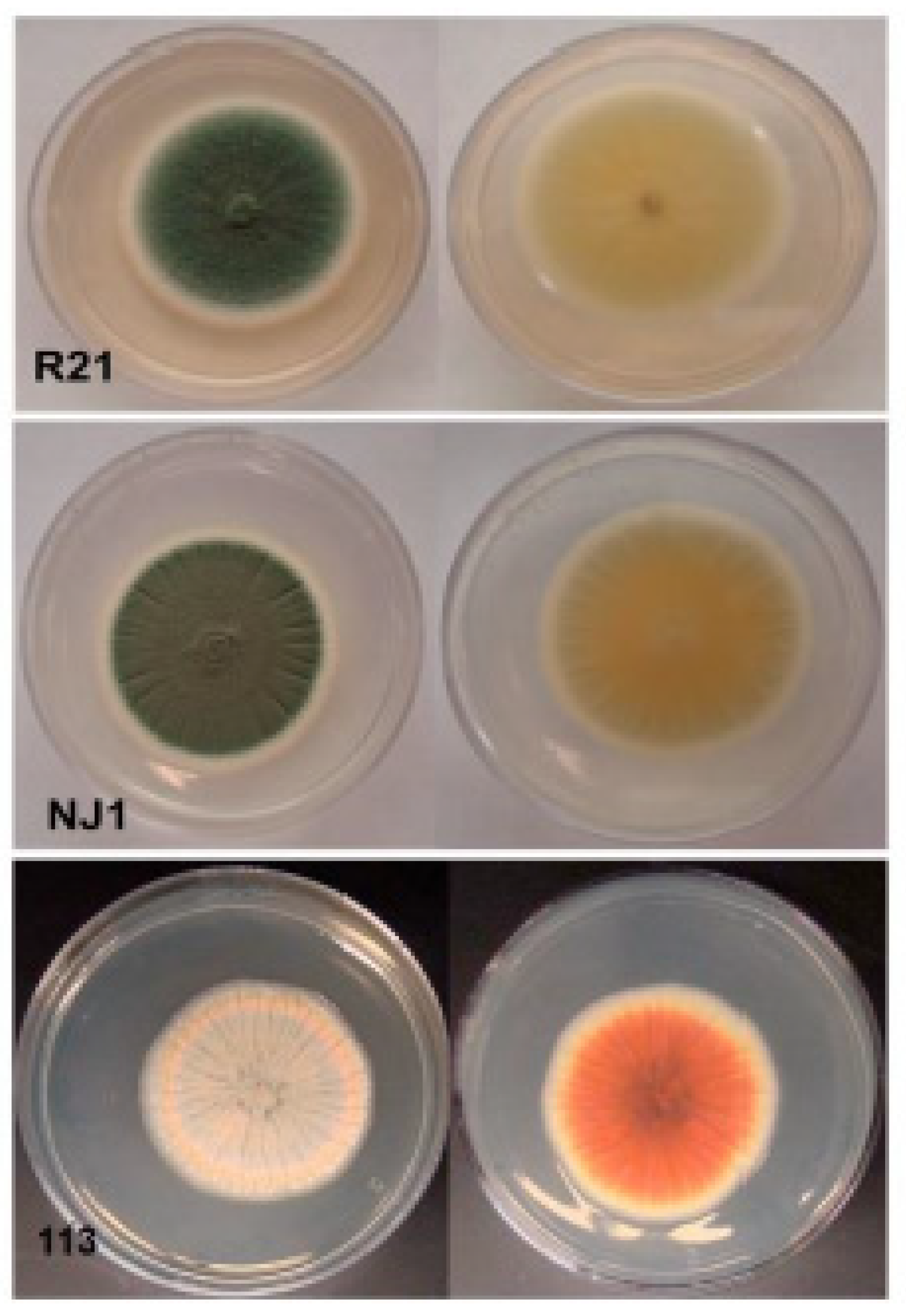
3.1. Fungal Isolation, Sequencing Data Statistics, Genome Assembly, and Gene Prediction

We selected three Penicillium species: P. expansum R21, P. crustosum NJ1, and P. maximae 113. For each strain, a 5 mm diameter plug of potato dextrose agar (PDA) was used as inoculum and cultivated for 7 days at 25 °C in the dark on 60 mm × 15 mm petri dish. The sizes, colors, and shapes of these three fungi after seven-day cultivation on PDA are shown in Figure 1. The insert sizes, read length, total bases, Q20%, and average depth are summarized in Table S1. Over 86% of the reads were high quality, and average sequencing depths of the three libraries were also above 67X. After trimming the raw sequencing data according to the approach in our previous study [49], the high-quality sequencing data still had genome coverages of at least 57X. We performed k-mer analyses using the 400–500 bp paired-end libraries to estimate the Penicillium spp. genomic sizes to be 32–35 Mb (Table S2). The total lengths of repeated sequences were 263–366 kb, contributing to 0.8–1.15% of the whole genomes (Table S3). Scaffolds assembled de novo and trimmed were used to determine the total number of predicted genes, gene lengths, and GC content (Table 1).

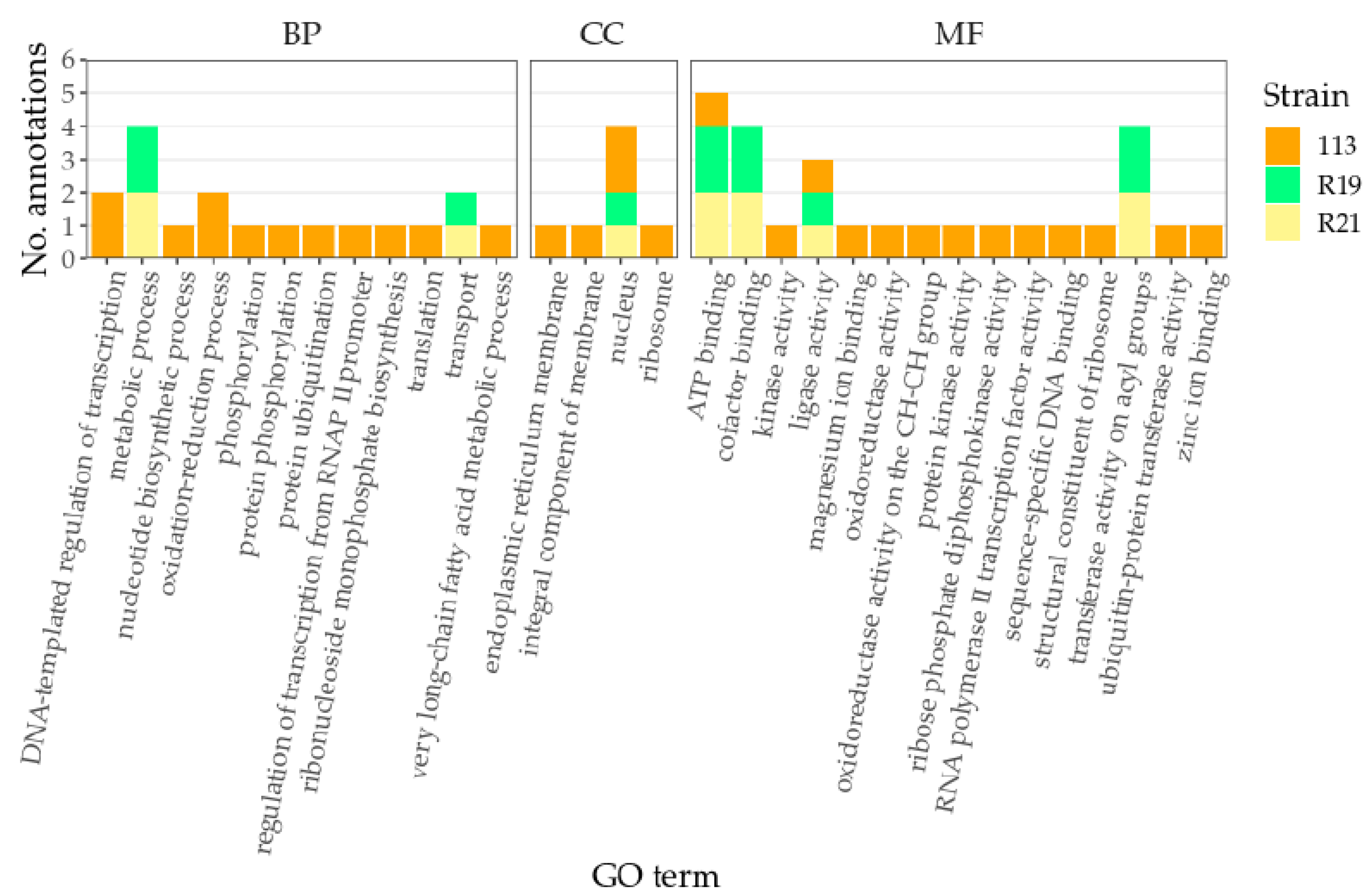
3.2. COG Analyses and GO Annotation

Clusters of Orthologous Groups (COGs) were constructed based on the comparisons of proteins from already sequenced genomes. COG database selected the main phylogenetic lineages, and each COG family consists of the proteins from at least three species. The carbohydrate transport and metabolism (G) had the most genes followed by predictions of general function (R), amino acid transport and metabolism (E), energy production and conversion (C), and secondary metabolites biosynthesis, transport, and catabolism (Q). The cell motility (N), extracellular structures (W), and nuclear structure (Y) had the fewest numbers of genes (Figure S1A,C,E). Taking 113 as an example, the predicted genes were annotated as a wide range of functional categories. The three most populous descriptive functional classes were “posttranslation modification, protein turnover, chaperones” (O, 427 genes), “secondary metabolites biosynthesis, transport and catabolism” (Q, 385 genes), and “energy production and conversion” (C, 372 genes). The fewest genes were annotated as being involved in “cell motility” (N, 2 genes) and extracellular structures (W, 4 genes). The category “general functional predictions only” (R) included the 751 genes that could not be functionally annotated (Figure S1E). In all three Penicillium strains, more than 655 putative genes were assigned to GO annotations; seven biological processes: cellular process, single-organism process, metabolic process, biological regulation, regulation of biological process, establishment of localization, and localization; two molecular functions: binding and catalytic activity; and six cellular components: organelle, cell compartment, membrane compartment, membrane, macromolecular complex, and cell (Figure S1B,D,F).

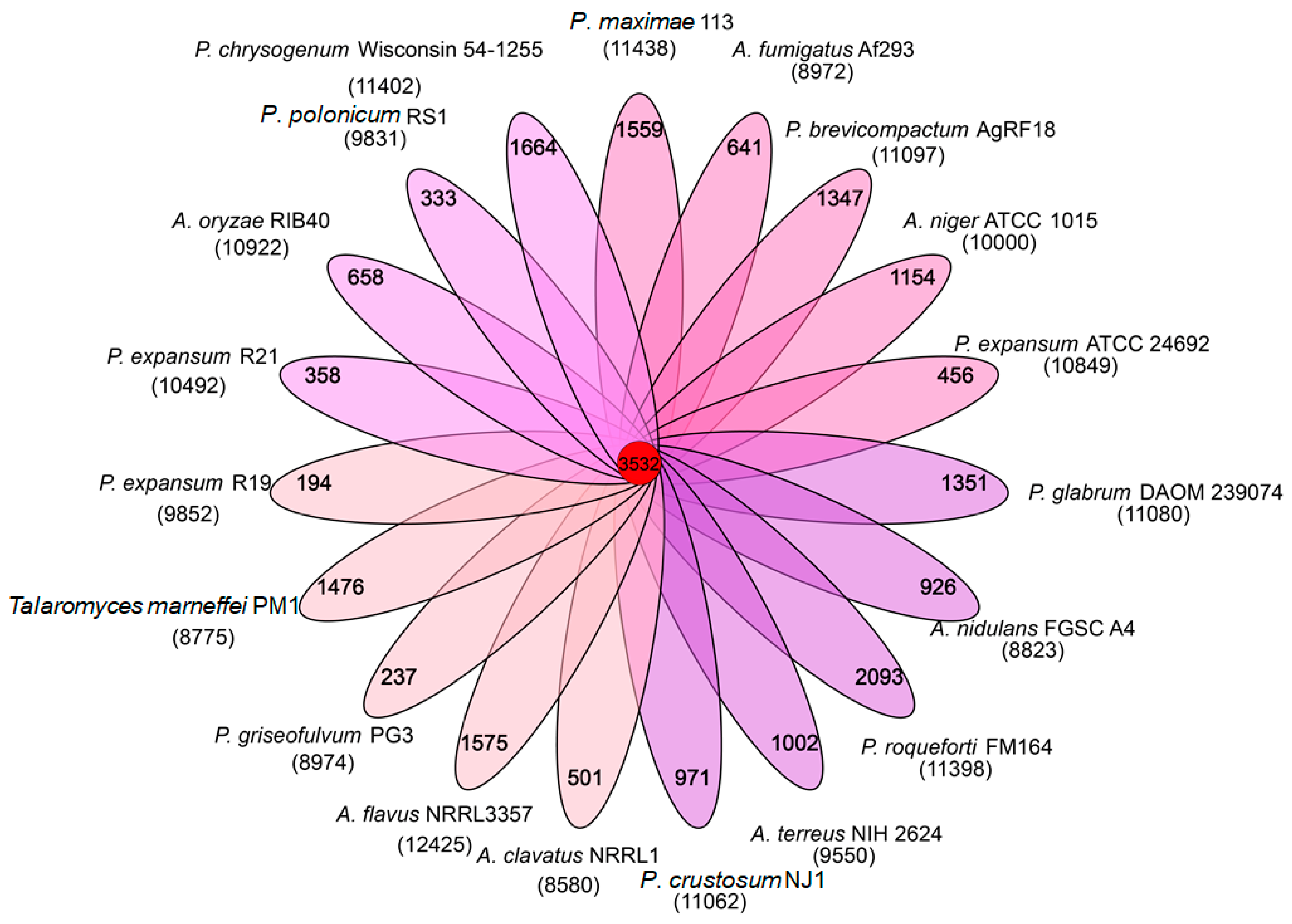
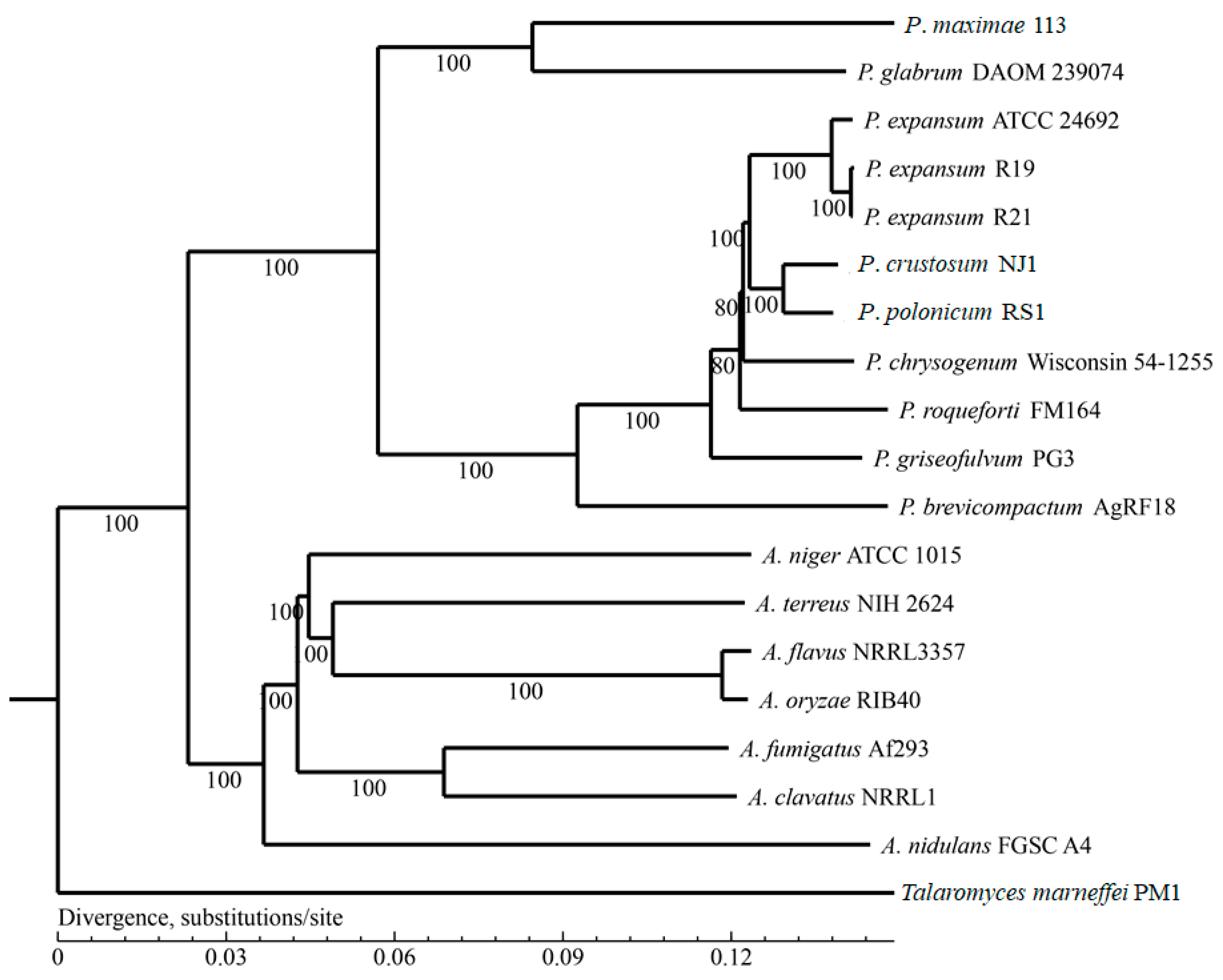
3.3. Homologous Genes and Phylogenetic Analysis

Nineteen genomes were chosen for analysis based on the relative detailed information and available genomes, and these filamentous fungal species shared 3532 common homologs. P. roqueforti FM164 had the highest number of non-homologous genes (2093), followed by P. chrysogenum Wisconsin 54-1255 (1664), Aspergillus flavus NRRL3357 (1575), 113 (1559), P. marneffei PM1 (renamed as Talaromyces marneffei, 1476) [3], and P. glabrum DAOM 239074 (1351). P. expansum R19 and P. griseofulvum PG3 had the fewer number of non-homologous genes, 194 and 237, respectively (Figure 2).
Based on the results from the homologous gene analysis, single-copy genes were selected for phylogenomic analysis. Three P. expansum strains (ATCC 24692, R19, and R21), P. polonicum RS1, and P. crustosum NJ1 strains were closely grouped into their own clades; 113 and P. glabrum DAOM 239074 were relatively distant from other strains. All seven Aspergillus species (ATCC1015, NIH2624, NRRL3357, RIB40, Af293, NRRL1, and FGSC A4) also closely grouped together (Figure 3).

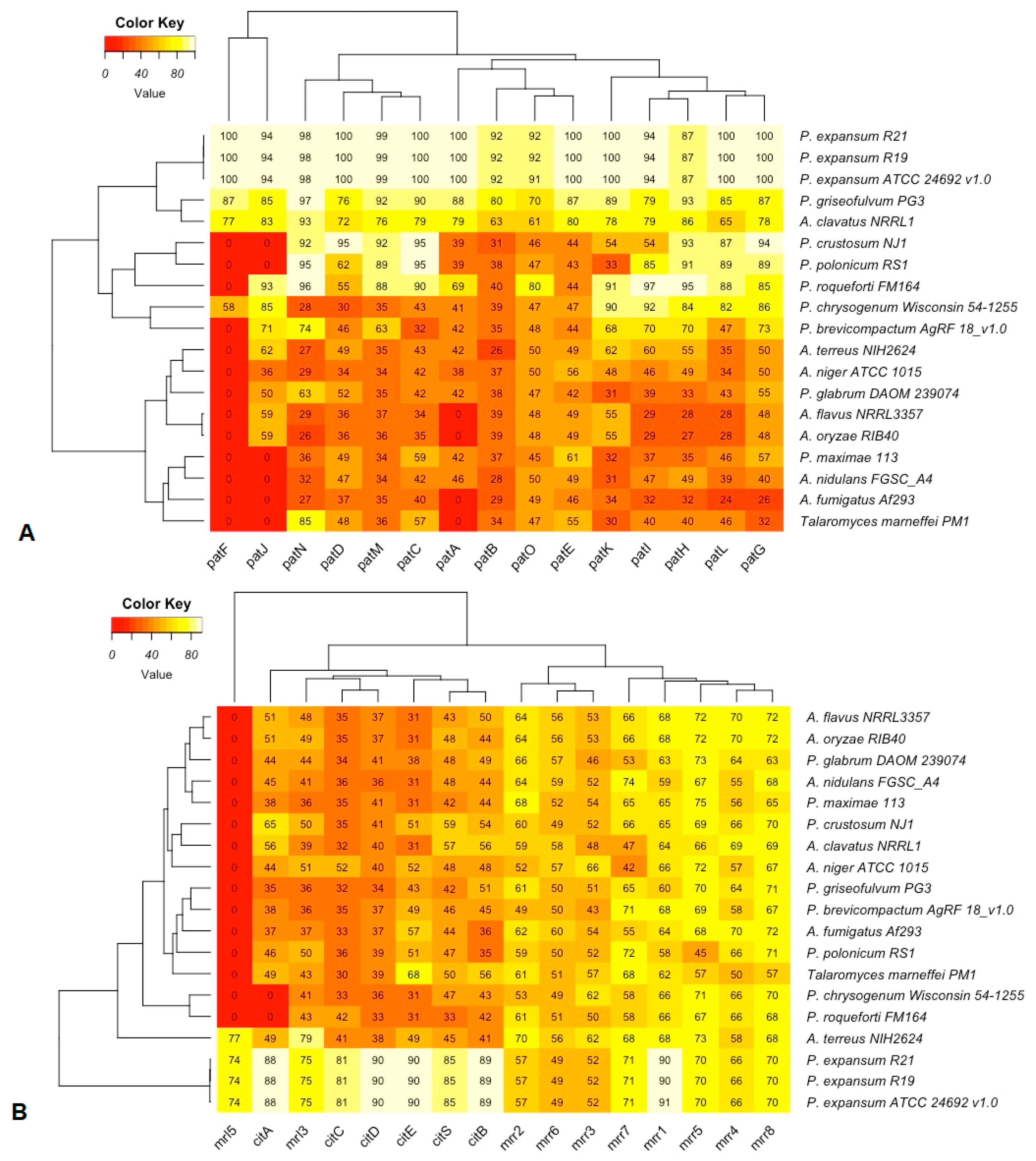
3.4. Patulin and Citrinin Gene Clusters

Functional conservation of patulin and citrinin gene clusters from 12 Penicillium and 7 Aspergillus strains were estimated by identities calculated based on TBLASTN known protein sequences against genome sequences (Figure 4). P. griseofulvum PG3 was confirmed to produce patulin by HPLC analysis [21]. Patulin production by P. expansum R21 was carried out via TLC, and R19 was confirmed by TLC and GC-MS [64,65]. As the patulin biosynthesis gene cluster of P. expansum ATCC24692 strain is highly similar to those strains R21 and R19, this strain may also produce this mycotoxin. Even though all the members in the patulin gene cluster could be detected from P. chrysogenum Wisconsin 54-1255 and A. clavatus NRRL1, these strains did not produce patulin due to the lower identities of some genes such as patD, patB, and patO [66]. Furthermore, the patF gene cannot be detected from most Aspergillus strains and the other seven Penicillium strains. Both RS1 and NJ1 strains also lacked the patJ gene and had low identity matches of the patO, patE, patA, patK, and patB genes (Figure 4A). The citrinin gene cluster could be detected from three Penicillium strains (P. expansum R19, R21, and ATCC24692) and one Aspergillus strain (A. terreus NIH2624) but cannot be detected from the other strains (lacking mrl5 gene). For all the three P. expansum strains (R19, R21, and ATCC24692), mrr2, mrr6, and mrr3 had lower than 60% identities; for the A. terreus NIH2624 strain, six genes in citrinin gene cluster (citA, citC, citD, citE, citS, and citB) had lower than 50% identities (Figure 4B).
Altogether, 59, 52, and 39 secondary metabolite gene clusters were found in strains R21, NJ1, and 113, respectively. The key enzymes of fungal secondary metabolite biosynthesis are non-ribosomal peptide synthetases (NRPS) and polyketide synthases (PKS). The NRPS and T1PKS secondary metabolite gene clusters with predicted core structures from these three Penicillium strains are shown in Table S4. P. polonicum and P. crustosum (7 in RS1 and 6 in NJ1 strains) had the highest numbers of gene clusters predicted for terpene synthesis; for P. expansum R19 and R21, and P. maximae 113, each had two gene clusters predicted for terpene synthesis (Table S5).

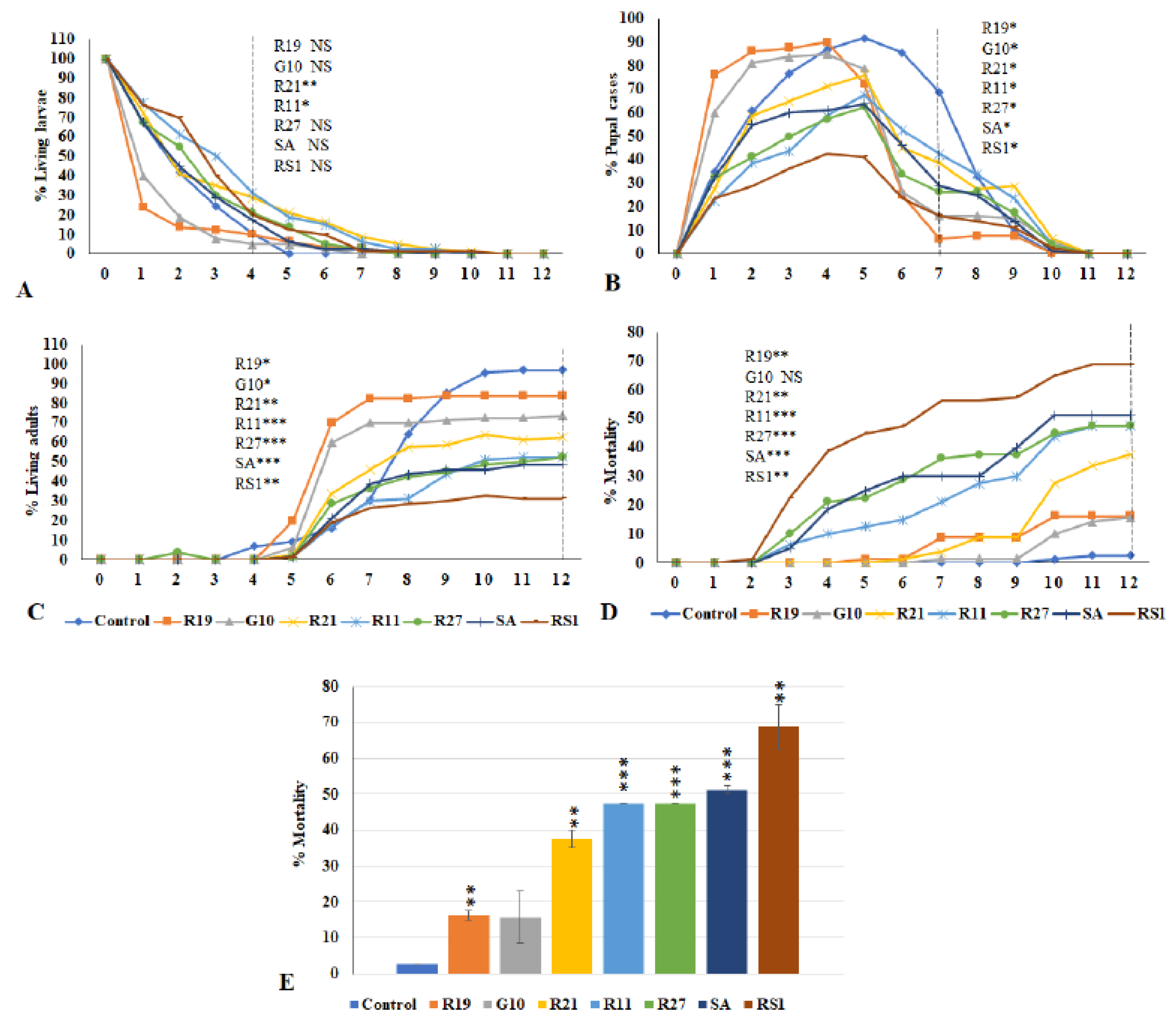
3.5. Effects of VOCs Emitted from Penicillium Species on the Third Instar Drosophila Larvae

To evaluate the effects of VOCs emitted from seven Penicillium strains, the numbers of larvae (Figure 5A), pupae (Figure 5B), adults (Figure 5C), and dead flies (Figure 5D) were counted daily for 12 days. Data are calculated as percentages of a given metamorphic stage during the Drosophila metamorphic cycle. Some flies never grew into adults; thus, the number of dead flies includes both dead larvae and dead pupae, which reflects the percent toxicity from exposure to the Penicillium VOCs mixtures (Figure 5E). Usually, control larvae pupate after 4 days; however, significant delays in pupation were observed for larvae exposed to VOCs emitted from Penicillium strains R21 (p < 0.005) and R11 (p < 0.05) (Figure 5A). By the 7th day, exposure to VOCs from all tested strains caused significant delays in pupation. Exposure to VOCs from strains R19 and G10 appeared to induce quicker transition to adulthood for surviving flies (Figure 5B). By the 10th to 12th day, 95–97% of the pupae from the controls had emerged as adults. The living adult percentages of flies exposed to VOCs from R19, G10, R21, R11, R27, SA, and RS1 by the 12th day were 84%, 74%, 63%, 53%, 53%, 49%, and 31%, respectively (Figure 5C). VOCs from P. solitum SA and P. polonicum RS1 were the most toxic, killing over 50% of the larvae; while P. expansum R19 and P. crustosum G10 were the least toxic, with an observed mortality of about 15% (Figure 5D,E). In summary, exposure of Drosophila larvae to VOCs emitted by Penicillium spp. VOCs negatively affected fly development and viability.

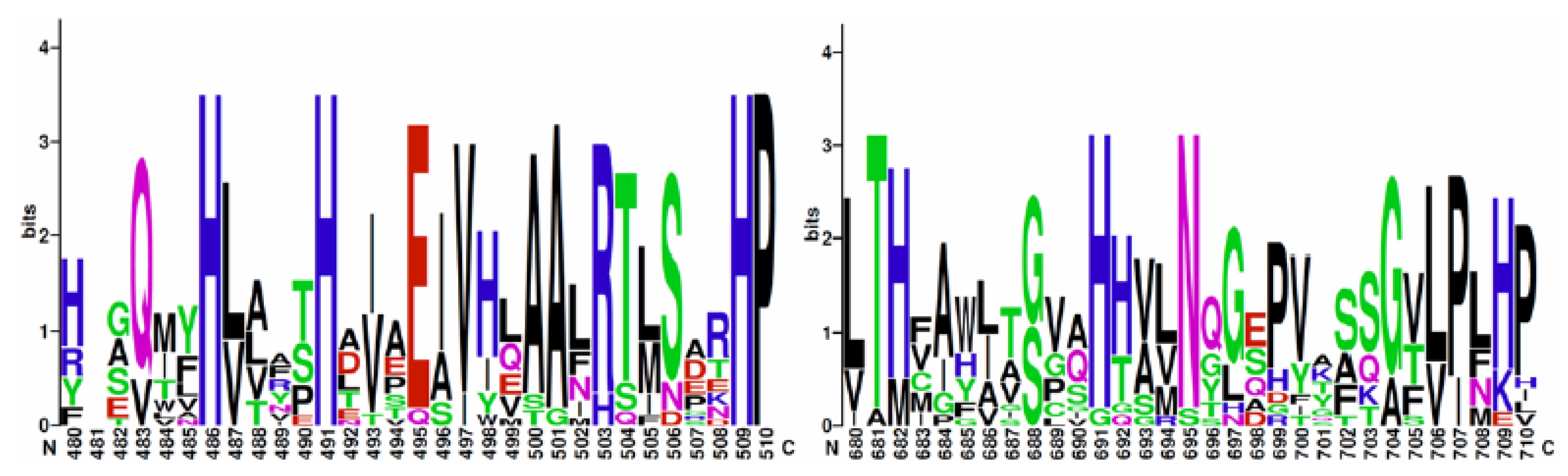
3.6. Identification of Putative Lipoxygenase Genes

A total of 46 fungal lipoxygenase protein sequences were retrieved from GenBank, 15 of which were specifically annotated as manganese lipoxygenases (Table S6). Probing for a biosynthetic operon, genes surrounding the lipoxygenase genes were functionally annotated. One manganese lipoxygenase gene was identified in strains P. maximae 113 and P. expansum R19 and R21, none of which appeared to be within a biosynthetic operon. Any similarities were likely evidence of orthologous genome architecture, especially between P. expansum strains R19 and R21. Around the lipoxygenase genes, functional annotations ATP binding function and nuclear localization were the most abundant (Figure 6). Other P. expansum, P. maximae, and P. crustosum sequences found to be similar to lipoxygenases were annotated as cytochrome P450s, Golgi SNAP receptors, or hypothetical proteins (Table S7). Multiple sequence alignments of manganese lipoxygenases revealed conserved amino acid residues within the sequences that were also conserved in the three putative lipoxygenase genes identified in this study (Figure 7).

4. Discussion

Genomic and transcriptomic analyses of Penicillium species have revealed several interesting genes and mechanisms likely involved in the adaption of this species to various environments [24]. The Penicillium genomes previously studied have estimated genome sizes, ranging from 25 Mb to 36 Mb, and a low proportion of interspersed repeats, ranging from 0.32 to 1.71%. The genomes of the three Penicillium strains studied here are consistent with the previous studies [24]. A direct relationship between host range and genome size has been proposed for fruit pathogens [13,17]. Compared to the genome sizes of P. digitatum (~25.7 Mb in average, only infects citrus fruits), P. italicum (~29 Mb, intermediate host range, mainly infects citrus fruits), and P. expansum (~31 Mb, infects pome and stone fruits), the genome sizes of P. expansum R21 (~34 Mb) and P. maximae strain 113 (~35 Mb) are fairly large. Penicillium species usually have four to eight chromosomes [69,70], and it is ideal to assemble raw sequencing data into complete individual chromosomes. Unfortunately, no complete reference P. maximae genome is available, so we performed a de novo assembly of strain 113. In the future, complementary genomic sequencing methods such as PacBio or other long-read sequencing may help assemble a more accurate genome [71,72].
The Food and Agriculture Organization of the United Nations has found that about one-fourth of the world’s food crops are contaminated with mycotoxins every year [73,74]. Penicillium produces a wide range of mycotoxins of which the most important are patulin, citrinin, and ochratoxin A [75,76]. Patulin, a potential carcinogen [36], is mainly produced by strains of P. expansum [13]. The patulin gene cluster in P. expansum consists of fifteen genes in the following order: patH, patG, patF, patE, patD, patC, patB, patA, patM, patN, patO, patL, patI, patJ, and patK [28,77]. The transcription factor patL and the patK gene coding the backbone enzyme are present in all analyzed strains but with diverse similarities (24–100% for patL and 30–100% for patK, Figure 4A) indicating that these two genes experienced different evolutionary histories during adaptation to different environments and hosts. In this study, the complete and functional patulin gene cluster can be detected from three strains of P. expansum (R21, R19, and ATCC24692). We confirmed the production of patulin from two of these strains. No patulin is produced by P. chrysogenum Wisconsin 54-1255 or A. clavatus NRRL1, which reflects the lower sequence identities of genes such as patD, patB, patO, patH, and patL. In the future, more strains from A. clavatus should be tested as it is a reported patulin producer [66].
Citrinin, a nephrotoxic mycotoxin, was first isolated from P. citrinum and is a less toxic substance than patulin [78]. Sixteen putative genes: pksCT, ctnI, orf3, ctnE, orf1, ctnH, ctnD, orf4, ctnA, ctnR, orf5, orf6, orf7, and orf8, may be involved in citrinin biosynthesis [79,80]. The complete citrinin gene cluster has been identified from three Penicillium strains (P. expansum R19, R21, and ATCC24692) and one Aspergillus strain (A. terreus NIH2624). Our analysis indicates that the citS gene is present in all the analyzed strains with identities from 33% to 85% (Figure 4B). One recent study suggests that it could be an essential gene in Penicillium steckii [81]. The mrl3/ctnA (P. citrinum/P. expansum) sequence is also present in the analyzed strains (identities from 36% to 79%, Figure 4B) and may be similar to the specific transcriptional factor patL gene, which is often located inside the toxin cluster and activates all the genes in the cluster [82]. We did not verify citrinin production in any strain. Complete patulin and citrinin gene clusters could not be identified from P. maximae 113. In summary, patulin and citrinin are mainly produced by strains of P. expansum. Our patulin gene cluster identity and analysis support previous predictions that the cluster in P. griseofulvum PG3 (lower identity) is likely an ancestral trait in Penicillium spp. (higher identities), predating the divergence of P. roqueforti FM164 (lacking the patF gene) [22].
VOCs are chemically diverse groups of low-molecular-weight compounds that evaporate easily at normal temperatures and serve biologically targeted and diverse functions as semiochemicals, antimicrobials, and defense response chemicals [83,84]. Eight carbon compounds such as 1-octen-3-ol, 3-octanol, and 3-octanone are among the most common fungal VOCs [39]. In previous studies, low concentrations of the vapor form of several eight carbon compounds including 1-octen-3-ol are toxic to Drosophila larvae and adult flies [41,85,86]. 1-Octen-3-ol selectively affects dopaminergic neurons in adult Drosophila brain and induces Parkinson’s-like behavioral alterations in a fly model for this disease [87,88,89]. Moreover, we showed that the R and S enantiomers of 1-octen-3-ol have differential effects on the transcriptome of P. chrysogenum [90]. We found that the VOCs emitted by Penicillium maximae 113 caused a relatively high toxicity (60%) in the Drosophila larvae toxicity assay. Solid-phase microextraction gas chromatography mass spectrometry analysis revealed that a higher diversity and concentration (up to 300 ng/L) of VOCs were emitted by this strain after longer periods of cultivation (18 days compared to 7 days) [14].
We also tested the toxic effects of VOCs from several Penicillium strains; our data revealed that VOCs from P. polonicum strains are more toxic than those emitted by the other strains (Figure 5). More research, including GC-MS analyses, to elucidate individual VOC profiles of different species are needed to better understand their toxic effects. The group of fatty acid dioxygenases called Ppos (psi (precocious sexual inducer)-producing oxygenases) and lipoxygenases (LOXs) are candidates for the enzymes involved in eight carbon volatile formation in fungi [91]. In this study, putative lipoxygenase genes that would contribute to production of VOCs were identified in Penicillium maximae strain 113 and P. expansum strains R19 and R21. We will clone these candidate genes to explore their functions in our future work.
In summary, our analyses showed that different P. expansum strains have different secondary metabolite gene clusters, and their numbers are very changeable within this species. Comparative genomic studies have revealed that there are interesting patterns of gains or losses of genes involved in the production of secondary metabolites that act as effectors in pathogens [92]. Horizontal gene transfer is an ongoing and successful ecological strategy for many fungal species to acquire the novel secondary metabolic gene clusters [5,93] and may be occurring in mycotoxigenic species of Penicillium. However, this hypothesis needs further experimental verification.

Supplementary Materials

The following are available online at https://www.mdpi.com/article/10.3390/jof7090743/s1, Figure S1: COG analyses and Go annotation of three Penicillium species, Table S1: Sequencing data statistical analysis, Table S2: 17-mer analysis, Table S3: Repeated sequence analysis, Table S4: NRPS and T1PKS secondary metabolite gene clusters, Table S5: Secondary metabolite gene clusters predicted for terpene synthesis in Penicillium species, Table S6: Putative lipoxygenase genes returned by HMMER searches and their GO biological processes and molecular functions terms, Table S7: Lipoxygenase amino acid sequences retrieved from NCBI GenBank.

Author Contributions

G.Y. and A.G. conceived and designed the experiments; G.Y. and H.Z. performed the experiments; G.Y., H.Z., and K.K.P. analyzed the data; all authors contributed reagents/materials/analysis tools; G.Y. drafted the paper, and all other authors contributed to the revisions of this manuscript. All authors have read and agreed to the published version of the manuscript.

Funding

This research was funded by Hainan province’s Major Scientific and Technological Project “Construction and Application of Resistance Service Platform of Major Southern Propagation Crops” (No. ZDKJ201901); Hainan province’s Major Scientific and Technological Project “Biosafety Prevention and Control in Southern Breeding Area” (No. ZDKJ202002); and the Central Public-interest Scientific Institution Basal Research Fund for Chinese Academy of Tropical Agricultural Sciences (No.17CXTD-24).

Institutional Review Board Statement

Not applicable.

Informed Consent Statement

Not applicable.

Acknowledgments

Use of a company or product name by the U.S. Department of Agriculture does not imply approval or recommendation of the product to the exclusion of others that may also be suitable.

Conflicts of Interest

The authors declare no conflict of interest. The founding sponsors had no role in the design of the study, in the collection, analyses, or interpretation of data, in the writing of the manuscript, and in the decision to publish the results.

References

  1. Visagie, C.; Houbraken, J.; Frisvad, J.C.; Hong, S.-B.; Klaassen, C.; Perrone, G.; Seifert, K.; Varga, J.; Yaguchi, T.; Samson, R. Identification and nomenclature of the genus Penicillium. Stud. Mycol. 2014, 78, 343–371. [Google Scholar] [CrossRef] [Green Version]
  2. Gupta, V.K. New and Future Developments in Microbial Biotechnology and Bioengineering: Aspergillus System Properties and Applications; Elsevier: Amsterdam, The Netherlands, 2016. [Google Scholar]
  3. Houbraken, J.; Kocsubé, S.; Visagie, C.; Yilmaz, N.; Wang, X.-C.; Meijer, M.; Kraak, B.; Hubka, V.; Bensch, K.; Samson, R. Classification of Aspergillus, Penicillium, Talaromyces and related genera (Eurotiales): An overview of families, genera, subgenera, sections, series and species. Stud. Mycol. 2020, 95, 5–169. [Google Scholar] [CrossRef]
  4. Shelton, B.G.; Kirkland, K.H.; Flanders, W.D.; Morris, G.K. Profiles of airborne fungi in buildings and outdoor environments in the United States. Appl. Environ. Microbiol. 2002, 68, 1743–1753. [Google Scholar] [CrossRef] [Green Version]
  5. Van den Berg, M.A.; Albang, R.; Albermann, K.; Badger, J.H.; Daran, J.M.; Driessen, A.J.; Garcia-Estrada, C.; Fedorova, N.D.; Harris, D.M.; Heijne, W.H.; et al. Genome sequencing and analysis of the filamentous fungus Penicillium chrysogenum. Nat. Biotechnol. 2008, 26, 1161–1168. [Google Scholar] [CrossRef] [PubMed] [Green Version]
  6. Gupta, A.K.; Cooper, E.A.; Bowen, J.E. Meta-Analysis: Griseofulvin efficacy in the treatment of tinea capitis. J. Drugs Dermatol. 2008, 7, 369–372. [Google Scholar] [PubMed]
  7. Molimard, P.; Spinnler, H. Review: Compounds involved in the flavor of surface mold-ripened cheeses: Origins and properties. J. Dairy Sci. 1996, 79, 169–184. [Google Scholar] [CrossRef]
  8. Lacey, J.; Crook, B. Fungal and actinomycete spores as pollutants of the workplace and occupational allergens. Ann. Occup. Hyg. 1988, 32, 515–533. [Google Scholar]
  9. Frisvad, J.C.; Samson, R.A. Penicillium subgenus Penicillium. A guide to identification of food and air-borne terverticillate Penicillia and their mycotoxins. Stud. Mycol. 2004, 1, 1–174. [Google Scholar]
  10. Chalutz, E.; Wilson, C. Postharvest biocontrol of green and blue mold and sour rot of citrus fruit by Debaryomyces hansenii. Plant Dis. 1990, 74, 134–137. [Google Scholar] [CrossRef]
  11. Eckert, J.; Sievert, J.; Ratnayake, M. Reduction of imazalil effectiveness against citrus green mold in California packinghouses by resistant biotypes of Penicillium digitatum. Plant Dis. 1994, 78, 971–974. [Google Scholar] [CrossRef]
  12. Li, B.; Zong, Y.; Du, Z.; Chen, Y.; Zhang, Z.; Qin, G.; Zhao, W.; Tian, S. Genomic characterization reveals insights into patulin biosynthesis and pathogenicity in Penicillium species. Mol. Plant-Microbe Interact. 2015, 28, 635–647. [Google Scholar] [CrossRef] [PubMed] [Green Version]
  13. Ballester, A.-R.; Marcet-Houben, M.; Levin, E.; Sela, N.; Selma-Lázaro, C.; Carmona, L.; Wisniewski, M.; Droby, S.; González-Candelas, L.; Gabaldón, T. Genome, transcriptome, and functional analyses of Penicillium expansum provide new insights into secondary metabolism and pathogenicity. Mol. Plant-Microbe Interact. 2015, 28, 232–248. [Google Scholar] [CrossRef] [Green Version]
  14. Zhao, G.; Yin, G.; Inamda, A.A.; Luo, J.; Zhang, N.; Yang, I.; Buckley, B.; Bennett, J.W. Volatile organic compounds emitted by filamentous fungi isolated from flooded homes after Hurricane Sandy show toxicity in a Drosophila bioassay. Indoor Air 2017, 27, 518–528. [Google Scholar] [CrossRef]
  15. Rao, C.Y.; Riggs, M.A.; Chew, G.L.; Muilenberg, M.L.; Thorne, P.S.; Van Sickle, D.; Dunn, K.H.; Brown, C. Characterization of airborne molds, endotoxins, and glucans in homes in New Orleans after Hurricanes Katrina and Rita. Appl. Environ. Microbiol. 2007, 73, 1630–1634. [Google Scholar] [CrossRef] [PubMed] [Green Version]
  16. Cacho, R.A.; Tang, Y.; Chooi, Y.-H. Next-Generation sequencing approach for connecting secondary metabolites to biosynthetic gene clusters in fungi. Front. Microbiol. 2015, 5, 774. [Google Scholar] [CrossRef] [PubMed] [Green Version]
  17. Marcet-Houben, M.; Ballester, A.-R.; de la Fuente, B.; Harries, E.; Marcos, J.F.; González-Candelas, L.; Gabaldón, T. Genome sequence of the necrotrophic fungus Penicillium digitatum, the main postharvest pathogen of citrus. BMC Genom. 2012, 13, 646. [Google Scholar] [CrossRef] [Green Version]
  18. Julca, I.; Droby, S.; Sela, N.; Marcet-Houben, M.; Gabaldón, T. Contrasting genomic diversity in two closely related postharvest pathogens: Penicillium digitatum and Penicillium expansum. Genome Biol. Evol. 2016, 8, 218–227. [Google Scholar] [CrossRef] [Green Version]
  19. Yu, J.; Jurick, W.M.; Cao, H.; Yin, Y.; Gaskins, V.L.; Losada, L.; Zafar, N.; Kim, M.; Bennett, J.W.; Nierman, W.C. Draft genome sequence of Penicillium expansum strain R19, which causes postharvest decay of apple fruit. Genome Announc. 2014, 2, e00635-14. [Google Scholar] [CrossRef] [Green Version]
  20. Eldarov, M.A.; Mardanov, A.V.; Beletsky, A.V.; Dzhavakhiya, V.V.; Ravin, N.V.; Skryabin, K.G. Complete mitochondrial genome of compactin-producing fungus Penicillium solitum and comparative analysis of Trichocomaceae mitochondrial genomes. FEMS Microbiol. Lett. 2012, 329, 9–17. [Google Scholar] [CrossRef] [Green Version]
  21. Cheeseman, K.; Ropars, J.; Renault, P.; Dupont, J.; Gouzy, J.; Branca, A.; Abraham, A.-L.; Ceppi, M.; Conseiller, E.; Debuchy, R. Multiple recent horizontal transfers of a large genomic region in cheese making fungi. Nat. Commun. 2014, 5, 2876. [Google Scholar] [CrossRef]
  22. Banani, H.; Marcet-Houben, M.; Ballester, A.-R.; Abbruscato, P.; González-Candelas, L.; Gabaldón, T.; Spadaro, D. Genome sequencing and secondary metabolism of the postharvest pathogen Penicillium griseofulvum. BMC Genom. 2016, 17, 19. [Google Scholar] [CrossRef] [PubMed] [Green Version]
  23. Woo, P.C.; Lau, S.K.; Liu, B.; Cai, J.J.; Chong, K.T.; Tse, H.; Kao, R.Y.; Chan, C.-M.; Chow, W.-N.; Yuen, K.-Y. Draft genome sequence of Penicillium marneffei strain PM1. Eukaryot. Cell 2011, 10, 1740–1741. [Google Scholar] [CrossRef] [PubMed] [Green Version]
  24. Ropars, J.; de La Vega, R.R.; López-Villavicencio, M.; Gouzy, J.; Dupont, J.; Swennen, D.; Dumas, E.; Giraud, T.; Branca, A. Diversity and mechanisms of genomic adaptation in Penicillium. In Aspergillus and Penicillium in the Post-Genomic Era; Caister Academic Press: Poole, UK, 2016. [Google Scholar]
  25. Sanderson, P.; Spotts, R. Postharvest decay of winter pear and apple fruit caused by species of Penicillium. Phytopathology 1995, 85, 103–110. [Google Scholar] [CrossRef]
  26. Frisvad, J.C.; Samson, R.A. Polyphasic taxonomy of Penicillium subgenus Penicillium. A guide to identification of food and air-borne terverticillate Penicillia and their mycotoxins. Stud. Mycol. 2004, 49, 1–174. [Google Scholar]
  27. Sutton, T.B.; Aldwinckle, H.S.; Agnello, A.M.; Walgenbach, J.F. Compendium of Apple and Pear Diseases and Pests; American Phytopath Society: St. Paul, MN, USA, 2014. [Google Scholar]
  28. Puel, O.; Galtier, P.; Oswald, I.P. Biosynthesis and toxicological effects of patulin. Toxins 2010, 2, 613–631. [Google Scholar] [CrossRef] [Green Version]
  29. HemaMoorthy, T.; Prakasam, V. First report of Penicillium expansum causing bulb rot of lilium in India. Am.-Eurasian J. Agric. Environ. Sci. 2013, 13, 293–295. [Google Scholar]
  30. Wang, C.; Ai, J.; Lv, H.; Qin, H.; Yang, Y.; Liu, Y.; Fan, S. First report of Penicillium expansum causing postharvest decay on stored kiwifruit (Actinidia arguta) in China. Plant Dis. 2015, 99, 1037. [Google Scholar] [CrossRef]
  31. Palou, L.; Montesinos-Herrero, C.; Taberner, V.; Vilella-Esplá, J. First report of Penicillium expansum causing postharvest blue mold of fresh date palm fruit (Phoenix dactylifera) in Spain. Plant Dis. 2013, 97, 846. [Google Scholar] [CrossRef]
  32. Pitt, J.; Spotts, R.; Holmes, R.; Cruickshank, R. Penicillium solitum revived, and its role as a pathogen of pomaceous fruit. Phytopathology 1991, 81, 1108–1112. [Google Scholar] [CrossRef]
  33. Amiri, A.; Bompeix, G. Diversity and population dynamics of Penicillium spp. on apples in pre-and postharvest environments: Consequences for decay development. Plant Pathol. 2005, 54, 74–81. [Google Scholar] [CrossRef]
  34. Andersen, B.; Thrane, U. Food-Borne fungi in fruit and cereals and their production of mycotoxins. In Advances in Food Mycology; Springer: Boston, MA, USA, 2006; pp. 137–152. [Google Scholar]
  35. Vico, I.; Gaskins, V.; Duduk, N.; Vasić, M.; Yu, J.; Peter, K.A.; Jurick, W.M. First report of Penicillium crustosum causing blue mold on stored apple fruit in Serbia. Plant Dis. 2014, 98, 1430. [Google Scholar] [CrossRef]
  36. Ostry, V.; Malir, F.; Toman, J.; Grosse, Y. Mycotoxins as human carcinogens—The IARC Monographs classification. Mycotoxin Res. 2017, 33, 65–73. [Google Scholar] [CrossRef] [PubMed]
  37. Tannous, J.; Keller, N.P.; Atoui, A.; El Khoury, A.; Lteif, R.; Oswald, I.P.; Puel, O. Secondary metabolism in Penicillium expansum: Emphasis on recent advances in patulin research. Crit. Rev. Food Sci. Nutr. 2018, 58, 2082–2098. [Google Scholar] [CrossRef] [PubMed]
  38. Luciano-Rosario, D.; Keller, N.P.; Jurick, W.M. Penicillium expansum: Biology, omics, and management tools for a global postharvest pathogen causing blue mould of pome fruit. Mol. Plant Pathol. 2020, 21, 1391–1404. [Google Scholar] [CrossRef]
  39. Bennett, J.W.; Inamdar, A.A. Are some fungal volatile organic compounds (VOCs) mycotoxins? Toxins 2015, 7, 3785–3804. [Google Scholar] [CrossRef] [Green Version]
  40. Almaliki, H.S.; Drazanin, E.; Sadovnik, R.; Yin, G.; Bennett, J.W. Eight carbon volatiles are more toxic to wild-type Drosophila melanogaster than to flies with blocked immune system mutations. Entomol. Exp. Appl. 2021. (accepted). [Google Scholar]
  41. Yin, G.; Padhi, S.; Lee, S.; Hung, R.; Zhao, G.; Bennett, J.W. Effects of three volatile oxylipins on colony development in two species of fungi and on Drosophila larval metamorphosis. Curr. Microbiol. 2015, 71, 347–356. [Google Scholar] [CrossRef]
  42. Yin, G.; Zhang, Y.; Hua, S.S.T.; Yu, J.; Bu, L.; Pennerman, K.K.; Huang, Q.; Guo, A.; Bennett, J.W. Genome sequencing and analysis of the postharvest fungus Penicillium expansum R21. Genome Announc. 2017, 5, e01516-16. [Google Scholar] [CrossRef] [Green Version]
  43. Yung, P.Y.; Grasso, L.L.; Mohidin, A.F.; Acerbi, E.; Hinks, J.; Seviour, T.; Marsili, E.; Lauro, F.M. Global transcriptomic responses of Escherichia coli K-12 to volatile organic compounds. Sci. Rep. 2016, 6, 19899. [Google Scholar] [CrossRef] [Green Version]
  44. Yu, J.; Wu, G.; Jurick, W.M.; Gaskins, V.L.; Yin, Y.; Yin, G.; Bennett, J.W.; Shelton, D.R. Genome sequence of Penicillium solitum RS1, which causes postharvest apple decay. Genome Announc. 2016, 4, e00363-16. [Google Scholar] [CrossRef] [PubMed] [Green Version]
  45. Yin, G.; Zhang, Y.; Pennerman, K.K.; Wu, G.; Hua, S.S.T.; Yu, J.; Jurick, W.M.; Guo, A.; Bennett, J.W. Characterization of blue mold Penicillium species isolated from stored fruits using multiple highly conserved loci. J. Fungi 2017, 3, 12. [Google Scholar] [CrossRef] [Green Version]
  46. Yin, G.; Zhang, Y.; Pennerman, K.K.; Hua, S.S.T.; Yu, J.; Guo, A.; Liu, Z.; Bennett, J.W. Draft genome sequence of the fungus Penicillium solitum NJ1. Genome Announc. 2016, 4, e01176-16. [Google Scholar] [CrossRef] [Green Version]
  47. Yin, G.; Zhang, Y.; Pennerman, K.K.; Hua, S.S.T.; Huang, Q.; Guo, A.; Liu, Z.; Bennett, J.W. Genome sequencing and analysis of the filamentous fungus Penicillium sclerotiorum 113, isolated after Hurricane Sandy. Genome Announc. 2016, 4, e01153-16. [Google Scholar] [CrossRef] [Green Version]
  48. Yin, G.; Jurick, W.M., II; Zhao, G.; Bennett, J.W. New names for three Penicillium strains based on updated barcoding and phylogenetic analyses. Microbiol. Resour. Announc. 2021. (Under review). [Google Scholar]
  49. Yang, F.; Zhang, Y.; Huang, Q.; Yin, G.; Pennerman, K.K.; Yu, J.; Liu, Z.; Li, D.; Guo, A. Analysis of key genes of jasmonic acid mediated signal pathway for defense against insect damages by comparative transcriptome sequencing. Sci. Rep. 2015, 5, 16500. [Google Scholar] [CrossRef] [PubMed] [Green Version]
  50. Simpson, J.T.; Wong, K.; Jackman, S.D.; Schein, J.E.; Jones, S.J.; Birol, I. ABySS: A parallel assembler for short read sequence data. Genome Res. 2009, 19, 1117–1123. [Google Scholar] [CrossRef] [Green Version]
  51. Bao, E.; Jiang, T.; Girke, T. AlignGraph: Algorithm for secondary de novo genome assembly guided by closely related references. Bioinformatics 2014, 30, i319–i328. [Google Scholar] [CrossRef] [Green Version]
  52. Luo, R.; Liu, B.; Xie, Y.; Li, Z.; Huang, W.; Yuan, J.; He, G.; Chen, Y.; Pan, Q.; Liu, Y. SOAPdenovo2: An empirically improved memory-efficient short-read de novo assembler. Gigascience 2012, 1, 2047-2217X-2041-2018. [Google Scholar] [CrossRef]
  53. Tempel, S. Using and understanding RepeatMasker. In Mobile Genetic Elements; Humana Press: Totowa, NJ, USA, 2012; pp. 29–51. [Google Scholar]
  54. Holt, C.; Yandell, M. MAKER2: An annotation pipeline and genome-database management tool for second-generation genome projects. BMC Bioinform. 2011, 12, 491. [Google Scholar] [CrossRef] [Green Version]
  55. Ye, W.; Chen, Y.; Zhang, Y.; Xu, Y. H-BLAST: A fast protein sequence alignment toolkit on heterogeneous computers with GPUs. Bioinformatics 2017, 33, 1130–1138. [Google Scholar] [CrossRef] [Green Version]
  56. Conesa, A.; Götz, S.; García-Gómez, J.M.; Terol, J.; Talón, M.; Robles, M. Blast2GO: A universal tool for annotation, visualization and analysis in functional genomics research. Bioinformatics 2005, 21, 3674–3676. [Google Scholar] [CrossRef] [PubMed] [Green Version]
  57. Blin, K.; Medema, M.H.; Kottmann, R.; Lee, S.Y.; Weber, T. The antiSMASH database, a comprehensive database of microbial secondary metabolite biosynthetic gene clusters. Nucleic Acids Res. 2016, 45, D555–D559. [Google Scholar] [CrossRef] [PubMed] [Green Version]
  58. Ter-Hovhannisyan, V.; Lomsadze, A.; Chernoff, Y.O.; Borodovsky, M. Gene prediction in novel fungal genomes using an ab initio algorithm with unsupervised training. Genome Res. 2008, 18, 1979–1990. [Google Scholar] [CrossRef] [Green Version]
  59. Pertea, G.; Pertea, M. GFF Utilities: GffRead and GffCompare. F1000Research 2020, 9. [Google Scholar] [CrossRef]
  60. Knight, R.D.; Freeland, S.J.; Landweber, L.F. Selection, history and chemistry: The three faces of the genetic code. Trends Biochem. Sci. 1999, 24, 241–247. [Google Scholar] [CrossRef]
  61. Sievers, F.; Wilm, A.; Dineen, D.; Gibson, T.J.; Karplus, K.; Li, W.; Lopez, R.; McWilliam, H.; Remmert, M.; Söding, J. Fast, scalable generation of high-quality protein multiple sequence alignments using Clustal Omega. Mol. Syst. Biol. 2011, 7, 539. [Google Scholar] [CrossRef]
  62. Finn, R.D.; Clements, J.; Eddy, S.R. HMMER web server: Interactive sequence similarity searching. Nucleic Acids Res. 2011, 39, W29–W37. [Google Scholar] [CrossRef] [Green Version]
  63. Lichtner, F.J.; Gaskins, V.L.; Cox, K.D.; Jurick, W.M. Global transcriptomic responses orchestrate difenoconazole resistance in Penicillium spp. causing blue mold of stored apple fruit. BMC Genom. 2020, 21, 574. [Google Scholar] [CrossRef]
  64. Pennerman, K.K.; Scarsella, J.B.; Yin, G.-H.; Hua, S.-S.T.; Hartman, T.G.; Bennett, J.W. Volatile 1-octen-3-ol increases patulin production by Penicillium expansum on a patulin-suppressing medium. Mycotoxin Res. 2019, 35, 329–340. [Google Scholar] [CrossRef]
  65. Jurick, W.M.; Peng, H.; Beard, H.S.; Garrett, W.M.; Lichtner, F.J.; Luciano-Rosario, D.; Macarisin, O.; Liu, Y.; Peter, K.A.; Gaskins, V.L. Blistering1 modulates Penicillium expansum virulence via vesicle-mediated protein secretion. Mol. Cell. Proteom. 2020, 19, 344–361. [Google Scholar] [CrossRef]
  66. Frisvad, J. A critical review of producers of small lactone mycotoxins: Patulin, penicillic acid and moniliformin. World Mycotoxin J. 2018, 11, 73–100. [Google Scholar] [CrossRef]
  67. Wennman, A.; Oliw, E.H.; Karkehabadi, S.; Chen, Y. Crystal structure of manganese lipoxygenase of the rice blast fungus Magnaporthe oryzae. J. Biol. Chem. 2016, 291, 8130–8139. [Google Scholar] [CrossRef] [PubMed] [Green Version]
  68. Crooks, G.E.; Hon, G.; Chandonia, J.-M.; Brenner, S.E. WebLogo: A sequence logo generator. Genome Res. 2004, 14, 1188–1190. [Google Scholar] [CrossRef] [Green Version]
  69. Fierro, F.; Gutiérrez, S.; Diez, B.; Martín, J.F. Resolution of four large chromosomes in penicillin-producing filamentous fungi: The penicillin gene cluster is located on chromosome II (9.6 Mb) in Penicillium notatum and chromosome 1 (10.4 Mb) in Penicillium chrysogenum. Mol. Gen. Genet. MGG 1993, 241, 573–578. [Google Scholar] [CrossRef]
  70. Yuen, K.-y.; Pascal, G.; Wong, S.S.; Glaser, P.; Woo, P.C.; Kunst, F.; Cai, J.J.; Cheung, E.Y.; Médigue, C.; Danchin, A. Exploring the Penicillium marneffei genome. Arch. Microbiol. 2003, 179, 339–353. [Google Scholar] [CrossRef]
  71. Lee, H.; Gurtowski, J.; Yoo, S.; Nattestad, M.; Marcus, S.; Goodwin, S.; McCombie, W.R.; Schatz, M. Third-Generation sequencing and the future of genomics. BioRxiv 2016, 048603. [Google Scholar] [CrossRef] [Green Version]
  72. Rhoads, A.; Au, K.F. PacBio sequencing and its applications. Genom. Proteom. Bioinform. 2015, 13, 278–289. [Google Scholar] [CrossRef] [PubMed] [Green Version]
  73. Negedu, A.; Atawodi, S.; Ameh, J.; Umoh, V.; Tanko, H. Economic and health perspectives of mycotoxins: A review. Cont. J. Biomed. Sci. 2011, 5, 5–26. [Google Scholar]
  74. Upadhaya, S.D.; Park, M.; Ha, J.-K. Mycotoxins and their biotransformation in the rumen: A review. Asian-Australas. J. Anim. Sci. 2010, 23, 1250–1260. [Google Scholar] [CrossRef]
  75. Perrone, G.; Susca, A. Penicillium species and their associated mycotoxins. In Mycotoxigenic Fungi; Humana Press: New York, NY, USA, 2017; pp. 107–119. [Google Scholar]
  76. Andersen, B.; Smedsgaard, J.; Frisvad, J.C. Penicillium expansum: Consistent production of patulin, chaetoglobosins, and other secondary metabolites in culture and their natural occurrence in fruit products. J. Agric. Food Chem. 2004, 52, 2421–2428. [Google Scholar] [CrossRef] [PubMed]
  77. Tannous, J.; El Khoury, R.; Snini, S.P.; Lippi, Y.; El Khoury, A.; Atoui, A.; Lteif, R.; Oswald, I.P.; Puel, O. Sequencing, physical organization and kinetic expression of the patulin biosynthetic gene cluster from Penicillium expansum. Int. J. Food Microbiol. 2014, 189, 51–60. [Google Scholar] [CrossRef] [PubMed]
  78. Iwahashi, H.; Kitagawa, E.; Suzuki, Y.; Ueda, Y.; Ishizawa, Y.-h.; Nobumasa, H.; Kuboki, Y.; Hosoda, H.; Iwahashi, Y. Evaluation of toxicity of the mycotoxin citrinin using yeast ORF DNA microarray and Oligo DNA microarray. BMC Genom. 2007, 8, 95. [Google Scholar] [CrossRef] [PubMed] [Green Version]
  79. Li, Y.-P.; Xu, Y.; Huang, Z.-B. Isolation and characterization of the citrinin biosynthetic gene cluster from Monascus aurantiacus. Biotechnol. Lett. 2012, 34, 131–136. [Google Scholar] [CrossRef] [PubMed]
  80. He, Y.; Cox, R.J. The molecular steps of citrinin biosynthesis in fungi. Chem. Sci. 2016, 7, 2119–2127. [Google Scholar] [CrossRef] [Green Version]
  81. Yao, G.; Chen, X.; Zheng, H.; Liao, D.; Yu, Z.; Wang, Z.; Chen, J. Genomic and chemical investigation of bioactive secondary metabolites from a marine-derived fungus Penicillium steckii P2648. Front. Microbiol. 2021, 12, 600991. [Google Scholar] [CrossRef] [PubMed]
  82. El Hajj Assaf, C.; Snini, S.P.; Tadrist, S.; Bailly, S.; Naylies, C.; Oswald, I.P.; Lorber, S.; Puel, O. Impact of veA on the development, aggressiveness, dissemination and secondary metabolism of Penicillium expansum. Mol. Plant Pathol. 2018, 19, 1971–1983. [Google Scholar] [CrossRef] [Green Version]
  83. Korpi, A.; Järnberg, J.; Pasanen, A. Microbial volatile organic compounds. Crit. Rev. Toxicol. 2009, 39, 139–193. [Google Scholar] [CrossRef]
  84. Hung, R.; Lee, S.; Bennett, J.W. Fungal volatile organic compounds and their role in ecosystems. Appl. Microbiol. Biotechnol. 2015, 99, 3395–3405. [Google Scholar] [CrossRef] [PubMed]
  85. Almaliki, H.S.; Angela, A.; Goraya, N.J.; Yin, G.; Bennett, J.W. Volatile organic compounds produced by human pathogenic fungi are toxic to Drosophila melanogaster. Front. Fungal Biol. 2021, 1, 1–11. [Google Scholar] [CrossRef]
  86. Macedo, G.E.; de Brum Vieira, P.; Rodrigues, N.R.; Gomes, K.K.; Martins, I.K.; Franco, J.L.; Posser, T. Fungal compound 1-octen-3-ol induces mitochondrial morphological alterations and respiration dysfunctions in Drosophila melanogaster. Ecotoxicol. Environ. Saf. 2020, 206, 111232. [Google Scholar] [CrossRef]
  87. Inamdar, A.A.; Hossain, M.M.; Bernstein, A.I.; Miller, G.W.; Richardson, J.R.; Bennett, J.W. Fungal-Derived semiochemical 1-octen-3-ol disrupts dopamine packaging and causes neurodegeneration. Proc. Natl. Acad. Sci. USA 2013, 110, 19561–19566. [Google Scholar] [CrossRef] [Green Version]
  88. Inamdar, A.A.; Masurekar, P.; Bennett, J.W. Neurotoxicity of fungal volatile organic compounds in Drosophila melanogaster. Toxicol. Sci. 2010, 117, 418–426. [Google Scholar] [CrossRef] [PubMed] [Green Version]
  89. Inamdar, A.A.; Bennett, J.W. A common fungal volatile organic compound induces a nitric oxide mediated inflammatory response in Drosophila melanogaster. Sci. Rep. 2014, 4, 3833. [Google Scholar] [CrossRef] [PubMed] [Green Version]
  90. Yin, G.; Zhang, Y.; Fu, M.; Hua, S.S.T.; Huang, Q.; Pennerman, K.K.; Wu, G.; Jurick, W.M.; Lee, S.; Bu, L. Influence of R and S enantiomers of 1-octen-3-ol on gene expression of Penicillium chrysogenum. J. Ind. Microbiol. Biotechnol. 2019, 46, 977–991. [Google Scholar] [CrossRef]
  91. Brodhun, F.; Feussner, I. Oxylipins in fungi. FEBS J. 2011, 278, 1047–1063. [Google Scholar] [CrossRef]
  92. Fierro, F.; Barredo, J.L.; Diez, B.; Gutierrez, S.; Fernandez, F.J.; Martin, J.F. The penicillin gene cluster is amplified in tandem repeats linked by conserved hexanucleotide sequences. Proc. Natl. Acad. Sci. USA 1995, 92, 6200–6204. [Google Scholar] [CrossRef] [Green Version]
  93. Wisecaver, J.H.; Rokas, A. Fungal metabolic gene clusters-caravans traveling across genomes and environments. Front. Microbiol. 2015, 6, 161. [Google Scholar] [CrossRef] [Green Version]
Figure 1. Morphology of P. expansum R21, P. crustosum NJ1, and P. maximae 113 colonies after cultivation for seven days on PDA at 25 °C in the dark on the forward (left) and reverse (right) sides.
Jof 07 00743 g001
Figure 2. A Venn diagram of the homologous genes from eighteen genomes of Penicillium and Aspergillus species. Twelve Penicillium and seven Aspergillus strains were used in the analysis.
Jof 07 00743 g002
Figure 3. Phylogenetic relationships of selected Penicillium and Aspergillus species.
Jof 07 00743 g003
Figure 4. Functional conservation of patulin (A) and citrinin (B) gene clusters are estimated by TBLASTN identity analysis. Eleven Penicillium and seven Aspergillus strains were used.
Jof 07 00743 g004
Figure 5. Development and viability of Drosophila larvae exposed to VOCs emitted by Penicillium species. The percentages of living larvae (A), pupae (B), living adults (C), and dead flies (D,E) are shown. Asterisks (*) indicate significant difference from control (* p < 0.05, ** p < 0.005, *** p < 0.0005). NS denotes not significant, and N = 90. Dashed vertical lines denote the days for statistical analyses. R19, R21, R11, and R27 are P. expansum species isolated from apple; P. crustosum G10 and P. polonicum RS1, P. solitum SA are species isolated from apple and peach seed, respectively.
Jof 07 00743 g005
Figure 6. GO annotation frequencies of genes surrounding putative lipoxygenase genes. Two sets of genetic neighbors were inspected for 113, compared to only one set for strains R19 and R21 each. Panels are labeled by GO parent terms: BP, biological process; CC, cellular component; MF, molecular function.
Jof 07 00743 g006
Figure 7. Sequence logos of putative metal-ligand binding sites of manganese lipoxygenases. Using the known structure of manganese lipoxygenase from Magnaporthe oryzae [67] as a guide, the metal-ligand binding residues in the multiple sequence alignment are located at H486, H491, H691, and N695. The three manganese lipoxygenase proteins identified in this study had the same residues. The sequence XM_007913037.1 from Togninia minima had a glycine and a serine at positions 691 and 695, respectively. The images were generated using WebLogo [68]. Colors represent chemical properties of amino acids: polar, green; basic, blue; acidic, red; hydrophobic, black; glutamine and asparagine are pink.
Jof 07 00743 g007
Table 1. The assembly statistics and gene predictions.
Penicillium spp. AssemblyR21NJ1113
No. of all scaffolds25791228
Bases in all scaffolds (bp)35,046,06931,778,96034,025,097
No. of large scaffolds (>1000 bp)24448135
Bases in large scaffolds (>1000 bp)35,037,78131,755,87033,977,500
Largest length (bp)1,693,8955,141,9954,666,273
Scaffold N50 (bp)377,9463,327,7631,432,551
Scaffold N90 (bp)791,8671,217,655346,259
GC content48.23%47.95%48.63%
N rate0.53%0.81%1.33%
No. of all contigs573954737
Bases in all contigs (bp)34,858,99831,522,74533,572,562
No. of large contigs (>1000 bp)556712511
Bases in large contigs (bp)34,847,61131,399,67333,460,720
Length of largest contig (bp)556,579346,776614,748
Contig N50 (bp)108,37693,897157,787
Contig N90 (bp)35,35926,54039,668
Penicillium spp. gene prediction
No. genes12,70712,05712,649
Gene total length (bp)18,790,87517,215,81917,946,096
Gene density (genes per kb)0.3620.3790.371
Gene average length147814271418
GC content in gene region (%)52.352.252.4
Gene/genome (%)53.654.252.7
Intergenetic region length (bp)16,255,19414,563,14116,079,001
GC content in intergenetic region (%)43.443.044.4
Intergenetic length/genome (%)46.445.847.3
Publisher’s Note: MDPI stays neutral with regard to jurisdictional claims in published maps and institutional affiliations.

