Article

Five Novel Taxa from Freshwater Habitats and New Taxonomic Insights of Pleurotheciales and Savoryellomycetidae

1
Innovative Institute for Plant Health, Zhongkai University of Agriculture and Engineering, Guangzhou 510225, China
2
Center of Excellence for Fungal Research, Mae Fah Luang University, Chiang Rai 57100, Thailand
3
Research Center of Microbial Diversity and Sustainable Utilization, Faculty of Science, Chiang Mai University, Chiang Mai 50200, Thailand
4
Department of Biology, Faculty of Science, Chiang Mai University, Chiang Mai 50200, Thailand
5
Department of Health Sciences, Faculty of Medicine and Health Sciences, University of Mauritius, Reduit 80837, Mauritius
6
Research Center in Bioresources for Agriculture, Industry and Medicine, Chiang Mai University, Chiang Mai 50200, Thailand
7
Faculty of Agriculture and Food, Kunming University of Science & Technology, Kunming 650500, China
8
School of Life Science and Technology, University of Electronic Science and Technology of China, Chengdu 611731, China
9
Faculty of Environmental Science and Engineering, Kunming University of Science & Technology, Kunming 650500, China
*
Author to whom correspondence should be addressed.
J. Fungi 2021, 7(9), 711; https://doi.org/10.3390/jof7090711
Submission received: 5 July 2021 / Revised: 3 August 2021 / Accepted: 4 August 2021 / Published: 30 August 2021
(This article belongs to the Special Issue Diversity and Classification of Environmental Fungi)

Abstract

:
Pleurotheciales is the largest order in Savoryellomycetidae with a large proportion of species known from freshwater habitats. In order to investigate the phylogenetic relationships of taxa within Pleurotheciales and contribute to their diversity, submerged wood was collected from freshwater habitats in China (Yunnan Province) and Thailand. Two dematiaceous, sporodochial hyphomycetes and one annulatascales-like ascomycete with unusual morphology as compared to extant ones were discovered. They were subjected to DNA-based phylogenetic analyses and the results revealed three distinct lineages in Savoryellomycetidae. This morpho-phylo taxonomic study supports the establishment of five novel taxa including two novel genera, Obliquifusoideum and Saprodesmium, and three novel species, Coleodictyospora muriformis, Obliquifusoideum guttulatum and Saprodesmium dematiosporum. Coleodictyospora muriformis and S. dematiosporum are placed in Pleurotheciales, while O. guttulatum is referred to Savoryellomycetidae genera incertae sedis. The phylogenetic relationships are also presented for Coleodictyospora and Pseudocoleodictyospora, which raises an intriguing taxonomic issue. These two genera are positioned in two different classes, viz Sordariomycetes and Dothideomycetes, although they are quite similar except for the presence of a conidial sheath. This study expands our knowledge of the fungal diversity of freshwater fungi, and also indicates that Pleurotheciales species are mostly found in freshwater habitats.

1. Introduction

Savoryellomycetidae currently accommodates four orders, Conioscyphales, Fuscosporellales, Pleurotheciales and Savoryellales. This is based on evidence from phylogenetic analyses and divergence time studies with the order having a stem age estimated as 268 MYA [1]. The four orders clustered as a robust clade in all studies [1,2,3]. Pleurotheciales, with a single-family Pleurotheciaceae [4], is the largest order in Savoryellomycetidae. Pleurotheciaceae species have mostly been isolated from decaying wood or plant debris as saprobes, while few species were also identified as opportunistic human pathogens (Phaeoisaria clematidis) [5]. Multi-locus phylogenetic relationships of Pleurotheciaceae species were investigated to better understand their taxonomy [3,4,6,7,8,9,10] and as a result, eleven genera were accepted in the family [11].
Taxa of Pleurotheciaceae have perithecial ascomata with asexual dematiaceous hyphomycetous stages. Coelomyceteous asexual morphs have not been reported in the family. Pleurotheciaceae is an assemblage of genera representing a highly diverse morphology, especially in the asexual morphs. Pleurotheciella and Pleurothecium (type) generally have macronematous, unbranched conidiophores, holoblastic, sympodially proliferating conidiogenous cells with a conspicuous rachis of denticles, and hyaline, septate conidia [4,9,10,12,13]. Anapleurothecium has macronematous, unbranched conidiophores, sympodial, denticulate conidiogenous cells which are the cases of Pleurotheciella and Pleurothecium, but it has botuliform to cylindrical and brown conidia with a paler basal cell [7]. Phaeoisaria has synnematous conidiophores with tiny aseptate conidia [13]. Phragmocephala also has synnematous conidiophores, but the conidia are relatively large, ellipsoidal to subglobose, with dark brown to black central cells and paler polar cells [14]. Sterigmatobotrys are distinct in the family by their well-defined stipe and a complex penicillate conidiophore head consisting of series of penicillate branches [6]. While some other genera lack conspicuous macronematous conidiophores, and the conidia directly arise from the hyphae on the host substrate or from micronematous, subhyaline conidiophores, such as Neomonodictys with subglobose to globose, muriform conidia [8] and Helicoascotaiwania with helicoid conidia [2,15].
The diversity of morphology is also reflected in some sexual morphs of Pleurotheciaceae. The genera Adelosphaeria, Melanotrigonum, Pleurotheciella and Pleurothecium generally have superficial ascomata with a short papilla, narrowly or broadly clavate asci with a distinct, refractive apical ring and ellipsoidal-fusiform, septate ascospores [3,4,9], while Phaeoisaria has immersed ascomata with a quite long neck, cylindrical asci and filiform, multiseptate ascospores. Helicoascotaiwania is easily distinguished in the family in having generally immersed ascomata lying horizontally or obliquely to the host substrate and fusiform, versicolorous ascospores with darker central cells and paler polar cells [2,3].
The asexual–sexual morph connections were investigated based on cultural studies with a combination of molecular data. Some hyphomycetes were linked as the life cycle of known sexual morphs. The asexual morph of Pleurothecium recurvatum was first reported from the artificial medium (WA) inoculated by an ascomycetous species Carpoligna pleurothecii [16]. Réblová et al. [9] also found the asexual morphs from another medium (PCA) inoculated by ascomycetous species Pleurotheciella rivularia and Pleurothecium semifecundum. With DNA sequence data, Luo et al. [10] linked the asexual–sexual morph of Pleurotheciella fusiformis based on two specimens collected from Erhai Lake, Yunnan, China.
Luo et al. [6] used multi-locus analysis to first report a sexual morph in Phaeoisaria, namely Ph. filiformis, which was characterized by immersed ascomata with a long, cylindrical neck, and cylindrical asci containing hyaline and filiform ascospores. Although the morphological traits associate Ph. filiformis as allied with Ceratosphaeria and Ophioceras in Magnaporthales, phylogenetic analysis placed Ph. filiformis in Pleurotheciales. Neither conidia nor conidiomatal structures were observed from the culture of Ph. filiformis [6].
The main objectives of this study were to revise the taxon diversity within Pleurotheciales, report on novel taxa and provide new insights into the systematics of Savoryellomycetidae. Two specimens of dematiaceous hyphomycetes were preliminarily identified as Pleurotheciales species with micronematous conidiophores, holoblastic conidiogenous cells and dark muriform conidia, but the morphologies were rather unusual as compared to other members of the family. One specimen resembled annulatascaceae-like taxa in Diaporthomycetidae but is similar to the taxa of Pleurotheciaceae in Savoryellomycetidae. In order to clarify the placement of these specimens, a multi-locus analysis of a concatenated nuc 28S rDNA (LSU), nuc18S rDNA (SSU), internal transcribed spacer (ITS) and second-largest subunit of RNA polymerase II (rpb2) dataset were performed, and phylogenetic relationships inferred.

2. Materials and Methods

2.1. Herbarium Material, Isolation and Morphology

Decayed woody twigs and branches submerged in freshwater streams in forests were randomly collected in Yunnan Province, China, as well as Satun and Songkhla provinces in Thailand where all places are in the Greater Mekong Subregion. Specimens were placed in zip-lock plastic bags containing moistened cotton and taken to the laboratory. Sediment on separated specimens was washed off with tap water and incubated in plastic boxes lined with moistened tissue paper at room temperature (20–25 °C) for 1–2 weeks. The ascomata and sporodochia developed on the specimens were examined with a Nikon SMZ-171 dissecting microscope. Fungal structures were captured with a Nikon ECLIPSE Ni compound microscope fitted with a Canon EOS 750D digital camera. Single spore isolations were made from ascospore or conidium on potato dextrose agar (PDA, Shanghai Bio-way technology Co., Ltd., Shanghai, China) at room temperature (20–25 °C). All morphological approaches used herein were modified from Chomnunti et al. [17] and Senanayake et al. [18]. Tarosoft (R) Image Frame Work program was used to measure the fungal structures. Images were processed with Adobe Photoshop CS5 software (Adobe Systems, San Jose, CA, USA). Herbarium specimens (dry wood with fungal materials) were deposited in the herbarium of Mae Fah Luang University (MFLU), Chiang Rai, Thailand and herbarium of Cryptogams, Kunming Institute of Botany Academia Sinica (HKAS), Kunming, China. Living cultures were deposited in the Mae Fah Luang University Culture Collection (MFLUCC) and Kunming Institute of Botany Culture Collection (KUMCC). The novel taxa were registered in the databases Facesoffungi (http://www.facesoffungi.org, accessed on 10 June 2021) [19] and Index Fungorum (http://www.indexfungorum.org/names/names.asp, accessed on 21 June 2021).

2.2. DNA Extraction, PCR Amplification and Sequencing

Fungal mycelia were scraped from the colonies on PDA. The Biospin Fungus Genomic DNA Extraction Kit (Bioer Technology Co., Hangzhou, China) was used to extract total genomic DNA. The polymerase chain reaction (PCR) technique was utilized for the amplification of target DNA fragments. The primer pairs LR0R/LR5, NS1/NS4, ITS5/ITS4 and fRPB2-5F/fRPB2-7cR were used to amplify LSU, SSU, ITS and rpb2 [20,21,22]. The amplifications were carried out as detailed in Dong et al. [23]. The PCR thermal cycle program for the amplification of LSU, SSU and ITS was provided as initially 94 °C for 3 min, followed by 35 cycles of denaturation at 94 °C for 30 s, annealing at 55 °C for 50 s, elongation at 72 °C for 90 s and a final extension at 72 °C for 10 min. The annealing was adjusted to 52 °C for rpb2. PCR products were checked on 1% agarose electrophoresis gels stained with Gel Red. The sequencing reactions were carried out by Shanghai Sangon Biological Engineering Technology and Services Co., Shanghai, China.

2.3. Molecular Phylogenetic Analyses

2.3.1. Sequence Selection and Phylogenetic Analyses Construction

The qualities of generated sequences were initially checked with Finch TV v. 1.4.0 and each gene was subjected to a BlastN search in NCBI’s GenBank to assess the confidence level. Phylogenetic placements of the unidentified fungi were resolved by analyzing four different datasets: (1) two multi-locus datasets of a concatenated LSU, SSU, ITS and rpb2 sequences; (2) two separate individual LSU and ITS datasets. The first multi-locus dataset was analysed to infer the phylogenetic positions of unidentified fungi within the Savoryellomycetidae along with sequences deposited from recent relevant publications [3,6,24]. In the second multi-locus dataset, we included the other subclasses in Sordariomycetes to infer the subclass status of an unidentified genus which could not be confirmed in any orders in the subclass Savoryellomycetidae. Besides, Pseudocoleodictyospora and its related taxa were also included to show the phylogenetic relationships between Coleodictyospora and Pseudocoleodictyospora. The individual LSU and ITS phylogenetic analyses were utilized to auxiliarily assess the phylogenetic relationships of fungi in Savoryellomycetidae. All sequences used in this study were listed in Table 1.

2.3.2. Maximum Likelihood Analyses

Each dataset was aligned with MAFFT v. 7.409 online version [25] and manually verified with BioEdit v. 7.2.5 Biological Sequence Alignment Editor (Ibis BioSciences, Carlsbad, CA, USA), and then concatenated with Mesquite v. 3.11. The maximum likelihood (ML) analyses were performed with RAxML-HPC v. 8 on XSEDE in CIPRES Science Gateway [26,27], with the following changes from the default settings: maximum hours to run: 5; model for bootstrapping phase: GTRGAMMA; analysis type: rapid bootstrap analysis/search for best-scoring ML tree (-f a); bootstrapping type: rapid bootstrapping (-x); bootstrap iterations: 1000 (the maximum value allowed).

2.3.3. Bayesian Inference Analyses

The Bayesian inference (BI) analyses were performed with MrBayes on XSEDE also in CIPRES Science Gateway [26,27]. In the first analysis of Savoryellomycetidae, the best-fit model was GTR+I+G for LSU, ITS and rpb2, and SYM+I+G for SSU. Six simultaneous Markov chains were run for 965,100 generations and trees were sampled every 100th generation. In total, 9651 trees were sampled and the first 25% of sampled trees representing the burn-in phase of the analyses were discarded and the remaining 7239 trees were used for calculating posterior probabilities (PP) in the majority rule consensus tree (critical value for the topological convergence diagnostic is 0.01) [28].
In the second analysis, the best-fit model was GTR+I+G for all datasets. Six simultaneous Markov chains were run for 685,100 generations and trees were sampled every 100th generation. In total, 6851 trees were sampled and the first 25% of sampled trees representing the burn-in phase of the analyses were discarded and the remaining 5139 trees were used for calculating posterior probabilities (PP) in the majority rule consensus tree (critical value for the topological convergence diagnostic is 0.01) [28].
Phylogenetic trees were viewed with FigTree v. 1.4.03 (http://tree.bio.ed.ac.uk/ accessed on 5 May 2021) and edited with Microsoft Office PowerPoint 2007 (Microsoft Corporation, WA, USA).

3. Results

3.1. Phylogenetic Analyses

In the first phylogenetic analysis (Figure 1), the representative homologous sequences of Conioscyphales, Fuscosporellales and Savoryellales and sequences from all genera of Pleurotheciales representing 90 isolates and two outgroup taxa (Doratomyces stemonitis AFTOL-ID 1380 and Microascus trigonosporus AFTOL-ID 914) were included. The matrix had 2205 distinct alignment patterns, with 41.06% of completely undetermined characters and gaps. In the RAxML tree, three distinct independent lineages were identified: (1) one new genus Obliquifusoideum (no bootstrap support); (2) one new genus Saprodesmium (100% ML BS/1.00 BI PP); (3) Coleodictyospora (62% ML BS/--) with one new species C. muriformis.
In the second multi-locus phylogenetic analysis (Figure 2), a total of seven subclasses (Diaporthomycetidae, Hypocreomycetidae, Lulworthiomycetidae, Pisorisporiomycetidae, Savoryellomycetidae, Sordariomycetidae and Xylariomycetidae) in Sordariomycetes, as well as Pseudocoleodictyospora and its relatives in Dothideomycetes were included in the dataset, representing 55 isolates and one outgroup taxon (Cheilymenia stercorea AFTOL 148). The matrix had 2068 distinct alignment patterns, with 45.12% of completely undetermined characters and gaps. In the RAxML tree, Coleodictyospora was phylogenetically distant from Pseudocoleodictyospora, although their morphology was quite similar [29]. The relationships of Obliquifusoideum were weak with four orders in Savoryellomycetidae (Figure 1), but it was shown to be a genus in Savoryellomycetidae with strong bootstrap support.
In order to assess the phylogenetic position of Obliquifusoideum in Savoryellomycetidae, we constructed individual LSU and ITS phylogenetic trees (shown as Supplementary Figures S1 and S2) to enable topological comparison with those derived from the multi-locus datasets. The matrix of the LSU sequence comprised 92 isolates and had 537 distinct alignment patterns, with 14% of undetermined characters or gaps. The matrix of ITS sequence comprised 70 isolates and had 615 distinct alignment patterns, with 30.9% of undetermined characters or gaps. The phylogenetic position of Obliquifusoideum was different in all RAxML trees: it clustered with Savoryellales in individual LSU tree (Supplementary Figure S1), clustered with Savoryellales and Pleurothecium species of Pleurotheciales in individual ITS tree (Supplementary Figure S2), and clustered with Pleurotheciales in the multi-locus phylogenetic tree (Figure 1); but without bootstrap support in all trees.

3.2. Taxonomy of Fungi Colonising Decaying Submerged Wood

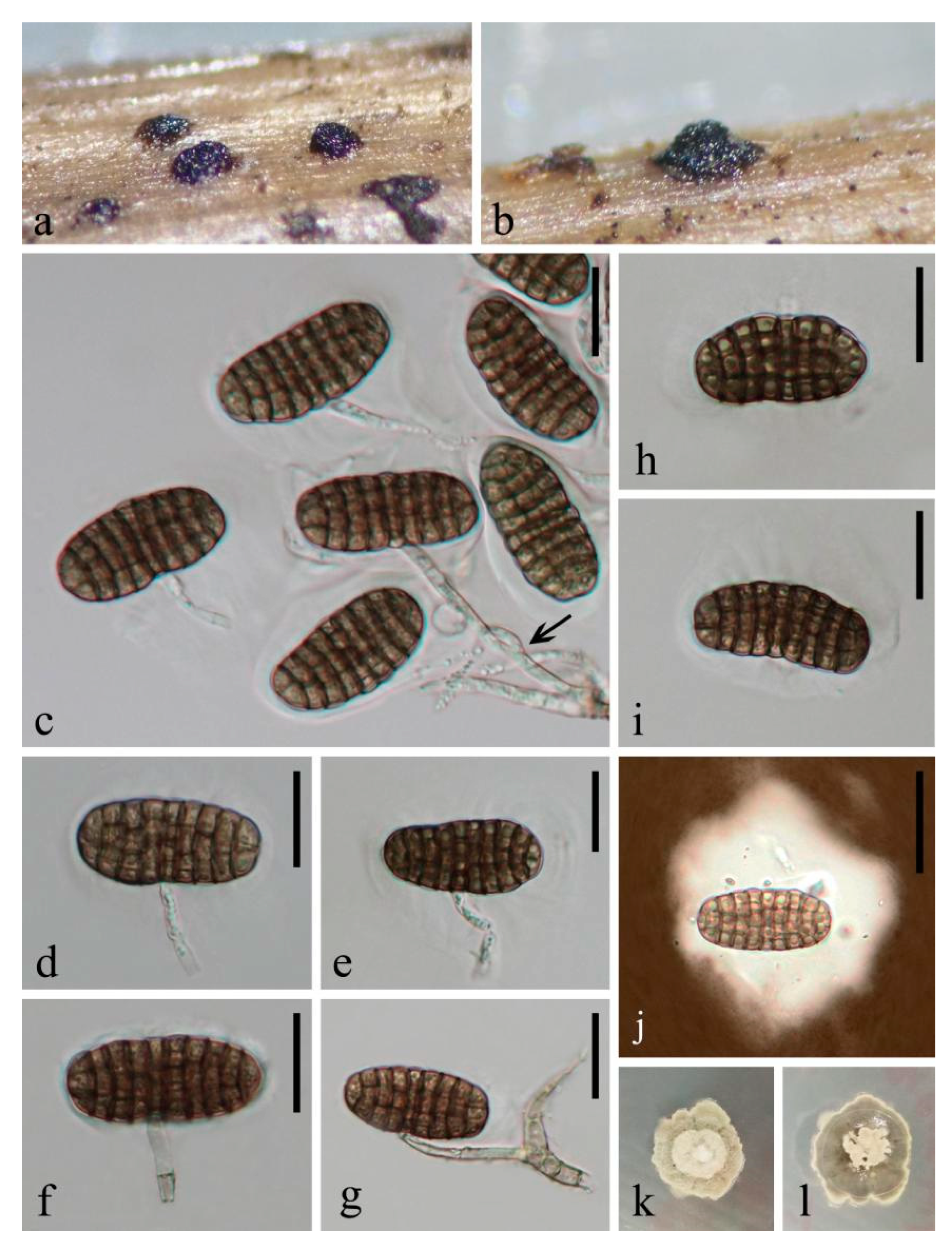
3.2.1. Novel Taxa in Pleurotheciaceae

In this section, one new genus and two new species are introduced in Pleurotheciaceae. These taxa are described alphabetically below.
Sordariomycetes O.E. Erikss. and Winka, Myconet 1(1): 10 (1997)
Savoryellomycetidae Hongsanan, K.D. Hyde and Maharachch., Fungal Diversity 84: 35 (2017)
Pleurotheciales Réblová and Seifert, in Réblová, Seifert, Fournier and Štěpánek, Persoonia 37: 63 (2016)
Pleurotheciaceae Réblová and Seifert, in Réblová, Seifert, Fournier and Štěpánek, Persoonia 37: 63 (2016)
Coleodictyospora Charles ex Matsush., Matsushima Mycological Memoirs 5: 8 (1987)
Type species: Coleodictyospora cubensis Charles ex Matsush.
Notes: Coleodictyospora was introduced by Charles [30] with a single species C. cubensis, but it lacked a Latin diagnosis. Matsushima [31] validated this genus and characterized it as having cylindrical, simple, septate and hyaline conidiophores, monoblastic conidiogenous cells, and transversely oblong or inverse reniform, muriform conidia surrounded by a semi-gelatinous, hyaline sheath. Berkleasmium micronesicum was then transferred to Coleodictyospora as C. micronesiaca based on its very similar morphological traits with C. cubensis, but it differs in having smaller conidia (30–42 × 15–18 μm vs. 42–50 × 20–22 μm) and reduced conidiophores [31].
Coleodictyospora cubensis was initially collected from North America [30] and subsequently recorded in Brunei [32] and Japan [33]. Nakagiri and Ito [33] named their specimen IFO 32,660 as C. cubensis based on the dimensions of conidiophores, conidia and conidial sheaths, as well as the number of conidial septa and the conidiophore attaching point, although it had overlapping conidial size with C. micronesiaca. Nakagiri and Ito [33] emphasized that IFO 32,660 might be a novel species considering the thinner conidia (28–48 × 13–19 μm vs. 42–50 × 20–22 μm) and less number of septa (7–11 vs. 8–14) than the type specimen of C. cubensis. However, this hypothesis could not be tested without the re-examination of the specimen IFO 32,660 or the molecular data from similar specimens in the same locality (Ishigaki Island, Japan).
Coleodictyospora micronesiaca is likely to be a cosmopolitan species as it was recorded in several countries worldwide, including China (Hong Kong, Taiwan) [31,34], Cuba [35], USA (Florida) [36], Mauritius [37], Mexico [38], Micronésia [39], Peru [40], Philippines [37] and Thailand [41]. However, these records were diagnosed solely based on the morphology and lack of support from molecular data, and the descriptions were omitted or briefly noted.
In this study, we isolated a Coleodictyospora species from decaying wood submerged in freshwater and provide sequence data for it. Since C. cubensis and C. micronesiaca lack sequence data in GenBank, we identified our new collection as a novel species in Coleodictyospora based on the comparison of their morphology.
Coleodictyospora muriformis W. Dong, Doilom and K.D. Hyde sp. nov. (Figure 3 and Figure 4a,b)
Index Fungorum number: IF558195; Facesoffungi number: FoF 09872
Etymology: in reference to the muriform conidia of the fungus
Holotype: MFLU 18-1544
Saprobic on decaying wood submerged in freshwater. Sexual morph: undetermined. Asexual morph: hyphomycetous. Colonies on natural substrate, effuse, gregarious, punctiform, sporodochial, raised, black. Mycelium partly immersed in natural substrate, consisting of branched, septate, thin-walled, smooth, pale brown to brown hyphae. Conidiophores up to 55 μm long, 3 μm wide, micronematous, mononematous, ascending from the basal mat of sporodochia, subcylindrical, branched, septate, hyaline to pale brown, smooth, thin-walled. Conidiogenous cells 7.5–20 × 2.5–3.5 μm ( x ¯ = 13.5 × 3 μm, n = 10), holoblastic, monoblastic, integrated, determinate, terminal, cylindrical, hyaline, smooth, thin-walled. Conidia 32–44 × 15.5–19 μm ( x ¯ = 38.5 × 17 μm, n = 100), solitary, acrogenous, generally produced in the middle position, occasionally laterally on conidiophores, and perpendicular to the conidiophores, mostly cylindro-ellipsoidal, sometimes reniform, muriform, dictyoseptate, with (7–)8–9 transverse and (2–)3 longitudinal septa, deeply constricted and with dark brown bands at the transverse septa, slightly constricted and brown to dark brown at the longitudinal septa, often distinctly constricted at the middle where conidiophore attaches to form reniform, brown, smooth, thin-walled, with a hyaline, semi-gelatinous sheath. Sheaths well-defined, ellipsoidal, thin at the beginning, 2 μm thick; becoming irregular-shaped, uneven, larger after being mounted in the water, up to 55 μm thick in Indian Ink.
Culture characteristics: on PDA, colony irregular, reaching 15 mm diam. in 25 days at room temperature (25–30 °C), surface rough, with dense mycelia, velvety, dry, umbonate in the middle from the side view, edge undulate; from above, dark gray at the margin, pale gray at the middle; from below, dark brown to black at the margin, pale gray at the middle; not producing pigmentation in culture.
Material examined: THAILAND, Satun Province, Khuan Kalong District, Thung Nui Sub-District (6°55′19″ N 100°08′17″ E), on decaying wood submerged in Chang stream originated from Panan Waterfall, 10 May 2018, W. Dong, hat284 (MFLU 18-1544, holotype), ex-type living culture MFLUCC 18-1243 = MFLUCC 18-1279; ibid., HKAS 105018, isotype, ex-isotype living culture KUMCC 19-0034 = KUMCC 19-0052.
Habitat and distribution: stream is located in tropical rainforest in Southern Thailand with hot and humid climate conditions, shallow and clear, flowing slowly from the Panan Waterfall, surrounded by angiosperms.
Notes: Coleodictyospora muriformis belongs in Coleodictyospora based on the punctiform, sporodochial colonies on the natural substrate, monoblastic conidiogenous cells, and cylindro-ellipsoidal, muriform conidia produced perpendicularly to the conidiophores and with a hyaline, semi-gelatinous sheath [30,31]. Coleodictyospora muriformis is easily distinguished from the type species C. cubensis by its shorter conidiophores (up to 55 μm long vs. 70–85 μm long), smaller conidia (32–44 × 15.5–19 μm vs. 42–50 × 20–22 μm) and fewer conidial transverse septa ((7–)8–9 vs. 8–14). The transverse septa of the conidia of C. muriformis are filled with dark brown bands, while they were neither described nor illustrated in C. cubensis [30,31,33,42]. Coleodictyospora muriformis has overlapping conidial dimensions with C. micronesiaca, but it differs in having long, branched conidiophores (up to 55 μm long) and longer conidiogenous cells (7.5–20 × 2.5–3.5 μm), and the conidiophores attach to the middle of the conidia. In contrast, C. micronesiaca lacks conidiophores and the conidiogenous cells are shorter (2–8 × 3–4 μm), which directly ascend from the basal mat of sporodochia; the conidiogenous cells often attach to the end of the conidia. In addition, the conidiogenous cells in C. micronesiaca are mostly short subulate, while they are long cylindrical in C. muriformis. We therefore introduce C. muriformis as new to the genus. A morphological comparison of Coleodictyospora species is summarized in Table 2 and a combined figure plate of three species is illustrated in Figure 4.
Saprodesmium W. Dong and Doilom gen. nov.
Index Fungorum number: IF558196; Facesoffungi number: FoF 09873
Etymology: “saprus” = saprobic, referring to the saprobic lifestyle of the fungus; “desmόs” = bond, link, referring to the aggregated conidia in sporodochia
Saprobic on decaying wood submerged in freshwater. Sexual morph: undetermined. Asexual morph: hyphomycetous. Colonies on natural substrate, effuse, gregarious, punctiform, sporodochial, raised, black. Mycelium partly immersed in natural substrate, consisting of branched, septate, thin-walled, smooth, pale brown to brown hyphae. Conidiophores micronematous, mononematous, unbranched, vesiculate, septate. Conidiogenous cells holoblastic, monoblastic, integrated, determinate. Conidia solitary, obovoid to ellipsoidal, clearly muriform, olivaceous when young, becoming quite blackish with age and obscuring the septa, with several subhyaline basal cells, smooth, thin-walled. Conidial secession schizolytic.
Type species: Saprodesmium dematiosporum W. Dong, Doilom and K.D. Hyde
Notes: The BlastN search of NCBI’s GenBank using the LSU sequence shows Saprodesmium dematiosporum has the closest hits with several genera in Pleurotheciaceae, i.e., Rhexoacrodictys erecta (KUMCC 20-0194, similarity = 96.68%), Neomonodictys muriformis (MFLUCC 16-1136, similarity = 94.11%) and Pleurothecium obovoideum (CBS 209.95, similarity = 93.73%). The closest hits using SSU sequence are Rhexoacrodictys erecta (KUMCC 20-0194, similarity = 99.54%), Dematipyriforma aquilaria (3-11-1, similarity = 99.49%) and Pleurothecium aquaticum (B-27, similarity = 99.06%). Based on ITS BlastN search, the closest relatives are however Phaeoisaria sp. (BAB-4787, similarity = 97.11%) and Pleurothecium recurvatum (CBS 138686, similarity = 96.11%). Saprodesmium dematiosporum clusters as an independent branch between Dematipyriforma and Rhexoacrodictys with high bootstrap support in concatenated LSU-SSU-ITS-rpb2 phylogeny (100% ML BS/1.00 BI PP, Figure 1) and individual ITS phylogeny (100% ML BS/1.00 BI PP, Supplementary Figure S2). Saprodesmium dematiosporum clusters with Rhexoacrodictys species in individual LSU phylogeny, but no bootstrap support (Supplementary Figure S1).
Dematipyriforma is an endophytic genus comprising a single species D. aquilaria [43]. Dematipyriforma shares similar morphological characteristics with Saprodesmium in having micronematous conidiophores, holoblastic conidiogenous cells and septate conidia. However, they are entirely different genera in the following aspects. The conidiophores of Dematipyriforma are hypha-like [43], while they are vesiculate in Saprodesmium which are also unique in the family Pleurotheciaceae. The conidia of Dematipyriforma are elongate pyriform, 4–5 transverse septate, sometimes 1–2 longitudinal septate, pale grey olivaceous to pale brown, and has rhexolytic conidial secession [43]. In contrast, the conidia of Saprodesmium are obovoid to ellipsoidal, irregularly muriform and olivaceous when young, becoming quite blackish with age and obscuring the septa, with several subhyaline basal cells, and has schizolytic conidial secession. In addition, Saprodesmium species is saprobe, while Dematipyriforma species is endophyte [43].
Rhexoacrodictys, typified by R. erecta, was introduced for several hyphomycetes characterized by macronematous, long cylindrical conidiophores with percurrent proliferating, monoblastic, integrated, terminal conidiogenous cells, and obovoid, oval or subspherical, muriform, brown to dark brown conidia often with a paler basal cell bearing a small marginal frill derived from the upper portion of the conidiophores and with rhexolytic conidial secession [44]. Saprodesmium shares some morphological traits with Rhexoacrodictys especially with regards to its muriform and obovoid conidia. Saprodesmium, however, has olivaceous conidia and when mature it has quite a blackish pigmentation obscuring the conidial septa. Rhexoacrodictys is featured by rhexolytic conidial secession with conidia that have a conspicuous paler basal cell bearing a small marginal frill, while the conidia of Saprodesmium secede schizolytically and it instead has several subhyaline, depressed subglobose cells at the base. Rhexoacrodictys has macronematous, long cylindrical conidiophores with percurrent proliferating [44,45], whereas Saprodesmium has micronematous, short, vesiculate, determinate conidiophores.
Based on the multi-locus and individual phylogenetic analyses, as well as the morphological comparison with the similar taxa in the family, we introduce Saprodesmium as a novel genus in Pleurotheciaceae.
Saprodesmium dematiosporum W. Dong, Doilom and K.D. Hyde sp. nov. (Figure 5)
Index Fungorum number: IF558197; Facesoffungi number: FoF 09874
Etymology: in reference to the dematiaceous conidia
Holotype: HKAS 101710
Saprobic on decaying wood submerged in freshwater. Sexual morph: undetermined. Asexual morph: hyphomycetous. Colonies on natural substrate, effuse, gregarious, punctiform, sporodochial, raised, black. Mycelium partly immersed in natural substrate, consisting of branched, septate, thin-walled, smooth, pale brown to brown hyphae. Conidiophores micronematous, mononematous, vesiculate, consisted of 1–4 subglobose, smooth, hyaline cells (each cell 8.5–12 μm diam.), unbranched, septate, constricted at the septa, smooth, thin-walled. Conidiogenous cells 8–11 μm diam. ( x ¯ = 9.7 μm, n = 10), holoblastic, monoblastic, integrated, determinate, terminal, subglobose, hyaline, smooth, thin-walled. Conidia 21–36 × 14.5–27 μm ( x ¯ = 27.5 × 21.5 μm, n = 70), solitary, acrogenous, obovoid to ellipsoidal, subglobose, clearly muriform, olivaceous when young, becoming quite blackish with age and obscuring the septa, with several subhyaline to pale brown basal cells, smooth, thin-walled. Conidial secession schizolytic.
Culture characteristics: on PDA, colony circular, reaching 50 mm diam. in 30 days at room temperature (25–30 °C), surface rough, with dense mycelia, dry, raised from the side view, edge entire; from above, dark gray at the margin, pale gray to white at the middle; from below, black at the margin, dark olivaceous at the middle; not producing pigmentation in culture.
Material examined: CHINA, Yunnan Province, Pingbian District (22°59′13″ N 103°40′30″ E), on decaying wood submerged in an unnamed stream originated from Dawei Mountain Nature Reserve, 20 September 2017, W. Dong, WF23A (HKAS 101710, holotype), ex-type living culture KUMCC 18-0059; ibid., MFLU 18-1165, isotype.
Habitat and distribution: stream is nearby Nature Reserve in Southern Yunnan of Yunnan-Kweichow Plateau, shallow and clear, flowing rapidly from the Dawei Mountain, surrounded by angiosperms.

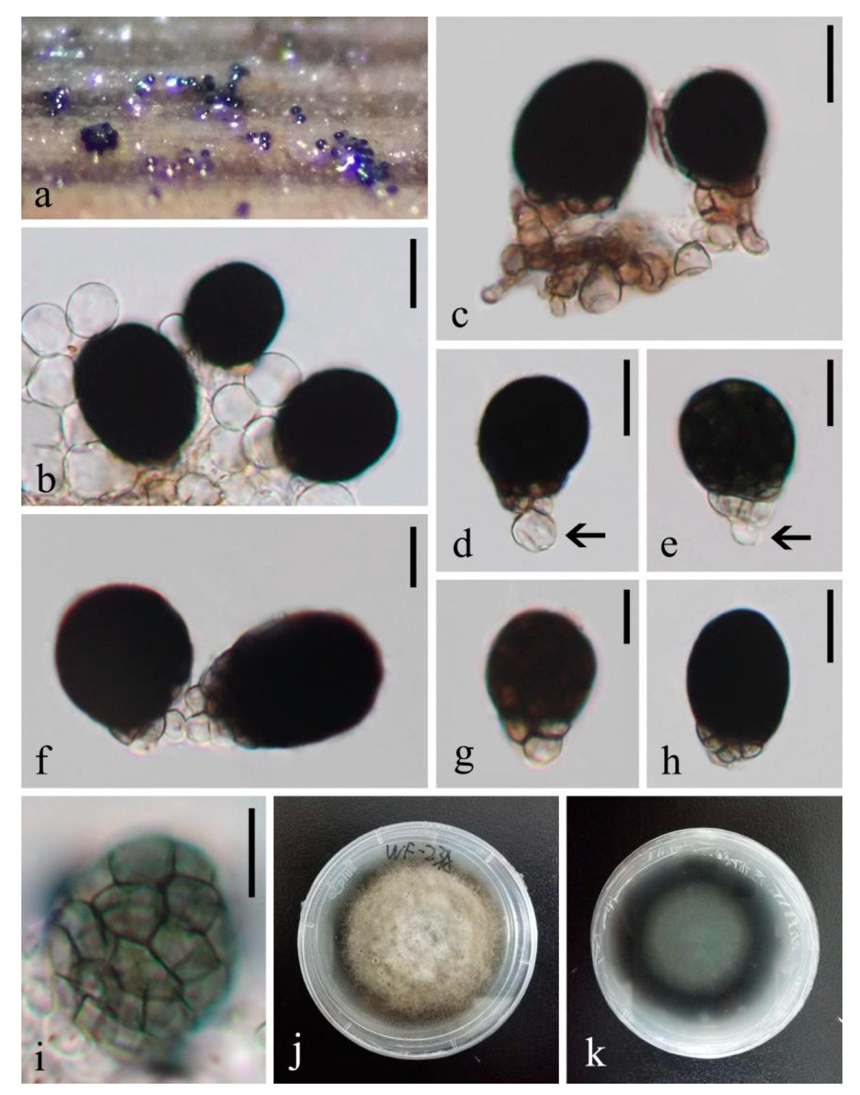
3.2.2. Novel Taxa in Savoryellomycetidae

In this section, one new genus with one new species are introduced and phylogenetically referred to Savoryellomycetidae genera incertae sedis.
Obliquifusoideum W. Dong, Doilom and K.D. Hyde gen. nov.
Index Fungorum number: IF558198; Facesoffungi number: FoF 09875
Etymology: in reference to its neck growing oblique to the host substrate and fusoid ascospores
Saprobic on decaying wood submerged in freshwater. Sexual morph: Ascomata superficial, ellipsoidal, black, coriaceous, ostiolate, with a lateral neck. Necks hyaline to dark, subcylindrical, oblique or horizontal to the host substrate. Peridium thin, soft, comprising several layers of brown, thin-walled cells of textura angularis. Paraphyses tapering towards the apex, dense, hypha-like, septate, unbranched, hyaline. Asci 8-spored, unitunicate, cylindrical, short pedicellate, with a small, refractive, barrel- or jar-shaped, apical ring, persistent. Ascospores uniseriate, fusoid, septate, hyaline, thin-walled. Asexual morph: undetermined.
Type species: Obliquifusoideum guttulatum W. Dong, Doilom and K.D. Hyde
Notes: The BlastN search of NCBIs GenBank using LSU sequence shows Obliquifusoideum guttulatum has the closest hits with several genera in Pleurotheciaceae, but with low percentage similarity, i.e., Melanotrigonum ovale (CBS 138743, similarity = 92.06%), Pleurotheciella saprophytica (MFLUCC 16-1251, similarity = 92.03%), Phaeoisaria annesophieae (MFLU 19-0531, similarity = 91.86%) and Sterigmatobotrys rudis (DAOM 229838, similarity = 91.83%). The closest hits using SSU sequence are several genera in Pleurotheciaceae, i.e., Dematipyriforma aquilaria (3-11-1, similarity = 98.13%), Phaeoisaria clematidis (MFLUCC 18-1017, similarity = 97.99%) and P. fasciculata (DAOM 230055, similarity = 97.92%). The closest hits using ITS sequence are however several genera in Conioscyphales and Pleurotheciales, i.e., Pleurothecium recurvatum (DAOM 230069, similarity = 98.75%), Conioscypha varia (CBS 604.70, similarity = 94.89%) and Neomonodictys muriformis (MFLUCC 16-1136, similarity = 93.19%).
The placement of Obliquifusoideum guttulatum is different in multi-locus and individual LSU and ITS phylogenetic trees and lacks significant support in all trees. Obliquifusoideum guttulatum is revealed as a sister taxon of Pleurotheciales in the multi-locus analysis of concatenated LSU-SSU-ITS-rpb2 matrix (Figure 1), while it clusters with Savoryellales in the individual LSU phylogeny (Supplementary Figure S1); Savoryellales and Pleurothecium species in the individual ITS phylogeny (Supplementary Figure S2). Obliquifusoideum is similar to Helicoascotaiwania in Pleurotheciales, and Ascotaiwania, Neoascotaiwania and Savoryella in Savoryellales. They generally have dark ascomata with a lateral neck, which is oblique or horizontal to the host substrate, and septate ascospores. However, the ascospores of the four genera are mostly ellipsoidal and versicolorous with dark middle cells and hyaline polar cells [2,3,4,46,47]. In contrast, Obliquifusoideum has fusoid and evenly hyaline ascospores. The morphological differences and the independent lineage in the multi-locus and individual phylogenetic trees therefore support Obliquifusoideum as a new genus.
Although the relationships of Obliquifusoideum were weak with four orders in Savoryellomycetidae (Figure 1), it was shown to be a genus in Savoryellomycetidae with strong bootstrap support (Figure 2). We consider it is wise to refer Obliquifusoideum to Savoryellomycetidae genera incertae sedis for now, until its phylogeny is better resolved with additional taxon sampling followed by divergence time estimates studies.
Obliquifusoideum guttulatum W. Dong, Doilom and K.D. Hyde sp. nov. (Figure 6)
Index Fungorum number: IF558199; Facesoffungi number: FoF 09876
Etymology: in reference to the guttulate ascospores of the fungus
Holotype: MFLU 18-1575
Saprobic on decaying wood submerged in freshwater. Sexual morph: Ascomata 100–120 μm high, 155–170 μm diam., scattered, superficial, ellipsoidal, black, coriaceous, ostiolate, with a lateral neck, ejecting asci and ascospores soon during incubation and becoming empty. Necks 160–180 μm long, 17–30 μm wide, hyaline to black, subcylindrical, oblique or horizontal to the host substrate. Peridium thin, 8–20 μm thick, soft, comprising several layers of pale brown, thin-walled cells of textura angularis, dark brown outwards. Paraphyses 3.5–5 μm wide, tapering towards the apex, dense, hypha-like, septate, unbranched, hyaline, embedded in a gelatinous matrix. Asci 97–110 × 7.3–7.7 μm ( x ¯ = 105 × 7.5 μm, n = 10), 8-spored, unitunicate, cylindrical, slightly narrower and truncate at the apex, short pedicellate, with a small, distinct, refractive, barrel- or jar-shaped, apical ring, 2 × 2.7 μm, persistent. Ascospores 14–17.5 × 4.3–5 μm ( x ¯ = 15.5 × 4.6 μm, n = 10), overlapping uniseriate, fusoid, straight or slightly curved, one median septate, with two additional obscure septate at two sides, guttulate, hyaline, thin and smooth-walled, without a gelatinous sheath. Asexual morph: undetermined.
Culture characteristics: on PDA, colony circular, reaching 8 mm diam. in 48 days at room temperature (25–30 °C), surface rough, with dense mycelia, dry, rigid, umbonate from the side view, edge entire; from above, creamy at the margin, dark grey to dark brown at the middle, brown at the center; dark brown from below; not producing pigmentation in culture.
Material examined: THAILAND, Songkhla Province, Rattaphum District, Khao Phra Sub-District (7°00′03″ N 100°08′33″ E), on decaying wood submerged in a stream originated from Borriphat Waterfall, 10 May 2018, W. Dong, hat138 (MFLU 18-1575, holotype), ex-type living culture MFLUCC 18-1233; ibid., HKAS 105007, isotype, ex-isotype living culture KUMCC 19-0023.
Habitat and distribution: stream is located in tropical rainforest in Southern Thailand with hot and humid climate conditions, shallow and clear, flowing slowly from the Borriphat Waterfall, surrounded by angiosperms.

4. Discussion

Doilom et al. [29] established a novel genus Pseudocoleodictyospora to accommodate three hyphomycetous species collected from the bark of living Tectona grandis (teak) and distinguished them from Coleodictyospora by the absence of a hyaline sheath. This establishment, however, lacks the support from the DNA sequence data of Coleodictyospora. The presence of conidial sheath as a criterion for delimiting two genera is interesting as this is often used for species delimitation in classification, such as species in Astrosphaeriella, Dictyosporium, Kirschsteiniothelia and Natipusilla [48]. In this study, we collected a freshwater hyphomycetous species which has very similar morphs to Pseudocoleodictyospora, but is characterized by a hyaline sheath. This peculiar phenotype further confirms it as a novel species in Coleodictyospora, namely C. muriformis (see notes of C. muriformis). On the basis of DNA-based phylogeny, Coleodictyospora muriformis is phylogenetically distant from Pseudocoleodictyospora (Pseudocoleodictyosporaceae, Pleosporales) and clusters as a member of the Pleurotheciales (Figure 2). This study further confirms the Doilom et al. [29] taxonomic assumption of establishing a novel genus based on the presence of conidial sheath with the support from the DNA sequence data. Amazingly, Coleodictyospora and Pseudocoleodictyospora are positioned in two different classes Sordariomycetes and Dothideomycetes, respectively (Figure 2), though they are quite similar except in terms of the conidial sheath. Nevertheless, it is not advisable to use conidial sheath as a criterion segregating species at a higher taxonomic level as it is often an unstable characteristic, especially among freshwater species such as Caryospora submersa and Pseudoastrosphaeriella bambusae [48].
In our multi-locus phylogenetic tree (Figure 1), Coleodictyospora is affiliated to Neomonodictys muriformis and Pleurothecium obovoideum. Coleodictyospora is similar to Neomonodictys in having muriform conidia, but they are entirely different genera. The conidia of Neomonodictys are subglobose to globose, comprising several subglobose cells, which are irregularly arranged in the conidia, pale brown when immature, producing black pigmentation and obscuring the conidial septa, with a protruding basal cell which attaches to the conidiophore [8]. In contrast, Coleodictyospora has cylindro-ellipsoidal conidia, with (7–)8–9 transverse and (2–)3 longitudinal septa, deeply constricted and with dark brown bands at the transverse septa, generally produced in the middle position and are perpendicular on the conidiophore. Pleurothecium obovoideum was proposed based on a known species, Ramichloridium obovoideum [49]. In the phylogenetic tree of Arzanlou et al. [49], they showed that the strain CBS 209.95 of R. obovoideum clustered with the sexual morph of Pleurothecium recurvatum (type species) and its morphological characteristics fit well with Pleurothecium and R. obovoideum was therefore transferred to Pleurothecium, namely P. obovoideum. However, with more species in Pleurothecium, P. obovoideum was reported to be distant from P. recurvatum and clustered with Neomonodictys in a well-supported clade [6,8]. The re-assessment of P. obovoideum is pending, however, its pleurothecium-like morphological characteristics [49] warrant it cannot be congeneric with Coleodictyospora.
Interestingly, we found that the conidia of Coleodictyospora are quite similar to the ascospores of a sexual species Boerlagiomyces websteri. Boerlagiomyces were recognized in Tubeufiaceae [11,50,51] and confirmed with DNA sequence data derived from a reference specimen of Boerlagiomyces macrospora [29]. However, B. websteri represented by a putative strain BCC 3834 clustered with several apothecial taxa in Pezizomycotina [52], and Boonmee et al. [50] had some doubts on this species because of its perithecial characteristic. Therefore, the accurate phylogenetic position of B. websteri is still questionable. Although the soft, membranous, setose ascomata and large dictyosporous ascospores of B. websteri fit with the features of Boerlagiomyces [50], the two-spored asci are unusual compared with the eight-spored asci of the type species B. velutinus [50]. Whether B. websteri has close phylogenetic relationships with Coleodictyospora in Savoryellomycetidae is pending and has to be resolved.
On the basis of morphology, we initially considered Obliquifusoideum as a member of Annulatascales due to its black ascomata with a lateral neck which is oblique or horizontal to the host substrate, hypha-like paraphyses with tapering apex, cylindrical asci with a distinct, refractive apical ring, and fusoid, hyaline ascospores. It is of interest, however, that Obliquifusoideum clusters in Savoryellomycetidae with relationships to Pleurotheciales and Savoryellales, which is distant from Annulatascales and annulatascales-like taxa in Diaporthomycetidae (Figure 2). It is not unexpected that Obliquifusoideum with annulatascales-like morphology can be discovered in another subclass Savoryellomycetidae as Annulatascales is commonly recognized to be polyphyletic and the species are often encountered from freshwater habitats [53,54]. It is reasonable that Obliquifusoideum is placed in Savoryellomycetidae due to its dark ascomata with an oblique or horizontal neck, and phragmoseptate conidia which are the sexual features of the other two members Pleurotheciales and Savoryellales.
DNA-based phylogeny has helped to provide better insights into the taxonomy of Pleurotheciales and a recommendation of species boundaries was established [55], leading to recent classification updates. Rhexoacrodictys, which was treated as a genus in Savoryellales by Xia et al. [56], was accepted in Pleurotheciales in a later phylogenetic study [6]. Our phylogenetic results corroborate those of Luo et al. [6], and our new genus Saprodesmium forms a well-supported lineage basal to Rhexoacrodictys in Pleurotheciales (Figure 1). The BlastN search of Saprodesmium using SSU sequence in NCBIs GenBank reveals a high similarity (99.49%) with a hyphomycetous species Dematipyriforma aquilaria. Dematipyriforma, typified by D. aquilaria, was isolated as an endophyte from the trunk of Aquilaria crassna, producing dark muriform conidia [43]. Dematipyriforma was placed in Savoryellales in the phylogenetic tree of Sun et al. [43], however, they did not include other related orders (Conioscyphales, Fuscosporellales and Pleurotheciales) in Savoryellomycetidae. Our multi-locus phylogeny places Dematipyriforma as a sister genus to Rhexoacrodictys and Saprodesmium with good bootstrap support in Pleurotheciales (99% ML BS/1.00 BI PP, Figure 1). On the other hand, the muriform conidia of Dematipyriforma are similar to Neomonodictys in Pleurotheciales. According to this morphological trait and phylogenetic result, we accept Dematipyriforma in Pleurotheciales. Besides saprobes and few opportunistic human pathogens, Dematipyriforma is the only presently known endophytic genus in the order, which increases our understanding of different life modes of Pleurotheciales.
Freshwater fungi are a unique group of organisms with a special ability to survive and grow on submerged wood in water by producing soft rot cavities [57,58]. There is very little overlap between the fungi growing on wood submerged in freshwater and those on adjacent stream sides [59,60]. Thus, we are continually finding novel taxa from this unique habitat and since streams are often disparate elements, we are likely to discover many more which will improve our understanding of fungal classification [61]. Freshwater appears to be an ecologically important niche for species in Pleurotheciales [62]. Almost all Pleurotheciella species were collected from freshwater [3,4,6,9,10,13], as well as some species from Helicoascotaiwania, Phaeoisaria, Pleurothecium and Sterigmatobotrys [6,10,12,15,63].

Supplementary Materials

The following are available online at https://www.mdpi.com/article/10.3390/jof7090711/s1, Figure S1: Phylogenetic tree generated from maximum likelihood analysis (RAxML) based on LSU sequence data, Figure S2: Phylogenetic tree generated from maximum likelihood analysis (RAxML) based on ITS sequence data.

Author Contributions

Conceptualization, W.D. and M.D.; resources, N.S., M.D. and Z.D.; writing—original draft preparation, W.D.; writing—review and editing, W.D., R.J., K.D.H., E.-F.Y., H.Z., X.Y., G.W., N.S., M.D. and Z.D.; supervision, N.S., M.D. and Z.D.; funding acquisition, Z.D. and N.S. All authors have read and agreed to the published version of the manuscript.

Funding

This research was funded by the Key Realm R&D Program of Guangdong Province, grant no: 2018B020205003. This work was partly supported by Chiang Mai University.

Data Availability Statement

The data generated from this study can be found in the Index Fungorum (http://www.indexfungorum.org/names/names.asp, accessed on 1 August 2021) and GenBank (https://www.ncbi.nlm.nih.gov/nuccore, accessed on 1 August 2021).

Acknowledgments

R.J. thanks the University of Mauritius for support. K.D.H thanks the Thailand Research grant entitled Impact of climate change on fungal diversity and biogeography in the Greater Mekong Subregion (grant no: RDG6130001). H.Z. thanks the National Natural Science Foundation of China (Project ID: NSF 31500017). W.D. thanks Shaun Pennycook (Landcare Research, New Zealand) for assistance in new epithets. Mahboob Mahboobullah is thanked for his preliminary analyses and suggestions on the manuscript.

Conflicts of Interest

The authors declare no conflict of interest.

References

  1. Hongsanan, S.; Maharachchikumbura, S.; Hyde, K.D.; Samarakoon, M.C.; Jeewon, R.; Zhao, Q.; Al-Sadi, A.; Bahkali, A.H. An updated phylogeny of Sordariomycetes based on phylogenetic and molecular clock evidence. Fungal Divers. 2017, 84, 25–41. [Google Scholar] [CrossRef]
  2. Dayarathne, M.C.; Maharachchikumbura, S.; Jones, E.B.G.; Dong, W.; Devadatha, B.; Yang, J.; Ekanayaka, A.H.; De Silva, W.; Sarma, V.V.; Al-Sadi, A.; et al. Phylogenetic Revision of Savoryellaceae and Evidence for Its Ranking as a Subclass. Front. Microbiol. 2019, 10, 840. [Google Scholar] [CrossRef] [PubMed] [Green Version]
  3. Réblová, M.; Restrepo, M.H.; Fournier, J.; Nekvindová, J. New insights into the systematics of Bactrodesmium and its allies and introducing new genera, species and morphological patterns in the Pleurotheciales and Savoryellales (Sordariomycetes). Stud. Mycol. 2020, 95, 415–466. [Google Scholar] [CrossRef]
  4. Réblová, M.; Seifert, K.; Fournier, J.; Štěpánek, V. Newly recognised lineages of perithecial ascomycetes: The new orders Conioscyphales and Pleurotheciales. Pers. Mol. Phylogeny Evol. Fungi 2016, 37, 57–81. [Google Scholar] [CrossRef] [Green Version]
  5. Guarro, J.; Vieira, L.A.; De Freitas, D.; Gené, J.; Zaror, L.; Hofling-Lima, A.L.; Fischman, O.; Zorat-Yu, C.; Figueras, M.J. Phaeoisaria clematidis as a Cause of Keratomycosis. J. Clin. Microbiol. 2000, 38, 2434–2437. [Google Scholar] [CrossRef] [PubMed]
  6. Luo, Z.-L.; Hyde, K.D.; Liu, J.-K.; Maharachchikumbura, S.; Jeewon, R.; Bao, D.-F.; Bhat, D.J.; Lin, C.-G.; Li, W.-L.; Yang, J.; et al. Freshwater Sordariomycetes. Fungal Divers. 2019, 99, 451–660. [Google Scholar] [CrossRef] [Green Version]
  7. Hernández-Restrepo, M.; Gené, J.; Castañeda-Ruiz, R.; Mena-Portales, J.; Crous, P.; Guarro, J. Phylogeny of saprobic microfungi from Southern Europe. Stud. Mycol. 2017, 86, 53–97. [Google Scholar] [CrossRef]
  8. Hyde, K.D.; Dong, Y.; Phookamsak, R.; Jeewon, R.; Bhat, D.J.; Jones, E.B.G.; Liu, N.-G.; Abeywickrama, P.D.; Mapook, A.; Wei, D.; et al. Fungal diversity notes 1151–1276: Taxonomic and phylogenetic contributions on genera and species of fungal taxa. Fungal Divers. 2020, 100, 5–277. [Google Scholar] [CrossRef] [Green Version]
  9. Réblová, M.; Seifert, K.A.; Fournier, J.; Štěpánek, V. Phylogenetic classification of Pleurothecium and Pleurotheciella gen. nov. and its dactylaria-like anamorph (Sordariomycetes) based on nuclear ribosomal and protein-coding genes. Mycologia 2012, 104, 1299–1314. [Google Scholar] [CrossRef]
  10. Luo, Z.-L.; Hyde, K.D.; Bhat, D.J.; Jeewon, R.; Maharachchikumbura, S.S.N.; Bao, D.-F.; Li, W.-L.; Su, X.-J.; Yang, X.-Y.; Su, H.-Y. Morphological and molecular taxonomy of novel species Pleurotheciaceae from freshwater habitats in Yunnan, China. Mycol. Prog. 2018, 17, 511–530. [Google Scholar] [CrossRef]
  11. Wijayawardene, N.N.; Hyde, K.D.; Al-ani, L.K.T.; Tedersoo, L.; Haelewaters, D.; Rajeshkumar, K.C.; Zhao, R.L.; Aptroot, A.; Leontyev, D.V.; Saxena, R.K.; et al. Outline of Fungi and fungus-like taxa. Mycosphere 2020, 11, 1–367. [Google Scholar] [CrossRef]
  12. Hyde, K.D.; Norphanphoun, C.; Abreu, V.P.; Bazzicalupo, A.; Chethana, K.W.T.; Clericuzio, M.; Dayarathne, M.C.; Dissanayake, A.J.; Ekanayaka, A.H.; He, M.-Q.; et al. Fungal diversity notes 603–708: Taxonomic and phylogenetic notes on genera and species. Fungal Divers. 2017, 87, 1–235. [Google Scholar] [CrossRef]
  13. Hyde, K.D.; Chaiwan, N.; Norphanphoun, C.; Boonmee, S.; Camporesi, E.; Chethana, K.W.T.; Dayarathne, M.C.; de Silva, N.I.; Dissanayake, A.J.; Ekanayaka, A.H.; et al. Mycosphere notes 169–224. Mycosphere 2018, 9, 271–430. [Google Scholar] [CrossRef]
  14. Su, H.-Y.; Udayanga, D.; Luo, Z.-L.; Manamgoda, D.; Zhao, Y.-C.; Yang, J.; Liu, X.-Y.; McKenzie, E.; Zhou, D.-Q.; Hyde, K. Hyphomycetes from aquatic habitats in Southern China: Species of Curvularia (Pleosporaceae) and Phragmocephala (Melannomataceae). Phytotaxa 2015, 226, 201–216. [Google Scholar] [CrossRef]
  15. Fallah, P.M.; Crane, J.L.; A Shearer, C. Freshwater ascomycetes: Two new species of Ascotaiwania from North America. Can. J. Bot. 1999, 77, 87–92. [Google Scholar]
  16. Fernández, F.A.; Lutzoni, F.M.; Huhndorf, S.M. Teleomorph-anamorph connections: The new pyrenomycetous genus Carpoligna and its Pleurothecium anamorph. Mycologia 1999, 91, 251–262. [Google Scholar] [CrossRef]
  17. Chomnunti, P.; Hongsanan, S.; Aguirre-Hudson, B.; Tian, Q.; Persoh, D.; Dhami, M.K.; Alias, A.S.; Xu, J.; Liu, X.; Stadler, M.; et al. The sooty moulds. Fungal Divers. 2014, 66, 1–36. [Google Scholar] [CrossRef]
  18. Senanayake, I.C.; Rathnayaka, A.R.; Marasinghe, D.S.; Calabon, M.S.; Gentekaki, E.; Lee, H.B.; Hurdeal, V.G.; Pem, D.; Dissanayake, L.S.; Wijesinghe, S.N.; et al. Morphological approaches in studying fungi: Collection, examination, isolation, sporulation and preservation. Mycosphere 2020, 11, 2678–2754. [Google Scholar] [CrossRef]
  19. Jayasiri, S.C.; Hyde, K.D.; Ariyawansa, H.; Bhat, J.; Buyck, B.; Cai, L.; Dai, Y.-C.; Abd-Elsalam, K.A.; Ertz, D.; Hidayat, I.; et al. The Faces of Fungi database: Fungal names linked with morphology, phylogeny and human impacts. Fungal Divers. 2015, 74, 3–18. [Google Scholar] [CrossRef]
  20. Vilgalys, R.; Hester, M. Rapid genetic identification and mapping of enzymatically amplified ribosomal DNA from several Cryptococcus species. J. Bacteriol. 1990, 172, 4238–4246. [Google Scholar] [CrossRef] [PubMed] [Green Version]
  21. White, T.J.; Bruns, T.; Lee, S.; Taylor, J. Amplification and Direct Sequencing of Fungal Ribosomal RNA Genes for Phylogenetics. In PCR Protocols: A Guide to Methods and Applications; Innis, M.A., Gelfand, D.H., Sninsky, J.J., White, T.J., Eds.; Academic Press: New York, NY, USA, 1990; pp. 315–322. [Google Scholar] [CrossRef]
  22. Liu, Y.J.; Whelen, S.; Hall, B.D. Phylogenetic relationships among ascomycetes: Evidence from an RNA polymerse II subunit. Mol. Biol. Evol. 1999, 16, 1799–1808. [Google Scholar] [CrossRef] [PubMed]
  23. Dong, W.; Hyde, K.D.; Doilom, M.; Yu, X.-D.; Bhat, D.J.; Jeewon, R.; Boonmee, S.; Wang, G.-N.; Nalumpang, S.; Zhang, H. Pseudobactrodesmium (Dactylosporaceae, Eurotiomycetes, Fungi) a Novel Lignicolous Genus. Front. Microbiol. 2020, 11, 456. [Google Scholar] [CrossRef] [PubMed] [Green Version]
  24. Dong, W.; Zhang, H.; Doilom, M.; Yu, X.-D.; Wang, G.-N.; Nalumpang, S. Rhexodenticula aquatica (Sordariomycetidae genera incertae sedis), a novel hyphomycete from freshwater in Thailand. Phytotaxa 2021, 483, 129–138. [Google Scholar] [CrossRef]
  25. Katoh, K.; Standley, D.M. A simple method to control over-alignment in the MAFFT multiple sequence alignment program. Bioinformatics 2016, 32, 1933–1942. [Google Scholar] [CrossRef]
  26. Miller, M.; Pfeiffer, W.; Schwartz, T. Creating the CIPRES Science Gateway for inference of large phylogenetic trees. In Proceedings of the Gateway Computing Environments Workshop, New Orleans, LA, USA, 14 November 2010; pp. 1–8. [Google Scholar] [CrossRef] [Green Version]
  27. Miller, M.A.; Schwartz, T.; Pickett, B.; He, S.; Klem, E.B.; Scheuermann, R.H.; Passarotti, M.; Kaufman, S.; O’Leary, M.A. A RESTful API for Access to Phylogenetic Tools via the CIPRES Science Gateway. Evol. Bioinform. 2015, 11, 43–48. [Google Scholar] [CrossRef] [PubMed]
  28. Larget, B.; Simon, D.L. Markov Chasin Monte Carlo Algorithms for the Bayesian Analysis of Phylogenetic Trees. Mol. Biol. Evol. 1999, 16, 750–759. [Google Scholar] [CrossRef]
  29. Doilom, M.; Dissanayake, A.J.; Wanasinghe, D.; Boonmee, S.; Liu, J.-K.; Bhat, D.J.; Taylor, J.E.; Bahkali, A.H.; McKenzie, E.H.C.; Hyde, K.D. Microfungi on Tectona grandis (teak) in Northern Thailand. Fungal Divers. 2017, 82, 107–182. [Google Scholar] [CrossRef]
  30. Charles, V.K. Coledictyospora, a new genus of dematiaceae. Phytopathology 1929, 19, 1051–1053. [Google Scholar]
  31. Matsushima, T. Matsushima mycological memoirs n. 5. Matsushima Fungus: Kobe. 1987. [Google Scholar]
  32. Fryar, S.; Davies, J.; Booth, W.; Hodgkiss, I.; Hyde, K. Succession of fungi on dead and live wood in brackish water in Brunei. Mycologia 2004, 96, 219–225. [Google Scholar] [CrossRef] [Green Version]
  33. Nakagiri, A.; Ito, T. Some dematiaceous hyphomycetes on decomposing leaves of Satakentia liukiuensis from Ishigaki Island, Japan. IFO Res. Comm. 1995, 17, 75–98. [Google Scholar]
  34. Ho, W.H.; Yanna; Hyde, K.D.; Hodgkiss, I.J. Seasonality and sequential occurrence of fungi on wood submerged in Tai Po Kau Forest Stream, Hong Kong. Fungal Divers. 2002, 10, 21–43. [Google Scholar]
  35. Hernández, A.; Mena, J. Hifomicetos asociados a Coccothrinax (Palmae) en diferentes localidades de la Provincia de Camagüey (Cuba). Bol. Soc. Micol. Madr. 1995, 20, 25–33. [Google Scholar]
  36. Delgado, G. South Florida microfungi: Veramycella bispora, a new palmicolous anamorphic genus and species, with some new records for the continental USA. Mycotaxon 2009, 107, 357–373. [Google Scholar] [CrossRef]
  37. Whitton, S.R.; McKenzie, E.H.C.; Hyde, K.D. Anamorphic fungi associated with Pandanaceae. In Fungi Associated with Pandanaceae; Whitton, S.R., McKenzie, E.H.C., Hyde, K.D., Eds.; Springer: Dordrecht, The Netherland, 2012; pp. 125–353. [Google Scholar]
  38. Becerra Hernández, C.I.; Heredia, G.; Arias, R.M.; Mena Portales, J.; Castañeda Ruiz, R.F. Los hongos anamorfos saprobios del estado de Tabasco. III. Rev. Mex. Micol. 2008, 28, 25–39. [Google Scholar]
  39. Matsushima, T. Matsushima mycological memories n. 2. Matsushima Fungus: Kobe. 1981. [Google Scholar]
  40. Matsushima, T. Matsushima mycological memories n. 7. Matsushima Fungus: Kobe. 1993. [Google Scholar]
  41. Pinnoi, A.; Lumyong, S.; Hyde, K.D.; Jones, E.B.G. Biodiversity of fungi on the palm Eleiodoxa conferta in Sirindhorn peat swamp forest, Narathiwat, Thailand. Fungal Divers. 2006, 22, 205–218. [Google Scholar]
  42. Seifert, K.A.; Morgan-Jones, G.; Gams, W.; Kendrick, B. The genera of Hyphomycetes; CBS-KNAW Fungal Biodiversity Centre: Utrecht, The Netherlands, 2011; pp. 1–997. [Google Scholar]
  43. Sun, L.Y.; Li, H.Y.; Sun, X.; Guo, L.D. Dematipyriforma aquilaria gen. et sp. nov., a new hyphomycetous taxon from Aquilaria crassna. Cryptogam. Mycol. 2017, 38, 341–351. [Google Scholar] [CrossRef] [Green Version]
  44. Baker, W.A.; Partridge, E.C.; Morgan-Jones, G. Notes on hyphomycetes LXXXVII. Rhexoacrodictys, a new segregate genus to accommodate four species previously classified in Acrodictys. Mycotaxon 2002, 82, 95–113. [Google Scholar]
  45. Xiao, Z.-J.; Li, X.-X.; Wang, H.-D.; Song, P.-Y.; Tang, L. Rhexoacrodictys broussonetiae sp. nov. from Guizhou, China. Mycotaxon 2018, 133, 149–152. [Google Scholar] [CrossRef]
  46. Sivanesan, A.; Chang, H. Ascotaiwania, a new amphisphaeriaceous ascomycete genus on wood from Taiwan. Mycol. Res. 1992, 96, 481–484. [Google Scholar] [CrossRef]
  47. Chang, H.; Hsieh, S.-Y.; Jones, E.; Read, S.; Moss, S. New freshwater species of Ascotaiwania and Savoryella from Taiwan. Mycol. Res. 1998, 102, 709–718. [Google Scholar] [CrossRef]
  48. Dong, W.; Wang, B.; Hyde, K.D.; McKenzie, E.H.C.; Raja, H.A.; Tanaka, K.; Abdel-Wahab, M.A.; Abdel-Aziz, F.A.; Doilom, M.; Phookamsak, R.; et al. Freshwater Dothideomycetes. Fungal Divers. 2020, 105, 319–575. [Google Scholar] [CrossRef]
  49. Arzanlou, M.; Groenewald, J.; Gams, W.; Braun, U.; Shin, H.-D.; Crous, P. Phylogenetic and morphotaxonomic revision of Ramichloridium and allied genera. Stud. Mycol. 2007, 58, 57–93. [Google Scholar] [CrossRef]
  50. Boonmee, S.; Rossman, A.Y.; Liu, J.-K.; Li, W.-J.; Dai, D.-Q.; Bhat, J.D.; Jones, E.B.G.; McKenzie, E.H.C.; Xu, J.-C.; Hyde, K.D. Tubeufiales, ord. nov., integrating sexual and asexual generic names. Fungal Divers. 2014, 68, 239–298. [Google Scholar] [CrossRef]
  51. Lu, Y.-Z.; Liu, J.-K.; Hyde, K.D.; Jeewon, R.; Kang, J.-C.; Fan, C.; Boonmee, S.; Bhat, D.J.; Luo, Z.-L.; Lin, C.-G.; et al. A taxonomic reassessment of Tubeufiales based on multi-locus phylogeny and morphology. Fungal Divers. 2018, 92, 131–344. [Google Scholar] [CrossRef]
  52. Kodsueb, R.; Jeewon, R.; Vijaykrishna, D.; McKenzie, E.H.C.; Lumyong, P.; Lumyong, S.; Hyde, K.D. Systematic revision of Tubeufiaceae based on morphological and molecular data. Fungal Divers. 2006, 21, 105–130. [Google Scholar]
  53. Zhang, H.; Dong, W.; Hyde, K.D.; Maharachchikumbura, S.; Hongsanan, S.; Bhat, D.J.; Al-Sadi, A.; Zhang, D. Towards a natural classification of Annulatascaceae-like taxa: Introducing Atractosporales ord. nov. and six new families. Fungal Divers. 2017, 85, 75–110. [Google Scholar] [CrossRef]
  54. Dong, W.; Hyde, K.D.; Jeewon, R.; Doilom, M.; Yu, X.D.; Wang, G.N.; Liu, N.G.; Hu, D.M.; Nalumpang, S.; Zhang, H. Towards a natural classification of annulatascaceae-like taxa II: Introducing five new genera and eighteen new species from freshwater. Mycosphere 2021, 12, 1–88. [Google Scholar]
  55. Jeewon, R.; Hyde, K.D. Establishing species boundaries and new taxa among fungi: Recommendations to resolve taxonomic ambiguities. Mycosphere 2016, 7, 1669–1677. [Google Scholar] [CrossRef]
  56. Xia, J.W.; Ma, Y.R.; Li, Z.; Zhang, X.G. Acrodictys-like wood decay fungi from southern China, with two new families Acrodictyaceae and Junewangiaceae. Sci. Rep. 2017, 7, 1–21. [Google Scholar] [CrossRef]
  57. Bucher, V.; Pointing, S.; Hyde, K.; Reddy, C. Production of Wood Decay Enzymes, Loss of Mass, and Lignin Solubilization in Wood by Diverse Tropical Freshwater Fungi. Microb. Ecol. 2004, 48, 331–337. [Google Scholar] [CrossRef]
  58. Hyde, K.D.; Fryar, S.; Tian, Q.; Bahkali, A.H.; Xu, J. Lignicolous freshwater fungi along a north–south latitudinal gradient in the Asian/Australian region; can we predict the impact of global warming on biodiversity and function? Fungal Ecol. 2016, 19, 190–200. [Google Scholar] [CrossRef]
  59. Fryar, S.C.; Booth, W.; Davies, J.; Hodgkiss, I.J.; Hyde, K.D. Distribution of fungi on wood in the Tutong River, Brunei. Fungal Divers. 2004, 17, 17–38. [Google Scholar]
  60. Kodsueb, R.; Lumyong, S.; McKenzie, E.; Bahkali, A.; Hyde, K. Relationships between terrestrial and freshwater lignicolous fungi. Fungal Ecol. 2016, 19, 155–168. [Google Scholar] [CrossRef]
  61. Hyde, K.D.; Jeewon, R.; Chen, Y.-J.; Bhunjun, C.S.; Calabon, M.S.; Jiang, H.-B.; Lin, C.-G.; Norphanphoun, C.; Sysouphanthong, P.; Pem, D.; et al. The numbers of fungi: Is the descriptive curve flattening? Fungal Divers. 2020, 103, 219–271. [Google Scholar] [CrossRef]
  62. Hyde, K.D.; Norphanphoun, C.; Maharachchikumbura, S.S.N.; Bhat, D.J.; Jones, E.B.G.; Bundhun, D.; Chen, Y.J.; Bao, D.F.; Boonmee, S.; Calabon, M.S.; et al. Refined families of Sordariomycetes. Mycosphere 2020, 11, 305–1059. [Google Scholar] [CrossRef]
  63. Ho, W.H.; Hyde, K.D.; Hodgkiss, J.I. Fungal communities on submerged wood from streams in Brunei, Hong Kong, and Malaysia. Mycol. Res. 2001, 105, 1492–1501. [Google Scholar] [CrossRef]
Figure 1. RAxML tree of Savoryellomycetidae with four orders, Conioscyphales, Fuscosporellales, Pleurotheciales and Savoryellales. The multi-locus tree is generated from combined LSU, SSU, ITS and rpb2 sequence data. Bootstrap support values for maximum likelihood (the first value) equal to or greater than 60% and Bayesian posterior probabilities (the second value) equal to or greater than 0.95 are placed near the branches as ML BS/BI PP. The asterisk “*” represents bootstrap support values with 100% ML BS and 1.00 BI PP. The tree is rooted to Doratomyces stemonitis AFTOL-ID 1380 and Microascus trigonosporus AFTOL-ID 914. The ex-type cultures are indicated using “T” after strain numbers and the new species introduced in this study are indicated in bold.
Jof 07 00711 g001aJof 07 00711 g001b
Figure 2. RAxML tree with taxa from two classes, Dothideomycetes and Sordariomycetes, to show the phylogenetic relationships between Coleodictyospora and Pseudocoleodictyospora. The illustrations of species in Coleodictyospora and Pseudocoleodictyospora are displayed near the generic names. The multi-locus tree is generated from combined LSU, SSU, ITS and rpb2 sequence data. Bootstrap support values for maximum likelihood (the first value) equal to or greater than 60% and Bayesian posterior probabilities (the second value) equal to or greater than 0.95 are placed near the branches as ML BS/BI PP. The asterisk “*” represents bootstrap support values with 100% ML BS and 1.00 BI PP. The tree is rooted to Cheilymenia stercorea AFTOL 148. The ex-type cultures are indicated using “T” after strain numbers and the new species introduced in this study are indicated in bold.
Jof 07 00711 g002
Figure 3. Coleodictyospora muriformis (MFLU 18-1544, holotype). (a,b) sporodochia with a mass of conidia on natural substrate; (c) conidia, conidiogenous cells and conidiophores (arrow shows branched conidiophore); (d,e) conidia with conidiogenous cells; (f,g) conidiophore bearing conidia; (h,i) reniform conidia with semi-gelatinous sheaths (h clearly shows the dark brown bands at the conidial transverse septa); (j) conidium in Indian Ink showing an irregular sheath; (k,l) colony on PDA (left-front, right-reverse). Scale bars, (ci) 20 μm; (j) 30 μm.
Jof 07 00711 g003
Figure 4. Coleodictyospora spp. (a,b from MFLU 18-1544, holotype. ce redrawn from Matsushima [31] and Seifert et al. [42]). (a,b) C. muriformis (conidia with branched conidiophores); (c) C. micronesica (conidia growing on a short subulate conidiogenous cell which directly ascends from the basal mat of sporodochia); (d,e) C. cubensis (d sporodochia bearing a mass of conidia. e conidia with a long conidiogenous cell). Scale bars, (ae) 30 μm.
Jof 07 00711 g004
Figure 5. Saprodesmium dematiosporum (HKAS 101710, holotype). (a) sporodochia with a mass of conidia and scattered conidia on natural substrate; (b) conidia and conidiophores; (c) conidia attach on pseudoparenchyma of sporodochia; (d,e) conidia with conidiogenous cells (arrows); (i) conidial surface showing muriform patttern; (j,k) colony on PDA (left-front, right-reverse). Scale bars, (be,h) 15 μm; (f,g,i) 10 μm.
Jof 07 00711 g005
Figure 6. Obliquifusoideum guttulatum (MFLU 18-1575, holotype). (a,b) ascomata lying on submerged wood; (c) section of ascoma; (d) peridium; (e) section of neck; (f) paraphyses; (g,h) unitunicate asci; (i,j) apical rings; (km) ascospoers (m ascospore in Indian Ink); (n,o) colony on PDA (up-front, down-reverse). Scale bars, (c) 50 μm; (d,f) 20 μm; (e) 30 μm; (g,h) 15 μm; (i,j) 5 μm; (km) 10 μm.
Jof 07 00711 g006
Table 1. Taxa used in the phylogenetic analyses and their corresponding GenBank accession numbers.
TaxonVoucher/CultureGenBank Accession Numbers
LSUSSUITSrpb2
Achroceratosphaeria potamiaCBS 125414GQ996538GQ996541MH863679KM588908
Adelosphaeria catenataCBS 138679T KT278707 KT278692 KT278721 KT278743
Anapleurothecium botulisporumFMR 11490TKY853483-KY853423-
Arecophila bambusaeHKUCC 4794AF452038AY083802--
Ascotaiwania latericollaICMP 22739TMN699407-MN699390MN704312
Ascotaiwania lignicolaNIL 00005 HQ446364 HQ446284 - HQ446419
Bactrodesmiastrum obovatumFMR 6482 FR870266 - - -
Bactrodesmiastrum pyriformeFMR 11931 HE646637 - - -
Bactrodesmium diversumCBS 144080MN699415MN699371MN699355MN704294
Bactrodesmium obovatumCBS 144078MN699425MN699376MN699396-
Bactrodesmium pallidumCBS 145349MN699429MN699380MN699364MN704302
Brachysporiella setosaHKUCC 3713 AF132334 - - -
Canalisporium exiguumSS 00809 GQ390281 GQ390266 - HQ446436
Canalisporium grenadoideumBCC 20507 GQ390267 GQ390252 - HQ446420
Cercophora caudataCBS 606.72AY999113DQ368659AY999135DQ368646
Cercophora newfieldianaSMH 3303AY780062--AY780167
Cercophora thailandicaMFLUCC 12-0845KU863127KU872131-KU940176
Cheilymenia stercoreaAFTOL 148AY544661AY544705--
Coleodictyospora muriformisMFLUCC 18-1243TMW981648MW981704MW981642-
Coleodictyospora muriformisMFLUCC 18-1279TMW981649MW981705MW981643-
Conioscypha japonicaCBS 387.84 AY484514 JQ437438 - -
Conioscypha lignicolaCBS 335.93T AY484513 JQ437439 - JQ429260
Conioscypha peruvianaILL 41202T KF781539 - - -
Conioscypha variaCBS 604.70MH871656-MH859869-
Cosmospora arxiiCBS 748.69MH871181-NR-145062HQ897725
Dematiosporium aquaticumMFLU 18-1641TMK835855--MN194029
Dematipyriforma aquilariaCGMCC 3.17268TKJ138623KJ138622KJ138621-
Diaporthe cyatheaeYMJ 1364JX570891JX570890-JX570893
Diaporthe eresAR 3538AF408350---
Diaporthe xishuangbanicaLC6744KY011862---
Doratomyces stemonitisAFTOL-ID 1380DQ836907DQ836901--
Entosordaria perfidiosaBW3MF488992--MF489002
Fuscosporella pyriformisMFLUCC 16-0570T KX550896 KX550900 - KX576872
Fusicolla aquaeductuumKUMCC 18-0015MH087221-MH087219-
Helicoascotaiwania farinosaDAOMC 241947JQ429230-JQ429145-
Helicoascotaiwania farinosaILLS 53605TAY094189---
Helicoascotaiwania farinosaP2-6AY316357---
Helicoascotaiwania lacustrisCBS 145963TMN699430MN699382MN699399MN704304
Helicoascotaiwania lacustrisCBS 145964MN699431MN699383MN699400MN704305
Helicoascotaiwania lacustrisCBS 146144MN699432MN699384MN699401MN704306
Melanotrigonum ovaleCBS 138744 KT278710 KT278697 KT278725 KT278746
Melanotrigonum ovaleCBS 138815 KT278711 KT278698 KT278722 KT278747
Melanotrigonum ovaleCBS 138743T KT278709 KT278696 KT278724 KT278745
Microascus trigonosporusAFTOL-ID 914DQ470958DQ471006DQ491513-
Mucispora obscuriseptataMFLUCC 15-0618T KX550892 KX550897 - KX576870
Nectria nigrescensAR 4211HM484720JN939647HM484707JQ014123
Neoascotaiwania fusiformisMFLUCC 15-0621TKX550893-MG388215KX576871
Neoascotaiwania limneticaCBS 126576KY853513KT278689KY853452MN704308
Neoascotaiwania terrestrisCBS 142291TKY853515KY853547KY853454-
Neomonodictys muriformisMFLUCC 16-1136TMN644485-MN644509-
Neoroussoella alishanenseMFLUCC 11-0190MN028398-MN028394-
Neoroussoella bambusaeMFLUCC 11-0124KJ474839-KJ474827KJ474856
Neotorula submersaKUMCC 15-0280KX789217-KX789214-
Neurospora crassaMUCL 19026AF286411X04971--
Obliquifusoideum guttulatumMFLUCC 18-1233TMW981650MW981706MW981645-
Paracremonium binnewijzendiiCBS 143277MG250174-NR-157491-
Parathyridaria percutaneaCBS 868.95KF366449KF366451KF322118KF366452
Phaeoisaria annesophieaeMFLU 19-0531MT559084-MT559109-
Phaeoisaria aquaticaMFLUCC 16-1298T MF399254 - MF399237 MF401406
Phaeoisaria clematidisMFLUCC 18-1017MW132065MW132063MW131990-
Phaeoisaria fasciculataCBS 127885T KT278705 KT278693 KT278719 KT278741
Phaeoisaria fasciculataDAOM 230055 KT278706 KT278694 KT278720 KT278742
Phaeoisaria pseudoclematidisMFLUCC 11-0393T KP744501 KP753962 KP744457 -
Phaeoisaria sedimenticolaCGMCC 3.14949T JQ031561 - JQ074237 -
Phragmocephala stemphylioidesDAOM 673211 KT278717 - KT278730 -
Pisorisporium cymbiformePRM 924378KM588902KM588899-KM588905
Pleurotheciella aquaticaMFLUCC 17-0464T MF399253 MF399220 MF399236 MF401405
Pleurotheciella centenariaDAOM 229631T JQ429234 JQ429246 JQ429151 JQ429265
Pleurotheciella erumpensCBS 142447TMN699435MN699387MN699406MN704311
Pleurotheciella fusiformisMFLUCC 17-0113T MF399250 MF399218 MF399233 MF401403
Pleurotheciella guttulataKUMCC 15-0442 MF399256 MF399222 MF399239 MF401408
Pleurotheciella guttulataKUMCC 15-0296T MF399257 MF399223 MF399240 MF401409
Pleurotheciella krabiensisMFLUCC 16-0852TMG837013MG837023MG837018-
Pleurotheciella lunataMFLUCC 17-0111T MF399255 MF399221 MF399238 MF401407
Pleurotheciella rivulariaCBS 125238T JQ429232 JQ429244 JQ429160 JQ429263
Pleurotheciella rivulariaCBS 125237 JQ429233 JQ429245 JQ429161 JQ429264
Pleurotheciella saprophyticaMFLUCC 16-1251T MF399258 MF399224 MF399241 MF401410
Pleurotheciella submersaDLUCC 0739 MF399259 MF399225 MF399242 MF401411
Pleurotheciella submersaMFLUCC 17-1709T MF399260 MF399226 MF399243 MF401412
Pleurotheciella sympodiaMFLUCC 18-1408MW981652-MW981644-
Pleurotheciella sympodiaMFLUCC 15-0996MW981651MW981703MW981641-
Pleurotheciella sympodiaMFLUCC 18-0658MT559086MT559094MT555418-
Pleurotheciella sympodiaMFLUCC 18-0983MT555425MT555734MT555419-
Pleurotheciella sympodiaKUMCC 19-0213MT555426-MT555420-
Pleurotheciella tropicaMFLUCC 16-0867TMG837015MG837025MG837020-
Pleurotheciella uniseptataDAOM 673210T KT278716 - KT278729 -
Pleurotheciella uniseptataKUMCC 15-0407 MF399248 - MF399231 MF401401
Pleurothecium aquaticumMFLUCC 17-1331T MF399263 - MF399245 -
Pleurothecium aquaticumB-27MK835854MK834786--
Pleurothecium floriformeMFLUCC 15-0628 KY697277 KY697279 KY697281 -
Pleurothecium obovoideumCBS 209.95 EU041841 - EU041784 -
Pleurothecium pulneyenseMFLUCC 16-1293 MF399262 MF399228 - MF401414
Pleurothecium recurvatumDAOM 230069JQ429238JQ429252JQ429157JQ429269
Pleurothecium semifecundumCBS 131271T JQ429240 JQ429254 JQ429159 JQ429270
Podosordaria tulasneiCBS 128.80KT281897---
Pseudoascotaiwania persoonii A57-14C AY094190 - - -
Pseudocoleodictyospora sukhothaiensisMFLUCC 12-0554KU764710KU712471KU712440KU712493
Pseudocoleodictyospora tectonaeMFLUCC 12-0385KU764709KU712461KU712443KU712491
Pseudocoleodictyospora tectonaeMFLUCC 12-0387KU764704KU712462KU712444KU712492
Pseudocoleodictyospora thailandicaMFLUCC 12-0565KU764701KU712472KU712441KU712494
Pseudoneurospora amorphoporcataCBS 626.80FR774287---
Rhexoacrodictys erectaHSAUPmyr4622KX033556KX033526KU999964-
Rhexoacrodictys erectaHSAUPmyr6489KX033555KX033525KU999963-
Rhexoacrodictys erectaKUMCC 20-0194MT559123-MT555421-
Rhexoacrodictys fimicolaHMAS47737KX033553KX033522KU999960-
Rhexoacrodictys fimicolaHMAS43690KX033550KX033519KU999957-
Roussoella nitidulaMFLUCC 11-0182KJ474843-KJ474835KJ474859
Saprodesmium dematiosporumKUMCC 18-0059TMW981647MW981707MW981646-
Savoryella aquaticaSS 03801 HQ446372 HQ446290 - HQ446441
Savoryella lignicolaNF00204HQ446378HQ446300HQ446357-
Savoryella longisporaSAT 00322HQ446380HQ446302HQ446359HQ446450
Savoryella paucisporaSAT 00866 HQ446381 HQ446303 - HQ446451
Savoryella verrucosaSS 00052HQ446374-HQ446353HQ446445
Savoryella yunnanensisMFLUCC 18-1395TMK411422MK411423--
Sordaria fimicolaCBS 508.50AY681160---
Sterigmatobotrys macrocarpaPRM 915682 GU017317 JQ429255 JQ429153 -
Sterigmatobotrys macrocarpaDAOM 230059 GU017316 - JQ429154 JQ429271
Sterigmatobotrys rudisDAOM 229838 JQ429241 JQ429256 JQ429152 JQ429272
Sterigmatobotrys uniseptataMFLUCC 15-0358TMK835850MK834784MK878379-
Subglobosporium tectonaeMFLUCC 12-0393KU764703KU712464KU712445KU712485
Subglobosporium tectonaeMFLUCC 12-0390KU764702KU712463KU712446KU712495
Thyridaria broussonetiaeCBS 141481KX650568-NR-147658KX650586
Torula aquaticaMFLUCC 16-1115MG208146-MG208167MG207977
Torula herbarumCPC 24114KR873288-KR873260-
Triadelphia uniseptataTA06NZ-142KT278718---
Tubakia seoraksanensisCBS 127490KP260499---
Xylaria hypoxylonCBS 122620KY610495-KY610407KY624231
Zalerion maritimaFCUL280207CP1JN886806KT347203KT347216-
Zalerion xylestrix309156EU848592EU848591--
The ex-type cultures are indicated using “T” after strain numbers and newly generated sequences are indicated in bold.
Table 2. Morphological comparison of Coleodictyospora species (on natural substrate) (update from Nakagiri and Ito [33]).
C. cubensisC. micronesicaC. muriformis
Conidiophores70–85 × 3.5–5 μmReduced, 2–8 × 3–4 μm (on CMA)Up to 55 μm long, 3 μm wide
Conidiophore attaching pointMiddleEndMiddle
Conidia42–50 × 20–22 μm30–40 × 13–16 μm32–44 × 15.5–19 μm
Conidial sheaths55–60 × 40–45 μmPresent, but not measuredUp to 55 μm thick in Indian Ink
Conidial transverse septa8–146–9(7–)8–9
Conidial septa with or without dark bandsNot mentionedNot mentionedWith dark brown bands
Publisher’s Note: MDPI stays neutral with regard to jurisdictional claims in published maps and institutional affiliations.

