Article

Genetic Diversity and Pathogenicity of Rhizoctonia spp. Isolates Associated with Red Cabbage in Samsun (Turkey)

1
Department of Plant Protection, Faculty of Agriculture, Ondokuz Mayis University, Atakum, 55139 Samsun, Turkey
2
Department of Plant Protection, Faculty of Agriculture, Kyrgyz Turkish Manas University, Bishkek 720044, Kyrgyzstan
3
Department of Plant Protection, Faculty of Agriculture, Bolu Abant Izzet Baysal University, 14030 Bolu, Turkey
4
Department of Agricultural Sciences, University of Helsinki, 00014 Helsinki, Finland
5
National Laboratory Astana, Nazarbayev University, Nur-Sultan 010000, Kazakhstan
6
Department of Plant Protection, Faculty of Agriculture, Ordu University, 52200 Ordu, Turkey
*
Authors to whom correspondence should be addressed.
J. Fungi 2021, 7(3), 234; https://doi.org/10.3390/jof7030234
Submission received: 27 February 2021 / Revised: 18 March 2021 / Accepted: 19 March 2021 / Published: 21 March 2021
(This article belongs to the Section Fungal Pathogenesis and Disease Control)

Abstract

:
A total of 132 Rhizoctonia isolates were recovered from red cabbage plants with root rot and wirestem symptoms in the province of Samsun (Turkey) between 2018 and 2019. Based on the sequence analysis of the internal transcribed spacer (ITS) region located between the 18S and 28S ribosomal RNA genes and including nuclear staining, these 124 isolates were assigned to multinucleate Rhizoctonia solani, and eight were binucleate Rhizoctonia. The most prevalent anastomosis group (AG) was AG 4 (84%), which was subdivided into AG 4 HG-I (81%) and AG 4 HG-III (3%), followed by AG 5 (10%) and AG-A (6%), respectively. The unweighted pair group method phylogenetic tree resulting from the data of 68 isolates with the inter-PBS amplification DNA profiling method based on interspersed retrotransposon element sequences confirmed the differentiation of AGs with a higher resolution. In the greenhouse experiment with representative isolates (n = 24) from AGs on red cabbage (cv. Rondale), the disease severity index was between 3.33 and 4.0 for multinucleate AG isolates and ranged from 2.5 to 3.17 for AG-A isolates. In the pathogenicity assay of six red cabbage cultivars, one isolate for each AG was tested using a similar method, and all cultivars were susceptible to AG 4 HG-I and AG 4 HG-III isolates. Redriver and Remale were moderately susceptible, while Rescue, Travero, Integro, and Rondale were susceptible to the AG 5 isolate. The results indicate that the most prevalent and aggressive AGs of Rhizoctonia are devastating pathogens to red cabbage, which means that rotation with nonhost-crops for these AGs may be the most effective control strategy. This is the first comprehensive study of Rhizoctonia isolates in red cabbage using a molecular approach to assess genetic diversity using iPBS-amplified DNA profiling.

1. Introduction

Turkey is the world’s fourth-largest vegetable producer with approximately one million hectares of cultivation area and an annual yield of 24.1 million tons [1]. Brassicas is a family of vegetables, including important and highly diverse crops that are widely grown due to their contribution to the human diet and other health benefits [2,3].
The most common cultivated species of the brassicas are broccoli (Brassica oleracea var. italica), Brussels sprouts (B. oleracea var. gemmifera), cauliflower (B. oleracea var. botrytis), Chinese cabbage (B. pekinensis), kale (B. oleracea var. acephala), radish (Raphanus sativus), red cabbage (B. oleracea var. capitata f. rubra), and white head cabbage (B. oleracea var. capitata f. alba), which are responsible for 4.5% of total vegetable production in Turkey. Red cabbage in Samsun province has about 52.266 da of a total cultivation area and 192.219 tons of annual production, accounting for 57% of the country’s total production [4]. Cruciferous vegetables in the family of Brassicaceae are cultivated throughout the world; however, they suffer from various bacterial, fungal, protozoan, and viral diseases [5]. Rhizoctonia spp. is an important and difficult-to-treat group among the myriad of fungal pathogens and associated with damping off, root rot, wirestem, foot rot, and the head rot disease complex of brassicas, which result in economic yield losses [5,6,7,8,9,10,11]. This phytopathogen as a complex group shows significant differences in cultural, molecular, and biochemical properties, and pathogenicity.
Rhizoctonia isolates can be classified into the groups of multinucleate Rhizoctonia (MNR) binucleate Rhizoctonia (BNR), and uninucleate Rhizoctonia (UNR), taking into account the average number of nuclei per young vegetative hyphal cell of the isolates [6]. Isolates of Rhizoctonia spp. are further divided into numerous anastomosis groups (AGs) on the basis of interactions of isolates with hyphal anastomosis that differed in genotypic and phenotypic properties. Thirteen AGs designated as AG1–AG13 and AG-BI have been assigned within MNR, Rhizoctonia solani J.G. Kuhn, whilst 19 AGs of BNR, referred to as AG A–AG W, have been designated based on hyphal fusion [12,13,14,15,16]. Some multinucleate AGs are further clustered into distinct subgroups [12,17].
The internal transcribed spacer (ITS) between the 18S and 28S ribosomal RNA genes, including ITS1, 5.8S rRNA, and ITS2, has been widely employed to evaluate genetic variation and characterize AG groups of Rhizoctonia isolates [13]. The resolution of hyphal anastomosis analysis is insufficient to distinguish subgroups within AG 1, AG 2, and AG 4, because anastomosis reaction occurs between isolates of various subgroups in the same group [12,18]. The analysis of the ITS sequences is a critical tool to overcome this bottleneck.
The isolates of MNR (AG 1 (-IB and -IC), AG 2 (-1, -2, -2IIIB, and -2IV), AG 3, AG 4 (HG-I, HG-II, and HG-III), AG 5, AG 9, AG 10), and BNR (AG-A, AG-E, AG-Fb, AG-Fc, and AG-K) have been noted to cause destructive diseases in Brassica spp. in Australia [19], Belgium [20], Canada [21], China [7], Japan [9,22,23], Brazil [18], North America [24,25], the United Kingdom [26], Turkey [10,11], and Vietnam [8].
However, the genetic conservationism of the internal transcribed spacer sequences for a particular species makes the study of these sequences for intraspecific diversity unsuitable. Ideal and simple approaches that may correspond to the assessment of genetic intraspecific diversity are based on DNA profiling methods. Such DNA profiling methods include all PCR-based DNA fingerprinting method variants of the Random Amplified Polymorphic DNA (RAPD) method, such as Inter Simple Sequence Repeat (ISSR), and others [27].
Fundamental components of all eukaryote genomes include mobile genetic elements and other interspersed repeats that can activate under stress conditions and indirectly promote survival under environmental stresses. Various PCR-based DNA fingerprinting methods are used to detect chromosomal changes related to recombination processes of mobile genetic elements [28,29,30]. These methods are based on interspersed repeat sequences and are an effective approach to assess the biological diversity of hosts and their variability. The assessment of genetic intraspecific diversity using mobile genetic elements or other interspersed repeats is simple and accessible as RAPD. Most disseminated repeats with one other during inter- and intrachromosomal recombination, which leads to the formation of inverted repeats that are close to one other and allow PCR amplification [27,31]. These interspersed repeat sequences arise from various families of long-terminal repeat (LTR) retrotransposons that have replicated through RNA reverse transcription and integration of resultant cDNA into another locus [32]. Highly conserved repeat sequences for these retrotransposons include the tRNA priming binding site (PBS) when initializing retrotransposon replication. Sequences of the PBS region are complementary to at least 12 nucleotides of the tRNA sequences, which are sufficient for use as PCR primers [33]. As retrotransposon sequences are frequently near one other in inverted orientation, PBS sequences are accessible when used for DNA amplification for most eukaryotic species with large genomes, such as plants and animals [28].
Genetic differences in the AGs and their subgroups were genetically evaluated using DNA profiling PCR methods based on a single-primer complementary to the PBS region downstream of the 5′ LTR for retrotransposons [33]. This DNA marker system based on retrotransposons, the Inter Primer Binding Site (iPBS) amplification technique, is ideal for the assessment of the genetic intraspecific diversity of all eukaryotes [33,34]. This PCR-based DNA fingerprinting method has permitted the genetic differentiation between mold and yeast at intraspecies levels, as well as the identification of the AGs of Rhizoctonia spp. [35,36,37,38,39,40]. Genetic differences in hypertension and their subgroups have been phylogenetically assessed using PCR-based DNA fingerprint techniques even in the absence of prior knowledge of the sequences [34,41,42,43,44]. Several studies have reported that Rhizoctonia isolates within many AGs have caused diseases in important crops in Turkey to date [11,45,46,47,48,49]; however, there are no detailed reports identifying the AGs and their subgroups of Rhizoctonia isolates causing the root rot and wirestem of red cabbage in Turkey. No information is available about the aggressiveness of the red cabbage isolates to red cabbage cultivars commonly cultivated in the country. The aims of this study were the following: (i) to conduct the molecular characterization of AGs and their subgroups of Rhizoctonia isolates recovered from diseased red cabbage plants showing root rot and wirestem symptoms; (ii) to apply DNA-profiling PCR methods based on iPBS amplification to discriminate AGs of Rhizoctonia spp.; (iii) to determine the pathogenicity of the isolates belonging to AGs determined in this study in red cabbage cv. Rondale; and (iv) to reveal the resistance status of six red cabbage cultivars against different AGs under greenhouse conditions.

2. Materials and Methods

2.1. Sample Collection and Isolation of Rhizoctonia Isolates

During the 2018 and 2019 growing seasons, diseased plants with typical symptoms of root rot and wirestem were sampled from 79 randomly selected commercial red cabbage fields in Samsun province between July and October. For each field, three diseased plants were quickly collected onto Whatman filter paper and shipped at room temperature to the laboratory.
Necrotic basal stem and root tissues were washed under tap water, rinsed in sterile distilled water, and cut into 1–2 cm sections. These segments were surface sterilized in 1% NaClO solution for 2 min, blotted-dry on sterile filter papers, and placed on 2% acidified water agar (pH 4.5) with 10% lactic acid. The plates were incubated at 23 °C in the dark for two days. Following two-day incubation, the hyphae of fungal colonies recovered from the sections were examined under a CX31 compound microscope (Olympus, Tokyo, Japan) at a magnification of 200×. Colonies showing Rhizoctonia-like growth [50] were subjected to a second transfer to potato dextrose agar (PDA; 213400, BD Difco, Sparks, MD, USA) and incubated at 23 °C for one week in the dark. The isolates were maintained at 4 °C on PDA slants.
To specify the number of nuclei per hyphal cell of Rhizoctonia isolates, the 2-day-old hyphal tips of each isolate were stained with 0.5% safranin O and 3% KOH solution as described by Bandoni [51] and observed immediately at 400× by the CX31 microscope.

2.2. DNA Extraction

The mycelial discs from the solid culture of isolates were transferred to 250 mL Erlenmeyer flasks each containing 80 mL potato dextrose broth (254920, BD Difco, Sparks, MD, USA) placed on a shaking incubator at 180 rpm and 23 °C for three days. Mycelia were harvested by filtration, washed three-times with sterile distilled water, blotted dry, and ground to a powder in liquid nitrogen. Approximately 50–100 mg of the mycelial powder for each isolate was subjected to genomic DNA extraction with the CTAB method, as described in [52]. The DNA pellets were dissolved in 1 × TE buffer (1 mM EDTA, 10 mM Tris-HCl, pH 8.0). DNA concentration was measured using a nanospectrophotometer (DS-11 FX+, Denovix Inc., Wilmington, DE, USA). The resultant DNA for each isolate was diluted with 1 × TE solution to 10 ng/μL and used as a template for PCR experiments.

2.3. ITS Sequencing

To amplify the ITS of genomic rDNA from Rhizoctonia isolates, the primer pair ITS1/ITS4 [53] was used. The amplifications were carried out in a 50 μL reaction volume containing 25 μL DreamTaq PCR Master Mix (2X) (Thermo Fischer Scientific, Waltham, MA, USA), 0.4 µM of each primer, and 10 ng template DNA. The PCR amplification was carried out as follows: 3 min at 95 °C; 30 cycles of 30 s at 95 °C, 30 s at 52 °C; 1 min at 72 °C; and a final extension of 5 min at 72 °C. The PCR products were purified and bidirectionally sequenced by the Macrogene Inc. Sequencing Service (Seoul, Korea).
Nucleotide sequence comparisons were conducted using the National Center for Biotechnology Information (NCBI) Basic Local Alignment Search Tool (BLAST) network services with ITS sequences as a query to determine the closest available reference sequences of various AGs in the complete NCBI nucleotide library.
The alignment of the sequences of the isolates derived in this study and representative Rhizoctonia isolates retrieved from GenBank (AG 4 HG-I: AB000012, HQ636466, AY387544; AG 4 HG-III: AF354077, JF701709, HQ185372; AG 5: HQ185737, MF070670, KJ170355; AG-A: AY927330, DQ102411, FR734301) was implemented in MEGA X [54] with the CLUSTAL W alignment method [55]. A Maximum Likelihood (ML) tree was constructed in a maximum parsimony (MP) analysis starting tree automatically generated by the software, and the robustness of phylogeny was assessed using 1000 bootstraps [56].

2.4. iPBS Amplification Analyses

A total of 7 PBS primers were selected for the assessment of genetic intraspecific diversity based on DNA profiling methods for 68 Rhizoctonia isolates belonging to different AGs (Table 1). The iPBS-amplification PCR was carried out in a 25 μL reaction volume containing 12.5 μL DreamTaq PCR Master Mix (2×), 0.04 unit of Pfu DNA Polymerase (Thermo Fischer Scientific, Waltham, MA, USA), 0.8 µM of primer, and 10 ng template DNA. The reaction program was set at 95 °C for 3 min, followed by 32 cycles of 30 s at 95 °C, 30 s at 50–62 °C (depending on primers), and 1 min at 72 °C, with a final extension at 72 °C for 5 min. The amplified DNA fragments were analyzed on 1.4% agarose gel using 1 × TAE buffer at 70 V for 5 h and staining with ethidium bromide. PCR products were visualized and imaged under ultraviolet (UV-B) light in the G: BOX F3 gel documentation system (Syngene, Synoptics Ltd., Cambridge, UK). The PCR reaction was conducted twice for each primer to ensure the reproducibility of the results.
A binary matrix was obtained reporting each specific iPBS fragment present (1) or absent (0). The values of polymorphism information content (PIC) and resolving power (RP) were estimated to assess the performance of iPBS markers according to Roldán-Ruiz et al. [57] and Prevost and Wilkinson [58], respectively. Estimates of similarity based on Jaccard’s similarity coefficient were created using the NTSYS-pc version 2.10e program [59]. The similarity matrix was then analyzed using the unweighted pair group method using the arithmetic average (UPGMA) clustering method. The R software package vegan ver. 2.4.4 [60] was used to construct Principal coordinate analyses (PCoA).
The population structure analysis of 68 Rhizoctonia isolates with 106 loci was inferred by Bayesian model-based clustering implemented in the program Structure v.2.3.4 [61]. The algorithm was run using a mixed model of 14 independent values (K) from 2 to 15 assumed groups, using 100,000 Markov chain Monte Carlo (MCMC) repetitions after a 100,000 burn-in period. The delta KK) model and the estimated likelihood values were used to estimate the most probable number of the clusters using STRUCTURE HARVESTER [62].

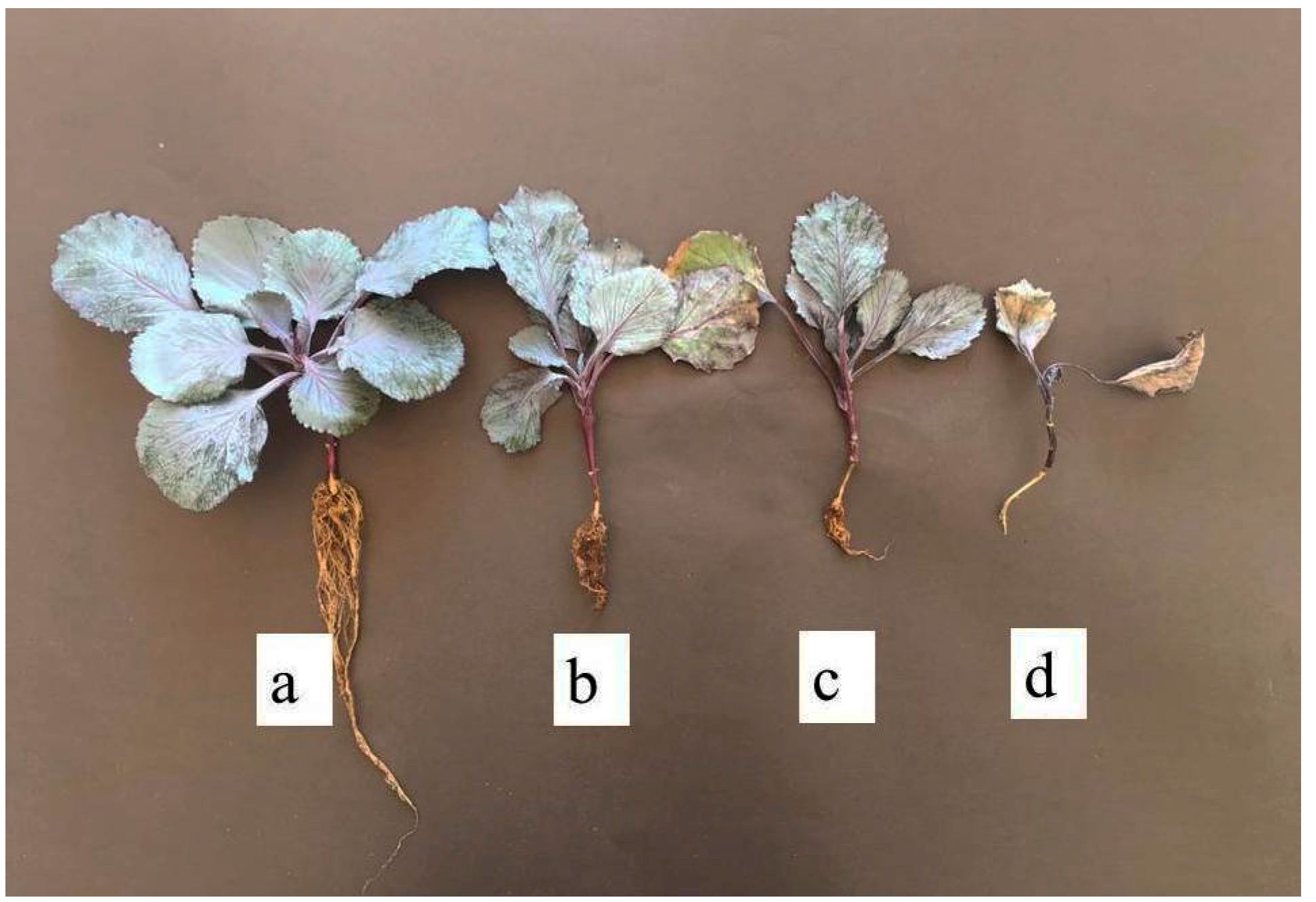
2.5. Pathogenicity Assays

Of 24 isolates, 14 belonged to AG 4 HG-I, 2 to AG 4 HG-III, 4 to AG 5, and 4 to AG-A of Rhizoctonia spp., selected on the basis of AGs, and geographical origin was tested for aggressiveness on red cabbage cv. Rondale under greenhouse conditions. The inoculum of each isolate was prepared on wheat seeds using the seed-colonization method of Brayford [63]. The seeds of red cabbage were superficially disinfected in 1% NaOCl solution for 3 min, washed twice with sterilized distilled water, and air-dried on sterile tissue paper in a laminar flow. Each single seed was sown in plastic pots (7 cm in length, 6.5 cm in diameter) filled with a sterilized peat:perlite (2:1, v:v) mixture. The pots were maintained at 20–25 °C with a 16 h photoperiod in a greenhouse.
Six-week-old seedlings at the three true leaf stage were singly transplanted into 10 cm diameter plastic pots (250 mL) containing a sterilized mixture of sandy loam soil:manure:washed sand (2:2:1, v:v:v), and inoculated with 5 g of colonized wheat seeds by placing into the pit dispersedly and surrounded seedling roots at the same time. Controls were inoculated with sterile wheat seeds. Plants were kept in the greenhouse at 20–25 °C. Six replicates were employed for each isolate, with an equal number of control plants. After inoculation, plants were kept in the greenhouse. Twenty-one days after inoculation, plants were gently uprooted, washed with tap water for 5 min, and evaluated for the disease. The disease severity index (DSI) was assessed by evaluating seedling/root symptoms using a slightly modified 1–4 disease severity scale: 1 = no symptom; 2 = basal rot on part of the stem; 3 = basal rot girdled stem superficially, but plants did not wilt; 4 = basal rot deeply girdled stem, stem base was constricted, roots were sometimes detached from stem, and plants wilted [9]. The values of plant height, shoot and root dry weights, and root length were also recorded for each plant. The experimental set-up was a completely randomized design with six red cabbage seedlings per treatment, and the experiment was repeated once. The isolates tested were re-isolated from the symptomatic tissues of the seedlings.

2.6. Resistance Response Classification

The reactions of six red cabbage cultivars (Rescue, Travero, Integro, Remale, Redriver, and Rondale) grown commonly in Turkey were estimated using the method described in the pathogenicity experiment against Rhizoctonia spp. The isolates of Rs-RC-20 (AG 4 HG-I), Rs-RC-109 (AG 4 HG-III), Rs-RC-118 (AG 5), and R-RC-131 (AG-A) representing each AG were selected for this experiment. The cultivars were grouped according to disease severity on stems and roots. The cultivars were considered resistant (R) if the mean disease severity was ≤1.50, moderately resistant (MR) if the mean disease severity ranged between 1.51 and 2.25, moderately susceptible (MS) if the mean disease severity ranged between 2.26 and 3.00, and susceptible (S) if the mean disease severity was ≥3.01.

2.7. Statistical Analysis

All data obtained from pathogenicity tests was subjected to Box-Cox transformation before statistical analyses, since they failed to meet the assumptions of normal distribution (Shapiro–Wilk test) and variance homogeneity (Levene’s test). Each plant growth parameter was separated individually at a probability level of p < 0.05 by Fisher’s least significant difference (LSD) test based on the transformed data. On the other hand, due to the nonnormal distribution of disease severity data, analysis was performed with the Dunn-test based on the rank sums using the Kruskal–Wallis pairwise multiple comparisons. To assess the effect of the isolate, cultivar, and isolate–cultivar on disease severity, a nonparametric test of two-way ANOVA, the Scheirer–Ray–Hare (SRH) test, was performed using R software. All other analyses were conducted using the XLSTAT program version 18.07 (Addinsoft Company, New York, NY, USA).

3. Results

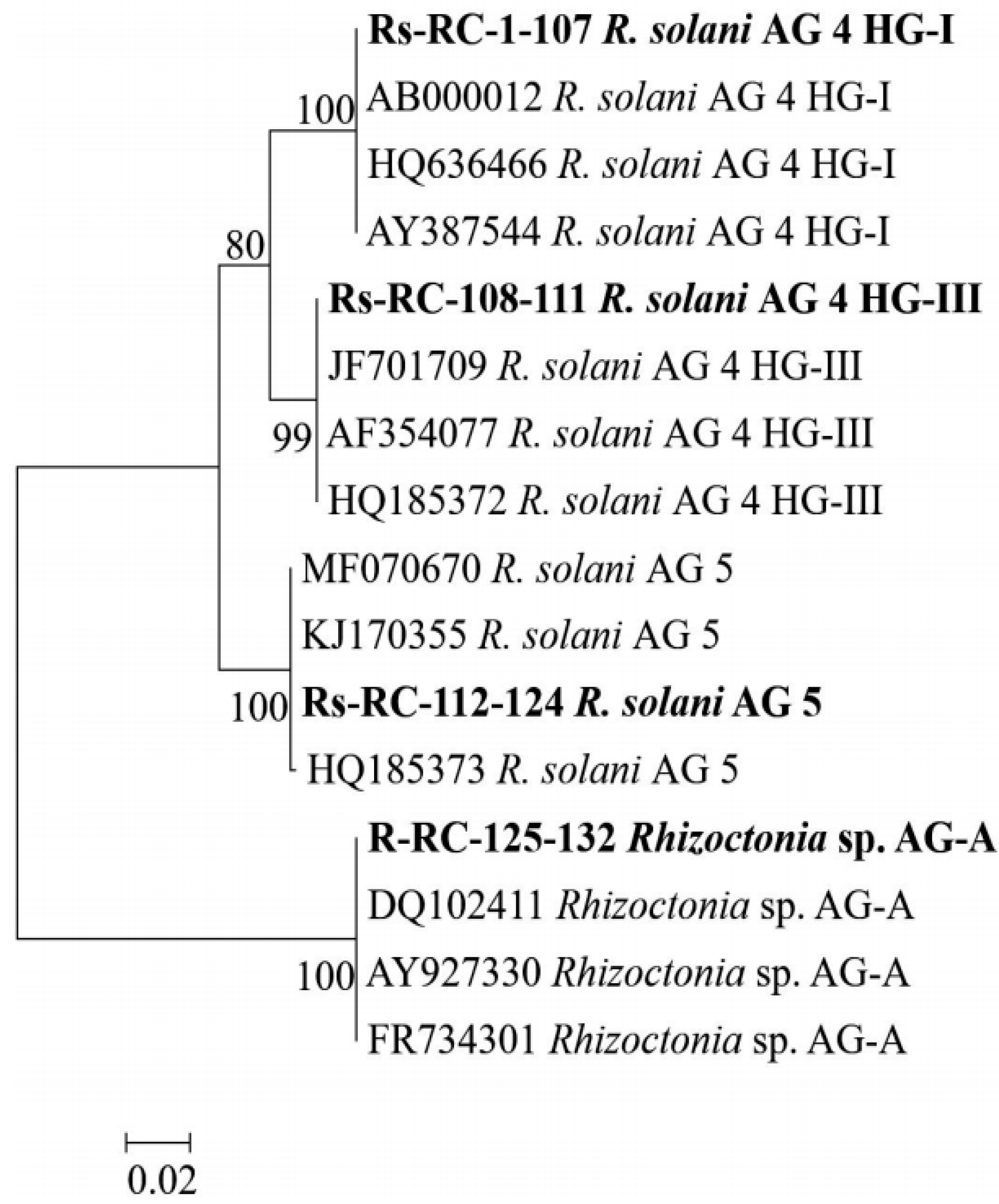
3.1. Rhizoctonia Species and AGs on Red Cabbage

Altogether, 132 isolates of Rhizoctonia spp. associated with dark lesions of underground parts of red cabbage plants were obtained from the fields planted in sixteen locations of Samsun province. Using the nuclear staining method, 124 of 132 isolates were classified as MNR, while the remaining isolates (n = 8) were BNR (Table 2).
The BLAST service at the NCBI website was used for analysis of ITS sequences. The BLAST analysis revealed that 107 isolates were MNR AG 4 HG-I, the most prevalent group (81%) in the investigated area, followed by AG 5, AG 4 HG-III, and BNR AG-A with 13 (10%), 4 (3%), and 8 (6%) isolates, respectively. No polymorphism was observed among the nucleotide sequences of isolates within the same AG. The ITS fragments amplified using primers ITS1 and ITS4 showed 100% similarity with those of the corresponding isolates belonging to different AGs from GenBank. The accession numbers provided by GenBank for the sequences obtained in this study are shown in Table 2. The Maximum Likelihood tree generated with the ITS sequences showed a clear clustering among anastomosis groups (Figure 1). All isolates obtained in this study and the known AGs retrieved from GenBank clustered into two major groups with strong bootstrap values (99–100%) according to their nuclei number within vegetative cells. Group I comprised multinucleate isolates, which were further grouped into three subgroups according to their anastomosis groups, whereas the isolates of AG-A formed Group II as the most diverse group.

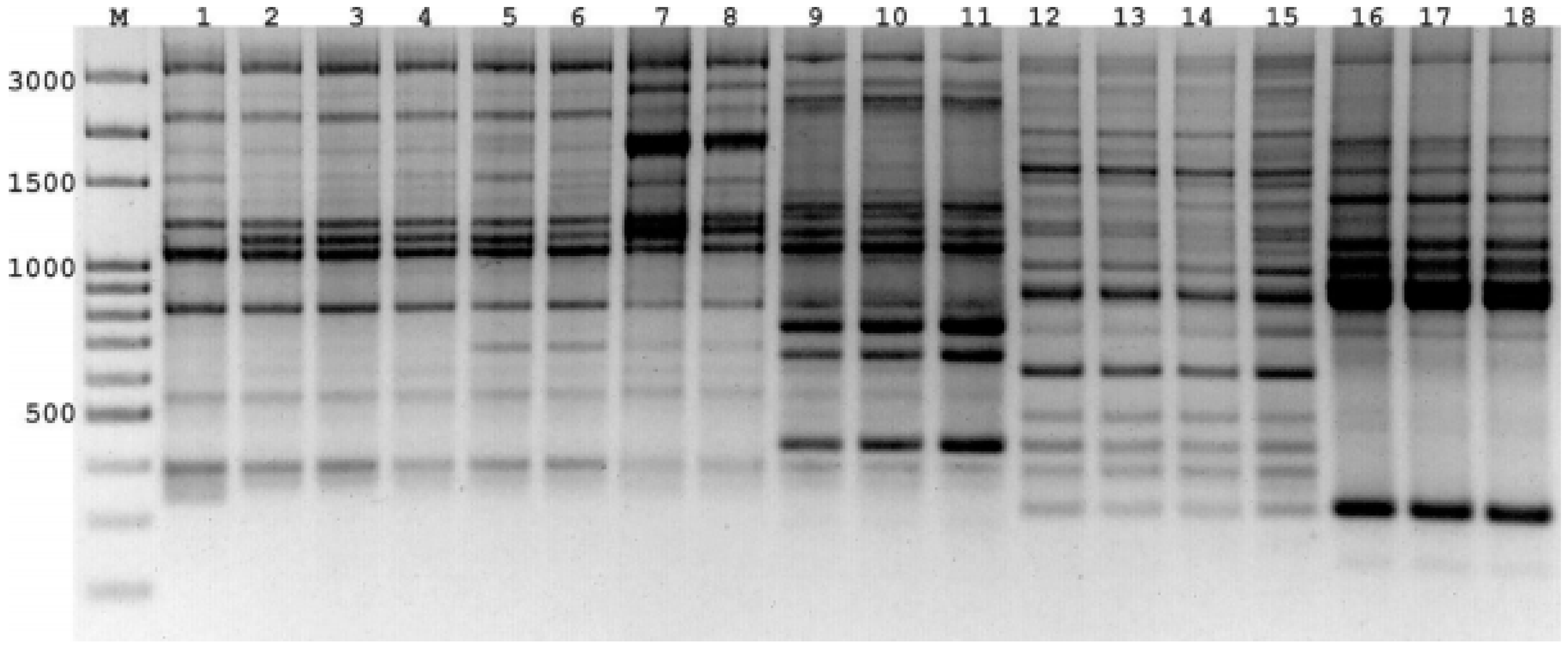
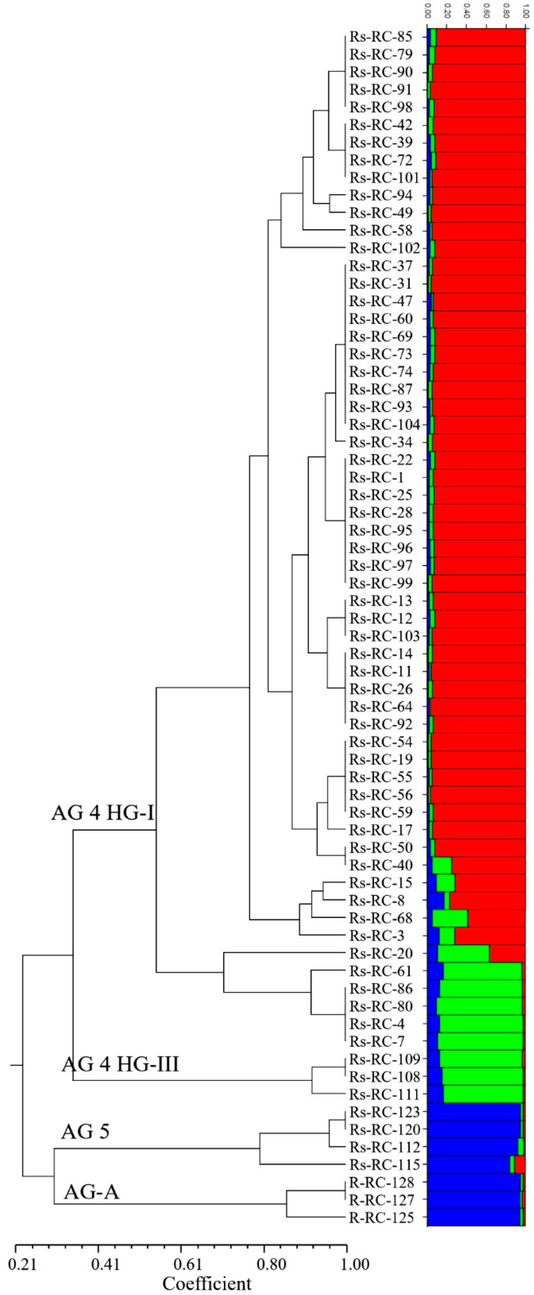
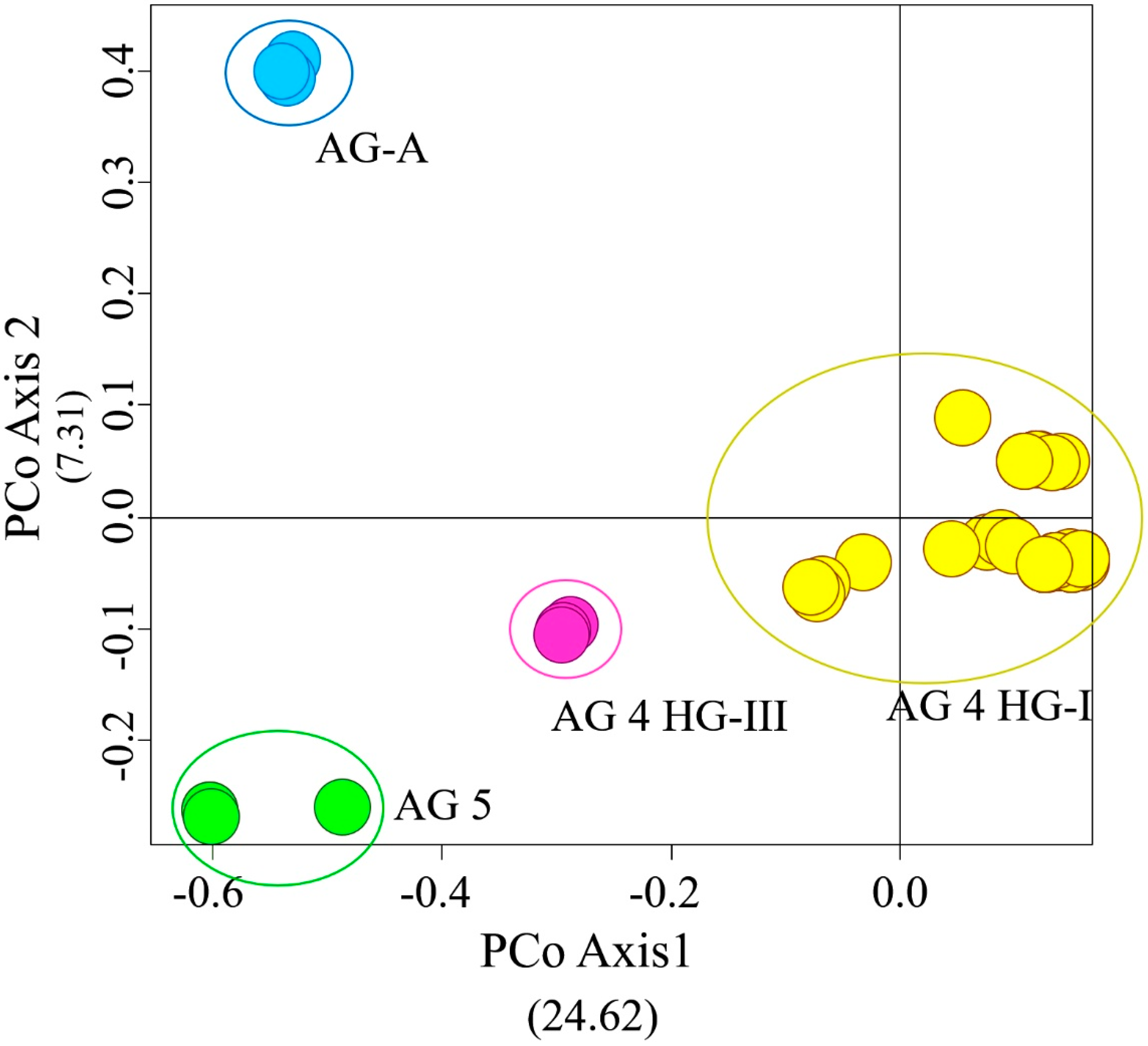
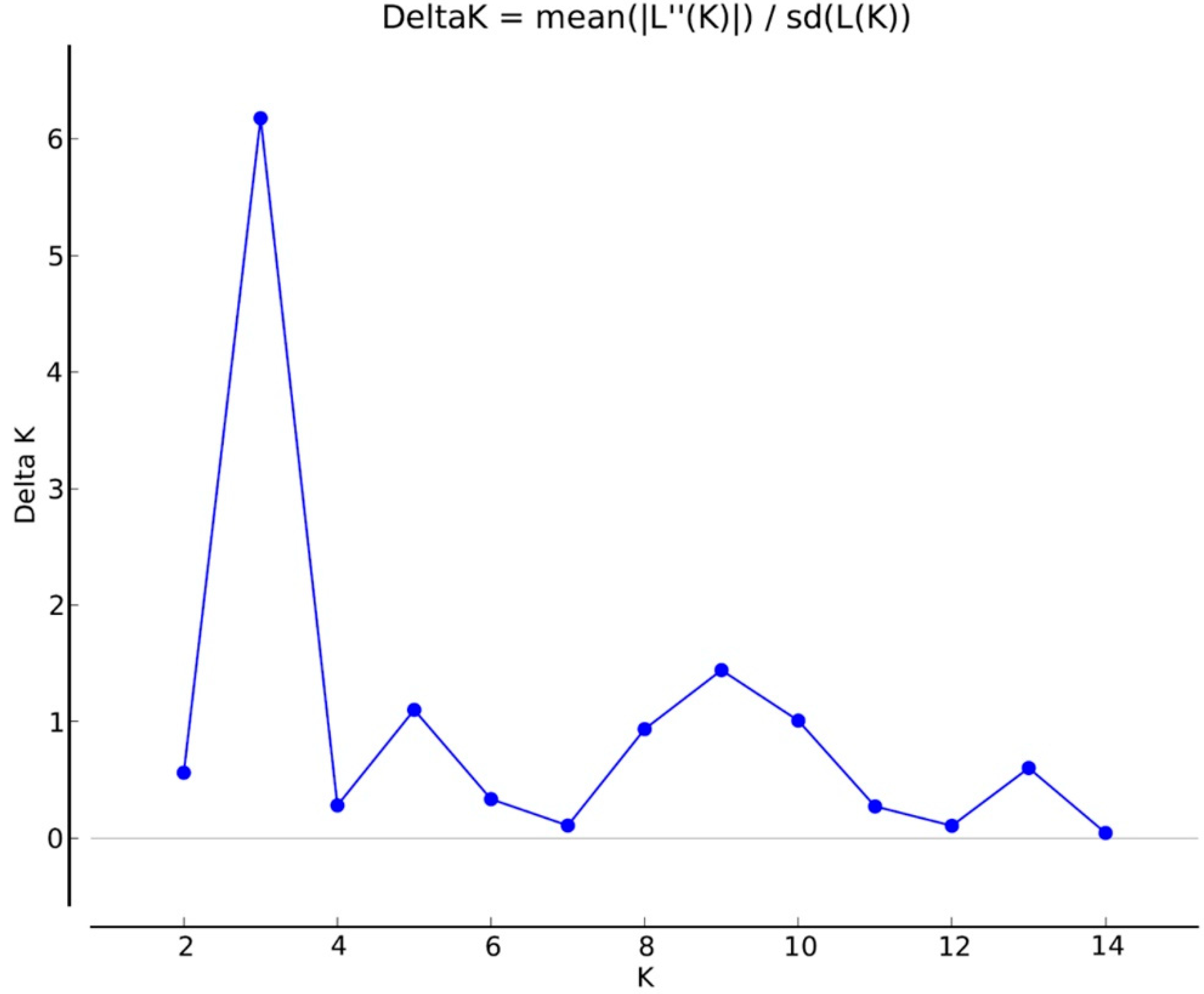
3.2. iPBS Amplification Analysis

The seven PBS primers in iPBS-amplification profiling generated 106 scorable and reproducible fragments to evaluate the extent of genetic variation between Rhizoctonia isolates. Ninety-six of those fragments (90%) were polymorphic. The number of fragments amplified with the primers varied between 11 (2219/2237) and 30 (2395), with a mean of 15 fragments per PBS primer. The values of the PIC and RP index estimated for the markers are listed in Table 1. The range of PIC was 0.37 (2219)–0.14 (2239), averaging 0.18 (Figure 2). The mean of the RP values, a parameter that indicates the discriminatory potential of the primers chosen, was 3.15 for all primers. The highest RP value of 5.32 was obtained from 2395, while the lowest RP value was recorded as 1.94 for 2239. The UPGMA dendrogram produced using the Jaccard’s similarity coefficient for iPBS profiling methods clustered distinctly 68 Rhizoctonia isolates into four major groups, which were completely conserved among isolates within the same AG assignment (Figure 3). Group I comprised 58 AG 4 HG-I isolates, and Group 2, 3, and 4 were composed of AG 4 HG-III (3), AG 5 (4), and AG-A (3) isolates, respectively. The groups were further clustered into subgroups at different similarity degrees. In addition, UPGMA cluster analysis based on iPBS-amplification profiling data supported the grouping of isolates based on ITS sequences. The PcoA analysis distinguished Rhizoctonia isolates according to their anastomosis groups and plotted the isolates to four major groups, which confirmed the UPGMA pattern (Figure 4). Structure runs were conducted for K = 2 to 15 based on the iPBS data. Ln values increased sharply at K = 3, after which the increase was slow without reaching the plateau (Figure 5). The highest peak was detected at K = 3, which implies that the isolates could be distributed into three separated clusters according to the ΔK criteria of Evanno et al. [64].

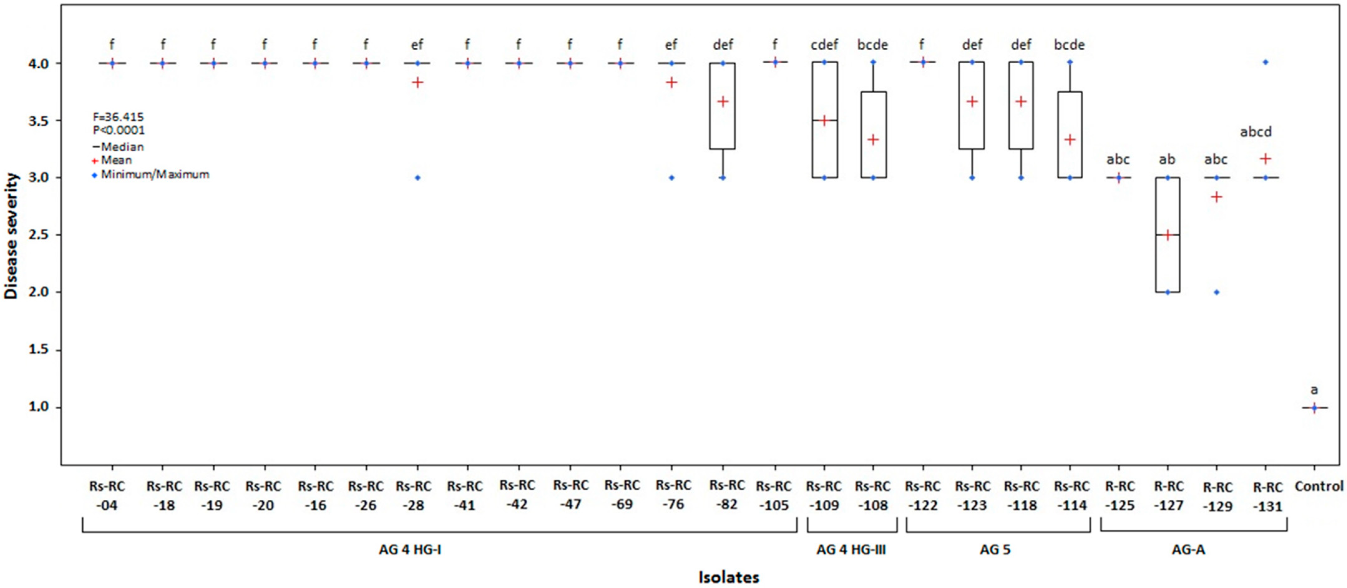
3.3. Virulence of the Isolates

The results of the pathogenicity test on seedlings of red cabbage cv. Rondale revealed a significant difference in virulence between the isolates tested (Kruskal–Wallis ANOVA, H24,125 = 106.81, p < 0.0001) (Figure 6). Disease symptoms developed in inoculated red cabbage seedlings were scored after twenty-one days of inoculation (Figure 7). The disease severity index (DSI) varied from 3.33 to 4.0 for all R. solani isolates. In particular, almost all AG 4 HG-I isolates, and isolate Rs-RC-122 of AG 5, caused severe necrosis symptoms in the seedlings, resulting in damping off. All AG-A isolates had a lower virulence ranging from 2.5 to 3.17. All isolates caused a significant reduction in the dry weight of shoots and roots compared to control plants (ANOVA, F24,125 = 7.002, p < 0.0001; F24,125 = 3.369, p < 0.0001, respectively) (Table 3). Compared to the control, some R. solani isolates and all AG-A isolates were found to have no adverse effects on root length or/and plant height (ANOVA F24,125 = 2.554, p < 0.0004; F24,125 = 4.051, p < 0.0001, respectively).

3.4. Host Response of Some Commercial Cultivars to Different AGs of Rhizoctonia

A statistically significant difference was observed among different AGs (AG 4 HG-I, -HG-III, and 5) of Rhizoctonia isolates in the severity of root rot in the cultivars (Rescue, Travero, Integro, Remale, Redriver, and Rondale) of red cabbage (SRH test: isolates: H4 = 135.85, p < 0.0001; cultivars: H5 = 11.45, p < 0.0432, respectively) (Table 4). In general, the AG-A isolate caused low disease severity in all cultivars, but R. solani isolates led to the development of severe root rot resulting in high disease severity values ranging from 2.5 to 4. All cultivars were susceptible to AG 4 HG-I and HG-III isolates of R. solani. Redriver and Remale to the AG 5 isolate were moderately susceptible, while Rescue, Travero, Integro, and Rondale were susceptible. On the other hand, Redriver was moderately resistant to AG-A; Remale, Rescue, and Travero were moderately susceptible; and Integro and Rondale were susceptible. No significant isolate–cultivar interaction was observed for root rot severity on red cabbages (SRH test: isolates:cultivars H20 = 15.09, p < 0.7712).

4. Discussion

This is the first characterization of Rhizoctonia isolates obtained from red cabbage growing areas in Turkey. To the best of our knowledge, no reports about the pathogenicity of Rhizoctonia spp. in red cabbage plants have been recorded in the world. The majority of the Rhizoctonia isolates were MNR AG 4 (84%). The sequence analysis of the ITS allowed further division of the 111 isolates belonging to MNR AG 4 into two subgroups, AG 4 HG-I (n = 107) and AG 4 HG-III (n = 4). The remaining isolates were classified into AG 5 (8 isolates) and BNR AG-A (13 isolates).
As indicated in the previous reports, both BNR and MNR isolates cause disease in Brassica crops, including white cabbage, kale, broccoli, cauliflower, oilseed rape, and canola in several countries in the world [7,8,9,10,11,18,20,22,23,26,65]. The most prevalent and damaging R. solani AGs were AG 2-1 and AG 4 among AGs determined for brassicas [19,24,25,66,67]. Recently, Türkkan, Kılıçoğlu and Erper [11] determined that 37% (11 isolates) of limited Rhizoctonia isolates (30 isolates) obtained from kale growing areas in Ordu province, Black Sea region of Turkey, belong to AG 2-1, followed by AG-A with 20% (9 isolates), and AG 4 HG-I with 10% (3 isolates). They isolated Rhizoctonia isolates during November–April when temperatures ranged from 7–16 °C. However, in the present study, surveys were conducted between July and October when the weather was warmer (25–33 °C). Previous studies have shown that the abundance of each AG may depend on the climate. For example, it has been reported in several studies that AG 4 HG-I is dominant in warmer periods, while AG 2-1, AG 1-IB, and AG-BI are dominant during colder periods [68,69,70]. Moreover, Yitbarek et al. [71] observed that AG 4 caused severe root rot in B. napus seedlings at temperatures varying between 26 and 35 °C, whereas the pathogenic activity of AG 2-1 was considerably reduced at these temperatures. The relationship between AG and temperature is probably the same for isolates in this study and may explain why we commonly isolate R. solani AG 4 HG-I during warmer periods. In addition, previous studies have reported that it is the most common pathogen on cucumber, winter squash, bean, and soybean in the Samsun province of Turkey, where brassicas (cauliflower, white cabbage, and red cabbage) are used as an effective rotational crop [47,72,73]. Given that the pathogen is polyphagous, these alternative hosts likely contributed to the prevalence of Rhizoctonia root rot in the fields, which is consistent with Keinath’s findings [74]. Cubeta and Vilgalys [75], and Sharon et al. [76] noted that DNA fingerprinting techniques for studying the genetic variation of Rhizoctonia are most suitable at the individual level rather than for the determination of AGs or subgroups within an AG. There is a lack of evidence, however, to support this hypothesis, since no DNA marker system has been tested in grouping a large number of Rhizoctonia isolates of different AGs. In the present study, the retrotransposon-based iPBS amplification DNA profiling method differentiated the isolates according to their anastomosis groups, including AG 4 HG-I, HG-III, AG 5, and AG-A, via phylogenetic analysis. A more detailed sub-grouping was observed among the isolates within each AG group formed in the phylogenetic tree, especially among AG 4 HG-I isolates. This is in accordance with other researchers who have grouped Rhizoctonia spp. isolates using these markers [35,36]. Pourmahdi and Taheri [35] showed the efficacy of this molecular marker system to determine the AGs of R. solani isolates from tomato as a novel universal molecular marker system. iPBS-amplification markers have also been successfully employed for the characterization of several other fungi and yeast [36,37,38,39,40,77,78]. To the best of our knowledge, this study is the first characterization of various Rhizoctonia spp. isolates belonging to different AGs obtained from red cabbage using the retrotransposon marker system for DNA fingerprinting.
The pathogenicity studies on red cabbage seedlings showed that the disease severity index (DSI) used in the evaluation of the aggressiveness of Rhizoctonia isolates varied widely between 2.5 and 4.0. In the MNR isolates, AG 4 HG-I isolates had the highest index value (DSI: 4.0) on the seedlings, with exceptions of the RS-RC-28, -76, and -82 isolates, and Rs-RC-122 isolate of AG 5. The AG 4 HG-I isolates caused wirestem symptoms by severe necrosis of the stem and root tissues. AG 4 HG-III and AG 5 isolates were more aggressive than isolates of BNR AG-A. Pannecoucque, Van Beneden, and Höfte [20] found that cauliflower plants were more susceptible to AG 1-IC, AG 2–1, AG 2–1 subset Nt, AG 2–2, and AG 4 HG-II isolates than AG 1-IB and AG 5 isolates. None of the AG-A isolates caused symptoms on Chinese and white cabbage, but AG-Fc isolates caused severe lesions [8]. Türkkan, Kılıçoğlu, and Erper [11] noted that except for several isolates within AG 2-1, AG 2-1 and AG 4 HG-I caused severe symptoms and deaths on seedlings of kale cv. Arzuman, but the isolates in AG 5, AG-E, AG-Fb, and AG-K were of relatively low and moderate virulence.
The results of pathogen–cultivar interactions in the current study are consistent with those of Keinath and Farnham [24], who found no cultivar by isolate interaction when testing cultivars for root rot resistance against AG 4 and AG 2-1 of R. solani. They also determined that AG 4 caused severe wirestem rot in the seedlings of 12 brassica cultivars (3 each of broccoli, cauliflower, cabbage, and collard) in all experiments, while AG 2-1 caused less root rot in the growth chamber but no root rot in the field. Chinese cabbage and white cabbage seedlings were highly susceptible to R. solani AG 4 HG-I in both in vitro and in vivo bioassays conducted by Hua, Bertier, Soltaninejad, and Hofte [8]. Türkkan et al. [11] reported that R. solani AG 2-1 and AG 4 HG-I isolates generally caused more severe root rot in the seedlings of kale cv. Arzuman. The present study revealed that the Redriver cultivar was moderately resistant to Rhizoctonia AG-A, although no cultivars tested were resistant to AGs of R. solani.
In conclusion, the Rhizoctonia spp. isolates, whether MNR or BNR, cause devastating diseases in many crops, resulting in economic crop losses. The AG classification of Rhizoctonia isolates is a critical approach to characterize the various groups that cause plant diseases. The iPBS-amplification DNA profiling analysis is a simple, effective, and powerful tool to identify AG subgroups. Further studies with other isolates belonging to more AGs are needed to comprehensively investigate the association between grouping within Rhizoctonia spp. isolates and iPBS-amplification DNA profiling.
In this study, we confirmed the presence of some virulent Rhizoctonia isolates associated with root rot and wirestem in red cabbage. Due to the persistence of Rhizoctonia in the soil for several years and the presence of different AGs that are aggressive in various host plants [6], crop rotational strategies alone are insufficient to control Rhizoctonia in the fields surveyed. The isolates of AG 4 HG-I cause problems for several crops cultivated in the agricultural areas of the Black Sea region, including the province of Samsun [10,11]. AG 4 HG-I, therefore, has the potential to damage Brassica crops. Although the use of chemical fungicides is environmentally undesirable, the most common method for protecting crops against Rhizoctonia isolates is the use of fungicides worldwide [79]. In Turkey, several fungicides are used to control Rhizoctonia species on different crops as seed or seedling treatments. An approach of integrated management of plant disease including applying chemical and biological fungicides, crop rotation, and breeding varieties with resistance is needed for the efficient control of the root rot and wirestem of red cabbage.

Author Contributions

Data curation, E.Y. and M.A.; formal analysis, G.O. and E.Y.; investigation, I.E., G.O., S.A., E.Y. and M.T.; methodology, R.K.; resources, G.O., S.A., E.Y., M.A. and M.T.; supervision, I.E.; validation, I.E., G.O., R.K.; statistical analysis, M.T.; writing—original draft, I.E., G.O. and M.T.; writing—review and editing, G.O. and R.K. All authors have read and agreed to the published version of the manuscript.

Funding

This study was founded by the Research and Development Unit (BAP) of Ondokuz Mayis University with the project no PYO.ZRT.1904.18.015, and by the Science Committee of the Ministry of Education and Science of the Republic of Kazakhstan (grant no. AP08855353) for RK.

Institutional Review Board Statement

Not applicable.

Informed Consent Statement

Not applicable.

Data Availability Statement

All relevant data are within the manuscript.

Acknowledgments

Open access funding provided by University of Helsinki, including Helsinki University Central Hospital. The authors wish to thank Jennifer Rowland (The University of Helsinki Language Centre) for outstanding editing.

Conflicts of Interest

The authors declare no conflict of interest.

References

  1. Food and Agriculture Organization (FAO). Food and Agriculture Organization of the United Nations. Available online: http://www.fao.org/faostat/en/#data (accessed on 1 March 2021).
  2. Monteiro, A.; Lunn, T. Trends and Perspectives of Vegetable Brassica Breeding World-Wide. In Proceedings of the WCHR-World Conference on Horticultural Research 495, Rome, Italy, 17–20 June 1998; pp. 273–280. [Google Scholar]
  3. Balkaya, A.; Yanmaz, R.; Apaydin, A.; Kar, H. Morphological characterisation of white head cabbage (Brassica oleracea var. capitata subvar. alba) genotypes in turkey. N. Z. J. Crop Hortic. Sci. 2005, 33, 333–341. [Google Scholar] [CrossRef] [Green Version]
  4. TUIK. Turkish Statistical Institute: Crop Production Statistics. Available online: https://biruni.tuik.gov.tr/medas (accessed on 1 March 2021).
  5. Rimmer, S.R.; Shattuck, V.I.; Buchwaldt, L. Compendium of Brassica Diseases; American Phytopathological Society (APS Press): St. Paul, MN, USA, 2007. [Google Scholar]
  6. Sneh, B.; Jabaji-Hare, S.; Neate, S.; Dijst, G. Rhizoctonia Species: Taxonomy, Molecular Biology, Ecology, Pathology and Disease Control; Springer Science & Business Media: Berlin/Heidelberg, Germany, 1996. [Google Scholar] [CrossRef]
  7. Yang, G.H.; Chen, J.Y.; Pu, W.Q. First report of head rot of cabbage and web blight of snap bean caused by Rhizoctonia solani AG-4 HGI. Plant Pathol. 2007, 56, 351. [Google Scholar] [CrossRef]
  8. Hua, G.K.; Bertier, L.; Soltaninejad, S.; Hofte, M. Cropping systems and cultural practices determine the Rhizoctonia anastomosis groups associated with Brassica spp. in Vietnam. PLoS ONE 2014, 9, e111750. [Google Scholar] [CrossRef] [Green Version]
  9. Misawa, T.; Kubota, M.; Sasaki, J.; Kuninaga, S. First report of broccoli foot rot caused by Rhizoctonia solani AG-2-2 IV and pathogenicity comparison of the pathogen with related pathogens. J. Gen. Plant Pathol. 2014, 81, 15–23. [Google Scholar] [CrossRef]
  10. Saygi, S.; Erper, I.; Ozer, G.; Turkkan, M. First report of Rhizoctonia solani AG 4 HG-I causing damping off and wirestem of white cabbage in Turkey. J. Plant Pathol. 2019, 102, 587. [Google Scholar] [CrossRef] [Green Version]
  11. Türkkan, M.; Kılıçoğlu, M.Ç.; Erper, I. Characterization and pathogenicity of Rhizoctonia isolates collected from Brassica oleracea var. acephala in Ordu, Turkey. Phytoparasitica 2020, 48, 273–286. [Google Scholar] [CrossRef]
  12. Carling, D.E.; Kuninaga, S.; Brainard, K.A. Hyphal anastomosis reactions, rDNA-internal transcribed spacer sequences, and virulence levels among subsets of Rhizoctonia solani anastomosis group-2 (AG-2) and AG-BI. Phytopathology 2002, 92, 43–50. [Google Scholar] [CrossRef] [PubMed] [Green Version]
  13. Sharon, M.; Sneh, B.; Kuninaga, S.; Hyakumachi, M.; Naito, S. Classification of Rhizoctonia spp. using rDNA-ITS sequence analysis supports the genetic basis of the classical anastomosis grouping. Mycoscience 2008, 49, 93–114. [Google Scholar] [CrossRef]
  14. Aiello, D.; Guarnaccia, V.; Formica, P.T.; Hyakumachi, M.; Polizzi, G. Occurrence and characterisation of Rhizoctonia species causing diseases of ornamental plants in Italy. Eur. J. Plant Pathol. 2017, 148, 967–982. [Google Scholar] [CrossRef]
  15. Dong, W.; Li, Y.; Duan, C.; Li, X.; Naito, S.; Conner, R.L.; Yang, G.; Li, C. Identification of AG-V, a new anastomosis group of binucleate Rhizoctonia spp. from taro and ginger in Yunnan province. Eur. J. Plant Pathol. 2017, 148, 895–906. [Google Scholar] [CrossRef]
  16. Zhao, C.; Li, Y.; Liu, H.; Li, S.; Han, C.; Wu, X. A binucleate Rhizoctonia anastomosis group (AG-W) is the causal agent of sugar beet seedling damping-off disease in China. Eur. J. Plant Pathol. 2019, 155, 53–69. [Google Scholar] [CrossRef]
  17. Ogoshi, A. Ecology and pathogenicity of anastomosis and intraspecific groups of Rhizoctonia solani Kuhn. Annu. Rev. Phytopathol. 1987, 25, 125–143. [Google Scholar] [CrossRef]
  18. Kuramae, E.E. Identification of Rhizoctonia solani AG 1-IB in lettuce, AG 4 HG-I in tomato and melon, and AG 4 HG-III in broccoli and spinach, in Brazil. Eur. J. Plant Pathol. 2003, 109, 391–395. [Google Scholar] [CrossRef]
  19. Khangura, R.K.; Barbetti, M.J.; Sweetingham, M.W. Characterization and pathogenicity of Rhizoctonia species on canola. Plant Dis. 1999, 83, 714–721. [Google Scholar] [CrossRef] [Green Version]
  20. Pannecoucque, J.; Van Beneden, S.; Höfte, M. Characterization and pathogenicity of Rhizoctonia isolates associated with cauliflower in belgium. Plant Pathol. 2008, 57, 737–746. [Google Scholar] [CrossRef]
  21. Yang, J. Characterization, virulence, and genetic variation of Rhizoctonia solani AG-9 in Alberta. Plant Dis. 1996, 80. [Google Scholar] [CrossRef]
  22. Sayama, A. Occurrence of damping-off disease caused by Rhizoctonia solani ag 2-2-iiib on cabbage plug seedlings. Annu. Rep. Soc. Plant Prot. N. Jpn. 2000, 51, 54–57. [Google Scholar]
  23. Misawa, T.; Aoki, M. First report of Rhizoctonia solani AG-1 IC causing head rot of cabbage in Japan. New Dis. Rep. 2017, 36, 12. [Google Scholar] [CrossRef] [Green Version]
  24. Keinath, A.P.; Farnham, M.W. Differential cultivars and criteria for evaluating resistance to Rhizoctonia solani in seedling Brassica oleracea. Plant Dis. 1997, 81, 946–952. [Google Scholar] [CrossRef] [PubMed] [Green Version]
  25. Paulitz, T.C.; Okubara, P.A.; Schillinger, W.F. First report of damping-off of canola caused by Rhizoctonia solani AG 2-1 in washington state. Plant Dis. 2006, 90, 829. [Google Scholar] [CrossRef]
  26. Budge, G.E.; Shaw, M.W.; Colyer, A.; Pietravalle, S.; Boonham, N. Molecular tools to investigate Rhizoctonia solani distribution in soil. Plant Pathol. 2009, 58, 1071–1080. [Google Scholar] [CrossRef]
  27. Kalendar, R.; Shustov, A.V.; Seppanen, M.M.; Schulman, A.H.; Stoddard, F.L. Palindromic sequence-targeted (PST) PCR: A rapid and efficient method for high-throughput gene characterization and genome walking. Sci. Rep. 2019, 9, 17707. [Google Scholar] [CrossRef]
  28. Kalendar, R.; Muterko, A.; Boronnikova, S. Retrotransposable elements: DNA fingerprinting and the assessment of genetic diversity. Methods Mol. Biol. 2021, 2222, 263–286. [Google Scholar] [CrossRef] [PubMed]
  29. Ghonaim, M.; Kalendar, R.; Barakat, H.; Elsherif, N.; Ashry, N.; Schulman, A.H. High-throughput retrotransposon-based genetic diversity of maize germplasm assessment and analysis. Mol. Biol. Rep. 2020, 47, 1589–1603. [Google Scholar] [CrossRef] [Green Version]
  30. Li, S.; Ramakrishnan, M.; Vinod, K.K.; Kalendar, R.; Yrjälä, K.; Zhou, M. Development and deployment of high-throughput retrotransposon-based markers reveal genetic diversity and population structure of Asian bamboo. Forests 2019, 11, 31. [Google Scholar] [CrossRef] [Green Version]
  31. Kalendar, R.; Raskina, O.; Belyayev, A.; Schulman, A.H. Long tandem arrays of Cassandra retroelements and their role in genome dynamics in plants. Int. J. Mol. Sci. 2020, 21, 2931. [Google Scholar] [CrossRef]
  32. Kalendar, R.; Amenov, A.; Daniyarov, A. Use of retrotransposon-derived genetic markers to analyse genomic variability in plants. Funct. Plant Biol. 2018, 46, 15–29. [Google Scholar] [CrossRef]
  33. Kalendar, R.; Antonius, K.; Smýkal, P.; Schulman, A.H. IPBS: A universal method for DNA fingerprinting and retrotransposon isolation. Theor. Appl. Genet. 2010, 121, 1419–1430. [Google Scholar] [CrossRef]
  34. Milovanov, A.; Zvyagin, A.; Daniyarov, A.; Kalendar, R.; Troshin, L. Genetic analysis of the grapevine genotypes of the Russian Vitis ampelographic collection using iPBS markers. Genetica 2019, 147, 91–101. [Google Scholar] [CrossRef] [Green Version]
  35. Pourmahdi, A.; Taheri, P. Genetic diversity of thanatephorus cucumeris infecting tomato in Iran. J. Phytopathol. 2015, 163, 19–32. [Google Scholar] [CrossRef]
  36. Özer, G.; Bayraktar, H.; Baloch, F.S. IPBS retrotransposons ‘a universal retrotransposons’ now in molecular phylogeny of fungal pathogens. Biochem. Syst. Ecol. 2016, 68, 142–147. [Google Scholar] [CrossRef]
  37. Ozer, G.; Sameeullah, M.; Bayraktar, H.; Göre, M.E. Genetic diversity among phytopathogenic Sclerotiniaceae, based on retrotransposon molecular markers. Phytopathol. Mediterr. 2017, 56, 251–258. [Google Scholar]
  38. Özer, G.; Bayraktar, H. Genetic diversity of Fusarium oxysporum f. sp. cumini isolates analyzed by vegetative compatibility, sequences analysis of the rDNA IGS region and iPBS retrotransposon markers. J. Plant Pathol. 2018, 100, 225–232. [Google Scholar] [CrossRef]
  39. Aydin, F.; Ozer, G.; Alkan, M.; Cakir, I. The utility of iPBS retrotransposons markers to analyze genetic variation in yeast. Int. J. Food Microbiol. 2020, 325, 108647. [Google Scholar] [CrossRef] [PubMed]
  40. Turzhanova, A.; Khapilina, O.N.; Tumenbayeva, A.; Shevtsov, V.; Raiser, O.; Kalendar, R. Genetic diversity of Alternaria species associated with black point in wheat grains. PeerJ 2020, 8, e9097. [Google Scholar] [CrossRef]
  41. Amaradasa, B.S.; Horvath, B.J.; Lakshman, D.K.; Warnke, S.E. DNA fingerprinting and anastomosis grouping reveal similar genetic diversity in species infecting turfgrasses in the transition zone of USA. Mycologia 2013, 105, 1190–1201. [Google Scholar] [CrossRef] [PubMed] [Green Version]
  42. Amaradasa, B.S.; Keenan Amundsen, D.L. AFLP fingerprinting for identification of infra-species groups of Rhizoctonia solani and Waitea circinata. J. Plant Pathol. Microbiol. 2015, 6. [Google Scholar] [CrossRef] [Green Version]
  43. Doungous, O.; Kalendar, R.; Filippova, N.; Ngane, B.K. Utility of iPBS retrotransposons markers for molecular characterization of African Gnetum species. Plant Biosyst. 2019, 154, 587–592. [Google Scholar] [CrossRef]
  44. Khapilina, O.; Raiser, O.; Danilova, A.; Shevtsov, V.; Turzhanova, A.; Kalendar, R. DNA profiling and assessment of genetic diversity of relict species Allium altaicum pall. on the territory of Altai. PeerJ 2021, 9, e10674. [Google Scholar] [CrossRef]
  45. Demirci, E.; Döken, M. Anastomosis groups and pathogenicity of Rhizoctonia solani Kuehn isolates from potatoes in Erzurum- Turkey. J. Turk. Phytopathol. 1993, 22, 95–102. [Google Scholar]
  46. Erper, I.; Karaca, G.H.; Turkkan, M.; Ozkoc, I. Characterization and pathogenicity of Rhizoctonia spp. from onion in Amasya, Turkey. J. Phytopathol. 2006, 154, 75–79. [Google Scholar] [CrossRef]
  47. Erper, I.; Kilicoglu, M.C.; Turkkan, M.; Onder, H. Characterization and pathogenicity of Rhizoctonia spp. isolated from winter squash in the Black Sea region of Turkey. Eur. J. Plant Pathol. 2016, 146, 683–697. [Google Scholar] [CrossRef]
  48. Özer, G.; Bayraktar, H. Characterization and sensitivity to fungicides of Rhizoctonia spp. recovered from potato plants in Bolu, Turkey. J. Phytopathol. 2015, 163, 11–18. [Google Scholar] [CrossRef]
  49. Erper, I.; Yildirim, E. Characterization and pathogenicity of Rhizoctonia spp. isolated from vegetable crops grown in greenhouses in Samsun province, Turkey. Biosci. J. 2017, 33. [Google Scholar] [CrossRef] [Green Version]
  50. Ogoshi, A. Grouping of Rhizoctonia solani Kühn and their perfect stages. Bull. Nat. Inst. Agric. Sci. Ser. C 1975, 30, 1–63. [Google Scholar]
  51. Bandoni, R.J. Safranin o as a rapid nuclear stain for fungi. Mycologia 2018, 71, 873–874. [Google Scholar] [CrossRef]
  52. Kalendar, R.; Boronnikova, S.; Seppanen, M. Isolation and purification of DNA from complicated biological samples. Methods Mol. Biol. 2021, 2222, 57–67. [Google Scholar] [CrossRef]
  53. White, T.J.; Bruns, T.; Lee, S.; Taylor, J. Amplification and direct sequencing of fungal ribosomal RNA genes for phylogenetics. PCR Protoc. A Guide Methods Appl. 1990, 18, 315–322. [Google Scholar]
  54. Kumar, S.; Stecher, G.; Li, M.; Knyaz, C.; Tamura, K. Mega X: Molecular evolutionary genetics analysis across computing platforms. Mol. Biol. Evol. 2018, 35, 1547–1549. [Google Scholar] [CrossRef] [PubMed]
  55. Thompson, J.D.; Higgins, D.G.; Gibson, T.J. Clustal W: Improving the sensitivity of progressive multiple sequence alignment through sequence weighting, position-specific gap penalties and weight matrix choice. Nucleic Acids Res. 1994, 22, 4673–4680. [Google Scholar] [CrossRef] [Green Version]
  56. Felsenstein, J. Confidence limits on phylogenies: An approach using the bootstrap. Evolution 1985, 39, 783–791. [Google Scholar] [CrossRef]
  57. Roldán-Ruiz, I.; Dendauw, J.; Van Bockstaele, E.; Depicker, A.; De Loose, M. AFLP markers reveal high polymorphic rates in ryegrasses (Lolium spp.). Mol. Breed. 2000, 6, 125–134. [Google Scholar] [CrossRef]
  58. Prevost, A.; Wilkinson, M.J. A new system of comparing PCR primers applied to ISSR fingerprinting of potato cultivars. Theor. Appl. Genet. 1999, 98, 107–112. [Google Scholar] [CrossRef]
  59. Rohlf, F.J. Ntsys-Pc: Numerical Taxonomy and Multivariate Analysis System Version 2.1; Exeter Publishing Setauket: New York, NY, USA, 2000. [Google Scholar]
  60. R-Team. The R Project for Statistical Computing. Available online: https://www.r-project.org/ (accessed on 1 March 2021).
  61. Pritchard, J.K.; Stephens, M.; Donnelly, P. Inference of population structure using multilocus genotype data. Genetics 2000, 155, 945–959. [Google Scholar]
  62. Earl, D.A.; vonHoldt, B.M. Structure harvester: A website and program for visualizing structure output and implementing the evanno method. Conserv. Genet. Resour. 2011, 4, 359–361. [Google Scholar] [CrossRef]
  63. Brayford, D. Methods for Research on Soilborne Phytopathogenic Fungi; Singleton, L.L., Mihail, J.D., Rush, C.M., Eds.; APS Press: St. Paul, MN, USA, 1993; pp. 103–106. [Google Scholar] [CrossRef]
  64. Evanno, G.; Regnaut, S.; Goudet, J. Detecting the number of clusters of individuals using the software structure: A simulation study. Mol. Ecol. 2005, 14, 2611–2620. [Google Scholar] [CrossRef] [Green Version]
  65. Rollins, P.A.; Keinath, A.P.; Farnham, M.W. Effect of inoculum type and anastomosis group of Rhizoctonia solani causing wirestem of cabbage seedlings in a controlled environment. Can. J. Plant Pathol. 1999, 21, 119–124. [Google Scholar] [CrossRef]
  66. Yitbarek, S.M.; Verma, P.R.; Morrall, R.A.A. Anastomosis groups, pathogenicity, and specificity of Rhizoctonia solani isolates from seedling and adult rapeseed/canola plants and soils in Saskatchewan. Can. J. Plant Pathol. 1987, 9, 6–13. [Google Scholar] [CrossRef]
  67. Verma, P.R. Biology and control of Rhizoctonia solani on rapeseed: A review. Phytoprotection 2005, 77, 99–111. [Google Scholar] [CrossRef] [Green Version]
  68. Balali, G.R.; Neate, S.M.; Scott, E.S.; Whisson, D.L.; Wicks, T.J. Anastomosis group and pathogenicity of isolates of Rhizoctonia solani from potato crops in South Australia. Plant Pathol. 1995, 44, 1050–1057. [Google Scholar] [CrossRef]
  69. Carling, D.E. Effect of temperature on virulence of Rhizoctonia solani and other Rhizoctonia on potato. Phytopathology 1990, 80, 930–934. [Google Scholar] [CrossRef]
  70. Harikrishnan, R.; Yang, X.B. Recovery of anastomosis groups of Rhizoctonia solani from different latitudinal positions and influence of temperatures on their growth and survival. Plant Dis. 2004, 88, 817–823. [Google Scholar] [CrossRef] [Green Version]
  71. Yitbarek, S.M.; Verma, P.R.; Gugel, R.K.; Morrall, R.A.A. Effect of soil temperature and inoculum density on pre-emergence damping-off of canola caused by Rhizoctonia solani. Can. J. Plant Pathol. 1988, 10, 93–98. [Google Scholar] [CrossRef]
  72. Erper, I.; Hatat Karaca, G.; Özkoc, I. Characterization of Rhizoctonia species causing root-rot of cucumber plants grown in greenhouses in Samsun, Turkey. Acta Hortic. 2002, 531–534. [Google Scholar] [CrossRef]
  73. Erper, I.; Ozkoc, I.; Karaca, G.H. Identification and pathogenicity of Rhizoctonia species isolated from bean and soybean plants in Samsun, Turkey. Arch. Phytopathol. Plant Prot. 2011, 44, 78–84. [Google Scholar] [CrossRef]
  74. Keinath, A.P. Relationships between inoculum density of Rhizoctonia solani, wirestem incidence and severity, and growth of cabbage. Phytopathology 1995, 85, 1487–1492. [Google Scholar] [CrossRef]
  75. Cubeta, M.A.; Vilgalys, R. Population biology of the Rhizoctonia solani complex. Phytopathology 1997, 87, 480–484. [Google Scholar] [CrossRef] [PubMed] [Green Version]
  76. Sharon, M.; Sneh, B.; Kuninaga, S.; Hyakumachi, M. The advancing identification and classification of Rhizoctonia spp. using molecular and biotechnological methods compared with the classical anastomosis grouping. Mycoscience 2006, 47, 299–316. [Google Scholar] [CrossRef]
  77. Ozer, G.; Imren, M.; Alkan, M.; Paulitz, T.C.; Bayraktar, H.; Palacioğlu, G.; Mehdiyev, I.; Muminjanov, H.; Dababat, A.A. Molecular and pathogenic characterization of Cochliobolus anamorphs associated with common root rot of wheat in Azerbaijan. Phytopathol. Mediterr. 2020, 59, 147–158. [Google Scholar]
  78. Šķipars, V.; Siaredzich, M.; Belevich, V.; Bruņeviča, N.; Brūna, L.; Ruņģis, D.E. Genetic differentiation of Phoma sp. isolates using retrotransposon-based iPBS assays. Environ. Exp. Biol. 2018, 16, 307–314. [Google Scholar] [CrossRef]
  79. Ogoshi, A. Introduction—The genus Rhizoctonia. In Rhizoctonia Species: Taxonomy, Molecular Biology, Ecology, Pathology and Disease Control; Springer: Dordrecht, The Netherlands, 1996; pp. 1–9. [Google Scholar] [CrossRef]
Figure 1. The Maximum Likelihood tree was generated using the ITS sequences of Rhizoctonia isolates from this study (bold) and reference isolates representing AGs from GenBank. The percentage of replicate trees in which the associated taxa clustered together in the bootstrap test (1000 replicates) is shown next to the branches.
Jof 07 00234 g001
Figure 2. The band profiles with PBS primer (2395) for Rhizoctonia isolates. 1–8-AG4 HG-I; 9–11-AG4 HG-III; 12–15–AG5; 16–18-AG-A. M: 100 bp DNA Ladder (Solis BioDyne, Tartu, Estonia).
Jof 07 00234 g002
Figure 3. UPGMA tree based on iPBS combined data matrix for 68 Rhizoctonia isolates. Each individual is represented by a horizontal bar broken into different colored genetic clusters, with length proportional to the probability of assignment to each cluster.
Jof 07 00234 g003
Figure 4. Association matrix for anastomosis groups, based on principal coordinate analyses.
Jof 07 00234 g004
Figure 5. Plot of delta K values from the structure analyses of 68 Rhizoctonia isolates.
Jof 07 00234 g005
Figure 6. Pathogenicity of Rhizoctonia spp. isolates obtained from red cabbage to seedlings of the cv. Rondale twenty-one days after inoculation. Seedling/root symptoms were evaluated 21 days after inoculation based on the following scale: 1 = no symptom; 4 = basal rot deeply girdled stem, stem base was constricted, roots were sometimes detached from stem, and plants wilted. Values followed by the same letter are not significantly different according to Dunn-test based on the rank sums using the Kruskal–Wallis (p < 0.0001).
Jof 07 00234 g006
Figure 7. Different disease symptoms caused by isolates belonged to different AGs of Rhizoctonia spp. on cv. Rondale seedlings after three-weeks of inoculation: a = healthy seedling (control); b = very little superficial lesions on part of stem (R-RC-127); c = basal rot girdled stem superficially, but plants did not dead (R-RC-125); d = severe basal rot deeply girdled stem, typical stem rot (wirestem), partially restricted root length, and dead plant (Rs-RC-20).
Jof 07 00234 g007
Table 1. The information of PBS primers used to evaluate Rhizoctonia spp. isolates.
IDPrimer Sequences (5′–3′)Ta (°C)GC (%)TBPBPICRP
2078GCGGAGTCGCCA62.875.017150.163.26
2095GCTCGGATACCA53.758.313120.182.82
2219GAACTTATGCCGATACCA53.044.41190.243.41
2222ACTTGGATGCCGATACCA53.050.012100.223.26
2237CCCCTACCTGGCGTGCCA55.072.211100.162.00
2239ACCTAGGCTCGGATGCCA55.061.112110.141.94
2395TCCCCAGCGGAGTCGCCA52.872.230290.155.32
Total 10696
Avg. 15.1413.710.183.15
Ta (°C), annealing temperature; TB, total band; PB, polymorphic band; PIC, polymorphism information content; RP, resolving power.
Table 2. Anastomosis groups, isolate numbers, and location of isolates of Rhizoctonia spp. including their accession numbers.
Anastomosis GroupIsolate Denomination *LocationGenBank
Accession Nos.
AG 4 HG-IRs-RC-1AğıllarMT068296
Rs-RC-2Ağıllar MT068297
Rs-RC-3AğıllarMT068298
Rs-RC-4AğıllarMT068299
Rs-RC-5Ağıllar MT068300
Rs-RC-6Ağıllar MT068301
Rs-RC-7AltınovaMT068302
Rs-RC-8Altınova MT068303
Rs-RC-9Altınova MT068304
Rs-RC-10Altınova MT068305
Rs-RC-11AltınovaMT068306
Rs-RC-12AltınovaMT068307
Rs-RC-13Altınova MT068308
Rs-RC-14AltınovaMT068309
Rs-RC-15BalıklarMT068310
Rs-RC-16Balıklar MT068311
Rs-RC-17ÇetinkayaMT068312
Rs-RC-18Çetinkaya MT068313
Rs-RC-19DedeliMT068314
Rs-RC-20DereköyMT068315
Rs-RC-21Dereköy MT068316
Rs-RC-22DoğancaMT068317
Rs-RC-23Doğanca MT068318
Rs-RC-24Doğanca MT068319
Rs-RC-25Doğanca MT068320
Rs-RC-26Karpuzlu MT068321
Rs-RC-27Karpuzlu MT068322
Rs-RC-28Karpuzlu MT068323
Rs-RC-29Karpuzlu MT068324
Rs-RC-30Karpuzlu MT068325
Rs-RC-31Karpuzlu MT068326
Rs-RC-32Karpuzlu MT068327
Rs-RC-33Karpuzlu MT068328
Rs-RC-34Karpuzlu MT068329
Rs-RC-35Karpuzlu MT068330
Rs-RC-36Karpuzlu MT068331
Rs-RC-37Karpuzlu MT068332
Rs-RC-38Karpuzlu MT068333
Rs-RC-39Karpuzlu MT068334
Rs-RC-40Karpuzlu MT068335
Rs-RC-41Karpuzlu MT068336
Rs-RC-42KaygusuzMT068337
Rs-RC-43Kaygusuz MT068338
Rs-RC-44Kaygusuz MT068339
Rs-RC-45Kaygusuz MT068340
Rs-RC-46Kaygusuz MT068341
Rs-RC-47KuşçularMT068342
Rs-RC-48Kuşçular MT068343
Rs-RC-49Osmanbeyli MT068344
Rs-RC-50Osmanbeyli MT068345
Rs-RC-51Osmanbeyli MT068346
Rs-RC-52Osmanbeyli MT068347
Rs-RC-53Osmanbeyli MT068348
Rs-RC-54Osmanbeyli MT068349
Rs-RC-55Osmanbeyli MT068350
Rs-RC-56Osmanbeyli MT068351
Rs-RC-57ÜçpınarMT068352
Rs-RC-58Osmanbeyli MT068353
Rs-RC-59Osmanbeyli MT068354
Rs-RC-60Osmanbeyli MT068355
Rs-RC-61Osmanbeyli MT068356
Rs-RC-62Osmanbeyli MT068357
Rs-RC-63Osmanbeyli MT068358
Rs-RC-64Osmanbeyli MT068359
Rs-RC-65Osmanbeyli MT068360
Rs-RC-66Osmanbeyli MT068361
Rs-RC-67Osmanbeyli MT068362
Rs-RC-68ŞeyhörenMT068363
Rs-RC-69ŞeyhörenMT068364
Rs-RC-70Şeyhören MT068365
Rs-RC-71Şeyhören MT068366
Rs-RC-72TürbeMT068367
Rs-RC-73TürbeMT068368
Rs-RC-74TürbeMT068369
Rs-RC-75Türbe MT068370
Rs-RC-76Türbe MT068371
Rs-RC-77Türbe MT068372
Rs-RC-78Türbe MT068373
Rs-RC-79TürbeMT068374
Rs-RC-80TürbeMT068375
Rs-RC-81Türbe MT068376
Rs-RC-82Türbe MT068377
Rs-RC-83Türbe MT068378
Rs-RC-84Türbe MT068379
Rs-RC-85TürbeMT068380
Rs-RC-86TürbeMT068381
Rs-RC-87TürbeMT068382
Rs-RC-88Türbe MT068383
Rs-RC-89Türbe MT068384
Rs-RC-90ÜçpınarMT068385
Rs-RC-91ÜçpınarMT068386
Rs-RC-92ÜçpınarMT068387
Rs-RC-93ÜçpınarMT068388
Rs-RC-94ÜçpınarMT068389
Rs-RC-95ÜçpınarMT068390
Rs-RC-96ÜçpınarMT068391
Rs-RC-97ÜçpınarMT068392
Rs-RC-98ÜçpınarMT068393
Rs-RC-99ÜçpınarMT068394
Rs-RC-100ÜçpınarMT068395
Rs-RC-101YeşilyazıMT068396
Rs-RC-102YeşilyazıMT068397
Rs-RC-103YeşilyazıMT068398
Rs-RC-104YeşilyazıMT068399
Rs-RC-105Yeşilyazı MT068400
Rs-RC-106Yeşilyazı MT068401
Rs-RC-107Yeşilyazı MT068402
AG 4 HG-IIIRs-RC-108KoşuköyMT068403
Rs-RC-109TürbeMT068404
Rs-RC-110Türbe MT068405
Rs-RC-111Altınova MT068406
AG 5Rs-RC-112KoşuköyMT068407
Rs-RC-113Koşuköy MT068408
Rs-RC-114Koşuköy MT068409
Rs-RC-115BalıklarMT068410
Rs-RC-116Balıklar MT068411
Rs-RC-117Balıklar MT068412
Rs-RC-118Balıklar MT068413
Rs-RC-119Balıklar MT068414
Rs-RC-120AğıllarMT068415
Rs-RC-121Ağıllar MT068416
Rs-RC-122Ağıllar MT068417
Rs-RC-123AğıllarMT068418
Rs-RC-124Ağıllar MT068419
AG-AR-RC-125BalıklarMT053135
R-RC-126Koşuköy MT053136
R-RC-127Koşuköy MT053137
R-RC-128TürbeMT053138
R-RC-129Türbe MT053139
R-RC-130Türbe MT053140
R-RC-131TürbeMT053141
R-RC-132Türbe MT053142
* R, Rhizoctonia sp.; Rs, Rhizoctonia solani.
Table 3. The effect of isolates of Rhizoctonia spp. obtained from red cabbage to seedlings of the cv. Rondale twenty-one days after inoculation.
Anastomosis GroupIsolate aRootShoot Dry Weight (g)Plant Height (cm)
Dry Weight (g)Length (cm)
AG 4 HG-IRs-RC-040.013 b ± 0.002 c cde d2.083 ± 0.523 h0.127 ± 0.014 h4.883 ± 0.190 ijk
Rs-RC-160.014 ± 0.004 cde3.083 ± 0.688 c–h0.200 ± 0.016 def4.700 ± 0.191 k
Rs-RC-180.010 ± 0.005 e2.000 ± 0.129 h0.148 ± 0.032 gh4.833 ± 0.211 jk
Rs-RC-190.013 ± 0.007 cde2.333 ± 0.247 gh0.182 ± 0.020 e-h5.000 ± 0.289 h–k
Rs-RC-200.017 ± 0.007 cde2.583 ± 0.300 e–h0.185 ± 0.020 efg5.333 ± 0.167 e–k
Rs-RC-260.020 ± 0.012 cde2.717 ± 0.415 d–h0.232 ± 0.023 b–e5.167 ± 0.333 f–k
Rs-RC-280.021 ± 0.005 cde3.167 ± 0.105 b–h0.212 ± 0.017 def5.250 ± 0.214 f–k
Rs-RC-410.019 ± 0.005 cde3.367 ± 0.851 b–h0.202 ± 0.016 def5.083 ± 0.539 g–k
Rs-RC-420.019 ± 0.007 cde3.500 ± 0.129 b-g0.195 ± 0.023 d–g5.667 ± 0.279 c–i
Rs-RC-470.013 ± 0.004 cde3.083 ± 0.455 b–h0.243 ± 0.010 b–e5.833 ± 0.307 b–f
Rs-RC-690.010 ± 0.002 e2.417 ± 0.375 fgh0.212 ± 0.017 def5.417 ± 0.300 d–k
Rs-RC-760.029 ± 0.005 bc3.583 ± 0.271 b–g0.237 ± 0.029 b–e5.750 ± 0.423 c–h
Rs-RC-820.023 ± 0.008 b3.750 ± 0.382 b–f0.220 ± 0.015 c–f5.917 ± 0.271 b–f
Rs-RC-1050.013 ± 0.005 cde3.000 ± 0.258 c–h0.178 ± 0.033 fgh4.950 ± 0.263 h–k
AG 4 HG-IIIRs-RC-1080.025 ± 0.009 bcd3.833 ± 0.211 a–e0.218 ± 0.010 c-f5.667 ± 0.401 c–i
Rs-RC-1090.026 ± 0.002 bcd4.083 ± 0.490 a–d0.198 ± 0.022 def5.550 ± 0.293 c–j
AG 5Rs-RC-1140.027 ± 0.009 bcd3.917 ± 0.712 a–e0.205 ± 0.020 def5.917 ± 0.300 b–f
Rs-RC-1180.018 ± 0.005 cde3.833 ± 0.833 b–f0.263 ± 0.023 bcd5.800 ± 0.163 b–g
Rs-RC-1220.018 ± 0.007 cde3.167 ± 0.558 b–h0.257 ± 0.021 bcd6.333 ± 0.247 abc
Rs-RC-1230.026 ± 0.013 bcd3.950 ± 0.419 a–e0.262 ± 0.013 bcd6.667 ± 0.401 ab
AG-AR-RC-1250.029 ± 0.006 bc4.000 ± 0.606 a–e0.297 ± 0.042 bc6.200 ± 0.100 a–d
R-RC-1270.057 ± 0.013 b4.500 ± 0.548 ab0.315 ± 0.041 b6.083 ± 0.239 a–e
R-RC-1290.046 ± 0.005 b4.167 ± 0.792 a–d0.292 ± 0.026 bc6.250 ± 0.310 a–d
R-RC-1310.027 ± 0.007 bcd4.250 ± 0.250 abc0.232 ± 0.031 cde6.183 ± 0.290 a–d
ControlControl0.203 ± 0.018 a5.333 ± 0.558 a0.558 ± 0.049a6.917 ± 0.271 a
aRhizoctonia spp. isolates obtained from red cabbage. b Values represent the mean of six replications for each isolate. c Mean values followed by standard error of the mean. d Means in a column followed by the same letter are not significantly different according to Fisher’s LSD test (p < 0.05).
Table 4. Host response of some commercial red cabbage cultivars to isolates belonging to different anastomosis groups (AGs) of Rhizoctonia spp. in vivo experiment.
IsolateDisease Severity Observed on Variety a
RescueTraveroIntegroRemalaRedriverRondale
AG 4 HG-I4.0 b ± 0.00 c b d4.0 ± 0.00 b4.0 ± 0.00 b4.0 ± 0.00 c4.0 ± 0.00 c4.0 ± 0.00 c
AG 4 HG-III4.0 ± 0.00 b B e4.0 ± 0.00 b B4.0 ± 0.00 b B3.3 ± 0.21 bc A3.5 ± 0.22 bc AB3.5 ± 0.22 bc AB
AG 54.0 ± 0.00 b B4.0 ± 0.00 b B4.0 ± 0.00 b B3.0 ± 0.00 b A2.5 ± 0.22 b A4.0 ± 0.00 c B
AG-A3.0 ± 0.26 a B3.0 ± 0.00 a B3.2 ± 0.31 b B2.7 ± 0.21 b AB2.3 ± 0.21 ab A3.2 ± 0.17 ab B
Control1.0 ± 0.00 a1.0 ± 0.00 a1.0 ± 0.00 a1.0 ± 0.00 a1.0 ± 0.00 a1.0 ± 0.00 a
a Root symptoms were evaluated on the following scale: 1 = no symptom; 2 = basal rot on part of stem; 3 = basal rot girdled stem superficially, but plants did not wilt; and 4 = basal rot deeply girdled stem, stem base was constricted, roots were sometimes detached from stem, and plants had wilted. b Values represent the mean of six replications for each isolate. c Mean values followed by standard error of the mean. d Means in a column followed by the same small letters are not significantly different according to Scheirer–Ray–Hare (SRH) test (p < 0.05). e Means in a row followed by the same capital letters are not significantly different according to Scheirer–Ray–Hare (SRH) test (p < 0.05).
Publisher’s Note: MDPI stays neutral with regard to jurisdictional claims in published maps and institutional affiliations.

