Review

Recent Molecular Tools for the Genetic Manipulation of Highly Industrially Important Mucoromycota Fungi

1
Colin Ratledge Center for Microbial Lipids, School of Agriculture Engineering and Food Science, Shandong University of Technology, Zibo 255000, China
2
Department of Botany and Microbiology, Faculty of Science, Al-Azhar University, Assiut 71524, Egypt
3
Department of Food Sciences, Faculty of Science and Technology, University Kebangsaan Malaysia, UKM Bangi 43600, Malaysia
*
Author to whom correspondence should be addressed.
These authors contributed equally to this paper as first authors.
J. Fungi 2021, 7(12), 1061; https://doi.org/10.3390/jof7121061
Submission received: 29 October 2021 / Revised: 27 November 2021 / Accepted: 6 December 2021 / Published: 10 December 2021
(This article belongs to the Special Issue Genetic Manipulation of Fungal Model Organisms)

Abstract

:
Mucorales is the largest and most well-studied order of the phylum Mucormycota and is known for its rapid growth rate and various industrial applications. The Mucorales fungi are a fascinating group of filamentous organisms with many uses in research and the industrial and medical fields. They are widely used biotechnological producers of various secondary metabolites and other value-added products. Certain members of Mucorales are extensively used as model organisms for genetic and molecular investigation and have extended our understanding of the metabolisms of other members of this order as well. Compared with other fungal species, our understanding of Mucoralean fungi is still in its infancy, which could be linked to their lack of effective genetic tools. However, recent advancements in molecular tools and approaches, such as the construction of recyclable markers, silencing vectors, and the CRISPR-Cas9-based gene-editing system, have helped us to modify the genomes of these model organisms. Multiple genetic modifications have been shown to generate valuable products on a large scale and helped us to understand the morphogenesis, basic biology, pathogenesis, and host–pathogen interactions of Mucoralean fungi. In this review, we discuss various conventional and modern genetic tools and approaches used for efficient gene modification in industrially important members of Mucorales.

1. Introduction

Mucoromycota comprise a diverse group of molds, including the common bread molds, such as Mucor and Rhizopus. The phylum Mucoromycota consists of three subphyla:
(1)
the Glomeromycotina, which form arbuscular mycorrhizae,
(2)
the Mortierellomycotina, which are typically root endophytes, and,
(3)
the Mucoromycotina, which include the orders Mucorales, Umbelopsidales, and Endogonales [1].
Mucorales is a fascinating group of filamentous fungi and the largest and most well-studied order of Mucoromycota, which is currently poorly recognized due to polyphyly. According to earlier morphological classifications, fungi reproducing via zygospores were classified in the phylum Zygomycota [2]. However, the phylum Zygomycota was abandoned because it was not supported in several molecular phylogenies that included a higher number of loci and taxa [3], a fact that resulted in many incorrect citations [1]. On the basis of recent and confirmed results, the phylum Mucoromycota was defined [4].
Members of the order Mucorales are occasionally described as pin molds. Mucoralean fungi are known for their rapid growth and large hyphae (long, filamentous structures) that lack septa. Most of the hyphae develop within the substrate. Sporangiophores are sac-like sporangia packed with asexual sporangiospores that are supported by upright hyphae. Mucorales are haploid microorganisms that normally reproduce asexually via sporangiospores; however, they are heterothallic, and can also reproduce sexually via zygospores, where meiosis takes place [5]. For reproduction via zygospores one (+)-strain and one (−)-strain are needed. Numerous fungi within Mucorales have been associated with human pathogenesis, while others are related to food and industry. A few members of the order Mucorales, such as Mucor, are dimorphic fungi, capable of isotropic and polarized forms; depending on the environmental conditions, they can grow as yeast or hyphae respectively [6]. Their predominant growth form is filamentation, but certain conditions promote yeast growth [7].
The calcineurin gene was found to be involved in the regulation of this dimorphism process [7,8]. This process was also found to contribute to virulence in species of the order Mucorales [7]. Fungi in the Mucorales order have shown many uses in research and in industrial and medical fields. For instance, Mucor species are used in biotechnology as bio-transformants to produce useful enzymes and diverse metabolites [9]. Additionally, some Mucorales species are biotechnological producers of extracellular hydrolytic enzymes (lipases and proteases) [10], organic acids such as fumaric, malic and lactic acid [11,12], alcohol and carotenoids [13,14], and steroids and terpenoids [5], while others are employed in oriental food fermentation processes as starter cultures [15]. Moreover, several of these species acts as plant parasites or spoilage organisms, and can cause life-threatening infections such as mucormycosis [1], which is known as the second-most common fungal deep mycosis after Aspergillosis [16]. Furthermore, certain members of Mucorales, such as Rhizopus arrhizus, Phycomyces blakesleeanus and M. circinelloides, are extensively used as model organisms for genetic and molecular investigation, including in light sensing [17], signal processing and molecular regulation [18], morphogenesis [19], sexual reproduction and differentiation [20], pathogenicity [21,22], and carotenogenesis [23].
Kosa et al. recently studied more than 100 strains of Mucoromycota and found that most of them showed a filamentous growth mode [24]. However, certain members of the order Mucorales could produce yeast, spores, and mycelia in response to environmental conditions [25]. It has been observed that the absence of oxygen and availability of hexose sugars can stimulate yeast growth while aerobic conditions can induce hyphal growth [26]. Genetic manipulation tools, such as gene deletion constructs, have identified genes that influence morphogenesis, and this information has led to the identification of interesting candidates for therapeutic development. For instance, the role of the calcineurin pathway in regulating the yeast–mycelium transition and virulence was determined in M. circinelloides by homologous recombination of upstream and downstream sequences flanking an auxotrophic marker such as pyrG [8]. Disruption of the CnbR gene (or the addition of calcineurin inhibitors) locked the fungus in yeast form, making it significantly less virulent in mice. In addition to calcineurin, the cyclic AMP (cAMP) pathway and its target protein kinase A (PKA) reportedly participate in controlling morphogenesis. Interestingly, calcineurin inhibits PKA, implying a connection between these two regulatory mechanisms [7]. Related gene deletions, including the deletion of heterotrimeric G proteins and ADP-ribosylation factors (Arfs), revealed other proteins involved in the control of M. circinelloides dimorphism [27].
Despite all these important contributions, Mucor spp. are less investigated than basidiomycetous or ascomycetous species, and their taxonomy, metabolism and ecology are less documented than their counterparts among the higher fungi [5]. However, scientists are beginning to make progress in overcoming the challenges of understanding Mucorales genetics. Although we are still far from fully comprehending the Mucorales, various methods for developing new tools to investigate the order have been documented in the literature [28]. Researchers have mainly focused on M. circinelloides, which has expanded the biological understanding of the genus and fungal biology in general, with particular advancements in the understanding of fungal dimorphism and light-mediated gene regulation [5].

2. Attributes of Model Organisms for Genetic Manipulation

The development of suitable and stable genetically modified organisms to serve as hosts and express desired target genes is necessary for successful molecular genetic analysis and protein synthesis. A model organism is studied to improve our knowledge of biological processes. Model organisms are appealing because they are industrially important, contain a broad genetic toolbox that permits rapid and simple modifications, and have been the subject of decades of research. However, transforming synthetic pathways into model organisms is often most successful with short heterologous pathways, whereas long synthetic and heterologous pathways are generally less resilient and can create a substantial metabolic burden.
The most common characteristics of model organisms include a short lifespan, an available genomic sequence, being well understood, and the abilities to grow quickly, to be easily controlled, and to produce a high number of offspring. They should be easy to store in a laboratory and non-pathogenic [29]. They must also resemble other organisms or systems. There are three different types of model organisms: genetic, genomic, and experimental.
-
Genetic model organisms are species that can be genetically modified, and large-scale genetic crosses are possible with them;
-
Genomic model organisms have a particular genomic size or composition that can be used, such as pufferfish;
-
Experimental model organisms may not be amenable to genetic modification; however, other advantages are specific to the experiment and the traits being sought.
The completion of the human genome project required the use of model organisms. These were used to create novel DNA sequencing technology, data analysis, and project management techniques [30]. Model organisms are also crucial in the development of metabolomics techniques [31]. A trade-off in the selection of a model organism is whether it can display the desired phenotype. The selection of organisms should also reflect the level of the research question. When researchers choose a model organism, they consider many characteristics, including size, generation time, accessibility, mechanism conservation, manipulation, genetics, and possible economic gain.
Certain members of Mucorales, such as R. arrhizus, P. blakesleeanus and M. circinelloides, are extensively used as model organisms for genetic and molecular investigation. The order Mucorales contains a few industrially important model organisms that are well-studied and amenable to genetic modification. Some of these are also used to investigate and understand pathogenesis and host–pathogen interactions in mucormycosis. Nevertheless, genetic understanding of Mucorales remains challenging due to the lack of dominant selection markers, limited transformation efficiency, and low frequency of chromosomal integration [32]. Therefore, our understanding of the genetics of most Mucorales species remains limited. Whole-genome duplication occurred early in the Mucoromycotina lineage and may have produced additional proteins that expanded the signaling and sensory pathways [33]. Therefore, in this review, we consider a few industrially important Mucorales model organisms due to the availability of genetic tools for them. We also highlight a few important genetic modification tools and approaches that are used to manipulate the Mucorales genome for useful metabolite production and gene regulatory studies. The following are the most well known Mucoromycota model organisms employed in various industries for bioactive compound production due to their ease of genetic manipulation.

2.1. Mucor circinelloides

Although the genetic study of the Mucor genus is at a very early stage compared with that of Neurospora or Aspergillus, this genus has unique characteristics that make it a source of desirable model organisms for genetic study and basic and applied research. A 36.6 Mb genomic sequence of M. circinelloides f. lusitanicus was the first publicly available Mucor genome (http://genome.jgi.doe.gov/Mucci2/Mucci2.home.html (accessed on 20 June 2016.) and the ongoing annotation of this fungus will help in deciphering its metabolism. It is also an attractive target for biotechnological development, due to its dimorphic nature [34] and the availability of an efficient transformation system, allowing improved carotene content production [35].
Van Heeswijck and Roncero (1984) were the first researchers to transform the protoplast of M. circinelloides with plasmids using CaCl2 and a polyethylene glycol-based method [36,37]. Since then, the process of transformation has been improved regularly, including the introduction of strategies such as transformation mediated by Agrobacterium tumefaciens (ATMT) [38] and biolistic [39] mediated transformation, but protoplast mediation remains the gold standard protocol for Mucor transformation [37]. Previously, self-replication plasmids were used for transformation in M. circinelloides [40]. Similarly, genomic integration could also be carried out either with ATMT [38] or restricted enzyme-mediated transformation (REMI) [41]. The availability of genetic tools and the ability to knock out desired genes in M. circinelloides has permitted important breakthroughs in fungal genetics, particularly in gene regulation [28].
Though Neurospora crassa can be a model for understanding blue light regulation, several reports on M. circinelloides have also proven highly informative to mycologists. Because it is dimorphic, this species is considered an excellent candidate to illuminate the transition from yeast to hyphal growth via mutants [7]. More specific progress has been made in understanding how the RNA-mediated gene silencing pathway or RNA interference (RNAi), a conserved defense mechanism against invasive nucleic acids, may regulate genes. The RNAi machinery of M. circinelloides not only functions as a defense mechanism but also plays an important role in the regulation of many endogenous genes [42].
M. circinelloides has also been exploited for enzymes, carotenoids [14], and other metabolites, and to understand the genetic and molecular foundations of Mucorales fungi’s pathogenicity [7,43]. M. circinelloides is considered a true “microbial factory” due to its intrinsic potential to produce enzymes, organic acids, essential amino acids, food pigments, ethanol, chitosan, polyphenols, and microbial lipids [44,45]. For instance, M. circinelloides can produce the nutritionally important polyunsaturated fatty acid (PUFA) gamma-linolenic acid (GLA, C18:3), which supports animal and human health. M. circinelloides is a pioneer microorganism used for commercial GLA production for human consumption, and has attracted much research attention as a result. Kabir et al. carried out the genetic modification of M. circinelloides for dihomo-gamma-linolenic acid (DGLA) production. They overexpressed the delta-6 elongase gene from M. alpina and found 5.72% DGLA production in the modified strain [46].
M. Circinelloides, being model organisms for carotenogenic studies, have also been employed for heterologous production. Previously, Papp et al [35] successfully inserted the crtW gene from a marine bacterium into M. circinelloides and produced canthaxanthin (6–13 μg/g) and astaxanthin (3 µg/g) [35]. To enhance astaxanthin production in Mucor spp., some researchers expressed the Paracoccus crtZ gene in canthaxanthin-producing M. circinelloides mutants. They obtained a maximum of 443 ± 71 μg/g of canthaxanthin and 14–35 μg/g of astaxanthin when the mutant was grown on an optimized medium [47]. Csernetics et al. also genetically manipulated M. circinelloides by introducing crtR and crtS from X. dendrorhous, a yeast, resulting in 190 μg/g of canthaxanthin [48].

2.2. Rhizopus arrhizus

The fungus R. arrhizus (and its close relative R. delemar) is a common filamentous fungus found on decaying organic matter. It belongs to the Mucorales order of the phylum Mucoromycota. Rhizopus stolonifer, formerly known as Rhizopus nigricans, was the first species to be described in the genus Rhizopus, and it is recognized for producing fermentation products such as ethanol, L(+)-lactic acid, fumaric acid, and L(+)-malic acid, to a lesser extent [49]. In Asia, R. arrhizus strains are commonly employed for food fermentation (as R. arrhizus is generally recognized as safe) to produce alcoholic drinks, ragi, or tempeh, and the strains are usually considered harmless. R. arrhizus is also an opportunistic human pathogen with a high incidence in mucormycosis infections [50]. R. arrhizus strains can be classified into two types based on the primary organic acid produced when grown on D-glucose [51]. The first group mainly produces lactic acid, and the second mainly fumaric and L(+)-malic acids. High yields of these chemicals are produced on a wide range of carbon sources, with L(+)-lactic acid and ethanol yields exceeding 85% of the theoretical yield and fumaric acid yields above 65% of the theoretical yield. The genome of R. arrhizus strain 99–880 (a type II strain) was published in 2004. This was a significant contribution and provided fresh insights into molecular approaches, as it was the first of the polyphyletic zygomycetes to be sequenced. Because of the whole-genome duplication, gene deletion and silencing techniques to genetically modify pathways are challenging to implement [49].
Recently, our understanding of heterologous gene expression in R. arrhizus has improved dramatically and three transformation systems have been identified: Agrobacterium tumefaciens-mediated transformation (ATMT) [52], protoplast generation and transformation with exogenous DNA via the CaCl2/PEG method [52], and a particle bombardment DNA delivery system [53]. Various metabolic engineering tools have recently become available, including random mutagenesis, RNAi, and gene knockout strategies. Furthermore, it is now possible to incorporate heterologous genes, leading to the development of new products and, eventually, complete pathways. Multiple studies have reported that various auxotrophic selection markers should be established to allow multiple-gene introduction. This should be combined with more research to discover the fate of the injected DNA, as it seldom integrates into the genome. In short, R. arrhizus is a versatile organism that has already been employed for a variety of purposes, and is expected to be used for more in the future [49].

2.3. Mortierella alpina

The order Mortierellales, which is part of the Mortierellomycotina subphylum (phylum: Mucoromycota), contains the Mortierellaceae family, which consists of more than 80 species in five genera. Mortierella species are soil-inhabiting fungi [4]. Mortierellales members are ecologically and physiologically varied, allowing them to be found all over the planet. M. alpina is an oleaginous filamentous fungus with a high capacity for fatty acid biosynthesis that may produce a wide range of biologically active PUFAs, such as arachidonic acid (ARA) and eicosapentaenoic acid (EPA), at an industrial scale [54]. Mortierella fungi (Mortierellomycotina, Mortierellales) are normally coenocytic; however, they have a stronger inclination toward septum formation than Mucor fungus. Mortierella species are typically non-pathogenic towards humans, plants, and animals. M. alpina was identified as a good candidate for the commercial production of high-value PUFAs in 1987, due to its triacylglycerol profiles abundant in high-value PUFAs [55]. Because of its high ARA content (>50% of its total fatty acid content), the lipid fraction generated by M. alpina is often utilized as a dietary supplement. The US Food and Drug Administration (FDA) has certified ARA-rich oils extracted from M. alpina as generally recognized as safe (GRAS). M. alpina can produce both n-3 and n-6 PUFAs and the yields can be controlled via fermentation and genetic manipulation.
This biotechnological importance has sparked interest in studying M. alpina lipid metabolism in order to leverage the species to generate certain fatty acids [56]. Since an efficient genetic toolbox was introduced, biotechnology-enabled genetic manipulation of M. alpina has increased its lipid accumulation and PUFA synthesis capacities [57,58]. Recent developments in omics technology and genetic engineering have exposed various critical parameters that regulate lipid yield and PUFA production in M. alpina, allowing genetic modification and the development of fermentation techniques [59,60]. A transcriptome analysis revealed that the expression pattern of lipogenesis-related genes in M. alpina was quite comparable to those of other oleaginous fungi [61,62]. Combining mutation or genetic modification with fermentation optimization is still a popular strategy for increasing M. alpina’s lipid yield [63,64]. Breeding and genetic engineering require research into the biosynthesis of ARA and related critical enzymes.
M. alpina has been effectively genetically transformed in the past few years. In 2000, Mackenzie published the first report on a M. alpina transformation system. Using the homologous histone (hisH4.1) promoter, the hygromycin resistance gene was transformed within the protoplast of M. alpina CBS 224.37 to produce a resistant strain using the traditional polythene glycol approach [65]. In addition, Ando et al. established the microprojectile bombardment and ATMT methods, which resulted in a transformation frequency of more than 400 transformants per 108 spores and mitotic stability in 60 to 80% of the transformants [57,66]. DNA from an exogenous source is integrated into the chromosome via homologous recombination (HR) and non-homologous end-joining (NHEJ) DNA double-strand break repair processes. The HR technique is frequently used for gene targeting, precisely introducing foreign genes to destroy target genes. Eliminating the critical proteins involved in NHEJ has been reported to improve efficiency [67].
Several genes involved in fatty acid synthesis and reducing power were found to be overexpressed in M. alpina. In one overexpression study, Δ12 desaturase was overexpressed in M. alpina JT-180 and ARA production and proportion were enhanced by 66.7% and 18% of total fatty acids, respectively, compared with wild-type strain 1S-4 [68].
Based on the understanding that increasing the supply of the reducing power NADPH could improve lipid and ARA production in M. alpina, Hao et al. overexpressed the malic enzymes malE1 and malE2, glucose-6-phosphate dehydrogenase (G6PD2), and 6-phosphate gluconate dehydrogenase in M. alpina. Overexpression of G6PD2 resulted in a 1.7-fold rise in total fatty acid production, while malE2 overexpression resulted in a 1.5-fold increase in ARA content [60]. In another study, the gamma-linolenic acid elongase gene, which encodes the fatty acid elongase EL2 that catalyzes the conversion of gamma-linolenic acid into dihomo-gamma-linolenic acid, was successfully introduced into a target strain of the fungus. This resulted in a twofold ARA production increase compared with the parent strain [69]. Hao et al. genetically manipulated the fungus to improve its utilization of glycerol. They overexpressed glycerol kinase (GK) and glycerol-3-phosphate dehydrogenase (G3PD) and found that G3PD did not significantly affect lipid accumulation, but GK improved the total fatty acid content by up to 35%. The co-overexpression of GK and malic enzyme (ME1) was found to increase fatty acid accumulation by 81% in M. alpina when it was cultivated with pure glycerol. They observed that ARA yield increased by 60% when the repeat batch approach was used to overcome the inhibitory effect of excessive raw glycerol content [70].
The aerobic fatty acid desaturase/elongase pathway catalyzes PUFA synthesis in M. alpina and requires exceptionally high oxygen consumption. Sufficient dissolved oxygen (DO) has been shown to play an essential role in enhancing the degree of fatty acid unsaturation [59]. One recent study on M. alpina ATCC 32222 promoted the heterologous overexpression of the VHb (Vitreoscilla hamoglobin) gene to boost the utilization of oxygen, and found that the modified strain produced four to eight times more total lipids and ARA than the original strain [71]. To investigate the function of beta-isopropylmalate dehydrogenase (IPMDH) in lipogenesis, Tangs et al. executed the homologous overexpression of MaLeuB, the gene that encodes IPMDH. They found that the total fatty acid content of the recombinant strain was 20.2% higher than that of the control strain, representing a fourfold increase in the MaLeuB transcriptional level [54].

2.4. Cunninghamella spp.

Cunninghamella are oleaginous fungi in the order Mucorales and the Cunninghamellaceae family. They are filamentous fungi that can be found in soil and plant matter, especially in Mediterranean and subtropical climates [72,73]. They have also been found in animal tissue, cheese, and Brazil nuts [74]. Because species of this genus metabolize a wide range of medications in ways that are similar to mammalian enzyme systems, members of this genus are frequently employed to investigate drug metabolism in an in vitro model [75]. Cunninghamella species use both phase I (oxidative) and phase II (conjugative) biotransformation processes to digest a wide array of xenobiotics [76]. This species is frequently utilized as a microbial model for the metabolism of aromatic xenobiotics in mammals [77]. More than 20 species are found in the genus Cunninghamella, among which C. elegans, C. bertholletiae, and C. echinulata are the most common species. However, other species were reported to have been studied in a majority of scientific surveys, including C. blakesleeana, C. echinulata var. echinulata, C. clavata, C. echinulata var. nodosa var. nov., C. intermedia, C. homothallica, C. septata sp. nov., and C. vesiculosa.
C. echinulata is frequently found as an air contaminant [78], and recently attracted the interest of the biotechnology industry because of its potential to produce γ-linolenic acid and its ability to bioconcentrate metals [79]. C. echinulata prefers a nitrogen-depleted medium with a C/N molar ratio of 169 to produce the greatest amount of GLA [80]. The literature reported that given a high C/N ratio of more than 100, C. echinulata could produce over 35% w/w cellular storage lipids and over 11% w/w GLA. GLA content in C. echinulata steadily increased after the accumulation of cellular lipids, with the greatest GLA yield up to 720 mg/L; 80 mg/g of dry biomass was reported with a C/N ratio of 163 [80]. Various studies have also found that ideal or optimized conditions are crucial for lipid quality and output and that GLA yield varied by dietary source. For instance, when C. echinulata ATHUM4411 was grown with tomato waste hydrolysate, it yielded 8.7 g/L of lipids and 1018 mg/L of GLA; potato starch yielded 3.8 g/L of lipids and 540 mg/L of GLA [81,82]. Using starch as a substrate, C. echinulate CCRC 31840 produced 964 mg/L of GLA, which could be enhanced to 1349 mg/L by optimizing the inoculum [83]. This fungus is now widely grown for its ability to generate GLA, with a preference for R- p-hydroxymexiletine (R-PHM) and S-hydroxymethyl mexiletine (S-HMM) syntheses [84]. The growth conditions for this strain to produce GLA have been thoroughly researched, including temperature, pH, and growth medium composition. In this genus, however, no gene involved in GLA formation has been elucidated. Therefore, to identify the function of delta-6 desaturase, Wan et al. cloned it from C. echinulata and characterized it with respect to its capacity to direct the synthesis of GLA from LA when heterologously produced in Pichia pastoris [85].
Occasionally, C. echinulata has been reported as a causative agent of mucormycosis if its spores are inhaled [86]. The fungus has a p450 cytochrome system that is comparable to that of humans, making it a potentially helpful model for studying drug metabolism in the liver [84]. C. echinulata has also been employed to transform cortexolone to hydrocortisone [87]. and has been examined for its ability to hydroxylate biphenyl oxide [88]. It can bioabsorb metals, with peak levels of bioabsorption occurring 5 to 15 min after contact with metal [79]. Pekala et al. used different Cunninghamella strains to assess the biotransformation pathways of 1-[3-(4-tert-butylphenoxy)- propyl] piperidine (DL76) and novel histamine H3 receptor antagonistin in an in vitro model, and assumed that the oxidation of the methyl group in the tert-butyl moiety leading to the production of a metabolite with alcohol I° characteristics was the most likely metabolic conversion for DL76 [89]. Table 1 summarizes the information regarding genetic manipulation in model Mucoralean fungi discussed above.

3. Genetic Tools and Approaches for Genetic Modification in Mucorales

The availability of suitable and efficient genetic modification tools, including reliable and efficient methodology, is not only a fundamental requirement for all molecular and cell biological studies but also for strain improvement strategies using metabolic and genetic engineering. Although many researchers have reported genetic transformation of various Mucorales through targeted gene disruption, gene deletion and the integration of exogenous DNA into the genome of the host organism have remained bottleneck operations in the field [40,99]. Currently, gene silencing with an RNAi-based method is reportedly the most common tool for genetic modification, rather than the typical manipulation of targeted genes by disruption [100,101]. Therefore, this review aims to provide updated information regarding the different genetic manipulation tools and methods in Mucorales and the recent progress made in the field of genetic modification.

3.1. Restriction Enzymes

Restriction enzymes are well known as reagents widely used by molecular biologists for genetic manipulation and analysis [102]. They are among the first generation of genome editing tools employed in medical mycology. These enzymes are also frequently used to verify the identity of a genome in order to enable suitable design for modifying it by cutting DNA molecules at specific locations and introducing new genes into the gaps [103]. The discovery of restriction endonucleases has engendered genetic engineering and advancements in molecular techniques in recent research. Restriction enzymes’ mechanism makes them a valuable tool for gene cloning. They have frequently been utilized to genetically manipulate medically important fungi since their discovery in the early 1970s. Restriction enzyme-mediated integration (REMI) has been used to develop mutants in medically important fungi such as Aspergillus fumigatus, Candida albicans, and A. nidulans [104,105]. Restriction enzymes’ ability to generate a sequence of genetic patterns has been used to investigate azole resistance mechanisms in pathogenic fungi such as Aspergillus and Candida spp. [106,107]. Moreover, restriction enzymes are used to digest fungal cell walls for protoplast generation [92,108], as the fungal cell wall is made up of glucan, chitin, and mannan. Cell wall components differ among fungal species; hence, different enzymes should be employed in combination to generate protoplasts.

3.2. Genetic Markers

Mucorales research is still in its early stages compared with research into other pathogenic fungi. Among the genetically amenable Mucorales, Mucor and Rhizopus spp. have shown greater drug resistance than others [28]. Conventional genetic modification involves drugs as dominant selection markers; as this is ineffective for these species, our ability to genetically manipulate Mucorales is significantly limited [109]. Another method is the utilization of auxotrophic markers for gene replacement in Rhizopus and Mucor. LeuA and pyrG are traditionally used for genetic manipulation in these two Mucorales. The leuA gene is an ortholog of the LEU1 gene in Saccharomyces cerevisiae, which codes for an isopropylmalate isomerase involved in the leucine biosynthesis process [110]. The pyrG gene is an ortholog of the URA3 gene in S. cerevisiae, which codes for the orotidine-5′-phosphate (OMP) decarboxylase involved in de novo pyrimidine biosynthesis [7,109]. The pyrF gene has been utilized as a selection marker in Rhizopus; pyrF is an ortholog of the URA5 gene, which codes for an orotate phosphoribosylatransferase that also participates in pyrimidine biosynthesis [111]. Many researchers have used these two selection markers to complement uracil and leucine auxotrophy in M. circinelloides [41,47,48]. For instance, Garcia et al. used the pyrG gene in Mucor as a reusable marker for successive gene deletions. Mutants with deleted pyrG could be selected by using 5-fluoroorotic acid (5-FOA), which was decarboxylated by the pyrG gene product, generating the toxic chemical 5-fluorouracil [109]. Mutants lacking the wild-type pyrG gene could thus survive on 5-FOA and uracil/uridine-containing media. Any mutants from which the pyrG tag was removed can be cultivated with this approach. A second gene can then be deleted from the mutants developed with the first transformation after the marker has been removed.

3.3. Random Mutagenesis

Random mutagenesis is the process of using mutagens to modify the DNA. A single mutagen or a combination of two or more mutagens may be applied to the target cells. This is a strong tool for disrupting gene function, generating enzymes, and improving metabolic pathway productivity. It is used to develop genes in vitro via an iterative recombinant generation approach. This strategy has been effectively utilized to overcome challenges in protein engineering when combined with powerful, high-throughput screening or selection tools. Chemical mutagenesis was employed in R. arrhizus to develop auxotrophic mutants using N-methyl-N-nitro-N-nitrosoguanidine [112] or to boost L(+)-lactic acid production with diethyl sulphate [113]. Radiation with UV light, gamma radiation with 60Co [113], and low-energy ion implantation have all been used to induce random mutations [114]. This technique’s disadvantages include the generation of multiple mutations and the time needed for screening and subsequent selection rounds. Effective screening methods are needed to assess mutants for desired features.
For complementation analysis of carotenogenic mutants, vegetative spore suspensions (approximately 106 spores/mL) of several strains of M. circinelloides were treated with N-methyl-N8-nitro-N-nitrosoguanidine. Mutants of the filamentous fungus with altered β-carotene production (genotype car) were identified by inspecting the color of the colonies obtained from mutagenized spores [115]. Similarly, in another study, spores of an M. circinelloides auxotrophic strain were subjected to UV radiation (50 mJ/cm2) or NTG in conditions that resulted in a 1–5% survival rate in an attempt to understand the underlying regulatory mechanism of carotenogenesis in this fungus via mutagenesis. Mutagenesis and screening resulted in 26 mutants with altered color [23]. Jin et al. used a UV mutated strain of M. alpina ME1 and an optimized mycelia aging technique to obtain 19 g/L of ARA production in a 5L fermenter [95]. The overexpression of the ω-3 fatty acid desaturase gene from Phytophthora parasitica (PpFADS17) in M. alpina suggests its potential as an EPA-producing strain under normal temperatures [96]. Takeno et al. used chemical mutagens such as N-methyl-N′-nitro-N-nitrosoguanidine to screen a uracil-deficient strain of M. alpina and found that it had no orotate phosphate ribosyl transferase activity. They then extracted the ura5 gene and used it as the selective gene to create the pDura5 vector [69].

3.4. Gene Knockout

In Mucorales, particularly Mucor and Rhizopus spp., traditional gene deletion constructs have also been designed to knock out a particular gene (Figure 1). To do so, 1-kb DNA fragments located upstream and downstream of a target gene are placed at the 5′or 3′ end of the marker gene (pyrG or leuA). The deletion construct is created using overlapping PCR or cloning techniques, and then transferred into the fungus via protoplast-mediated transformation, electroporation, or biolistic transformation for gene replacement based on homologous recombination [92,109,116]. The homologous recombination between the gene of interest and the marker is subsequently enhanced by juxtaposing 1-kb segments of the target gene [90]. This traditional gene deletion strategy has contributed to our knowledge of Mucorales. For example, the mechanism of light-sensing in Mucor was explained by deleting the related white color gene complex [115]. Similarly, disrupting the crgA gene increased the understanding of its negative role in carotenogenesis in M. circinelloides. This crgA gene is reportedly involved in light-induced carotenoid biosynthesis. This regulatory role was confirmed by the development of a crgA null mutant through gene replacement strategies [23,117].
In M. circinelloides, the two most advantageous loci carRP and carB [118,119] are well known for the targeted integration of a gene that codes for three enzyme activities involved in the production of β-carotene. The region 1 kb up- and downstream of carRP helped the disruption of this particular gene and the integration of a targeted gene that resulted in white-colored mutant strains which could be easily distinguished from the yellow phenotype of wild-type M.circinelloides [37,46,92]. These gene knockout methods have also been used to demonstrate functions of the sexM gene in sexual development; of Argnaute genes, RNA-dependent RNA polymerase (RdRP) genes, and Dicer genes in gene silencing; and of calcineurin genes in dimorphism and virulence [8,120]. Similarly, the roles of protein kinase A and factors involved in ADP ribosylation in morphogenesis were described through relevant gene deletions [27,121]. Genes can be knocked out through double crossing-over events, which can help in investigating the loss of a specific gene function or its effect on metabolic pathways. A similar technique was employed in Rhizopus to produce a deletion construct, which was then transformed to generate a deletion allele [122]. Ibrahim et al. employed this conventional gene knockout approach to elucidate the role of the high-affinity iron permease gene (FRT1) in iron uptake and pathogenicity [21].

3.5. Gene Silencing Using RNA Interference

RNAi is another elegant way to downregulate gene expression. It is a recently developed method in which double-stranded RNA initiates the breakdown of homologous sequences of messenger RNA, resulting in the reduction or complete cessation of translation of related proteins [123]. Several proteins are required for the RNAi machinery: Dicer, Argonaute proteins, and RdRP. The Dicer cuts double-stranded RNA (dsRNA) into double-stranded short interference RNA (siRNA). Argonaute then binds to the siRNA fragments, retaining the single-stranded RNA. The process of silencing involves two different RNA polymerase enzymes in fungi. RdRP1 is the first polymerase that starts the silencing by generating antisense RNA transcripts from the target gene, whereas RdRP2 is the second polymerase that produces new dsRNA molecules from the target mRNA template by strengthening the silencing signal [124]. The RNase III Dicer processes dsRNA, molecules obtained from exogenous sequences to form siRNAs, which are attached to an Argonaute protein inside the RNAi silencing complex (RISC) (Figure 2). These siRNAs are then used as a guide to detect complementary target RNA molecules for gene silencing or degradation [125].
Mucor species possess a conserved RNAi pathway that is involved in the post-transcriptional regulation of genes [126,127,128]. Endogenous short RNAs (esRNAs) are produced by the cleavage of double-stranded RNA (dsRNA) and regulate endogenous gene expression in Mucor by degrading their associated mRNAs [129]. The identification of two different types of small antisense RNAs associated with silencing was achieved by transforming self-replicating plasmids or vectors involved in the expression of inverted repeat transgenes [126,130]. Short antisense RNAs (21 nt) are only detected in the RNA of spores, whereas long antisense RNAs (25 nt) are only synthesized or detected in the early vegetative growth stage (late-stage).
In one study, gene silencing was triggered with hairpin RNA, which showed that a single Dicer gene (dcl-2) was required for gene silencing and vegetative growth [131]. Synthetic siRNAs have also been used to initiate the RNAi response. The product of dcl-2 split the dsRNA molecules into the two types of siRNAs, which were then added to a single Argonaute gene (ago-1) to mediate endogenous and exogenous gene silencing in M. circinelloides [128].
Figure 2. Hypothetical model of the RNAi pathway in fungi. The Dicer ribonuclease III enzyme (DCR) cleaves exogenous dsRNAs into ~21–24 nucleotide siRNAs. The guide siRNA is then loaded onto the major catalytic component, called Argonaute (Ago), and other proteins generating the RISC. The siRNA, along with RISC, complementarily pair with messenger mRNA, resulting in degradation of mRNAs [132].
Jof 07 01061 g002
One investigation of mRNA profiling revealed the role of RNAi machinery in controlling the response to certain environmental signals [42]. In a recent study, RNAi processes reportedly caused epimutations in fungal pathogens by temporarily silencing certain genes in response to stressful stimuli. These epimutations were produced by two interacting RNAi pathways in M. circinelloides: the canonical RNAi pathway and a novel RNAi degradative pathway. The second is regarded as a non-canonical RNAi pathway (NCRIP) because it used RdRPs and a novel ribonuclease III-like (R3B2) protein to degrade the targeted transcripts [133]. M. circinelloides has been found to have a sort of genetic mutation that contributes to rapamycin and FK506 resistance via the RNAi pathway [101].
The gene silencing mechanism has been extensively investigated and is also used as a molecular tool for genetic modification in this fungus. For instance, Trieu et al. developed a whole-genome silencing library using the silencing vector pMAT1700 [100]. As plasmids can be modified to silence any target gene, this self-replicating plasmid is a useful tool to screen for functional genomics. It usually contains a pyrG selection marker and a silencing cassette. The silencing cassette has two convergent promoters (Pgpdh1 and Pzrt1) flanked by a multiple cloning site (MCS) that generates dsRNA that can activate the gene silencing mechanism of Mucor and Rhizopus spp. (Figure 3). The carB gene, which is responsible for carotene synthesis and also functions as a silencing reporter, rests between the two promoters. A vector within the target gene can then be transformed via electroporation. As the carB gene is silenced at the same time as the target gene, successful silencing of the cloned genomic segment results in easily visible white colonies. Using this method, researchers discovered two new virulence factors: phospholipase D and myosin 5 [100]. Thus, gene silencing can be used as a tool to screen and identify unique phenotypes or any other characteristics distinct from the wild-type strain, and then functional validation can be conducted using knockdown strains.
A gene silencing system was also employed to genetically manipulate Rhizopus as a molecular tool. The genomic sequencing of R. arrhizus strain 99–880 suggested the presence of two Argonaute copies, one Dicer, and five RdRP-encoding genes. Ibrahim et al. used RNAi to silence the high-affinity iron permease-encoding gene (ftr1) and evaluate its effectiveness. A plasmid construct was produced with sense and antisense (both 450 bp) versions of the ftr1 gene separated by a spacer element [21]. After the construct was transcribed, a hairpin structure (hpRNA) was generated, starting the RNAi process. This technique successfully silenced the gene, and the iron uptake of the transformants developed through this construct was lowered by approximately 50% [21]. In a similar study, siRNA was used to knockout lactate dehydrogenase (ldhA and ldhB) genes, which resulted in an 85.7% reduction in the production of lactic acid [98]. To do so, synthetic siRNAs were created for a specific region in the LDHA-encoding gene with the highest sequence similarity to ldhB. These 25-nucleotide siRNAs were utilized to transform R. arrhizus CCUG 28958 protoplasts. A glucose-related protein (GRP78) was recently revealed to act as an endothelial cell receptor to which Mucorales species might bind during host cell invasion [22]. A CotH3, CotH2 Rhizopus mutant was created as a result of this research (RNAi: CotH2,3). The above findings showed that RNAi can be employed to downregulate gene expression. Gene silencing has several advantages over gene knockout in Mucorales, the most important of which is the ability to examine essential genes. It can also compensate for the lack of selection markers in Mucorales.
Takeno et al. increased the FA content of M. alpina with RNAi. They were able to silence the 12-desaturase gene, which is important in FA synthesis, and increase the concentration of FAs produced. More crucially, the FA composition was not affected by the RNAi-based silencing [97]. However, the major hurdle for successful heterologous gene expression is genomic integration of the vector DNA. Furthermore, there are no dominant selection markers and few auxotrophic markers present. The effect of siRNA-mediated gene silencing is temporary, making it unsuitable for industrial applications, and it can have unpredictable off-target effects [134].
Figure 3. Gene silencing in Mucor and Rhizopus spp. Two convergent promoters flank a multiple cloning site that can be digested, where a target gene can be placed. Amp and pyrG act as selectable markers, and the presence of the carB gene adjacent to the target gene allows for the selection of white colonies post-transformation. The dsRNA resulting from this self-replicative vector can silence the target gene. Data taken from [28].
Jof 07 01061 g003

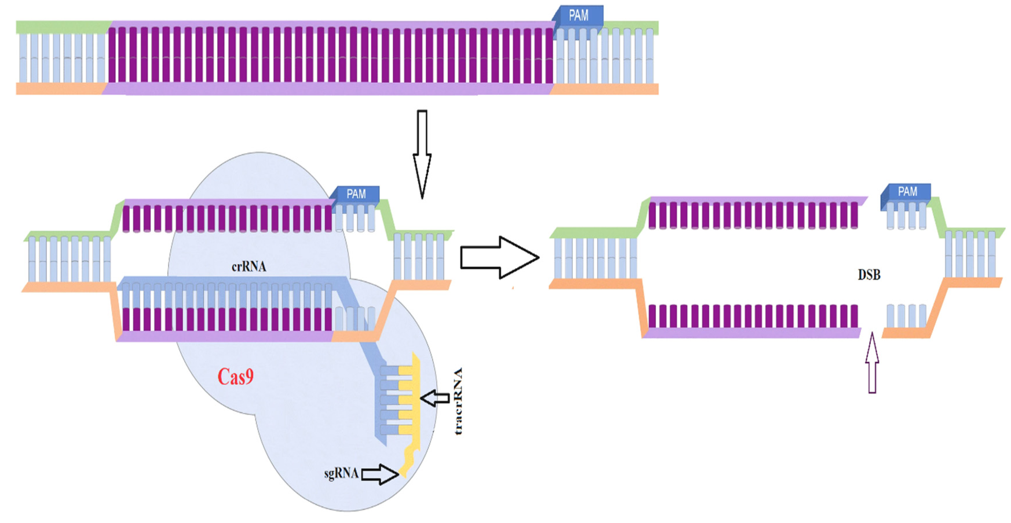
3.6. CRISPR-Cas9 Based Genome Modification

Clustered regularly interspaced short palindrome repeats (CRISPR) systems are DNA sequences present in bacteria that protect bacteriophages [135]. The Streptococcus pyogenes Type II CRISPR-Cas system has been widely investigated, and CRISPR-associated protein 9 (Cas9) is required for all stages of S. pyogenes immunity [136]. The CRISPR-Cas9 genome editing system is a novel and versatile method that has already been used to target and disrupt genes in both yeast and filamentous fungi and to perform gene editing in plants [137], embryos [100], and microorganisms [138,139,140]. The CRISPR-Cas9 system consists of two key components: a single guide RNA (sgRNA) and a Cas9 nickase [141]. The sgRNA is obtained by linking CRISPR RNA (crRNA), which is complementary to a particular part of the target gene, with trans-activating crRNA (tracrRNA). The sgRNA instructs the Cas9 nuclease to create a double-stranded nick with a protospacer adjacent motif (PAM) in the 20-bp target DNA (Figure 4). Chemical changes at specific sites of the sgRNA have also been found to boost the specificity of the Cas9 enzyme(s) and reduce off-target effects [142]. In filamentous fungi, low gene editing frequencies offer a major problem for genetic modification, particularly in Aspergillus spp. [139,143], Neurospora crassa [144], and Tichoderma reseei [145]. CRISPR-Cas9 has been used to solve this problem, and recently gained popularity in the field of genetic manipulation of microorganisms that cause mucormycosis.
CRISPR-Cas9-based genome editing in filamentous fungi can be conducted via a variety of approaches. Multiple plasmids can be constructed and transformed by an in vivo expression vector that contains the Cas9 gene and sequences the encoding parts of the guide RNA (gRNA), including crRNA, tracrRNA, and the protospacer fragment, ensuring their expression. Although these techniques are effective, using expression vectors for CRISPR-Cas9 genome editing has certain disadvantages. In addition, creating the required plasmid expressing cas9 and the proper gRNA can be time-consuming and difficult, and these systems allow the plasmids to persist and replicate after the genome editing event, which can limit the applicability of this approach and generate mutants. These mutants may contain bacterial antibiotic resistance genes and other bacterial sequences utilized for plasmid selection during development that are contraindicated in many uses. Furthermore, the presence of plasmid and foreign DNA over the long term may create off-target effects as well as undesired reorganization and degradation of the exogenous DNA and the genome, which are frequently reported in Mucorales fungi [40,99]. In the plasmid-free method, a ribonucleoprotein (RNP) complex generated by the Cas9 enzyme and in vitro transcribed gRNA is transformed into a cell to engineer the gene of interest. One of the advantages of this approach is that off-target effects can be avoided using this strategy; however, the RNP can be readily degraded. Recently, a novel CRISPR-Cas9 system was designed by Nagy et al. and proved as a tool for site-specific mutagenesis of M. circinelloides without the use of plasmids or in vitro RNP synthesis [146]. Their team employed this plasmid-free approach for targeted gene disruption of carB and hmgR2 genes in M. circinelloides, resulting in the generation of stable mutants. They directly introduced the guide RNA, Cas9 enzyme, and in the case of homology-directed repair (HDR), the template DNA into the recipient strain [146]. This plasmid-free technique has several advantages over the in vivo strategy, including the elimination of additional cloning processes, the avoidance of off-target effects due to the temporary exposure to Cas9 in transformed cells, and the ability of Cas9 to be active immediately after being transformed into the cells.
Similarly, for targeted gene disruption in Rhizopus, a plasmid-based approach was employed by Baldin et al., combining all of the necessary components into a single vector [147]. Based on the well-established fact that polyketide synthase (PKS) is an important enzyme that produces toxins in filamentous fungi [148], and its disruption results in transgenic fungi with a considerable reduction in their harmful effect on their hosts [149], Fuller and coworkers used a CRISPR system for targeting loss of function in PKS to study gene-related functions. They observed high editing efficiency (25–53%) and established that Cas9 expression is useful for Aspergillus fumigatus growth and other characteristics [139]. According to several studies, genetic modification using CRISPR-Cas9 in the fungus A. fumigatus has been successfully undertaken to demonstrate the importance of various genes in Aspergillus [150,151]. For example, Umeyama and coworkers applied CRISPR to replace the cyp51A gene in azole-resistant clinical isolates of A. fumigatus [150]. In this investigation, a Cas9-gRNA ribonucleoprotein complex and a donor template were supplied into cells simultaneously, followed by azole susceptibility testing in transformants that revealed enhanced susceptibility due to the replacement of Ser138 with glycine [150]. More recent studies have also indicated the applications of CRISPR-Cas9 technology for the synthesis of recombinant proteins in Aspergilli species [152,153]. These investigations showed that CRISPR-Cas9 performs well in Aspergillus research and that components of the system have a wide range of usefulness.
CRISPR-based techniques have been proven beneficial among the various technologies due to several reasons: they allow a simpler design procedure and more economical and faster execution compared with nuclease platforms, making them highly desirable engineering tools for industrially important microorganisms that do not have genetic tools for modification. The CRISPR system’s multiplexing features [154,155,156,157] can help it to perform better in the future in a variety of ways; for example, in the establishment of fungal cell factories for the synthesis of high-value added products and metabolites [157] or novel antifungal bioactive compounds [158], the system can be made more economical by techniques such as genome reduction to reduce undesired products [159] and developing sets of deletion mutants over fungal gene interaction systems, respectively [160].
Figure 4. Cas9 induced double-strand break in a target gene. The sgRNA comprises tracrRNA and crRNA. The crRNA is specific to the target DNA, and the Cas9 enzyme creates a double strand break upstream of the PAM motif [28].
Jof 07 01061 g004

4. Conclusions

Fungi of the phylum Mucoromycota have been reported with various functions and applications in research and industrial and medical fields. Ecologically, Mucoralean fungi represent a diverse group, but ecological roles and geographic distribution remain unidentified for the majority of taxa. Various recent advances have been made in understanding the genetics of Mucoromycota fungi, for instance, construction of recyclable markers for genome manipulation and silencing vectors, gene silencing with RNAi, and the revolution of the CRISPR-Cas9 gene editing system. This review discusses several important genetic modification tools and approaches which are used to manipulate the genome of model organisms of the order Mucorales for useful metabolite production and gene regulatory studies. These genetic tools have been successfully used to obtain valuable bioactive compounds from these industrially important fungi. Though progress has been made by overcoming challenges to genetic manipulation in these species, we are still far from fully understanding the Mucorales order. Genetic manipulation of Mucorales has some limitations due to the limited availability of genetic tools. There is a need to further explore the potential of these fungi and tune beneficial pathways for increased titer and yield of a variety of valuable compounds.

Author Contributions

Conceptualization, H.M. and Y.S.; methodology, H.M. and T.N.; software, J.Y.; validation, A.M.S. and Y.N.; formal analysis, H.M. and T.N.; investigation, J.Y.; resources, H.M. and T.N.; data curation, Y.N.; writing—original draft preparation, H.M. and T.N.; writing—review and editing, H.M., T.N. and Y.S.; visualization, A.M.S.; supervision, Y.S.; project administration, Y.S.; funding acquisition, Y.S. All authors have read and agreed to the published version of the manuscript.

Funding

This research was funded by TaiShan Industrial Experts Programme (tscy 20160101), National Natural Science Foundation of China (Grants No. 31972851 and 32101927) and Shandong Postdoctoral Innovation Project (202103031).

Institutional Review Board Statement

Not applicable.

Informed Consent Statement

Not applicable.

Conflicts of Interest

The authors declare no conflict of interest.

References

  1. Walther, G.; Wagner, L.; Kurzai, O. Updates on the taxonomy of mucorales with an emphasis on clinically important taxa. J. Fungi 2019, 5, 106. [Google Scholar] [CrossRef] [PubMed] [Green Version]
  2. Benny, G.L.; Humber, R.A.; Morton, J.B. Zygomycota: Zygomycetes. In Systematics and Evolution; Springer: Berlin/Heidelberg, Germany, 2001; pp. 113–146. [Google Scholar]
  3. Hibbett, D.S.; Binder, M.; Bischoff, J.F.; Blackwell, M.; Cannon, P.F.; Eriksson, O.E.; Huhndorf, S.; James, T.; Kirk, P.M.; Lücking, R.; et al. A higher-level phylogenetic classification of the Fungi. Mycol. Res. 2007, 111, 509–547. [Google Scholar] [CrossRef] [PubMed]
  4. Spatafora, J.W.; Chang, Y.; Benny, G.L.; Lazarus, K.; Smith, M.E.; Berbee, M.L.; Bonito, G.; Corradi, N.; Grigoriev, I.; Gryganskyi, A.; et al. A phylum-level phylogenetic classification of zygomycete fungi based on genome-scale data. Mycologia 2016, 108, 1028–1046. [Google Scholar] [CrossRef] [PubMed] [Green Version]
  5. Morin-Sardin, S.; Nodet, P.; Coton, E.; Jany, J.L. Mucor: A Janus-faced fungal genus with human health impact and industrial applications. Fungal Biol. Rev. 2017, 31, 12–32. [Google Scholar] [CrossRef]
  6. Lübbehüsen, T.L.; Nielsen, J.; McIntyre, M. Characterization of the Mucor circinelloides life cycle by on-line image analysis. J. Appl. Microbiol. 2003, 95, 1152–1160. [Google Scholar] [CrossRef] [Green Version]
  7. Lee, S.C.; Li, A.; Calo, S.; Heitman, J. Calcineurin Plays Key Roles in the Dimorphic Transition and Virulence of the Human Pathogenic Zygomycete Mucor circinelloides. PLoS Pathog. 2013, 9, e1003625. [Google Scholar] [CrossRef] [Green Version]
  8. Lee, S.C.; Li, A.; Calo, S.; Inoue, M.; Tonthat, N.K.; Bain, J.M.; Louw, J.; Shinohara, M.L.; Erwig, L.P.; Schumacher, M.A.; et al. Calcineurin orchestrates dimorphic transitions, antifungal drug responses and host-pathogen interactions of the pathogenic mucoralean fungus Mucor circinelloides. Mol. Microbiol. 2015, 97, 844–865. [Google Scholar] [CrossRef] [Green Version]
  9. Iwen, P.C.; Thapa, I.; Bastola, D. Review of methods for the identification of zygomycetes with an emphasis on advances in molecular diagnostics. Lab. Med. 2011, 42, 260–266. [Google Scholar] [CrossRef] [Green Version]
  10. Thakur, A.; Pahwa, R.; Singh, S.; Gupta, R. Production, purification, and characterization of polygalacturonase from mucor circinelloides ITCC 6025. Enzyme Res. 2010, 2010, 1–7. [Google Scholar] [CrossRef] [Green Version]
  11. Magnuson, J.K.; Lasure, L.L. Organic Acid Production by Filamentous Fungi. In Advances in Fungal Biotechnology for Industry, Agriculture, and Medicine; Springer: Boston, MA, USA, 2004. [Google Scholar]
  12. Zhang, K.; Yang, S.T.; Chalmers, J.J.; Wood, D. Fumaric Acid Fermentation by Rhizopus oryzae with Integrated Separation Technologies; The Ohio State University: Columbus, OH, USA, 2012. [Google Scholar]
  13. Papp, T.; Nyilasi, I.; Csernetics, Á.; Nagy, G.; Takó, M.; Vágvölgyi, C. Improvement of Industrially Relevant Biological Activities in Mucoromycotina Fungi. In Gene Expression Systems in Fungi: Advancements and Applications. Fungal Biology; Schmoll, M., Dattenböck, C., Eds.; Springer: Cham, Switzerland, 2016; pp. 97–118. [Google Scholar]
  14. Naz, T.; Nosheen, S.; Li, S.; Nazir, Y.; Mustafa, K.; Liu, Q.; Garre, V.; Song, Y. Comparative analysis of β-carotene production by mucor circinelloides strains CBS 277.49 and WJ11 under light and dark conditions. Metabolites 2020, 10, 38. [Google Scholar] [CrossRef] [Green Version]
  15. Dinesh Babu, P.; Bhakyaraj, R.; Vidhyalakshmi, R. A Low Cost Nutritious Food “Tempeh”—A Review. World J. Dairy Food Sci. 2009, 4, 22–27. [Google Scholar]
  16. Riley, T.T.; Muzny, C.A.; Swiatlo, E.; Legendre, D.P. Breaking the Mold: A Review of Mucormycosis and Current Pharmacological Treatment Options. Ann. Pharmacother. 2016, 50, 747–757. [Google Scholar] [CrossRef]
  17. Corrochano, L.M.; Garre, V. Photobiology in the Zygomycota: Multiple photoreceptor genes for complex responses to light. Fungal Genet. Biol. 2010, 47, 893–899. [Google Scholar] [CrossRef]
  18. Ruiz-Vázquez, R.M.; Nicolás, F.E.; Torres-Martínez, S.; Garre, V. Distinct RNAi Pathways in the Regulation of Physiology and Development in the Fungus Mucor circinelloides. Adv. Genet. 2015, 91, 55–102. [Google Scholar] [CrossRef]
  19. Wang, L.; Lin, X. Morphogenesis in Fungal Pathogenicity: Shape, Size, and Surface. PLoS Pathog. 2012, 8, e1003027. [Google Scholar] [CrossRef] [Green Version]
  20. Wöstemeyer, J.; Schimek, C.; Wetzel, J.; Burmester, A.; Voigt, J.; Schulz, E.; Ellenberger, S.; Siegmund, L. 10 Pheromone Action in the Fungal Groups Chytridiomycetes and Zygomycetes and in the Oophytes. In Growth, Differentiation and Sexuality; Springer: Singapore, 2016. [Google Scholar]
  21. Ibrahim, A.S.; Gebremariam, T.; Lin, L.; Luo, G.; Husseiny, M.I.; Skory, C.D.; Fu, Y.; French, S.W.; Edwards, J.E.; Spellberg, B. The high affinity iron permease is a key virulence factor required for Rhizopus oryzae pathogenesis. Mol. Microbiol. 2010, 77, 587–604. [Google Scholar] [CrossRef] [Green Version]
  22. Gebremariam, T.; Liu, M.; Luo, G.; Bruno, V.; Phan, Q.T.; Waring, A.J.; Edwards, J.E.; Filler, S.G.; Yeaman, M.R.; Ibrahim, A.S. CotH3 mediates fungal invasion of host cells during mucormycosis. J. Clin. Investig. 2014, 124, 237–250. [Google Scholar] [CrossRef] [Green Version]
  23. Zhang, Y.; Navarro, E.; Cánovas-Márquez, J.T.; Almagro, L.; Chen, H.; Chen, Y.Q.; Zhang, H.; Torres-Martínez, S.; Chen, W.; Garre, V. A new regulatory mechanism controlling carotenogenesis in the fungus Mucor circinelloides as a target to generate β-carotene over-producing strains by genetic engineering. Microb. Cell Fact. 2016, 15, 1–14. [Google Scholar] [CrossRef] [Green Version]
  24. Kosa, G.; Zimmermann, B.; Kohler, A.; Ekeberg, D.; Afseth, N.K.; Mounier, J.; Shapaval, V. High-throughput screening of Mucoromycota fungi for production of low- and high-value lipids. Biotechnol. Biofuels 2018, 11, 66. [Google Scholar] [CrossRef]
  25. Orlowski, M. Mucor dimorphism. Microbiol. Rev. 1991, 55, 234–258. [Google Scholar] [CrossRef]
  26. Wolff, A.M.; Appel, K.F.; Petersen, J.B.; Poulsen, U.; Arnau, J. Identification and analysis of genes involved in the control of dimorphism in Mucor circinelloides (syn. racemosus). FEMS Yeast Res. 2002, 2, 203–213. [Google Scholar] [CrossRef] [Green Version]
  27. Patiño-Medina, J.A.; Maldonado-Herrera, G.; Pérez-Arques, C.; Alejandre-Castañeda, V.; Reyes-Mares, N.Y.; Valle-Maldonado, M.I.; Campos-García, J.; Ortiz-Alvarado, R.; Jácome-Galarza, I.E.; Ramírez-Díaz, M.I.; et al. Control of morphology and virulence by ADP-ribosylation factors (Arf) in Mucor circinelloides. Curr. Genet. 2018, 64, 853–869. [Google Scholar] [CrossRef]
  28. Garcia, A.; Vellanki, S.; Lee, S.C. Genetic Tools for Investigating Mucorales Fungal Pathogenesis. Curr. Clin. Microbiol. Rep. 2018, 5, 173–180. [Google Scholar] [CrossRef]
  29. Ankeny, R.A.; Leonelli, S. What’s so special about model organisms? Stud. Hist. Philos. Sci. Part A 2011, 42, 313–323. [Google Scholar] [CrossRef]
  30. Wilson, R.K. How the worm was won: The C. elegans genome sequencing project. Trends Genet. 1999, 15, 51–58. [Google Scholar] [CrossRef]
  31. Clendinen, C.S.; Stupp, G.S.; Wang, B.; Garrett, T.J.; Edison, A.S. Current Metabolomics BENTHAM SCIENCE Send Orders for Reprints to reprints@benthamscience.ae 13 C Metabolomics: NMR and IROA for Unknown Identification. Curr. Metab. 2016, 4, 116–120. [Google Scholar]
  32. Uppuluri, P.; Alqarihi, A.; Ibrahim, A.S. Mucormycoses. In Encyclopedia of Mycology; Zaragoza, Ó., Casadevall, A.B.T.-E., Eds.; Elsevier: Oxford, UK, 2021; pp. 600–612. ISBN 978-0-323-85180-0. [Google Scholar]
  33. Corrochano, L.M.; Kuo, A.; Marcet-Houben, M.; Polaino, S.; Salamov, A.; Villalobos-Escobedo, J.M.; Grimwood, J.; Álvarez, M.I.; Avalos, J.; Bauer, D.; et al. Expansion of Signal Transduction Pathways in Fungi by Extensive Genome Duplication. Curr. Biol. 2016, 26, 1577–1584. [Google Scholar] [CrossRef] [Green Version]
  34. McIntyre, M.; Breum, J.; Arnau, J.; Nielsen, J. Growth physiology and dimorphism of Mucor circinelloides (syn. racemosus) during submerged batch cultivation. Appl. Microbiol. Biotechnol. 2002, 58, 495–502. [Google Scholar] [CrossRef]
  35. Papp, T.; Velayos, A.; Bartók, T.; Eslava, A.P.; Vágvölgyi, C.; Iturriaga, E.A. Heterologous expression of astaxanthin biosynthesis genes in Mucor circinelloides. Appl. Microbiol. Biotechnol. 2006, 69, 526–531. [Google Scholar] [CrossRef]
  36. van Heeswijck, R.; Roncero, M.I.G. High frequency transformation of Mucor with recombinant plasmid DNA. Carlsberg Res. Commun. 1984, 49, 691–702. [Google Scholar] [CrossRef] [Green Version]
  37. Garre, V.; Barredo, J.L.; Iturriaga, E.A. Transformation of Mucor circinelloides f. lusitanicus Protoplasts. Fungal Biol. 2015, 49–59. [Google Scholar]
  38. Nyilasi, I.; Ács, K.; Papp, T.; Nagy, E.; Vágvölgyi, C. Agrobacterium tumefaciens-mediated transformation of Mucor circinelloides. Folia Microbiol. 2005, 50, 415–420. [Google Scholar] [CrossRef] [PubMed]
  39. González-Hernández, G.A.; Herrera-Estrella, L.; Rocha-Ramírez, V.; Roncero, M.I.G.; Gutierrez-Corona, J.F. Biolistic transformation of Mucor circinelloides. Mycol. Res. 1997, 101, 953–956. [Google Scholar] [CrossRef]
  40. Papp, T.; Csernetics, Á.; Nyilasi, I.; Ábrók, M.; Vágvólgyi, C. Genetic transformation of zygomycetes fungi. In Progress in Mycology; Springer: Singapore, 2010; ISBN 9789048137138. [Google Scholar]
  41. Papp, T.; Csernetics, Á.; Nagy, G.; Bencsik, O.; Iturriaga, E.A.; Eslava, A.P.; Vágvölgyi, C. Canthaxanthin production with modified Mucor circinelloides strains. Appl. Microbiol. Biotechnol. 2013, 97, 4937–4950. [Google Scholar] [CrossRef]
  42. Nicolás, F.E.; Vila, A.; Moxon, S.; Cascales, M.D.; Torres-Martínez, S.; Ruiz-Vázquez, R.M.; Garre, V. The RNAi machinery controls distinct responses to environmental signals in the basal fungus Mucor circinelloides. BMC Genom. 2015, 16, 237. [Google Scholar] [CrossRef] [Green Version]
  43. Li, C.H.; Cervantes, M.; Springer, D.J.; Boekhout, T.; Ruiz-Vazquez, R.M.; Torres-Martinez, S.R.; Heitman, J.; Lee, S.C. Sporangiospore size dimorphism is linked to virulence of mucor circinelloides. PLoS Pathog. 2011, 7, e1002086. [Google Scholar] [CrossRef]
  44. Nosheen, S.; Yang, J.; Naz, T.; Nazir, Y.; Ahmad, M.I.; Fazili, A.B.A.; Li, S.; Mustafa, K.; Song, Y. Annotation of AMP-activated protein kinase genes and its comparative transcriptional analysis between high and low lipid producing strains of Mucor circinelloides. Biotechnol. Lett. 2020, 43, 1–10. [Google Scholar] [CrossRef]
  45. Hameed, A.; Hussain, S.A.; Ijaz, M.U.; Ullah, S.; Muhammad, Z.; Suleria, H.A.R.; Song, Y. Antioxidant activity of polyphenolic extracts of filamentous fungus Mucor circinelloides (WJ11): Extraction, characterization and storage stability of food emulsions. Food Biosci. 2020, 34, 100525. [Google Scholar] [CrossRef]
  46. Khan, M.A.K.; Yang, J.; Hussain, S.A.; Zhang, H.; Liang, L.; Garre, V.; Song, Y. Construction of DGLA producing cell factory by genetic modification of Mucor circinelloides. Microb. Cell Fact. 2019, 18, 64. [Google Scholar] [CrossRef] [Green Version]
  47. Csernetics, Á.; Tóth, E.; Farkas, A.; Nagy, G.; Bencsik, O.; Manikandan, P.; Vágvölgyi, C.; Papp, T. Expression of a bacterial ß-carotene hydroxylase in canthaxanthin producing mutant Mucor circinelloides strains. Acta Biol. Szeged. 2014, 58, 139–146. [Google Scholar]
  48. Csernetics, Á.; Tóth, E.; Farkas, A.; Nagy, G.; Bencsik, O.; Vágvölgyi, C.; Papp, T. Expression of Xanthophyllomyces dendrorhous cytochrome-P450 hydroxylase and reductase in Mucor circinelloides. World J. Microbiol. Biotechnol. 2015, 31, 321–336. [Google Scholar] [CrossRef] [Green Version]
  49. Meussen, B.J.; De Graaff, L.H.; Sanders, J.P.M.; Weusthuis, R.A. Metabolic engineering of Rhizopus oryzae for the production of platform chemicals. Appl. Microbiol. Biotechnol. 2012, 94, 875–886. [Google Scholar] [CrossRef] [Green Version]
  50. Roden, M.M.; Zaoutis, T.E.; Buchanan, W.L.; Knudsen, T.A.; Sarkisova, T.A.; Schaufele, R.L.; Sein, M.; Sein, T.; Chiou, C.C.; Chu, J.H.; et al. Epidemiology and outcome of zygomycosis: A review of 929 reported cases. Clin. Infect. Dis. 2005, 41, 634–656. [Google Scholar] [CrossRef] [Green Version]
  51. Oda, Y.; Yajima, Y.; Kinoshita, M.; Ohnishi, M. Differences of Rhizopus oryzae strains in organic acid synthesis and fatty acid composition. Food Microbiol. 2003, 20, 371–375. [Google Scholar] [CrossRef]
  52. Michielse, C.B.; Salim, K.; Ragas, P.; Ram, A.F.J.; Kudla, B.; Jarry, B.; Punt, P.J.; Van Den Hondel, C.A.M.J.J. Development of a system for integrative and stable transformation of the zygomycete Rhizopus oryzae by Agrobacterium-mediated DNA transfer. Mol. Genet. Genom. 2004, 271, 499–510. [Google Scholar] [CrossRef]
  53. Klein, T.M.; Sanford, J.C.; Wolf, E.D.; Wu, R. High-velocity microprojectiles for delivering nucleic acids into living cells. Nature 1987, 327, 70–73. [Google Scholar] [CrossRef]
  54. Tang, X.; Chang, L.; Gu, S.; Zhang, H.; Chen, Y.Q.; Chen, H.; Zhao, J.; Chen, W. Role of beta-isopropylmalate dehydrogenase in lipid biosynthesis of the oleaginous fungus Mortierella alpine. Fungal Genet. Biol. 2021, 152, 103572. [Google Scholar] [CrossRef]
  55. Totani, N.; Oba, K. The filamentous fungus mortierella alpina, high in arachidonic acid. Lipids 1987, 22, 1060–1062. [Google Scholar] [CrossRef]
  56. Ge, C.; Chen, H.; Mei, T.; Tang, X.; Chang, L.; Gu, Z.; Zhang, H.; Chen, W.; Chen, Y.Q. Application of a ω-3 desaturase with an arachidonic acid preference to eicosapentaenoic acid production in Mortierella alpina. Front. Bioeng. Biotechnol. 2018, 5, 89. [Google Scholar] [CrossRef] [Green Version]
  57. Ando, A.; Sumida, Y.; Negoro, H.; Suroto, D.A.; Ogawa, J.; Sakuradani, E.; Shimizu, S. Establishment of Agrobacterium tumefaciens-mediated transformation of an oleaginous fungus, Mortierella alpina 1S-4, and its application for eicosapentaenoic acid producer breeding. Appl. Environ. Microbiol. 2009, 75, 5529–5535. [Google Scholar] [CrossRef] [Green Version]
  58. Hao, G.; Chen, H.; Wang, L.; Gu, Z.; Song, Y.; Zhang, H.; Chen, W.; Chen, Y.Q. Role of malic enzyme during fatty acid synthesis in the oleaginous fungus Mortierella alpina. Appl. Environ. Microbiol. 2014, 80, 2672–2678. [Google Scholar] [CrossRef] [Green Version]
  59. Lu, H.; Chen, H.; Tang, X.; Yang, Q.; Zhang, H.; Chen, Y.Q.; Chen, W. Time-resolved multi-omics analysis reveals the role of nutrient stress-induced resource reallocation for TAG accumulation in oleaginous fungus Mortierella alpina. Biotechnol. Biofuels 2020, 13, 1–17. [Google Scholar] [CrossRef]
  60. Hao, G.; Chen, H.; Gu, Z.; Zhang, H.; Chen, W.; Chen, Y.Q. Metabolic engineering of Mortierella alpina for enhanced arachidonic acid production through the NADPH-supplying strategy. Appl. Environ. Microbiol. 2016, 82, 3280–3288. [Google Scholar] [CrossRef] [Green Version]
  61. Chen, H.; Hao, G.; Wang, L.; Wang, H.; Gu, Z.; Liu, L.; Zhang, H.; Chen, W.; Chen, Y.Q. Identification of a critical determinant that enables efficient fatty acid synthesis in oleaginous fungi. Sci. Rep. 2015, 5, 11247. [Google Scholar] [CrossRef]
  62. Vongsangnak, W.; Ruenwai, R.; Tang, X.; Hu, X.; Zhang, H.; Shen, B.; Song, Y.; Laoteng, K. Genome-scale analysis of the metabolic networks of oleaginous Zygomycete fungi. Gene 2013, 521, 180–190. [Google Scholar] [CrossRef]
  63. Kawashima, H.; Akimoto, K.; Higashiyama, K.; Fujikawa, S.; Shimizu, S. Industrial production of dihomo-γ-linolenic acid by a Δ5 desaturase-defective mutant of Mortierella alpina 1S-4 fungus. J. Am. Oil Chem. Soc. 2000, 77, 1135. [Google Scholar] [CrossRef]
  64. Li, X.; Liu, R.; Li, J.; Chang, M.; Liu, Y.; Jin, Q.; Wang, X. Enhanced arachidonic acid production from Mortierella alpina combining atmospheric and room temperature plasma (ARTP) and diethyl sulfate treatments. Bioresour. Technol. 2015, 177, 134–140. [Google Scholar] [CrossRef]
  65. Mackenzie, D.A.; Wongwathanarat, P.; Carter, A.T.; Archer, D.B. Isolation and use of a homologous histone H4 promoter and a ribosomal DNA region in a transformation vector for the oil-producing fungus Mortierella alpina. Appl. Environ. Microbiol. 2000, 66, 4655–4661. [Google Scholar] [CrossRef] [Green Version]
  66. Takeno, S.; Sakuradani, E.; Murata, S.; Inohara-Ochiai, M.; Kawashima, H.; Ashikari, T.; Shimizu, S. Establishment of an overall transformation system for an oil-producing filamentous fungus, Mortierella alpina 1S-4. Appl. Microbiol. Biotechnol. 2004, 65, 419–425. [Google Scholar] [CrossRef]
  67. Zhang, H.; Cui, Q.; Song, X. Research advances on arachidonic acid production by fermentation and genetic modification of Mortierella alpina. World J. Microbiol. Biotechnol. 2021, 37, 1–9. [Google Scholar] [CrossRef]
  68. Sakuradani, E. Advances in the production of various polyunsaturated fatty acids through oleaginous fungus Mortierella alpina breeding. Biosci. Biotechnol. Biochem. 2010, 74, 908–917. [Google Scholar] [CrossRef] [PubMed]
  69. Takeno, S.; Sakuradani, E.; Tomi, A.; Inohara-Ochiai, M.; Kawashima, H.; Shimizu, S. Transformation of oil-producing fungus, Mortierella alpina 1S-4, using Zeocin, and application to arachidonic acid production. J. Biosci. Bioeng. 2005, 100, 617–622. [Google Scholar] [CrossRef] [PubMed]
  70. Hao, G.; Chen, H.; Gu, Z.; Zhang, H.; Chen, W.; Chen, Y.Q. Metabolic engineering of Mortierella alpina for arachidonic acid production with glycerol as carbon source. Microb. Cell Fact. 2015, 14, 1–14. [Google Scholar] [CrossRef] [PubMed] [Green Version]
  71. Zhang, H.; Feng, Y.; Cui, Q.; Song, X. Expression of Vitreoscilla hemoglobin enhances production of arachidonic acid and lipids in Mortierella alpina. BMC Biotechnol. 2017, 17, 68. [Google Scholar] [CrossRef]
  72. de Hoog, G.S.; Guarro, J.; Gené, J.; Figueras, M.J. Atlas of Clinical Fungi; Amer Society for Microbiology: Baarn, The Netherlands, 2000; ISBN 9070351439. [Google Scholar]
  73. Larone, D.H. Medically Important Fungi-A Guide to Identification, 3rd ed.; ASM Press: Washington, DC, USA, 1995. [Google Scholar]
  74. Sutton, D.A.; Fothergill, A.W.; Rinaldi, M.G. Guide to Clinically Significant Fungi; Williams & Wilkins: Baltimore, MD, USA, 1998; ISBN 0683182749. [Google Scholar]
  75. Asha, S.; Vidyavathi, M. Cunninghamella-A microbial model for drug metabolism studies—A review. Biotechnol. Adv. 2009, 27, 16–29. [Google Scholar] [CrossRef]
  76. Zhang, D.; Yang, Y.; Leakey, J.E.A.; Cerniglia, C.E. Phase I and phase II enzymes produced by Cunninghamella elegans for the metabolism of xenobiotics. FEMS Microbiol. Lett. 1996, 138, 221–226. [Google Scholar] [CrossRef]
  77. Parshikov, I.A.; Muraleedharan, K.M.; Avery, M.A.; Williamson, J.S. Transformation of artemisinin by Cunninghamella elegans. Appl. Microbiol. Biotechnol. 2004, 64, 782–786. [Google Scholar] [CrossRef]
  78. Skinner, C.E.; Emmons, C.W.; Tsuchiya, H.M. Molds, Yeasts, and Actinomycetes. Soil Sci. 1947, 63, 418. [Google Scholar] [CrossRef]
  79. El-Morsy, E.S.M. Cunninghamella echinulata a new biosorbent of metal ions from polluted water in Egypt. Mycologia 2004, 96, 1183–1189. [Google Scholar] [CrossRef]
  80. Gema, H.; Kavadia, A.; Dimou, D.; Tsagou, V.; Komaitis, M.; Aggelis, G. Production of γ-linolenic acid by Cunninghamella echinulata cultivated on glucose and orange peel. Appl. Microbiol. Biotechnol. 2002, 58, 303–307. [Google Scholar] [CrossRef]
  81. Fakas, S.; Papanikolaou, S.; Galiotou-Panayotou, M.; Komaitis, M.; Aggelis, G. Organic nitrogen of tomato waste hydrolysate enhances glucose uptake and lipid accumulation in Cunninghamella echinulata. J. Appl. Microbiol. 2008, 105, 1062–1070. [Google Scholar] [CrossRef]
  82. Papanikolaou, S.; Galiotou-Panayotou, M.; Fakas, S.; Komaitis, M.; Aggelis, G. Lipid production by oleaginous Mucorales cultivated on renewable carbon sources. Eur. J. Lipid Sci. Technol. 2007, 109, 1060–1070. [Google Scholar] [CrossRef]
  83. Chen, H.C.; Liu, T.M. Inoculum effects on the production of γ-linolenic acid by the shake culture of Cunninghamella echinulata CCRC 31840. Enzyme Microb. Technol. 1997, 21, 137–142. [Google Scholar] [CrossRef]
  84. Freitag, D.G.; Foster, R.T.; Coutts, R.T.; Pickard, M.A.; Pasutto, F.M. Stereoselective metabolism of rac-mexiletine by the fungus Cunninghamella echinulata yields the major human metabolites hydroxymethylmexiletine and p-hydroxymexiletine. Drug Metab. Dispos. 1997, 25, 685–692. [Google Scholar]
  85. Wan, X.; Zhang, Y.; Wang, P.; Huang, F.; Chen, H.; Jiang, M. Production of gamma-linolenic acid in Pichia pastoris by expression of a delta-6 desaturase gene from Cunninghamella echinulata. J. Microbiol. Biotechnol. 2009, 1098–1102. [Google Scholar] [CrossRef]
  86. LeBlanc, R.E.; Meriden, Z.; Sutton, D.A.; Thompson, E.H.; Neofytos, D.; Zhang, S.X. Cunninghamella echinulata causing fatally invasive fungal sinusitis. Diagn. Microbiol. Infect. Dis. 2013, 76, 506–509. [Google Scholar] [CrossRef]
  87. Manosroi, J.; Chisti, Y.; Manosroi, A. Biotransformation of cortexolone to hydrocortisone by molds using a rapid color-development assay. Appl. Biochem. Microbiol. 2006, 42, 479–483. [Google Scholar] [CrossRef]
  88. Seigle-Murandi, F.M.; Krivobok, S.M.A.; Steiman, R.L.; Benoit-Guyod, J.L.A.; Thiault, G.A. Biphenyl Oxide Hydroxylation by Cunninghamella echinulata. J. Agric. Food Chem. 1991, 39, 428–430. [Google Scholar] [CrossRef]
  89. Pekala, E.; Kubowicz, P.; Lazewska, D. Cunninghamella as a microbiological model for metabolism of histamine H3 receptor antagonist 1-[3-(4-tert-butylphenoxy)propyl]piperidine. Appl. Biochem. Biotechnol. 2012, 168, 1584–1593. [Google Scholar] [CrossRef] [Green Version]
  90. Zhang, Y.; Luan, X.; Zhang, H.; Garre, V.; Song, Y.; Ratledge, C. Improved γ-linolenic acid production in Mucor circinelloides by homologous overexpressing of delta-12 and delta-6 desaturases. Microb. Cell Fact. 2017, 16, 113. [Google Scholar] [CrossRef]
  91. Yang, J.; Li, S.; Kabir Khan, M.A.; Garre, V.; Vongsangnak, W.; Song, Y. Increased lipid accumulation in Mucor circinelloides by overexpression of mitochondrial citrate transporter genes. Ind. Eng. Chem. Res. 2019, 58, 2125–2134. [Google Scholar] [CrossRef]
  92. Nosheen, S.; Naz, T.; Yang, J.; Hussain, S.A.; Fazili, A.B.A.; Nazir, Y.; Li, S.; Mohamed, H.; Yang, W.; Mustafa, K. Role of Snf-β in lipid accumulation in the high lipid-producing fungus Mucor circinelloides WJ11. Microb. Cell Fact. 2021, 20, 1–11. [Google Scholar] [CrossRef]
  93. Csernetics, Á.; Nagy, G.; Iturriaga, E.A.; Szekeres, A.; Eslava, A.P.; Vágvölgyi, C.; Papp, T. Expression of three isoprenoid biosynthesis genes and their effects on the carotenoid production of the zygomycete Mucor circinelloides. Fungal Genet. Biol. 2011, 48, 696–703. [Google Scholar] [CrossRef]
  94. Naz, T.; Yang, J.; Nosheen, S.; Sun, C.; Nazir, Y.; Mohamed, H.; Fazili, A.A.; Ullah, S.; Li, S.; Yang, W. Genetic modification of Mucor circinelloides for canthaxanthin production by heterologous expression of β-carotene ketolase gene. Front. Nutr. 2021, 718. [Google Scholar] [CrossRef]
  95. Jin, M.J.; Huang, H.; Xiao, A.H.; Gao, Z.; Liu, X.; Peng, C. Enhancing arachidonic acid production by Mortierella alpina ME-1 using improved mycelium aging technology. Bioprocess. Biosyst. Eng. 2009, 32, 117–122. [Google Scholar] [CrossRef]
  96. Huang, M.; Chen, H.; Tang, X.; Lu, H.; Zhao, J.; Zhang, H.; Chen, Y.; Chen, W. Two-stage pH control combined with oxygen-enriched air strategies for the highly efficient production of EPA by Mortierella alpina CCFM698 with fed-batch fermentation. Bioprocess. Biosyst. Eng. 2020, 43, 1725–1733. [Google Scholar] [CrossRef]
  97. Takeno, S.; Sakuradani, E.; Tomi, A.; Inohara-Ochiai, M.; Kawashima, H.; Ashikari, T.; Shimizu, S. Improvement of the fatty acid composition of an oil-producing filamentous fungus, Mortierella alpina 1S-4, through RNA interference with Δ12-desaturase gene expression. Appl. Environ. Microbiol. 2005, 71, 4992–4997. [Google Scholar] [CrossRef] [Green Version]
  98. Gheinani, A.H.; Jahromi, N.H.; Feuk-Lagerstedt, E.; Taherzadeh, M.J. RNA silencing of lactate dehydrogenase gene in Rhizopus oryzae. J. RNAi Gene Silenc. 2011, 7, 443–448. [Google Scholar]
  99. Ibrahim, A.S.; Skory, C.D. Genetic Manipulation of Zygomycetes. In Medical Mycology: Cellular and Molecular Techniques; Wiley: New York, NY, USA, 2007; ISBN 9780470019238. [Google Scholar]
  100. Trieu, T.A.; Navarro-Mendoza, M.I.; Pérez-Arques, C.; Sanchis, M.; Capilla, J.; Navarro-Rodriguez, P.; Lopez-Fernandez, L.; Torres-Martínez, S.; Garre, V.; Ruiz-Vázquez, R.M.; et al. RNAi-Based Functional Genomics Identifies New Virulence Determinants in Mucormycosis. PLoS Pathog. 2017, 13, e1006150. [Google Scholar] [CrossRef] [Green Version]
  101. Calo, S.; Shertz-Wall, C.; Lee, S.C.; Bastidas, R.J.; Nicolás, F.E.; Granek, J.A.; Mieczkowski, P.; Torres-Martínez, S.; Ruiz-Vázquez, R.M.; Cardenas, M.E.; et al. Antifungal drug resistance evoked via RNAi-dependent epimutations. Nature 2014, 513, 555–558. [Google Scholar] [CrossRef] [PubMed] [Green Version]
  102. Murray, N.E. Type I Restriction Systems: Sophisticated Molecular Machines (a Legacy of Bertani and Weigle). Microbiol. Mol. Biol. Rev. 2000, 64, 412–434. [Google Scholar] [CrossRef] [PubMed] [Green Version]
  103. Shankar, S.; Uddin, I.; Afzali, S.F. Restriction endonucleases. In Plant Biotechnology, Volume 1: Principles, Techniques, and Applications; CRC Press Book: Boca Raton, FL, USA, 2017; ISBN 9781351819510. [Google Scholar]
  104. Brown, D.H.; Slobodkin, I.V.; Kumamoto, C.A. Stable transformation and regulated expression of an inducible reporter construct in Candida albicans using restriction enzyme-mediated integration. Mol. Gen. Genet. 1996, 251, 75–80. [Google Scholar] [CrossRef] [PubMed]
  105. Brown, J.S.; Aufauvre-Brown, A.; Holden, D.W. Insertional mutagenesis of Aspergillus fumigatus. Mol. Gen. Genet. 1998, 259, 327–335. [Google Scholar] [CrossRef] [PubMed]
  106. Warrilow, A.G.S.; Parker, J.E.; Price, C.L.; Nes, W.D.; Kelly, S.L.; Kelly, D.E. In Vitro biochemical study of CYP51-mediated azole resistance in Aspergillus fumigatus. Antimicrob. Agents Chemother. 2015, 59, 7771–7778. [Google Scholar] [CrossRef] [Green Version]
  107. Jiang, C.; Dong, D.; Yu, B.; Cai, G.; Wang, X.; Ji, Y.; Peng, Y. Mechanisms of azole resistance in 52 clinical isolates of Candida tropicalis in China. J. Antimicrob. Chemother. 2013, 68, 778–785. [Google Scholar] [CrossRef]
  108. Hussain, S.A.; Hameed, A.; Khan, M.A.K.; Zhang, Y.; Zhang, H.; Garre, V.; Song, Y. Engineering of fatty acid synthases (FASs) to boost the production of medium-chain fatty acids (MCFAs) in Mucor circinelloides. Int. J. Mol. Sci. 2019, 20, 786. [Google Scholar] [CrossRef] [Green Version]
  109. Garcia, A.; Adedoyin, G.; Heitman, J.; Lee, S.C. Construction of a recyclable genetic marker and serial gene deletions in the human pathogenic mucorales Mucor circinelloides. G3 Genes Genomes Genet. 2017, 7, 2047–2054. [Google Scholar] [CrossRef] [Green Version]
  110. Roncero, M.I.; Jepsen, L.P.; Stroman, P.; van Heeswijck, R. Characterization of a leuA gene and an ARS element from Mucor circinelloides. Gene 1989, 84, 335–343. [Google Scholar] [CrossRef]
  111. Skory, C.D.; Ibrahim, A.S. Native and modified lactate dehydrogenase expression in a fumaric acid producing isolate Rhizopus oryzae 99-880. Curr. Genet. 2007, 52, 23–33. [Google Scholar] [CrossRef]
  112. Skory, C.D.; Freer, S.N.; Bothast, R.J. Production of L-lactic acid by Rhizopus oryzae under oxygen limiting conditions. Biotechnol. Lett. 1998, 20, 191–194. [Google Scholar] [CrossRef]
  113. Bai, D.M.; Zhao, X.M.; Li, X.G.; Xu, S.M. Strain improvement of Rhizopus oryzae for over-production of L(+)-lactic acid and metabolic flux analysis of mutants. Biochem. Eng. J. 2004, 18, 41–48. [Google Scholar] [CrossRef]
  114. Ge, C.M.; Gu, S.B.; Zhou, X.H.; Yao, J.M.; Pan, R.R.; Yu, Z.L. Breeding of L(+)-lactic acid producing strain by low-energy ion implantation. J. Microbiol. Biotechnol. 2004, 14, 363–366. [Google Scholar]
  115. Velayos, A.; López-Matas, M.A.; Ruiz-Hidalgo, M.J.; Eslava, A.P. Complementation analysis of carotenogenic mutants of Mucor circinelloides. Fungal Genet. Biol. 1997, 22, 19–27. [Google Scholar] [CrossRef]
  116. Vellanki, S.; Navarro-Mendoza, M.I.; Garcia, A.; Murcia, L.; Perez-Arques, C.; Garre, V.; Nicolas, F.E.; Lee, S.C. Mucor circinelloides: Growth, Maintenance, and Genetic Manipulation. Curr. Protoc. Microbiol. 2018, 49, e53. [Google Scholar] [CrossRef]
  117. Navarro, E.; Lorca-Pascual, J.; Quiles-Rosillo, M.; Nicolás, F.; Garre, V.; Torres-Martínez, S.; Ruiz-Vázquez, R. A negative regulator of light-inducible carotenogenesis in Mucor circinelloides. Mol. Genet. Genom. 2001, 266, 463–470. [Google Scholar] [CrossRef]
  118. Velayos, A.; Blasco, J.L.; Alvarez, M.I.; Iturriaga, E.A.; Eslava, A.P. Blue-light regulation of phytoene dehydrogenase (carB) gene expression in Mucor circinelloides. Planta 2000, 210, 938–946. [Google Scholar] [CrossRef]
  119. Velayos, A.; Eslava, A.P.; Iturriaga, E.A. A bifunctional enzyme with lycopene cydase and phytoene synthase activities is encoded by the carRP gene of Mucor circinelloides. Eur. J. Biochem. 2000, 267, 5509–5519. [Google Scholar] [CrossRef] [Green Version]
  120. Lee, S.C.; Blake Billmyre, R.; Li, A.; Carson, S.; Sykes, S.M.; Huh, E.Y.; Mieczkowski, P.; Ko, D.C.; Cuomo, C.A.; Heitman, J. Analysis of a food-borne fungal pathogen outbreak: Virulence and genome of a Mucor circinelloides isolate from yogurt. MBio 2014, 5, e01390–e01414. [Google Scholar] [CrossRef] [Green Version]
  121. Perina, D.; Mikoč, A.; Ahel, J.; Ćetković, H.; Žaja, R.; Ahel, I. Distribution of protein poly(ADP-ribosyl)ation systems across all domains of life. DNA Repair 2014, 23, 4–16. [Google Scholar] [CrossRef] [Green Version]
  122. Skory, C.D. Homologous recombination and double-strand break repair in the transformation of Rhizopus oryzae. Mol. Genet. Genom. 2002, 268, 397–406. [Google Scholar] [CrossRef]
  123. Fire, A.; Xu, S.; Montgomery, M.K.; Kostas, S.A.; Driver, S.E.; Mello, C.C. Potent and specific genetic interference by double-stranded RNA in caenorhabditis elegans. Nature 1998, 391, 806–811. [Google Scholar] [CrossRef]
  124. Calo, S.; Nicolás, F.E.; Vila, A.; Torres-Martínez, S.; Ruiz-Vázquez, R.M. Two distinct RNA-dependent RNA polymerases are required for initiation and amplification of RNA silencing in the basal fungus Mucor circinelloides. Mol. Microbiol. 2012, 83, 379–394. [Google Scholar] [CrossRef]
  125. Carthew, R.W.; Sontheimer, E.J. Origins and Mechanisms of miRNAs and siRNAs. Cell 2009, 136, 642–655. [Google Scholar] [CrossRef] [Green Version]
  126. Nicolás, F.E.; Torres-Martínez, S.; Ruiz-Vázquez, R.M. Two classes of small antisense RNAs in fungal RNA silencing triggered by non-integrative transgenes. EMBO J. 2003, 22, 3983–3991. [Google Scholar] [CrossRef] [Green Version]
  127. Billmyre, R.B.; Calo, S.; Feretzaki, M.; Wang, X.; Heitman, J. RNAi function, diversity, and loss in the fungal kingdom. Chromosom. Res. 2013, 21, 561–572. [Google Scholar] [CrossRef] [Green Version]
  128. Cervantes, M.; Vila, A.; Nicolás, F.E.; Moxon, S.; de Haro, J.P.; Dalmay, T.; Torres-Martínez, S.; Ruiz-Vázquez, R.M. A Single Argonaute Gene Participates in Exogenous and Endogenous RNAi and Controls Cellular Functions in the Basal Fungus Mucor circinelloides. PLoS ONE 2013, 8, e69283. [Google Scholar] [CrossRef] [Green Version]
  129. Nicolas, F.E.; Moxon, S.; de Haro, J.P.; Calo, S.; Grigoriev, I.V.; Torres-MartÍnez, S.; Moulton, V.; Ruiz-Vázquez, R.M.; Dalmay, T. Endogenous short RNAs generated by Dicer 2 and RNA-dependent RNA polymerase 1 regulate mRNAs in the basal fungus Mucor circinelloides. Nucleic Acids Res. 2010, 38, 5535–5541. [Google Scholar] [CrossRef]
  130. Nicolás, F.E.; Ruiz-Vázquez, R.M. Functional diversity of RNAi-associated sRNAs in fungi. Int. J. Mol. Sci. 2013, 14, 15348–15360. [Google Scholar] [CrossRef] [Green Version]
  131. De Haro, J.P.; Calo, S.; Cervantes, M.; Nicolás, F.E.; Torres-Martinez, S.; Ruiz-Vázquez, R.M. A single dicer gene is required for efficient gene silencing associated with two classes of small antisense RNAs in mucor circinelloides. Eukaryot. Cell 2009, 8, 1486–1497. [Google Scholar] [CrossRef] [Green Version]
  132. Nargesi, S.; Kaboli, S.; Thekkiniath, J.; Heidari, S.; Keramati, F.; Seyedmousavi, S.; Hedayati, M.T. Recent advances in genome editing tools in medical mycology research. J. Fungi 2021, 7, 257. [Google Scholar] [CrossRef]
  133. Pérez-Arques, C.; Navarro-Mendoza, M.I.; Murcia, L.; Navarro, E.; Garre, V.; Nicolás, F.E. A non-canonical RNAi pathway controls virulence and genome stability in Mucorales. PLoS Genet. 2020, 16, e1008611. [Google Scholar] [CrossRef] [PubMed]
  134. Alic, N.; Hoddinott, M.P.; Foley, A.; Slack, C.; Piper, M.D.W.; Partridge, L. Detrimental Effects of RNAi: A Cautionary Note on Its Use in Drosophila Ageing Studies. PLoS ONE 2012, 7, e45367. [Google Scholar] [CrossRef] [PubMed]
  135. Rath, D.; Amlinger, L.; Rath, A.; Lundgren, M. The CRISPR-Cas immune system: Biology, mechanisms and applications. Biochimie 2015, 117, 119–128. [Google Scholar] [CrossRef] [PubMed]
  136. Marraffini, L.A. The CRISPR-Cas System of Streptococcus Pyogenes: Function and Applications; University of Oklahoma Health Sciences Center: Oklahoma City, OK, USA, 2016. [Google Scholar]
  137. Bortesi, L.; Fischer, R. The CRISPR/Cas9 system for plant genome editing and beyond. Biotechnol. Adv. 2015, 33, 41–52. [Google Scholar] [CrossRef]
  138. Zheng, Y.M.; Lin, F.L.; Gao, H.; Zou, G.; Zhang, J.W.; Wang, G.Q.; Chen, G.D.; Zhou, Z.H.; Yao, X.S.; Hu, D. Development of a versatile and conventional technique for gene disruption in filamentous fungi based on CRISPR-Cas9 technology. Sci. Rep. 2017, 7, 9250. [Google Scholar] [CrossRef] [Green Version]
  139. Fuller, K.K.; Chen, S.; Loros, J.J.; Dunlap, J.C. Development of the CRISPR/Cas9 system for targeted gene disruption in Aspergillus fumigatus. Eukaryot. Cell 2015, 14, 1073–1080. [Google Scholar] [CrossRef] [Green Version]
  140. Shi, T.Q.; Liu, G.N.; Ji, R.Y.; Shi, K.; Song, P.; Ren, L.J.; Huang, H.; Ji, X.J. CRISPR/Cas9-based genome editing of the filamentous fungi: The state of the art. Appl. Microbiol. Biotechnol. 2017, 101, 7435–7443. [Google Scholar] [CrossRef]
  141. Ran, F.A.; Hsu, P.D.; Wright, J.; Agarwala, V.; Scott, D.A.; Zhang, F. Genome engineering using the CRISPR-Cas9 system. Nat. Protoc. 2013, 8, 2281–2308. [Google Scholar] [CrossRef] [Green Version]
  142. Ryan, D.E.; Taussig, D.; Steinfeld, I.; Phadnis, S.M.; Lunstad, B.D.; Singh, M.; Vuong, X.; Okochi, K.D.; McCaffrey, R.; Olesiak, M.; et al. Improving CRISPR-Cas specificity with chemical modifications in single-guide RNAs. Nucleic Acids Res. 2018, 46, 792–803. [Google Scholar] [CrossRef]
  143. Katayama, T.; Tanaka, Y.; Okabe, T.; Nakamura, H.; Fujii, W.; Kitamoto, K.; Maruyama, J. ichi Development of a genome editing technique using the CRISPR/Cas9 system in the industrial filamentous fungus Aspergillus oryzae. Biotechnol. Lett. 2016, 38, 637–642. [Google Scholar] [CrossRef]
  144. Matsu-ura, T.; Baek, M.; Kwon, J.; Hong, C. Efficient gene editing in Neurospora crassa with CRISPR technology. Fungal Biol. Biotechnol. 2015, 2, 1–7. [Google Scholar] [CrossRef] [Green Version]
  145. Liu, R.; Chen, L.; Jiang, Y.; Zhou, Z.; Zou, G. Efficient genome editing in filamentous fungus Trichoderma reesei using the CRISPR/Cas9 system. Cell Discov. 2015, 1, 15007. [Google Scholar] [CrossRef] [Green Version]
  146. Nagy, G.; Szebenyi, C.; Csernetics, Á.; Vaz, A.G.; Tóth, E.J.; Vágvölgyi, C.; Papp, T. Development of a plasmid free CRISPR-Cas9 system for the genetic modification of Mucor circinelloides. Sci. Rep. 2017, 7, 16800. [Google Scholar] [CrossRef] [Green Version]
  147. Baldin, C.; Soliman, S.; Jeon, H.; Skory, C.; Edwards, J.; Ibrahim, A. Optimization of the CRISPR/Cas9 System to Manipulate Gene Function in Rhizopus delemar. Open Forum Infect. Dis. 2017, 4, S116. [Google Scholar] [CrossRef] [Green Version]
  148. Langfelder, K.; Jahn, B.; Gehringer, H.; Schmidt, A.; Wanner, G.; Brakhage, A.A. Identification of a polyketide synthase gene (pksP) of Aspergillus fumigatus involved in conidial pigment biosynthesis and virulence. Med. Microbiol. Immunol. 1998, 187, 79–89. [Google Scholar] [CrossRef]
  149. Yang, G.; Rose, M.S.; Turgeon, B.G.; Yoder, O.C. A polyketide synthase is required for fungal virulence and production of the polyketide T-toxin. Plant Cell 1996, 8, 2139–2150. [Google Scholar] [CrossRef] [Green Version]
  150. Umeyama, T.; Hayashi, Y.; Shimosaka, H.; Inukai, T.; Yamagoe, S.; Takatsuka, S.; Hoshino, Y.; Nagi, M.; Nakamura, S.; Kamei, K.; et al. CRISPR/Cas9 genome editing to demonstrate the contribution of Cyp51A Gly138Ser to azole resistance in aspergillus fumigatus. Antimicrob. Agents Chemother. 2018, 62, e00894–e00918. [Google Scholar] [CrossRef] [Green Version]
  151. Gu, Y.; Gao, J.; Cao, M.; Dong, C.; Lian, J.; Huang, L.; Cai, J.; Xu, Z. Construction of a series of episomal plasmids and their application in the development of an efficient CRISPR/Cas9 system in Pichia pastoris. World J. Microbiol. Biotechnol. 2019, 35, 1–10. [Google Scholar] [CrossRef]
  152. Dong, L.; Lin, X.; Yu, D.; Huang, L.; Wang, B.; Pan, L. High-level expression of highly active and thermostable trehalase from Myceliophthora thermophila in Aspergillus niger by using the CRISPR/Cas9 tool and its application in ethanol fermentation. J. Ind. Microbiol. Biotechnol. 2020, 47, 133–144. [Google Scholar] [CrossRef]
  153. Rojas-Sánchez, U.; López-Calleja, A.C.; Millán-Chiu, B.E.; Fernández, F.; Loske, A.M.; Gómez-Lim, M.A. Enhancing the yield of human erythropoietin in Aspergillus niger by introns and CRISPR-Cas9. Protein Expr. Purif. 2020, 168, 105570. [Google Scholar] [CrossRef]
  154. Zalatan, J.G.; Lee, M.E.; Almeida, R.; Gilbert, L.A.; Whitehead, E.H.; La Russa, M.; Tsai, J.C.; Weissman, J.S.; Dueber, J.E.; Qi, L.S.; et al. Engineering complex synthetic transcriptional programs with CRISPR RNA scaffolds. Cell 2015, 160, 339–350. [Google Scholar] [CrossRef] [Green Version]
  155. Zhang, J.L.; Peng, Y.Z.; Liu, D.; Liu, H.; Cao, Y.X.; Li, B.Z.; Li, C.; Yuan, Y.J. Gene repression via multiplex gRNA strategy in Y. lipolytica. Microb. Cell Fact. 2018, 17, 1–13. [Google Scholar] [CrossRef]
  156. Ronda, C.; Maury, J.; Jakočiunas, T.; Baallal Jacobsen, S.A.; Germann, S.M.; Harrison, S.J.; Borodina, I.; Keasling, J.D.; Jensen, M.K.; Nielsen, A.T. CrEdit: CRISPR mediated multi-loci gene integration in Saccharomyces cerevisiae. Microb. Cell Fact. 2015, 14, 1–11. [Google Scholar] [CrossRef] [Green Version]
  157. Nielsen, J.C.; Nielsen, J. Development of fungal cell factories for the production of secondary metabolites: Linking genomics and metabolism. Synth. Syst. Biotechnol. 2017, 2, 5–12. [Google Scholar] [CrossRef]
  158. Calderone, R.; Sun, N.; Gay-Andrieu, F.; Groutas, W.; Weerawarna, P.; Prasad, S.; Alex, D.; Li, D. Antifungal drug discovery: The process and outcomes. Future Microbiol. 2014, 9, 791–805. [Google Scholar] [CrossRef] [Green Version]
  159. Hara, S.; Jin, F.J.; Takahashi, T.; Koyama, Y. A further study on chromosome minimization by protoplast fusion in Aspergillus oryzae. Mol. Genet. Genom. 2012, 287, 177–187. [Google Scholar] [CrossRef]
  160. Roemer, T.; Boone, C. Systems-level antimicrobial drug and drug synergy discovery. Nat. Chem. Biol. 2013, 9, 222–231. [Google Scholar] [CrossRef]
Figure 1. Representation of gene deletion in Mucor and Rhizopus spp. (A) ~1 kb of the 5′ and 3′ end of the target gene is incorporated on their respective sides to the pyrG marker gene. This is accomplished via overlapping PCR or cloning methods. (B) The deletion construct is delivered into the fungal cells via protoplastic transformation, electroporation, or biolistic transformation for homologous gene replacement. (C). To construct a recyclable marker, the pyrG gene is flanked on either side with a 237-bp repeat, resulting in the pyrG-dpl237 marker. The tandem repeats of 237 bp around the pyrG gene facilitate the excision of the pyrG marker after target gene deletion [28].
Jof 07 01061 g001
Table 1. Representative microorganisms of some selected Mucoromycota and employed engineering strategies for their production of valuable bioactive compounds.
OrganismGene NameStrategy EmployedProduct ObtainedReferences
M. circinelloides CBS 277.49Delta 12 and delta 6 desaturaseOverexpressionGLA[90]
M. circinelloidesDelta-6 elongaseOverexpressionDGLA[46]
M. circinelloidesCitrate transporterOverexpression44% lipid content elevation[91]
M. circinelloidesSnf-βOverexpression and knockout32% lipid content elevation[92]
M. circinelloidescrgAMutagenesis and deletion of crgA gene4 mg/g β-carotene
268–527 µg/g β-carotene
[23,93]
M. circinelloidescrtS and crtROverexpression190 µg/g canthaxanthin[48]
M. circinelloidescrtW and crtZCo-expression145–443 µg/g canthaxanthin, 35 µg/g astaxanthin[47]
M. circinelloidesbktOverexpression576 µg/g canthaxanthin[94]
M. alpinagamma linolenic acid elongaseOverexpressionTwofold greater production of ARA[69]
M. alpinaGK and ME1Co-overexpressionFatty acid accumulation by 81%[70]
M. alpinaN/AUV mutagenesis (optimized mycelia ageing technique)19 g/L of ARA production[95]
M. alpinaω-3 fatty acid desaturaseHeterologous expressionEPA[96]
M. alpinaG6PD2Overexpression1.7-fold rise in total FA production[60]
malE21.5-fold increase in ARA content
M. alpinaVHbHeterologous overexpressioneight times more total lipid and ARA[71]
M. alpina12-desaturase geneRNA interference19.02 g/L of ARA[97]
M. alpinaMaLeuBHomologous overexpression20.2% higher total FA[54]
R.arrhizusldhA and ldhBSmall interfering RNA15.4% increment in ethanol yield[98]
C. echinulata-Grown on tomato waste and potato starch1018 and 540 mg/L of GLA,
respectively
[81,82]
Publisher’s Note: MDPI stays neutral with regard to jurisdictional claims in published maps and institutional affiliations.

