Article

Genome Sequence Analysis of Auricularia heimuer Combined with Genetic Linkage Map

1
Lab of genetic breeding of edible mushromm, Horticultural, College of Horticulture, Jilin Agricultural University, Changchun 130118, China
2
Engineering Research Centre of Chinese Ministry of Education for Edible and Medicinal Fungi, Jilin Agricultural University, Changchun 130118, China
*
Authors to whom correspondence should be addressed.
J. Fungi 2020, 6(1), 37; https://doi.org/10.3390/jof6010037
Submission received: 3 March 2020 / Revised: 12 March 2020 / Accepted: 12 March 2020 / Published: 16 March 2020

Abstract

:
Auricularia heimuer is one of the most popular edible fungi in China. In this study, the whole genome of A. heimuer was sequenced on the Illumina HiSeq X system and compared with other mushrooms genomes. As a wood-rotting fungus, a total of 509 carbohydrate-active enzymes (CAZymes) were annotated in order to explore its potential capabilities on wood degradation. The glycoside hydrolases (GH) family genes in the A. heimuer genome were more abundant than the genes in the other 11 mushrooms genomes. The A. heimuer genome contained 102 genes encoding class III, IV, and V ethanol dehydrogenases. Evolutionary analysis based on 562 orthologous single-copy genes from 15 mushrooms showed that Auricularia formed an early independent branch of Agaricomycetes. The mating-type locus of A. heimuer was located on linkage group 8 by genetic linkage analysis. By combining the genome sequence analysis with the genetic linkage map, the mating-type locus of A. heimuer was located on scaffold45 and consisted of two subunits, α and β. Each subunit consisted of a pair of homeodomain mating-type protein genes HD1 and HD2. The mapping revealed conserved synteny at the whole mating-type loci and mirror symmetry relations near the β subunit between A. heimuer and Exidia glandulosa. This study proposed the potential for the bioethanol production by consolidated bioprocessing of A. heimuer. It will promote understanding of the lignocellulose degradation system and facilitate more efficient conversion of the agricultural wastes used for mushroom cultivation. It also will advance the research on the fruiting body development and evolution of A. heimuer.

1. Introduction

Auricularia heimuer is one of the most popular edible fungi in China and has high economic and medicinal value [1]. The fruiting body of wood ear is earlike and rich in gelatin. Its cultivation history in China can be traced back more than 1100 years. The traditional cut-log cultivation method of A. heimuer was described in Compendium of Materia Medica, which was published in 1578 by Li Shizhen [2]. The modern cultivation method of A. heimuer is mushroom bag cultivation (cultivation with intermittent mist under sunlight) [3], which has made it possible for the formation of huge industrial chains.
Under wild conditions, A. heimuer grows mostly on fallen trees and stumps (Vilela et al. 1982), indicating that A. heimuer is a wood-rotting fungus [4]. The sawdust of broad-leaved trees rich in lignin is often used for wood ear cultivation. With the increasing awareness of eco-environmental protection, the exploitation of forest resources has been restricted in China, resulting in a decrease in the supply of sawdust and an increase in the cost of mushroom cultivation. A large amount of crop straw is burned every year in China because of its inefficient utilization, and this causes serious air pollution. Compared with sawdust, crop straw is rich in cellulose and hemicellulose, which is not optimum for the cultivation of wood rot mushrooms [5]. However, sawdust resources are limited, research into alternative resources has gradually increased, and the ability of A. heimuer to degrade lignocellulose is still not clear at a molecular level. Energy security is becoming an important worldwide topic and bioethanol is one of the renewable resources that could help to ensure energy security. The edible fungi Flammulina velutipes [6], Pleurotus ostreatus, Tricholoma matsutake, and Agaricus blazei [7] have been reported to have the ability to convert lignocellulose into bioethanol, but similar studies on A. heimuer have not been reported so far. Auricularia belongs to the family Agaricomycetes, but is different from other mushrooms in this family [8]. In Auricularia there is no differentiation of stalks and caps, indicating that Auricularia is evolutionarily distant from other species in Agaricomycetes. Traditionally, molecular evolutionary studies have used phylogenetic trees that were constructed of one or several molecular markers, so the results may have some limitations. High-throughput sequencing technologies have been used to sequence the genomes of important edible fungi [9,10] and model fungi [11,12] in Agaricomycetes. The availability of these sequences has made it possible to study A. heimuer at the genomic level. Sexual reproduction of A. heimuer is an important part of its sexual life cycle, genetics, and breeding. Classical genetics has shown that the mating system of A. heimuer is bipolar heterothallism [13], but little is known about the structure of the locus and coding sequences of this mating-type.
To further explore the characteristics of A. heimuer, the whole genome sequence of A. heimuer was obtained by next-generation high-throughput sequencing. The potential of A. heimuer for lignocellulose decomposition and ethanol conversion was investigated by analyzing related genes. A phylogenomic tree was constructed of single-copy orthologous genes in the genome. The structure of the mating-type locus was analyzed. The coding sequences of mating-type genes were determined.

2. Materials and Methods

2.1. Strains and Culture Conditions

Monokaryotic A. heimuer strains A14-8 and A14-5 came from dikaryotic strain A14 (Qihei No. 1), which originated from a wild strain collected in AnTu county, Jilin province. Strain A18-119 came from dikaryotic strain A18 (Qihei No. 2) [14]. All strains were preserved in the College of Horticulture, Jilin Agricultural University. Strain A14-8 was used for the de novo genome sequencing and strains A14-5 and A18-119 were used for the re-sequencing. The genome sequence of A. heimuer strain Dai 13782 was obtained from GenBank (accession number NEKD00000000.1). The three monokaryotic strains A14-8, A14-5, and A18-119 were isolated from A. heimuer fruiting bodies by single-spore isolation. The strains were inoculated onto potato dextrose agar medium in 90-mm Petri dishes and cultured for 7 days at 25 °C. Five mycelial blocks of 0.5 mm3 were inoculated into agar-free potato dextrose liquid medium and cultured in shakers at 120 rpm for 7 days in the dark. F1 generations were obtained by crossing A14-5 and A18-119. The monokaryotic strains were selected from the F1 generation and used as the mapping population to construct a genetic linkage map [14].

2.2. Genomic DNA Extraction

Genomic DNA was extracted from strains A14-8, A14-5, and A18-119 using a QIAamp DNA Mini Kit (Qiagen, Dusseldorf, Germany). The quality of the extracted genomic DNA was determined using a NanoDrop 2000 UV-Vis spectrophotometer (Thermo Scientific, Waltham, MA USA) and Qubit 2.0 fluorometer (Life Technologies, Waltham, MA USA).

2.3. DNA Library Construction and Illumina Deep Sequencing

Two genome sequencing libraries with 500 bp and 2 kb of A14-8 were constructed for de novo sequencing using the NEBNext Ultra DNA Library Prep Kit for Illumina (E7370L, NEB, Ipswich, MA, USA) following the manufacturer’s instructions. Paired-end (2 × 150 bp) sequencing of the DNA libraries was performed on the Illumina HiSeq X system (Illumina, USA).

2.4. Gene Assembly, Prediction, and Functional Annotation

The clean sequence reads were obtained by filtering the raw data to remove reads that contained adapters The reads were assembled using the CLC Genomics Workbench (version 6.5) with the default parameters. Genes were predicted using GeneMark version 4.3 [15], Fgenesh [16], and Glimmer HMM (version 3.0) [17]. GeneWise [18] was used to predict the gene structure through searches against similar protein sequences in the UniProt database. Finally, EVidenceModeler was used to integrate the gene predictions to obtain the final set of predicted genes [19,20]. For functional annotation of the predicted genes, the genes were compared using BLAST (version 2.7.1) with the Kyoto Encyclopedia of Genes and Genomes (KEGG; http://www.genome.jp/kegg/) protein databases. The E-value cutoff for BLAST comparison was 0.001. The protein domains were identified using InterProScan [21]. Lineage-specific gene expansion and contraction were estimated using the CAFÉ software.

2.5. Annotation of Genes Encoding Carbohydrate-Active Enzymes (CAZymes)

The annotated A. heimuer genome and related genomes from GenBank (Table S1) were analyzed using the CAZy annotation pipeline [22]. Searches against the Pfam protein families’ database were performed to functionally annotate carbohydrate-active modules in the annotated genes, namely glycoside hydrolases (GHs), glycosyltransferases (GTs), polysaccharide lyases (PLs), and carbohydrate esterases (CEs). Auxiliary activity (AA) modules were identified using dbCAN2 [23].

2.6. Phylogenomic Analysis of A. heimuer and Related Genomes

Single-copy genes in OrthoDB 7 [24] were aligned using MAFFT version 6.7 [25] with the “genafpair” option and default parameters. A hidden Markov model (HMM) was constructed for each gene family using HMMER version 3.0 [26]. Orthologous gene families from 16 mushroom genomes A. heimuer, Tremella fuciformis, Laccaria bicolor, A. subglabra, Lentinula edodes, Coprinus cinereus, F. velutipes, Exidia glandulosa, P. ostreatus, Serpula lacrymans, Schizophyllum commune, Agaricus bisporus, Morchella conica, Volvariella volvacea, Ganoderma lucidum, and Tremella mesenterica (Table S1) were selected using the HMMs of the genes with E values < 1 × 10−50 and the highest HMM scores. Conserved regions were detected using Gblocks version 0.91b [27]. ProtTest version 3.2 [28] was used to select the appropriate amino acid replacement models with the maximum likelihood method. The phylogenomic tree was built using RAxML version 8 [29].

2.7. Analysis of Gene Families Expansion and Contraction

The program of CAFÉ 4.2.1 was used to estimate the CAZymes gene families expansion and contraction [30]. The birth and death (λ) rates were estimated by using the program lambda with the -s option. Two more parameters were set as the P value threshold (0.05), and the number of random samples used the default value (1000) following the Monte Carlo re-sampling procedure.

2.8. Preparation of Mating-type Mapping Population and Mapping

Single sequence repeat (SSR) markers for the linkage map were developed according to the whole genome sequence (GenBank NCVV00000000), which was sequenced earlier by our research team [14]. One of the monokaryotic strains from the F1 of 119-5 was selected randomly from the mapping population for pairwise mating with the other strains. The mating-type mapping population was determined by identifying clamp cell connections under an optical microscope. The mapping population was separated into two groups, one that shared mating compatibility with Gx and one that did not. JoinMap version 4.0 [31] was used for the linkage analysis of the mating-type locus. The mating-type locus was mapped onto the A. heimuer genetic linkage map, named MAT-A. The genetic linkage map of A. heimuer was constructed as described previously [14].

2.9. Analysis of Genomic Structure of the Mating-type Locus in A. heimuer

The nucleotide sequences of the homeodomain mating-type protein genes (HD1 and HD2), mitochondrial intermediate peptidase gene (mip), and the beta-flanking genes (beta-fg) C. cinereus (X79687.1, X79688.1, AF126786.1), S. commune (X77949.1), Pleurotus djamor (AY462112.1, AY462110.1), Pholiota nameko (AB435542.1), and F. velutipes (HQ630588.1, HQ630589.1) were used as query sequences. The obtained genomic sequences were aligned by blastp and tblastn with E-values < 1 × 10−50. Fgenesh was used to predict genes that were not detected using blastp and tblastn.

2.10. Gene Synteny Analysis of MAT-A of A. heimuer, A. subglabra, and E. glandulosa

The gene synteny of MAT-A of A. heimuer, A. subglabra, and E. glandulosa was analyzed using the MCScanX toolkit [32] with the default parameters. The gene synteny map was drawn using MCScanX [32].

3. Results

3.1. The Genome Features of A14-8

The obtained A14-8 genome sequence was 43.6 Mb in length (NCVV01000001) with 57% GC content. All the sequence data were assembled into 535 scaffolds with a N50 of 269,985 bp. The longest scaffold was 964,551 bp long. The assembled genome was predicted to contain 15,997 genes by combining the results from AUGUSTUS [33] and Fgenesh [34,35]. The average length of the A. heimuer genes was 1946 bp, and the average length of the coding sequences was 1588 bp including exons (average 270 bp) and introns (average 73 bp) (Table 1). The results of assignment against protein databases such as KEGG, InterPro signature, signal peptide, transmembrane domain and pfam alignment are shown in Tables S2–S5.

3.2. Lignocellulolysis

We detected 509 CAZymes in the A14-8 genome by searching the CAZy database (Table S3) [22], including 60 carbohydrate-binding modules (CBMs), 18 CEs, 265 GHs, 51 GTs, and 11 PLs. The A14-8 genome had the largest number of CAZyme coding genes among the 13 fungi genomes that were analyzed (Table 2). Further, the number of CAZyme coding genes in the A14-8 genome was 1.56 times higher than the average number of these genes in the eight wood-rotting fungi (T. fuciformis, L. edodes, F. velutipes, P. ostreatus, S. commune, G. lucidum, S. lacrymans, and T. mesenterica) and 1.36 times higher than the average number of these genes in the three straw-rotting fungi (A. bisporus, V. volvacea, and C. cinereus). The A14-8 genome also had more genes encoding GHs (265) and CBMs (60) than all the other genomes except the C. cinereus and V. volvacea genomes (Table 2). These results showed that A. heimuer has a large amount of candidate genes related to cellulose and hemicellulose degradation, as reflected mainly by the hydrolysis and oxidative reactions [36].
Floudas [37] identified 17 CAZyme families involved in wood degradation. Twelve of these families comprising 109 genes were found in A. heimuer (Table 3); the genes encoding GH74, CE1, CE12, CE15, and CE16 were not detected. The abundance of crystalline cellulose is an important marker for identifying white rot fungi [10]. Genes encoding key enzymes that reflect the nutrient utilization characteristics of A. heimuer were also detected, including 2, 5, 43, and 19 genes that encode members of the GH6, GH7, CBM1, and A9 subfamilies, respectively. These numbers were 29, 30, 40, and 30 higher than the numbers of the corresponding genes of the wood-rotting fungi L. edodes, F. velutipes, S. commune, and G. lucidum, and 40 higher than for the straw-rotting fungus A. bisporus (Table 3). In terms of the total gene numbers in wood-rotting fungi that are related to crystalline cellulose degradation, P. ostreatus has the largest number, then A. subglabra and A. heimuer. V. volvacea and C. cinereus have the largest gene numbers among all the wood-rotting and straw-rotting fungi. The results are consistent with the strong ability of straw-rotting fungi on the crystalline cellulose degradation.
As a contiguous amino acid sequence within a carbohydrate-active enzyme, CBMs have carbohydrate-binding activity. According to the results of Table 3, genes numbers of CBM1 accounted for 71.7% of total CBM genes. All identified CMB1 in A. heimuer had cellulose-binding modules. The length of CBM in the A. heimuer genome were all 29 amino acid in length according to the results of alignment (Table S6). Five GH7 genes and two GH6 genes were identified. It was found that six of these genes had cellulose-binding modules combined with glycoside hydrolase family (Table S6). Nineteen genes encoding members of the AA9 family were identified in the A. heimuer genome (Table S7). They accounted for 18.3% of the total number of genes encoding AA enzymes. AA9 (formerly GH61) proteins are copper-dependent lytic polysaccharide monooxygenases (LPMOs) that are an important oxidative enzyme for the decomposition of polysaccharides such as cellulose [36]. There were six and four more AA9 genes in the A. heimuer genome than there were in the L. edodes and G. lucidum genomes, respectively. The two possible ways of polysaccharide decomposition, hydrolysis and oxidation, indicate the diversity and efficiency of this process in A. heimuer.
A. heimuer is a wood-rotting fungus. The A. heimuer genome contained 104 genes that encode members of the AA families including AA1–AA9 and AA12 (Table S7). This number of genes is more than the numbers of corresponding gene in the F. velutipes, S. commune, and G. lucidum genomes. The AA1 enzymes are multicopper oxidases that use diphenols as electron donors and oxygen as the acceptor, and are one of the most important CAZyme families that degrade lignin. One ferroxidase (AA1_2)-encoding gene and four laccase-like multicopper oxidase (AA1_2 and AA1_3) encoding genes were found in the A. heimuer genome. The organization of genes in the A. heimuer genome that encodes AA enzymes is similar to that found in ascomycetes [38]. Class II lignin-modifying peroxidases are also important CAZymes for lignin degradation and they are used as markers to discriminate white rot fungi and brown rot fungi. A total of 21 AA2 subfamilies-encoding genes were identified in the A. heimuer genome. Twelve genes were identified as cytochrome-c peroxidase, and five genes were identified as 1,3 exoglucanase based on the annotation of funcat. Eleven genes were identified as peroxidase MNP2, one gene was identified as cytochrome c, one gene was identified as peroxidase, three genes were identified as cytochrome c peroxidase, three genes were identified as generic peroxidase, and one gene was identified as ascorbate oxidase (Table S8). The total number of AA2 subfamilies genes was 2.1 times more than in the G. lucidum and L. edodes genomes and 2.3 times more than in the P. ostreatus genome. The results for the genes that encode members of theAA1 and AA2 subfamilies indicates that lignin degradation by A. heimuer depends mainly on the aiding of AA2 subfamilies.
Thirty genes that encode members of the AA3 family were detected, which is the largest number detected among the AA families. Among the 30 genes, at least 22 genes were AA3-2 family genes that encode the aryl-alcohol oxidases and glucose 1-oxidases that are required for AA2 family genes to oxidize lignin [38], eight of which can form multi-modular structures with enzymes in the AA8 family. The large number of genes encoding AA2 and AA3-2 family enzymes forms a solid foundation for efficient lignin degradation in A. heimuer. Genes encoding three AA12 enzymes with pyrroloquinoline quinone-dependent reductase activity also have been found in the A. heimuer genome [39], but not in the L. edodes, S. lacrymans, or G. lucidum genomes, and only one copy was found in P. ostreatus.

3.3. Gene Families Expansion and Contraction of CAZymes

From the perspective of different classes, the expansions and contractions of CAZyme families are different in evolution (Figure 1). Compared with the species of Agaricomycetes, Tremellomycetes species (T. fuciformis and T. mesenterica) went through a more dramatic change in gene families, losing 85 gene families and gaining eight. In Agaricomycetes (including the species of Auriculariales, Polyporales, Boletales, and Agaricales), the Auricularia evolved first (Figure 1). According to the results of expansions and contracts, Agaricales obtained more gene families (47) and lost fewer gene families, while other species belonging to different orders obtained four gene families and lost 17 as a single branch.
Analysis of changes in gene families of CAZymes revealed six expended gene families and 20 contracted gene families in the A. heimuer genome (Figure 1). The contracted gene families included five AA families, 12 GH families, two CBM families and one GT family, three of which were identified as rapidly evolving families. The expended gene families included four GH families and two CBM families, and no rapidly evolving families were found (Table S9). According to the results, the AA3 gene family was the contraction family found the fastest (p value = 5.93 × 10⁻⁵), and the number of contraction genes was eight (Table S9). In the previous analysis, it was demonstrated that AA3 was the family with the largest number (30 genes). Among the 30 genes, only three AA3-3 genes, one AA3-4 gene and zero AA3-1 genes were identified. AA3-2 genes accounted for 66.7%. Based on the previous analysis in this research, lignin degradation by A. heimuer depends mainly on the aiding of AA2 famliy genes. It is known that AA3-2 family genes encode the aryl-alcohol oxidases and glucose 1-oxidases that are required for AA2 famliy genes to oxidize lignin. Although the AA3 family has been under contraction, a large amount of AA3-2 genes was left and performed the function of lignin degradation with AA2 famliy genes. The other family genes in AA3 having been lost in the evolution. These observations indicate that even the AA3 family was subject to contraction evolution, with A. heimuer retaining the largest number of AA3-2 famliy genes. This might have been due to better adaption to utilize lignin in the process of evolution.

3.4. Alcohol Dehydrogenases

Alcohol dehydrogenases (ADHs) are a large family of enzymes that are in charge of the reversible oxidation of alcohols to aldehydes through the concomitant reduction of NAD1 or NADP1 [40]. A total of 102 genes that encode class III, IV, and V ethanol dehydrogenases were identified in the A. heimuer genome, which was 1.76 times more than the corresponding genes (58) in the F. velutipes genome [41]. This results indicate that A. heimuer had the potential to synthesize bioethanol by converting lignocellulose to polysaccharides, then to sugars, and finally to ethanol. (Tables S10 and S11). It is known that cellulose can be degraded to cellobiose then glucose by fungi [42]. The glucose can be converted to pyruvate via the Embden–Meyerhof–Parnas (EMP) pathway [7], then be converted to acetaldehyde and, finally, ethanol for the function of ADHs. According to the annotation of KEGG pathway, we checked A. heimuer genes during the conversion from cellulose to ethanol (Figure 2 and Figure 3), which showed that acetaldehyde can be conversed to ethanol for the function of ADHs (EC 1.1.1.1).

3.5. Phylogenomic Analysis

Phylogenomic analysis was carried out using the published sequences of 562 single-copy orthologous genes of 15 mushroom species belonging to orders Auriculariales, Boletales, Polyporales, Agaricales, and Tremellales (Table S1). The orthologous genes of M. conica were used as the outgroup. The species in Agaricales and Tremellales were separated into two clades. The two Auricularia species formed an earlier independent branch in the Agaricomycetes clade (Figure 4). Other mushrooms that showed differentiation of stipe and pileus formed a separate branch, which is consistent with the morphological classification.

3.6. Mating-Type Analysis of Mapping Population

One-hundred and thirty-eight strains were identified from F1 119-5 as monokaryotic strains due to the presence of a clamp connection under an optical microscope. One of the 138 strains was designated G1 and selected to mate with the remaining 137 strains in the mapping population. The compatible mating-types with G1 were designated as A2. A total of 72 compatible mating reactions and 65 incompatible mating reactions were detected, indicating the mating-type of 72 strains was A1 and the mating-type of 65 strains was A2 (Table S12).

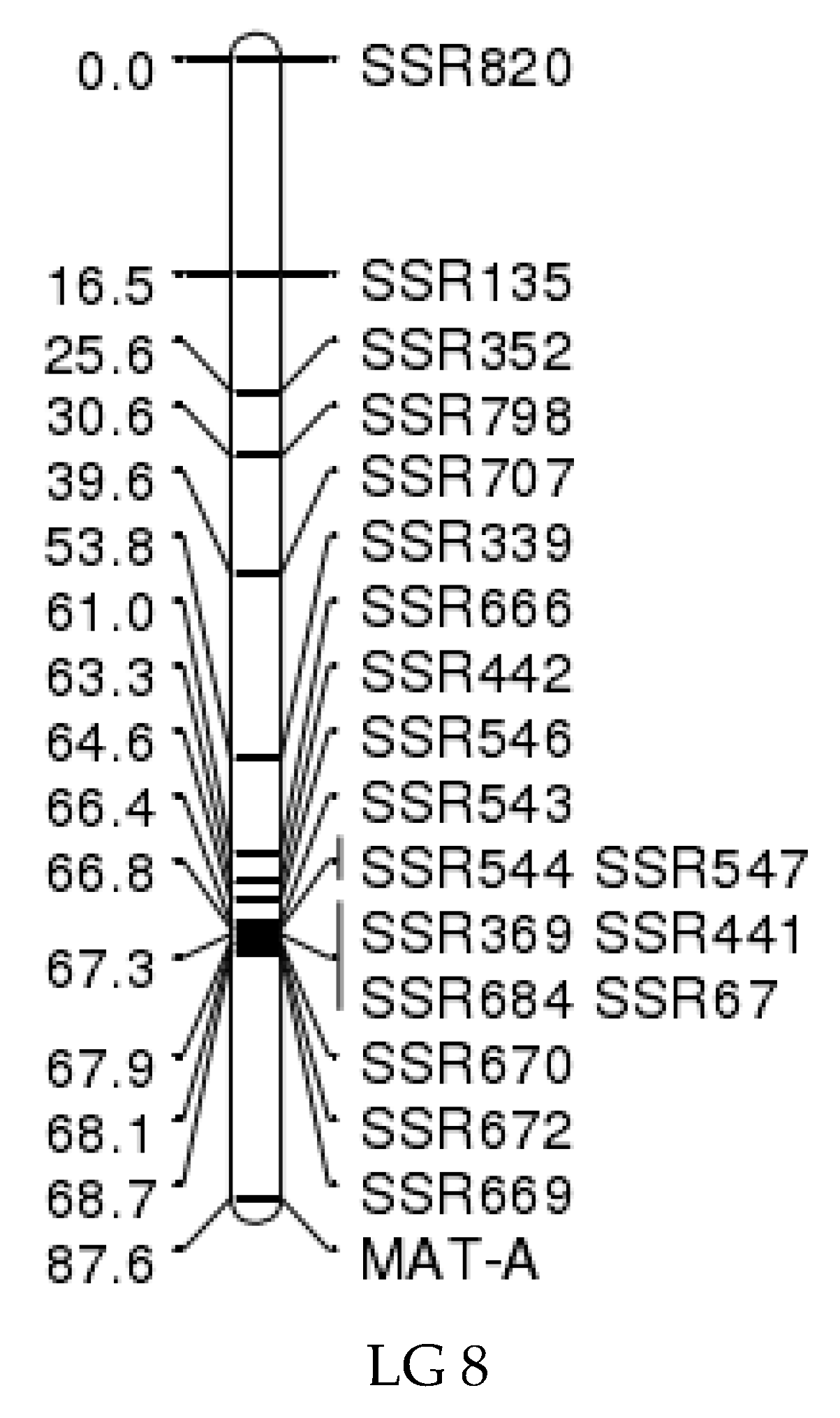
3.7. Mating-Type Location on the Genetic Linkage Map

The results obtained using JoinMap version 4.0 showed that mating-type strains were located at the locus at 87.6 cM on linkage group 8 (Figure 5) [14]. This indicates that the simple sequence repeat (SSR) molecular marker SSR669 (scaffold45_size276651_(CTG)6_18_209253_209270) was linked to the mating-type locus (MAT-A), which was 18.9 cM distant from the mating-type locus (Figure 5).

3.8. Structure of the Mating-type Locus

The blastp and tblastn alignments indicated that the mating-type genes of strain A14-8 were on scaffold45, which is consistent with the linkage map positioning results. The SSR marker SSR669 that was linked closely to MAT-A was also located on scaffold45. SSR669 was located downstream of HD148-2-1 (GenBank: MN267021) at a distance of 88.535 kb. There were two pairs of homeodomain mating-type protein genes HD148-1-1 (GenBank: MN267022) and HD148-2-1, HD148-1-2 (GenBank: MN267023), and HD148-2-2 (GenBank: MN267024) on scaffold45. HD148-1-1 and HD148-2-1 encoded α subunits of 683 and 693 amino acids, respectively, and the distance between the two genes was 153 bp. HD148-1-2 and HD148-2-2 encoded β subunits of 626 and 628 amino acids, respectively. The distance between the two genes was 294 bp. There was 36.725 kb between the genes encoding the α and β subunits (Table 4).
The results for strains A14-5, 18-119, and Dai 13782 show that the mating-type locus of A. heimuer contained two pairs of genes that encoded two subunits of the homeodomain proteins. The HD1 genes varied in length from 593 to 715 bp, and the HD2 genes varied from 587 to 650 bp. The distance between genes encoding the α and β subunits varied in length from 36 to 41 kb (Table 4 and Figure 6).
The genes encoding mitochondrial intermediate peptidase (mip) and beta-fg were linked closely to the mating-type locus in A14-8, which is similar to what was found for other edible mushrooms [43]. The genes 148mip (GenBank: MN267034) and 148beta-fg (GenBank: MN267037) were on scaffold45 and encoded proteins of 767 and 231 amino acids, respectively. But unlike in other fungi genomes, in the A. heimuer genomes these two genes were on the one side of the mating-type locus and not flanking it (Figure 6). The 148beta-fg gene was downstream of HD148-1-2 at a distance of 51.913 kb and the 148mip was downstream of beta-fg at a distance of 6.32 kb (Table S13).

3.9. Analysis of the Synteny of MAT-A in the A. heimuer, A. subglabra, and E. glandulosa Genomes

MAT-A of the A. heimuer strain A14-8 was on scaffold45 at the locus from positions 110,367 to 230,620 bp, a total of about 12 kb. The mating-type genes HD1-2 and HD1-1 that encode α subunits were located upstream of the MAT-A locus, and the mating-type genes HD2-2 and HD2-1 that encode β subunits were in the middle of the MAT-A locus. The results show that the genes in the MAT-A locus were highly conserved between A. heimuer and A. subglabra (Figure 7).
A total of 51 genes were detected in the MAT-A locus of A. heimuer and 43 (84.3%) of them were homologous to A. subglabra genes. Both mip and beta-fg were found downstream of MAT-A. There was conserved synteny between the homologous genes in the MAT-A locus of A. heimuer and A. subglabra, indicating the synteny in this locus were highly conserved. A total of 33 (64.71%) homologous genes were detected in the MAT-A locus of E. glandulosa. The mating-type genes flanking the β subunit showed mirrored symmetry, indicating the genes at this site were reversed during evolution (Figure 7). Therefore, the number of homologous genes and conserved synteny indicate that the homology between A. heimuer and A. subglabra was higher than it was between A. heimuer and E. glandulosa, which is consistent with the phylogenomic relationship of the three species.
Different color bars represent different genes, and their direction represents the direction of gene transcription. Light blue stripes represent homologous genes shared by three species, green stripes represent homologous genes shared by two species, and pink stripes represent non-homologous genes between the three species. Homologous genes are linked by black lines, dark blue stripes represent the HD1 gene, red stripes represent the HD2 gene, and subunits re marked by black lines above them. The purple strip represents the gene encoding mitochondrial intermediate peptidase. Numbers represent the number of different genes on MAT-A, and SSR669 represents the closest SSR marker to the mating-type locus on linkage map.

4. Discussion

4.1. Genome Features

The strain A14-8 used in this study was from the cultivar A14 that originated from a wild strain collected in AnTu county, Jilin Province. The strain Dai 13782 was from a wild strain found in Heilongjiang Province. Both Heilongjiang and Jilin Province are the two major production areas of these strains, so the two genomes can be considered as representative of different major production areas in China. The size of the A14-8 genome was 43.6 Mb, which was about 6 Mb shorter than the Dai 13782, and the scaffold numbers of A14-8 were four times more than the numbers of Dai 13782. The main reason for this should be lying in the different sequencing methods.

4.2. CAZymes

The Pfam database was used to identify the CAZymes and 120 more genes were identified in this study compared with the number reported by Yuan et al. [1]. The result of Dai 13782 was based on a comparison of BLASTP against the CAZy database (http://www.cazy.org/). The results of B14-8 were based on dbCAN2, a toolkit developed for CAZymes combined with Pfam. The differences between the two strain results could be mainly because of annotation pipelines. The correspondence between CAZy families and those in PFAM/INTERPRO or DBCAN is far from perfect [22]. In other words, the different results provided an alternative reference to exploit CAZymes and supply more candidates for further study. Genes encoding laccase-like multicopper oxidases (AA1_3) and ferroxidases (AA1_2) were found in A. heimuer and A. subglabra genomes, suggesting these genes may be common in the Auriculariaceae family. These results are very similar to those obtained for ascomycetes and support the speculation that genes encoding AA1_3 enzymes may have originated from the common ancestor of basidiomycetes and ascomycetes, or by the horizontal transfer of genes between species [38]. Quantitative differences between the AA1 and AA2 families indicates there were more genes encoding AA1 enzymes than AA2 enzymes in the genomes of L. bicolor, L. edodes, C. cinereus, F. velutipes, P. ostreatus, S. lacrymans, A. bisporus, M. conica, V. volvacea, G. lucidum, and T. mesenterica. However, there were 4.2 times more genes encoding AA2 enzymes than AA1 enzymes in the A. heimuer genome, indicating that the AA2 famliy genes play crucial roles in the lignin degradation ability of A. heimuer [38]. According to the ability of wood rot fungi to degrade lignin in plant cell walls, they can be classified as white rot fungi or brown rot fungi. The main differences between these two nutritional types were the higher presence of the PODs and the larger number of enzymes that act on crystalline cellulose in white rot fungi compared with brown rot fungi [10]. Comparative genomics analysis revealed the existence of intermediate types between white rot fungi and brown rot fungi, such as Botryobasidium botryosum and Jaapia argillacea. A. heimuer was identified as a typical white rot fungus in this study because of the presence of PODs and the large number of enzymes that act on crystalline cellulose. Moreover, the genes that encode enzymes in the A2, CBM1, GH6, GH7, and AA9 families are important for the lignocellulose degradation of A. heimuer and can be considered as core candidate genes for the nutritional utilization and development of molecular markers for breeding high-yield cultivars of A. heimuer. To efficiently utilize these genes in future studies, the transcriptomes of A. heimuer should to be analyzed at different developmental stages to detect the most active lignocellulose degrading genes.

4.3. Expansion and Contraction of CAZymes

Based on the analysis of CAZyme gene family expansion and contraction, we found that the number of contracted gene families (20) in A. heimuer were significantly more than the number of expanding gene families (6) in its evolution. Twenty-five of them were related to lignocellulose degradation, while only one gene family was involved in the synthesis of cell wall. This suggests that A. heimuer enhanced its nutrition utilization ability by changing the composition and quantity of genes related to lignocellulose degradation during its evolution. The most rapidly evolving family was AA3, with eight genes contracted. It seemed that this kind of evolutionary process is not beneficial to the nutritional utilization of A. heimuer, but through statistical analysis most of the AA3-2 family genes are kept in its evolutionary process. Its function is to participate in the degradation of lignin with AA2 family genes, and the number of these two gene families accounts for 49% of the total AA families’. In addition, the gene families related to starch degradation contracted in A. heimuer included CBM20, GH13, GH15, and GH27. However, these families are very rich in L. edodes [44]. This shows that different species may take different evolutionary approaches to nutrition utilization. Based on the above analysis, it is speculated that A. heimuer has economic and effective ways of improving lignin utilization in evolution.

4.4. Alcohol Dehydrogenases

Alcohol dehydrogenases can be used for the large-scale production of bioethanol, and this process has been studied extensively in Saccharomyces cerevisiae [40]. Mushrooms can convert cellulose into bioethanol because ethanol dehydrogenases can catalyze cellulose to acetaldehyde then ethanol. This has been identified in F. velutipes, P. ostreatus, and Agaricus blazei, and has been used in the production of alcoholic beverages [7]. The F. velutipes genome showed a strong potential for alcohol conversion, encoding 58 homologs to alcohol dehydrogenase genes covering a broad range of possible substrates. A total of 102 genes that encode alcohol dehydrogenases were identified in the A. heimuer genome, which was 1.76 times more than was identified in the F. velutipes genome. Further, a large number of genes that could decompose lignocellulose were found in the A. heimuer genome. The presence in A. heimuer of the consolidated bioprocessing steps from cellulose to bioethanol, including cellulose conversion to polysaccharides, hydrolysis of polysaccharides to sugars, and fermentation of sugars to alcohol, indicates that A. heimuer has the potential to synthesize bioethanol. According to a study of Tokumitsu et al., alcohol fermentation by Agaricus blazei depends on both the EMP pathway and ED pathway [7]. Based on analysis of the KEGG pathway in this study, only genes involved in EMP were identified, and no genes were found in the Entner–Doudoroff pathway. Until now, there have only been a few studies on alcohol dehydrogenases and none on the production of bioethanol in A. heimuer. To clarify which genes are involved in ethanol production, further studies on the ethanol dehydrogenases in the A. heimuer transcriptome and the expression of ethanol dehydrogenases in different tissues and at different developmental stages are required to evaluate the feasibility of bioethanol production by A. heimuer.

4.5. Phylogenomic Analysis

The results by phylogenomic analysis showed that A. heimuer, A. subglabra, and E. glandulosa formed an Auricularia-related clade, which indicates that Auricularia differentiated earlier than the other genera in Agaricomycetes. This conclusion can also be drawn from the morphological differences of the fruiting body between A. heimuer and other genera in Agaricomycetes. Wood-rotting and straw-rotting fungi clustered into two groups based on their nutritional types. In this study, 562 single-copy orthologous genes were used in the evolutionary analysis. This is more than the genes number used to analyze the phylogenies of V. volvacea [45], G. lucidum [46], and 31 fungi identified by Dimitrios [37].

4.6. Mating-type Locus of A. heimuer

In this study, the genetic linkage map and genomics data of the mating-type locus of A. heimuer were combined to locate the mating-type locus and clarify its gene composition. The results show that the two methods could confirm each other, indicating that the results were accurate. A mating-type locus was located on linkage group 8 on scaffold45, which was consistent with the results of the genomics analysis. The distance between marker SSR669 and the mating-type locus on the linkage map was 18.9 cM, and the corresponding distance on scaffold45 was 88.535 kb. By comparing and analyzing the mating-type sequences of different strains A14-8, A14-5,18-119 and Dai 13782, it was found that the mating-type loci of different strains had the same compositions and structures. All of the mating-type structures were composed of two subunits, α and β, but the gap between the two subunits was quite different, from 145 to 2198 bp. The bioinformatics analysis also detected pheromone and pheromone receptor genes in the A. heimuer genome. In basidiomycetes with a tetrapolar mating-type system, these genes worked together to control nuclear migration and the fusion of clamp cells with subapical cells [47]. Three hypotheses of the evolutionary relationship between bipolar and tetrapolar mating-system types have been proposed: (1) Factor A and B, which originally regulated mating incompatibility on different chromosomes, are linked by gene conversion or recombination, demonstrating that the mating-type system is bipolar [48,49,50]; (2) Factor A and B are located on different chromosomes and one has lost its function in regulating mating (such as in Coprinellus disseminates [51], P. nameko [52], and Phanerochaete chrysosporium [53]), and although pheromone and receptor genes are present, only transcription factors (HD genes) control the mating; and (3) The function of one mating-type gene is replaced by another gene. A. heimuer is speculated to be the second type, which means that although the pheromone and pheromone receptor genes are found, they no longer regulate mating incompatibility.4.7. Synteny of A. heimuer on MAT-A
The mip and beta-fg genes of A. heimuer and A. subglabra were found to be located on one side of MAT-A, the same as was found for L. edodes [54], but the distance between the two genes in HD148-2-1 was 51.91 kb, which is much longer than the distance in L. edodes (0.76 kb). In the F. velutipes [43,54], C. cinereus [43,45,54], L. bicolor [49,53,54], Coprinellus disseminatus [53], P. djamor [54], P. chrysosporium [53], P. nameko [45], A. bisporus [45], V. volvacea [45], and Postia placenta [53] genomes, the mip and beta-fg genes flanked the mating-type genes. The tight linkage between the mip locus and the genes of MAT-A of homobasidiomycetes were reported to be conserved within <1 kb, for example, in the genomes of the model mushroom C. cinereus [55] and S. commune [56], whereas in A. heimuer strains A14-8 and Dai 13782 the distances were 59.5 and 67.6 kb, respectively, which are much longer than those of C. cinereus [55] and S. commune. The mating-type genes of mushroom-forming fungi are essential for the formation of dikaryotic mycelia and the development of fruiting bodies. The research on the structure and synteny of MAT-A provided the reference for the evolution study of different species in Agaricomycetes.

5. Conclusions

The efficient utilization of substrate is one of the key factors in improving mushroom yield. One of the essential questions affecting the utilization of substrates is how the lignocellulose was degraded [57]. Therefore, in-depth studies on the composition and mechanism of CAZyme can lay the groundwork for breeding edible mushrooms that are efficient in the degradation of lignocellulose. On the basis of the A. heimuer genome sequence, we analyzed the genes involved in degrading lignocellulolytic enzymes, screening out the candidate genes for the nutritional utilization. The results can be used to develop molecular markers for breeding high-yield cultivars of A. heimuer. Bioethanol production is an important topic worldwide due to energy security. This study has identified alcohol dehydrogenase in the A. heimuer genome and showed that A. heimuer has the potential to produce ethanol as a reactor by consolidated bioprocessing. The mating loci of A. heimuer were mapped to a genetic linkage map and the structure and coding sequences of the mating loci were analyzed using genome-wide sequence information. Research on the structure and synteny of MAT-A provided the reference for the evolution study of different species in Agaricomycetes. In sum, these results will contribute to further studies of the genetics, evolution, breeding, bioethanol development and cultivation of A. heimuer.

Supplementary Materials

The following are available online at https://www.mdpi.com/2309-608X/6/1/37/s1.

Author Contributions

Conceptualization, F.Y. and Y.Z.; methodology, M.F.; software, M.F. and P.W.; validation, X.W., Y.C., and L.L.; formal analysis, M.F. and P.W.; investigation, X.W. and P.W.; resources, Y.C., Y.Z., and J.L.; data curation, M.F.; writing—original draft preparation, M.F. and Y.C.; writing—review and editing, F.Y.; visualization, M.F. and X.W.; supervision, Y.Z. and F.Y.; project administration, F.Y.; funding acquisition, F.Y. All authors have read and agree to the published version of the manuscript.

Funding

This research was funded by the China Agriculture Research System, grant number CARS-20 and National Natural Science Foundation of China, grant number 31272219.

Acknowledgments

We thank Margaret Biswas, PhD, from Liwen Bianji, Edanz Group China (www.liwenbianji.cn/ac) for editing the English text of a draft of this manuscript.

Conflicts of Interest

The authors declare no conflict of interest.

References

  1. Yuan, Y.; Wu, F.; Si, J.; Zhao, Y.-F.; Dai, Y.-C. Whole genome sequence of Auricularia heimuer (Basidiomycota, Fungi), the third most important cultivated mushroom worldwide. Genomics 2018, 111, 50–58. [Google Scholar] [CrossRef] [PubMed]
  2. Lou, L.H. Cultivation of Auricularia on Logs in China. In Tropical Mushrooms Biological Nature and Cultivation Methods; Chang, S.T., Quimio, T.H., Eds.; The Chinese University Press: Hong Kong, China, 1982; pp. 437–441. [Google Scholar]
  3. Ying, C.; Fang-jie, Y.; Gui-juan, L.; Hai-ying, W.; Yan, L. The Announcements and Suggestions of Auricularia auricula Cultivation Using Substitute Media. Edible Fungi China 2010, 29, 55–58. [Google Scholar]
  4. Yao, F.J.; Lu, L.X.; Wang, P.; Fang, M.; Zhang, Y.M.; Chen, Y.; Zhang, W.T.; Kong, X.H.; Lu, J.; Honda, Y. Development of a Molecular Marker for Fruiting Body Pattern in Auricularia auricula-judae. Mycobiology 2018, 46, 72–78. [Google Scholar] [CrossRef] [PubMed] [Green Version]
  5. Fang-jie, Y.; You-min, Z.; Ying, C. The Industrial Development Situation of Auricularia auricular of China. North. Hortic. 2010, 18, 209–211. [Google Scholar]
  6. Maehara, T.; Ichinose, H.; Furukawa, T.; Ogasawara, W.; Takabatake, K.; Kaneko, S. Ethanol production from high cellulose concentration by the basidiomycete fungus Flammulina velutipes. Fungal Biol-Uk 2013, 117, 220–226. [Google Scholar] [CrossRef]
  7. Okamura-Matsui, T.; Tomoda, T.; Fukuda, S.; Ohsugi, M. Discovery of alcohol dehydrogenase from mushrooms and application to alcoholic beverages. J. Mol. Catal. B Enzym. 2003, 23, 133–144. [Google Scholar] [CrossRef]
  8. Wu, F.; Yuan, Y.; Malysheva, V.F.; Du, P.; Dai, Y.-C. Species clarification of the most important and cultivated Auricularia mushroom “Heimuer”: Evidence from morphological and molecular data. Phytotaxa 2014, 186, 241–253. [Google Scholar] [CrossRef]
  9. Morin, E.; Kohler, A.; Baker, A.R.; Foulongne-Oriol, M.; Lombard, V.; Nagy, L.G.; Ohm, R.A.; Patyshakuliyeva, A.; Brun, A.; Aerts, A.L.; et al. Genome sequence of the button mushroom Agaricus bisporus reveals mechanisms governing adaptation to a humic-rich ecological niche. Proc. Natl. Acad. Sci. USA 2012, 109, 17501–17506. [Google Scholar] [CrossRef] [Green Version]
  10. Riley, R.; Salamov, A.A.; Brown, D.W.; Nagy, L.G.; Floudas, D.; Held, B.W.; Levasseur, A.; Lombard, V.; Morin, E.; Otillar, R.; et al. Extensive sampling of basidiomycete genomes demonstrates inadequacy of the white-rot/brown-rot paradigm for wood decay fungi. Proc. Natl. Acad. Sci. USA 2014, 111, 9923–9928. [Google Scholar] [CrossRef] [Green Version]
  11. Ohm, R.A.; de Jong, J.F.; Lugones, L.G.; Aerts, A.; Kothe, E.; Stajich, J.E.; de Vries, R.P.; Record, E.; Levasseur, A.; Baker, S.E.; et al. Genome sequence of the model mushroom Schizophyllum commune. Nat. Biotechnol. 2010, 28, 957–963. [Google Scholar] [CrossRef] [Green Version]
  12. Stajich, J.E.; Wilke, S.K.; Ahrén, D.; Au, C.H.; Birren, B.W.; Borodovsky, M.; Burns, C.; Canbäck, B.; Casselton, L.A.; Cheng, C.K.; et al. Insights into evolution of multicellular fungi from the assembled chromosomes of the mushroom Coprinopsis cinerea (Coprinus cinereus). Proc. Natl. Acad. Sci. USA 2010, 107, 11889–11894. [Google Scholar] [CrossRef] [PubMed] [Green Version]
  13. Whitehouse, H. Multiple-allelomorph heterothallism in the fungi. New Phytol. 1949, 48, 212–244. [Google Scholar] [CrossRef]
  14. Lu, L.-X.; Yao, F.-J.; Wang, P.; Fang, M.; Zhang, Y.-M.; Zhang, W.-T.; Kong, X.-H.; Lu, J. Construction of a genetic linkage map and QTL mapping of agronomic traits in Auricularia auricula-judae. J. Microbiol. 2017, 55, 792–799. [Google Scholar] [CrossRef] [PubMed]
  15. Ter-Hovhannisyan, V.; Lomsadze, A.; Chernoff, Y.O.; Borodovsky, M. Gene prediction in novel fungal genomes using an ab initio algorithm with unsupervised training. Genome. Res. 2008, 18, 1979–1990. [Google Scholar] [CrossRef] [PubMed] [Green Version]
  16. Solovyev, V.; Kosarev, P.; Seledsov, I.; Vorobyev, D. Automatic annotation of eukaryotic genes, pseudogenes and promoters. Genome. Biol. 2006, 7, S10. [Google Scholar] [CrossRef] [Green Version]
  17. Delcher, A.L.; Bratke, K.A.; Powers, E.C.; Salzberg, S.L. Identifying bacterial genes and endosymbiont DNA with Glimmer. Bioinformatics 2007, 23, 673–679. [Google Scholar] [CrossRef]
  18. Madeira, F.; Park, Y.M.; Lee, J.; Buso, N.; Gur, T.; Madhusoodanan, N.; Basutkar, P.; Tivey, A.R.N.; Potter, S.C.; Finn, R.D.; et al. The EMBL-EBI search and sequence analysis tools APIs in 2019. Nucleic Acids Res. 2019, 47, W636–W641. [Google Scholar] [CrossRef] [Green Version]
  19. Haas, B.J.; Salzberg, S.L.; Zhu, W.; Pertea, M.; Allen, J.E.; Orvis, J.; White, O.; Buell, C.R.; Wortman, J.R. Automated eukaryotic gene structure annotation using EVidenceModeler and the Program to Assemble Spliced Alignments. Genome Biol. 2008, 9, R7. [Google Scholar] [CrossRef] [Green Version]
  20. Luo, X.; Cao, J.; Junkai, H.; Wang, Z.; Guo, Z.; Chen, Y.; Ma, S.; Liu, J. Genome sequencing and comparative genomics reveal the potential pathogenic mechanism of Cercospora sojina Hara on soybean. DNA Res. 2017, 25, 1–13. [Google Scholar] [CrossRef] [Green Version]
  21. Quevillon, E.; Silventoinen, V.; Pillai, S.; Harte, N.; Mulder, N.; Apweiler, R.; Lopez, R. InterProScan: Protein domains identifier. Nucleic Acids Res. 2005, 33, W116–W120. [Google Scholar] [CrossRef] [Green Version]
  22. Lombard, V.; Golaconda Ramulu, H.; Drula, E.; Coutinho, P.M.; Henrissat, B. The carbohydrate-active enzymes database (CAZy) in 2013. Nucleic Acids Res. 2014, 42, D490–D495. [Google Scholar] [CrossRef] [Green Version]
  23. Zhang, H.; Yohe, T.; Huang, L.; Entwistle, S.; Wu, P.; Yang, Z.; Busk, P.K.; Xu, Y.; Yin, Y. dbCAN2: A meta server for automated carbohydrate-active enzyme annotation. Nucleic Acids Res. 2018, 46, W95–W101. [Google Scholar] [CrossRef] [PubMed] [Green Version]
  24. Waterhouse, R.M.; Tegenfeldt, F.; Li, J.; Zdobnov, E.M.; Kriventseva, E.V. OrthoDB: A hierarchical catalog of animal, fungal and bacterial orthologs. Nucleic Acids Res. 2013, 41, D358–D365. [Google Scholar] [CrossRef] [PubMed]
  25. Katoh, K.; Toh, H. Recent developments in the MAFFT multiple sequence alignment program. Brief Bioinform. 2008, 9, 286–298. [Google Scholar] [CrossRef] [PubMed] [Green Version]
  26. Wheeler, T.J.; Eddy, S.R. nhmmer: DNA homology search with profile HMMs. Bioinformatics 2013, 29, 2487–2489. [Google Scholar] [CrossRef] [Green Version]
  27. Dereeper, A.; Guignon, V.; Blanc, G.; Audic, S.; Buffet, S.; Chevenet, F.; Dufayard, J.F.; Guindon, S.; Lefort, V.; Lescot, M. Phylogeny.fr: Robust phylogenetic analysis for the non-specialist. Nucleic Acids Res. 2008, 36, W465–W469. [Google Scholar] [CrossRef]
  28. Darriba, D.; Taboada, G.; Doallo, R.; Posada, D. ProtTest 3: Fast selection of best-fit models of protein evolution. Bioinformatics 2011, 27, 1164–1165. [Google Scholar] [CrossRef] [Green Version]
  29. Stamatakis, A. RAxML version 8: A tool for phylogenetic analysis and post-analysis of large phylogenies. Bioinformatics 2014, 30, 1312–1313. [Google Scholar] [CrossRef]
  30. Han, M.; Thomas, G.; Lugo-Martinez, J.; Hahn, M. Estimating Gene Gain and Loss Rates in the Presence of Error in Genome Assembly and Annotation Using CAFE 3. Mol. Biol. Evol. 2013, 30, 1987–1997. [Google Scholar] [CrossRef]
  31. Van Ooijen, J.W. Multipoint maximum likelihood mapping in a full-sib family of an outbreeding species. Genet. Res. 2011, 93, 343–349. [Google Scholar] [CrossRef]
  32. Wang, Y.; Tang, H.; Debarry, J.D.; Tan, X.; Li, J.; Wang, X.; Lee, T.H.; Jin, H.; Marler, B.; Guo, H.; et al. MCScanX: A toolkit for detection and evolutionary analysis of gene synteny and collinearity. Nucleic Acids Res. 2012, 40, e49. [Google Scholar] [CrossRef] [PubMed] [Green Version]
  33. Stanke, M.; Morgenstern, B. AUGUSTUS: A web server for gene prediction in eukaryotes that allows user-defined constraints. Nucleic Acids Res. 2005, 33, W465–W467. [Google Scholar] [CrossRef] [PubMed] [Green Version]
  34. Salamov, A.A.; Solovyev, V.V. Ab initio Gene Finding in Drosophila Genomic DNA. Genome Res. 2000, 10, 516–522. [Google Scholar] [CrossRef] [PubMed] [Green Version]
  35. Radakovits, R.; Jinkerson, R.E.; Fuerstenberg, S.I.; Tae, H.; Settlage, R.E.; Boore, J.L.; Posewitz, M.C. Draft genome sequence and genetic transformation of the oleaginous alga Nannochloropsis gaditana. Nat Commun. 2012, 3, 1–10. [Google Scholar] [CrossRef] [PubMed] [Green Version]
  36. Vaaje-Kolstad, G.; Westereng, B.; Horn, S.J.; Liu, Z.; Zhai, H.; Sørlie, M.; Eijsink, V.G.H. An Oxidative Enzyme Boosting the Enzymatic Conversion of Recalcitrant Polysaccharides. Science 2010, 330, 219–222. [Google Scholar] [CrossRef] [PubMed]
  37. Floudas, D.; Binder, M.; Riley, R.; Barry, K.; Blanchette, R.A.; Henrissat, B.; Martínez, A.T.; Otillar, R.; Spatafora, J.W.; Yadav, J.S.; et al. The Paleozoic Origin of Enzymatic Lignin Decomposition Reconstructed from 31 Fungal Genomes. Science 2012, 336, 1715–1719. [Google Scholar] [CrossRef] [Green Version]
  38. Levasseur, A.; Drula, E.; Lombard, V.; Coutinho, P.M.; Henrissat, B. Expansion of the enzymatic repertoire of the CAZy database to integrate auxiliary redox enzymes. Biotechnol. Biofuels 2013, 6, 41. [Google Scholar] [CrossRef] [Green Version]
  39. Matsumura, H.; Umezawa, K.; Takeda, K.; Sugimoto, N.; Ishida, T.; Samejima, M.; Ohno, H.; Yoshida, M.; Igarashi, K.; Nakamura, N. Discovery of a Eukaryotic Pyrroloquinoline Quinone-Dependent Oxidoreductase Belonging to a New Auxiliary Activity Family in the Database of Carbohydrate-Active Enzymes. PLoS ONE 2014, 9, e104851. [Google Scholar] [CrossRef] [Green Version]
  40. De Smidt, O.; Preez, J.C.D.; Albertyn, J. The alcohol dehydrogenases of Saccharomyces cerevisiae: A comprehensive review. Fems. Yeast Res. 2010, 8, 967–978. [Google Scholar] [CrossRef] [Green Version]
  41. Park, Y.J.; Baek, J.H.; Lee, S.; Kim, C.; Rhee, H.; Kim, H.; Seo, J.S.; Park, H.R.; Yoon, D.E.; Nam, J.Y.; et al. Whole genome and global gene expression analyses of the model mushroom Flammulina velutipes reveal a high capacity for lignocellulose degradation. PLoS ONE 2014, 9, e93560. [Google Scholar] [CrossRef]
  42. Hatakka, A.; Hammel, K.E. Fungal biodegradation of lignocelluloses. In Industrial Applications; Hofrichter, M., Ed.; Springer: Berlin/Heidelberg, Germany, 2011; pp. 319–340. [Google Scholar]
  43. Van Peer, A.F.; Park, S.Y.; Shin, P.G.; Jang, K.Y.; Yoo, Y.B.; Park, Y.J.; Lee, B.M.; Sung, G.H.; James, T.Y.; Kong, W.S. Comparative genomics of the mating-type loci of the mushroom Flammulina velutipes reveals widespread synteny and recent inversions. PLoS ONE 2011, 6, e22249. [Google Scholar] [CrossRef] [Green Version]
  44. Chen, L.; Gong, Y.; Cai, Y.; Liu, W.; Zhou, Y.; Xiao, Y.; Xu, Z.; Liu, Y.; Lei, X.; Wang, G.; et al. Genome Sequence of the Edible Cultivated Mushroom Lentinula edodes (Shiitake) Reveals Insights into Lignocellulose Degradation. PLoS ONE 2016, 11, e0160336. [Google Scholar] [CrossRef] [PubMed]
  45. Bao, D.; Gong, M.; Zheng, H.; Chen, M.; Zhang, L.; Wang, H.; Jiang, J.; Wu, L.; Zhu, Y.; Zhu, G.; et al. Sequencing and Comparative Analysis of the Straw Mushroom (Volvariella volvacea) Genome. PLoS ONE 2013, 8, e58294. [Google Scholar] [CrossRef]
  46. Chen, S.; Xu, J.; Liu, C.; Zhu, Y.; Nelson, D.R.; Zhou, S.; Li, C.; Wang, L.; Guo, X.; Sun, Y. Genome sequence of the model medicinal mushroom Ganoderma lucidum. Nat. Commun. 2012, 3, 913. [Google Scholar] [CrossRef] [PubMed] [Green Version]
  47. Swiezynski, K.M.; Day, P.R. Heterokaryon formation in Coprinus lagopus. Genet. Res. Camb. 1960, 1, 114–128. [Google Scholar] [CrossRef]
  48. Fraser, J.A.; Diezmann, S.; Subaran, R.L.; Allen, A.; Lengeler, K.B.; Dietrich, F.S.; Heitman, J. Convergent Evolution of Chromosomal Sex-Determining Regions in the Animal and Fungal Kingdoms. PLoS Biol. 2004, 2, 2243–2255. [Google Scholar] [CrossRef] [PubMed] [Green Version]
  49. Bakkeren, G.; Kronstad, J.W. Linkage of mating-type loci distinguishes bipolar from tetrapolar mating in basidiomycetous smut fungi. Proc. Natl. Acad. Sci. USA 1994, 91, 7085–7089. [Google Scholar] [CrossRef] [PubMed] [Green Version]
  50. Lee, N.; Bakkeren, G.; Wong, K.; Sherwood, J.E.; Kronstad, J.W. The mating-type and pathogenicity locus of the fungus Ustilago hordei spans a 500-kb region. Proc. Natl. Acad. Sci. USA 1999, 96, 15026–15031. [Google Scholar] [CrossRef] [PubMed] [Green Version]
  51. James, T.Y.; Srivilai, P.; Kües, U.; Vilgalys, R. Evolution of the bipolar mating system of the mushroom Coprinellus disseminatus from its tetrapolar ancestors involves loss of mating-type-specific pheromone receptor function. Genetics 2006, 172, 1877–1891. [Google Scholar] [CrossRef] [Green Version]
  52. Yi, R.; Tachikawa, T.; Mukaiyama, H.; Mochida, Y.; Ishikawa, M.; Aimi, T. DNA-mediated transformation system in a bipolar basidiomycete, Pholiota microspora (P. nameko). Mycoscience 2009, 50, 123–129. [Google Scholar] [CrossRef]
  53. James, T.Y.; Lee, M.; van Diepen, L.T.A. A Single Mating-Type Locus Composed of Homeodomain Genes Promotes Nuclear Migration and Heterokaryosis in the White-Rot Fungus Phanerochaete chrysosporium. Eukaryot Cell 2011, 10, 249–261. [Google Scholar] [CrossRef] [Green Version]
  54. Au, C.H.; Wong, M.C.; Bao, D.; Zhang, M.; Song, C.; Song, W.; Law, P.T.W.; Kües, U.; Kwan, H.S. The genetic structure of the A mating-type locus of Lentinula edodes. Gene 2014, 535, 184–190. [Google Scholar] [CrossRef] [PubMed]
  55. Casselton, L.A.; Asanteowusu, R.N.; Banham, A.H.; Kingsnorth, C.S.; Kües, U.; O’Shea, S.F.; Pardo, E.H. Mating type control of sexual development in Coprinus cinereus. Can. J. Bot. 1995, 73, 266–272. [Google Scholar] [CrossRef]
  56. Specht, C.A.; Stankis, M.M.; Giasson, L.; Novotny, C.P.; Ullrich, R.C. Functional analysis of the homeodomain-related proteins of the A alpha locus of Schizophyllum commune. Proc. Natl. Acad. Sci. USA 1992, 89, 7174–7178. [Google Scholar] [CrossRef] [PubMed] [Green Version]
  57. Zhilin, L.; Ruili, Z. Advances of Genomics-assisted Cultivation and Breedingof Edible and Medicinal Mushrooms. Acta Edulis Fungi 2018, 25, 93–106. [Google Scholar]
Figure 1. The expansions and contractions of the CAZyme gene families. Numbers associated with each branch designate the numbers of gene families that have expanded (red) or contracted (green) since the split from the common ancestor. The color bars correspond to different orders.
Jof 06 00037 g001
Figure 2. The cellulose degradation pathway of A. heimuer. The red and green boxes indicate the substrates and existing homologous genes of the enzyme, respectively. The white boxes mean the enzymes that were not identified in A.heimuer genome. The blue box indicates the core region of the function. The figure was generated by a KEGG mapper.
Jof 06 00037 g002
Figure 3. Pathway of the biosynthesis of ethanol. The red and green boxes indicate the substrates and existing homologous genes of the enzyme, respectively; The white boxes mean the enzymes that were not identified in A.heimuer genome. The blue box indicates the core region of the function. The figure was generated by a KEGG mapper.
Jof 06 00037 g003
Figure 4. Phylogenomic tree of 15 mushroom species based on single-copy orthologous genes. The mushroom species separated into three main groups. Morchella conica was used as the outgroup. The different orders are indicted by different colored bars. Tree scale = 0.1. The numbers under bars indicate 100% bootstrap.
Jof 06 00037 g004
Figure 5. MAT-A mapping on the genetic linkage map. The bar in the middle represents the linkage group, the number on the left represents the genetic distance, the different SSR markers used for the construction of linkage map in this research are listed on the right according to the linkage relation. MAT-A represents the mating-type loci.
Jof 06 00037 g005
Figure 6. The mating-type structure of 14-8. The blue lines indicate the mating-type locus, the red arrows indicate the transcriptional direction of the genes, and the gray bar represents the region of scaffold45 that contains the mating-type genes. The numbers below are the positions of the two ends of the mating-type genes on scaffold45. The gap between the two subunits is 32.726 kb.
Jof 06 00037 g006
Figure 7. Synteny of genes on MAT-A of A. heimuer, A. subglabra, and E. glandulosa.
Jof 06 00037 g007
Table 1. Features of the Auricularia heimuer genome.
General Features.Properties of Predicted Gene Models
Genome size (Mb)43.6KEGG alignment2257
GC content (%)57.09Interpro signature9254
Scaffolds N50 (bp)276,651Pfam alignment12,279
Number of scaffolds535CAZyme alignment509
Number of predict gene models14,094Signal peptide1694
Average of gene length (bp)1737.54Transmembrane domain2441
Average of cds length (bp)1390
Average of exon number5.71
Average of intron number4.71
Average of exon length (bp)244
Average of intron length (bp)74
Repetitive sequences (%)3.73
Table 2. The number of CAZymes and AAs of A. heimuer compared with those of other fungi.
CAZy FamilyA.heiT.fucA.subL.edoF.velP.ostS.comG.lucS.lacC.cinA.bisT.mesV.vol
CBM60115630225420281271291066
CE18141811191412872318811
GH26510729220919021220620512517815256195
GT51535162574854523751415051
PL1121132018116264219
AA104141248591133789941124908118
sum509201552400399479381398224453334134460
CAZy Family: GH, glycoside hydrolase; GT, glycosyltransferase; PL, polysaccharide lyases; CE, carbohydrate esterases; CBM, carbohydrate-binding modules; AA, auxiliary activities. Species abbreviations and genome references: A.hei is A. heimuer; A.sub is A. subglabra; T.fuc is T. fuciformis; L.bic is L. bicolor; L.edo is Lentinula edodes; C.cin is C. cinereus; F.vel is F. velutipes; P.ost is P. ostreatus; S.lac is S. lacrymans; S.com is S. commune; A.bis is A. bisporus; V.vol is V. volvacea; G.luc is G. lucidum; T.mes is T. mesenterica.
Table 3. Genes numbers of CAZyme families involved in the wood degradation of A. heimuer.
SubstateCAZy FamilySpecies
A. hei.A.subL. edoF. velP. ostS. com.S. lac.G. lucA. bisV. volL. bicC. cin.
Crystalline celluloseCBM1434323153148201654150
GH6221231111405
GH756331620311206
AA91920131928225151131334
sum697140397829143929101495
LigninAA1_10014811241311111017
AA1_2111210211040
AA22121103921105934
AA3_1100100001001
AA3_22234172036157262826827
AA3_3364444544422
AA3_4131001000000
AA4111312211100
AA5_1575715231084106
AA6442324224223
AA71514111923128121326718
AA89112311215103
sum83102687310345368081844681
CAZy Family: GH, glycoside hydrolase; GT, glycosyltransferase; PL, polysaccharide lyases; CE, carbohydrate esterases; CBM, carbohydrate-binding modules; AA, auxiliary activities. Species abbreviations and genome references: A.hei is A. heimuer; A.sub is A. subglabra; T.fuc is T. fuciformis; L.bic is L. bicolor; L.edo is Lentinula edodes; C.cin is C. cinereus; F.vel is F. velutipes; P.ost is P. ostreatus; S.lac is S. lacrymans; S.com is S. commune; A.bis is A. bisporus; V.vol is V. volvacea; G.luc is G. lucidum. The background color from dark blue to dark red showed the increased numbers.
Table 4. α and β subunit structure of the mating-type locus in the A. heimuer genomes.
StrainMating-Type LocusGeneLength of Amino Acid (aa)SubunitThe Gap in the Same Subunit (bp)The Gap Between α and β Subunit (kb)
A14-8scaffold45HD1-1(MN267022)683α subunit15336.725
HD1-2(MN267023)626
A14-5contig976HD1-1(MN267026)593α subunit29436.716
HD1-2(MN267027)683
18-119contig313HD1-1(MN267028)684α subunit114337.563
HD1-2(MN267029)633
Dai 13782NEKD01000007.1HD1-1(MN442077)615α subunit17141.16
HD1-2(MN442078)613
StrainMating-type LocusGeneLength of Amino Acid (aa)SubunitThe Gap in the Same Subunit (bp)
A14-8scaffold45HD2-1(MN267021)693β subunit2198
HD2-2(MN267024)628
A14-5contig976HD2-1(MN267030)628β subunit153
HD2-2(MN267031)626
18-119contig313HD2-1(MN267032)622β subunit411
HD2-2(MN267033)650
Dai 13782NEKD01000007.1HD2-1(MN442079)715β subunit145
HD2-2(MN442080)599

